Embed Size (px)
DESCRIPTION
Nanocrystalline diamond films, which comprise the so called nanocrystalline diamond (NCD) and ultrananocrystalline diamond (UNCD), represent a class of biomaterials possessing outstanding mechanical, tribological, and electrical properties, which include high surface smoothness, high corrosion resistance, chemical inertness, superior electrochemical behavior, biocompatibility, and nontoxicity. These properties have positioned the nanocrystalline diamond films as an attractive class of materials for a range of therapeutic and diagnostic applications in the biomedical field. Consequently, the interaction of nanocrystalline diamond films with biomolecules and cells has been focus of intense research during the last years, with many studies focused in tailoring the properties of the films for the control of their biological performance. In this chapter, the current knowledge regarding the biological performance of nanocrystalline diamond films is reviewed from an application-specific perspective, covering topics such as enhancement of cellular adhesion, anti-fouling coatings, non-thrombogenic surfaces, micropatterning of cells and proteins, and immobilization of biomolecules for bioassays. In order to better understand the terminology used in the literature, which is related to the fabrication and surface functionalization of this class of materials, some of the most common approaches for synthesis and modification of CVD diamond films is introduced. Although many challenges still remain, it is envisioned that the application of this unique class of materials will significantly influence the next generation of biomedical devices.
Citation preview
Send Orders for Reprints to [email protected]
70 Frontiers in Biomaterials, Vol. 1, 2014, 70-100
Shunsheng Cao & Huijun Zhu (Eds) All rights reserved-© 2014 Bentham Science Publishers
CHAPTER 3 Nanocrystalline Diamond Films for Biomedical Applications Cristian Pablo Pennisi* and María Alcaide
Department of Health Science and Technology, Aalborg University, Aalborg, Denmark
Abstract: Nanocrystalline diamond films, which comprise the so called nanocrystalline diamond (NCD) and ultrananocrystalline diamond (UNCD), represent a class of biomaterials possessing outstanding mechanical, tribological, and electrical properties, which include high surface smoothness, high corrosion resistance, chemical inertness, superior electrochemical behavior, biocompatibility, and nontoxicity. These properties have positioned the nanocrystalline diamond films as an attractive class of materials for a range of therapeutic and diagnostic applications in the biomedical field. Consequently, the interaction of nanocrystalline diamond films with biomolecules and cells has been focus of intense research during the last years, with many studies focused in tailoring the properties of the films for the control of their biological performance. In this chapter, the current knowledge regarding the biological performance of nanocrystalline diamond films is reviewed from an application-specific perspective, covering topics such as enhancement of cellular adhesion, anti-fouling coatings, non-thrombogenic surfaces, micropatterning of cells and proteins, and immobilization of biomolecules for bioassays. In order to better understand the terminology used in the literature, which is related to the fabrication and surface functionalization of this class of materials, some of the most common approaches for synthesis and modification of CVD diamond films is introduced. Although many challenges still remain, it is envisioned that the application of this unique class of materials will significantly influence the next generation of biomedical devices.
Keywords: Antifouling surfaces, biocompatibility, bionanotechnology, biosensors, blood compatibility, cell adhesion, cell growth, cell-material interface, chemical vapor deposition, diamond films, differentiation, immobilization, implant, micropatterning, nanocrystalline diamond, protein adsorption, surface functionalization, synthetic diamonds, ultrananocrystalline diamond, wettability.
*Address correspondence to Cristian Pablo Pennisi: Laboratory for Stem Cell Research, Department of Health Science and Technology, Aalborg University, Fredrik BajersVej 3B, DK-9220, Aalborg Ø, Denmark; Tel: +45 9940 2419; Fax: +45 9940 7816; E-mail: [email protected]
Nanocrystalline Diamond for Biomedical Applications Frontiers in Biomaterials, Vol. 1 71
1. INTRODUCTION
The nanocrystalline diamond films constitute a unique class of materials within the group of synthetic diamonds. Although these films span a wide range of morphologies and properties, they can be essentially defined as a thin continuous layer of nanometer-sized diamond crystallites (smaller than 100 nm), which are synthesized by using the chemical vapor deposition (CVD) technique [1]. These kind of diamond films possess some unique mechanical, tribological, and electrical properties, which combined with their surface smoothness, high corrosion resistance, chemical inertness, absence of toxicity, and biocompatibility have positioned them as an attractive class of materials for both therapeutic and diagnostic applications in the biomedical field. There are many proposed biomedical applications for which nanocrystalline diamond filmsholdan unprecedented potential, such as orthopedic and dental implants [2-4], cardiovascular and ophthalmologic devices [5, 6], neural prostheses [7-9], and biosensors [10, 11].
In this chapter, we will review the current knowledge about the biological performance of nanocrystalline diamond films from an application-specific perspective. In order to better understand the terminology used in the different studies, some of the most common approaches forsynthesis and modification of CVD diamond films will be introduced. Also, the synthesis of diamond particles will be briefly described in the context of the historical development of synthetic diamonds and due to their application in the process of CVD growth of nanocrystalline diamond films. We do not include here studies involving diamond-like carbon (DLC) films, which are a class of amorphous carbon materials thatfind widespread application in the biomedical field [12].
2. DIAMOND SYNTHESIS
2.1. General Overview
The exploitation of naturally occurring diamonds for industrial applications is not practically viable, not only due to their scarcity but also for technical reasons. Therefore, for many years, methods for the fabrication of artificial diamonds were investigated. Nowadays, it is possible to obtain synthetic diamonds in form of
72 Frontiers in Biomaterials, Vol. 1 Pennisi and Alcaide
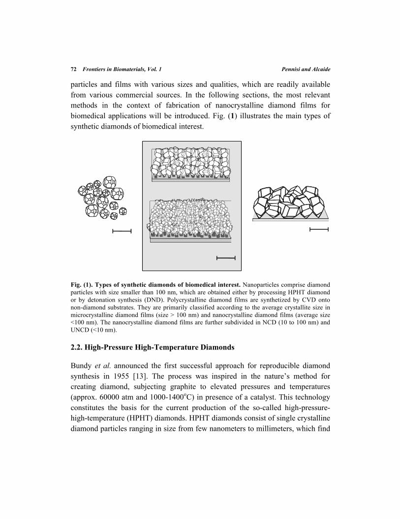
particles and films with various sizes and qualities, which are readily available from various commercial sources. In the following sections, the most relevant methods in the context of fabrication of nanocrystalline diamond films for biomedical applications will be introduced. Fig. (1) illustrates the main types of synthetic diamonds of biomedical interest.
Fig. (1). Types of synthetic diamonds of biomedical interest. Nanoparticles comprise diamond particles with size smaller than 100 nm, which are obtained either by processing HPHT diamond or by detonation synthesis (DND). Polycrystalline diamond films are synthetized by CVD onto non-diamond substrates. They are primarily classified according to the average crystallite size in microcrystalline diamond films (size > 100 nm) and nanocrystalline diamond films (average size <100 nm). The nanocrystalline diamond films are further subdivided in NCD (10 to 100 nm) and UNCD (<10 nm).
2.2. High-Pressure High-Temperature Diamonds
Bundy et al. announced the first successful approach for reproducible diamond synthesis in 1955 [13]. The process was inspired in the nature’s method for creating diamond, subjecting graphite to elevated pressures and temperatures (approx. 60000 atm and 1000-1400oC) in presence of a catalyst. This technology constitutes the basis for the current production of the so-called high-pressure-high-temperature (HPHT) diamonds. HPHT diamonds consist of single crystalline diamond particles ranging in size from few nanometers to millimeters, which find
Nanocrystalline Diamond for Biomedical Applications Frontiers in Biomaterials, Vol. 1 73
many diverse industrial applications, such as cutting tools and abrasives in the mechanical industry, optical components, etc. HPHT diamonds have been applied in the fabrication of CVD substrates for the assessment of cell compatibility of single crystalline [9] and polycrystalline diamond films [14, 15].
2.3. Detonation Nanodiamonds
Scientists from the former USSR discovered an alternative method for the fabrication of synthetic diamonds in the 1960’s [16]. The approach basically consists in the conversion of a carbon containing explosive (such as trinitrotoluene, TNT) to diamond nanoparticles under a controlled detonation taking place inside a closed container. This class of synthetic diamonds is known as detonation nanodiamond (DND), which have raised tremendous interest during the last years due to their outstanding mechanical, chemical, electronic and optical properties [17]. In particular, DNDs are subject of intense research in the biomedical field for application as drug delivery agents, markers for bioimaging, and components of composite biomaterials. This topic is covered by several detailed reviews in the literature [17-22]. In the context of fabrication of nanocrystalline diamond films, similarly to the HPHT nanoparticles, DNDs are used as seeds for the CVD growth of polycrystalline diamond films onto non-diamond substrates [23, 24].
2.4. Diamond Films
2.4.1. Growth by Chemical Vapor Deposition
Another approach for the synthesis of diamond consists in the thermal decomposition of a carbonaceous gas, such as methane, in presence of hydrogen. The carbon atoms derived from the chemical reactions taking place in the gas phase are deposited onto a solid surface; thereby the process is called chemical vapor deposition. This process was initially developed by the Union Carbide Corporation in the 1950’s [25]. One of the major limitations of the original method was its low rate of growth. A major breakthrough in the technique was achieved by researchers from the Japanese Institute for Research in Inorganic Materials (NIRIM) in the 1980’s, who were able to obtain good quality diamond
74 Frontiers in Biomaterials, Vol. 1 Pennisi and Alcaide
films at reasonably high rates [26]. Since then, the interest in the CVD synthesis has grown tremendously and significant development in the technique has been reported [27, 28]. Several methodologies are currently available that allow growing nanocrystalline diamond films on a variety of substrates. These methods share in common the fact that all require a source of energy to activate the chemical reactions in the gas phase. Based on the source used for the activation, the most common methods comprise the hot filament-assisted CVD (HF-CVD) [26], the microwave plasma-enhanced CVD (MP-CVD) [29-31], and the radiofrequency-assisted CVD (RF-CVD) [32]. A detailed review and a comparative analysis of the different modalities can be found in the literature [28].
2.4.2. Types of CVD Diamond Films
By adjusting the growth environment used in the CVD process, it is possible to obtain a range of films with distinct microstructure and physical properties [1]. Two basic forms of diamond films can be synthesized according to the substrate material: single-crystalline diamond (SCD) and poly-crystalline diamond (PCD). SCD films are grown on HPHT substrates and natural diamond. These films are said to be homoepitaxial, since no lattice mismatch exist between the substrate and the newly deposited material. Although the quality of the SCD films produced by CVD is quite high, the need for a monocrystalline diamond substrate makes this procedure rather expensive and also limits the range of applications for which the films can be employed. The cost is reduced and the range of possibilities is expanded when films are grown on non-diamond substrates, such as silicon, titanium or quartz. In this situation the resulting films are polycrystalline, and said to be heteroepitaxial.
PCD films can be divided in categories according to the final crystallite size. Typically, if the crystallites possess an average size that exceeds 100 nm, the films are defined as microcrystalline diamond (MCD). Although MCD films preserve most of the properties of monocrystalline diamonds, they suffer from several disadvantages that limit their application in the biomedical field. The most critical is the very large surface roughness, which not only leads to poor tribological properties but also hinders the homogeneous deposition of films on substrates with surface topographies smaller than the crystallite size. The
Nanocrystalline Diamond for Biomedical Applications Frontiers in Biomaterials, Vol. 1 75
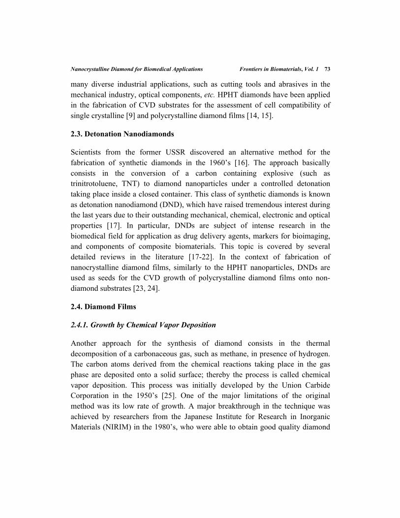
application of MCD films is therefore limited to the fabrication of abrasive and cutting tools used in dentistry and orthopedic surgery [33, 34]. However, it is expected that the nanocrystalline films, those in which crystallite size is below 100 nm, will reach a much larger impact within the biomedical field. Nanocrystalline diamond films are further subdivided in nanocrystalline diamond (NCD) or ultrananocrystalline diamond (UNCD) according to a criterion that will be discussed in the following section. Fig. (2) presents scanning electron micrographs displaying the typical morphology of PCD films. As previously mentioned, nanocrystalline diamond films and their biomedical applications constitute the main focus of this chapter.
Fig. (2). Typical morphology of the polycrystalline diamond films grown by CVD. Top and cross sectional view scanning electron micrographs of different types of polycrystalline diamond films grown on Si substrates. (a) MCD film displaying clearly visible micrometer-sized grains and columnar growth. (b) NCD film of approx. 400 nm of thickness grown from hydrogen rich plasma. (c) UNCD film of approx. 1 µm in thickness grown from Ar rich plasma. Modified from ref. [11], by Vermeeren et al.
76 Frontiers in Biomaterials, Vol. 1 Pennisi and Alcaide
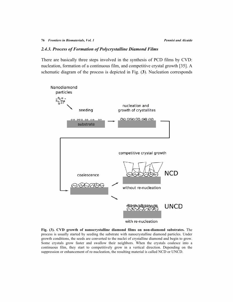
2.4.3. Process of Formation of Polycrystalline Diamond Films
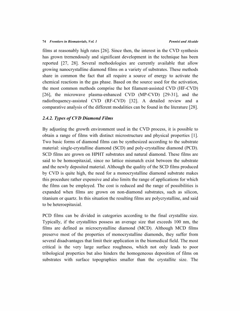
There are basically three steps involved in the synthesis of PCD films by CVD: nucleation, formation of a continuous film, and competitive crystal growth [35]. A schematic diagram of the process is depicted in Fig. (3). Nucleation corresponds
Fig. (3). CVD growth of nanocrystalline diamond films on non-diamond substrates. The process is usually started by seeding the substrate with nanocrystalline diamond particles. Under growth conditions, the seeds are converted to the nuclei of crystalline diamond and begin to grow. Some crystals grow faster and swallow their neighbors. When the crystals coalesce into a continuous film, they start to competitively grow in a vertical direction. Depending on the suppression or enhancement of re-nucleation, the resulting material is called NCD or UNCD.
Nanocrystalline Diamond for Biomedical Applications Frontiers in Biomaterials, Vol. 1 77
to the formation of the smallest thermodynamically stable diamond nuclei at the substrate surface. Since this initial step could take prohibitively long time in non-diamond substrates due to the absence of initial sites for a stable carbon binding, diverse strategies have been employed to provide a template for the diamond film [36]. One of the most useful strategiesconsists in the use of diamond nanoparticles (DND or HPHT), which are pre-adsorbed in the substrate to facilitate the nucleation [37]. Depending on the growth conditions, the nuclei will grow into larger grains and eventually coalesce onto a continuous film, in which some of these grains have fused together. Subsequently, the grains start to grow in a direction perpendicular to the substrate, leading to a columnar structure in which faster growing structures overgrow the slower ones. Diverse parameters, such as initial seeding density, growth chemistry and surface temperature will determine the relative growth rate of the columns and the final mechanical, morphological and chemical properties of the film [23]. Films that are grown with a very high initial nucleation density, in a conventional diamond growth process (in which the ratio of hydrogen to methane is high), and whose thickness does not typically exceed few microns, tend to have grainssmaller than 100 nm and are designated as nanocrystalline diamond (NCD) [38]. Other types of films, which are grown in Ar-rich plasma with little or no hydrogen content, display a significant interruption of the crystal formation process. This is caused by the appearance of new nucleation sites, meaning that grain coarsening does not occur. This type of diamond films, in which a high re-nucleation rate keeps the grain size in the range of few nanometers independently of the film thickness, are denominated ultrananocrystalline diamond (UCND) [39].
3. CONTROLLING THE PROPERTIES OF NANOCRYSTALLINE DIAMOND FILMS
3.1. Surface Termination
Surface properties such as wettability, reactivity, and surface conductivity of a material are strongly dependent on the surface termination. As-grown nanocrystalline diamond films present a hydrogen-terminated surface, which is stable under atmospheric conditions and displays a contact angle around 93o [40]. In addition, H-terminated diamond surfaces display a high surface conductivity
78 Frontiers in Biomaterials, Vol. 1 Pennisi and Alcaide
[41]. Upon appropriate physical or chemical treatment, the surface termination can be changed by several functional groups. Thus, O-terminated diamond surfaces can be obtained by exposure to an O2 rich plasma [4, 24, 42, 43] or by UV oxidation [44]. Upon oxidation diamond surfaces are rendered hydrophilic and the surface potential vanishes. Depending on the oxidation level, the water contact angle can be reduced below 5o. On the contrary, by exposing the diamond surface to fluorine radicals, such as by using CF4 plasmas, superhydrophobic diamond surfaces can be obtained [45].
The primary aim of surface modification is to tailor the surface properties of the diamond. As a general principle, as it will be shown later, hydrophilic surfaces are preferred to promote cell attachment while hydrophobic and superhydrophobic surfaces are used to prevent biofouling of the material. However, modification of the diamond surface termination is also desired when the aim is to promote the covalent attachment of a variety of molecules such as proteins, DNA, or macromolecular complexes, as it will be described in the following sections.
3.2. Surface Functionalization
Covalent grafting of diverse organic or bioorganic molecules can extend the functionalities offered by the different surface termination groups described in the previous section. Molecular monolayers grafted to the diamond surfaces can provide the moieties for the covalent coupling of a wide variety of biomolecules, such as DNA or proteins [46]. In addition, the presence of an intermolecular layer between the material and the bioorganic molecules can help preventing non-specific adsorption. Most of the approaches for the covalent grafting of organic molecular layers on diamond have been developed for H-terminated surfaces. The most common method consists in photochemical grafting of alkene-based molecules [47]. Functionalization can also be induced by means of simple chemical or electrochemical reactions, grafting the organic layers via reaction with diazonium salts [48, 49]. For the functionalization of O-terminated diamond surfaces, some of the primary functionalization methodsinclude silanization [50] and esterification [51], both dependent on the reaction with surface hydroxyl groups present in the surface upon oxidation.
Nanocrystalline Diamond for Biomedical Applications Frontiers in Biomaterials, Vol. 1 79
The molecular monolayers grafted on the diamond surface provide specific reactive groups to which the bioorganic molecules can covalently attach. For the covalent attachment of proteins, the reactive groups are usually selected to react with the amine of surface lysine residues or the thiol of surface cysteine residues [52].
3.3. Doping
The crystalline structure of diamond is constituted by a three dimensional lattice of carbon atoms forming very strong C-C bonds, displaying a with a wide 5.5 eV band gap. This makes diamond an excellent electrical insulator, with a typical conductivityabove109 Ωcm. Polycrystalline CVD films are also insulating in nature, though conductivity is increased due to the presence of graphitic carbon, impurities and lattice defects. Extrinsic conductivity can be induced by means of doping, which can produce diamond CVD films displaying a wide range of electrical properties, from semiconductor to semi-metallic behavior depending on the level of doping [23, 27].
NCD films can achieve a p-type semiconductor behavior by means of boron doping. Boron doping is realized by addition of a boron containing gas such as diboraneor trimethylboron during the CVD process. Heavily boron doped NCD films represent an attractive material for use as electrode in bioelectrochemical applications [53, 54]. The main advantages of this type of electrodes are the low and stable capacitive background current, which allows larger sensitivities, and the very wide potential window for water stability (up to 3.2 V in water, in contrast to 1.3 V for platinum). Although the synthesis and characterization of boron doped NCD films has been extensively studied, the fabrication of n-type doped diamond films remains a challenge [53, 55, 56].
In the case of UNCD films, doping is achieved by incorporation of nitrogen in the gas mixture during the CVD deposition [57]. Nitrogen does not act as conventional dopant, but it is believed that nitrogen atoms are incorporated into the grain boundaries, giving rise to graphitic grain boundary conduction [58].
80 Frontiers in Biomaterials, Vol. 1 Pennisi and Alcaide
5. BIOMEDICAL APPLICATIONS OF NANOCRYSTALLINE DIAMOND FILMS
5.1. Surfaces Supporting Cellular Adhesion
In the context of cell material interactions, adhesion is the process involving the development of anchorage contacts between the cells and material surface. Although in the absence of serum proteins cells can adhere by nonspecific interactions on surfaces, here we only consider relevant to discuss approaches in which integrin mediated anchorage is involved, since interaction between integrins and cell adhesion molecules regulates cell behaviours like proliferation, migration, and differentiation [59-61]. As it will be described in the following sections, nanocrystalline diamond films provided by appropriate chemistries or cell-adhesion ligands seem to be an optimal approach for a variety of applications in which cell adhesion to a material surface is desired, both in vitro and in vivo.
5.1.1. Interfacing Bonetissue for Orthopaedic Applications
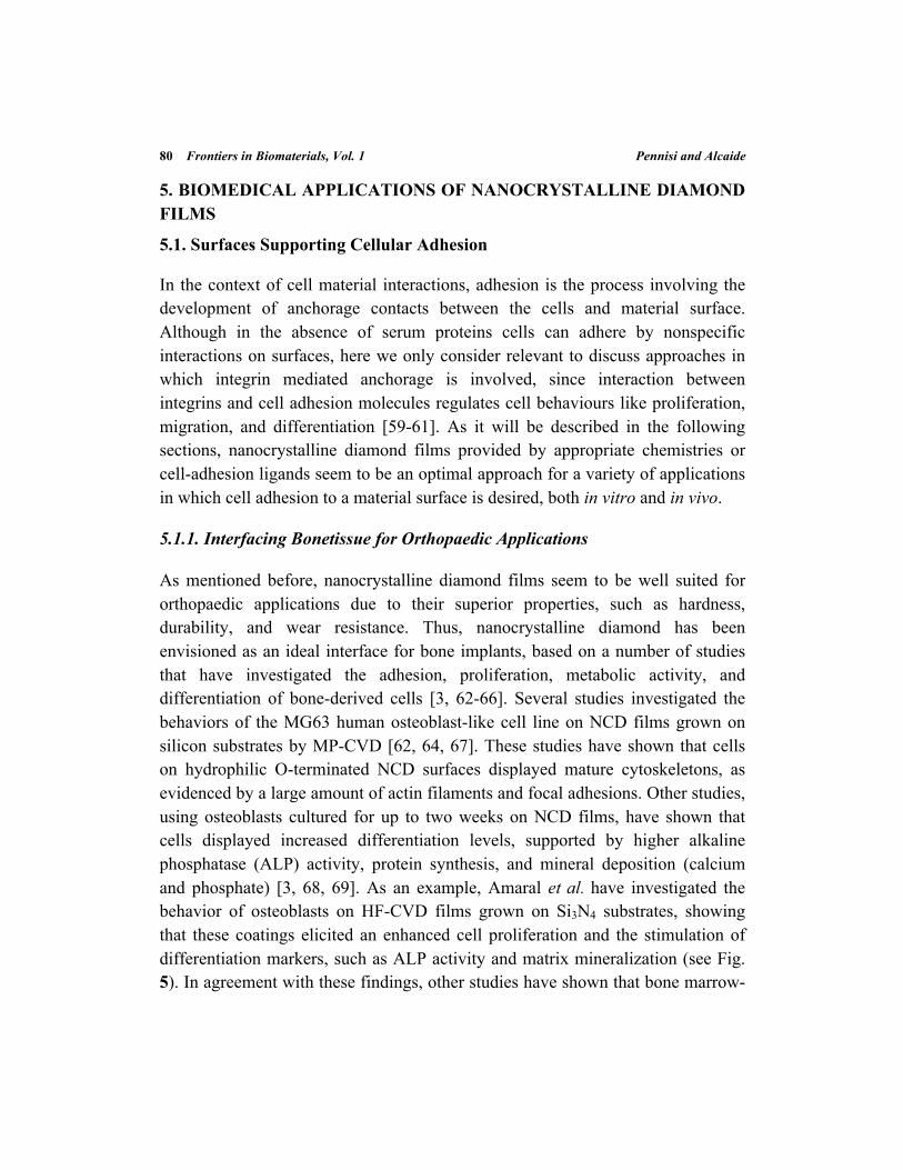
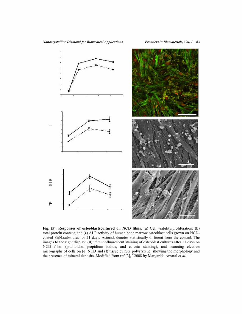
As mentioned before, nanocrystalline diamond films seem to be well suited for orthopaedic applications due to their superior properties, such as hardness, durability, and wear resistance. Thus, nanocrystalline diamond has been envisioned as an ideal interface for bone implants, based on a number of studies that have investigated the adhesion, proliferation, metabolic activity, and differentiation of bone-derived cells [3, 62-66]. Several studies investigated the behaviors of the MG63 human osteoblast-like cell line on NCD films grown on silicon substrates by MP-CVD [62, 64, 67]. These studies have shown that cells on hydrophilic O-terminated NCD surfaces displayed mature cytoskeletons, as evidenced by a large amount of actin filaments and focal adhesions. Other studies, using osteoblasts cultured for up to two weeks on NCD films, have shown that cells displayed increased differentiation levels, supported by higher alkaline phosphatase (ALP) activity, protein synthesis, and mineral deposition (calcium and phosphate) [3, 68, 69]. As an example, Amaral et al. have investigated the behavior of osteoblasts on HF-CVD films grown on Si3N4 substrates, showing that these coatings elicited an enhanced cell proliferation and the stimulation of differentiation markers, such as ALP activity and matrix mineralization (see Fig. 5). In agreement with these findings, other studies have shown that bone marrow-
Nanocrystalline Diamond for Biomedical Applications Frontiers in Biomaterials, Vol. 1 81
derived mesenchymal stem cells also increase their metabolic activity on NCD films, even though their adhesion is similar to polystyrene control surfaces [70]. From these studies, it appears to be a correlation between surface properties (specific topography and chemistry) of the nanocrystalline diamond films and osteoblast functions, since cells show enhanced cell functions on O-terminated and NH3-terminated surfaces rather than H-terminated surfaces [4, 68]. In addition, it has also been suggested that osteoblast functions may be enhanced on NCD surfaces presenting a surface topography similar to that of the bone [70].
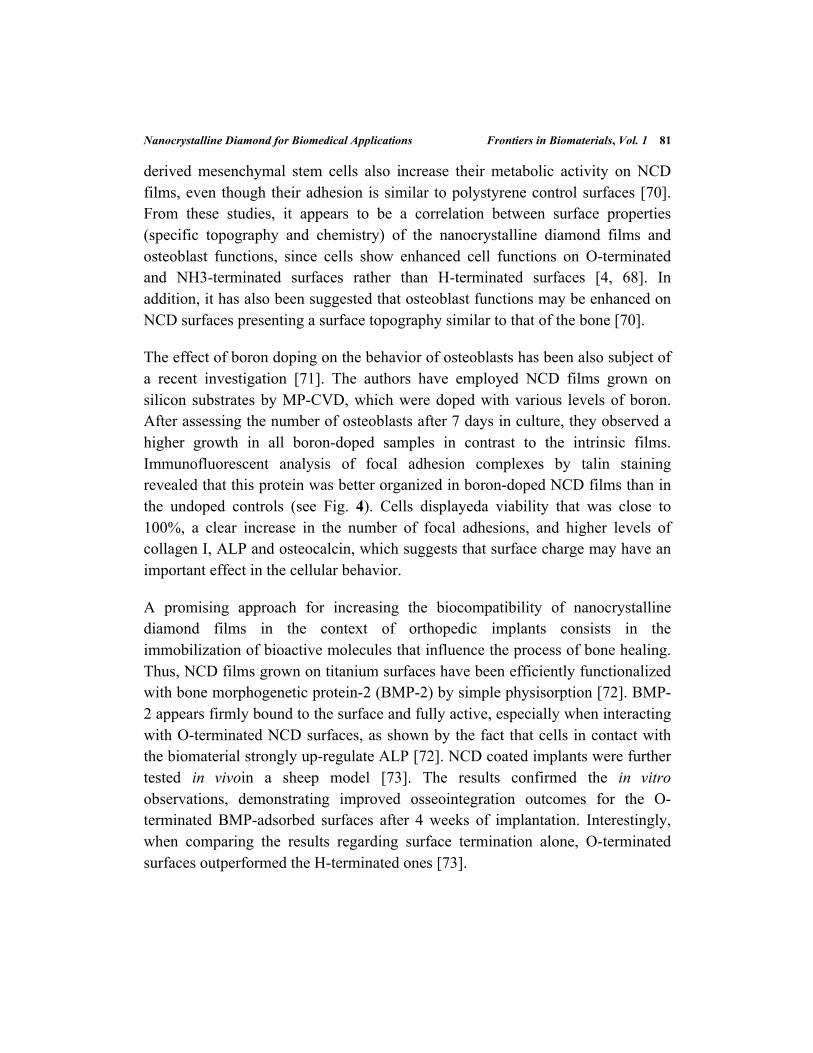
The effect of boron doping on the behavior of osteoblasts has been also subject of a recent investigation [71]. The authors have employed NCD films grown on silicon substrates by MP-CVD, which were doped with various levels of boron. After assessing the number of osteoblasts after 7 days in culture, they observed a higher growth in all boron-doped samples in contrast to the intrinsic films. Immunofluorescent analysis of focal adhesion complexes by talin staining revealed that this protein was better organized in boron-doped NCD films than in the undoped controls (see Fig. 4). Cells displayeda viability that was close to 100%, a clear increase in the number of focal adhesions, and higher levels of collagen I, ALP and osteocalcin, which suggests that surface charge may have an important effect in the cellular behavior.
A promising approach for increasing the biocompatibility of nanocrystalline diamond films in the context of orthopedic implants consists in the immobilization of bioactive molecules that influence the process of bone healing. Thus, NCD films grown on titanium surfaces have been efficiently functionalized with bone morphogenetic protein-2 (BMP-2) by simple physisorption [72]. BMP-2 appears firmly bound to the surface and fully active, especially when interacting with O-terminated NCD surfaces, as shown by the fact that cells in contact with the biomaterial strongly up-regulate ALP [72]. NCD coated implants were further tested in vivoin a sheep model [73]. The results confirmed the in vitro observations, demonstrating improved osseointegration outcomes for the O-terminated BMP-adsorbed surfaces after 4 weeks of implantation. Interestingly, when comparing the results regarding surface termination alone, O-terminated surfaces outperformed the H-terminated ones [73].
82 Frontiers in Biomaterials, Vol. 1 Pennisi and Alcaide
Fig. (4). Effect of boron doping on cell adhesion. Immunofluorescence staining of talin in MG 63 osteoblast-like cells on day 3 after seeding on: (A) glass coverslips, (B) undoped NCD, (C) NCD films doped with boron in concentrations of 133 ppm, (D) 1000 ppm, and (E) 6700 ppm. The cell nuclei are counterstained with propidium iodide. Calibration bar = 10 µm. Modified from ref. [69], ©2011 by Grausova et al.
In summary, surface properties of the NCD films such as chemistry, topography and charge have a strong influence on the fate of cultured osteoblasts, which reflects the fact that cell behavior is highly dependent on the microenvironmental conditions. As previously mentioned, the surface properties of NCD films can be easily tailored, which could be used to modulate osteoblast proliferation and bone growth in orthopedic implants and other bone tissue engineering applications.
5.1.2. Interfacing Soft Tissue
NCD films also represent an attractive alternative for biomedical implants in which an improvement of soft-tissue attachment is required, such as in the transmucosal part of osseointegrated dental implants. In these applications, the aim is to create a soft tissue seal to prevent infections and implant loosening [74]. Since the tissue seal involves both epithelial and connective tissue components, several studies have investigated in vitro the adhesion of epithelial cell types and fibroblasts on nanocrystalline films.
Nanocrystalline Diamond for Biomedical Applications Frontiers in Biomaterials, Vol. 1 83
Fig. (5). Responses of osteoblastscultured on NCD films. (a) Cell viability/proliferation, (b) total protein content, and (c) ALP activity of human bone marrow osteoblast cells grown on NCD-coated Si3N4substrates for 21 days. Asterisk denotes statistically different from the control. The images to the right display: (d) immunofluorescent staining of osteoblast cultures after 21 days on NCD films (phalloidin, propidium iodide, and calcein staining), and scanning electron micrographs of cells on (e) NCD and (f) tissue culture polystyrene, showing the morphology and the presence of mineral deposits. Modified from ref [3], ©2008 by Margarida Amaral et al.
84 Frontiers in Biomaterials, Vol. 1 Pennisi and Alcaide
Bajaj and coworkers have compared cell adhesion, proliferation, and growth of HeLa cells and fibroblasts on UNCD films. They have found an increased attachment and proliferation of cells on UNCD films, when comparing to other substrates like platinum or silicon [75]. Accordingly, other studies have found that primary fibroblastsdisplayed the maximum cytoplasmic and nuclear area on UNCD films, indicating a greater affinity of the cells for the material [44]. Studies by Lechleitner et al. have shown the importance of surface termination in controlling epithelial cell adhesion to NCD surfaces. They have shown that attachment and proliferation of these cells is enhanced on O-terminated surfaces, in contrast to H- terminated surfaces, suggesting that the lack of functional polar groups prevents adherent cells from settling on the NCD surface [76]. On the other hand, Amaral et al. have assessed the behavior of L929 fibroblasts and human gingival fibroblasts cultured on NCD films grown on Si3 N4 ceramic substrates by HF-CVD [77]. After analyzing cell adhesion, viability, and proliferation for 8 days, they have shown that cell proliferation is slightly higher for both cell types compared to polystyrene controls. NCD coatings offered a suitable surface for cell attachment, spreading and proliferation and were completely covered with continuous cell layers after few days. Remarkably, their films were used “as prepared”, which indicates that cell adhesion to the NCD surface was enhanced even without a hydrophilic treatment or surface functionalization. Smisdom et al. have assessed the growth and viability of Chinese hamster ovary (CHO) cells cultured on H- and O- terminated MP-CVD MCD and NCD films [78]. They have shown absence of toxicity andan equivalent growth of cells on all surfaces irrespective of the topographyor surface termination. This apparent lack of effect from surface chemistry and topography might be explained by recent studies from Klauser and coworkers. They have assessed the behavior of epithelial cells on O-terminated NCD coatings, showing that cells are able to attach after 24 to 72 hours with and without addition of fetal bovine serum (FBS) to the growth medium. Remarkably, the experiments performed on hydrophobic surfaces (H-NCD and F-NCD) have shown that cell-adhesion is only possible in the presence of FBS, suggesting that the proteins contained in the serum are important mediators of cell adhesion, independently of their chemical termination [24].
Nanocrystalline Diamond for Biomedical Applications Frontiers in Biomaterials, Vol. 1 85
One of the first reports of the in vivo biocompatibility of NCD films has been carried out using a subcutaneous implantation rat model [79]. This study aimed to assess the tissue responses to implanted titanium discs in which NCD was grown by HF-CVD. Their results show that hydrophilicity has a positive influence on the tissue healing at the implant surfaces, revealed by an increase in the number of cells and the attachment of connective tissue to the O-terminated implant surfaces. Interestingly, the inflammatory response was also reduced in the peri-implant area of the O-terminated implants.
5.1.3. Neural Interfacing Applications
Nanocrystalline diamond films are also being investigated as promising materials to improve the stability of neural interfaces, for applications ranging from in vitro platforms used for neurophysiologic studies to implantable neural prosthesis [6, 9, 80]. The interaction of neural cells with nanocrystalline diamond films and particles has been primarily studied in vitro using neural cell lines. These studies have demonstrated that cells are able to attach, display high viability, low content of reactive oxygen species, and lack of alteration of the mitochondrial membrane [81-83]. Ariano et al. have also observed that neural cell lines can adhere and maintain their functionality when cultured on H-terminated NCD substrates despite its hydrophobic nature, revealing the spontaneous electrical activity by capacitive coupling with the cell membrane [9]. Other studies investigated the behavior of primary neuronson functionalized NCD films, assessing parameters such as neuronal morphology, outgrowth, synaptic activity, ion channel availability, and calcium signals during stimulation [84, 85]. These studies have shown that primary neurons can be successfully cultured for several days on the NCD films, display ingneurite extensions, and keeping their electrical properties, including spontaneous action potentials. However, unlike transformed cell lines or other primary cell types such as fibroblasts, surface functionalization was needed to successfully promote neural cell attachment.
UNCD films also represent promising substrates for neural applications, since it has been shown that they are able to spontaneously induce neuronal differentiation of neural stem cells [8]. Using MP-CVD UNCD films grown on quartz substrates, both H- and O- terminated, it was found that stem cell
86 Frontiers in Biomaterials, Vol. 1 Pennisi and Alcaide
differentiation is promoted even in absence of differentiation reagents. In the case of H-terminated samples, the mechanism seems to be mediated by the absorption of fibronectin, which activates an integrin β1-Fak-Erk signaling pathway. Furthermore, the different terminations, -H or -O, possess different neural differentiation abilities towards neurons and oligodendrocytes respectively [7, 8]. These observations suggest that UNCD could be used as a potential material for central nervous system applications in tissue engineering or cell transplantation
5.2. Antifouling Surfaces
The progress on implant technologies requires special attention to possible surface colonization by microorganisms. It is estimated that around 60% of all microbial infections are caused by biofilms that create a reservoir for immunologically quiet bacteria, release toxic substances and can cause high resistance to antibiotics. Bacterial adhesion and biofilm formation are both mediated by proteins adsorbed onto the material surface. In this direction, several studies have focused on the analysis of biomaterial susceptibility to bacterial adhesion and infection and their capacity to reduce colonization and formation of biofilms. Observations on the bactericide and bacterial anti-adhesive properties of different materials when incubated with E. Coli cells, with and without pre-treatment with proteins, have evidenced that bacterial colonization depends on the previous adhesion of proteins and that CVD-NCD surfaces present the highest resistance to it, even in the presence of plasma proteins, compared to other materials such as stainless steel, and very similar to titanium [94]. Furthermore, Medina et al. have analysed the ability of Pseudomona aeruginosa to colonize different surfaces, including H-terminated HF-CVD NCD [86]. NCD exhibits bactericidal effects within 24 hours and shows the lowest bacterial colonization density after Cu. Surface roughness and free energy are parameters expected to influence the rate of bacterial colonization of the different surfaces. However, not all data show a correlation between them and the anti-adhesive activity of the materials and, even though it is generally believed that hydrophobic surfaces with a large contact angle favour anti-bacterial properties, results here are contradictory, with cases when a decrease of bacterial adhesion is correlated by a reduction in the hydrophobicity. As previously mentioned, interaction of cells with NCD is favoured by O-terminated surfaces and bacteria require similar conditions for their survival, it is
Nanocrystalline Diamond for Biomedical Applications Frontiers in Biomaterials, Vol. 1 87
expected that H-terminated NCD substrates will not favour their colonization. It appears to be an association of the anti-bacterial properties of NCD and its semiconductivity, so that the electrically active surface of NCD interacts with the bacterial membrane altering its morphology and avoiding adhesion and colonization [86].
Another major problem of the adhesion of surface-active materials, such as proteins, is the interference with working electrodes that lead to alterations and even suppression of the recording signals. The resistance of diamond electrodes to protein adhesion has been evidenced by voltammetry measurements in the presence of various proteins and surfactants showing that, specifically in the case of boron-doped diamond films, this material displays insignificant fouling effects and is very resistant to surfactant interferences, making it ideal for electro analytical applications [87].
5.3. Patterning of Cells and Proteins
Micropatterning of living cells on solid substrates has recently attracted much attention due to its extensive range of applications, especially for cell-based bioassays, tissue-engineering and fundamental studies of cell biology [88-90]. Micrometer-sized patterns allow the study of cell sensitivity for fine spatial cues and thus enable more complex investigations of morphogenesis, cell polarity, and cell division axis. Most cell micropatterning methods focus on how interactions between cells and surfaces control cell adhesion.
A variety of techniques have been developed for patterning cellular growth. The most commonly used methods include micro-contact printing (MCP), photolithography, inkjet printing and stencil-assisted patterning techniques. These techniques generally involve patterning proteins or factors that either attract (extra-cellular matrix proteins, such as poly-lysine) or repel cellular attachment(anti-biofouling agents), although some techniques have focused on the direct placement of cells. MCP has been used to produce patterns of laminin and poly-lysine for spatially directing the growth of primary hippocampal and cortical neurons on many different substrates, including glass, polystyrene and diamond [81]. Inkjet printing has been widely used for depositing patterns of liquids and
88 Frontiers in Biomaterials, Vol. 1 Pennisi and Alcaide
suspensions onto surfaces at micrometer-scale resolutions. Laser micromachining uses a high-power focused laser beam to directly write patterns into the substrate surface, or into the polymer adhesion layer upon which the cells subsequently grow. UV-directed light micropatterning has been recently introduced for its ability either to oxidize surface coatings, and thus destroy their anti-adhesive properties, or to de-protect or activate photosensitive linkers [91].
Most of these techniques are being used in central nervous system applications, where the aim is to direct neuronal growth in an ordered manner while reducing glial scarring, thus improving the recorded signal quality to increase the long-term performance of devices such as brain computer interfaces (BCI) [80]. Electrodes fail very often when implanted into the central nervous system because of the glial encapsulation, known as glial scarring, that limits their long-term functionality.
Due to its excellent properties, CVD diamond has attracted much attention as a base material for neural implant coatings to achieve reduced levels of glial scarring and spatially control growth of neurons on different surfaces. Its mechanical and chemical properties make it very suitable for improving the long-term performance of the invasive electrodes systems used in BCIs, as it does the fact that can be modified with dopants. In the case of boron doping, it increases the electrical conductivity of diamond making it more suitable for electrode applications. Therefore boron-doped NCD has been tested in a number of studies for spatially directing neural cell growth and limiting the attachment of cells involved in the immune response and foreign body reaction. May et al. have successfully cultured neurons on H- and O- terminated boron-doped NCD surfaces by laser micro machining, achieving a spatially directed neuronal growth along predesigned pathways. Cells grow well across the coatings avoiding crossing over the etched areas and forming a spatially defined surface [92]. Regan et al. have tried different micropatterning techniques including inkjet printing and laser micro-machining to control neural adhesion and modify inflammatory cell attachment onto boron-doped NCD surfaces resulting in an improvement in neural connectivity [80]. Marcon et al. report a new method using UV-directed light to pattern cells in a spatially controlled manner exploiting the superhydrophobic/superhydrophilic wetting contrast of chemically functionalized boron- doped NCD nanowires that has a great impact on cell adhesion. This

Nanocrystalline Diamond for Biomedical Applications Frontiers in Biomaterials, Vol. 1 89
approach enables the production of single-cell arrays without any geometrical constraints, opening a wide range of possible applications in the development of cell-based biological assays in well-controlled and biologically relevant environments [91].
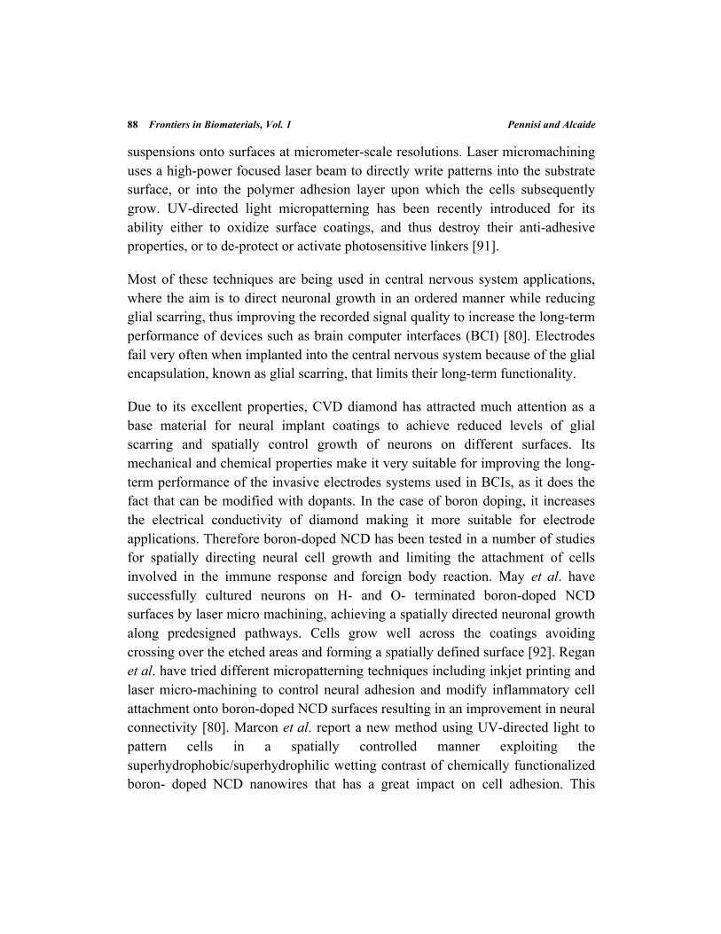
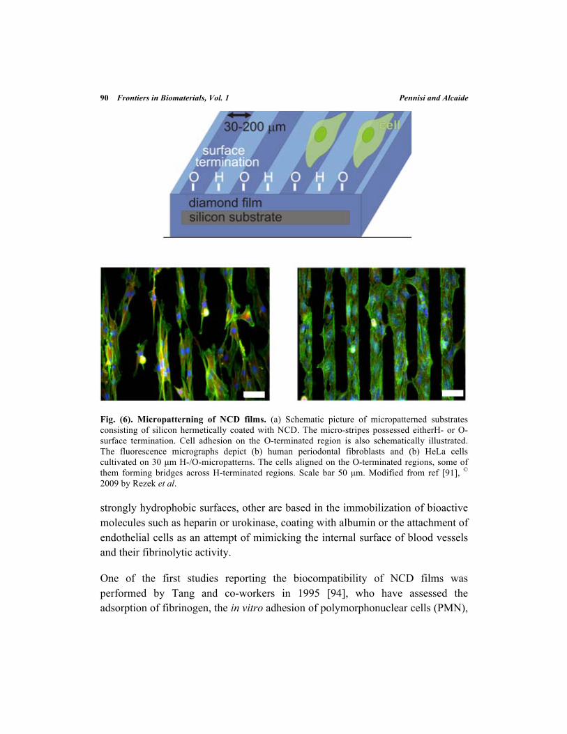
Although most work has been done with neural cells, some other studies have used other cells, like osteoblasts, showing a selective adhesion and arrangement of these cells on NCD grown on silicon substrates by CVD, which was microscopically patterned with H- and O- terminated regions (see Fig. 6) [93]. Control of cellular density and serum concentration in the cell medium influences colonization of the substrates with preference for O-terminated regions and a lower evolution of cell morphology in H-terminated areas. In this case, to reach an optimal status, cells communicate and secrete ECM, modifying the surface. This mechanism enables to overgrow electrically conductive H-terminated surfaces when surrounded by O-terminated regions at small dimensions, making it very attractive for bio-electronic applications [93]. Although this area of research is still in development, the results obtained so far indicate that microtailored NCD substrates could provide great benefits for applications in bioelectronics, tissue engineering and biotechnology.
5.4. Development of Non-Thrombogenic Coatings
Hemocompatibility is a characteristic of great importance for a biomaterial, since blood is usually the first body fluid contacting with implants or other medical devices. The interaction of blood with biomaterials includes, primarily, adsorption of plasma proteins on their surfaces right after implantation followed by platelet adhesion that contributes to a surface-induced thrombosis. An inflammatory reaction takes place as well, with the recruitment of leukocytes, fibroblast proliferation and collagen synthesis. Thus, a long exposure to blood can cause embolization, calcification and changes in the biomaterial properties that can compromise the stability and functionality of the implanted devices.
To reduce clotting, materials and surfaces with high protein-resistant properties have been investigated. Some strategies have developed strongly hydrophilic and
90 Frontiers in Biomaterials, Vol. 1 Pennisi and Alcaide
Fig. (6). Micropatterning of NCD films. (a) Schematic picture of micropatterned substrates consisting of silicon hermetically coated with NCD. The micro-stripes possessed eitherH- or O- surface termination. Cell adhesion on the O-terminated region is also schematically illustrated. The fluorescence micrographs depict (b) human periodontal fibroblasts and (b) HeLa cells cultivated on 30 µm H-/O-micropatterns. The cells aligned on the O-terminated regions, some of them forming bridges across H-terminated regions. Scale bar 50 µm. Modified from ref [91], © 2009 by Rezek et al.
strongly hydrophobic surfaces, other are based in the immobilization of bioactive molecules such as heparin or urokinase, coating with albumin or the attachment of endothelial cells as an attempt of mimicking the internal surface of blood vessels and their fibrinolytic activity.
One of the first studies reporting the biocompatibility of NCD films was performed by Tang and co-workers in 1995 [94], who have assessed the adsorption of fibrinogen, the in vitro adhesion of polymorphonuclear cells (PMN),
Nanocrystalline Diamond for Biomedical Applications Frontiers in Biomaterials, Vol. 1 91
and the inflammatory responses after 1 week of implantation in a mice model. The amount of adsorbed fibrinogen to NCD films was similar than to other biomaterials, including stainless steel and titanium. The adhesion of PMN to plasma pre-incubated samples was similar to that on stainless steel but 40% lower than that on titanium. Similar to the other biomaterials, minimal inflammatory responses were found on the implanted NCD samples [94].
Okroj et al. have shown that NCD-coated steel exhibits a higher level of resistance to blood platelet adhesion and thrombus formation than the bare material, presenting a practically free-of adhered platelets surface after 1 hour incubation with whole blood. In addition, the NCD coating can also prevent ion release [95]. These observations correspond well with other in vitro studies showing that NCD films cause reduced plasma protein adsorption [96] and that aggregation of proteins or platelets barely occurs on the surface of the films [97]. Further indication of the excellent hemocompatibility of NCD films has been recently provided by an in vivo study, which has shown that NCD coated stents are able to significantly reduce the neointimal hyperplasia in a pig coronary artery model [98]. In perspective, it is expected that NCD films will be established as blood contacting material for use in cardiovascular applications such as heart valves and vascular stents.
5.5. Immobilization of Biomolecules for Biosensing Applications
A biosensor is a device that uses biological receptors for the detection of an analyte that usually is a biological substance too. In general, the detection is performed by selective, biological receptors such as enzymes, antibodies, nucleic acids, membranes or cells, whereas biomimetic sensors use synthetic receptors, such as molecularly imprinted polymers. Biosensors are devices designed to detect or quantify biochemical molecules, and they have been widely used as powerful analytical tools in areas such as medical diagnostics, food industry, and environmental monitoring. A biosensor is usually fabricated by immobilizing a biological receptor material on the surface of a suitable transducer that converts the biochemical signal into quantifiable electronic signals, which can be used to detect proteins, nucleic acids or antigen-antibody interactions. An ideal biosensor should combine nature’s sensitivity and specificity with the advantages of modern microelectronics [10, 99, 100].
92 Frontiers in Biomaterials, Vol. 1 Pennisi and Alcaide
Thanks to its dual role as a substrate for bio-functionalization, presenting high strength of C-C bonds, and as an electrode, with the capacity to promote different electron transfer reactions, its low background current and its large electrochemical potential window, nanocrystalline diamond films are particularly attractive substrates for biosensor applications. Recent advances in the synthesis of highly conducting NCD thin films have lead to an entirely new class of electrochemical biosensors and bioinorganic interfaces. Diamond nanowires as well can be a new approach towards next generation electrochemical gene sensor platforms.
The role of NCD has been investigated in the fabrication of biosensors binding very different molecules. Studies assessing different substrates for DNA immobilization have demonstrated that NCD films has superior properties to those of other materials such as glass, gold or silicon [99]. The general principle of the diamond-based DNA sensor research is to develop a prototype biosensor for diagnostic purposes based on DNA covalently attached to CVD diamond with the same sensitivity and specificity as the commonly used methods, such as blotting techniques and microarrays, but with the particular benefits of allowing real time and label-free measurements for optical, electronic and acoustic read-out [101, 102]. Other studies have tried to immobilize RNA and proteins in order to determine RNA-protein and RNA-RNA interactions on NCD surfaces [103]. The aim is to apply these experimental conditions based on RNA biosensor systems for a variety of biotechnological applications, such as screening approaches for early diagnosis of cancer.
Diamond films are also excellent platforms for studies using supported lipid bilayers to investigate physiological processes such as ligand-receptor interactions, membrane disruption and cell signaling. Optically transparent diamond films offer a unique combination of transparency and surface conductance, allowing detection of permeation events based on optical and field-effect properties, which have been used to investigate the effects of an antimicrobial peptide on the permeability of supported artificial lipid bilayers [104]. Other approach for highly sensitive measurement of peptide-induced membrane disruption consists in the use of conducting boron-doped diamond
Nanocrystalline Diamond for Biomedical Applications Frontiers in Biomaterials, Vol. 1 93
films, which are employed as active electrodes for the electrochemical impedance spectroscopy measurements [105].
Other biosensors that have been around for many years involve the use of many different types of enzymes, immobilization chemistries and substrates. Thus, glucose sensors for example, work using this principle, and the covalent immobilization of glucose oxidase or similar enzymes forms the basis for commercial devices. Garrido et al. have analyzed the immobilization of the enzyme catalase on diamond, showing promising results for the use of this material also for enzyme-based biosensors [99]. Another interesting application of nanocrystalline diamond films in the field of biosensors is that of linking antibodies to diamond surfaces (UNCD) for the detection of bacterial pathogens [106]. The UNCD film minimizes the desorption of antibody from the surface due to the strong chemical bond, while the activity is maintained due to reduced water density and reduced antibody-surface interactions. The ability to selectively capture bacterial cells can be kept stable even after exposure to buffer solutions at 37oC for periods up to 2 weeks [106].
6. SUMMARY
In this chapter, the interaction of nanocrystalline diamond films with biomolecules and cells, as well as approaches to control these interactions, have been reviewed. It has been shown that properties such as wettability, electric conductivity, and surface chemistry have a significant impact in the control of these interactions. Accordingly, it has been shown that the properties of nanocrystalline diamond films can be tailored to facilitate the control of biological responses such as adsorption and immobilization of proteins, cellular adhesion, and thrombogenicity. Although many correlations have been clearly established, some discrepancies still remain, as for instance regarding the effect of surface wettability on the protein and cellular attachment. These discrepancies are probably caused by the diversity in growth conditions, substrate preparation methods, surface pretreatment, and biological models that are employed by each research group. Therefore, one of the focus areas for further research will be related to standardization of the conditions for film growth and modification. For some of the intended applications the full potential of nanocrystalline diamond
94 Frontiers in Biomaterials, Vol. 1 Pennisi and Alcaide
films will be revealed only when appropriate proof-of-concept experiments are carried out, including relevant in vitro and in vivo assays.
LIST OF ABBREVIATION
CVD = Chemical vapor deposition
DND = Detonation nanodiamond
HF-CVD = Hot filament chemical vapor deposition
HPHT = High-pressure high-temperature diamond
MCD = Microcrystalline diamond
MP-CVD = Microwave plasma enhanced chemical vapor deposition
NCD = Nanocrystalline diamond
PCD = Poly-crystalline diamond
RF-CVD = Radiofrequency-assisted chemical vapor deposition
SCD = Single-crystalline diamond
UNCD = Ultrananocrystalline diamond
ACKNOWLEDGEMENTS
We are grateful to Andy Taylor for critically reading the manuscript and providing helpful comments. Also, we thank Veronique Vermeeren, Margarida Amaral, and Bohuslav Rezek for providing the original images reproduced in this work. This work was supported by the EU through the project MERIDIAN (Micro and Nano Engineered Bi-Directional Carbon Interfaces for Advanced Peripheral Nervous System Prosthetics and Hybrid Bionics), contract n. 280778-02.
CONFLICT OF INTEREST
The authors confirm that this chapter contents have no conflict of interest.
Nanocrystalline Diamond for Biomedical Applications Frontiers in Biomaterials, Vol. 1 95
REFERENCES
[1] Williams OA. Nanocrystalline diamond. Diam Relat Mater2011; 20: 621-40. [2] Yang L, Sheldon BW, Webster TJ. Orthopedic nano diamond coatings: Control of surface
properties and their impact on osteoblast adhesion and proliferation. J. Biomed. Mater. Res.2008; 91: 548-56.
[3] Amaral M, Gomes PS, Lopes MA, Santos JD, Silva RF, Fernandes MH. Nanocrystalline Diamond as a Coating for Joint Implants: Cytotoxicity and Biocompatibility Assessment. J Nanomater2008; 2008: 1-9.
[4] Yang L, Li Y, Sheldon BW, Webster TJ. Altering surface energy of nanocrystalline diamond to control osteoblast responses. J. Mater. Chem.2011; 22: 205.
[5] Xiao X, Wang J, Liu C, Carlisle JA, Mech B, Greenberg R, et al. In vitro and in vivo evaluation of ultrananocrystalline diamond for coating of implantable retinal microchips. J. Biomed. Mater. Res.2006; 77: 273-81.
[6] Hadjinicolaou AE, Leung RT, Garrett DJ, Ganesan K, Fox K, Nayagam DAX, et al. Electrical stimulation of retinal ganglion cells with diamond and the development of an all diamond retinal prosthesis. Biomaterials2012; 33: 5812-20.
[7] Chen YC, Lee DC, Hsiao CY, Chung YF, Chen HC, Thomas JP, et al. The effect of ultra-nanocrystalline diamond films on the proliferation and differentiation of neural stem cells. Biomaterials2009; 30: 3428-35.
[8] Chen YC, Lee DC, Tsai TY, Hsiao CY, Liu JW, Kao CY, et al. Induction and regulation of differentiation in neural stem cells on ultra-nanocrystalline diamond films. Biomaterials2010; 31: 5575-87.
[9] Ariano P, Budnyk O, Dalmazzo S, Lovisolo D, Manfredotti C, Rivolo P, et al. On diamond surface properties and interactions with neurons. Eur Phys J E Soft Matter2009; 30: 149-56.
[10] Wenmackers S, Vermeeren V, Vandeven M, Ameloot M, Bijnens N, Haenen K, et al. Diamond‐based DNA sensors: surface functionalization and read‐out strategies. Physica Status Solidi A Appl Res2009; 206: 391-408.
[11] Vermeeren V, Wenmackers S, Wagner P, Michiels L. DNA sensors with diamond as a promising alternative transducer material. Sensors2009; 9: 5600-36.
[12] Roy RK, Lee K-R. Biomedical applications of diamond-like carbon coatings: A review. J. Biomed. Mater. Res.2007; 83B: 72-84.
[13] Bundy FP, Hall HT, Strong HM, Wentorf RH. Man-made diamonds. Nature1955; 176: 51-5.
[14] Liu YL, Sun KW. Protein Functionalized Nanodiamond Arrays. Nanoscale Res Lett2010; 5: 1045-50.
[15] Girard HA, Perruchas S, Gesset C, Chaigneau M, Vieille L, Arnault J-C, et al. Electrostatic grafting of diamond nanoparticles: a versatile route to nanocrystalline diamond thin films. ACS Appl Mater Interfaces2009; 1: 2738-46.
[16] Danilenko VV. On the history of the discovery of nanodiamond synthesis. Phys Solid State2004; 46: 595-9.
[17] Mochalin VN, Shenderova O, Ho D, Gogotsi Y. The properties and applications of nanodiamonds. Nature Nanotech2011; 7: 11-23.
96 Frontiers in Biomaterials, Vol. 1 Pennisi and Alcaide
[18] Lam R, Ho D. Nanodiamonds as vehicles for systemic and localized drug delivery. Expert Opin. Drug Deliv.2009; 6: 883-95.
[19] Schrand AM, Suzanne A, Hens C, Shenderova OA. Nanodiamond particles: properties and perspectives for bioapplications. CRC Cr Rev Sol State2009; 34: 18-74.
[20] Krueger A. Beyond the shine: recent progress in applications of nanodiamond. J. Mater. Chem.2011; 21: 12571-8.
[21] Zhu Y, Li J, Li W, Zhang Y, Yang X, Chen N, et al. The biocompatibility of nanodiamonds and their application in drug delivery systems. Theranostics2012; 2: 302.
[22] Krueger A, Lang D. Functionality is Key: Recent Progress in the Surface Modification of Nanodiamond. Adv. Funct. Mater.2012; 22: 890-906.
[23] Williams OA, Nesládek M, Daenen M, Michaelson S, Hoffman A, Osawa E, et al. Growth, electronic properties and applications of nanodiamond. Diam Relat Mater2008; 17: 1080-8.
[24] Klauser F, Hermann M, Steinmüller Nethl D, Eiter O, Pasquarelli A, Bertel E, et al. Direct and Protein‐Mediated Cell Attachment on Differently Terminated Nanocrystalline Diamond. Chem Vapor Depos2010; 16: 42-9.
[25] Eversole WG. Synthesis of Diamond. USPTO; 3, 030, 187, 1958. [26] Matsumoto S, Sato Y, Tsutsumi M, Setaka N. Growth of diamond particles from methane-
hydrogen gas. J Mater Sci1982; 17: 3106-12. [27] Butler JE, Sumant AV. The CVD of nanodiamond materials. Chem Vapor Depos2008; 14:
145-60. [28] Gracio JJ, Fan QH, Madaleno JC. Diamond growth by chemical vapour deposition. J Phys
D Appl Phys2010; 43: 374017. [29] Kamo M, Sato Y, Matsumoto S, Setaka N. Diamond synthesis from gas phase in
microwave plasma. J Cryst Growth1983; 62: 642-4. [30] Bachmann PK. Microwave plasma CVD and related techniques for low pressure diamond
synthesis. In: Thin Film Diamond. Dordrecht: Springer Netherlands; 1994. pages 31-53. [31] Taylor A, Fendrych F, Fekete L, Vlcek J, Rezacova V, Petrak V, et al. Novel high
frequency pulsed MW-linear antenna plasma-chemistry: Routes towards large area, low pressure nanodiamond growth. Diam Relat Mater2011; 20: 613-5.
[32] Meyer DE. Radio-frequency plasma chemical vapor deposition growth of diamond. J. Vac. Sci. Technol. A1989; 7: 2325.
[33] Trava-Airoldi VJ, Corat EJ, Leite NF, do Carmo Nono M, Ferreira NG, Baranauskas V. CVD diamond burrs — Development and applications. Diam Relat Mater1996; 5: 857-60.
[34] Amar M, Ahmed W, Sein H, Jones AN, Rego CA. Chemical vapour deposition of diamond coatings onto molybdenum dental tools. J Phys: Condens Matter2003; 15: S2977-82.
[35] May PW. Diamond thin films: a 21st-century material. Philos T Roy Soc A2000; 358: 473-95.
[36] Liu H, Dandy DS. Studies on nucleation process in diamond CVD: an overview of recent developments. Diam Relat Mater1995; 4: 1173-88.
[37] Das D, Singh RN. A review of nucleation, growth and low temperature synthesis of diamond thin films. Int. Mat. Rev.2007; 52: 29-64.
[38] Philip J, Hess P, Feygelson T, Butler JE, Chattopadhyay S, Chen KH, et al. Elastic, mechanical, and thermal properties of nanocrystalline diamond films. J. Appl. Phys.2003; 93: 2164.
[39] Gruen DM. Nanocrystalline diamond films. Annu. Rev. Mater. Sci.1999; 29: 211-59.
Nanocrystalline Diamond for Biomedical Applications Frontiers in Biomaterials, Vol. 1 97
[40] Ostrovskaya L, Perevertailo V, Ralchenko V, Saveliev A, Zhuravlev V. Wettability of nanocrystalline diamond films. Diam Relat Mater2007; 16: 2109-13.
[41] Maier F, Riedel M, Mantel B, Ristein J, Ley L. Origin of Surface Conductivity in Diamond. Phys. Rev. Lett.2000; 85: 3472-5.
[42] Michalikova L, Rezek B, Kromka A, Kalbacova M. CVD diamond films with hydrophilic micro-patterns for self-organisation of human osteoblasts. Vacuum2009; 84: 61-4.
[43] Kromka A, Grausova L, Bacakova L, Vacik J, Rezek B, Vanecek M, et al. Semiconducting to metallic-like boron doping of nanocrystalline diamond films and its effect on osteoblastic cells. Diam Relat Mater2010; 19: 190-5.
[44] Chong KF, Loh KP, Vedula SRK, Lim CT, Sternschulte H, Steinmüller D, et al. Cell Adhesion Properties on Photochemically Functionalized Diamond. Langmuir2007; 23: 5615-21.
[45] Freedman A, Stinespring CD. Fluorination of diamond (100) by atomic and molecular beams. Appl. Phys. Lett.1990; 57: 1194.
[46] Szunerits S, Boukherroub R. Different strategies for functionalization of diamond surfaces. J Solid State Electr2008; 12: 1205-18.
[47] Hamers RJ, Butler JE, Lasseter T, Nichols BM, Russell JN Jr., Tse K-Y, et al. Molecular and biomolecular monolayers on diamond as an interface to biology. Diam Relat Mater2005; 14: 661-8.
[48] Kuo T-C, McCreery RL, Swain GM. Electrochemical Modification of Boron‐Doped Chemical Vapor Deposited Diamond Surfaces with Covalently Bonded Monolayers. Electrochem Solid St1999; 2: 288-90.
[49] Wang J, Firestone MA, Auciello O, Carlisle JA. Surface Functionalization of Ultrananocrystalline Diamond Films by Electrochemical Reduction of Aryldiazonium Salts. Langmuir2004; 20: 11450-6.
[50] Notsu H, Fukazawa T, Tatsuma T, Tryk DA, Fujishima A. Hydroxyl groups on boron-doped diamond electrodes and their modification with a silane coupling agent. Electrochem Solid St2001; 4: H1-H3.
[51] Delabouglise D, Marcus B, Mermoux M, Bouvier P, Chane-Tune JRM, Petit J-P, et al. Biotin grafting on boron-doped diamond. Chem. Commun.2003; : 2698.
[52] Garrido JA. Biofunctionalization of Diamond Surfaces: Fundamentals and Applications. In: Sussmann/CVD Diamond for Electronic Devices and Sensors. Chichester, UK: John Wiley & Sons, Ltd; 2009. pages 399-437.
[53] Vanhove E, de Sanoit J, Mailley P, Pinault MA, Jomard F, Bergonzo P. High reactivity and stability of diamond electrodes: The influence of the B‐doping concentration. Physica Status Solidi A Appl Res2009; 206: 2063-9.
[54] Wei JJ, Li CM, Gao XH, Hei LF, Lvun FX. The influence of boron doping level on quality and stability of diamond film on Ti substrate. Appl Surf Sci2012; 258: 6909-13.
[55] Kraft A. Doped diamond: a compact review on a new, versatile electrode material. Int. J. Electrochem. Sci2007; 2: 355-85.
[56] Luong JHT, Male KB, Glennon JD. Boron-doped diamond electrode: synthesis, characterization, functionalization and analytical applications. Analyst2009; 134: 1965-79.
[57] Bhattacharyya S, Auciello O, Birrell J, Carlisle JA, Curtiss LA, Goyette AN, et al. Synthesis and characterization of highly-conducting nitrogen-doped ultrananocrystalline diamond films. Appl. Phys. Lett.2001; 79: 1441.
98 Frontiers in Biomaterials, Vol. 1 Pennisi and Alcaide
[58] Birrell J, Gerbi JE, Auciello O, Gibson JM, Gruen DM, Carlisle JA. Bonding structure in nitrogen doped ultrananocrystalline diamond. J. Appl. Phys.2003; 93: 5606.
[59] Giancotti FG, Ruoslahti E. Integrin signaling. Science1999; 285: 1028-33. [60] Cavalcanti-Adam EA, Micoulet A, Blümmel J, Auernheimer J, Kessler H, Spatz JP. Lateral
spacing of integrin ligands influences cell spreading and focal adhesion assembly. Eur J Cell Biol 85: 219-24.
[61] Pennisi CP, Dolatshahi-Pirouz A, Foss M, Chevallier J, Fink T, Zachar V, et al. Nanoscale topography reduces fibroblast growth, focal adhesion size and migration-related gene expression on platinum surfaces. Colloids and Surfaces B: Biointerfaces2011; 85: 189-97.
[62] Bacakova L, Grausova L, Vacik J, Fraczek A, Blazewicz S, Kromka A, et al. Improved adhesion and growth of human osteoblast-like MG 63 cells on biomaterials modified with carbon nanoparticles. Diam Relat Mater2007; 16: 2133-40.
[63] Grausova L, Kromka A, Bacakova L, Potocky S, Vanecek M, Lisa V. Bone and vascular endothelial cells in cultures on nanocrystalline diamond films. Diam Relat Mater2008; 17: 1405-9.
[64] Kalbacova M, Michalikova L, Baresova V, Kromka A, Rezek B, Kmoch S. Adhesion of osteoblasts on chemically patterned nanocrystalline diamonds. Phys Status Solidi B Basic Solid State Phys2008; 245: 2124-7.
[65] Rezek B, Ukraintsev E, Kromka A, Ledinský M, Broz A, Nosková L, et al. Assembly of osteoblastic cell micro-arrays on diamond guided by protein pre-adsorption. Diam Relat Mater2010; 19: 153-7.
[66] Bacakova L, Filova E, Parizek M, Ruml T, Svorcik V. Modulation of cell adhesion, proliferation and differentiation on materials designed for body implants. Biotechnol. Adv.2011; 29: 739-67.
[67] Babchenko O, Kromka A, Hruska K, Kalbacova M, Broz A, Vanecek M. Fabrication of nano‐structured diamond films for SAOS‐2 cell cultivation. Physica Status Solidi A Appl Res2009; 206: 2033-7.
[68] Kalbacova M, Broz A, Babchenko O, Kromka A. Study on cellular adhesion of human osteoblasts on nano‐structured diamond films. Phys Status Solidi B Basic Solid State Phys2009; 246: 2774-7.
[69] Yang L, Sheldon BW, Webster TJ. The impact of diamond nanocrystallinity on osteoblast functions. Biomaterials2009; 30: 3458-65.
[70] Broz A, Baresova V, Kromka A, Rezek B, Kalbacova M. Strong influence of hierarchically structured diamond nanotopography on adhesion of human osteoblasts and mesenchymal cells. Physica Status Solidi A Appl Res2009; 206: 2038-41.
[71] Grausova L, Kromka A, Burdikova Z, Eckhardt A, Rezek B, Vacik J, et al. Enhanced growth and osteogenic differentiation of human osteoblast-like cells on boron-doped nanocrystalline diamond thin films. PLoS ONE2011; 6: e20943.
[72] Steinmüller-Nethl D, Kloss FR, Najam-Ul-Haq M, Rainer M, Larsson K, Linsmeier C, et al. Strong binding of bioactive BMP-2 to nanocrystalline diamond by physisorption. Biomaterials2006; 27: 4547-56.
[73] Kloss FR, Gassner R, Preiner J, Ebner A, LARSSON K, Hächl O, et al. The role of oxygen termination of nanocrystalline diamond on immobilisation of BMP-2 and subsequent bone formation. Biomaterials2008; 29: 2433-42.
[74] Berglundh T, Lindhe J, Ericsson I, Marinello CP, Liljenberg B, Thornsen P. The soft tissue barrier at implants and teeth. Clin Oral Implants Res1991; 2: 81-90.
Nanocrystalline Diamond for Biomedical Applications Frontiers in Biomaterials, Vol. 1 99
[75] Bajaj P, Akin D, Gupta A, Sherman D, Shi B, Auciello O, et al. Ultrananocrystalline diamond film as an optimal cell interface for biomedical applications. Biomed Microdevices2007; 9: 787-94.
[76] Lechleitner T, Klauser F, Seppi T, Lechner J, Jennings P, Perco P, et al. The surface properties of nanocrystalline diamond and nanoparticulate diamond powder and their suitability as cell growth support surfaces. Biomaterials2008; 29: 4275-84.
[77] Amaral M, Gomes PS, Lopes MA, Santos JD, Silva RF, Fernandes MH. Cytotoxicity evaluation of nanocrystalline diamond coatings by fibroblast cell cultures. Acta Biomater2009; 5: 755-63.
[78] Smisdom N, Smets I, Williams OA, Daenen M, Wenmackers S, Haenen K, et al. Chinese hamster ovary cell viability on hydrogen and oxygen terminated nano‐and microcrystalline diamond surfaces. Physica Status Solidi A Appl Res2009; 206: 2042-7.
[79] Kloss FR, Steinmüller Nethl D, Stigler RG, Ennemoser T, Rasse M, Hächl O. In vivo investigation on connective tissue healing to polished surfaces with different surface wettability. Clin Oral Implants Res2011; 22: 699-705.
[80] Regan EM, Taylor A, Uney JB, Dick AD, May PW, McGeehan J. Spatially controlling neuronal adhesion and inflammatory reactions on implantable diamond. IEEE J. Emerg. Sel. Topics Power Electron. 2011; 1: 557-65.
[81] Specht CG, Williams OA, Jackman RB, Schoepfer R. Ordered growth of neurons on diamond. Biomaterials2004; 25: 4073-8.
[82] Schrand AM, Huang H, Carlson C, Schlager JJ, Ōsawa E, Hussain SM, et al. Are Diamond Nanoparticles Cytotoxic? J. Phys. Chem. B2007; 111: 2-7.
[83] Frewin CL, Jaroszeski M, Weeber E, Muffly KE, Kumar A, Peters M, et al. Atomic force microscopy analysis of central nervous system cell morphology on silicon carbide and diamond substrates. J. Mol. Recognit.2009; 22: 380-8.
[84] Ariano P, Baldelli P, Carbone E, Gilardino A, Giudice Lo A, Lovisolo D, et al. Cellular adhesion and neuronal excitability on functionalised diamond surfaces. Diam Relat Mater2005; 14: 669-74.
[85] Thalhammer A, Edgington RJ, Cingolani LA, Schoepfer R, Jackman RB. The use of nanodiamond monolayer coatings to promote the formation of functional neuronal networks. Biomaterials2010; 31: 2097-104.
[86] Medina O, Nocua J, Mendoza F, Gómez-Moreno R, Ávalos J, Rodríguez C, et al. Bactericide and bacterial anti-adhesive properties of the nanocrystalline diamond surface. Diam Relat Mater2012; 22: 77-81.
[87] Shin D, Tryk DA, Fujishima A, Merko i A, Wang J. Resistance to Surfactant and Protein Fouling Effects at Conducting Diamond Electrodes. Electroanalysis2005; 17: 305-11.
[88] Chen CS, Mrksich M, Huang S, Whitesides GM, Ingber DE. Micropatterned Surfaces for Control of Cell Shape, Position, and Function. Biotechnol. Prog.1998; 14: 356-63.
[89] Kane RS, Takayama S, Ostuni E, Ingber DE, Whitesides GM. Patterning proteins and cells using soft lithography. Biomaterials1999; 20: 2363-76.
[90] Shen CJ, Fu J, Chen CS. Patterning cell and tissue function. Cel. Mol. Bioeng.2008; 1: 15-23.
[91] Marcon L, Addad A, Coffinier Y, Boukherroub R. Cell micropatterning on superhydrophobic diamond nanowires. Acta Biomater2013; 9: 4585-91.
[92] May PW, Regan EM, Taylor A, Uney J, Dick AD, McGeehan J. Spatially controlling neuronal adhesion on CVD diamond. Diam Relat Mater2012; 23: 100-4.
100 Frontiers in Biomaterials, Vol. 1 Pennisi and Alcaide
[93] Rezek B, Michalikova L, Ukraintsev E, Kromka A, Kalbacova M. Micro-pattern guided adhesion of osteoblasts on diamond surfaces. Sensors2009; 9: 3549-62.
[94] Tang L, Tsai C, Gerberich WW, Kruckeberg L, Kania DR. Biocompatibility of chemical-vapour-deposited diamond. Biomaterials1995; 16: 483-8.
[95] Okroj W, Kamińska M, Klimek L, Szymański W, Walkowiak B. Blood platelets in contact with nanocrystalline diamond surfaces. Diam Relat Mater2006; 15: 1535-9.
[96] Walkowiak B, Jakubowski W, Okroj W, Kochmanska V, Kroliczak V. Interaction of body fluids with carbon surfaces. 2001. pages 75-6.
[97] Narayan RJ, Wei W, Jin C, Andara M, Agarwal A, Gerhardt RA, et al. Microstructural and biological properties of nanocrystalline diamond coatings. Diam Relat Mater2006; 15: 1935-40.
[98] Kocka V, Jirasek T, Taylor A, Fendrych F, Rezek B, Simunkova S, et al. Novel Nanocrystalline Diamond Coating of Coronary Stents Reduces Neointimal Hyperplasia in Pig Model. Exp Clin Cardiol2014; 20: 65-76.
[99] Härtl A, Schmich E, Garrido JA, Hernando J, Catharino SCR, Walter S, et al. Protein-modified nanocrystalline diamond thin films for biosensor applications. Nat Mater2004; 3: 736-42.
[100] Qureshi A, Gurbuz Y, Howell M, Kang WP, Davidson JL. Nanocrystalline diamond film for biosensor applications. Diam Relat Mater2010; 19: 457-61.
[101] Vermeeren V, Bijnens N, Wenmackers S, Daenen M, Haenen K, Williams OA, et al. Towards a Real-Time, Label-Free, Diamond-Based DNA Sensor. Langmuir2007; 23: 13193-202.
[102] Vermeeren V, Wenmackers S, Daenen M, Haenen K, Williams OA, Ameloot M, et al. Topographical and Functional Characterization of the ssDNA Probe Layer Generated Through EDC-Mediated Covalent Attachment to Nanocrystalline Diamond Using Fluorescence Microscopy. Langmuir2008; 24: 9125-34.
[103] Popova B, Kulisch W, Popov C, Hammann C. Immobilization of RNA and Protein Biomolecules on Nanocrystalline Diamond for the Development of New Biosensors. In: Functional Properties of Nanostructured Materials. Springer; 2006. pages 515-20.
[104] Ang PK, Loh KP, Wohland T, Nesladek M, Van Hove E. Supported Lipid Bilayer on Nanocrystalline Diamond: Dual Optical and Field-Effect Sensor for Membrane Disruption. Adv. Funct. Mater.2009; 19: 109-16.
[105] Petrak V, Grieten L, Taylor A, Fendrych F, Ledvina M, Janssens SD, et al. Monitoring of peptide induced disruption of artificial lipid membrane constructed on boron-doped nanocrystalline diamond by electrochemical impedance spectroscopy. Physica Status Solidi A Appl Res2011; 208: 2099-103.
[106] Radadia AD, Stavis CJ, Carr R, Zeng H, King WP, Carlisle JA, et al. Control of Nanoscale Environment to Improve Stability of Immobilized Proteins on Diamond Surfaces. Adv. Funct. Mater.2011; 21: 1040-50.