Embed Size (px)
Citation preview


Nanobiomaterials inClinical Dentistry
Edited by
Karthikeyan Subramani
Waqar Ahmed
James K. Hartsfield, Jr.
AMSTERDAM • BOSTON • HEIDELBERG • LONDONNEW YORK • OXFORD • PARIS • SAN DIEGO
SAN FRANCISCO • SINGAPORE • SYDNEY • TOKYOWilliam Andrew is an imprint of Elsevier

William Andrew is an imprint of Elsevier225 Wyman Street, Waltham, 02451, USAThe Boulevard, Langford Lane, Kidlington, Oxford OX5 1GB, UK
First edition 2013
Copyright r 2013 Elsevier Inc. All rights reserved
No part of this publication may be reproduced or transmitted in any form or by any means, electronic ormechanical, including photocopying, recording, or any information storage and retrieval system, withoutpermission in writing from the publisher. Details on how to seek permission, further information about thePublisher’s permissions policies and arrangements with organizations such as the Copyright Clearance Centerand the Copyright Licensing Agency, can be found at our website: www.elsevier.com/permissions.
This book and the individual contributions contained in it are protected under copyright by the Publisher(other than as may be noted herein).
NoticeKnowledge and best practice in this field are constantly changing. As new research and experience broaden ourunderstanding, changes in research methods, professional practices, or medical treatment may become necessary.
Practitioners and researchers must always rely on their own experience and knowledge in evaluating and using anyinformation, methods, compounds, or experiments described herein. In using such information or methods theyshould be mindful of their own safety and the safety of others, including parties for whom they have a professionalresponsibility.
To the fullest extent of the law, neither the Publisher nor the authors, contributors, or editors, assume any liabilityfor any injury and/or damage to persons or property as a matter of products liability, negligence or otherwise, orfrom any use operation of any methods, products, instructions, or ideas contained in the material herein.
Library of Congress Cataloging-in-Publication DataA catalog record for this book is available from the Library of Congress
British Library Cataloguing-in-Publication DataA catalogue record for this book is available from the British Library
ISBN: 978-1-4557-3127-5
For information on all Elsevier publicationsvisit our website at elsevierdirect.com
Typeset by MPS Limited, Chennai, Indiawww.adi-mps.com
Printed and bound in United States of America
13 14 15 11 10 9 8 7 6 5 4 3 2 1
Contents
Foreword by Professor C.N.R. Rao ......................................................................................... xviiForeword by Professor Peixuan Guo ....................................................................................... xixAcknowledgments ................................................................................................................. xxi
SECTION 1 INTRODUCTION
CHAPTER 1 Introduction to Nanotechnology ........................................................................... 3Waqar Ahmed, Abdelbary Elhissi and Karthikeyan Subramani
1.1 Introduction ..................................................................................................... 31.2 Approaches to nanotechnology....................................................................... 41.3 Nanotechnology on a large scale and volume................................................ 5
1.3.1 Top-down approach ................................................................................... 51.3.2 Bottom-up approach .................................................................................. 6
1.4 Applications .................................................................................................. 111.5 Future considerations .................................................................................... 151.6 Nanobiomaterials in clinical dentistry.......................................................... 15References.............................................................................................................. 16
CHAPTER 2 Nanotechnology and Nanobiomaterials in Dentistry............................................. 17Seyed Shahabeddin Mirsasaani, Mehran Hemati, Tina Tavasoli,Ehsan Sadeghian Dehkord, Golnaz Talebian Yazdi and Danesh Arshadi Poshtiri
2.1 Introduction ................................................................................................... 172.2 Nanoscale materials ...................................................................................... 18
2.2.1 Nanoparticles ........................................................................................... 202.2.2 Characterization ....................................................................................... 202.2.3 Nanofibers................................................................................................ 21
2.3 Nanodentistry ................................................................................................ 212.4 Nanobiomaterials in dentistry ....................................................................... 212.5 Nanobiomaterials in preventive dentistry..................................................... 222.6 Nanobiomaterials in restorative dentistry..................................................... 25
2.6.1 Dental nanocomposites ............................................................................ 252.6.2 Silver nanoparticles in restorative dental materials ................................ 29
2.7 Nanocomposites in bone regeneration.......................................................... 292.8 Conclusions ................................................................................................... 30References.............................................................................................................. 30
v
SECTION 2 NANOSCALE MATERIALS AND THEIR APPLICATIONSIN DENTAL BIOMATERIALS
CHAPTER 3 Carbon Nanotube-Based Materials—Preparation,Biocompatibility, and Applications in Dentistry ................................................... 37
Mrinal Bhattacharya and Wook-Jin Seong3.1 Introduction ................................................................................................... 373.2 Preparation of CNT composites.................................................................... 38
3.2.1 Melt processing of CNT composites....................................................... 403.2.2 Solution processing of CNT composites................................................. 423.2.3 In situ polymerization technique ............................................................. 423.2.4 Electrospinning ........................................................................................ 433.2.5 Layer-by-layer assembly ......................................................................... 46
3.3 Conductivity .................................................................................................. 473.4 CNT cytotoxicity........................................................................................... 483.5 CNT applications in dentistry....................................................................... 50
3.5.1 Dental restorative materials..................................................................... 503.5.2 Bony defect replacement therapy............................................................ 523.5.3 Protein, gene, and drug delivery ............................................................. 54
3.6 Summary and conclusions ............................................................................ 56References.............................................................................................................. 56
CHAPTER 4 Dental and Skeletal Applications of Silica-Based Nanomaterials ......................... 69Shin-Woo Ha, M. Neale Weitzmann and George R. Beck Jr.
4.1 Introduction ................................................................................................... 704.2 Silica nanoparticles ....................................................................................... 704.3 Synthesis of silica-based nanomaterials ....................................................... 71
4.3.1 Methods.................................................................................................... 724.3.2 Dispersibility and purification................................................................. 734.3.3 Composites and functionalization ........................................................... 74
4.4 Physicochemical properties of silica-based nanomaterials .......................... 754.4.1 Size........................................................................................................... 754.4.2 Shape........................................................................................................ 764.4.3 Surface properties and modifications...................................................... 77
4.5 Dental applications of silica-based nanomaterials ....................................... 784.5.1 Composite resins...................................................................................... 784.5.2 Surface topography: roughness, polishing, and
antimicrobial properties ........................................................................... 824.6 Skeletal applications of silica-based nanomaterials ..................................... 83
4.6.1 Skeletal modeling and remodeling, osteoblast, and osteoclasts ............. 834.6.2 Silica and osteoblasts............................................................................... 844.6.3 Silica nanoparticles and bone metabolism.............................................. 844.6.4 Osseointegration ...................................................................................... 854.6.5 Biocompatibility/toxicology .................................................................... 85
vi Contents
4.7 Conclusions ................................................................................................... 85Acknowledgments ................................................................................................. 86References.............................................................................................................. 86
CHAPTER 5 Nanoparticles, Properties, and Applications in Glass Ionomer Cements ................ 93Abdul Samad Khan, Maria Khan and Ihtesham Ur Rehman
5.1 Introduction ................................................................................................... 935.2 Smart dental materials .................................................................................. 945.3 Nanotechnology and dentistry ...................................................................... 945.4 Glass ionomer cement................................................................................... 955.5 Modified GIC ................................................................................................ 975.6 Resin-modified nano-glass ionomer composites .......................................... 985.7 Nanoparticles-based GIC ............................................................................ 1035.8 Conclusions ................................................................................................. 105References............................................................................................................ 106
CHAPTER 6 Nanostructured Dental Composites and Adhesives withAntibacterial and Remineralizing Capabilities for Caries Inhibition .................... 109
Hockin H.K. Xu, Lei Cheng, Ke Zhang, Mary Anne S. Melo,Michael D. Weir, Joseph M. Antonucci, Nancy J. Lin, Sheng Lin-Gibson,Laurence C. Chow and Xuedong Zhou
6.1 Introduction ................................................................................................. 1096.2 Development of antibacterial nanocomposite with
CaP nanoparticles........................................................................................ 1116.3 Durability of antibacterial nanocomposite in water-aging......................... 1146.4 Antibacterial dentin primer......................................................................... 1186.5 Antibacterial adhesive................................................................................. 1206.6 Antibacterial and remineralizing adhesive containing NACP ................... 1236.7 Summary and conclusions .......................................................................... 125Acknowledgments ............................................................................................... 126Disclaimer............................................................................................................ 126References............................................................................................................ 126
CHAPTER 7 Nanotechnology and Nanoparticles in Contemporary Dental Adhesives............... 131Mohammad Nassif and Farid El Askary
7.1 Introduction ................................................................................................. 1327.2 Brief history of dental adhesives ................................................................ 1327.3 Contemporary adhesive systems................................................................. 133
7.3.1 Etch-and-rinse adhesives ....................................................................... 1357.3.2 Self-etching adhesives ........................................................................... 1357.3.3 Glass ionomer adhesive ......................................................................... 137
7.4 Chemical compositions of contemporary adhesives .................................. 1387.4.1 Resin monomers .................................................................................... 1397.4.2 Solvents.................................................................................................. 140
viiContents
7.4.3 Fillers ..................................................................................................... 1407.5 Ketac nanoprimer ........................................................................................ 1457.6 Nanoleakage ................................................................................................ 1477.7 Atomic force microscopy in the field of dental adhesion.......................... 1497.8 How can nanoscience and nanotechnology improve the outcome of
clinical adhesive dentistry?......................................................................... 1517.8.1 Use of hydrophobic coating .................................................................. 1517.8.2 Extended polymerization time............................................................... 1527.8.3 Use of MMPs inhibitors ........................................................................ 1527.8.4 Improved impregnation ......................................................................... 1537.8.5 Wet ethanol bonding approach.............................................................. 1537.8.6 Improving dental collagen network mechanical properties
prior to bonding ..................................................................................... 1547.8.7 Enhancing biomimetic remineralization ............................................... 155
7.9 Future prospective of nanotechnology in the field of adhesive
dentistry ....................................................................................................... 1557.9.1 On-demand antibacterial adhesives....................................................... 1557.9.2 Improving adhesive polymerization through catalytic
activity of nanoparticles ........................................................................ 1557.9.3 Antibacterial orthodontic adhesives containing nanosilver .................. 1567.9.4 Radiopaque dental adhesives................................................................. 1567.9.5 Self-adhesive composites....................................................................... 1567.9.6 Self-healing adhesives ........................................................................... 1567.9.7 High-speed AFM ................................................................................... 157
7.10 Conclusions and future directions ............................................................ 159References............................................................................................................ 160
SECTION 3 NANOBIOMATERIALS IN PREVENTIVEAND RESTORATIVE DENTISTRY
CHAPTER 8 Nanobiomaterials in Preventive Dentistry ......................................................... 167Matthias Hannig and Christian Hannig
8.1 Introduction—current challenges in preventive dentistry .......................... 1678.2 The ubiquitous phenomenon of bioadhesion on dental hard tissues ......... 1688.3 Main strategies for implementation of nanosized
materials in dental prophylaxis................................................................... 1708.4 Modulation of bioadhesion and biofilm management ............................... 1708.5 Effects on de- and remineralization............................................................ 1748.6 Erosion......................................................................................................... 1768.7 Nanosized calcium fluoride ........................................................................ 1778.8 Dentin hypersensitivity ............................................................................... 1778.9 Regeneration of dental hard substances ..................................................... 1788.10 Discussion and clinical recommendations.................................................. 179
viii Contents
8.11 Conclusions ................................................................................................. 180References............................................................................................................ 181
CHAPTER 9 Silver and Phosphate Nanoparticles: Antimicrobial Approachand Caries Prevention Application ................................................................... 187
D.B. Barbosa, D.R. Monteiro, A.S. Takamyia, E.R. Camargo,A.M. Agostinho, A.C.B. Delbem and J.P. Pessan
9.1 Welcome to nanoworld ............................................................................... 1879.2 Nanoparticles X “BUGS” ........................................................................... 190
9.2.1 Microbial biofilms and tolerance .......................................................... 1909.2.2 Silver nanoparticles and biofilms.......................................................... 192
9.3 Phosphates micro- and nanoparticles and caries prevention...................... 1959.3.1 De- and remineralization process.......................................................... 1959.3.2 Nanophosphates and microbial adhesion .............................................. 195
9.4 Pros and cons of nanoparticles toward biological-dental application ....... 1969.4.1 Toxicity .................................................................................................. 1969.4.2 Processing costs ..................................................................................... 1979.4.3 Potential use in dental field and future directions ................................ 198
9.5 Conclusions ................................................................................................. 198References............................................................................................................ 199
CHAPTER 10 Nanoparticles and the Control of Oral Biofilms................................................ 203Robert Patrick Allaker
10.1 Introduction ............................................................................................... 20410.2 Biofilms and oral infections ..................................................................... 205
10.2.1 Formation and properties of oral biofilms ........................................ 20510.2.2 Oral biofilms and disease .................................................................. 20610.2.3 Control of oral biofilms ..................................................................... 207
10.3 Antimicrobial nanoparticles and oral biofilm control.............................. 20710.3.1 Nanoparticulate metals as antimicrobial agents................................ 20710.3.2 Nanoparticulate metal oxides as antimicrobial agents...................... 21110.3.3 Oral applications of nanoparticulate metals and metal oxides ......... 21310.3.4 Quaternary ammonium compounds................................................... 215
10.4 Antiadhesive nanoparticles and oral biofilm control ............................... 21610.4.1 Chitosan nano- and microparticles .................................................... 21610.4.2 Silica and silicon nanoparticles ......................................................... 21610.4.3 Hydroxyapatite and other calcium phosphate-based systems........... 217
10.5 Photodynamic therapy and the use of nanoparticles
to control oral biofilms ............................................................................. 21810.6 Biocompatibility of nanoantimicrobials within the oral cavity ............... 21910.7 Conclusions ............................................................................................... 221Acknowledgments ............................................................................................... 223References............................................................................................................ 223
ixContents
SECTION 4 NANOBIOMATERIALS IN ORTHODONTICS
CHAPTER 11 Nanotechnology in Orthodontics�1: The Past, Present,and a Perspective of the Future ..................................................................... 231
Karthikeyan Subramani, Sarandeep Huja, G. Thomas Kluemper,Lorri Morford and James K. Hartsfield Jr.
11.1 Introduction ............................................................................................... 23111.2 Nanoindentation and atomic force microscopy studies on orthodontic
brackets and archwires.............................................................................. 23311.3 Friction reducing nanocoatings on orthodontic archwires ....................... 23411.4 Nanoparticles in orthodontic adhesives.................................................... 23611.5 Nanoparticle delivery from orthodontic elastomeric ligatures ................ 24111.6 Developing and future applications of nanotechnology in
dentistry and orthodontics......................................................................... 24211.6.1 The use of shape-memory polymer in orthodontics ......................... 24211.6.2 BioMEMS/NEMS for orthodontic tooth movement
and maxillary expansion .................................................................... 24211.6.3 Nanorobot delivery for oral anesthesia and improved
oral hygeine........................................................................................ 24411.7 Temporary anchorage devices .................................................................. 24411.8 Conclusions ............................................................................................... 245References............................................................................................................ 245
CHAPTER 12 Nanotechnology in Orthodontics�2: Facts andPossible Future Applications ......................................................................... 249
Tarek El-Bialy12.1 Introduction ............................................................................................... 24912.2 Nanoscale in orthodontics......................................................................... 25012.3 Nanotechnology and gene therapy in orthodontics .................................. 25112.4 Nanofabricated ultrasound device for orthodontics ................................. 25212.5 Nanomechanical sensors for orthodontic forces
and moments measurement....................................................................... 25312.6 Future applications of nanotechnology in orthodontics ........................... 25512.7 Conclusions ............................................................................................... 256Acknowledgment ................................................................................................. 256References............................................................................................................ 256
CHAPTER 13 Nanoparticle Coating of Orthodontic Appliances for Friction Reduction............. 259Meir Redlich and Reshef Tenne
13.1 Introduction ............................................................................................... 26013.2 Friction in orthodontics............................................................................. 26013.3 Materials considerations: fullerene-like nanoparticles............................. 263
x Contents

13.3.1 Prelude: inorganic fullerene-like nanoparticles
of WS2 and MoS2 .............................................................................. 26313.3.2 Synthesis............................................................................................. 26413.3.3 Self-lubricating surfaces .................................................................... 265
13.4 Orthodontic appliances coated with nanoparticles................................... 26613.4.1 Challenges in designing the experimental setup............................... 26613.4.2 Coating process and tribological measurements ............................... 26713.4.3 Coating adhesion and wear................................................................ 26813.4.4 In vitro friction force tests ................................................................. 269
13.5 Safety: toxicity and biocompatibility ....................................................... 27413.6 Conclusions: from the lab to the clinic .................................................... 276Acknowledgments ............................................................................................... 277References............................................................................................................ 277
SECTION 5 NANOBIOMATERIALS IN PROSTHODONTICS
CHAPTER 14 Characterization of Silver Nanoparticles Incorporated Acrylic-BasedTissue Conditioner with Antimicrobial Effect and Cytocompatibility .................. 283
Ki Young Nam and Chul Jae Lee14.1 Introduction ............................................................................................... 28314.2 Preparation and identification of silver nanoparticles.............................. 28514.3 Acrylic tissue conditioner combined with silver nanoparticles ............... 287
14.3.1 Fabrication of Ag�-tissue conditioner composites ............................ 28714.3.2 In vitro antimicrobial effects on S. aureus,
Streptococcus mutans, and C. albicans ............................................. 28714.3.3 Cytotoxic test on human gingival cell line ....................................... 289
14.4 Characterization of Ag�-tissue conditioner composites ........................... 28914.4.1 Determination of eluted Ag1 from the specimens ........................... 28914.4.2 Microstructure of Ag�-tissue conditioner composites....................... 291
14.5 Conclusions ............................................................................................... 292References............................................................................................................ 293
SECTION 6 NANOBIOMATERIALS IN PERIODONTICS, IMPLANTDENTISTRY, AND DENTAL TISSUE ENGINEERING
CHAPTER 15 Bioactive Glass Nanoparticles for Periodontal Regenerationand Applications in Dentistry ......................................................................... 299
Sandhra M. Carvalho, Agda A.R. Oliveira, Elke M.F. Lemosand Marivalda M. Pereira
15.1 Introduction ............................................................................................... 29915.2 Composition and synthesis of bioactive glass nanoparticles ................... 30015.3 Bioactivity of glass nanoparticles............................................................. 30315.4 Bioactive glass in dentistry....................................................................... 30515.5 Bioactive glass nanoparticles in periodontal regeneration....................... 309
xiContents
15.6 Bioactive glass nanocomposites ............................................................... 31215.7 Bioactive glass nanocomposite applications in bone
regeneration and dental implants.............................................................. 31415.8 The future of bioactive glass nanoparticles in dentistry .......................... 31515.9 Conclusions ............................................................................................... 317Acknowledgments ............................................................................................... 317References............................................................................................................ 318
CHAPTER 16 Impact of Nanotechnology on Dental Implants ................................................ 323Sandrine Lavenus, Julie Roze, Guy Louarn and Pierre Layrolle
16.1 Introduction ............................................................................................... 32316.2 Nanoscale surface modifications .............................................................. 32616.3 Interactions of surface dental implants with blood .................................. 32716.4 Interactions between surfaces and mesenchymal stem cells.................... 328
16.4.1 Origin of mesenchymal stem cells .................................................... 32916.4.2 Migration, adhesion, and proliferation .............................................. 32916.4.3 Differentiation .................................................................................... 329
16.5 Tissue integration...................................................................................... 33016.6 Conclusions ............................................................................................... 332Acknowledgments ............................................................................................... 332References............................................................................................................ 332
CHAPTER 17 Titania Nanotube Coatings on Dental Implants with EnhancedOsteogenic Activity and Anti-Infection Properties............................................ 337
Lingzhou Zhao, Kaifu Huo and Paul K. Chu17.1 Introduction ............................................................................................... 33717.2 Fabrication of NTs on Ti .......................................................................... 33917.3 Factors influencing the bioactivity of the NTs ........................................ 340
17.3.1 Influence of sterilization on the bioactivity of the NTs ................... 34017.3.2 Influence of cell phenotype on the bioactivity of the NTs............... 34217.3.3 Influence of protein concentration in culture medium on
the bioactivity of the NTs.................................................................. 34217.3.4 Influence of protein distribution pattern on the
bioactivity of the NTs........................................................................ 34417.4 In vitro bioactivity of the NTs and in vivo osseointegration................... 346
17.4.1 In vitro bioactivity of the NTs........................................................... 34617.4.2 In vivo osseointegration of the NTs.................................................. 348
17.5 Drug-loading NTs for better bioactivity and antibacterial properties ..... 35017.6 Conclusions ............................................................................................... 355Acknowledgments ............................................................................................... 355References............................................................................................................ 355
xii Contents
CHAPTER 18 Carbon Nanomaterials for Implant Dentistry and Bone Tissue Engineering ........ 359Qing Cai, Karthikeyan Subramani, Reji Mathew and Xiaoping Yang
18.1 Introduction ............................................................................................... 35918.2 Enhanced functions of osteoblasts on carbon nanomaterials................... 36118.3 CNT/CNF applications in dentistry .......................................................... 36718.4 Fabrication of carbon nanomaterials ........................................................ 370
18.4.1 Carbon nanotubes (CNTs) ................................................................. 37118.4.2 Carbon nanofibers (CNFs)................................................................. 371
18.5 Cytotoxicity of carbon nanomaterials....................................................... 37218.6 Fabrication of carbon nanomaterials with
improved osteogenic bioactivity............................................................... 37418.6.1 Biomineralization............................................................................... 37418.6.2 Sol�gel/electrospinning..................................................................... 376
18.7 Unique properties of CaP nanoparticles�embedded
CNFs for bone tissue engineering ............................................................ 38118.8 Conclusions ............................................................................................... 383References............................................................................................................ 383
CHAPTER 19 Nanoceramics for Bone Regeneration in the Oral andCraniomaxillofacial Complex ......................................................................... 389
R. Dziak, K. Mohan, B. Almaghrabi and Y. Park19.1 Introduction ............................................................................................... 38919.2 Nanoceramics and bone repair ................................................................. 39119.3 Hydroxyapatite .......................................................................................... 39219.4 Nano-HA�collagen composites ............................................................... 39719.5 Hydrogels and nano-HA ........................................................................... 39719.6 Chitosan and nano-bioactive glass composites ........................................ 39819.7 Nanocalcium sulfate.................................................................................. 39819.8 Conclusions ............................................................................................... 405Acknowledgment ................................................................................................. 406References............................................................................................................ 406
CHAPTER 20 Biomimetics Using Nanotechnology/Nanoparticles in DentalTissue Regeneration ..................................................................................... 411
Shengbin Huang, Tingting Wu and Haiyang Yu20.1 Introduction ............................................................................................... 41120.2 Nanotechnology for craniofacial bone and cartilage
tissue engineering...................................................................................... 41220.3 Nanotechnology for periodontal regeneration.......................................... 41420.4 Nanotechnology for tooth regeneration.................................................... 417
20.4.1 Nanomaterials in biomimetic enamel regeneration .......................... 41720.4.2 Nanomaterial in enamel and dentine remineralization ..................... 41920.4.3 Nanomaterial in dentin�pulp complex regeneration........................ 421
xiiiContents

20.5 Conclusions ............................................................................................... 423References............................................................................................................ 423
SECTION 7 NANOBIOMATERIALS IN ENDODONTICS
CHAPTER 21 Scope of Nanotechnology in Endodontics ....................................................... 431Sami M.A. Chogle, Bassam M. Kinaia and Harold E. Goodis
21.1 Introduction ............................................................................................... 43221.2 Clinical applications.................................................................................. 433
21.2.1 Instrument modifications ................................................................... 43321.2.2 Enhancement of canal disinfection.................................................... 43421.2.3 Material modifications ....................................................................... 436
21.3 Applications for repair and pulp regeneration ......................................... 44021.4 Repair and regeneration ............................................................................ 44121.5 Nanotechnology applications for repair and pulp regeneration............... 44221.6 Conclusion................................................................................................. 444Acknowledgments ............................................................................................... 445References............................................................................................................ 445
SECTION 8 SALIVARY DIAGNOSTICS AND NANOPARTICLESAS DENTAL DRUG DELIVERY SYSTEMS ANDTHEIR BIOCOMPATIBILITY/TOXICITY
CHAPTER 22 Saliva as an Emerging Biofluid for Clinical Diagnosis and Applicationsof MEMS/NEMS in Salivary Diagnostics.......................................................... 453
Chamindie Punyadeera and Paul D. Slowey22.1 Introduction ............................................................................................... 454
22.1.1 Saliva—A miracle biofluid?.............................................................. 45422.1.2 Saliva production and bimolecular transport .................................... 455
22.2 Saliva as a biofluid for disease detection ................................................. 45622.2.1 Saliva diagnostic assays in the market to date.................................. 45822.2.2 Saliva research update ....................................................................... 462
22.3 Applications of saliva for early detection of ischemic heart
disease and in head and neck cancers ...................................................... 46322.3.1 Salivary C-reactive protein levels as a proxy to diagnose ischemic
heart disease...................................................................................... 46322.3.2 Salivary DNA methylation as a proxy to diagnose
head and neck cancer......................................................................... 46322.3.3 Applications of Micro Electromechanical Systems (MEMS)/
Nano Electromechanical Systems (NEMS) in salivary diagnostics . 46522.4 Future outlook and conclusions ................................................................ 466Acknowledgments ............................................................................................... 468References............................................................................................................ 468
xiv Contents

CHAPTER 23 Nanoparticles as Dental Drug-Delivery Systems .............................................. 475E. Pinon-Segundo, N. Mendoza-Munozand D. Quintanar-Guerrero
23.1 Introduction ............................................................................................... 47523.2 Definitions ................................................................................................. 47923.3 Dental applications of nanoparticles......................................................... 482
23.3.1 Polymeric nanoparticles..................................................................... 48423.3.2 Nonpolymeric nanoparticles .............................................................. 489
23.4 Future trends.............................................................................................. 491Acknowledgments ............................................................................................... 491References............................................................................................................ 491
CHAPTER 24 Orally Delivered Nanoparticle Drug Delivery Systems for DentalApplications and Their Toxicity on Systemic Organs........................................ 497
Yashwant Pathak and Charles Preuss24.1 Introduction ............................................................................................... 49724.2 Dental applications of nano drug delivery ............................................... 499
24.2.1 Nanoparticulate drug delivery systems local anesthesia................... 49924.2.2 Curing the hypersensitivity in oral treatments .................................. 50024.2.3 Nanorobotic dentifrices...................................................................... 50024.2.4 Dental durability and cosmetics applications in dentistry ................ 50124.2.5 Nanophase alumina for dental applications ...................................... 50124.2.6 Nanoparticulate drug delivery in dental applications ....................... 50124.2.7 Treatment of oral cancer using nanoparticulate drug
delivery system .................................................................................. 50224.3 Toxicity of nanoparticles .......................................................................... 502
24.3.1 Toxicokinetics .................................................................................... 50324.3.2 Acute and chronic toxicity................................................................. 50424.3.3 Genotoxicity and carcinogenicity ...................................................... 50524.3.4 Reproductive and developmental toxicity......................................... 505
24.4 Conclusions ............................................................................................... 506References............................................................................................................ 506
Index.................................................................................................................................. 509
xvContents
CHAPTER
17Titania Nanotube Coatings onDental Implants with EnhancedOsteogenic Activity andAnti-Infection Properties
Lingzhou Zhaoc, Kaifu Huoa,b and Paul K. ChubaSchool of Materials and Metallurgy, Wuhan University of Science and Technology, Wuhan, China
bDepartment of Physics and Materials Science, City University of Hong Kong, Tat Chee Avenue, Kowloon,
Hong Kong, ChinacDepartment of Periodontology and Oral Medicine, School of Stomatology,
The Fourth Military Medical University, West Changle Road, Xi’an, China
CHAPTER OUTLINE
17.1 Introduction ............................................................................................................................... 337
17.2 Fabrication of NTs on Ti ............................................................................................................. 339
17.3 Factors influencing the bioactivity of the NTs .............................................................................. 340
17.3.1 Influence of sterilization on the bioactivity of the NTs.............................................. 340
17.3.2 Influence of cell phenotype on the bioactivity of the NTs.......................................... 342
17.3.3 Influence of protein concentration in culture medium on the bioactivity of the NTs .... 342
17.3.4 Influence of protein distribution pattern on the bioactivity of the NTs........................ 344
17.4 In vitro bioactivity of the NTs and in vivo osseointegration ........................................................... 346
17.4.1 In vitro bioactivity of the NTs................................................................................. 346
17.4.2 In vivo osseointegration of the NTs ......................................................................... 348
17.5 Drug-loading NTs for better bioactivity and antibacterial properties .............................................. 350
17.6 Conclusions............................................................................................................................... 355
Acknowledgments ............................................................................................................................... 355
References ......................................................................................................................................... 355
17.1 IntroductionDental implants are an ideal option for people in good general oral health who have lost a tooth or
teeth due to periodontal disease, an injury, or some other reasons. Titanium (Ti) and its alloys are
337Nanobiomaterials in Clinical Dentistry.
© 2013 Elsevier Inc. All rights reserved.

widely used as dental implant materials due to their good mechanical properties, excellent corrosion
resistance, and biocompatibility. The long-term normal functions of dental implants are related to
their early and rigid osseointegration. Although the spontaneously formed thin TiO2 passive layer
provides some corrosion resistance and biocompatibility for the Ti implant, it is not able to induce
bone formation effectively. Providing that the natural extracellular matrix (ECM) has a hierarchical
nanostructure [1], the formation of a nanostructured surface on an implant is a good strategy to
achieve enhanced osseointegration. Grit blasting, anodization, acid etching, chemical grafting,
and ion implantation are some common techniques used to modify Ti implants to enhance bone�implant contact (BIC) and improve their clinical performance. Anodization is a newly developed
method to form an oxide nanotube (NT) coating on metals including Ti [2�5]. By adjusting the
anodization conditions such as the voltage and time, nanoscale properties can be controlled [2,3].
The nanotubular topography mimics the dimensions of collagen fibril in bones to some extent [6]
and has elasticity similar to that of bones [7]. The NT coatings on Ti have been found to foster the
growth of nanostructured hydroxyapatite in simulated body fluids (SBF) [8,9], enhance ECM secre-
tion, mineralization, and other functionalities of osteoblasts, and even induce the commitment of
mesenchymal stem cells (MSCs) toward bone lineage in the absence of extra osteogenic supple-
ments (OS) [2,3,10�12]. Emerging in vivo evidence also suggests the ability of the NT coatings
to enhance osseointegration [13�16].
Implant-associated infection which is another issue impairing the normal function of dental
implants is usually difficult to treat and sometimes requires implant removal and repeated revi-
sion of surgeries [17]. Various means such as thorough sterilization and stringent aseptic surgical
protocols have been proposed to mitigate bacterial contamination. However, bacterial invasion
usually occurs after surgery, and complications can arise from infection of nearby tissues or a
hematogenous source at a later time [18]. Infections associated with dental implants are character-
ized by bacterial colonization and biofilm formation on the implanted device and infection of the
adjacent tissues (periimplantitis). Bacteria in the biofilm are far more resistant to antibiotics
resulting in persistent infection despite aggressive antibiotic therapy [19]. As emerging antibiotic
resistance becomes more challenging, developing novel implants or surface modification methods
with dual functions of excellent bone-bonding ability and long-lasting antibacterial ability
through a procedure ready for industrial production and clinical application is the need for the
hour in implant dentistry.
NTs on dental Ti implants not only provide a nanotopographical surface to induce bone forma-
tion effectively but also serve as a good drug loading and delivering platform for various targeted
agents to attain extra functions. Many kinds of agents including antibacterial agents, bone growth
favoring agents, and anti-inflammatory agents have been incorporated into NTs to enhance the
implants in clinical applications. It is our belief that NTs are more suitable for loading and delivery
of the inorganic agents that are stable and have very low effective doses. Hence, we have loaded
silver [5], strontium [20], zinc and so on into the NTs to achieve long-term functions concerning
antibacterial ability and osteogenesis induction.
In this chapter, we summarize the latest progress on TiO2 NT coatings and review the factors
influencing the NT bioactivity, the effects of the NTs on bone cell functionalities in vitro, osseoin-
tegration in vivo, and drug loading and delivery. This chapter will not describe in detail the prepa-
ration and mechanism of TiO2 NT by anodization because there are already some reviews in the
literature regarding the fabrication of anodized NT arrays on Ti implants.
338 CHAPTER 17 Titania Nanotube Coatings on Dental Implants

17.2 Fabrication of NTs on TiUniform and highly ordered NT arrays can be readily fabricated by anodization of a Ti foil in
F-containing electrolytes. The as-anodized NTs, which grow in situ on the Ti substrate, are highly
oriented perpendicular to the Ti surface [21]. The NTs are generally fabricated in the aqueous
hydrofluoric acid (HF) electrolyte in a two-electrode electrochemical cell at a constant potential
between 5 and 35 V [4,8,10]. At a low anodization voltage of 5 V, the morphology of the anodized
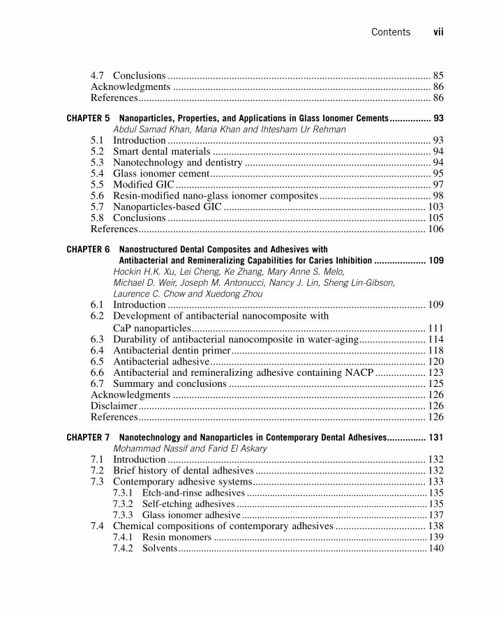
film is sponge-like with a typical pore size of 25 nm. At 20 V, hollow and cylindrical tube-like fea-
tures with an inner diameter of 80 nm form (Figure 17.1). Besides the HF/H2O electrolyte, many
other aqueous electrolytes have also been developed to fabricate NTs, for example, H2SO4/NaF/
(A) (B)
(C) (D)
FIGURE 17.1
(A and B) NTs formed at 5 and 20 V in 0.5 wt% aqueous HF solution with tube diameters of 25 and 80 nm,
respectively. (C and D) NTs formed at 10 and 40 V in an EG solution with 0.5 wt% NH4F,
5 vol% CH3OH, and 5 vol% H2O with tube diameters of 30 and 80 nm, respectively.
Reprinted with permission from Ref. [4].
33917.2 Fabrication of NTs on Ti

H2O [7], NaH2PO4/HF [9], and Na2SO4/HF [9]. The diameter of the NTs can be regulated to vary
from 15 to 140 nm and the length can range from 200 to 1000 nm by adjusting the electrolyte con-
tent and anodization voltage [21]. When using polar organic electrolytes, much longer NTs of hun-
dreds of micrometers can be fabricated [22]. Ethylene glycol (EG) is the commonly used organic
solvent to fabricate NTs. The typical NTs formed by anodization of a Ti foil in an EG solution
with 0.5 wt% NH4F, 5 vol% CH3OH, and 5 vol% H2O at 10 V for 1 h and 40 V for 40 min are
shown in Figure 17.1C and D, respectively.
17.3 Factors influencing the bioactivity of the NTsVarious factors can influence the bioactivity of the NTs during their preparation and cell culture
process. A better understanding of these factors helps to optimize the NTs for better biological
performance. Here, we summarize the factors such as the sterilization process, phenotype of cells,
protein concentration in the culture medium, and smoothness of the top ends of the tube walls that
affect the bioactivity of the NTs.
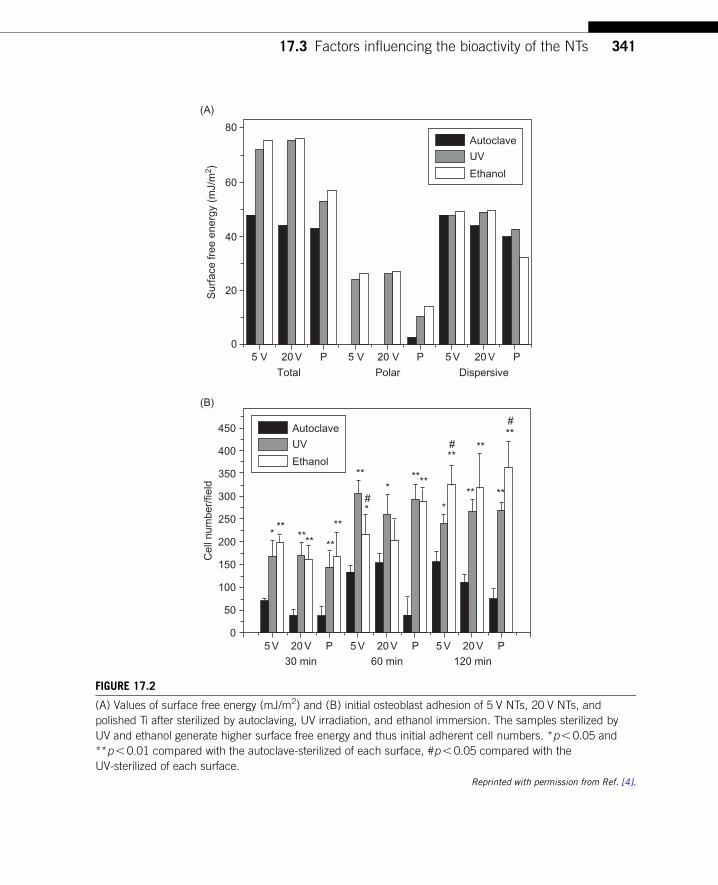
17.3.1 Influence of sterilization on the bioactivity of the NTsSterilization process is essential for the in vitro bioactivity assay and finally in vivo applications
of dental implants. However, many researchers choose sterilization methods arbitrarily when study-
ing the biocompatibility of NTs. In some experiments, autoclaving is used [10�13,23], whereas in
some others, ultraviolet (UV) irradiation or ethanol immersion is used [24�26]. Sterilization meth-
ods can be considered as a posttreatment of the samples, and the various sterilization methods can
change the surface properties of the samples and corresponding bioactivity. We have compared the
effects of three commonly used sterilization methods, namely autoclaving, UV irradiation, and eth-
anol immersion on the bioactivity of NTs.
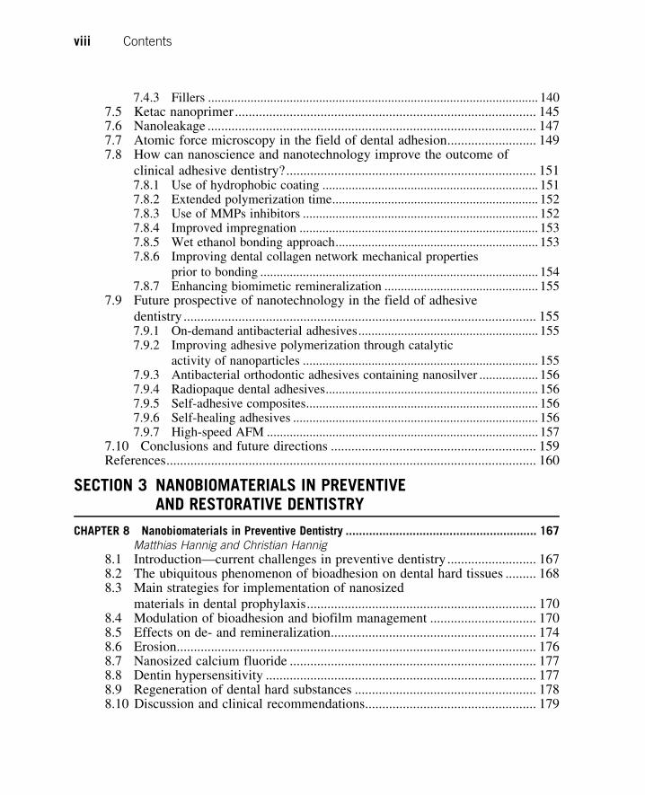
We have found that the sterilization process can modify the surface of the biomaterials. Strong
discoloration of samples is occasionally observed after autoclaving, but it is usually not found after
UV or ethanol sterilization. The most significantly modified characteristics after sterilization are
surface chemistry and wettability. Autoclaving decreases the surface free energy of biomedical
implants, but contrarily UV treatment dramatically enhances that of Ti (Figure 17.2A). The
decrease in hydrophilicity by autoclaving is related to the deposition of hydrophobic contaminants
on the implant surfaces [27]. The enhancement in the surface free energy of Ti by UV treatment is
associated with the molecular structure alteration of surface titania with abundant Ti�OH group
formation and removal of surface hydrophobic contaminants especially hydrocarbons [28,29].
The changes in the surface properties subsequently lead to differential cell responses.
UV and ethanol sterilizations induce higher initial adherent cell numbers (Figure 17.2B) and cell
proliferation than autoclaving, and UV irradiation leads to the best cell functionalities including
adhesion, proliferation, as well as differentiation represented by related gene expressions. UV steril-
ization appears to be the optimal sterilization method from the viewpoint of eliminating surface
contamination.
Oh et al. [30] have investigated the influence of two sterilization methods of wet autoclaving
versus dry autoclaving on the functionalities of osteoblasts cultured on the NTs. Their results
340 CHAPTER 17 Titania Nanotube Coatings on Dental Implants
5 V 20 V
Total
0
20
40
Sur
face
free
ene
rgy
(mJ/
m2 )
60
80
(A)
Polar Dispersive
P 5 V 20 V P 5 V 20 V P
Autoclave
UV
Ethanol
5 V 20 V30 min
0
Cel
l num
ber/
field
450
400
350
*
*
**#
#
#
****** **
**
** ****
****
**
****300
250
200
150
100
50
(B)
P 5 V 20 V60 min
P 5 V 20 V120 min
P
Autoclave
UV
Ethanol
FIGURE 17.2
(A) Values of surface free energy (mJ/m2) and (B) initial osteoblast adhesion of 5 V NTs, 20 V NTs, and
polished Ti after sterilized by autoclaving, UV irradiation, and ethanol immersion. The samples sterilized by
UV and ethanol generate higher surface free energy and thus initial adherent cell numbers. *p, 0.05 and
**p, 0.01 compared with the autoclave-sterilized of each surface, #p, 0.05 compared with the
UV-sterilized of each surface.
Reprinted with permission from Ref. [4].
34117.3 Factors influencing the bioactivity of the NTs
indicate that the adhesion, proliferation, and alkaline phosphatase (ALP) activity of osteoblasts
cultured on the larger 70 and 100 nm NTs are dramatically changed by the different sterilization
conditions at a low cell seeding density of 10,000 cells/well in 12-cell culture well. The different
autoclaving methods create huge differences in cell adherence on 70 and 100 nm NTs compared to
30 and 50 nm NTs. These results reveal that the nanofeatures of proteins adhered on the NTs can
be altered by different sterilization methods.
17.3.2 Influence of cell phenotype on the bioactivity of the NTsNTs have been assayed for various biological purposes such as bone implants [2,3,10�12,31],
transcutaneous part of the implants [32], and vascular prostheses [24]. There is much evidence
from the various primary cell phenotypes including primary osteoblasts, osteoblast cell lines,
MSCs, endothelial cells (ECs), vascular smooth muscle cells (VSMCs), dermal fibroblasts, and
epidermal keratinocytes suggesting that different cell phenotypes respond differently to the NTs.
We have observed differential responses of primary rat calvarial osteoblasts to the NTs compared
to those of the osteoblast cell lines [10,31]. Our results to some degree corroborate the report by
Brammer et al. [33]. They have compared the effects of TiO2 and C-coated NT surface chemistries
on osteoblast and osteoprogenitor cell behaviors. The TiO2 NT surface induces an increase in osteo-
blast functionalities in terms of ALP activity. In contrast, it is the carbon chemistry that results in
increased bone mineral deposition and bone matrix protein expression of osteoprogenitor cells.
More significant evidence unambiguously demonstrating the phenotypic dependence of cell
responses to the NTs is obtained from ECs/VSMCs and dermal fibroblasts/epidermal keratinocytes.
Peng et al. [24] have found that the NTs significantly enhance EC proliferation but decrease
VSMC proliferation (Figure 17.3). Smith et al. [32] have reported increased dermal fibroblast and
decreased epidermal keratinocyte adhesion, proliferation, and differentiation on the NTs.
The evidence reminds us that when comparing the reports on the bioactivity of the NTs from
different sources, it should be borne in mind that the responses of cells to the NTs are phenotypic
dependent. In addition, the differential response of the different cell phenotypes to the NTs provides
a good approach for tissue specific implants that selectively benefit from the desired tissue integra-
tion while simultaneously inhibiting the unwanted response.
17.3.3 Influence of protein concentration in culture mediumon the bioactivity of the NTsThe proteins adsorbed to the implant surface play a key role in cell/implant interactions. We have
compared the influence of the serum concentration in the culture medium on the change in the pro-
tein adsorption amount and the consequent initial cell spreading on the NTs and flat Ti [2].
Different serum concentrations do not influence cell adhesion on flat Ti control and 25 nm NTs
but seriously affect that on 80 nm NTs (Figure 17.4). The cells attach and spread well on the
80 nm NTs when cultured with 5% or 10% serum while 2% serum leads to poor cell adhesion.
This phenomenon can be explained by the cell adhesion mechanism. A requirement for normal cell
functionalities on biomaterials is stable adhesion or else cell apoptosis will occur [34]. Therefore,
the amount of adsorbed proteins is very important for the biological performance of biomaterials.
The amounts of proteins on the nanostructured surface increase with serum concentrations from
342 CHAPTER 17 Titania Nanotube Coatings on Dental Implants

2% to 10% (Figure 17.4B, F, G, J, and K). With regard to the flat surface and 25 nm NTs, because
large microscale focal adhesion can form, the cells can attach and spread well in 10%, 5%, or 2%
serum. As for the large NTs with size of 50�100 nm, because they constrain cell focal adhesion to
the top of the tube walls, high quality is needed for the small focal adhesion to support stable cell
adhesion. As shown in Figure 17.4G, H, K, and L), when cultured in 10% or 5% serum, the
adsorbed proteins not only widen the intertubular areas but also provide adequate integrin adhesion
sites, thus giving rise to enough high-quality focal adhesion and good cell adhesion. In 2% serum,
the smaller amount of adsorbed proteins results in narrow tube walls, a low density of integrin
adhesion sites, low-quality small focal adhesion, and poor cell adhesion (Figure 17.4C and D).
2
1.4
1.2
1
0.8
0.6
0.4
0.2
0
NT Flat(A)
(B)
1.8
1.6
1.4
1.2
Nor
mal
ized
rat
io o
f EdU
+ E
Cs
1
0.8
0.6
0.4
0.2
0
Nor
mal
ized
rat
io o
f EdU
+ V
SM
C
Day 1
*
Day 3
Day 1
**
**
Day 3
NT Flat
FIGURE 17.3
Ratio of 5-ethynyl-20-deoxyuridine (EdU) positive (A) ECs and (B) VSMCs on flat or NT substrate normalized
by the average proportion of positive cells on flat surfaces on day 1 and 3. Data are presented as
average6standard deviation. *P, 0.05, **P, 0.01 versus same day flat control.
Reprinted with permission from Ref. [24].
34317.3 Factors influencing the bioactivity of the NTs

Long and thin cell fillopodia are observed on 80 nm NTs cultured in 2% serum indicating that cells
cannot form stable adhesion (Figure 17.4D), while strong and thick lamellipodia are observed from
cells cultured in 5% or 10% serum demonstrating stable cell adhesion (Figures 17.4H and L). We
have also observed many cell fragments on 80 nm NTs in 2% serum on the cell retraction path
(Figure 17.4D). This may partly account for cell apoptosis on the surface. In Park et al.’s [25,35]
experiments, a low-medium serum concentration of 2% is used in cell cultures and should account
for the unfavorable effect of larger NTs on MSC functions observed by them. Since there are abun-
dant proteins in vivo, the results obtained from 10% serum should reflect the in vivo performance
of the NTs more accurately. Our results indicate the influence of protein concentration in the cul-
ture medium on the evaluated bioactivity of the NTs, which should be of concern when comparing
different reports on the NTs bioactivity.
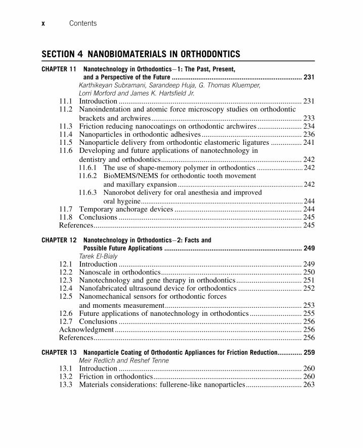
17.3.4 Influence of protein distribution pattern on the bioactivity of the NTsAs aforementioned, the proteins adsorbed onto the biomaterials mediate cell adhesion and follow
functions on the biomaterials and play a crucial role in conveying the biological effects of the topo-
graphical cue. Besides the amount, other aspects of the adsorbed proteins such as species, confor-
mation, and orientation have also been reported to influence the cell/biomaterials interaction. We
notice that the NTs formed in an inorganic electrolyte have relatively flat top ends of NT walls
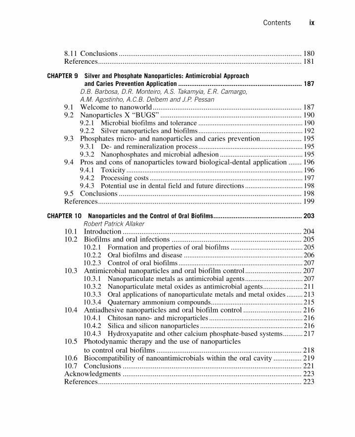
Flat Ti2
%5
%10
%5 V NT 20 V NT 20 V NT
(A) (B) (C) (D)
(E) (F) (G) (H)
(I) (J) (K) (L)
FIGURE 17.4
Cell shape on flat Ti, 25 and 80 nm NTs cultured with 2%, 5% or 10% serum for 12 h. The insets in (B), (C),
(F), (G), (J), and (K) show the ECM deposition along the nanotopographies.
Reprinted with permission from Ref. [2].
344 CHAPTER 17 Titania Nanotube Coatings on Dental Implants
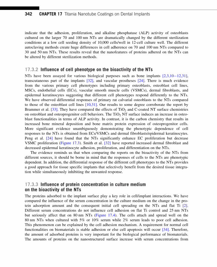
(Figure 17.5A), and consequently induce an even distribution of proteins along the tube walls and
intimate cell attachment (Figure 17.5C). On contrary, for the NTs formed in an EG solution with
0.5 wt% NH4F, 5 vol% CH3OH, and 5 vol% H2O, the top ends of the NT walls are not completely
smooth and have flakes (Figure 17.5B). Although the NTs formed in the EG solution induce more
protein deposition forming thicker protein layers, the adsorbed proteins do not distribute evenly but
form pillars (Figure 17.5D). Tall protein pillars with relatively small top dimensions mostly
,50 nm are distributed at a certain distance from each other. The uneven protein distribution can
be ascribed to the unsmooth top ends of the NT walls with flakes, which provide local nucleation
(A) (B)
(C) (D)
FIGURE 17.5
(A) NTs formed at 20 V in 0.5 wt% HF aqueous solution; (B) NTs formed at 40 V in an EG solution with 0.5 wt
% NH4F, 5 vol% CH3OH, and 5 vol% H2O. Red arrows indicate the unsmooth top ends of nanotube walls
with flakes; (C) protein adsorption on the NTs shown in (A) and the detail of cell interaction with them;
(D) protein adsorption on the NTs shown in (B) and details of cell interaction with them. Yellow arrows show
the protein pillars formed on them. (For interpretation of the references to color in this figure legend, the
reader is referred to the web version of this book.)
Parts of the figure are reprinted with permission from Ref. [2].
34517.3 Factors influencing the bioactivity of the NTs
sites for protein aggregation leading to pillar formation. Collectively, the evidence demonstrates that
even subtle changes in the nanotopography can lead to dramatic alteration in the protein deposition
pattern. The immediate substrate that cells interact with is the adsorbed proteins rather than the primi-
tive nanotopographies. The notably uneven protein deposition will change the nanotopography and
make the ultimate topographical cues exposed to cells to be quite different from the primitive nano-
topographies, thereby altering the biological performance. The uneven protein distribution con-
sequently leads to compromised cell focal adhesion and following functions including proliferation
and differentiation. It is thus strongly suggested that the protein adsorption pattern should also be
carefully inspected when studying the bioactivity of nanoscale biomaterials.
17.4 In vitro bioactivity of the NTs and in vivo osseointegration17.4.1 In vitro bioactivity of the NTsNTs can foster the growth of nanostructured hydroxyapatite. Oh et al. [8] have treated the NTs with
NaOH solution to investigate their bioactivity. NTs induce the growth of extremely fine-scale (B8 nm
feature) nanofibers of bioactive sodium titanate on the top edge of the B15 nm thick NT wall. After
immersion in a SBF, the nanoscale sodium titanate can induce the nucleation and growth of nanodi-
mensioned HA phase. The kinetics of HA formation can be significantly accelerated by the presence
of the NTs. Pittrof et al. [9] have developed micropatterned NT layers surrounded by compact oxide
via an optimized process. By immersing such patterns in SBF, selective and dense apatite deposition
occurs only on the NT surfaces (Figure 17.6). These results verify the strong ability of the NTs to
induce apatite deposition. Although there is still debate whether SBF can predict the bioactivity of
biomaterials [36], the strong ability of the NTs to foster nanostructured hydroxyapatite deposition to
a certain degree demonstrates their bone formation favoring properties.
(A) (B)
Apatite
Apatite
Apatite
FIGURE 17.6
Patterned samples after immersion in 1.53 SBF. (A and B) Field emission scanning electron microscopy
(FE-SEM) micrographs showing selective apatite nucleation exclusively on nanotube regions.
Reprinted with permission from Ref. [9].
346 CHAPTER 17 Titania Nanotube Coatings on Dental Implants

Osteoblasts are responsible for bone formation, and the effects of NTs on osteoblast functions
have been widely observed on primary rat calvarial osteoblasts [3,4,31] and MC3T3-E1 mouse
osteoblasts [10,26]. There is some controversy on the effects of the NTs on some of the osteoblast
functions due to the differential experimental conditions in the literature. However, it seems that
the NTs are effective in promoting ECM secretion and mineralization. The NTs with diameters of
25 and 80 nm formed in 0.5 wt% aqueous HF solution can induce more collagen secretion and min-
eral deposition in primary rat calvarial osteoblast cultures [4]. Hierarchical hybrid micro/nanotex-
tured titanium surface topographies with NTs produced by our group by simple acid etching
followed by anodization mimic the hierarchical structures of bone tissues, thereby inducing more
collagen secretion compared to the microrough and flat Ti surfaces (Figure 17.7) [3].
(A) (B)
(C) (D)
FIGURE 17.7
Collagen secretion by osteoblasts on samples after 7 days of incubation. (A) Hierarchical hybrid micro/
nanotextured surface with 15 nm NTs, (B) hierarchical hybrid micro/nanotextured surface with 80 nm NTs,
(C) acid-etched microstructured surface, and (D) flat Ti surface.
Reprinted with permission from Ref. [3].
34717.4 In vitro bioactivity of the NTs and in vivo osseointegration

MSCs play a crucial role in bone regeneration and bony fixation of implanted biomaterials. Most
of the osteoblastic cells that colonize the implant surface to induce bone growth originate from MSCs
and hence, in order to accomplish good osseointegration, it is critical to induce the differentiation of
MSCs preferentially toward osteogenitor cells and then into osteoblasts in lieu of other cell lineages.
We find that the NTs significantly promote MSC attachment and spreading (Figure 17.8), collagen
secretion and ECM mineralization (Figure 17.9), as well as osteogenesis-related gene expression in
the absence of extra OS [2]. The osteogenesis-inducing ability of the 80 nm NTs is higher than that
of the 25 nm ones. Oh et al. [37] have also observed that small 30 nm NTs promote MSC adhesion
without noticeable differentiation whereas larger ones of 70�100 nm elicit selective MSC differentia-
tion to osteoblasts. Moon et al. [38] have recently assessed the size effect of NTs on the behavior and
osteogenic functionality of human MSCs. After incubation for 2 weeks, expression of ALP, osteopon-
tin, integrin-β, and protein kinase R-like endoplasmic reticulum kinase genes are significantly higher
in cells cultured on 70 nm NTs than those cultured on 30, 50, and 100 nm NTs and Ti. The evidence
demonstrates that NTs with a suitable tube size have osteogenesis-inducing ability.
The osteogenesis-inducing ability of the NTs arises from their modulating effect on cell shape and
focal adhesion. This will lead to changes in the mechanotransduction including the indirect one, that
is integrin-dependent signal pathways, and the direct one that is gene expression originating from the
cell nucleus distortion by force transferred via the cytoskeleton [39,40]. The shape of stem cells
on biomaterials is closely related to the high cytoskeletal tension such as the well-spread stem cell
and that with the proper aspect ratio undergoing osteogenesis with the poorly spread stem cell becom-
ing adipocytes [41]. Therefore, the effects of the NTs on promoting MSC spreading constitute an
important mechanism for the osteogenesis-inducing ability. The higher osteogenesis-inducing ability
rendered by the 80 nm NTs than the 25 nm ones can be explained by the influence of the nanotopo-
graphy on the cell focal adhesion size, distribution, and related mechanotransduction. The presenta-
tion of integrin ligation sites at a distance larger than a certain value (about 50�70 nm) perturbs
integrin clustering, focal adhesion assembly, and organization of the actin stress fiber anchored to the
focal adhesion [42]. Accordingly, the 25 nm NTs do not, or slightly, influence the integrin clustering
and focal adhesion formation. Instead, the 80 nm NTs constrain the cell focal adhesion to the inter-
tubular area. In this way, the 80 nm NTs modulate the size, shape, and distribution of focal adhesion
to a nanoscale periodic occurrence. On one hand, it triggers more integrin-related signals, and on the
other hand, it induces a nanoscale periodic distribution of the cytoskeletal actin and stress leading to
extensive nucleus distortion and related direct mechanotransduction signals.
17.4.2 In vivo osseointegration of the NTsThe good bone-favoring properties of the NTs with suitable size have also been verified by
various in vivo studies. Bjursten et al. [13] have investigated the in vivo bone bonding between
80 nm NTs and grit-blasted TiO2. Four weeks after implantation into rabbit tibias, the NTs improve
the bone bonding strength by as much as ninefolds compared to the grit-blasted TiO2 surface. The
histological analysis confirms greater BIC areas, new bone formation, and calcium and phosphorus
levels on the NTs. Von Wilmowsky et al. [14] have reported that a NT structured implant surface
with a diameter of 30 nm can influence bone formation and bone development by enhancing the
osteoblast functionalities and the NT coatings resist shearing forces evoked by implant insertion.
They have recently reported a significantly higher value of the BIC for the 50, 70, and 100 nm NTs
348 CHAPTER 17 Titania Nanotube Coatings on Dental Implants
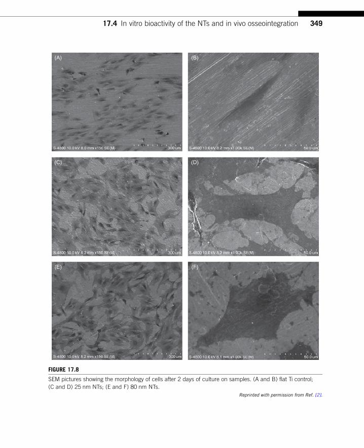
(A) (B)
(C) (D)
(E) (F)
FIGURE 17.8
SEM pictures showing the morphology of cells after 2 days of culture on samples. (A and B) flat Ti control;
(C and D) 25 nm NTs; (E and F) 80 nm NTs.
Reprinted with permission from Ref. [2].
34917.4 In vitro bioactivity of the NTs and in vivo osseointegration
compared to the pristine Ti controls [15]. The bone morphogenetic protein 2 (BMP-2) expression
within the 50, 70, and 100 nm groups is statistically different compared to the control group. In
addition, a significant difference is found from the osteocalcin expression in the 70 nm group.
Wang et al. [16] have investigated the effects of the NTs with different diameters of 30, 70, and
100 nm on the biological attachment mechanism of implants to bone in vivo. When comparing to
machined Ti implants, a significant increase in BIC (Figure 17.10) and gene expression level is
found in the bone attached to implants with the NTs, especially with the 70 nm diameter ones. The
evidence demonstrates the strong ability of the NTs to induce better osseointegration, and the NTs
with a size of about 70 nm may be the optimal ones for osseointegration.
17.5 Drug-loading NTs for better bioactivity and antibacterial propertiesThe nanotubular structure of the NTs provide space for drug loading, thereby opening the possibil-
ity of endowing the implant surface with extra properties by loading targeted agents. Desai’s group
(A) (B) (C)
FIGURE 17.9
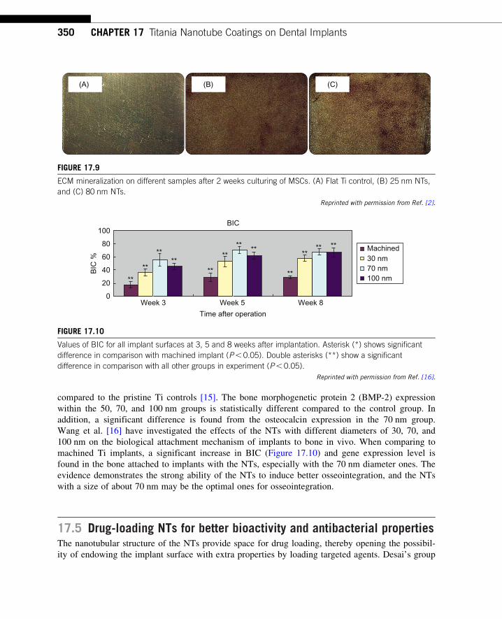
ECM mineralization on different samples after 2 weeks culturing of MSCs. (A) Flat Ti control, (B) 25 nm NTs,
and (C) 80 nm NTs.
Reprinted with permission from Ref. [2].
100
80
60
BIC
%
40
20
0Week 3 Week 5 Week 8
Time after operation
BIC
****
**
**
****
**
**
******
**
Machined30 nm70 nm100 nm
FIGURE 17.10
Values of BIC for all implant surfaces at 3, 5 and 8 weeks after implantation. Asterisk (*) shows significant
difference in comparison with machined implant (P, 0.05). Double asterisks (**) show a significant
difference in comparison with all other groups in experiment (P, 0.05).
Reprinted with permission from Ref. [16].
350 CHAPTER 17 Titania Nanotube Coatings on Dental Implants

first tested the suitability of the NTs to serve as a potential drug loading and delivering platform
[43,44]. They used bovine serum albumin and lysozyme as model proteins to investigate the load-
ing and release efficiencies from the NT platforms. They demonstrated the efficacy of using NTs
as drug eluting coatings for implantable devices. Various amounts of drugs can be incorporated
into the NTs and their release can be adjusted by varying the tube length, diameter, and wall
thickness [43]. Another report demonstrated that the NTs can control small molecule delivery
within weeks and larger molecules in months [44]. Various agents have been experimentally loaded
into the NTs to attain better bioactivity and extra properties such as antibacterial ability. There are
many reports on the incorporation of growth factors or antibiotics to the NTs and certain bioactivity
and antibacterial ability. However, we believe that the NTs are ideal for loading and delivering
targeted inorganic agents such as silver (Ag), strontium (Sr), and zinc (Zn). First of all, these are
much smaller molecules than growth factors and antibiotics and function at very low doses. Long-
lasting activity can be achieved by increasing the loaded amounts and controlling the release rate
appropriately. Secondly, these agents are stable due to their inorganic nature, thereby facilitating
the use of loading processes and loading methods that tend to have harsh conditions. Thirdly, the
stable properties of the agents may also permit relatively long storage after fabrication of the
implants and it is important to commercial adoption.
As mentioned in the introduction section, postoperation infection remains one of the most com-
mon and serious complications for a dental implant, and so a surface boasting long-term antibacte-
rial ability is highly desirable in order to prevent implant-associated infection. We have fabricated
Ag nanoparticles incorporated NTs (NT�Ag) on Ti implants to achieve this purpose [5]. The
Ag nanoparticles adhere tightly to the wall of the NTs prepared by immersion in a AgNO3 solution
followed by UV light irradiation (Figure 17.11). The amount of Ag introduced to the NTs can be
controlled by changing the processing parameters such as the AgNO3 concentration and immersion
time. The NT�Ag can kill all the planktonic bacteria in the suspension during the first several
days, and the ability of the NT�Ag to prevent bacterial adhesion is maintained without obvious
decline for 30 days, which are normally long enough to prevent postoperation infection in the early
and intermediate stages and perhaps even late infection around the implant. The ability of the
NT�Ag to prevent viable bacterial colonization is vividly displayed by fluorescence staining
(Figure 17.12). After 7 days of repeated bacterial invasion every 24 h, there are large amounts of
viable bacteria on the flat Ti and smaller amounts on the TiO2�NTs. In comparison, the amounts
of viable bacteria are obviously smaller on the NT�Ag samples due to the Ag loading amount.
Although the NT�Ag structure shows some cytotoxicity, it can be reduced by properly controlling
the Ag release rate. This NT�Ag structure with relatively long-term antibacterial ability has prom-
ising applications in bone implants after eradicating the cytotoxicity by properly controlling the
Ag release.
Sr shows the effect to modulate bone turnover toward osteogenesis by enhancing osteoblast func-
tions and inhibiting osteoclast functions, and so Sr-loaded nanotubular structures (NT�Sr) that allow
controlled and long-term Sr release are expected to yield favorable osteogenic effects. Well-ordered
SrTiO3 NT arrays capable of Sr release at a small rate and for a long time have been successfully fab-
ricated on titanium by simple hydrothermal treatment of anodized titania NTs (Figure 17.13) [20].
This surface architecture combines the functions of nanoscale topography and Sr release to enhance
osseointegration while at the same time leaving space for loading of other functional substances. In
vitro experiments reveal that the SrTiO3 NT arrays possess good biocompatibility (Figure 17.13) and
35117.5 Drug-loading NTs for better bioactivity and antibacterial properties

(A) (B)
(C) (D)
(E)
(F)
FIGURE 17.11
SEM images of the samples. (A) NTs, (B�E) Ag incorporating NTs formed in AgNO3 solutions of different
concentrations (0.5, 1, 1.5, and 2 M). The inset in (A) is the side-view SEM image revealing that the length of
the nanotubes is about 7 μm. The red arrows in (B�E) indicate the Ag nanoparticles. (F) TEM image
acquired from the Ag incorporating NTs formed in 1 M AgNO3 shows that the Ag nanoparticles attached to
the inner wall of the NTs have a diameter of about 10�20 nm. (For interpretation of the references to color in
this figure legend, the reader is referred to the web version of this book.)
Reprinted with permission from Ref. [5].
352 CHAPTER 17 Titania Nanotube Coatings on Dental Implants

(A) (B)
(C) (D)
(E) (F)
FIGURE 17.12
Representative images showing viability of the bacteria after 7 days of incubation on samples: (A) Ti, (B) NTs,
(C�F) Ag incorporating NTs formed in AgNO3 solutions of different concentrations (0.5, 1, 1.5, and 2 M).
Live bacteria appear green while dead ones appear orange. (For interpretation of the references to color in
this figure legend, the reader is referred to the web version of this book.)
Reprinted with permission from Ref. [5].
35317.5 Drug-loading NTs for better bioactivity and antibacterial properties

can induce precipitation of hydroxyapatite from SBF. Recently, we have found that the NT�Sr coat-
ing can dramatically improve MSC spreading as well as proliferation and enhance MSC differentia-
tion toward osteogenitor cells and subsequently osteoblasts. The inorganic NT�Sr gives rise to good
osteogenic activity without the need to apply foreign complex biomolecules. The materials are easy
to fabricate and have good stability that would facilitate large-scale industrial production, storage,
transport, sterilization, and clinical use. They are thus very attractive to bone implants, especially
osteoporotic bone implants for clinical applications.
In addition to Sr, there are many other agents showing attractive properties in bone�implant
applications, e.g., Zn. Besides the effects of positively regulating bone turnover, Zn has good
antibacterial activity and anti-inflammation ability. Hence, Zn is a good candidate for implant
surface loading and long-term release to attain better clinical performance. We have also developed
Zn loaded NTs (NT�Zn) using a method similar to that of NT�Sr. The NT�Zn can also promote
(A) (B)
(C) (D)
FIGURE 17.13
SEM micrographs of (A) titania nanotube arrays and (B) SrTiO3 nanotube arrays. Cell morphology on SrTiO3
nanotube arrays after 5 days of culturing: (C) low magnification and (D) high magnification.
Reprinted with permission from Ref. [20].
354 CHAPTER 17 Titania Nanotube Coatings on Dental Implants

MSC attachment and spread and induce MSC osteogenic differentiation. In addition, the NT�Zn
samples also exhibit excellent antibacterial effects to prevent bacterial colonization.
17.6 ConclusionsMany reports have revealed the effectiveness of NTs in promoting the functions of osteoblasts and
MSCs and MSC osteogenic differentiation in vitro and enhancing implant osseointegration in vivo.
Our studies demonstrate the suitability of the NTs to load and deliver some inorganic bioactive
agents such as Ag, Sr, and Zn to achieve antibacterial- and/or osteogenesis-inducing abilities. The
NT surfaces, especially those loaded with suitable inorganic bioactive agents, have huge promise in
fabricating dental implants with better clinical performance.
AcknowledgmentsThis work was jointly supported by National Natural Science Foundation of China Nos. 50902104 and
31200716, City University of Hong Kong Applied Research Grant (ARG) No. 9667066, and the Opening
Project of State Key Laboratory of High Performance Ceramics and Superfine Microstructure (SKL201103SIC).
References[1] J.Y. Rho, L. Kuhn-Spearing, P. Zioupos, Mechanical properties and the hierarchical structure of bone,
Med. Eng. Phys. 20 (1998) 92�102.
[2] L. Zhao, L. Liu, Z. Wu, Y. Zhang, P.K. Chu, Effects of micropitted/nanotubular titania topographies on
bone mesenchymal stem cell osteogenic differentiation, Biomaterials 33 (2012) 2629�2641.
[3] L. Zhao, S. Mei, P.K. Chu, Y. Zhang, Z. Wu, The influence of hierarchical hybrid micro/nano-textured
titanium surface with titania nanotubes on osteoblast functions, Biomaterials 31 (2010) 5072�5082.
[4] L. Zhao, S. Mei, W. Wang, P.K. Chu, Z. Wu, Y. Zhang, The role of sterilization in the cytocompatibility
of titania nanotubes, Biomaterials 31 (2010) 2055�2063.
[5] L. Zhao, H. Wang, K. Huo, L. Cui, W. Zhang, H. Ni, et al., Antibacterial nano-structured titania coating
incorporated with silver nanoparticles, Biomaterials 32 (2011) 5706�5716.
[6] M. Tzaphlidou, The role of collagen in bone structure: an image processing approach, Micron 36 (2005)
593�601.
[7] G.A. Crawford, N. Chawla, K. Das, S. Bose, A. Bandyopadhyay, Microstructure and deformation behav-
ior of biocompatible TiO2 nanotubes on titanium substrate, Acta Biomater. 3 (2007) 359�367.
[8] S.H. Oh, R.R. Finones, C. Daraio, L.H. Chen, S.H. Jin, Growth of nano-scale hydroxyapatite using
chemically treated titanium oxide nanotubes, Biomaterials 26 (2005) 4938�4943.
[9] A. Pittrof, S. Bauer, P. Schmuki, Micropatterned TiO2 nanotube surfaces for site-selective nucleation of
hydroxyapatite from simulated body fluid, Acta Biomater. 7 (2011) 424�431.
[10] K.S. Brammer, S. Oh, C.J. Cobb, L.M. Bjursten, H.V. Heyde, S. Jin, Improved bone-forming functional-
ity on diameter-controlled TiO2 nanotube surface, Acta Biomater. 5 (2009) 3215�3223.
[11] K. Das, S. Bose, A. Bandyopadhyay, TiO2 nanotubes on Ti: influence of nanoscale morphology on bone
cell-materials interaction, J. Biomed. Mater. Res. A 90 (2009) 225�237.
355References
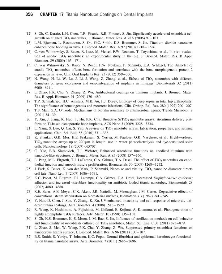
[12] S. Oh, C. Daraio, L.H. Chen, T.R. Pisanic, R.R. Finones, S. Jin, Significantly accelerated osteoblast cell
growth on aligned TiO2 nanotubes, J. Biomed. Mater. Res. A 78A (2006) 97�103.
[13] L.M. Bjursten, L. Rasmusson, S. Oh, G.C. Smith, K.S. Brammer, S. Jin, Titanium dioxide nanotubes
enhance bone bonding in vivo, J. Biomed. Mater. Res. A 92 (2010) 1218�1224.
[14] C. von Wilmowsky, S. Bauer, R. Lutz, M. Meisel, F.W. Neukam, T. Toyoshima, et al., In vivo evalua-
tion of anodic TiO2 nanotubes: an experimental study in the pig, J. Biomed. Mater. Res. B Appl.
Biomater. 89 (2009) 165�171.
[15] C. von Wilmowsky, S. Bauer, S. Roedl, F.W. Neukam, P. Schmuki, K.A. Schlegel, The diameter of
anodic TiO2 nanotubes affects bone formation and correlates with the bone morphogenetic protein-2
expression in vivo, Clin. Oral Implants Res. 23 (2012) 359�366.
[16] N. Wang, H. Li, W. Lu, J. Li, J. Wang, Z. Zhang, et al., Effects of TiO2 nanotubes with different
diameters on gene expression and osseointegration of implants in minipigs, Biomaterials 32 (2011)
6900�6911.
[17] L. Zhao, P.K. Chu, Y. Zhang, Z. Wu, Antibacterial coatings on titanium implants, J. Biomed. Mater.
Res. B Appl. Biomater. 91 (2009) 470�480.
[18] T.P. Schmalzried, H.C. Amstutz, M.K. Au, F.J. Dorey, Etiology of deep sepsis in total hip arthroplasty.
The significance of hematogenous and recurrent infections, Clin. Orthop. Rel. Res. 280 (1992) 200�207.
[19] T.F. Mah, G.A. O’Toole, Mechanisms of biofilm resistance to antimicrobial agents, Trends Microbiol. 9
(2001) 34�39.
[20] Y. Xin, J. Jiang, K. Huo, T. Hu, P.K. Chu, Bioactive SrTiO3 nanotube arrays: strontium delivery plat-
form on Ti-based osteoporotic bone implants, ACS Nano 3 (2009) 3228�3234.
[21] L. Yang, S. Luo, Q. Cai, S. Yao, A review on TiO2 nanotube arrays: fabrication, properties, and sensing
applications, Chin. Sci. Bull. 55 (2010) 331�338.
[22] K. Shankar, G.K. Mor, H.E. Prakasam, S. Yoriya, M. Paulose, O.K. Varghese, et al., Highly-ordered
TiO2 nanotube arrays up to 220 μm in length: use in water photoelectrolysis and dye-sensitized solar
cells, Nanotechnology 18 (2007) 065707.
[23] C. Yao, E.B. Slamovich, T.J. Webster, Enhanced osteoblast functions on anodized titanium with
nanotube-like structures, J. Biomed. Mater. Res. A 85 (2008) 157�166.
[24] L. Peng, M.L. Eltgroth, T.J. LaTempa, C.A. Grimes, T.A. Desai, The effect of TiO2 nanotubes on endo-
thelial function and smooth muscle proliferation, Biomaterials 30 (2009) 1268�1272.
[25] J. Park, S. Bauer, K. von der Mark, P. Schmuki, Nanosize and vitality: TiO2 nanotube diameter directs
cell fate, Nano Lett. 7 (2007) 1686�1691.
[26] K.C. Popat, M. Eltgroth, T.J. Latempa, C.A. Grimes, T.A. Desai, Decreased Staphylococcus epidermis
adhesion and increased osteoblast functionality on antibiotic-loaded titania nanotubes, Biomaterials 28
(2007) 4880�4888.
[27] R.E. Baier, A.E. Meyer, C.K. Akers, J.R. Natiella, M. Meenaghan, J.M. Carter, Degradative effects of
conventional steam sterilization on biomaterial surfaces, Biomaterials 3 (1982) 241�245.
[28] Y. Han, D. Chen, J. Sun, Y. Zhang, K. Xu, UV-enhanced bioactivity and cell response of micro-arc oxi-
dized titania coatings, Acta Biomater. 4 (2008) 1518�1529.
[29] R. Wang, K. Hashimoto, A. Fujishima, M. Chikuni, E. Kojima, A. Kitamura, et al., Photogeneration of
highly amphiphilic TiO2 surfaces, Adv. Mater. 10 (1998) 135�138.
[30] S. Oh, K.S. Brammer, K.-S. Moon, J.-M. Bae, S. Jin, Influence of sterilization methods on cell behavior
and functionality of osteoblasts cultured on TiO2 nanotubes, Mater. Sci. Eng. C 31 (2011) 873�879.
[31] L. Zhao, S. Mei, W. Wang, P.K. Chu, Y. Zhang, Z. Wu, Suppressed primary osteoblast functions on
nanoporous titania surface, J, Biomed. Mater. Res. A 96 (2011) 100�107.
[32] B.S. Smith, S. Yoriya, T. Johnson, K.C. Popat, Dermal fibroblast and epidermal keratinocyte functional-
ity on titania nanotube arrays, Acta Biomater. 7 (2011) 2686�2696.
356 CHAPTER 17 Titania Nanotube Coatings on Dental Implants
[33] K.S. Brammer, C. Choi, C.J. Frandsen, S. Oh, G. Johnston, S. Jin, Comparative cell behavior on carbon-
coated TiO2 nanotube surfaces for osteoblasts vs. osteo-progenitor cells, Acta Biomater. 7 (2011)
2697�2703.
[34] L. Zhao, L. Hu, K. Huo, Y. Zhang, Z. Wu, P.K. Chu, Mechanism of cell repellence on quasi-aligned
nanowire arrays on Ti alloy, Biomaterials 31 (2010) 8341�8349.
[35] J. Park, S. Bauer, P. Schmuki, K. von der Mark, Narrow window in nanoscale dependent activation of
endothelial cell growth and differentiation on TiO2 nanotube surfaces, Nano Lett. 9 (2009) 3157�3164.
[36] T. Kokubo, H. Takadama, How useful is SBF in predicting in vivo bone bioactivity? Biomaterials 27
(2006) 2907�2915.
[37] S. Oh, K.S. Brammer, Y.S. Li, D. Teng, A.J. Engler, S. Chien, et al., Stem cell fate dictated solely by
altered nanotube dimension, Proc. Natl. Acad. Sci. U. S. A. 106 (2009) 2130�2135.
[38] K.-S. Moon, S.-H. Yu, J.-M. Bae, S. Oh, Biphasic osteogenic characteristics of human mesenchymal
stem cells cultured on TiO2 nanotubes of different diameters, J. Nanomater. (2012) doi: 10.1155/2012/
252481.
[39] A.S. Curtis, M. Dalby, N. Gadegaard, Cell signaling arising from nanotopography: implications for nano-
medical devices, Nanomedicine 1 (2006) 67�72.
[40] L.E. McNamara, R.J. McMurray, M.J. Biggs, F. Kantawong, R.O. Oreffo, M.J. Dalby,
Nanotopographical control of stem cell differentiation, J. Tissue Eng. (2010) 120623.
[41] R. McBeath, D.M. Pirone, C.M. Nelson, K. Bhadriraju, C.S. Chen, Cell shape, cytoskeletal tension, and
RhoA regulate stem cell lineage commitment, Dev. Cell 6 (2004) 483�495.
[42] E.A. Cavalcanti-Adam, T. Volberg, A. Micoulet, H. Kessler, B. Geiger, J.P. Spatz, Cell spreading and
focal adhesion dynamics are regulated by spacing of integrin ligands, Biophys. J. 92 (2007) 2964�2974.
[43] K.C. Popat, M. Eltgroth, T.J. LaTempa, C.A. Grimes, T.A. Desai, Titania nanotubes: a novel platform
for drug-eluting coatings for medical implants? Small 3 (2007) 1878�1881.
[44] L. Peng, A.D. Mendelsohn, T.J. LaTempa, S. Yoriya, C.A. Grimes, T.A. Desai, Long-term small mole-
cule and protein elution from TiO2 nanotubes, Nano Lett. 9 (2009) 1932�1936.
357References