Embed Size (px)
Citation preview

NanoBio Summit 2014 The University of Alabama October 23-24, 2014
Connect with Colleagues to Discover the Latest Research, Innovations, and Implementation of Nanobiotechnology
Conference Topics
Nanobioscience Nanoengineering Nanomaterials
Nanomedicine Nanobiotechnology Biomedical Engineering
Collaboration ! Discovery ! Commercialization

Welcome to NanoBio Summit 2014
The University of Alabama and the NanoBio Summit 2014 Organizing Committee are pleased to welcome you to the NanoBio Summit 2014.
Conference themes this year include nanobioscience, nanoengineering, nanomaterials, nanomedicine, nanobiotechnology, and biomedical engineering. These research areas continue to bring surprising insights and innovative technologies and materials for industrial, environmental, and human health applications. In addition, the focus on collaboration, discovery, and commercialization is designed to bring together researchers from many disciplines, to showcase emerging areas of research and to provide participants with knowledge on the process from “bench discovery” to commercial products. We look forward to the exciting science and engineering information that will be presented at the conference!
The University of Alabama is a student-centered research institution, serving 36,155 undergraduate and graduate students from every state in the nation and from more than 90 countries. The incoming freshman class included 6,856 students with a 26 average ACT and 3.65 average high school GPA. Nearly 30% of the incoming class had a 4.0 GPA, and over 31% scored 30 or higher on the ACT (a 94% increase since 2010). Our nearly 5,000 graduate students study in nationally-ranked masters and doctoral programs, and UA annually ranks in the top three among the 50 flagship universities in diversity and student support. The education and experiences our students receive at UA provide them with the skills, training, knowledge of cultural inclusivity, and networks to become leaders in their fields and world citizens in the global, knowledge-based economy and society.
We hope that you will have time to explore Tuscaloosa and our beautiful campus while you are at the conference. This year, The University of Alabama’s Quad was recognized as one of 14 “most beautiful and iconic” quads in the U.S. by Business Insider Magazine.
Enjoy the conference and the locale!
Patricia A. Sobecky Carl A. Pinkert Associate Provost for Academic Affairs Vice President for Research The University of Alabama and Economic Development
The University of Alabama

Platinum Supporters
The Office of the Vice President for Research and Economic Development The University of Alabama
Center for NanoBiotechnology Research at Alabama State University
Gold Supporters
The University of Alabama Center for Economic Development Tuskegee University
The Department of Biological Sciences The University of Alabama
Silver Supporter
Tescan USA
Bronze Supporters
THANK YOU TO OUR SUPPORTERS
National Science Foundation National Institutes of Health
ICE Publishing Institution of Civil Engineers
VWR International
Eppendorf
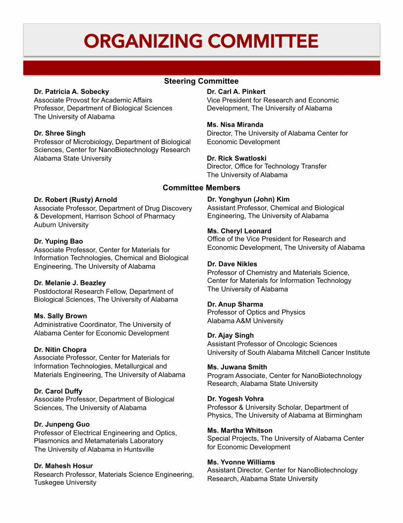
ORGANIZING COMMITTEE
Dr. Patricia A. Sobecky Associate Provost for Academic Affairs Professor, Department of Biological Sciences The University of Alabama
Dr. Shree Singh Professor of Microbiology, Department of Biological Sciences, Center for NanoBiotechnology Research Alabama State University
Dr. Carl A. Pinkert Vice President for Research and Economic Development, The University of Alabama
Ms. Nisa Miranda Director, The University of Alabama Center for Economic Development
Dr. Rick Swatloski Director, Office for Technology Transfer The University of Alabama
Steering Committee
Committee Members Dr. Robert (Rusty) Arnold Associate Professor, Department of Drug Discovery & Development, Harrison School of Pharmacy Auburn University
Dr. Yuping Bao Associate Professor, Center for Materials for Information Technologies, Chemical and Biological Engineering, The University of Alabama
Dr. Melanie J. Beazley Postdoctoral Research Fellow, Department of Biological Sciences, The University of Alabama
Ms. Sally Brown Administrative Coordinator, The University of Alabama Center for Economic Development
Dr. Nitin Chopra Associate Professor, Center for Materials for Information Technologies, Metallurgical and Materials Engineering, The University of Alabama
Dr. Carol Duffy Associate Professor, Department of Biological Sciences, The University of Alabama
Dr. Junpeng Guo Professor of Electrical Engineering and Optics, Plasmonics and Metamaterials Laboratory The University of Alabama in Huntsville
Dr. Mahesh Hosur Research Professor, Materials Science Engineering, Tuskegee University
Dr. Yonghyun (John) Kim Assistant Professor, Chemical and Biological Engineering, The University of Alabama
Ms. Cheryl Leonard Office of the Vice President for Research and Economic Development, The University of Alabama
Dr. Dave Nikles Professor of Chemistry and Materials Science, Center for Materials for Information Technology The University of Alabama
Dr. Anup Sharma Professor of Optics and Physics Alabama A&M University
Dr. Ajay Singh Assistant Professor of Oncologic Sciences University of South Alabama Mitchell Cancer Institute
Ms. Juwana Smith Program Associate, Center for NanoBiotechnology Research, Alabama State University
Dr. Yogesh Vohra Professor & University Scholar, Department of Physics, The University of Alabama at Birmingham
Ms. Martha Whitson Special Projects, The University of Alabama Center for Economic Development
Ms. Yvonne Williams Assistant Director, Center for NanoBiotechnology Research, Alabama State University

PROGRAM Thursday, October 23, 2014
MORNING – Sellers Auditorium
9:00 am Dr. Patricia A. Sobecky – Welcome and Opening Remarks Session Theme: Nanobioscience, Nanobiotechnology, Biomedical Engineering, and Nanomedicine
Moderator: Dr. Ajay Singh – University of South Alabama 9:30 am Dr. Carol Duffy – The University of Alabama
Development of HSV-1 as a Nanoparticle Delivery Vector 10:05 am Dr. Joseph D. Ng – The University of Alabama in Huntsville
Synthetic Biology for Nano- and Atomic-Scale Macromolecular Structure Determination 10:40 am Coffee Break
Moderator: Dr. Anup Sharma – Alabama A&M University 11:00 am Dr. Eugenia Kharlampieva – The University of Alabama at Birmingham
Polymer Nanomaterials for Drug Delivery and Cell Transplantation 11:35 am Dr. Michael H. Irwin – Auburn University
A Proteoliposome Nanocarrier for Mitochondrial Gene Delivery
Session Theme: Nanoengineering and Nanomaterials Moderator: Dr. J.K. Vishwanatha – University of North Texas Health Science Center
9:30 am Dr. Nitin Chopra – The University of Alabama Multi-Functional and Heterostructured Nanosystems for Advanced Nanobiotechnologies
10:05 am Dr. Satilmus Budak – Alabama A&M University Design of Nano-Structures for Energy Efficient Devices
10:40 am Coffee Break Moderator: Dr. Robert D. Arnold – Auburn University
11:00 am Dr. Srinivas Palanki – University of South Alabama Effect of Size and Concentration of Gold and Silver Nanoparticles on Skin Caner Chemoprevention
11:35 am Dr. Mahesh V. Hosur – Tuskegee University An Overview on Recent Advances in NSF-EPSCoR Nano-Biomaterials Research Thrust
MORNING – Rast Conference Room
LUNCH – Sellers Auditorium
12:10 pm Lunch Break (1 hour 35 minutes) 12:40 pm Val Walton – Communications Director,
Economic Development Partnership of Alabama, Alabama Launchpad Program
Lunch Keynote Speaker Dr. Andrew D. Penman
Vice President, Drug Development Southern Research Institute
Birmingham, Alabama Drug Development – A Perspective on Changing Times in the Industry

PROGRAM Thursday, October 23, 2014
AFTERNOON – Sellers Auditorium
Session Theme: Collaboration, Discovery, and Commercialization Moderator: Dr. Shree Singh – Alabama State University
1:45 pm Dr. Jean M. Feugang – Mississippi State University Application of Nanotechnology in Assisted Reproduction in Farm Animals
2:20 pm Dr. David E. Nikles – The University of Alabama Building and Managing a Multidisciplinary Material Research Science and Engineering Center at The University of Alabama
2:55 pm Dr. Jacob Jordan – Defense Advanced Research Projects Agency (DARPA) Innovation in Biological Technologies at DARPA
3:30 pm Refreshments
Poster Session 3:45 pm – 6:00 pm
AFTERNOON – Rast Conference Room
EVENING – Sellers Auditorium
Banquet 7:00 pm – 9:00 pm Keynote Speaker
Dr. Richard M. Myers President and Science Director
HudsonAlpha Institute for Biotechnology Huntsville, Alabama
Using Genomics and Genetics to Understand Human Health and Disease

PROGRAM Friday, October 24, 2014
MORNING – Sellers Auditorium
8:30 am Welcome and Opening Remarks
Dr. Carl A. Pinkert – Moderator Vice President for Research and Economic Development
The University of Alabama
Dr. Judy Bonner President
The University of Alabama
Jo Bonner Vice Chancellor for Government Relations and Economic Development
The University of Alabama System
Bill Taylor President
Economic Development Partnership of Alabama
Angela Till Deputy Secretary of Commerce
Alabama Department of Commerce Accelerate Alabama – The State of Alabama’s Strategic Plan for Growth and Development
9:30 am Coffee Break
10:00 am Awards Ceremony
Undergraduate and Graduate Student Poster Presentation Awards
11:15 am Conference Closes

SPEAKERS
Dr. Satilmus Budak Associate Professor, Department of Electrical Engineering and Computer Science
Alabama A&M University Dr. S. Budak is an Associate Professor in the Department of Electrical Engineering and Computer Science of the Alabama A&M University (AAMU). He has been currently working on high efficient thermoelectric devices from the nanolayered thin film systems modified by high energy ion beam bombardment and thermal annealing at different temperatures for energy harvesting from waste of heat since 2005. His group reached remarkable results on the efficient thermoelectric devices. His research background and studies include also colossal magneto-resistivity, Electron Spin Resonance (ESR) and Ferromagnetic Resonance (FMR) Spectroscopy, magnetic properties of materials, semiconductor materials, Chemical Vapor Deposition (CVD), pulsed laser deposition (PLD), molecular beam epitaxy (MBE), Ion Beam Assisted Deposition (IBAD), thermoelectric materials and characterization techniques, Rutherford Backscattering (RBS), Scanning Electron Microscopy (SEM+ EDS), high and low energy ion implantation, DC plasma processing, DC/RF magnetron sputtering, optical absorption spectroscopy, growth and characterization of semiconductor nanowires, Fluoro-polymer films and their modification with high ion beam bombardment, radiation effects on ETFE polymer, proton beam effects on Phenolic-based composites reinforced with nano-powders, polymeric thermal analysis of C+H ion implanted UHMWPE samples, cell adhesion study of the titanium alloys, surface morphology of UHMWPE for biomedical implants, microelectronic device and nano-bio sensor fabrication, microprocessors. He will give a talk on design of nanostructures for energy efficient devices including his current research and his colleagues’ research overviews from the Alabama A&M University.
Abstract Design of Nano-Structures for Energy Efficient Devices
Thermoelectric materials are increasingly becoming more important due to their applications in thermoelectric power generation as heat harvesting and microelectronic cooling devices [1, 2]. The theory of thermoelectric power generation and thermoelectric refrigeration was first presented by Altenkirch in 1990 [3]. The efficiency of the thermoelectric devices and materials is determined by the figure of merit [4] where S is the Seebeck coefficient, σ is the electrical conductivity, T is the absolute temperature, and κ is the thermal conductivity [5, 6]. Effective thermoelectric materials and devices have a low thermal conductivity and a high electrical conductivity [7]. Solid state thermoelectric devices are reliable energy converters since they do not have noise or vibration due to not having mechanical moving parts [8]. Therefore, thermoelectric materials (TEM) are attracting worldwide attention now, for use of exhaust waste heat from power plant or automobile [9]. Recent years witnessed remarkable growing interest in thermoelectric nano-composite for energy conversion applications [10]. We have been working on over 20 different nanostructured thermoelectric thin film systems. The thin film systems have been prepared using high vacuum deposition techniques like DC/RF Magnetron sputtering and Ion Beam Assisted Deposition. In order to form nano-structures (nano dots and / or nano clusters) in the multilayers, we used thermal annealing and MeV Si ion bombardments performed with the Pelletron ion beam accelerator at the Alabama A&M University Materials Research Laboratory (AAMU-MRL). The prepared multi-nano-layered thin film systems were characterized using Seebeck coefficient, van der Pauw electrical resistivity, thermal conductivity, SEM+EDS, AFM, Raman, Optical absorption, XPS, RBS measurement techniques. The findings from some of the ongoing researches in AAMU will be presented. References: [1] Budak,S., Muntele, C., Zheng, B., Ila, D., 2007, MeV Si ion Bombardment Effect on Thermoelectric Properties of Sequentially Deposited SiO2/AuxSiO2(1-x) Nanolayers, Nuc. Instr. and Meth. B 261, 1167-1170. [2] Budak, S., Guner, S., Muntele, C., Ila, D., 2009, Nuc. Instr. and Meth. B 267, 1592-1595. [3] Hongxia, Xi, Lingai, Luo, Gilles, Fraisse, 2007, Renewable and Sustainable Energy Reviews 11, 923-936. [4] Guner, S., Budak, S., Minamisawa, R. A., Muntele, C., Ila, D., 2008, Nuc. Instr. and Meth. B 266, 1261. [5] T.M. Tritt; ed., Recent Trends in Thermoelectrics, in Semiconductors and Semimetals, 71, (2001). [6] Huang, B. C. -K., Lim, J. R., Herman, J., Ryan, M. A., Fleural, J. -P., Myung, N. V., 2005, Electrochemical Acta, 50, 4371. [7] Scales, Brian C., 2002, Science 295, 1248. [8] Budak, S., Guner, S., Minamisawa, R. A., Muntele, C. I., Ila, D., 2014, Thermoelectric Properties of Zn4Sb3/CeFe(4-x)CoxSb12 Nano-layered Superlattices Modified by MeV Si ions Beam, Applied Surface Science 310, 226-229. [9] Kawaharada, Y., Kurosaki, K., Uno, M., et al., 2001, Thermoelectric properties of CoSb3, Journal of Alloys and Compounds 315, 193-197. [10] Liu, W., Yanm, X., Chen, G., et al., 2011, Recent advances in thermoelectric nanocomposites, Nano Energy, doi: 10.1016/j.nanoen.2011.10.001.

SPEAKERS
Dr. Nitin Chopra Associate Professor
Department of Metallurgical and Materials Engineering Center for Materials for Information Technologies
The University of Alabama
Nitin Chopra earned his undergraduate engineering degree (Materials and Metallurgical Engineering) from the Indian Institute of Technology, Kanpur (India) in 2001, and a doctoral degree in Materials Science and Engineering from the University of Kentucky (2005). After completing his PhD, Nitin worked as a postdoctoral fellow at the University of Kentucky (Department of Chemistry, 2005-2009) where he studied nanostructures and soft biomaterials for applications in drug delivery, chemical and biological sensors, supercapacitors, and biofuel cells. His research from 2001 led to several breakthroughs in the area of carbon nanotubes and nanomaterials, nanoporous membranes, and shadow lithography method. Nitin is currently an Associate Professor in the Department of Metallurgical and Materials Engineering at the University of Alabama and an Adjunct in Department of Biological Sciences. His research interests include development of nanoscale heterostructures, studying their growth mechanisms, materials characterization, and applications in biomaterials, sensors, complex architectures, and devices utilizing materials chemistry at nanoscale. Nitin is the recipient of The Minerals, Metals, and Materials Society (TMS) Young Leader Professional Development Award in 2010, 2011 TMS Young Leader International Scholar Award, and was inducted into the 2010 class of Emerging Professionals in ASM International. He is an honorary fellow of the Australian Institute of High Energetic Materials. He is also the founding Editor-in-chief of “Nanomaterials and Energy” journal (ICE Publishing, UK). He serves on various technical and programming committees in TMS, ASM International, and serves ACS as the 2013 Past Chair of the local Alabama section. He has published more than 75 peer-reviewed research articles, including publications in Nature and Science, over 90 conference presentations, and 100 invited talks nationally and internationally. His work is cited more than 2500 times and have an h-index more than 20. He is also a recipient of the University of Alabama’s 2013 Faculty Fellow Service Learning Award. He also serves on industrial and technical advisory boards.
Abstract Multi-Functional and Heterostructured Nanosystems for Advanced Nanobiotechnologies
Multi-functionality at nanoscale for biological applications is a critical challenge due to limitations of material and architecture development at nanoscale. This also implies that developing multi- component nanomaterials is necessary as synergistic effects of various materials and nanostructures can be achieved in such nanosystems. In this talk, I will be discussing some of the major advancements from my research group in the area of nanoscale heterostructures and their potential for advanced nano-biotechnologies. The emphasis will be on 1-D nanostructures coated with metal or metal oxide nanoparticles, their fundamental growth mechanisms, and characterization. Furthermore, such heterostructures were impregnated within soft matrix for applications in molecular detection, separation, and delivery. In regard to the nanoparticle coating on 1-D nanostructure surface, a unique material development will be discussed emphasizing efforts in combining 1-D nanostructure, graphene, and plasmonic nanostructures. The studies relating to their applications in chemical and biological sensing, plasmonics, and higher order bio-assemblies will be presented. The talk will strongly indicate the University of Alabama’s abilities and research infrastructure in the area of exotic material development and impact on nano-biotechnologies.

SPEAKERS
Dr. Carol Duffy Associate Professor
Department of Biological Sciences The University of Alabama
Dr. Carol Duffy is an Associate Professor in the Department of Biological Sciences at the University of Alabama. Dr. Duffy’s background is in molecular virology with both bacteriophage and animal viruses. Her lab is currently working on a number of projects to 1) elucidate the roles of tegument proteins in HSV-1 replication, and 2) determine the contribution of HSV-1 to chronic illnesses such as Alzheimer’s disease, fibromyalgia, and chronic gastrointestinal disorders. In addition, the Duffy lab has been collaborating with Dr. Yuping Bao (UA) to develop methodologies for the biofunctionalization of inorganic nanoparticles and investigate the potential of HSV-1 as a multifunctional vector for medical applications. Her talk will focus on the development of HSV-1 as an inorganic nanoparticle delivery vector.!
Abstract
Development of HSV-1 as a Nanoparticle Delivery Vector
Brandon Hill, Yaolin Xu, Yuping Bao, Carol Duffy
Herpes simplex virus type 1 (HSV-1) is an enveloped dsDNA virus. Due to its relatively large genome, HSV-1 has been investigated for use in gene therapy. In addition, genetic modifications of this virus have enabled its safe and successful use as an oncolytic cancer therapy agent. Together, these studies have shown HSV-1 is amenable to gene delivery and cell-specific targeting. Further development of this virus as a magnetic nanoparticle delivery vector will be valuable for both diagnostic and therapeutic applications. To this end, we are developing a methodology for the attachment of inorganic nanoparticles to the viral envelope. This methodology allows for the specific and directional attachment of inorganic nanoparticles to proteins under physiological conditions, minimizing structural damage to the conjugated proteins and, thus, enhancing biological functionality. Because HSV-1 contains an envelope, it is much more amenable to surface protein changes than naked viruses. Thus, we can mutate some of the envelope glycoproteins for nanoparticle attachment, and others for cell-specific targeting. Our long-term goal is the development of HSV-1 as a single vector that can be utilized for gene therapy, oncolytic therapy, magnetic hyperthermia, and enhanced diagnostics.
SPEAKERS
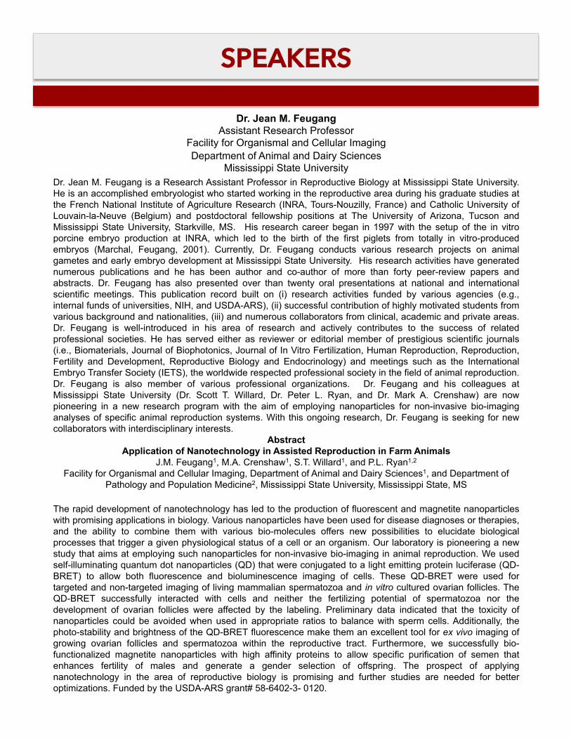
Dr. Jean M. Feugang Assistant Research Professor
Facility for Organismal and Cellular Imaging Department of Animal and Dairy Sciences
Mississippi State University Dr. Jean M. Feugang is a Research Assistant Professor in Reproductive Biology at Mississippi State University. He is an accomplished embryologist who started working in the reproductive area during his graduate studies at the French National Institute of Agriculture Research (INRA, Tours-Nouzilly, France) and Catholic University of Louvain-la-Neuve (Belgium) and postdoctoral fellowship positions at The University of Arizona, Tucson and Mississippi State University, Starkville, MS. His research career began in 1997 with the setup of the in vitro porcine embryo production at INRA, which led to the birth of the first piglets from totally in vitro-produced embryos (Marchal, Feugang, 2001). Currently, Dr. Feugang conducts various research projects on animal gametes and early embryo development at Mississippi State University. His research activities have generated numerous publications and he has been author and co-author of more than forty peer-review papers and abstracts. Dr. Feugang has also presented over than twenty oral presentations at national and international scientific meetings. This publication record built on (i) research activities funded by various agencies (e.g., internal funds of universities, NIH, and USDA-ARS), (ii) successful contribution of highly motivated students from various background and nationalities, (iii) and numerous collaborators from clinical, academic and private areas. Dr. Feugang is well-introduced in his area of research and actively contributes to the success of related professional societies. He has served either as reviewer or editorial member of prestigious scientific journals (i.e., Biomaterials, Journal of Biophotonics, Journal of In Vitro Fertilization, Human Reproduction, Reproduction, Fertility and Development, Reproductive Biology and Endocrinology) and meetings such as the International Embryo Transfer Society (IETS), the worldwide respected professional society in the field of animal reproduction. Dr. Feugang is also member of various professional organizations. Dr. Feugang and his colleagues at Mississippi State University (Dr. Scott T. Willard, Dr. Peter L. Ryan, and Dr. Mark A. Crenshaw) are now pioneering in a new research program with the aim of employing nanoparticles for non-invasive bio-imaging analyses of specific animal reproduction systems. With this ongoing research, Dr. Feugang is seeking for new collaborators with interdisciplinary interests.
Abstract Application of Nanotechnology in Assisted Reproduction in Farm Animals
J.M. Feugang1, M.A. Crenshaw1, S.T. Willard1, and P.L. Ryan1,2 Facility for Organismal and Cellular Imaging, Department of Animal and Dairy Sciences1, and Department of
Pathology and Population Medicine2, Mississippi State University, Mississippi State, MS
The rapid development of nanotechnology has led to the production of fluorescent and magnetite nanoparticles with promising applications in biology. Various nanoparticles have been used for disease diagnoses or therapies, and the ability to combine them with various bio-molecules offers new possibilities to elucidate biological processes that trigger a given physiological status of a cell or an organism. Our laboratory is pioneering a new study that aims at employing such nanoparticles for non-invasive bio-imaging in animal reproduction. We used self-illuminating quantum dot nanoparticles (QD) that were conjugated to a light emitting protein luciferase (QD-BRET) to allow both fluorescence and bioluminescence imaging of cells. These QD-BRET were used for targeted and non-targeted imaging of living mammalian spermatozoa and in vitro cultured ovarian follicles. The QD-BRET successfully interacted with cells and neither the fertilizing potential of spermatozoa nor the development of ovarian follicles were affected by the labeling. Preliminary data indicated that the toxicity of nanoparticles could be avoided when used in appropriate ratios to balance with sperm cells. Additionally, the photo-stability and brightness of the QD-BRET fluorescence make them an excellent tool for ex vivo imaging of growing ovarian follicles and spermatozoa within the reproductive tract. Furthermore, we successfully bio-functionalized magnetite nanoparticles with high affinity proteins to allow specific purification of semen that enhances fertility of males and generate a gender selection of offspring. The prospect of applying nanotechnology in the area of reproductive biology is promising and further studies are needed for better optimizations. Funded by the USDA-ARS grant# 58-6402-3- 0120.

SPEAKERS
Dr. Mahesh Hosur Professor
Department of Materials Sciences & Engineering Tuskegee University
Mahesh Hosur is a Professor in Materials Science and Engineering department at Tuskegee University. He has led research efforts in advanced fiber reinforced composites, sandwich composites, nanophased composites, advanced green composites in areas of processing, process sensing, low-cost manufacturing, static and dynamic characterization, fatigue and fracture, structural analysis, development of characterization of flexible armors for extremities protection, repair of thick section composites, damage tolerant design, environmental effects, morphological characterization, microstructural characterization, nondestructive evaluation in particular ultrasonic and thermography techniques. To date he has led research efforts worth over $22M as PI and over $31.5M as Co-PI. He has supervised about 50 graduate students and nearly 50 undergraduate students besides mentoring junior faculty members. He has authored or coauthored 3 books, 3 book chapters, over 275 articles in journals and conference proceedings besides numerous technical reports. He has received many honors which includes recognition as Fellow of American Society for Mechanical Engineers.
Abstract
An Overview on Recent Advances in NSF-EPSCoR Nano-Biomaterials Research Thrust
The presentation will focus on the recent advances through NSF-EPSCoR Nano and Biomaterials Research thrust being carried out at six doctoral granting institutions in Alabama. The Nano and Biomaterials Research Thrust involves the development of new nanostructured materials with enhanced thermal, physical, mechanical, and biodegradable properties. The tasks being carried out are divided in three main areas: polymeric nanocomposites, advanced green composites, and synthesis of nanoparticles for drug delivery applications. In polymeric nanocomposites area research is being carried out to include nanoparticles like nanoclay, single and multiwalled carbon nanotubes, metal and metal oxide nanoparticles to improve the performance of polymers which are then used for fabrication of fiber reinforced composites for different high technology applications. Advanced green composites research is looking at the use of plant based polymers and natural fibers as viable alternates to synthetic polymers and fibers which are petroleum based and non-biodegradable. In the studies on synthesis of nanoparticles for drug delivery applications different types of nanoparticles are being synthesized using microwave and sonochemical methods.

SPEAKERS
Dr. Michael H. Irwin Research Associate Professor Department of Pathobiology
College of Veterinary Medicine Auburn University
Abstract
A Proteoliposome Nanocarrier for Mitochondrial Gene Delivery
MH Irwin1, BN Augsburger1 and CA Pinkert2 1Department of Pathobiology, College of Veterinary Medicine, Auburn University
2Department of Biological Sciences, College of Arts and Sciences, The University of Alabama
A number of significant hurdles must be overcome to enable the manipulation of mitochondrial genetics. The mitochondrial genome is effectively sequestered within two lipid bilayers: the outer mitochondrial membrane (OMM) and the inner mitochondrial membrane (IMM), making access to mitochondrial DNA (mtDNA) difficult. Cells contain hundreds to thousands of copies of mtDNA, and mutations in mitochondrial genes are typically heteroplasmic. Exceeding a threshold level of heteroplasmy (typically around 70% for many mtDNA mutations) can cause cells to rapidly degenerate from normal to a disease phenotype. An effective mitochondrial gene therapy requires technology to deliver complete, healthy mitochondrial genomes to the mitochondrial matrix, thereby shifting the level of heteroplasmy below the threshold level. Where possible, mimicking natural cellular mechanisms and components is desirable. We propose to engineer a nanocarrier with the capability to cross the plasma membrane and to fuse with both the OMM and the IMM, providing effective payload delivery to the mitochondrial matrix. This proteoliposome nanocarrier will consist of an outer liposomal shell enveloping inner concentric shells designed for sequential OMM and IMM fusion. Fusion of the OMMs of adjacent mitochondria is mediated by the transmembrane GTPases, mitofusin 1 (Mfn1) and mitofusin 2 (Mfn2). The first part of the project, presented here, focuses specifically on developing a red fluorescently labelled proteoliposome incorporating recombinant Mfn2 for in vitro fusion with the OMM of green fluorescent protein (GFP)-labelled mitochondria isolated from stably transfected NIH-3T3 fibroblasts. Flow cytometry will be used to detect Mfn2-mediated liposomal-mitochondrial fusion through recognition of green-red fluorescence colocalization.

SPEAKERS
Dr. Jacob Jordan Contractor Support, DARPA/BTO
Defense Advanced Research Projects Agency (DARPA) Biological Technologies Office
Formed in April 2014, the mission of the Defense Advanced Research Projects Agency Biological Technologies Office (DARPA/BTO) is to foster, demonstrate, and transition breakthrough fundamental research, discoveries, and applications that integrate biology, engineering, and computer science for national security. This presentation will provide an overview of the BTO vision, general strategies for innovation within DARPA programs, and a few specific examples of ongoing work within the field.

SPEAKERS
Dr. Eugenia Kharlampieva Assistant Professor of Polymer Chemistry
Department of Chemistry The University of Alabama at Birmingham
Eugenia Kharlampieva is an Assistant Professor of Polymer Chemistry at the Department of Chemistry at the University of Alabama at Birmingham. She received her Ph.D. in Polymer Science from the Stevens Institute of Technology and postdoctoral training in Materials Science and Engineering at the Georgia Institute of Technology. Her research centers at the intersection of polymer chemistry, nanotechnology, and biomedical science and includes synthesis and assembly of polymers and nanostructures as novel platforms for therapeutic applications such as controlled delivery and regenerative medicine. She has authored more than 60 articles and has been recently awarded NSF CAREER.
Abstract
Polymer Nanomaterials for Drug Delivery and Cell Transplantation
Eugenia Kharlampieva, University of Alabama at Birmingham, AL 35294, USA
Bio-inspired fabrication of biologically-active and stimuli-sensitive nanostructured materials are of increasing interest in bio- and nanotechnology. This talk will focus on functional ultrathin coatings and hollow microcontainers (capsules) obtained by hydrogen-bonded layer-by-layer assembly of synthetic and biological macromolecules on inorganic templates and living cells. We will discuss pH-triggered volume and shape transitions in these materials to be used for controlled drug delivery. We will also address the application of nanostructured coatings in cell-based transplantation therapy. We will introduce nanothin immunomodulatory coatings with diminished inflammatory immune responses deposited on surfaces of mammalian pancreatic islet cells. These materials provide prolonged cell viability and function to be used in diabetes treatment.

SPEAKERS
Dr. Richard M. Myers President and Science Director
HudsonAlpha Institute for Biotechnology Huntsville, Alabama
Banquet Keynote Speaker
Abstract
Using Genomics and Genetics to Understand Human Health and Disease
Technologies for collecting very large amounts of genomic and genetic data have dramatically increased in throughput and efficiency in recent years, so that it is now possible to determine whole and partial genome sequences and measure quantities and qualities of every type and source of nucleic acid that is produced by cells, tissues and organisms. This talk will describe the development of some of these approaches, and how they are applied on a large scale to study a variety of problems in human health and disease. Results of applying whole genome and targeted DNA sequencing to large numbers of research and clinical samples for childhood and adult diseases, acquired cancers, and interactions of our bodies and cells to the environment, including differential responses to drug treatments, will be discussed.

SPEAKERS
Dr. Joseph D. Ng Associate Professor
Director of the Biotech Science & Engineering Program Department of Biological Sciences & Lab of Structural Biology
The University of Alabama in Huntsville
Dr. Ng, a biotechnology scientist, received his Doctorate in Biochemistry from the University of California, Riverside in 1992 and did his postdoctoral work studying protein crystal growth and X-ray crystallography of macromolecules at The National Center for Scientific Research (CNRS) in France. As a postdoctoral fellow, Dr. Ng was contracted with the French Space Agency (CNES) to work with NASA on biological experiments in space where he was responsible for coordinating scientists all over Europe to conduct biological experiments on the International Space Station. In 1998, Dr. Ng joined the University of Alabama in Huntsville (UAH) as a faculty member in the Department of Biological Science. Presently, Dr. Ng is the Director of the Biotechnology Science and Engineering Program at UAH and a principal investigator for a research team studying structural and synthetic biology. He has over 60 research publications, won several research awards and has been recognized for academic excellence in the scientific community including being a member of the U.S. National Committee for Crystallography of the National Academies. Dr. Ng is also a co-founder and President of iXpressGenes Inc. He has been instrumental in advancing the Biotechnology community in Huntsville AL in his involvement and partnership with the UAH business school, private companies and government laboratories.
Abstract
Synthetic Biology for Nano- and Atomic-Scale Macromolecular Structure Determination
Synthetic biology is an emerging area that involves the design and construction of new or existing biological parts. Gene synthesis and assembly have been coupled to X-ray crystallographic techniques as a strategic synthetic biology approach for the structural determination of macromolecules. A PCR-based gene synthesis method will be described on how coding region assembly of proteins potential for drug targets can be easily performed. The gene synthesis procedure is based on sequential assembly such that homogeneous DNA products can be obtained after each synthesis step without extensive manipulation or purification requirements. Coupling the gene synthesis procedure to in vivo homologous recombination techniques allows efficient subcloning and site-directed mutagenesis for error correction. Recombinant proteins important for pharmaceutical leads have been assembled or modified using synthetic biology techniques for recombinant expression and crystallization for structure determination by X-ray and Neutron crystallography. In particular, the soluble inorganic pyrophosphatase (IPPase) has been one of the proteins of focus. IPPase in an enzyme that catalyzes the hydrolysis of inorganic pyrophosphate (PPi) to form orthophosphate (Pi). The action of this enzyme shifts the overall equilibrium in favor of synthesis during a number of ATP-dependent cellular processes such as in the polymerization of nucleic acids, production of coenzymes and proteins and sulfate assimilation pathways. The structures determined include the recombinant IPPase bound to Mg+2, Ca+2, Br-, SO2
-2 or PO4-2 involving those with non-
hydrolyzed and hydrolyzed pyrophosphate complexes. All the crystallographic structures provide snapshots of the active site corresponding to different stages of the hydrolysis of inorganic pyrophosphate. As a result, a structure-based model of IPPase catalysis is devised showing the enzyme's low-energy conformations, hydration states, movements and nucleophile generation within the active site.

SPEAKERS
Dr. David E. Nikles Professor
Department of Chemistry and Center for Materials for Information Technology
The University of Alabama
Abstract
Building and Managing a Multidisciplinary Materials Research Science and Engineering Center at The University of Alabama
The Materials Research Science and Engineering Centers program the Division of Materials Science at the National Science Foundation supports multidisciplinary university research centers to pursue cutting edge materials research and conduct education and outreach programs. From 1994 to 2008 the Center for Materials for Information Technology at the University of Alabama won three consecutive MREC competitions. The MRSEC supported basic research in the materials underpinning magnetic recording science and technology. The Center was divided into two different interdisciplinary research groups IRG-1 Materials for Recording Heads and IRG-2 Materials for Recording Media. This presentation will describe the genesis of the Center, including team building, preparation of pre-proposals and full proposals and reverse site visits. The management of the highly diverse research teams of faculty, visiting faculty, post docs, graduate students, undergraduate research assistants, high school students and high school teachers will be discussed. The theme of the presentation is lessons learned. The program solicitation for the next MRSEC competition will begin in 2016. Now is the time for identifying teams of faculty in anticipation of submitting a pre-proposal in the fall of 2016.

SPEAKERS
Dr. Srinivas Palanki Professor and Chair
Department of Chemical and Biomolecular Engineering University of South Alabama
Dr. Srinivas Palanki joined the Department of Chemical and Biomolecular Engineering at the University of South Alabama as Professor and Chair in 2007. Prior to joining USA, he was a Professor of Chemical and Biomedical Engineering at Florida State University. He has been a visiting professor at Ecole Polytechnique Federale de Lausanne (Switzerland), Ecole Nationale Superieure Des Industries Chimiques (France) and National University of Singapore (Singapore). He obtained his Ph.D in Chemical Engineering from the University of Michigan (Ann Arbor) and his B.Tech in Chemical Engineering from the Indian Institute of Technology (Delhi). His research focuses on the application of systems analysis tools to problems in engineering and biology.
Abstract Effect of Size and Concentration of Gold and Silver Nanoparticles on Skin Cancer
Chemoprevention
Srinivas Palanki1, Sumit Arora2, Rohan Palanki2, Lilia Rusu1, Ajay P. Singh2, Seema Singh2 1Department of Chemical and Biomolecular Engineering, University of South Alabama,
Mobile, Alabama 36688, USA 2Department of Oncologic Sciences, Mitchell Cancer Institute, University of South Alabama,
Mobile, Alabama 36604, USA
Skin cancer is the most commonly diagnosed malignancy in America. The traditional approach to protect against the harmful effects of ultraviolet (UV) radiation has been to use sunscreen lotion as a direct barrier on the skin. However, recent studies have shown that zinc oxide (ZnO) nanoparticles and titanium dioxide (TiO2) nanoparticles, which are used as UV filters in sunscreens, can have inflammatory/toxic effects on normal skin cells. For this reason, it is necessary to look for novel nanoparticles that are effective for skin cancer chemoprevention with minimal side-effects on normal skin cells. In this research, the effect of size and concentration of gold nanoparticles (AuNPs) and silver nanoparticles (AgNPs) were tested for skin cancer chemoprevention against UV-induced cell damage. Cell viability analysis indicated that AuNPs and AgNPs in the size range 10-100 nm and concentration range 1-10 mg/L are not toxic to nontumerigenic HaCaT cells. Dot-blot assay results indicate that UV-B radiation causes considerable DNA damage to HaCaT cells and this damage is significantly reduced in the presence of AgNPs in the size range 10-40 nm. FACS results indicate that cells without AgNPs undergo significant early apoptosis in the presence of UV radiation. However, AgNPs of size 10-40 nm provide significant protection (4-5 fold reduction in apoptosis), thereby proving the chemopreventive effect of AgNPs. On the other hand, AuNPs do not provide any chemoprevention to UVB-induced DNA damage. Cell cycle studies show that treatment with AgNPs in the size range 10-40 nm prior to UVB exposure caused significant accumulation of HaCaT cells in the G1/S phase (~ 9.0 fold higher as compared to AgNPs-untreated UVB-exposed cells).

SPEAKERS
Andrew D. Penman, Ph.D., FRSC Vice President
Drug Development Southern Research Institute
Lunch Keynote Speaker
In November 2008, Andrew D. Penman, Ph.D., was named Vice President of the Drug Development Division at Southern Research. In this position, he leads the preclinical contract research operations in toxicology, bioanalytical sciences, infectious diseases, cancer therapeutics, and immunology. Before joining Southern Research, Dr Penman served as vice president of Preclinical Development for Angiotech Inc. in Canada where he directed the company's global preclinical research and development activities focusing on a number of different therapeutic areas. He also led the company's Vascular WrapTM project team for a product in Phase III clinical trials. Prior to that, Dr Penman was President of Preclinical Technologies at Aptuit. He also held scientific management positions at Pharmacia (now part of Pfizer) in the US, Cephac Europe SA in France, and Quintiles in the UK and US. Dr. Penman earned his Bachelor's degree in chemistry from Heriot-Watt University in Edinburgh, Scotland and his Doctorate degree from the University of Kent in the United Kingdom. He is a Fellow of the Royal Society of Chemistry, has made numerous scientific presentations and is author or co-author on a number of scientific publications.

POSTER ABSTRACTS

Poster # G-500
Double Receptor Targeting Multifunctional Iron Oxide Nanoparticles Drug Delivery System for the Treatment and Imaging of Prostate Cancer
Md. Shakir Uddin Ahmed1*, Mohamed Abdalla2, and Timothy Turner1
1Tuskegee University, Department of Biology & Center for Cancer Research,
Tuskegee, AL 2Tuskegee University, Department of Chemistry & Center for Cancer Research,
Tuskegee, AL
As an alternative to the drawbacks of current advanced prostate cancer chemotherapy, we propose a multifunctional double targeting drug delivery system that utilizes the combination of two cancer-targeting peptides: a modified luteinizing hormone releasing hormone (LHRH), the ligand for luteinizing hormone releasing hormone receptor (LHRH-R), and AE105, the ligand for urokinase type plasminogen activator receptor (uPAR) and loaded with the anticancer drug Paclitaxel (PTX) as the payload. The results obtained from dynamic light scattering (DLS) indicates that conjugation of peptides on IONPs resulted in an increase in the average hydrodynamic size of targeted IONPs (16.34 nm) as compared to non-targeted IONPs (12.51 nm), as well as a decrease of zeta potential from -70.43 mV to -58.06 mV, respectively. Prussian blue staining demonstrated that both, LHRH and AE105 targeted IONPs were internalized efficiently by the human prostate cancer cell line, PC3. In vitro magnetic resonance imaging (MRI) results showed that double-targeted IONPs significantly maintained T2 MRI contrast effect and reduction of T2 values upon internalization by PC3 cells. In vitro MRI imaging confirmed the preferential binding and accumulation of double-targeted IONPs in PC3 cells when compared to normal prostate epithelial cells (RC77N/E). PTX loaded double-targeted IONPs showed an approximately 2-fold reduction in PC3 cell viability when compared to non- targeted IONPs. These were also stable at physiological pH and efficiently released around pH 4. In addition, IONPs system is capable of reducing drug concentration. Our results indicate that we have developed a LHRH-R and uPAR targeted IONPs drug delivery system that potentially provides a MRI tractable delivery of cancer therapeutics such as PTX to PC3 cells. Therefore, our optimized double-targeted IONPs drug delivery system has the potential to significantly improve the health outcomes and quality of life for cancer patients as a novel type of targeted nanomedicine therapy.

Poster # O-100 Chemo-Mechanical Investigation of Tungsten Exposed Bio-Monitoring Systems,
Gastropod (Otala lactea) Shells
Paul G Allison1,2, Jen Seiter2, Alfredo Diaz3, Jay Lindsay2, Robert Moser2, R.V. Tappero4, Alan Kennedy2
1University of Alabama, Department of Mechanical Engineering
2US Army Engineer Research & Development Center 3University of Puerto at Mayaguez, Department of Mechanical Engineering
4National Synchrotron Light Source at Brookhaven National Laboratory Environmental concerns with military use of metallic tungsten (W), which was initially assumed to be an environmentally friendly alternative to lead, arose due to previous investigations that identified fishing weights and munitions containing elemental W can fragment and oxidize into complex monomeric and polymeric tungstate (WO4) species in the environment. The speciation leads to increased solubility and mobility in soils resulting in a greater increase of the potential for toxicity and bioaccumulation into plant and animal tissues. In this study, we expand on our previous research that identified tungsten toxicity, bioaccumulation, and compartmentalization into organisms, and correlate through depth-sensing nanoindentation that the bioaccumulation of W degrades the mechanical properties by over 50% in the gastropod (Otala lactea) shell. Synchrotron- based X-ray fluorescence maps and X-ray diffraction measurements confirmed the bioaccumulation of W and integration into the shell matrix with the observed changes in shell biomechanical properties, mineralogical composition, and crystal orientation.

Poster # G-501
Silver-Polyvinyl Pyrrolidone (Ag-PVP) Nanoparticles Inhibition of Chlamydia trachomatis Inflammatory Mediators in Macrophages is Partly Due to Down-
Regulation of Expression of its Major Outer Membrane Protein
D’Andrea Ashmore, Saurabh Dixit, Shree R. Singh and Vida A. Dennis
Center for NanoBiotechnology & Life Sciences Research, Alabama State University, Montgomery, Alabama
Chlamydia trachomatis, the most sexually transmitted diseases globally, affects both men and women and poses a huge economic burden on the population due to its asymptomatic nature. C. trachomatis causes symptoms that range from burning during urination, discharge and urethritis to name a few. Also, it can often lead to pelvic inflammatory disease and infertility in women and sterility in men without early intervention. We recently published that Ag-PVP nanoparticles exerted anti-inflammatory actions in mouse macrophages by inhibiting several inflammatory mediators, including receptors, chemokines and cytokines. Here we hypothesized that Ag-PVP anti- inflammatory action in macrophages maybe due to its ability to inhibit C. trachomatis bacterial load. Mouse J774 macrophages were exposed to various concentrations of Ag- PVP (3-12 µg/mL) and infected with C. trachomatis at a multiplicity of infection (MOI) of 2.5 to 20 for up to 48 hr. We used TaqMan qRT-PCR to quantify the mRNA gene expression of C. trachomatis major outer membrane protein (MOMP) as a marker of bacterial load. Our qRT-PCR data showed that Ag-PVP reduced MOMP expression by 70% with no toxicity to cells at all tested concentrations of Ag-PVP, suggesting that nanoparticles potentially reduced C. trachomatis bacterial load in macrophages. We further demonstrated by IL-6 cytokine specific ELISA that Ag-PVP inhibited IL-6 in a C. trachomatis concentration-dependent manner, correlating with its modulation of MOMP expression. Our data shows that the ability of Ag-PVP to inhibit C. trachomatis bacterial load maybe a potential mechanism of its anti-inflammatory actions in macrophages.

Poster # U-500
Designing Liposomes for Protein Encapsulation
Brandi Barlow1, Elisabeth Kastner2, Shree Singh1, Yvonne Perrie2, Komal Vig1
1Center for Nanobiotechnology Research, Alabama State University, Montgomery, AL 2Aston Pharmacy School, Aston University, Aston Triangle, Birmingham, B4 7ET
A liposome is a vesicle, made out of the same material as a cell membrane. Membranes are made of phospholipid bilayer. Phospholipids are molecules that have a hydrophilic head group and a hydrophobic tail group. In the present study, liposomes were prepared for protein encapsulation and delivery. The liposomes were created through multilamellar vesicles (MLV), small unilamellar vesicles (SUV), and dehydration-rehydration vesicles (DRV) method. To prepare liposome, chloroform:methanol (9:1) surfactant solution was used. Synthetic phospholipid- Dipalmitoylphosphatidylcholine (DPPC) (100mg) was dissolved in 1ml of the surfactant solution to prepare the liposome. To stabilize the liposome cholesterol (Chol) was added at the rate of 10 mg/mL in the surfactant solution. The mixture containing DPPC and Chol (16 µmoles each) were placed in a round bottom flask and placed on a rotary evaporator to make a dry lipid film. The film was further flushed with Nitrogen to ensure complete solvent removal. The dried film was rehydrated in PBS (2mL) to get MLV. To prepare SUV liposomes, prepared MLV solution (2ml) was placed in a beaker and sonicated for 5mins to get 100 nm diameter SUV. The SUV suspension was centrifuged at 3,500 g for 10 minutes to remove any debris from the sonicator probe. To encapsulate protein, SUV suspension was placed in a beaker and the Bovine Serum Albumin (BSA) protein (200 ug /2000ul) was added. The mixture was freezed at -20oC for an hr and transferred to freeze-drier overnight. To further prepare DRV, the freeze dried mixture was rehydrated in 100 µl of deionized water. We prepared liposomes with BSA, and blank liposomes with either water or PBS. Work is currently going on to encapsulate drugs in the liposomes for delivery purposes.

Poster # G-200 Effect of Gold Nanorod Functionalization on the Inhibition of Human Respiratory
Syncytial Virus
Swapnil Subhash Bawage, Pooja Munnilal Tiwari, Vida Dennis, Shree Ram Singh
Center for NanoBiotechnology Research, Alabama State University Montgomery, Alabama
Human respiratory syncytial virus (RSV) is a Paramyxovirus causing respiratory tract infections in the infants, children, old adults and immunosuppressed individuals. It is observed that almost every child below the age of 2 years would have had an RSV infection. RSV can lead to pneumonia and bronchiolitis in the pediatric and geriatric populations. There is neither active vaccine against RSV nor any effective drug, except broad spectrum anti-viral drug ribavirin. Recent trends in the RSV research indicate nanomedicine as an alternative option. Functionalization of gold nanorods with drugs or cell penetrating peptides provides longer retention, controlled release, protection from degrading enzymes and enhanced cellular delivery. Here, we have conjugated gold nanorods (GNR) with ribavirin (GNR-R), cell penetrating peptide HIV-TAT (GNR-T) and both together (GNR-RT). The nanoparticles were then characterized for functionalization by dynamic light scattering, zeta potential, Fourier transform- infrared spectroscopy, UV-Visible spectroscopy and transmission electron microscopy. RSV inhibition was assessed by immuno-fluorescence microscopy, real time PCR and plaque assay. Our results show that, RSV was inhibited by GNR. However, functionalization of GNR with ribavirin or/and TAT peptide reduced the RSV inhibition.

Poster # G-201 Evaluation of Antimicrobial Activity of Therapeutic Peptide Conjugated with Gold
Nanoparticles
Courtnee’ R. Bell, Shanese Jasper, Kasha Casey, Atul Chaudari, Vida Dennis, Shreekumar Pillai and Shree Singh
Center for Nanobiotechnology Research, Alabama State University, Montgomery, AL
Antibiotics are traditionally used to treat bacterial infections; however, many strains of bacteria have evolved and show a propensity to exhibit antibiotic resistance. This overwhelming problem creates a need for a new type of antimicrobial such as Gold (Au) Nanoparticles, which are becoming popular as antibacterials. These nanoparticles have promising effects as antimicrobial nanocomposites, but can be toxic in high levels. To alleviate this problem, the nanoparticles can be combined with antimicrobial peptides to decrease toxicity and increase antibacterial properties. In this study we evaluated the antimicrobial properties of peptide, p557 against Streptococcus pyogenes and E.coli using the Minimum Inhibitory Concentration (MIC). We then evaluated the antimicrobial properties of Gold nanoparticles using the same assay. The MIC of p557 for S. pyogenes was shown to be 1.9µg/mL when only 1mg/mL stock solution of the peptide was used. When 1mg/mL was used against E.coli, p557 showed inhibition at 3.9µg/mL. Evaluation of the Au-Nanoparticle against both S.pyogenes and E.coli revealed that the MIC was much higher and gave a variable range of 250µg/mL-125µg/mL. Our results show that both the peptide and the Au nanoparticle inhibited both gram-positive and gram-negative bacteria. However, the peptide inhibited both bacteria at a lower concentration than the Au nanoparticle. Given the functionality of the peptide and the Au nanoparticle we believe that this shows potential for complete microbial inhibition when conjugated. The interaction of p557 and Au nanoparticles with bacterial cells will be further investigated by Atomic Force Microscopy (AFM), standard plate count, growth- curve assay and real-time PCR.

Poster # U-100
Electrospinning Poly (ε-Caprolactone) Nanofiber for Bone-Tissue Regeneration
Brandi Bethune1, Elijah Nyairo2 and Derrick R Dean3
1Center for Nanobiotechnolgy Research, Alabama State University, Montgomery, AL 2Department of Physical Sciences, Alabama State University, Montgomery, AL 3Biomedical Engineering Program, Alabama State University, Montgomery, AL
This research aims to develop polymeric nanofibers that can be used as tissue scaffolds. Nanoscale fiber scaffolds provide an optimal template for cells to seed, migrate and grow. The goal is for the cells to attach to the scaffolds, then replicate, differentiate and organize into normal healthy tissues as the scaffold degrades. In this study, non-woven poly (ε-caprolactone) (PCL) and hydroxyapatite (HA) nanofibers with different wt % compositions were prepared by electrostatic co-spinning technology. It was hypothesized that PCL and PCL/HA scaffolds will mimic the nano-features of the natural extracellular matrix (ECM). To test if these scaffolds mimic the properties of natural ECM, we used TRAMP C2 cell lines derived from transgenic adenocarcinoma of mouse prostate (TRAMP) mice. The scaffolds were analyzed by MTT assay at different time points to verify cell toxicity/proliferation. Characterization for morphology of the electrospun fibers were observed using scanning electron Microscopy (SEM) and SEM micrographs were analyzed using image analysis software. The fibers were characterized for thermal behavior using Differential Scanning Calorimetry (DSC), and for chemical structure using Fourier Transform Infrared Spectroscopy (FTIR). Thus, our objective is to develop biodegradable scaffolds for bone tissue that mimics the size scale and chemistry of the ECM with an interconnected pore structure, and enhanced mechanical properties.

Poster # G-202 Polyvinyl Pyrrolidone (PVP) Coated Silver Nanoparticles Demonstrates a Capsule
Dependent Antimicrobial Effect Against Streptococcus pneumoniae
Ronda K. Bibbs1 and Mamie Coats2
CREST Center for Nanobiotechnology Research & Biological Sciences Department Alabama State University
Both the antimicrobial nature and toxicity of metals are well documented in literature. Advances in nanotechnology have extended the usefulness of metals as antimicrobials while decreasing their toxic effects. Our study examined the effectiveness of metallic nanoparticles on the growth of pathogenic Streptococcus pneumoniae. the need for novel antimicrobials which can control pneumococcal growth is becoming more pressing due to increasing numbers of isolates which are resistant to current therapies. Nanospheres of gold (AuNP), titantium dioxide (TiO2), or polyvinyl pyrrolidone (PVP) coated silver (Ag-PVP) were tested for their ability to inhibit the growth of the pneumococcal model strain D39. Of the formulations test, Ag-PVP was found to be the most effective and provided the most consistent results. The inhibitory effect of Ag-PVP was seen for serotypes 4, 19F, and 3 in addition to serotype 2. The examination of S. pneumoniae strains that completely lack a capsular polysaccharide showed that the lack of the capsule made bacteria more resistant to the action of Ag-PVP. These data demonstrate a serotype independent, capsule dependent bactericidal activity for metallic nanoparticles includes of S. pneumoniae. [This work was supported by NSF- CREST (HRD-1241701) and NSF-HBCU-UP (HRD-1135863) at Alabama State University, Montgomery, AL 36104].

Poster # U-300
Multiscale Modeling of Electrical Properties of Carbon Nanotube-Based Composites
Ben Binderow* and Vinu Unnikrishnan+
*Undergraduate Student, +Assistant Professor
Aerospace Engineering and Mechanics, The University of Alabama The relationship between the structure and its property is essential to understand the mechanical characteristics of nano-materials like Carbon Nanotubes (CNT), and based nano-composite systems. In addition to the high tensile strength, carbon nanotubes are also known for their low electrical resistance and their ability to be effective electrical and thermal conductors. Carbon nanotubes are now beginning to show as promising materials for use in integrated electronics, primarily due to their potential to develop flexible nano-electronic systems compared to silicon based electronic systems. In this work, we develop a computational model to understand the thermal and electrical properties of carbon nanotube reinforced composite systems and the computational model would be compared with experimentally published data. One of the major advancement of the developed computational model is the inclusion of the interfacial electrical resistance of carbon nanotubes and study of the effect of the overall electrical conductivity of the nanocomposite using a multi-phase approach. Given the conductivity of the matrix and the nanotube, the model would be able to predict the effective electrical (or thermal) conductivity, for a specific volumetric fraction of the nanotube. The model will also be able to calculate the conductivity of a multi-phased nano-composite for which the CNTs are initially coated with a separate material and then combined into a matrix, using a multi-phase approach.

Poster # U-105
Influence of Fe2O3 Nanoparticles Treatment on the Arabidopsis thaliana
Sergey Bombin, Mitchell LeFebvre, Katrina Ramonell
Department of Biological Sciences, University of Alabama Iron oxide nanoparticles are widely used for biomedical and industrial purposes and are present in our environment. Interestingly, there are few studies observing the potential impact of nanoparticles on plant and animal species. The goal of this project is to identify the effects of charged Arabidopsis thaliana. To achieve this goal, we treated Arabidopsis plants with both positively and negatively charged iron oxide nanoparticles dissolved in distilled water. Previous experiments showed that the model plant - Fe2O3 nanoparticles on the growth and development of Fe2O3 nanoparticles are readily absorbed by the root system of Arabidopsis and are rapidly dispersed throughout the plant’ tissue including transport into the reproductive structures. In this study, we investigated the effect of iron oxide nanoparticles on plant’s physiology and reproductive abilities. Results describing the effect of iron oxide nanoparticles on seed production, seed germination, pollen viability and root development will be presented.

Poster # G-502
A Simple Method for the 360-Degree Acquisition of Bioluminescence, Fluorescence, or X-Ray Data using a Mouse Imaging Spinner (MiSpinner)*
Andrew Brannen, Matthew Eggert, Robert Arnold, Peter Panizzi
Department of Drug Discovery and Development, Harrison School of Pharmacy,
Auburn University Optical imaging modalities are powerful tools for the in vivo assessment of drug delivery and therapeutic strategies. Current 2-dimensional imaging approaches have inherent limitations that result from the scattering of light as it passes through animal tissues, the interference of that signal by the absorptive properties of the tissue and blood, and the relative orientation of the light source in the animal during longitudinal studies. To account for this, it is commonplace to acquire images from multiple orientations of a single subject, in order to obtain the most accurate representation of a given region of interest (ROI). However, this method leads to subjectivity and inconsistency when manually turning or manipulating the subject in the field of view. We have invented a device that works with existing optical imaging systems that eliminates the subjectivity of the animal positioning through the use of an actuated motor and animal holder mechanism. Our device provides a rapid means of acquiring multiple images in precise intervals around the subject animal in photographic, bioluminescent, fluorescent, and X-ray modalities. Here, we demonstrate the application of this device through imaging a bioluminescent PC3-Fluc xenograph tumor in NCRNu mouse, and graphically illustrate the change in bioluminescent ROI intensity as it relates to the orientation of the mouse around a 360 degrees axis. The ROI intensity data plotted as a function of the degrees of rotation results in a bell-shaped curve, with the peak representing maximal bioluminescent intensity from the tumor source. *Provisional patent US62/020,056; trademark pending

Poster # U-400
Exposition of Foreign Peptides on Qβ Coliphage for Au Nanoparticle Binding
Alexandria Brooks, R. Singleton, Carrie A. Sanders and A. B. Waffo
1Department of Biological Sciences, Alabama State University 2Centers for NanoBiotechnology Research, Alabama State University, Montgomery, AL It is known that Au can be used in the treatment of cancer in nano form as a probe to both target and treat cancerous tissues. And although Au itself can be used in the treatment, an anti-cancer biodrug can be coated onto the Au. However, two problems exist. One, nanoparticles tend to aggregate in vivo and are cleared by the immune system. The only way to prevent aggregation is to keep the nanoparticles separated in vivo. The second problem arises with the nature of biodrugs themselves. Biodrugs are made from the same biomolecules that make up the body, as such; they are also subjected to the same enzymes that degrade biomolecules in the body. This leads to indiscriminate distribution, degradation, and a risk of under-medicating. To compensate, a larger dose of the biodrug is given; however, toxicity becomes the risk. Since it is known that certain peptides (nano-tags) bind Au, we hypothesize that displaying these nano-tags on the surface of our bacteriophage Qβ, and allowing them to bind Au will prevent aggregation. This Au can then be coated with an anti-cancer biodrug, and the Au will convey protection to the biodrug. To achieve this, the genes of three gold-binding peptides: Au0 (LKAHLPPSRLPS), Au1 (VSGSSPDS), and Au2 (TGTSVLIATPYV) were inserted separately into the genome of Qβ at the end of the A1 gene. The resulting recombinant phages, pQβAu0, pQβAu1 and pQβAu2, were transformed with HB 101 E. coli. The plaque assay provided the titer and phage morphology, and RT-PCR confirmed the tag gene size for each construct. A binding assay allowed different concentrations of Au to bind to the recombinant phage. Confirmation and visualization of the phage-nanoparticle complex was verified via Transmission Electron Microscopy (TEM). The next phase is to focus on coating the Au nanoparticles with chemotherapeutic biodrugs.

Poster # U-501
Proteomic Studies of Antibacterial Peptides
Stephen H. Brown1, Atul A Chaudhari2, Shree R. Singh2, Shreekumar R. Pillai2
1College of Science and Mathematics, Auburn University, Auburn, AL 2Center for Nanobiotechnology Research, Alabama State University, Montgomery, AL
Resistance to antibiotics is of grave concern and there is an urgent need for development of new antimicrobial strategies. Antimicrobial peptides (APs) could be a suitable approach as certain antimicrobial peptides have shown antibacterial activity against some of the antibiotic- resistant bacteria. It is also of interest to investigate the mechanism of action. In the present study, we evaluated the antimicrobial effect of two commercially available peptides TP493 and TPOB1013 from Therapeutic Peptides Inc. Minimum inhibitory concentrations (MIC) of both the peptides were investigated against a foodborne pathogen, Salmonella enterica serovar Typhimurium. Post exposures to peptides, the morphological changes were captured by scanning electron microscopy (SEM). The protein profile of outer membrane (OM) and periplasm of the bacteria exposed to both the peptides was also investigated and compared to untreated bacteria. The MICs for TP493 and TPOB1013 were between 3.9 - 7.8 µg/ml, and 1.9- 3.9 µg/ml, respectively. SEM analysis revealed the formation of pores in bacteria, damage to the outer membranes, and lysis of the cells. The protein profiles of OM and periplasm of the peptides-treated bacteria showed significant difference compared to untreated bacteria. It was observed that some of the proteins were either significantly expressed or were not observed in peptide treated bacteria compare to untreated bacteria. The results suggest that TP493 and TPOB1013 have antimicrobial potential and could be considered for development of new antimicrobials. This work was supported by NSF-REU (DBI-1358923) to Dr. Komal Vig (PI) and by NSF-CREST (HRD-1241701) to Dr. Shree S. Singh (PI).

Poster # G-100 MicroRNA Sponge Production using PCR-based Concatemerization of Short DNA
Oligonucleotides
SE Cardin, TJ Perry, GM Borchert
Department of Biology, University of South Alabama, Mobile, AL
MicroRNAs (miRs) are small, noncoding RNAs encoded within our genome that regulate gene expression by silencing messenger RNA (mRNA) transcripts. Since being discovered in humans in 2001, much has been learned concerning the many cellular activities that miRs can affect. MiRs have been implicated in almost every cellular activity, including cell fate determination, stress response, metabolism, apoptosis, and carcinogenesis. Importantly, alterations in microRNA activity due to mutation or misexpression have been repeatedly shown to result in tumorigenesis and disease. However, because of their relatively recent discovery, therapeutic tools to suppress miRs are just now being developed. One extremely promising way to inhibit miRs is to use microRNA sponges which consist of ~15 miR “target” sites that can specifically bind and inactivate particular miRs. Unfortunately, microRNA sponge production has proven to be problematic thus far as current production methods involve either costly commercial synthesis or low throughput ligation-based cloning. However, we have recently developed an entirely novel, PCR-based method that allows sponge production in a much more rapid manner. In all, PCR amplification, cloning, and sequence confirmation are readily attainable in less than two weeks and at a significantly lower cost than commercial inhibitor synthesis.

Poster # G-503
Peptide-Conjugated SPIONs for Salmonella Biofilms
K.K.Casey, K.Hussain, A. A. Chaudhari, S. Jasper, C. Bell, S. Pillai
Center for Nanobiotechnology Research, Alabama State University, Montgomery, AL Biofilms created by foodborne pathogens are difficult to eradicate with traditional antibiotics due to challenges that are faced with penetrating biofilms. Therefore, the development of a novel delivery system is urgent. Superparamagnetic iron oxide nanoparticles (SPIONs) are promising candidates in the area of biomedical applications, especially targeted drug delivery. Here, we evaluate the hypothesis that SPIONs combined with antimicrobial peptides can be effective in penetrating biofilms and delivering the peptide to a specific site for treatment. SPIONs can be targeted to the infection sites using an external magnetic field, causing deep penetration of the biofilm. In our present study, we have evaluated a proprietary peptide TP556 from Therapeutic Peptide Inc. as well as carboxylated SPIONs for antimicrobial effects on the planktonic form of the bacteria. The minimum inhibitory concentration (MIC) of TP556 and SPIONs against Salmonella enterica serovar Typhimurium (S. typhimurium) was assessed. The MIC of TP556 was between 50 and 62.5 µg/mL. SPIONs did not retard bacteria alone even at concentrations of 250 µg/mL. We have also begun to evaluate to effects of TP556 conjugated SPIONs against biofilms in the presence of a magnetic field. Magnetically concentrated TP556-SPIONs cause some bacterial killing in the established biofilm.

Poster # U-200
PLGA Nanoparticles as a Delivery Agent for Cancer Cells
India Chaney1, Leandra Jones2, Komal Vig2
1Huntingdon College, Montgomery, AL 2Center for Nanobiotechnology Research, Alabama State University, Montgomery, AL
Historically, it has been difficult to combat cancer in any form because of its ability to elude traditional forms of medicine, such as chemotherapy, radiation, and surgery. In fact, cancer treatments negatively affect the patient and are at times ineffective against preventing cancer entirely. Hopefully, a new form of treatment can be more effective and less hazardous for the patient. In the present study, we wanted to evaluate nanoparticles as drug delivery agents to cancer cells. Anti-cancer drug, Doxorubincin Hydrochloride (DOX) was encapsulated in poly (DL-lactide-glycolide) PLGA nanoparticles to allow low doses of the drug to be released into the body over an extended period of time in order to kill cancer cells. Drug encapsulated PLGA and PLGA nanoparticles were prepared using “emulsion” method. The nanoparticles were characterized using Zeta sizer, TEM, SEM, and FTIR. PLGA-DOX nanoparticles were around 220 nm with a charge of -29 mV. Our results showed 90% drug encapsulation of nanoparticles. FTIR analysis confirmed that DOX was successfully encapsulated in the PLGA nanoparticles. Release of the drug from nanoparticle was evaluated in PBS by continuous shaking and estimating absorbance at 480 nm. Drug was released gradually over the period of time. We observed almost 40% cell death of A549 cells at 250 ug/mL PLGA-DOX. The current results are promising; however, further research is necessary to achieve uniform sized nanoparticles, to increase drug nanoparticles, and to increase drug load so as to have more efficient way to kill cancer cells.

Poster # O-500 Pegylation of Silver Coated Single Walled Carbon Nanotubes Reduces Toxicity to
Human Cells at Their Antibacterial Concentrations
Atul A. Chaudhari1, Shanese L. Jasper1, Ejovwoke Dosunmu1, Michael E. Miller2, Robert D. Arnold3, Shree R. Singh1, Shreekumar R. Pillai1*
1Center for Nanobiotechnology Research, Alabama State University, Montgomery, AL
2Research Instrumentation Facility, Auburn University, AL 3Department of Drug Discovery and Development, Auburn University, AL
Silver coated single walled carbon nanotubes (AgCNTs) are well known for their antibacterial activity. However their toxicity to human cells is a serious public health concern. This can be avoided by the process called as functionalization using non- toxic material such as polyethylene glycol (PEG). This may however reduce their antibacterial activity as pegylation may cover the silver coating on carbon nanotubes. Present study was attempted to investigate whether pegylation of AgCNTs reduces their toxicity in vitro, without affecting their antimicrobial activity. For this purpose, AgCNTs were pegylated using phospholipid polyethylene glycol (PL-PEG) and were characterized by zeta potential measurement, Fourier transmission infra-red spectroscopy (FT-IR) and electron microscopy (EM). In vitro cell toxicity assay was carried out using human lung carcinoma (A549), hepatocellular carcinoma (Hep2) and murine macrophages (J774) whereas antibacterial activity was investigated against Salmonella enterica serovar Typhimurium. Shift in the charge (-41.8 to 8) using zeta potential measurements and characteristic FT-IR peaks on pAgCNTs similar to PL-PEG confirmed the pegylation which was further evidenced by electron microscopy which showed increase in size of AgCNTs upon pegylation. More importantly, pAgCNTs were relatively non -toxic to human cells at their anti- bacterial concentraions (62.5 µg/mL) compared to plain AgCNTs. We are further evaluating the antibacterial activity of pAgCNTs compared to plain AgCNTs at molecular levels.

Poster # G-504
Investigating the Biological Effects of Iron Oxide Nanoparticle Exposure Using Drosophila melanogaster
Evan A. Chavers1, Ben W. Henderson1, Rami R. Ajjuri1, Yuping Bao2 and
Janis M. O’Donnell1
1Department of Biological Sciences, University of Alabama 2Department of Chemical Engineering, University of Alabama
The past two decades have brought a myriad of novel medical applications for nanoparticle technology. As the clinical utilities for nanoparticles expand, so too does the need for a comprehensive understanding of their toxicology. To this point, recent reports have confirmed a wide range of deleterious effects associated with the use of gold, silver, and aluminum nanoparticles. These studies, however, have been mostly conducted in complex rodent models or in vitro using cultured cells. There remains a need for a practical, whole-organism model to study nanoparticle toxicology in vivo and assess the genetic and reproductive effects. Using Drosophila melanogaster, our research focuses on evaluating the potential toxic effects of polyacrylic acid (PAA)-coated iron oxide nanoparticles (IONPs) on larval development, reproduction, and the immune response. Interestingly, we found that exposure to low concentrations (0.5- 10µg/mL) of PAA-coated IONPs resulted in a higher larval mortality rate than exposure to higher concentrations. Additionally, low-dose exposure caused reproductive defects in adults leading to a reduction in fertility but not fecundity. When investigating the morphology of reproductive tissues, we found abnormalities in the ovarian cells of adults that had been dosed with PAA-coated IONPs as larva. Additionally, we have identified a dose-dependent threshold of IONP exposure required to activate the immune response, which may explain the deleterious effects of only low-dose treatments. Overall, our investigation reveals a tightly-regulated dose-response relationship required for understanding the adverse effects of nanoparticle uses, as well as provides researchers with a robust whole-organism model for assessing nanoparticle toxicology and teratology.

Poster # G-401
Understanding Graphene/Carbon/Gold Hybrids for Advanced Bionanotechnologies
Nitin Chopra*1,2, Yuan Li1, Junchi Wu1, Wenwu Shi1, Larry Summerville1,3, John West4,
Todd Gilliam4, Siddhantha Chandra5
1Department of Metallurgical and Materials Engineering, Box 870202, The University of Alabama, Tuscaloosa, AL
2Department of Biological Sciences, The University of Alabama, Tuscaloosa, AL 3NSF-REU Fellow
4NSF-REU Fellow, Department of Chemical and Biological Engineering, The University of Alabama, Tuscaloosa, AL,
5Hillcrest High School, Tuscaloosa, AL *[email protected]
Controlled growth and uniform patterning of graphene/carbon shells encapsulated gold nanoparticles (GNPs) on silicon wafer or on high curvature nanostructures is reported. Gold nanoparticles were uniformly patterned on these substrates via dewetting process. Surface oxidation of these patterned nanoparticles rendered them as sacrificial catalysts for the chemical vapor deposition (CVD) growth of graphene/carbon shells. The shell morphological evolution and thickness as well as surface migration of nanoparticles during the CVD process were studied. It is proposed that graphene/carbon shell growth is controlled by Ostwald’s ripening, surface gold oxide, and reducing CVD growth environment. Furthermore, complex heterostructures based on CNTs coated with GNPs were fabricated. Finally, Discrete Dipole Approximation (DDA) method was utilized to simulate optical properties of GNPs and comparisons were made with gold nanoparticles on various substrates. Tunable optical characteristics of GNPs with resonance peak wavelengths in visible light range were estimated.

Poster # G-203
Genipin as Anticancer Drug Against Human PC3 Cells
Brennetta Crenshaw1, Robert D. Arnold2, Allan E. David3, Shree R. Singh1, and Komal Vig1
1Center for Nanobiotechnology Research, Alabama State University, Montgomery, AL
2Harrison School of Pharmacy 3College of Engineering, Auburn University, Auburn, AL
The use of natural products have shown tremendous therapeutic prospects as carriers for drug delivery. Natural products, such as genipin, can act as an anticancer drug to treat various forms of cancer. In the present study, the inhibitory effects of genipin were evaluated against human prostate cancer cell line PC3. PC3 cells were seeded in 96-well culture plates and treated with genipin for 72 hours. Cell viability was determined by MTT Assay. 72 hours after incubation, 20 µl of 5 mg/ml MTT solution was added to each well with PC3 cells. The cells were futher incubated for 2 hours at 37ºC in culture hood. Media was removed and 200 µl of DMSO was added. Absorbance was read at 590 nm. Genipin effect on cellular protein was determined by SRB Assay. After 72 hours of incubation, media was removed and 200 µl of 10% TCA was added to the cells. Protein-bound dye was dissolved in 10 mM Tris base solution. Our studies showed that genipin reduced cell viability by 50% at 87ng in 72hours as shown by MTT assay. SRB assay indicated an IC50 value of 221.7ng in 72hours. Work is currently in progress on the encapsulation of genipin in a liposome for efficient delivery to cells and to increase its efficiency.

Poster # U-201
Polyvinyl Alcohol (PVA) Inluences Polylactic Acid-Polyethylene Glycol (PLA- PEG) Copolymer Morphology and Encapsulation Efficiency Characteristics
Marcus D. Davis, Saurabh Dixit, Shree R. Singh, and Vida A. Dennis
Center for NanoBiotechnology Research (CNBR), Alabama State University,
Montgomery, AL, USA Biodegradable nanoparticles (NPs) have a wide range of possibilities and advantages when compared to other drug delivery systems. Biodegradable polymers typically employed for fabrication of NPs provide controlled release, low toxicity, high encapsulation efficiency, sub-cellular size and bio-distribution in vivo. They are also excellent for vaccines delivery and can be fabricated to evoke immune responses, either by direct stimulation of antigen presenting cells (APCs) or delivering antigens to specific cellular compartments. However, problems associated with NPs formulation include their homogeneity, and rapid protein release (burst effect). Much research has been conducted on the block copolymer polyethylene glycol and polylactic acid (PLA-PEG). Polyvinyl alcohol (PVA) is the most commonly used emulsifier in the formulation of NPs. PVA provides non-aggregating, emulsifying and adhesive properties to NPs and an interconnected network with the polymer at the interface which ultimately can affect their homogeneity, release profile and size. The objective of this study was to determine the concentration-dependent effect of PVA on the size, morphology and release profiles of the modeled protein, bovine serum albumin (BSA) encapsulated within PLA-PEG. NPs were formulated by the Water/Oil/Water double emulsion method using concentrations of PVA ranging from 0.5-3%. Scanning electron microscopy (SEM) analyses revealed that use of 0.5, 1.0 and 2% PVA resulted in smaller NPs (105 - 228 nm). In contrast, 3% PVA resulted in larger NPs (246 - 332 nm). The encapsulation efficiency (EE) of NPs with 0.5, 2 and 3 % PVA was 10-30%, while that of 1% PVA was 40%. Selection of 1% PVA for formulation of NPs with 2X polymer (200 mg polymer with 4 mg BSA) and less BSA (2 mg BSA and 100 mg polymer) resulted in 55% and 64% EE, respectively. These interesting observations will permit the selection of sizes of PLA- PEG NPs formulations for maximal interaction with APCs, but more importantly for vaccine delivery.

Poster # G-300
Processing of Nanoclay/CNT Hybrid CFRP/Epoxy Composites
Morgan Perry Davis Jr., Alfred Tcherbi-Narteh , Mahesh V. Hosur, Shaik Jeelani
Materials Science and Engineering, Tuskegee University, Tuskegee, AL 36088 Carbon fiber reinforced polymer (CFRP) composites materials have been identified as one of advanced material systems that has been at the forefront of next generation structural materials in aerospace and automotive industries for a number of reasons. Partly due to factors such as high strength-to-weight-ratio and high stiffness and overall cost saving over time. Properties of these materials are further enhanced with the addition of different nanoparticles fillers. With these promising enhancements in properties, there are concerns about their vulnerability to damages caused by constant impact from random foreign objects. In the current research studies, high strain rate tests were conducted on CFRP to understand the mechanical response of CFRP materials subjected to different strain rate compression loading using Split Hopkinson pressure bar setup. Samples used in the study were fabricated using combination of montmorillonite nanoclay (MMT) and carbon nanotube (CNT) hybrid and SC-15 epoxy resin infused into 8 harness satin weave carbon fabric using hand-layup and compression molding. CNT used were carboxylic functionalized multi-walled carbon nanotubes (COOH-MWCNT) and I.30E MMT nanoclay at 0.3 wt. % CNT/SC-15; 2 wt. % MMT/SC-15 and hybrid combination of 2 wt. % MMT and 0.1 wt. % CNT.

Poster # G-101
A Streamlined Protocol for the Differentiation of Antibiotic Mechanisms Using Bioluminescence Imaging
Richard W. Davis IV, Heather Eggleston, and Peter Panizzi
Department of Drug Discovery and Development, Harrison School of Pharmacy,
Auburn University, AL 36849 Antibiotic resistance is on the rise, leading to what many refer to as the “post-antibiotic era.” Exacerbated by limited new antibiotic types over the past 30 years, there is a need for more rapid, sensitive protocols for high-throughput examination of antibiotic candidates. One major determinant in the characterization of antibiotics is cost-effective analysis of the underlying mechanism by which hit-induced antibiosis occurs; bactericidal agents, which kill the bacterial cell, are far less likely to develop antibiotic resistance than bacteriostatic agents, which merely inhibit cellular growth. Within, we describe a technique that combines classic antibiotic diffusion tests with the use of genetically modified, constitutively active bioluminescent strains of Staphylococcus aureus. We have previously shown that this pathogen-based bioluminescence is a surrogate for cellular and physiological changes, as the cellular level of metabolites directly relates to light production by the engineered luciferase. We demonstrate that this new protocol is able to streamline the differentiation of antibiotic mechanisms, as delayed growth in sub-clinical levels of bacteriostatic agents led to signal recovery of up to 100% untreated cells once antibiosis is reprieved, versus cells that are placed in the same concentration of bactericidal agents with 28% recovery. These differences are mechanism-dependent and due to physiological changes that occur in the nano-scale. Use of this protocol should provide for a more streamlined and cost-effective means of evaluating novel antibiotic candidates, such as coformulations, delivery systems, and peptidomimetics, alleviating barriers which have restricted antibiotic discovery for the past three decades.

Poster # G-505
The Effects of Positively Charged Iron Oxide Nanoparticles on Survival and Fertility in Drosophila melanogaster
Hunter B. Dean1, Benjamin W. Henderson1, Yuping Bao2, Janis M. O'Donnell1
1Department of Biological Sciences, The University of Alabama
2Department of Chemical and Biological Engineering, The University of Alabama Superparamagnetic nanoparticles have gained interest in medicine for magnetic resonance imaging contrast, cell targeting, controlled drug release, hyperthermia, and tissue repair. Much of this focus has recently shifted toward use of iron oxide nanoparticles (IONP) due to suggestions that iron oxides may be safer for biological applications than comparable nickel and cobalt nanoparticles; however, this safety is also subject to the surface coatings of the particles. While much of the previous work examining this toxicity has used in vitro models, this study utilizes Drosophila melanogaster as a whole organism model of IONP toxicology. Maghemite IONPs coated with polyethylenimine were fed over 24 hours to 2nd instar Drosophila larvae. Fed larvae were then assayed for immune activity, survivorship through feeding, pupation, and eclosion, and adult fertility. Though no effects were found on survivorship at any stage of growth, concentration-dependent deleterious effects on fertility were found in both males and females.

Poster # O-501
Macrophage Uptake of Chlamydia trachomatis Outer Membrane Peptide Encapsulated in PLA-PEG Nanoparticles and Induction of Mucosal Antibodies
Saurabh Dixit, MurtadaTaha, Shree R. Singh and Vida A. Dennis
Center for NanoBiotechnology & Life Sciences Research, Alabama State University,
915 South Jackson Street, Montgomery, AL, 36104, USA In quest of a vaccine against Chlamydia trachomatis, the most reported bacterial sexually transmitted infection, we previously reported that encapsulation of M278, a peptide derived from the major outer protein of C. trachomatis within poly (lactic acid)-b- Poly (ethylene glycol) (PLA-PEG) nanoparticles triggered enhanced systemic adaptive immune responses in mice. Because PLA-PEG can facilitate uptake of antigens by antigen presenting cells (APCs) or by increasing the influx of APCs in to the injection site, here in this study, we investigated the potential of encapsulated-M278 to stimulate production of Th1 cytokines by mouse and human APCs. Our results revealed that mouse J774 macrophages and human THP-1 monocytes exposed in vitro to different concentrations of PLA-PEG-PBS (PBS encapsulated PLA-PEG) and PLA-PEG-M278 (encapsulated-M278) induced marked production of IL-6 and IL-12 Th1 cytokines, suggesting the ability of encapsulated-M278 to directly activate APCs. We next immunized four groups of BALB/c mice subcutaneously with PLA-PEG-PBS, PLA-PEG- M278, M278 or PBS and two weeks after the last immunization vaginal wash samples were collected for mucosal antibody analyses. PLA-PEG-M278 immunized mice produced higher IgG and IgG2b M278-specific antibodies as compared to bare M78 immunized mice, suggesting that PLA-PEG potentiated the capacity of M278 to induce mucosal antibody responses in mice. Collectively, encapsulated-M278 holds promise as a vaccine candidate against Chlamydia by triggering both systemic and mucosal antibody immune responses in mice. Studies are ongoing to determine the mechanisms involved in uptake of nanoparticles by APCs and for enhancement of immune responses in mice.

Poster # G-406
Antimicrobial Effect of Silver Carbon Nanotubes Against Mucoid and Non-Mucoid P. aeruginosa
Ejovwoke Dosunmu, Atul Chaudhari, Michael Miller, Shree R. Singh, Vida A. Dennis,
and Shreekumar R. Pillai*
Center for NanoBiotechnology & Life Sciences Research, Alabama State University, 915 South Jackson Street, Montgomery, AL, 36104, USA
Pseudomonas aeruginosa is a major opportunistic Gram-negative bacterium that causes a wide array of human infections particularly in immunocompromised patients. It frequently colonizes mops, waterlines and faucet heads in hospitals, and if not properly disinfected, they could become a reservoir for continuous re-colonization. Due to the high occurrence of P. aeruginosa resistance to commonly used antimicrobials, the identification of novel antimicrobials is needed. One area of rapidly growing interest and study is the use of antimicrobial nanoparticles including nanomaterials coupled with metals. Silver particles coated carbon nanotubes (AgCNTs) are well known for their antimicrobial properties. In the present study we hypothesized that AgCNTs may have bactericidal activity against mucoid and non-mucoid P. aeruginosa. We determined the antimicrobial effect of AgCNT against the viability of both strains of P. aeruginosa, effect on bacterial growth in the presence of AgCNTs, its microbicidal mode of action and effect on the essential genes of P. aeruginosa. Our results demonstrated that antimicrobial activity was significant at minimum inhibitory concentration (MIC) of 62.5 – 31.25 µg/mL for both strains. Scanning and transmission electron microscopy analysis showed disruption of cell membrane integrity and leaking of cytoplasmic content. The outer membrane porin gene, OprD, LasA protease gene, LasA, ClpX protease gene, ClpX and inner membrane protein gene, CreD were significantly down-regulated in the AgCNT-treated mucoid and non-mucoid strain. These results show that AgCNT possess antimicrobial activity against mucoid and non-mucoid strain of P. aeruginosa.and down regulate essential genes of both strains of P. aeruginosa. This study indicates that AgCNT can be applicable to antimicrobial coating, disinfecting surfaces and for water treatment and purification due to its insolubility in water.

Poster # G-102 Differential Expression of Suppressor of Cytokine Signaling (SOCS) 1 and SOCS3
Proteins in Mouse Macrophages Following Exposure to Chlamydia trachomatis and the Anti-Inflammatory Cytokine Interleukin-10
Skyla Duncan, Saurabh Dixit, Shree R. Singh and Vida A. Dennis
Center for NanoBiotechnology and Life Sciences Research, Alabama State University, 915 South Jackson Street, Montgomery, AL, 36104, USA
Inflammation induced by exposure to Chlamydia trachomatis, the major etiological agent of bacterial sexually transmitted infections, plays an integral role in its disease pathogenesis. Recently we showed that the anti-inflammatory cytokine, interleukin-10 (IL-10) inhibits pro- inflammatory mediators triggered by C. trachomatis and its recombinant major outer membrane protein (rMOMP) in mouse J774 macrophages. The objective of this study is to understand the molecular mechanism(s) by which IL-10 regulates inflammation induced by C. trachomatis in macrophages. We focused primarily on the suppressor of cytokine signaling (SOCS) 1 and SOCS3 because they are the potential mediators of the anti-inflammatory effects of IL-10 in macrophages. Herein we employed TaqMan qRT-PCR and demonstrate by concentration- dependent and time-kinetics studies the mRNA gene expression levels of SOCS1 and SOCS3 in J774 macrophages following their exposure to rMOMP in the presence and absence of IL-10. Time-kinetics studies revealed that rMOMP induced marked up-regulation of SOCS3 expression, which was further augmented in the presence of IL-10. Concentration-dependent studies showed that SOCS1 and SOCS3 were induced by as little as 0.1 µg/mL of rMOMP in macrophages. Notably, in the presence of IL-10, SOCS 1 was markedly down-regulated, suggesting regulation of SOCS1 and IL-10 by SOCS3. Up-regulation of SOCS3 resulted in a marked down-regulation of SOCS1 expression except at 24 hr post-macrophage exposure, coinciding with reduced SOCS3expression. Our data shows that SOCS1 and SOCS3 are differentially expressed in mouse macrophages with an ensuing effect on the IL-10-mediated inhibition of pro-inflammatory mediators.

Poster # G-410
Chemical Ordering in Metal-Based Composite Nanocrystals
Gregory Dye, Shane Street
Department of Chemistry, University of Alabama, Tuscaloosa, AL, U.S.A. The production of nano-scale materials with predictable and reproducible composition and morphology has been a continuing problem for researchers in nanomaterials. Hyper-branched polyethyleneimine, as a polymer scaffold, was complexed in aqueous solution with Co2+ and Pt4+ in various ratios to form metal alloy CoxPt1-x nanomaterials. Chemical reduction of dendrimer-metal precursor complexes results in the kinetically-favored crystalline, amorphous phase. This research examines whether photo-reduction of the precursor complex results in a thermodynamically-favored chemically-ordered crystalline phase, while also achieving a narrow size distribution. Bulk sample and grain-by-grain measurements are conducted in order to ascertain the effect of chemical versus photo-reduction and molar ratios of precursor compounds on composition and structure. Preliminary results show that the size control previously reported in the literature is achievable through photo- and chemical reduction, and that the polymer prevents aggregation in solution. The binding of the metal to the polymer has been studied via nuclear magnetic resonance spectroscopy, ultraviolet-visible spectroscopy, fluorescence spectroscopy, and cyclic voltammetry in order to understand the process of metal uptake into the polymer, and to produce functional nanomaterials.

Poster # G-506
Enhanced Targeting and Uptake of Liposome Nanoparticles Via Phospholipase A2 Receptor in Prostate Cancer
Matthew W. Eggert1, Anna M. Burcham1, Nhat D. Quach2, Natalie E. Scholpa2,
Brian S. Cummings2, Robert D. Arnold1
1Deparment of Drug Discovery & Development, Harrison School of Pharmacy, Auburn University
2Department of Pharmaceutical and Biomedical Sciences, College of Pharmacy, University of Georgia
Secretory phospholipase responsive liposomes (SPRL) offer the combined capacity to identify and target cancerous tissue and effectively deliver chemotherapeutic drugs. These lipid-based nanoparticles are degraded selectively by secretory phospholipase A2 (sPLA2), an enzyme that is over expressed in many tumors and is associated with cancer metastasis and malignancy. We have shown that a membrane protein for sPLA2, M-type phospholipase A2 receptor (PLA2R1), can alter the uptake and drug delivery of SRPL formulations. Flow cytometry and fluorescence microscopy were used to evaluate the uptake of two liposome formulations modified with a fluorescent lipid membrane probe, and the binding/uptake of two fluorescently labelled targeting peptides, expected to interact with the active site of PLA2R1, in a trio of human prostate cancer cell lines, i.e., wild prostate cancer PC-3, a PLA2R shRNA knock down variant (PC-3-PLA2R-KD) and a control - shRNA scrambled knock down (PC-3-PLA2R-SCR) cells. At 72 hr, the PLA2R-KD cell line demonstrated an increase in mean fluorescence versus the regular PC-3 and scrambled shRNA cell line. More importantly, fluorescent signal indicated SPRL liposome uptake was significantly greater (p<0.05) vs. SSL beyond 24hrs. Additionally, our peptide data revealed a consensus sequence peptide with increased fluorescence and binding affinity to the cellular membrane at lower concentration in regular PC3, while the KD cell line demonstrated a marked decrease in fluorescence signal among the peptides. The identification and use of a targeting peptide for PLA2R1 may be used to distinguish indolent vs. malignant disease, aid in detecting metastatic cancers and improve targeted drug delivery.

Poster # G-301
Surface Redistribution Kinetics of H/Li on Graphene for Advanced Energy Storage
Aiesha Ethridge2, Mitchell Ong1, Tae Wook Heo1, Y. Morris Wang1, Michael Curry2,
Brandon C. Wood1
1Lawrence Livermore National Laboratory, Livermore, CA 94550 2Department of Materials Science & Engineering, Tuskegee University,
Tuskegee, AL 36088 The motivation for this project is to create advanced materials for energy storage in battery electric and fuel cell electric vehicles, which show great promise as future energy solutions. One of the best candidate materials for enabling this technology is high surface area carbon. In order to leverage the high surface area in batteries and hydrogen storage tanks, investigations on how hydrogen and lithium interact with carbon substrates are required. Thus, in this study, we use first principles calculations to determine the kinetics and thermodynamics of the redistribution of hydrogen and lithium on graphene. We find that hydrogen and lithium move and pattern differently on the surface which we are able to explain in terms of fundamental differences in bonding mechanisms. For future research, we plan to investigate the homogeneity, concentration, and voltage dependency of hydrogen and lithium on graphene.
Poster # G-507
Development and High Throughput Screening of Targeted Anticancer Nanomedicines
James W. Gillespie1, Amanda Hazi1, Tao Wang2, Lixia, Wei1, James Diskin1, Ben
Shelley1, John Snellgrove1, Vladimir P. Torchilin2, and Valery A. Petrenko1
1Department of Pathobiology, College of Veterinary Medicine, Auburn University, AL 36849 USA
2Department of Pharmaceutical Sciences, Northeastern University, Boston, MA 02115 USA Center for Translational Cancer Nanomedicine (CTCN) at
Northeastern University CCNE The past decade has seen an explosion of nanomedicines created to increase the therapeutic index of cytotoxic drugs across different types of cancer, including lung and pancreatic cancers that remain among the leading causes of mortality across the nation. Currently, the clinical prognosis for patients with these forms of cancer is poor with little option of curative treatment. Liposomes have been introduced to the clinical setting with great success and have significantly increased the therapeutic index of existing chemotherapies by encapsulating them within a lipid bilayer, protecting them from degradation and early elimination. Liposomes are then allowed to accumulate by passive transport mechanisms, such as the EPR effect, to enhance the accumulation of drug at the tumor site. Here we show that the use of phage fusion proteins bearing cancer cell-specific peptide fusions inserted into pre-formed, drug-loaded liposomes can increase the activity of nanomedicines in cancer cells. Two landscape phage display libraries were used to identify approximately 85 different families of peptides across >200 different phage clones following selection. A novel insertion method was developed to allow a 10-fold increase in screening throughput of targeted liposomes. This approach allowed us to identify targeted liposomes with greater activity than unmodified preparations. We also show the utility of phage fusion peptides to target various types of nanomedicines by increasing the toxicity of paclitaxel micelles following incorporation of cancer cell-specific peptides into micelles loaded with paclitaxel. Our work shows that phage fusion proteins can be used to significantly increase screening of targeted nanomedicine.

Poster # U-202
Mechanical and Thermal Characterization of Cellulose Nanofiber Reinforced Polyvinyl Alcohol Composite Film
Raju Gupta1, MD Nuruddin2, Mahesh V. Hosur2, Shaik Jeelani2
1Department of Aerospace Engineering,
2Department of Materials Science and Engineering, Tuskegee University The aim of this research work was to prepare poly vinyl alcohol (PVA) thin film with different loading of cellulose nanofibers (CNFs) (1, 3, 5 and 7% loading) and also study the effect of cellulose nanofibers (CNFs) on the mechanical and thermal properties of poly vinyl alcohol (PVA) nano-composite films. With increasing CNFs content, the tensile and thermal properties also were increased up to 5% loading. For 7% loading of CNFs, the properties of composites films were degraded most probably due to the poor dispersion and agglomeration of CNFs. Biodegradability test of the samples showed that with increasing CNFs content increases biodegradability of the thin film.
Poster # G-103
A Novel Mechanism for the Directional Attachment of Proteins to Inorganic Nanoparticles Under Physiological Conditions
Brandon Hill1, Yaolin Xu2, and Yuping Bao2, Carol Duffy1
1Department of Biological Sciences, University of Alabama
2Department of Chemical Engineering, University of Alabama Biological functionalization of inorganic nanoparticles is of great interest for biomedical applications. One current challenge in nanomedicine is developing multifunctional nanoparticles that will allow for the directional attachment of biological molecules to nanoparticle surfaces without damaging their structure, rendering them nonfunctional for their designed purpose. We have engineered a novel E/K coiled-coil mechanism for conjugation of peptides and proteins to iron oxide nanoparticle surfaces using red and green fluorescent proteins as a model system. This novel conjugation mechanism provides advances over current technologies in two ways. First, proteins can be specifically and directionally attached to the surfaces of nanoparticles, allowing for improved medical functionalization. Second, nanoparticle attachment can be performed under physiological conditions, minimizing structural damage to the conjugated proteins and, thus, enhancing biological functionality. In the present study, we present the expression and purification of several proteins functionalized for nanoparticle attachment and show the specificity of nanoparticle attachment provided by our technology. We also show that our mechanism allows for the potential attachment of multiple different proteins on the same nanoparticle. Future studies will apply this conjugation method to expand the use of inorganic nanoparticles as a platform for biomedical applications.

Poster # G-404 Chemical Functionalization and Characterization of Crystalline Cellulose Derived
from Agricultural Waste Products
Chemar J. Huntley1*, Kristy D. Crews1, and Michael L. Curry1,2
1Department of Materials Science and Engineering, Tuskegee University, Tuskegee, AL 36088
2Department of Chemistry, Tuskegee, AL 36088 Due to the abundance of agricultural waste products interest has intensified in its use as a biomass source for the extraction of cellulose. However, the extraction of cellulose from different biomasses is a rigorous process and, often, modification of its structure is needed to obtain the desired chemical and physical structural properties. In this research report, we have extracted cellulose from the agricultural waste products wheat straw and peanut shell via acid hydrolysis and, subsequently, subjected its structure to chemical functionalization using the Albright-Goldman and Jones oxidation reactions. Apparently, x-ray diffraction and scanning electron microscopy analyses reveal a structural rearrangement – conversion from CI to CII – for the modified cellulose. Furthermore, thermal analyses indicate a slight improvement in the thermal stability for the modified cellulose when compared to its unmodified counterpart. Acknowledgements: The authors gratefully acknowledge the National Science Foundation under Grant Nos. NSF EPS-1158862, NSF HRD-1137681, and NSF IGERT on Sustainable Electronics DGE-1144843 for support of this research.

Poster # G-405
Low Velocity Impact Characterization of MMT/MWCNT Binary Nanoparticle Modified Carbon/Epoxy Composites Subjected to Marine Environmental
Conditioning
Md Ekramul Islam, Mahesh V Hosur, Alfred Tcherbi-Narteh, Shaik Jeelani
Department of Materials Science and Engineering, Tuskegee University, Tuskegee, AL 36088
Low velocity impact response of carbon fiber reinforced epoxy nanocomposites after submerging to marine environmental conditioning was investigated. Carbon/Epoxy composites reinforced with 0.3% loading of COOH-MWCNT, 2% of MMT and 0.1% MWCNT/2% MMT binary nanoparticles by weight of epoxy were tested and the results were compared with neat carbon/epoxy composite. At first, dry samples without degradation were tested at 30J and 40J energy levels. The damage area for all modified and control samples were observed using thermographic imaging technique. Modified and control samples were submerged in saline water and tested after 1 month and 6 months of conditioning. From experimental data significant improvement was observed for the binary nanoparticles reinforced composite panels.

Poster # U-204
Fast and Facile Synthesis of Stable and Biocompatible Silver Nanoparticles Stabilized by Polyethylene Glycol
Sapna Jain1, S. Singh2, Gabrielle Webber1, Atul Chaudhari2 and S. Pillai2
1Department of Physical Sciences, Alabama State University, Montgomery, AL
2Center for NanoBiotechnology and Research, Alabama State University, Montgomery, AL
The role of green synthesis methods of nanoparticles is very significant in the field of nanotechnology. Herein, we report, the synthesis of stable and biocompatible silver nanoparticles by a fast and facile, one-step process involving polyethylene glycol. Silver nanoparticles show enhanced properties, when supported on a substrate and incorporated into an organic or inorganic matrix. Silver nanoparticles were prepared using silver nitrate (AgNO3) as a precursor in an aqueous solution of polyethylene glycol (PEG) which acted as both a reducing and stabilizing agent. The reducing reactivity of PEG is sensitive to its molecular weight, thus a study has been made on establishing the optimum length of PEG that exhibits maximum reducing abilities. Therefore, different molecular weight PEG, ranging from 200 to 8000 daltons have been be tried. Ethylene glycol and PEG 200 were used as a reducing agent and were found to be ineffective in their role in synthesis of silver nanoparticle even at high temperatures (> 150° C). However, under the same conditions, PEG 1000 was able to reduce Ag+ to silver nanoparticles. Further studies demonstrated that the reducing properties of PEG increased with the chain length of the polymer chain of PEG. The size of the nanoparticles depended on the reaction temperature and concentration of the precursor apart from the chain length of PEG. The properties of the synthesized silver nanoparticles were studied at different reaction times. The ultraviolet-visible spectra were in excellent agreement with the obtained nanoparticle studies performed by scanning electron microscopy (SEM), transmission electron microscopy (TEM), atomic force microscopy (AFM) and size distributions. The silver nanoparticles were characterized by using Fourier transform infrared (FT-IR) and zeta potential measurements. The use of biocompatible reagents, such as PEG provides green and economic features to this work. Keywords: size controlled silver nanoparticles, green synthesis, polyethylene glycol

Poster # G-204
Synthesis and Characterization of Peptide Conjugated Silver Nanoparticles for Use as Antibacterials
Shanese L. Jasper1, Conner S. Dobson2, Robert D. Arnold2, Allan E. David3, Mona
Bakeer4, Shreekumar R. Pillai1
1Center for NanoBiotechnology (CNBR), Alabama State University 2Department of Drug Discovery and Development, Harrison School of Pharmacy,
Auburn University 3Department of Chemical Engineering, Auburn University
4LSU Health Science Center, School of Allied Health, Louisiana State University The ongoing trend of antibiotic resistant bacteria makes it necessary to develop novel therapeutic agents. Silver nanoparticles are of great interest for use as antibacterial agents due to their known antimicrobial properties. However, these nanoparticles have proven to be toxic. Another promising alternative to classical drug treatments are antimicrobial peptides. Antimicrobial peptides are a part of the innate immune system and the use of such peptides alone or in conjunction with metal nanoparticles may decrease the toxicity and provide enhanced antibacterial effects. In this study, bioconjugates of silver nanoparticles and TP373, a proprietary peptide from Therapeutic Peptides Inc., have been synthesized and characterized by UV–vis spectroscopy, transmission electron microscopy (TEM) and zeta (ζ) potential. The antibacterial efficacy of the prepared bioconjugates has been tested against Salmonella enterica serovar Typhimurium using the microtiter broth dilution method for determination of the minimum inhibitory concentration (MIC). The conjugates of silver nanoparticles and peptide TP373 exhibit antibacterial activity against S. typhimurium. Results presented provide insight on future strategies for the development and application of silver nanoparticle-based antibacterials.

Poster # U-102 Observations of the VP22/VP16 Interaction of a Mutated HSV-1 by Use of SEM and
Confocal Microscopy
AnnaCarrol M. Jones, Kirstin Witmire, Kara Whelply, Dr. Carol Duffy, Dr. Kim Lackey
The Department of Biological Sciences, University of Alabama, Tuscaloosa, AL Herpes simplex virus type 1 (HSV-1) virions contain a layer of proteins between the capsid and envelope called the tegument. The HSV-1 tegument is made of ~20 different proteins that are delivered to the host cell upon infection and play vital roles in virus replication. The VP22, VP16, and vhs tegument proteins form a complex via VP22-VP16 and vhs-VP16 interactions. Vhs is an RNase that serves to degrade host mRNAs immediately upon infection. VP16 works as a transcription factor at early times in infection, and at late times suppresses vhs’s RNase activity. VP22 promotes viral spread, viral protein synthesis late in infection, and is hypothesized to modulate vhs’s RNase activity. The roles of the various proteins were identified by studying individual deletion mutants. To determine if the VP22- VP16 interaction is necessary for activities previously attributed to the individual proteins, a recombinant virus was made in which this interaction was disrupted (VP22L251A/L252A). Disruption of the VP22-VP16 interaction resulted in decreased plaque size and a change in plaque morphology similar to that observed with the VP22-null virus, suggesting the VP22-VP16 interaction may be as important to viral spread as the VP22 protein itself. Using Scanning Electron Microscopy, we are examining plaque structure in cells infected with the WT, VP22-null, and L251A/L252A recombinant viruses. Confocal microscopy will be performed to examine the cytoskeletal arrangement in cells infected with these viruses. The ultimate objective is to determine the role of the VP22-VP16 interaction in plaque morphology and HSV-1 cell-cell spread.

Poster # G-205
PLGA Encapsulated Anti-Viral Drugs Used as Potential Inhibitors Against Respiratory Syncytial Virus
Leandra B. Jones, Shree Singh, Komal Vig
Center for Nanobiotechnology Research, Alabama State University,
Montgomery, AL 36104 RSV is the most common cause of bronchiolitis and pneumonia in infants in the U.S. and the cause of respiratory infections in elder adults. Currently, RSV has no reliable treatment; thus, patients suffering are unable to combat the virus with any drug. As general methods of treatment, infants can be given oxygen, have mucus suctioned from airways, or intubation with mechanical ventilation. An alternative method for treatment using nanotechnology will be essential for RSV treatment. Ribavirin has shown promise of inhibit influenza virus by acting as a guanine analog. Encapsulation of Ribavirin and other anti-viral drugs with PLGA nanoparticles will inhibit RSV infection. PLGA is both biodegradable and biocompatible, and since both monomers are natural substances it has minimal toxicity. Our results will be further supported by verification assays such as ZetaSizer, SEM, TEM, and MTT assay. Preliminary work shows that this new drug delivery system will change the course of treatment for RSV, by providing a new drug that will be able to be given to infants as well as adults.

Poster # G-302
Enhanced Optical Absorption by Localized and Non-Localized Plasmon Resonance Modes in Patterned Metal-Insulator-Metal Nanostructures
Wonkyu Kim, Boyang Zhang, and Junpeng Guo*
Department of Electrical and Computer Engineering,
University of Alabama in Huntsville, Huntsville, AL 35899, USA *[email protected]
We have simulated and fabricated the patterned metal-insulator-metal structure devices for enhancing optical absorption in metasurface in the visible and near-infrared wavelength range. The metal-insulator-metal structure consists of a gold nano-disk array on an aluminum nitride layer deposited on a thick gold film. The thick gold film was pre-deposited on a silicon wafer substrate. Here we find that both localized plasmon resonance and non-localized plasmon resonance can cause the strong light absorption in visible and near infra-red wavelength regime. Absorptions caused by both plasmon resonance modes strongly depend on the size and period of gold nano-disk array.

Poster # G-104
Characterization of a Suppressor Mutation of the Listeria monocytogenes Molecular Chaperone PrsA2
Nicole M. LaRue1, Laty A. Cahoon2, Francis Alonzo III2 and Nancy E. Freitag2
1Department of Biological Sciences, Alabama State University, Montgomery Alabama
2Department of Microbiology and Immunology, University of IL at Chicago
Listeria monocytogenes is a Gram positive rod shaped bacterium that typically lives in nature but has the ability to infect humans. PrfA is a transcriptional activator in Listeria that is upregulated during infection. PrfA causes the upregulation of PrsA2, a molecular chaperone that regulates the activity of secreted proteins that contribute to bacterial virulence. As a result, we sought to characterize a PrsA2 suppressor mutant that can compensate for some but not all PrsA2-associated phenotypes when prsA2 is deleted. Complete genome sequencing of the suppressor mutant revealed an E81V mutation in lmo1507, the response regulator of a two component regulatory system for which lmo1508 is the predicted sensor kinase. The lmo1507 E81V/∆prsA2 mutant was found to have swimming motility and red blood cell hemolysis activities that were equal to or better than those of the wildtype strain suggesting that lmo1507 E81V is a gain of function mutation. However, mouse models of infection comparing the organ load of the lmo1507/∆prsA2 mutant strain to the wildtype showed decreased bacterial levels of the mutant strain. GUS reporter gene assays were used to determine binding sites within the prsA2 promoter for the response regulator Lmo1507 and PrfA, and we found that Lmo1507 site A as well as the PrfA binding site had a positive effect on transcription, while the Lmo1507 site B had a negative effect. However, when all three sites were mutated, transcription still occurred indicating that additional regulatory sites may exist and that prsA2 is regulated by multiple systems.

Poster # G-402
Controlled Growth of Silicon-Gold Nanoscale Heterostructures and Their Chemical Sensing Applications
Yuan Li1, Nitin Chopra1,2*
1Department of Metallurgical and Materials Engineering, Box 870202, The University of
Alabama, Tuscaloosa, AL 35401, USA 2Department of Biological Sciences, Box 870344, The University of Alabama,
Tuscaloosa, AL 35401, USA Email: [email protected]; Tel: 205-348-4153
Silicon nanowire-gold nanoparticle heterostructures have been of recent interest for sensors. Here, we demonstrate the fabrication of such heterostructures in a facile and scalable approach. Fundamental understanding of growth of heterostructures were developed with a special emphasis on interface development. These nanowire heterostructures were characterized using high resolution electron microscopy, X-ray photoelectron spectroscopy, and Raman spectroscopy. Finally, plasmonic sensing using silicon nanowire-gold nanoparticle heterostructures with improved sensitivity and detection limits was demonstrated.

Poster # G-400
Highly Ordered Assembly of Graphene-Encapsulated Gold Nanoparticle for Surface-Enhanced Raman Scattering Sensor
Yuan Li1, Kelly Burnham2, John Dykes3, Nitin Chopra1,4*
1Department of Metallurgical and Materials Engineering, Box 870202, The University of
Alabama, Tuscaloosa, AL 35401, USA 2NSF-REH Fellow, Northridge High School, Tuscaloosa, AL 35406, USA
3Department of Mathematics, The University of Alabama, Tuscaloosa, AL, 35407, USA 4Department of Biological Sciences, Box 870344, The University of Alabama,
Tuscaloosa, AL 35401, USA Email: [email protected]; Tel: 205-348-4153
The graphene-molecule interactions are of great importance for applications in Raman-based chemical and biological sensing. Herein we demonstrate controlled fabrication of graphene- encapsulated gold nanoparticles (GNPs) for SERS applications. The Au nanoparticle size control in a wet-chemical process allowed for controlled growth of graphene shells around them. The influence of GNP size and density on the SERS sensitivity was demonstrated for model molecule detection. An electro-optical device was also realized using GNPs.

Poster # G-403
Oxidation of Graphene-Encapsulated Gold Nanoparticles and Their Further Bio-Functionalization
Yuan Li1, Wenwu Shi1, Junchi Wu1, Nitin Chopra1,2*
1Department of Metallurgical and Materials Engineering, Box 870202, The University of
Alabama, Tuscaloosa, AL 35401, USA 2Department of Biological Sciences, Box 870344, The University of Alabama,
Tuscaloosa, AL 35401, USA Email: [email protected]; Tel: 205-348-4153
The combination of graphene and gold nanoparticles is an exciting topic for bio-optical applications. Here, we utilized a novel CVD method to produce graphene shell supported on gold nanoparticles. Further plasma treatment was used for their surface oxidation and bio-functionaization. The oxidation process effectively removed amorphous carbon, created carboxyl groups, and optimized the graphene thickness. The optimized nano-hetero-structure presented unique bio-chemical properties and opportunities. The bio-chemical functionalization approaches used allowed for integrating plasmonic nanoparticles, DNA, and other nanostructures to the surface of graphene. Both double-strand λ-DNA and single-strand NH2-DNA were attached to the graphene shell and demonstrates the ability of such hybrid nano-architectures for bio-applications. This combination will be remarkably importance in the future DNA detection/recognition and bio-device applications.

Poster # U-203
Patterned Architectures of Graphene-Encapsulated Gold Nanoparticles Tagged With Carbon Nanotubes
Yuan Li1, John Dykes2, Todd Gilliam3, Nitin Chopra1,4*
1Department of Metallurgical and Materials Engineering, The University of Alabama,
Tuscaloosa, AL 2Department of Mathematics, The University of Alabama, Tuscaloosa, AL
3Department of Chemical Engineering, The University of Alabama, Tuscaloosa, AL 4Department of Biological Sciences, The University of Alabama, Tuscaloosa, AL
Email: [email protected]; Tel: 205-348-4153 Integrating highly conductive noble metal nanoparticles and carbon nanostructures such as graphene, carbon nanotubes (CNTs) are of great interest for advanced bio-electronic systems. Here we report the fabrication of a single-walled CNTs based composite network filled with uniformly dispersed graphene encapsulated gold nanoparticles (GNPs). This involved the chemical vapor deposition method and functional biochemistry to achieve the hybrid architectures. Morphology and structure of the architecture was observed using high resolution microscopy and spectroscopy. The influence of GNPs incorporation in the CNT network and the introduction of biotin- streptavidin was demonstrated in detail and the properties were validated using mathematical modeling methods. This study provides interesting view for understanding the fundamental biochemistry of the CNT-GNP combination.

Poster # U-401
Molecular Simulation of Bismuth Telluride Exfoliation in an Ionic Liquid Solvent
Thomas Ludwig1, Zhongtao Zhang1, Haley Gordon2, Hung-Ta Wang1, C. Heath Turner1
1The University of Alabama/Chemical and Biological Engineering 2St. Mary’s College /University of Notre Dame/Department of Chemical and
Biomolecular Engineering Nanosheets of bismuth telluride (Bi2Te3) are of great research interest because of their potential applications in diverse material applications, such as thermoelectrics, heterogeneous catalysis, and superconductors. One proposed method of their fabrication is by ionic liquid (IL) assisted exfoliation. Ionic liquids display unique and tunable solvation properties, and they are considered to be green alternatives to conventional solvents. In this work, molecular simulations are used to probe the mechanism of exfoliation of nanosheets of Bi2Te3 in the ionic liquid [BMIM]Cl. We study the thermodynamics, kinetics, and structure of the exfoliation process over a wide temperature range. We observe spontaneous exfoliation without artificial forces, allowing a kinetics-based estimate of the apparent activation energy of exfoliation. We also estimate the surface free energy of Bi2Te3, and the results are comparable to experiment. The structure of the IL solvation layers is found to be strongly ordered, with density oscillations near the interface alternating in charge. This is consistent with the observed stability of experimental dispersions of Bi2Te3, as well as previous simulation results. We find that surface free energy is independent of the size of the Bi2Te3 model used, which implies that the size of the system studied is adequate to describe experimental scale systems and to predict effective exfoliation conditions.

Poster # U-502
Isothermal Release of Dibucaine from Polymer Micelles
Benjamin J. McCormick1, Lindsey N. Cobb1, Jesse A. Gettinger1, Sarah M. Nikles1, Amanda L. Stafford-Glover1, Jacqueline A. Nikles2, Christopher S. Brazel3 and
David E. Nikles1
1Department of Chemistry, The University of Alabama 2Department of Chemistry, The University of Alabama at Birmingham
3Department of Chemical & Biological Engineering, The University of Alabama Our multidisciplinary team is building a targeted, thermally triggered drug delivery system for cancer chemotherapy. The delivery system consists of polymer micelles made from either poly(ethylene glycol-b-caprolactone) diblock polymers or poly(ethylene glycol-b-caprolactone-b-lactic acid) triblock polymers. The poly(ethylene glycol) block was hydrophilic and formed a corona around the core of the micelles. The polycaprolactone block or polycaprolactone-poly(lactic acid) blocks were hydrophobic and formed the core of the micelles. The polycaprolactone block was semi-crystalline with a melting endotherm in the range of 40 to 50 °C. Dibucaine, a local anesthetic, was used as an inexpensive surrogate for more expensive cancer drugs. Dibucaine was loaded into the core of the polymer micelles. The loaded micelles were soaked either at 27, 37, 57 or 67 °C and periodically sampled to determine the percent release. This gave curves of percent release as a function of time for each temperature. The curves were fitted to Crank’s model from release of a small molecule from a sphere, giving values of D/r2, where D was the diffusion coefficient and r was the radius of the micelle core. For each case D/r2 increased when the temperature was above the melting point of the micelle core. The diffusion coefficient was higher when the core melted. The melting of the core was the trigger for thermal release of dibucaine.

Poster # U-103
Automated, Tablet-Based Microscope for Imaging 3D Culture Models of Breast Epithelial Growth and Behavior
Sarah McFann1, Benjamin Ricca2, Mike D’Ambrosio2, Johnathan Wang2, and Daniel
Fletcher2
1Department of Chemical and Biological Engineering, The University of Alabama 2Department of Bioengineering, The University of California, Berkeley
Mammary epithelial cells within the body reside in a chemically and mechanically complex 3D environment known as the extracellular matrix (ECM). Though the ECM has been shown to play an important role in both the development and maintenance of breast architecture, the role of external forces that deform the matrix surrounding the epithelial cells is still unclear. In this study, we applied a compressive strain that generated a transient force to a 3D malignant breast epithelial culture and found that single malignant cells could be “phenotypically reverted” to develop into growth-arrested, polarized structures. To further characterize the inputs that lead to this type of mechanically induced phenotypic reversion, the effectiveness of mechanically induced phenotypic reversion, and the pathway through which mechanically induced phenotypic reversion occurs, we needed a high-throughput method capable of imaging multiple developing 3D cell cultures in real-time. To address this need, we developed an inexpensive, tablet-based, automated microscope to collect time- lapse images of the growth and behavior of 3D breast epithelial models.

Poster # G-110 Influence of Different Peroxide Initiators on the Cure Behavior of Bio-Based and
Recycled Unsaturated Polyester Resins
Shatori Meadows, Mahesh Hosur, Alfred Tcherbi-Narteh, and Shaik Jeelani
Department of Materials Science and Engineering, Tuskegee University Effects of four different peroxide initiators on the cure behavior and thermal properties of unsaturated bio-based EnvirezTM 70301 and recycled polyester resins EnvirezTM 50380 provided by Ashland Incorporated were studied. Peroxide initiators used in the study were Methyl Ethyl Ketone Peroxide (MEKP), Tert-butyperoxy benzoate (TBPB), Luperox® CU80 and Luperox® IS-300 measured at one part per hundred for initiation of polymerization process during curing. Most reactions of polyester resins involving initiators may be uncontrollable, and lead to thermal runaway of the reactions; hence the objective of this study was to investigate initiator with most controllable reaction and their effects on cure behaviors and development of composite material properties. Differential scanning calorimetry (DSC) was utilized in dynamic and isothermal modes and results of the scans analyzed using Flynn-Wall-Ozawa, Kissinger and Kamal models of kinetic analysis. Kinetic parameters such as activation energy, pre- exponential factor, reaction rate constants and orders were determined and used in selecting the most suitable initiator for each resin with a controlled reaction. Activation energy determined from Flynn-Wall-Ozawa model for EnvirezTM 70301 and EnvirezTM 50380 were 84, 106, 81 and 97 kJ/mol; and 71, 102, 84, and 104 kJ/mol for MEKP, TBPB, Luperox® CU80, and Luperox® IS-300 respectively. The results also showed MEKP has a much broader and more relaxed exothermic peak, while TBPB showed the highest and narrow exothermic peak compared to the other initiators. These results indicate Luperox® CU80 as most viable initiator to achieve fast reaction rate constant for fast line speed used in pultrusion applications.

Poster # G-111
Moisture Absorption, Thermal and Mechanical Studies of Jute Fiber and Jute Fiber-Reinforced PHBV Bio Composites With/Without Chemical Treatments
Maria N. Poupard1, Shaik Zainuddin1*, Mahesh V. Hosur1, Shaik Jeelani1,2
1Department of Materials Science and Engineering, Tuskegee University,
Tuskegee, AL 36088 2Department of Mechanical Engineering, Tuskegee University, Tuskegee, AL 36088
The main aim of this study is to understand the moisture absorption on the thermal and mechanical properties of jute fiber reinforced PHBV bio composites. Moisture absorption, mechanical and thermal properties of jute fiber-reinforced PHBV bio composites was investigated. Aging plays a crucial role in natural fibers when they are exposed to the environment. Natural fibers will degrade due to the absorption of water molecules. Chemical treatments on the fiber can be done in order to increase the fiber moisture resistance properties and also to improve the fiber matrix adhesion. Three types of chemical treatments were done. Jute fibers were treated with 1, 3, 5 and 7% solution of NaOH, another batch was treated with 2% in addition to the fibers previously treated with 5 % of NaOH. The third batch of fibers were treated with only Silane in 1, 2, and 3% solutions. Moisture absorption studies for a period of 3 months for chemical treated and untreated fibers were performed. Natural fiber reinforced bio composites were fabricated using chemically treated and untreated fibers and Poly (3-hydroxybutyrate-co-3-hydroxyvalerate) PHBV as the matrix. X-ray diffraction, thermo gravimetric and dynamic mechanical analysis were performed. Moisture absorption results showed that fibers treated with NaOH were more sensitive to moisture since alkaline treatment modify the fiber surface and may create voids where water can penetrate. XRD results showed that NaOH treatment changes the crystalline structure of the jute fiber, 1, 3 and 5% of NaOH showed an increment in crystallinity. However, 7% of NaOH showed a decrease. Thermo gravimetric analysis also revealed an increment in the decomposition temperature for 1, 3, and 5% of NaOH, and a decrease for 7% of NaOH.

Poster # G-508 Quantification of Disposition, Intracellular Uptake and Degradation of Lipid-Based
Nanomedicines by LC-MS/MS
Ben Nie, Guodong Zhu, Brian S. Cummings, Robert D. Arnold
Department of Drug Discovery and Development, Harrison School of Pharmacy, Auburn University
One challenge associated with the development and optimization of lipid-based nanomedicines is the ability to extract them from biological milieu and distinguish them and their metabolites form endogenous phospholipids. We developed an acidified Bligh-Dyer (BD) extraction method in combination with LC-MS/MS and applied this approach to intracellular uptake of secretory phospholipase A2 (sPLA2) responsive liposomes (SPRL), into human prostate cancer cells (PC-3). Acidification of samples prior to and/or after BD extraction significantly improved the recovery of anionic lipids DSPA (79.4% to 94.0%), DSPS (78.6% to 89.5%) and DSPG (83.8% to 91.6%) without any negative impact on the extraction of zwitterionic lipids DPPC, DSPC and DSPE. Further, the acidified procedure allowed for accurate, simultaneous quantification of lipid components in a complex liposome formulation where the lipid concentrations differed by 10-fold. To quantify uptake and track degradation of formulations, deuterated lipid (d70-DSPC) was included in SPRL liposome preparations in different amounts (10%, 50% and 80% lipid mole %). Formulations, cell culture media and PC-3 cells were extracted and lipids and metabolites were quantified by LC-MS/MS. We demonstrated a correlation between the uptake of d70-DSPC and d35-steric acid in cells and the increased amount of d70-DSPC in SPRL formulations. These data suggest that d70-DSPC and d35-steric acid are good probes for quantifying the uptake of SPRL. This study suggests that pre- and post-acidification overcomes some drawbacks of conventional BD extraction for anionic phospholipids, and BD extraction combined with LC-MS/MS is a useful approach for tracking the disposition, uptake and degradation of lipid-based nanoparticles.

Poster # G-303
Surface Area and Toluene Adsorption Capacity for Fabricated Single-Walled Carbon Nanotube (SWNT) Buckypaper
Jonghwa Oh1, Claudiu. T. Lungu1 and Evan. L. Floyd2
1Department of Environmental Health Sciences, University of Alabama at Birmingham
2Department of Occupational and Environmental Health, University of Oklahoma To use single-walled carbon nanotubes (SWNTs) in volatile organic compound (VOC) passive samplers, SWNT buckypaper were fabricated in three different ways and their adsorption efficiencies were examined. 200 mL of the SWNT solution (arc discharge SWNTs suspended in 1% sodium cholate and sodium dodecyl sulfate) was diluted in 400 mL of acetone, vacuum-filtered through a polytetrafluoroethylene membrane filter, and buckypaper was obtained by delaminating the layer of SWNTs from the filter (not cleaned). A cleaning process was added to the above fabrication process. After SWNT solution was vacuum-filtered, SWNTs on the filter were cleaned with 250 mL of deionized water and 50 mL of acetone (acetone-cleaned). As another cleaning process, methanol was used to dilute and clean SWNTs (methanol-cleaned). The fabricated buckypapers were investigated for surface area and toluene adsorption isotherm. As a result, the cleaning process increased BET surface area; 43, 217, and 348 m2/g Brunauer, Emmett and Teller (BET) surface area for not cleaned, acetone-, and methanol-cleaned buckypapers, respectively. The adsorption capacity increased with increasing surface area of buckypapers; 52, 58, and 69 mg (toluene)/g (buckypaper) for not cleaned, acetone-, and methanol-cleaned buckypapers, respectively. Methanol- cleaned buckyapers were the most adsorptive, hinting for further investigation in desorption efficiency for the application to VOC passive samplers.

Poster # O-300
Self-Assembly of Protein Cages with Polymer and Nanoparticles
Soubantika Palchoudhury1, Ziyou Zhou1,2, Franklin Okirie2, Gregory J. Bedwell3, Peter E. Prevelige3, and Arunava Gupta1,2
1Center for Materials for Information Technology, The University of Alabama
2Department of Chemical & Biological Engineering, The University of Alabama 3Department of Microbiology, University of Alabama at Birmingham
Protein cage based multicomponent, hierarchical nano-architectures hold enormous potential in the fields of energy, catalysis, and bio-applications owing to their tunable material properties. In particular, the bacteriophage P22 offers vast possibilities for bottom-up self-assembly because it is easily produced, can be genetically modified, and contains surface features or portal protein complex for potential interaction with other synthetic components. Additionally, the structure of P22 has been well-studied and is considered a hallmark example of viral self-assembly. Multiple ligands are compiled within a single P22 unit to provide versatile targeting capacity. Here, the self-assembly of P22 with inorganic nanoparticles and dendrimers is demonstrated for the first time. The assembly of P22 with other components in aqueous phase was systematically investigated using freshly-synthesized cationic inorganic nanoparticles (iron oxide and Au) and polyamidoamine dendrimers (PAMAM) as model systems. To build binary three-dimensional self-assembly structures, the negatively charged P22 surfaces were targeted with water-soluble cationic nanoparticles via controlled electrostatic attraction. The size and surface charge of the nanoparticles were found to play key roles in the self-assembly architectures. In addition, the PAMAM dendrimer facilitated ordered assembly of P22 procapsids, likely via interaction of terminal amine groups of the dendrimer with P22 surfaces. The mass ratio of dendrimer to P22 procapsids and the electrolyte concentration in the aqueous medium were found to affect the size of self- assembly structures. These bio-inspired, P22-based ordered nano-architectures will be particularly useful to develop novel and environment-friendly materials of tunable properties for different applications.

Poster # G-206
The Susceptibility of Streptococcus pnuemoniae to Clindamycin Encapsulated PLGA Nanoparticles
Veolanda A. Peoples, Ronda Bibbs, Shree R. Singh, Mamie T. Coats
Center for NanoBiotechnology Research Alabama State University, Montgomery, AL
Streptococcus pneumoniae is a major respiratory pathogen which remains a major cause of morbidity and mortality worldwide in spite of available vaccines and antibiotics. The development of a novel antimicrobial drug is needed in order to conquer existing mutating S. pneumoniae isolates. Our goal was to investigate the usefulness of clindamycin loaded PLGA (poly lactic-co-glycolic acid) nanoparticles in inhibiting the growth of planktonic S. pneumoniae. PLGA nanoparticles were prepared by emulsification-diffusion method. The synthesized smooth and spherical PLGA and clindamycin encapsulated PLGA (PLGA-Clin) nanoparticles measured approximately 150-200nm in diameter. The inhibitory potential for PLGA and PLGA-Clin were examined in pneumococcal isolates with varying sensitivity to clindamycin. The encapsulate (PLGA-Clin) showed reduced viability when compared to PLGA in pneumococcal strains that are sensitive or resistant to clindamycin. Therefore, the novel encapsulate antibiotic shows promise in combating antibiotic resistant strains of S. pneumoniae. [This work was supported by NSF-CREST (HRD-1241701) at Alabama State University, Montgomery, AL 36104].

Poster # U-503
Development and Characterization of a Gold-Lipidic Nanocomposite Chemotherapeutic Delivery System
Christina Pickering1,2, Connor Dobson1,2, Allan David2, Peter Panizzi1, Robert Arnold1
1Department of Drug Discovery and Development, Harrison School of Pharmacy,
Auburn University, Auburn, AL 2Department of Chemical Engineering, Samuel Ginn College of Engineering,
Auburn University, Auburn, AL Traditional chemotherapeutic drugs such as doxorubicin are cytotoxic agents that are effective at killing cancer cells but are toxic to non-target healthy cells. For many, their selectivity is based upon their ability to inhibit the growth of rapidly-dividing cells, leading to undesirable effects in naturally rapidly-dividing cells such as those found in the gastrointestinal tract or bone marrow. Liposomal drug carriers are an effective strategy for improving efficacy and reducing toxicity, but their clinical use has been limited due to non-optimal drug release, leading to poor exposure and ineffective treatment. We hypothesized that nanocomposite systems consisting of functionalized gold nanoparticles encapsulated within liposomes may be used to improve the efficacy of traditional chemotherapy and permit non-invasive imaging. Here we present the synthesis of 2 nm glutathione-capped gold clusters, which are stable in aqueous media, readily conjugated using simple chemical techniques, and retain characteristic fluorescence and absorbance properties of gold nanoparticles. We then demonstrate the formulation of the liposomal nanocomposites comprised of gold clusters encapsulated with a standard “stealth” liposome. These nanocomposites exhibit minimal cytotoxicity, are readily taken up by PC-3 human prostate cancer cells in vitro, and demonstrate tumor-imaging capabilities in vivo in a human xenograft mouse tumor model. These studies support our hypothesis that composite gold-lipidic nanoparticles can be prepared and used to improve cancer detection and treatment. On-going research with these nanocomposite systems includes determining the effect of the gold nanoparticles on doxorubicin loading and release and functionalization of the gold nanoparticles with paclitaxel for dual-drug delivery capabilities.

Poster # G-105
Effects of Potassium Hydroxide on the Thermal and Mechanical Properties of Surface Treated Woven Flax Fiber Bio-Based Composites
Vertonica F. Powell-Rose, Mahesh Hosur, Alfred Tcherbi-Narteh, Shaik Jeelani
Materials Science and Engineering Department and Center for Advanced Materials
Tuskegee University, Tuskegee, AL 36088 Current research objectives are to find optimal uses for natural fibers and to address known challenges, such as a high degree of moisture absorption and poor thermal stability. In this research study, chemical treatment were used on a commercially obtained woven flax fiber. Alkali chemical treatment was carried out at various times and concentrations. Thermogravimetric analysis (TGA) display samples treated for 1, 2 and 4 hours showed an increase of 20° C or 6% in the peak decomposition temperature compared to the neat. There was an 18.17%reduction in the amount of residue compared the neat samples at 18.17%. These results indicate that the treatment causes the amount of moisture to reduce and removal of the lignin from the fiber structure which leaves less char. Scanning Electron Microscope (SEM) was used to study specific treatment time and concentration where the fibers began to dissolve the outer surface of the cell walls. From the SEM results, each treatment increases the roughness of the fiber surface which will lead to better interfacial adhesion between the fiber and the matrix. Thermal properties showed a ~ 10° C improvement in the peak decomposition and at 50% weight loss. DMA displays a 27% and 38% increase in the storage modulus and loss modulus respectively. Flexure showed enhancement at the 4 hours treatment time. From this study, the most improvement in the properties, at a treatment time of 4 hours with KOH concentration of 3% is the best.

Poster # U-403 Transformation of Cellulose Into Glucose and Value Added Chemicals Using Ionic
Liquids
Alexis Rogers, Sapna Jain
Department of Physical Sciences, Alabama State University Montgomery, Alabama
An ionic liquid-based chemical hydrolysis strategy was developed to obtain high-yielding soluble sugars and 5-hydroxymethylfurfural (HMF), a promising platform chemical derived from cellulose. The efficiency of two biomass conversion technologies, dilute acid hydrolysis and dissolution in ionic liquids (ILs), are compared in terms of delignification, saccrification efficiency and saccharide yields with crystalline cellulose serving as a model biomass. Several ILs such as 1-ethyl-3-methylimidazolium chloride [EMIM], 1-butyl-3-methylimidazolium chloride [BMIM], 1-ethyl-3-methylimidazolium chloride aluminum chloride [EMIM] AlCl3 were tried and analyzed for their conversion efficiency and selectivity for various products such as glucose, fructose, HMF, and formic acid etc. Initial ILs dissolution and subsequent acid hydrolysis improved the dissolution and release of total sugars from cellulose. The amount of mineral acid load was also reduced considerably by initial ILs dissolution of cellulose. [EMIM] treatment on the long fibers of cellulose had no significant effect on the cellulose morphology as evident by scanning electron microscopy (SEM), and optical microscopy analysis (OM). Acid catalyzed hydrolysis and dissolution in ILs resulted in higher conversion of sugars which was evident by Benedict’s test’, analysis for reducing sugars. Conversion of various products was also analyzed by Fourier Transform-infrared spectroscopy (FT-IR). The condition including the ratio of Ils to cellulose, temperature and time for cellulose conversion was optimized. This ionic liquid-based hydrolysis strategy showed the great potential to produce soluble sugars and HMF from cellulose.

Poster # G-207
Selected Material Binding Peptides Displayed on Qβ Coliphage Surface
Carrie Sanders, Rana Singleton, Alexandria Brooks, and Alain Bopda Waffo
Department of Biological Sciences, Alabama State University It is known that nanoparticles are of great interest when it comes to biological applications, specifically as an aid in biodrug delivery. So the combination of nanoparticles and biodrugs come to mid; but there are a couple of problems that arise. First is with the nanoparticles themselves. Nanoparticles tend to aggregate in vivo, when this occurs the immune system will clear these aggregated particles. Second is with the biodrugs. Biodrugs are made from the same biomolecules that make up the body, as such; they are also subjected to the same enzymes that degrade biomolecules in the body. To compensate, a larger dose of the biodrugs are given which can lead to toxicity issues. Since it is known that certain peptides bind to nanoparticles, we hypothesize that displaying these nano-tags on the surface of our bacteriophage Qβ, and allowing them to bind to nanoparticles that will prevent aggregation. These nanoparticles can be coated with biodrugs. To achieve this, the genes of material binding peptides: Co (HSVRWLLPGAHP)), Ag (AYSSGAPPMPPF), and Au (TGTSVLIATPYV) were inserted separately into the genome of Qβ at the end of the A1 gene. The resulting recombinant phages, pQβCo, pQβAg and pQβAu, were transformed with HB 101 E. coli. The plaque assay provided the titer and phage morphology, and RT-PCR confirmed the tag gene size for each construct. Confirmation and visualization of the phage-nanoparticle complex can be verified via Transmission Electron Microscopy (TEM). The next phase is to focus on coating the material binding nanoparticles with biodrugs.

Poster # G-407
Polymeric Thermo-Sensitive Systems for Cancer Hyperthermia Therapy
Rhythm R. Shah1, David E. Nikles2, Christopher S. Brazel1
1Department of Chemical and Biological Engineering, University of Alabama 2Department of Chemistry, University of Alabama
To avoid patient discomfort and prevent serious side-effects associated with cancer therapy, nanoparticle-loaded temperature-sensitive polymeric gels and micelles were devised to deliver localized chemotherapy and hyperthermia in response to external stimuli. Polymeric systems were conceptualized to include hyperthermia therapy through the use of magnetic nanoparticles combined with localized chemotherapy triggered by magnetic heating. This system should efficiently generate heat inside human body, release the desired quantity of drug triggered by temperature rise, be completely non-toxic in vivo, and possess the ability to target cancer tumors. Here we investigate the design and performance of thermo-sensitive hydrogels and block co-polymer micelles with iron oxide nanoparticles. We also experimentally evaluate the heating efficiency, effect of viscosity on temperature rise, drug release efficiency, and toxicity of these drug delivery systems.

Poster # G-208 Antibody Conjugated Iron Oxide Nanoparticles for Highly Efficient Neuroblastoma
Targeting
Jennifer Sherwood1, Yaolin Xu1, Dana C. Baiu2, Mario Otto*2, and Yuping Bao*1
1Department of Chemical Engineering, University of Alabama 2Department of Pediatrics, University of Wisconsin School of Medicine and
Public Health
The potential for simultaneous imaging and drug therapy point to iron oxide nanoparticles as an extremely effective material for biological applications. In order to achieve the full potential of iron oxide nanoparticles, specific targeting is key. Here, highly efficient neuroblastoma cell targeting was achieved by modifying the nanoparticle surface with the anti-GD2 antibody. The nanoparticles were first functionalized with dopamine; the amine group attached to the nanoparticle surfaces and the quinone structure available for further surface modification. Activation of the surface through basic conditions allowed for conjugation of the antibody via catechol reactions. Fourier transform infrared spectroscopy (FTIR) and dynamic light scattering (DLS) were utilized to verify successful surface modification and stability of the nanoparticles. Targeting efficiency of the antibody-conjugated nanoparticles to GD2-positive neuroblastoma cells were verified through fluorescence microscopy, Prussian blue staining, and transmission electron microscopy. The conjugated nanoparticles quickly attached to the GD2-positive cells within 4 hours, but did not attach to cells without the antibody. These detailed studies implied the anti-GD2 antibody active surface retained functionality after conjugation and was solely responsible for cellular uptake. Perhaps, more importantly, this conjugation method may be used for a variety of targeting moieties and further surface modification can allow for successful drug delivery to a targeted area. This work is in part supported by NSF DMR-0907204 and CAREER: DMR 1149931

Poster # U-101
Light and Temperature Responsive Hydrogels Embedded With Nanoscale Heterostructures
Wenwu Shi1, Colton Herren2, Adrika Venkatanarayanan3, and Nitin Chopra1,3
1Metallurgical and Materials Engineering, The University of Alabama,
Tuscaloosa, AL 35401 2Hillcrest High School, Tuscaloosa, AL
3Department of Biological Sciences, The University of Alabama, Tuscaloosa, AL 35401 Development of multifunctional hydrogels for controlled release is critical for designing programmable delivery devices. We report a surfactant-free approach to fabricate carbon nanotubes (CNTs) decorated with one or more kinds of noble metal nanoparticles. These heterostructures were embedded in a temperature-responsive poly N-isopropylacrylamide (NIPAAm) hydrogel. These nanocomposites hydrogels could swell and shrink under light illumination due to optical to thermal energy conversion and generating temperature gradients between ~3.2 × 10⁻³ and 3.8 × 10⁻³ ºC/s. Controlled delivery of model molecules in optical cycles followed by a burst release under a temperature cycle was demonstrated. Such nanocomposite hydrogels represents a truly multifunctional controlled release system activated by multiple triggers and compatible with physiological conditions. The use of low-energy light illumination as a trigger mechanism makes this a safe delivery approach.

Poster # G-106 Biomechanics of Blood Flow Through Cerebral Artery Under Elevated Intracranial
Pressure Due to Traumatic Brain Injury
Hasson B.Q. Syed, Vinu Unnikrishnan
Department of Aerospace Engineering and Mechanics, The University of Alabama Elevated intracranial pressure (ICP) is a major contributor to morbidity and mortality especially in severe head injury or traumatic brain injuries. It has been found that significant intracranial damage occurs for elevated ICP > 20 mm Hg in almost 72% of the patients. To examine the effect of the intracranial pressure (ICP) waveforms on the wall shear stress distribution in cerebral artery, fully coupled fluid structural interaction (FSI) simulations are carried out. Three varying ICP waveforms and three constant ICP profiles are analyzed with the arterial wall considered as elastic material and with inlet velocity and outlet pressure conditions. The results of the analysis demonstrate that the arterial wall experiences significant deformation depending on the ICP waveforms, but no significant effect has been found for the arterial wall shear stress (WSS) except a small change at the peak systole with different ICP Profiles. In addition, since the blood pressure is peak at peak systole of the cardiac cycle, the results demonstrate that the strains on the cerebral arterial wall are also near maximum. Similarly, the strains are minimum at the low diastole. Results describing the importance of wall shear stress and intracranial pressure on blood flow through arteries will also be presented.

Poster # G-107
The Role of Suppressor of Cytokine Signaling (SOCS) 1 Protein in Chlamydia trachomatis-Induced Inflammation in Mouse Macrophages
Will Taite, Saurabh Dixit, Shree R. Singh, Vida A. Dennis*
Center for NanoBiotechnology Research (CNBR), Alabama State University,
Montgomery, AL USA
Chlamydia trachomatis (CT) is the most frequently reported bacterial sexually transmitted infection, inflicting asymptomatic genital tract infections which contribute to propagation between unaware sexual partners. It is also the leading cause of infectious blinding trachoma caused by chronic conjunctival inflammation. An obligate intracellular pathogen, CT has the properties of manipulating inflammatory immune responses by regulating the expression of various immunological molecules. Suppressor of cytokine signaling (SOCS) proteins are negative feedback inhibitors for cytokines where their inhibitory effects are derived from interaction between cytokine receptors and/or Janus kinases (JAKs), thereby prohibiting recruitment of signal transducers and activators of transcription (STATs) to the signaling complex. This study investigated the role that SOCS proteins may play in regulating inflammation as induced by CT in innate immune cells. Mouse J774 macrophages were stimulated with the recombinant major outer membrane protein (rMOMP) of CT. RNA was extracted at time points up to 90 minutes to quantify SOCS1 expression levels. TaqMan qRT-PCR was utilized to determine that macrophages incubated with MOMP augmented SOCS1 expression levels (by up to a 50-fold increase) in a kinetic fashion, suggesting that SOCS1 may potentially act as direct regulators of inflammatory cytokine signaling pathways. The primary objective of this study was to determine the mechanism(s) by which CT regulates cytokine signaling to avoid the innate immune response and the underlying pathways in which this method is accomplished. Funding: NIH- MBRS-RISE (1R25GM106995-01) and NSF-CREST (HRD# 1241701)

Poster # O-101
A Bio-Gel Enriched Fibro-Porous Small Diameter Vascular Nano-Scaffolds
Vinoy Thomas1,2, Danna Nozik1, Harsh Patel2, Raj Singh3, Yogesh Vohra2
1Department of Materials Science and Engineering 2Center for Nanoscale Materials & Biointegration, University of Alabama at Birmingham
3Vivo Bioscience Inc. There is a huge demand for small-diameter vascular grafts, as the majority of vascular disease cases involve small-caliber blood vessels. Recently, electrospinning has gained attention as a valuable technique for the fabrication of scaffolds for blood vessel engineering, as electrospinning produces nanofibers that closely approximate the structure of native extracellular matrix (ECM). Electrospun scaffolds for tissue engineering applications must have the bioactivity necessary for cell adhesion, as well as high mechanical properties matching those of native blood vessels. Accordingly, electrospun scaffolds were fabricated in 3D tubular structure from a blend of the synthetic polymers, viscoelastic and durable polycaprolactone (PCL) and relatively fast degrading shape-memory poliglecaprone (PGC), and coated with a physiological biogel matrix, containing protein-cocktail of collagens, laminin, and proteoglycans. The biohybrid fibro-porous graft exhibited mechanical properties comparable to those of native blood vessels, and the HuBiogelTM coating imparts the necessary bioactivity for the growth of vascular cells. The scaffolds were crosslinked using two crosslinking agents, the traditional crosslinker EDC and the natural crosslinker genipin, to improve the stability of the coating in aqueous environments. The effect on mechanical, structural, and morphological properties was evaluated for application in vascular tissue engineering. Additionally, the effect of crosslinking on coating stability was investigated to assure the presence of protein matrix on scaffold for effective cell-matrix interactions. This research was supported by the National Center for Advancing Translational Sciences of NIH under R41TR001009. The content is solely the responsibility of the authors and does not necessarily represent the official views of the NIH.

Poster # G-304
Surface Plasmon Resonance Spectrometer Biochemical Sensor With Patterned Gold Nanodisk Gratings
Xueli Tian1, Hong Guo1, Junpeng Guo1,*, Ketan Bhatt2, and Yi Wang2
1Department of Electrical and Computer Engineering,
University of Alabama in Huntsville 2CFD Research Corporation
*email: [email protected] We investigate and have demonstrated coherent surface plasmon resonance spectrometer biochemical sensor with an e-beam patterned gold nanodisk array grating. The patterned gold nanodisk array grating is a two dimensional gold nanodisk array with a small nanodisk array period and a large grating period. Due to the coherent surface plasma radiation from the patterned gold nanodisks, surface plasmon resonance spectrum can be measured in the first order diffraction with a CCD. Larger resonance shift was measured in first order diffraction than in zeroth order transmission after a layer of Bovine serum albumin (BSA) protein was applied. A new surface plasmon resonance spectrometer sensor with a patterned gold nanodisk grating has been demonstrated for bio-chemical sensor application.

Poster # G-305 Effect of Eggshell Nanopowder on the Thermal and Mechanical Properties of Eco-
Friendly Polymer Matrix
Boniface J. Tiimob, Vijaya K. Rangari and Shaik Jeelani
Department of Materials Science and Engineering, Tuskegee University, AL 36088 In order to examine the effect of a bio sourced filler material on a high green content polymer formulation, eggshell nanopowder (ENPs) was prepared using mechanical attrition and ultrasound techniques. This was characterized with XRD and TEM. Bio based epoxy/ENPs composites were then fabricated and characterized using AFM, thermal, thermomechanical and mechanical testings equipment. The results showed significant improvements of 7-22 % in the storage moduli as well as 3-17 % reduction in coefficient of thermal expansion (CTE). Also, major delays in 5 % decomposition temperatures and increases in char yields were realized. The flexure strength, modulus and toughness significantly increased by 6-31 %, 11-37 % and 10-36 % respectively due to the addition of ENPs. Microstructure analysis of fractured surfaces showed deflected crack paths which contributed to improve the toughness.

Poster # G-509 Functionalized Gold Nanoparticles Inhibit Respiratory Syncytial Virus and Down
Regulate RIG-1-Like Receptor Signaling Genes
Pooja Munnilal Tiwari, Erdal Eroglu, Swapnil Subhash Bawage, Komal Vig, Shreekumar Pillai, Vida A. Dennis, Shree Ram Singh
Center for NanoBiotechnology Research and Department of Biological Sciences,
Alabama State University, Montgomery, AL 36101 Gold nanoparticles (GNPs) have been effectively used for biomedical applications including antibacterial and antiviral therapy. We here show the applications of gold nanoparticles in the inhibition of respiratory syncytial virus (RSV). RSV is one of the most significant causes of infantile respiratory infections, yet there is no licensed vaccine. We used GNPs and anti-RSV F fusion inhibitor peptide(s) (RF-482 and RF- 491) functionalized GNPs (fGNPs) against RSV. The GNPs and fGNPs did not bear any cytotoxic effects on HEp-2 cells. RSV inhibition was found to be 90% with GNPs, the 88% with peptide RF-482 and 89% with RF-482-GNP respectively. RF-491 did not inhibit RSV significantly; however, RF-491-gold nano-conjugate did inhibit RSV. These results were confirmed by qPCR and immunofluorescence and Western blotting. Immune response was studied in HEp-2 cells for MCP-1, IP-10, RANTES, IL-8 and IL-6. We also, performed antiviral immune response PCR array and found that RSV up regulates the RIG-1-like receptor signaling pathway. The genes ATG5, AZI2, CASP10, CASP8, CHUK, DDX3X, IKBKB, MAPK14, IL18 and FOS were up regulated by RSV; however, these genes were down regulated upon treatment with peptides, GNPs or fGNPs. Other pathways significantly altered include NOD-like receptor signaling and TLR receptor signaling. The study is currently underway to determine the further effects of these receptors and their consequences on the host cells.

Poster # G-108 Characterization of Circulating Tumor Cells and Cancer Stem Cells in Controlled
Microfluidic Shear Stress
Ursula Triantafillu and Yonghyun Kim
Department of Chemical and Biological Engineering, University of Alabama
The presented work focuses on the effects of fluid shear stress on circulating tumor cells (CTCs). CTCs are cells that enter blood circulation during the process of cancer metastasis. Detection of CTCs proves to be challenging due to the low number of CTCs in circulation. Lately, CTCs have been identified as either identical to or as a subset of cancer stem cells (CSCs). CSCs are a subpopulation of tumor cells that are self- renewing and have the ability to form new tumor cells. Due to these characteristics, CSCs are proposed to be the basis of relapse and metastasis in patients. Therefore, if CTCs are indeed specialized CSCs, there should be an overlap or co-expression of CTC- and CSC-markers. Recent studies exploring CTCs and CSC have not extensively studied in fluid shear stress environment which more properly addresses the role of CSC in metastasis. This work uses controlled microfluidic shear stress to determine whether CTCs share or co-express CSC markers. We hypothesize that the application of microfluidic shear stress can preferentially select and enrich CTCs/CSCs in-vitro. A programmable syringe pump attached to non-adherent polymeric tubing will be used in which CSC-enriched populations will be subject to precisely controlled fluid shear stress based on fundamental fluid dynamics principles. Physiological shear stresses, which arise from predominantly laminar flow with low Reynolds number, will be used. If our hypothesis is validated, there is potential to expand our detection strategies to include CSCs and thereby improve the detection of rare CTCs.

Poster # O-400
Theoretical Investigation and Mesoscopic Modelling of Thermal Conductivity of Carbon Nanotube Materials
Alexey N. Volkov1, Bernard K. Wittmaack2, Richard N. Salaway2, and Leonid V. Zhigilei2
1Department of Mechanical Engineering, University of Alabama
2Department of Materials Science and Engineering, University of Virginia The effective thermal conductivity of materials composed of carbon nanotubes (CNTs) is investigated analytically and in mesoscopic simulations. The mesoscopic simulations of CNT materials are performed with a model that represents individual nanotubes as chains of stretchable cylindrical segments and describes van der Waals interaction between nanotubes by the mesoscopic tubular potentials. The model of the heat transfer accounts for the contact heat transfer between nanotubes, the finite thermal conductivity of individual CNTs, and the thermal resistance of bending buckling kinks. The mesoscopic model of heat transfer is parameterized based on results of atomistic simulations of heat transfer performed for several representative systems composed of (10,10) CNTs. The results of mesoscopic simulations reveal a strong effect of the finite thermal conductivity of individual nanotubes on the conductivity of the CNT materials. The physical origin of this effect is explained in a theoretical analysis of systems composed of straight randomly dispersed CNTs. An analytical equation for quantitative description of the effect of finite thermal conductivity of individual CNT on the effective conductivity of CNT materials is obtained. Contrary to the common assumption of the dominant effect of the contact conductance, the contribution of the finite conductivity of individual nanotubes is found to control the value of conductivity of CNT films and vertically aligned arrays at material densities and nanotube lengths typical of real CNT materials.

Poster # G-109 Synthesis and Characterization of Novel Phenolic Resin System Based on Lignin
Extracted from Different Biomass Resources
Dereca Watkins, Md. Nuruddin, Alfred Tcherbi-Narteh, Mahesh Hosur, and Shaik Jeelani
Department of Materials Science and Engineering, Tuskegee University
Resole phenol formaldehyde resins have been used in various applications due to their outstanding physical and chemical properties of flame retardancy, solvent resistance, and thermal stability. However, major disadvantages are associated with the synthesis of resole phenol formaldehyde resins. One disadvantage includes the toxic effects of the phenol and formaldehyde chemical precursors on the human body. Many studies are using renewable resources, lignin, as partial replacement with the phenolic synthesis starting precursors to produce less hazardous materials. Lignin is a three- dimensional, highly cross-linked macromolecule composed of three types of substituted phenols which include: coniferyl, sinapyl, and p-coumaryl alcohols by enzymatic polymerization yielding a vast number of functional groups and linkage. As a natural and renewable raw material, obtainable at an affordable cost, and great chemical and physical properties, lignin's substitution potential extends to any products currently sourced from petrochemical substances. In order to produce less hazardous materials, the goal of the current research is geared towards the synthesis of novel resole phenolic type systems based on the best thermally stable lignin extracted from different biomass resources. Hence, lignin was extracted from wheat straw, pine straw, alfalfa, and flax fiber by formic/acetic acid treatment followed by peroxyformic/acetic acid treatment. Resulting lignin samples were characterized by Fourier transform infrared spectroscopy (FTIR), Thermogravimetric analysis (TGA) and Differential scanning calorimetry (DSC) analyses to compare thermal properties and chemical composition. In addition, void- free, homogenous, solid novel resole phenolic type resin systems have been synthesized using various ratios of the extracted lignin for the phenol precursor.

Poster # U-104 Development of a Molecularly Imprinted Polyacrylamide Polymer for Applications
in Biosensing
Ethan J. White, S. Singh, R. D. Collins, R. A. Almotir, S. A. Catledge
Department of Physics, University of Alabama in Birmingham
Molecular imprinting of polymers is a maturing technology which has been shown to be an effective approach which synthetically replicates natural biological recognition processes. Molecularly imprinted polymers (MIPs) are essentially synthetic receptors, capable of binding target molecules with high specificity. Typical recognition systems for proteins or biomarkers rely upon highly specific immunohistochemistry techniques. However, these antibody based assays have problems with stability and standardization, and might not be available for a particular protein or biomarker of interest. In addition, the most frequently used methods of MIP synthesis are not suitable for biosensing applications involving proteins, because they use solvents, such as dimethylsulfoxide, chloroform or tetrohydrofuran, which disrupt the three dimensional structures of naturally aqueous proteins. We propose that patterning arrays of pico-liter MIP sensing elements, followed by in situ polymerization, would be desirable over bulk polymerization by offering a higher surface area to volume ratio of the MIP, thereby increasing the number of available binding sites, potentially leading to the development of an MIP based, “lab-on-a-chip” biosensing tool. We investigated parameters influential in the generation of arrays of highly ordered, aqueous, Polyacrylamide P(Aam) MIPs synthesized via scanning probe ‘dip-pen’ nanolithography (DPN) techniques. We investigated the incorporation of polyethylene glycol and polvinylalcohol linear polymers into the crosslinked P(Aam) MIP, the resultant effects on surface wetting properties, and mechanical stability of the polymer. Using a fluorescent model template, we demonstrate proof-of-concept imprinting of the polymer dots, illustrating the potential of a P(Aam)/polymer blend MIP biosensing array.
Poster # U-402
Synthesis of Molecular Electronic Components for Self-Assembly Onto Metal Electrodes
Kindle S. Williams1, Joseph E. Meany2, and Stephen A. Woski2
1Department of Chemical & Biological Engineering, The University of Alabama
2Department of Chemistry, The University of Alabama Molecular electronics is the synthesis and study of single-molecule electronic devices. Such devices have the potential to be smaller and more efficient than their silicon-based analogs. Our present goal is to design and synthesize a carbon-based molecular diode. Current work is centered on molecules bearing an electron-rich dimethoxybenzene donor ring and an electron-poor quinone acceptor ring separated by a single bond. We have successfully synthesized a template molecule, dibromohemibiquinone – which can then be converted to aminohemibiquinone – that will allow for the substitution of functional groups at its attachment sites. One purpose of such groups is bonding to metal electrode surfaces. Through deprotonation and acylation at the amino site of aminohemibiquinone, it may be possible for attachment groups to be added to the hemibiquinone framework. Some particularly useful groups for bonding to metal surfaces are nitriles and thiols. A nitrile moiety has been incorporated via cyanobenzoyl chloride. In order to append a thiol group, we have prepared 4,4’-dithiodibenzoic acid. Activation of the acid group then allows for acylation of the aminohemibiquinone molecule. Once these substitutions are completed, surface studies will be conducted in order to determine the geometry of a monolayer of self-assembled molecules on a gold surface. Ideal surface geometry would be near perpendicular, as the purpose of the diode is to allow unidirectional electron flow between two surfaces via attachment to each. In addition, molecules similar to the hemibiquinones have been synthesized for electrochemical and spectral comparison.

Poster # U-205
Using LIBS for Nanomaterials Analyzation and Quantification
Chapman Wilson Jr1, Komal Vig2, Cleon Barnett2
1Dept. of Biological Sciences, Alabama State University, Montgomery, Al 36106 2Center for Nanobiotechnology Research
Laser induced breakdown spectroscopy (LIBS) has been used since the late 1980s as an analytical breakdown technique. LIBS evaluates the relative abundance of each constituent element, or to monitor the presence of impurities. LIBS may be used to detect the major and minor elements of a particular material. The technique can be used to analyze solids, liquids, aerosols, and other materials. In the present study, LIBS was used to detect, analyze, and quantify silver nanoparticles. A pulsed Neodymium Yttrium Aluminum Garnet (Nd:YAG) laser operating at 532 nm was used to perform the experiments. The laser has a pulse length of approximately 8 ns. The silver nanoparticles were prepared and deposited onto pure silicon and aluminum substrates. In this experiment we used five different concentrations of Ag nanoparticles which included 1.0, 0.50, 0.25, 0.10, and 0.050 µg/mL. The wavelengths 328.07 nm, 338.29 nm, 520.91 nm were used for the analysis of the Ag nanoparticles. The typical precisions using Andor Shamrock 303i ranged from 5% to as high as 39% Relative Standard Deviation (RSD). The precision using a non-intensified CCD Avantes Spectrometer was within the same range using silicon as a substrate. The precision ranged from 19% to as high as 55% RSD using pure aluminum as the substrate. The calibration curves for Ag nanoparticles gave linear results with r-squared values ranging from 0.91 to 0.99 from both spectrometers. An ongoing study is also in preparation to determine if LIBS could be used for the analysis of nanoparticles inside of human cells. This work was supported by NSF-REU (DBI-1358923) to Dr. Komal Vig (PI) and by NSF-CREST (HRD-1241701) to Dr. Shree S. Singh (PI).

Poster # G-408
Highly Stratified Stimuli-Responsive Multilayer Hydrogels for Sensing
Oleksandra Zavgorodnya, Veronika Kozlovskaya and Eugenia Kharlampieva*
Department of Chemistry, University of Alabama at Birmingham, Birmingham, AL, 901 14th St South CHEM201, Birmingham, AL, USA
[email protected] Polymer coatings with stimuli-triggered significant thickness change are of considerable interest for development of chemical and biological sensors. We report on (poly-N-vinylcaprolactam) (PVCL)-containing nanothin multilayer hydrogels with highly reversible volume transitions in response to temperature variations. The hydrogels were produced by glutaraldehyde-assisted cross-linking of hydrogen-bonded multilayers of PVCL-co-NH2 and poly(methacrylic acid). A layer of glutathione–stabilized gold nanoparticles was introduced within the PVCL hydrogel to initiate an optical response in the presence of anions. We found the signal intensity of (PVCL)81-Au hydrogels and the plasmon band position to be largely controlled by ion type and concentration when the temperature reversibly changed from 20° to 50°C. The band consistently shifted to lower wavelengths with an increase in chloride concentration. In contrast, a red shift was observed with increasing iodide concentration. The (PVCL)81-Au hybrid hydrogels afforded a clear and fast optical monitoring of hydrogel temperature-triggered response at varied ion concentrations. We also found that the pH-triggered swelling of ultrathin poly(methacrylic acid) multilayer hydrogels can be controlled by controlling their internal structure. The architecture of poly(methacrylic acid) hydrogels was tailored from well-stratified to highly intermixed by regulating deposition conditions for layer-by-layer templates used in the hydrogel fabrication. PMAA multilayer hydrogels capable of swelling up to 18 times the original dry thickness at pH=7.5 were obtained from well-stratified ‘spin-assisted’ templates; while hydrogels swelling only one third of this were formed from highly intermixed “dipped” templates. We believe that regulating architecture at the nanoscale is crucial for developing hydrogel-based materials for sensing.

Poster # G-409
Virus Constructed Au/CdS Plasmonic Photocatalytic Nanostructures
Ziyou Zhou1, Soubantika Palchoudhury1, Gregory J. Bedwell2, Peter E. Prevelige2, and Arunava Gupta1
1The University of Alabama, Tuscaloosa, AL
2University of Alabama at Birmingham, Birmingham, AL Bacteriophages have emerged as useful biotemplates for material synthesis due to their uniquerecognition ability for inorganic components. Bacteriophages, which are assembled from proteinsubunits into precise 3D nanoscale structures and amenable to express foreign protein insets through geneticengineering, can be employed for the controlled growth of desired inorganic nanostructures.We report the use of the bacteriophage P22 virus-like particles (VLP) as a platform for the biotemplated synthesis of photocatalytic CdS constrained inside VLP, along with selective growth of plasmonic gold nanoparticles on the shell of VLP. The confined synthesis of CdS inside VLP is realized by fusing CdS specific binding peptides into the scaffolding proteins of P22, which are positioned in the inner cavity of P22. Gold nanoparticles are then selectively synthesized on the shell of VLP. As compared to CdS inside P22 alone, the VLP constructed Au/CdS plasmonic photocatalytic nanostructures exhibit greatly enhanced photoactivity for the photodegradation of methylene blue using solar simulated radiation.

WELCOME TO THE 2ND ANNUAL NanoBio SUMMIT 2014 THANK YOU!
Please join us in 2015 for the
3rd Annual NanoBio Summit
Hosted by The University of Alabama at
Birmingham