Embed Size (px)
Citation preview
Analyst
MINIREVIEW
Cite this: Analyst, 2015, 140, 3872
Received 15th December 2014,Accepted 11th March 2015
DOI: 10.1039/c4an02304h
www.rsc.org/analyst
Nano-enabled bioanalytical approaches toultrasensitive detection of low abundance singlenucleotide polymorphisms
Lorico D. S. Lapitan Jr.,a,b Yuan Guoa and Dejian Zhou*a
Single nucleotide polymorphisms (SNPs) constitute the most common types of genetic variations in the
human genome. A number of SNPs have been linked to the development of life threatening diseases
including cancer, cardiovascular diseases and neurodegenerative diseases. The ability for ultrasensitive
and accurate detection of low abundant disease-related SNPs in bodily fluids (e.g. blood, serum, etc.)
holds a significant value in the development of non-invasive future biodiagnostic tools. Over the past two
decades, nanomaterials have been utilized in a myriad of biosensing applications due to their ability of
detecting extremely low quantities of biologically important biomarkers with high sensitivity and accuracy.
Of particular interest is the application of such technologies in the detection of SNPs. The use of various
nanomaterials, coupled with different powerful signal amplification strategies, has paved the way for a
new generation of ultrasensitive SNP biodiagnostic assays. Over the past few years, several ultrasensitive
SNP biosensors capable of detecting specific targets down to the ultra-low regimes (ca. aM and below)
and therefore holding great promises for early clinical diagnosis of diseases have been developed. This
mini review will highlight some of the most recent, significant advances in nanomaterial-based ultrasensi-
tive SNP sensing technologies capable of detecting specific targets on the attomolar (10−18 M) regime or
below. In particular, the design of novel, powerful signal amplification strategies that hold the key to the
ultrasensitivity is highlighted.
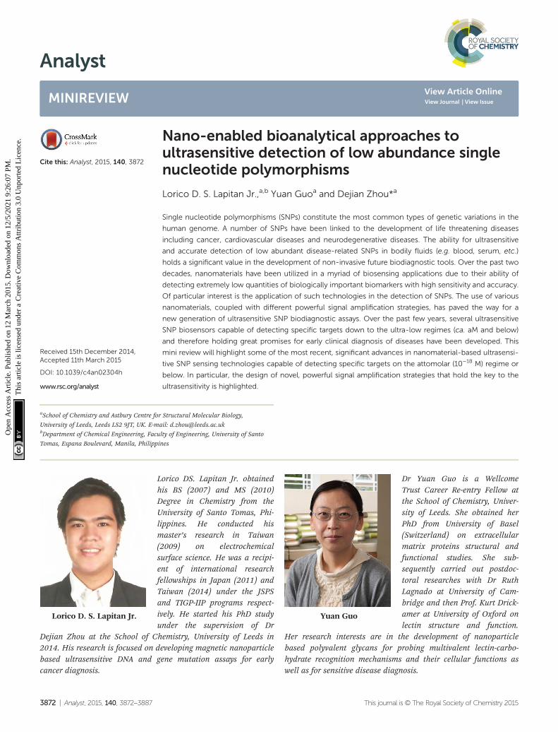
Lorico D. S. Lapitan Jr.
Lorico DS. Lapitan Jr. obtainedhis BS (2007) and MS (2010)Degree in Chemistry from theUniversity of Santo Tomas, Phi-lippines. He conducted hismaster’s research in Taiwan(2009) on electrochemicalsurface science. He was a recipi-ent of international researchfellowships in Japan (2011) andTaiwan (2014) under the JSPSand TIGP-IIP programs respect-ively. He started his PhD studyunder the supervision of Dr
Dejian Zhou at the School of Chemistry, University of Leeds in2014. His research is focused on developing magnetic nanoparticlebased ultrasensitive DNA and gene mutation assays for earlycancer diagnosis.
Yuan Guo
Dr Yuan Guo is a WellcomeTrust Career Re-entry Fellow atthe School of Chemistry, Univer-sity of Leeds. She obtained herPhD from University of Basel(Switzerland) on extracellularmatrix proteins structural andfunctional studies. She sub-sequently carried out postdoc-toral researches with Dr RuthLagnado at University of Cam-bridge and then Prof. Kurt Drick-amer at University of Oxford onlectin structure and function.
Her research interests are in the development of nanoparticlebased polyvalent glycans for probing multivalent lectin-carbo-hydrate recognition mechanisms and their cellular functions aswell as for sensitive disease diagnosis.
aSchool of Chemistry and Astbury Centre for Structural Molecular Biology,
University of Leeds, Leeds LS2 9JT, UK. E-mail: [email protected] of Chemical Engineering, Faculty of Engineering, University of Santo
Tomas, Espana Boulevard, Manila, Philippines
3872 | Analyst, 2015, 140, 3872–3887 This journal is © The Royal Society of Chemistry 2015
Ope
n A
cces
s A
rtic
le. P
ublis
hed
on 1
2 M
arch
201
5. D
ownl
oade
d on
12/
5/20
21 9
:26:
07 P
M.
Thi
s ar
ticle
is li
cens
ed u
nder
a C
reat
ive
Com
mon
s A
ttrib
utio
n 3.
0 U
npor
ted
Lic
ence
.
View Article OnlineView Journal | View Issue

1. Introduction
The completion of the human genome project in 20031 has ledto several important discoveries relating to structure of thehuman genome: it is characterized by variations in DNAnucleotide sequences of one or more bases in genes of thesame population.2,3 These variations in DNA impart certainphenotypic traits that distinguish an organism from another.If such variations occur in greater than one percent of thehuman population, they are collectively referred to as poly-morphisms. On the other hand, variations occurring in lessthan one percent of the population are often termed as“mutations” which are associated with a detrimental pheno-type such as those linked with various cases of cancer.3 Themost common sequence variation occurring in the humangenome is the stable substitution of a single nucleotide, alsoknown as single nucleotide polymorphisms (SNPs). Thehuman genome has been estimated to contain more than10 million SNPs which are distributed across the humangenome at an estimated frequency of at least one nucleotideevery 1000 base pairs but with apparent regional differences.4,5
While most SNPs do not alter the metabolic function andexpression levels of a gene, some do result in differences inpredisposition to certain heritable diseases,6–8 response todrugs (pharmacogenetics),9,10 and perception of pain.11
Several studies have shown that SNPs are closely associatedwith many types of cancer and presupposes the risk of cancerprogression.12–15 For instance, it was observed that singlepoint mutations in the KRAS gene at codons 12, 13, and 61 areassociated with the development of certain pancreatic14 andlung cancers.15 Moreover, numerous detrimental andinheritable diseases such as diabetes,7 vascular diseases,16
some forms of neurodegenerative and mental illness17,18 havealso been linked to SNPs. On the other hand, microRNAs(miRNAs) which are 17–24 base short RNA molecules playing
important roles in numerous cellular processes such as differ-entiation, cellular growth, and apoptosis. Point mutations inmiRNAs have been associated with several forms of cancers,affecting the cancer risk and treatment efficacy in some non-small cell lung cancer patients.19,20 These are just a fewexamples highlighting the association of SNPs in relation tohuman diseases and treatment responses, demonstrating thetremendous value of SNPs in biomedical research. Indeed,SNPs are considered as an important class of biomarkers thatcould allow scientists and medical practitioners to betterunderstand certain diseases, develop novel non-invasive diag-nostics tool and ultimately allow for a more personalizedapproach to disease treatment and therapies.
The most established techniques for point mutation detec-tions in DNA have largely relied on the amplification power ofthe polymerase chain reaction (PCR) coupled with quantitativefluorescence detection and/or DNA sequencing techniquessuch as pyro-/next generation sequencing of the amplifiedproduct. Although highly sensitive and widely used, suchmethods can sometimes introduce errors during the PCR expo-nential amplification process which is sensitive to contami-nations and hence may affect diagnostic accuracy.21,22 PCR isalso often regarded as labor-intensive and time-consuming,making it unsuitable for rapid, point-of-care diagnostics.Moreover, it also requires an expensive thermal cycler andthereby the cost per analysis might be high for developingcountries. These limitations have largely hampered the wideuse of PCR for rapid, on-site diagnostics. In the case of pointmutation detections involving miRNAs, the real-time quanti-tative reverse transcription polymerase chain reaction(qRT-PCR) is often the method of choice due to its high sensi-tivity. However, several drawbacks do exist for the qRT-PCR,including complex processes such as reverse transcription,multiple primer design, and precise temperature control.23
Unsurprisingly, many of the recent developments have focusedon developing PCR-free alternatives in SNP biosensing. Thereliable detection of low abundance specific SNPs without PCRpre-amplification represents an extremely challenging albeitexciting research area of the bioanalytical science.
Over the past few years, enormous efforts have beendirected towards the development of novel, ultrasensitive PCR-free SNP assays and a number of techniques have beendeveloped.22,24–27 The detection and quantification of knownSNPs are primarily based on the specificity of target hybridiz-ation28,29 and enzyme discriminations, such as specific enzy-matic cleavage,30 and single base primer extension.31,32 Ofparticular interest in the early stages of PCR-free SNP bio-sensing development has been based on using water-soluble,cationic conjugated polymers (CCPs) as fluorescence transdu-cers. For example, the Leclerc group33,34 has pioneered anddeveloped DNA detection based on the electrostatic attractionbetween a cationic polythiophene and DNA. The colour andfluorescence changes of the polymer in the presence of single-stranded and double-stranded DNAs were used as the basis forbiosensing. They have reported the first detection of SNPsdirectly from clinical samples without the need of DNA pre-
Dejian Zhou
Dejian Zhou completed his PhDdegree from Peking University(China) with Prof. Chun-HuiHuang on functional organisedultra-thin film materials in1995. He subsequently carriedout postdoctoral researches withProf. Geoffrey Ashwell at Cran-field University and then withProfs. Chris Abell, David Klener-man and Trevor Rayment onnanochemistry at University ofCambridge. Zhou took up hisacademic appointment at Univer-
sity of Leeds in 1997. His research interests are focused on devel-oping functional nanoparticle based earlier disease diagnosticassays as well as multifunctional nanomedicine for efficient, tar-geted cancer treatment.
Analyst Minireview
This journal is © The Royal Society of Chemistry 2015 Analyst, 2015, 140, 3872–3887 | 3873
Ope
n A
cces
s A
rtic
le. P
ublis
hed
on 1
2 M
arch
201
5. D
ownl
oade
d on
12/
5/20
21 9
:26:
07 P
M.
Thi
s ar
ticle
is li
cens
ed u
nder
a C
reat
ive
Com
mon
s A
ttrib
utio
n 3.
0 U
npor
ted
Lic
ence
.View Article Online

amplification with an impressive detection limit of 3 zeptomo-lar. The Bazan and Jaeger groups have also detected specificDNA sequences via CCP sensitised Förster resonance energytransfer (FRET) to dye-labelled probes.35,36 The electrostaticattraction between the cationic CCPs and the anionic DNAsleads to short distance donor (CCP)–acceptor (DNA dye-label),hence strong FRET.37 A substantive review of such work hasalready been provided by Swager et al.38 and hence this willnot be main topic here.
Recently, the use of various nanomaterials has provided thecapability of ultrasensitivity and high specificity in SNP detec-tion. Nanomaterials have been well-studied primarily becauseof their unique, size dependent physical and chemical pro-perties. In terms of biosensing applications, such useful pro-perties include extremely large surface area, tunable surfacechemical composition that allow easy and controlled immobil-ization of stable bioreceptors for efficient transduction oftarget binding into strong readout signals. These propertiesare advantageous for achieving ultrasensitivities, allowing easyintegration into miniaturized devices ushering an era of nextgeneration cost-effective SNP diagnostics.
Nanomaterials such as metallic nanoparticles (NPs),39–44,55
quantum dots (QDs),45–48 magnetic nanoparticles (MNPs),49–54
and carbon based nanomaterials56 have been combined withbiomolecules such as enzymes, oligonucleotides and DNA-zymes to develop sensors for detection and quantification ofcancer-specific SNPs. Such nanomaterial-based SNP assayshave been coupled with a number of different readout strat-egies, including colorimetry, fluorimetry, surface enhancedRaman spectroscopy (SERS), electrochemical and electro-chemiluminescence etc. For example, Boudreau and colleaguesrecently reported the use of FRET-based CCP transducers bycombining them with highly fluorescent core–shell nanoparti-cles. The Ag nanoparticle core was used to amplify the opticalsignal generated upon target recognition. This plasmonicenhancement resulted to the direct detection of unamplifiedtarget nucleic acid at femtomolar concentration regime.57–59
Table 1 summarizes some of the recently developed ultra-sensitive SNP assays using different nanomaterials coupledwith different probe and/or signal amplification strategies. Thesensitivities and specificities of these assays are already com-parable to, or even surpass those of many PCR based tech-niques, demonstrating the high suitability of nanomaterialsfor ultrasensitive SNP detection. Despite significant develop-ments, it is important to consider some other burgeoningchallenges in clinical diagnosis, particularly in the quantifi-cation of extremely low abundant SNPs in an overwhelmingbackground of wild-type genes in clinical settings. Moreover,the ability of simultaneous detection of multiple targets is alsoimportant for high diagnostic accuracy because “no tumormarker identified to date is sufficiently sensitive or specific to beused on its own to screen for cancer”.60,61 The practical impor-tance of these relies on the fact that most cancer cases are onlydiagnosed in the late stage and when the chances of patientsurvival are already slim. As a result, early diagnosis is of para-mount importance for improving the survival and prognosis ofcancer patients. All these necessitate the development of ultra-sensitive assays with multiplexing capability that can detectthe extremely low concentrations of cancer-specific SNPs inclinically media.
In general, three different approaches are widely employedto improve the assay detection limits: target, probe, and signalamplification. Among these, target amplification is mainlyachieved via enzyme-mediated replication of target nucleicacid sequences, leading to ca. 108–109 fold amplification oftarget concentration to make it high enough to be readilydetected by traditional approaches such as gel electrophor-esis.22 An excellent example here is the well established poly-merase chain reaction (PCR). More recently, isothermal targetamplification strategies have also been developed24–27,62 anddisplayed sensitivities comparable to that of the PCR. In con-trast, probe amplification does not change the copy number ofthe target, but instead the probe sequence which is comp-lementary to the target is amplified and detected by conven-
Table 1 Comparison of the sensing performance of some ultrasensitive nano-enabled DNA detection assaysa
First author ref. Nanomaterial platform Amplification strategy Transducer/analytical tool LOD
PCR based methodsY. Zhao66 Au NP + Ag NP PCR Circular dichroism 17 zMF. Patolsky84 Magnetic NP PCR Chemiluminescence 8.6 aMPCR-free methodsW. Shen40 Au NP LCR Colorimetric 20 aMD. Kato75 Au NP and magnetic NP LCR Colorimetric 50 zMJ. Ji46 CdTe QD Target recycling + RCA Electrochemical 11 aMS. Bi50 Magnetic NP RCA + DNAzyme Colorimetric 71 aMY. P. Zeng78 QD Primer generation + RCA Fluorimetric 50.9 aMJ. W. Nam80 Au NP + magnetic NP Bio-bar-code + silver amplification Scanometric 0.5 aMJ. Hu82 CdS QD CdS QD loaded in SiO2 particles H2S generation + atomic fluorescence
microscopy0.8 aM
C. Ding83 Au NP + CuS NP CuS NP loaded Au NP Electrochemical 19 aMR. D’Agata85 Au NP Short DNA modified Au NP Surface plasmon resonance imaging 2.6 aMJ. Zhou86 QD QD entrapped in liposome Single particle counting 1–2.5 aM
a PCR: polymerase chain reaction; LCR: ligation chain reaction; LOD: limit of detection.
Minireview Analyst
3874 | Analyst, 2015, 140, 3872–3887 This journal is © The Royal Society of Chemistry 2015
Ope
n A
cces
s A
rtic
le. P
ublis
hed
on 1
2 M
arch
201
5. D
ownl
oade
d on
12/
5/20
21 9
:26:
07 P
M.
Thi
s ar
ticle
is li
cens
ed u
nder
a C
reat
ive
Com
mon
s A
ttrib
utio
n 3.
0 U
npor
ted
Lic
ence
.View Article Online

tional hybridization method. Signal amplification strategiesare approaches that amplify the analytical signal generated byeach labeled or unlabelled probe so as to increase the assaysensitivity. Various signal amplification strategies have beenused to increase the sensitivity of the probe-based assays.
In this mini-review, we will highlight some of the mostrecent advance in the field of nanomaterial-based SNP detec-tion, with a particular focus on those techniques capable ofdetecting and quantifying extremely low target concentrationsdown to the attomolar regimes or below. Given the high speci-ficity, the hybridization-based method remains one of themost popular methods for nanomaterial based SNP assays. Ingeneral, the ultra-sensitivity of these assays have been achievedthrough one of the following five different strategies, (i) nano-particle-target assisted PCR amplification, (ii) nanoparticle-target assisted probe amplification, (iii) target recycling coupledwith probe and/or signal amplification, (iv) tandem amplifica-tion coupled with signal catalytic cascades, and (v) nanomaterialenhanced signal amplification. Each of these classes will be dis-cussed in this review with a focus on the assay sensitivity andcomment on their applicability to real biological matrices. More-over, a critical categorization of such approaches on the basis ofdifferent amplification strategies is proposed.
1.1 Nanoparticle-target assisted PCR amplification strategy
An essential first step in this approach is to pre-amplify theextremely low abundant SNP target into sufficiently highnumber of copies so that it can be readily detected by conven-tional methods. Given its extremely powerful exponentialamplification power, it is unsurprising that PCR-based SNPbiosensing assays have been reported to have high sensi-tivities.63 In particular, the incorporation of plasmonic nano-materials such as Au and Ag nanoparticles has been recentlycombined with PCR pre-amplification of the target DNA,leading to unprecedented sensitivities. For example, Kotov andcolleagues have reported an unexpected chirality of bridgedmetallic nanoparticle dimers and have exploited this in ultra-sensitive biosensing. They have prepared different sized Auand Ag NPs and functionalized them with specific antibodiesso that they can sandwich bind the specific target protein (e.g.prostate specific antigen PSA, a cancer marker) and assembleinto hetero-nanoparticle dimers. These biomolecule bridgedNP heterodimers are chiral, leading to an observable changein circular dichroism (CD) spectra over 350–600 nm region.The authors found that this assay was extremely sensitive,capable to detecting PSA down to the sub-aM level with anextremely impressive detection limit (DL) of 1.5 × 10⁻²⁰ M anda linear dynamic range of ∼3 orders of magnitude. Such ahighly impressive sensitivity has been attributed to plasmonicenhancement of intrinsic chirality of biomolecules, strongoptical coupling of photons with twisted NP heterodimers,and bisignate nature of the CD spectra.64,65
This assay has been recently modified to suit the detectionof ultralow concentrations of DNA66 by applying a PCR amplifi-cation of the dsDNA with primers linked to Au NPs (25 nm AuNP-linked forward primer and 10 nm Au NP-linked reverse
primer) in the presence of Taq polymerase and dNTPs (seeFig. 1). The DNA-bridged Au NP heterodimers displayed circu-lar dichroism (CD) bands at 260 and 525 nm, attributed to thechiral secondary structures of duplex DNA and the chiral NPdimers, respectively. These dimers displayed unexpected chir-ality due to the ellipsoidal shape of the NPs and a scissor-likeconfiguration with two long NP axes forming a dihedral angleof 10°. Coating of the Au NP dimers with Ag or Au are found toaffect their chiro-plasmonic activity: as the thickness of the Agshell increased, the intensity of the CD bands also increasedand shifted from 525 to 418 nm. In the case of Au coating,increasing the Au shell thickness resulted in a color change ofthe dispersion from pink to purple and the CD bands exhibi-ted a 61 nm red shift. More importantly, Au coating of DNA-bridged Au NP dimers gave fairly narrow spectra with highoptical anisotropy. Optimization of assay parameters such asspecific design of primers and number of PCR cycles hasyielded ultrasensitive DNA assay with a linear dynamic rangespanning 7 orders of magnitude (from 160 zM to 1.6 pM) andan extremely low DL of 17 zM. Currently, this method reportedthe highest sensitivity for DNA detection using nanomaterialsbased sensing platforms, and hence appears to have goodpotential for genetic based early diagnostic applications.Despite such great sensitivity, the applicability of this assaytowards the detection and discrimination of specific SNP’s incomplex media such as the wild-type DNA background has yetto be demonstrated. Moreover, given the fact that this assayrequires PCR pre-amplification, it is less well-suited to appli-cations that require rapid results such as the point-of-carediagnostics.
1.2 Nanoparticle-target assisted probe amplification strategy
Rolling circle amplification (RCA)67–69 and ligase chain reac-tion (LCR)70–72 are two of the most extensively used probeamplification strategies in SNP detections. Recently, nano-materials have been combined with these probe amplificationassays, leading to further improvements in detection limits.These assays typically use a complementary oligonucleotidecapture probe chemically linked to an Au nanoparticle,quantum dot, or magnetic nanoparticle to directly interactwith their specific analytes, thereby amplifying molecular reco-gnition events such as DNA hybridization. In addition, mag-netic nanoparticles have been further used for target capture,enrichment and purification which can further enhance theassay sensitivity.
Unlike PCR which requires thermal cycles, RCA is an iso-thermal probe amplification strategy using a single-strandedDNA as a padlock probe (Fig. 2A). The 5′-(phosphate modified)and 3′-terminal fragments of the padlock probe are specificallydesigned to be complementary to the target DNA. Hybridis-ation of the target to the padlock probe then brings its 5′- and3′-terminus close to each other, forming a nicked circle whichcan be covalently ligated together by a DNA ligase if thepadlock and target sequences are fully-matched, forming a cir-cularized padlock probe. The circularised padlock probe isthen amplified by a ϕ29 polymerase in the presence of dNTP’s,
Analyst Minireview
This journal is © The Royal Society of Chemistry 2015 Analyst, 2015, 140, 3872–3887 | 3875
Ope
n A
cces
s A
rtic
le. P
ublis
hed
on 1
2 M
arch
201
5. D
ownl
oade
d on
12/
5/20
21 9
:26:
07 P
M.
Thi
s ar
ticle
is li
cens
ed u
nder
a C
reat
ive
Com
mon
s A
ttrib
utio
n 3.
0 U
npor
ted
Lic
ence
.View Article Online

producing a greatly elongated single-stranded DNA containingnumerous copies of repeat tandem sequence units comp-lementary to the circular padlock probe. In contrast, the pres-ence of a single-base mismatch at the nicked target/padlockduplex probe can prevent the specific ligation and formationof the circular padlock probe, and therefore no RCA amplifica-tion (Fig. 2B). The stringent requirement of full sequencecomplementary between the padlock and target to form theligated circular padlock probe allows the RCA strategy to haveexcellent specificity for SNP detection.42,77
Similar to PCR, the ligase chain reaction (LCR) also requiresmultiple thermal cycles to achieve specific ligation of twoshort DNA probes catalysed by a DNA ligase into a singlestrand templated by a full-match DNA target.24–27 In a typicalSNP assay, LCR uses two pairs of probes each containing twoshort complementary oligonucleotides, but the overallsequence is complementary to the target DNA sequence(Fig. 3). After thermal denaturation followed by annealing, apair of the probes are hybridized to one of the denaturedtarget DNA strand. They are subsequently ligated by a DNAligase if the probes and target sequences are fully complemen-tary at the nick site. On the other hand, the presence of single-
base mismatch at the nick site can prevent specific ligation,allowing for the LCR to have high SNP selectivity. After thefirst ligation round, each ligated product can then function asa new template to ligate further probes in the followingthermal cycles. Repeating the thermal cycles thus leads to anexponential increase of the ligated probes. Ligation reactionshave better single-base mismatch discriminatory ability thanprimer extension methods, making LCR more specific for SNPdetection than PCR based methods.24
A particularly attractive SNP assay has been the colorimetricsensing using Au NPs because of their extremely strong surfaceplasmon resonance (SPR) absorption at ∼520 nm (ε > 109 M−1
cm−1 for a 20 nm Au NP, 4–5 orders of magnitudes strongerthan typical organic dyes), making it strongly colored even atlow nM concentrations. Moreover, its SPR band is also sensi-tive to aggregation, isolated Au NPs appear red but aggregatedones are blue or purple. The resulting color change is distinctand clearly visible by the naked eye.27 Here specific DNAtargets that can induce the aggregation of Au NPs have beenused to detect SNPs, taking the full advantage of the verysharp melting transition for the sandwich duplex formedbetween the target DNA and a pair of probe DNA (with
Fig. 1 Chiroplasmonic core–shell DNA-bridged nanoparticle heterodimers. (A) Schematic illustration of the PCR based assembly of Au nanoparticledimers. (B) Deposition of Au and Ag on the DNA-bridged Au nanoparticle dimers leading to single and multiple core–shell heterodimers. (C) CD andUV–vis spectra of Au coated heterodimers with DNA concentrations varying from 16 zM to 1.6 pM. (D) Calibration curve relating the intensity of CDbands of Au coated heterodimers and the concentration of DNA. Reprinted with permission from ref. 66, copyright©2014, American ChemicalSociety.
Minireview Analyst
3876 | Analyst, 2015, 140, 3872–3887 This journal is © The Royal Society of Chemistry 2015
Ope
n A
cces
s A
rtic
le. P
ublis
hed
on 1
2 M
arch
201
5. D
ownl
oade
d on
12/
5/20
21 9
:26:
07 P
M.
Thi
s ar
ticle
is li
cens
ed u
nder
a C
reat
ive
Com
mon
s A
ttrib
utio
n 3.
0 U
npor
ted
Lic
ence
.View Article Online

sequence complementary to each half of the DNA target) modi-fied Au NPs. The sharp melting transition means a single-basemismatch between the target and probes can be distinguisheddue to their slightly different thermal stability. This leads todistinct color changes at elevated temperatures that can beexploited for SNP detection.73,74 Combined with various ampli-fication strategies, such assays have achieved pretty low detec-tion limits, down to the femtomolar region.
More recently, LCR has been combined with Au NP forultrasensitive colorimetric SNP detection by the Gao group.40
The assay involves the real time ligation of oligonucleotidecoated Au NPs templated by the complementary SNP target(Fig. 4A). In each LCR cycle, there is an increasing amount ofAu NPs being ligated that are subsequently used to templatethe ligation of further Au NPs, leading to an exponentialincrease in the amount of covalently linked Au NPs. Since theSPR band of Au NPs is sensitive to NP assembly, this resultedin a color change that can be directly monitored by UV-vis(Fig. 4B). This assay has an impressive linear dynamic range of6 orders of magnitude and is capable of detecting specificDNA targets down to 20 aM. This assay also has high selecti-vity: it can specifically detect the wild-type (WT) KRAS gene inthe presence of large excesses of genomic DNAs even at1 : 100 000 (KRAS : genomic DNA) base pair ratios when read-ings were taken at 90 °C. At this temperature, all non-chemi-cally ligated DNA–Au NP conjugates were dislodged, leavingonly the covalently ligated Au NPs in the solution. Thus, theincorporation of ligation allowed for easy elimination of inter-ferences from coexisting DNA and a reduced background. Thisassay has been further detected the WT-KRAS in the presenceof KRAS single point mutants, yielding an impressive SNPselectivity factor of 2000. This assay can be used as an efficientapproach for detecting specific mutant DNAs by simply re-designing the sequences of capture probes and signal probes,and thus has great potential for ultrasensitive detection ofvarious disease-related SNPs. Despite such great promises, therelatively long assay time and requirement of multiple thermalcycles may limit its use in rapid, point-of-care applications.
Although the LCR strategy appears to be a highly attractivealternative to PCR amplification, an apparent weakness hasbeen its low amplification efficiency when being combinedwith nanomaterials. This is due to the restricted accessibilityof the ligases to the ligation site in oligonucleotide modifiedAu NPs. In an attempt to overcome the limitations of conven-tional LCR, a process called enzyme-free click chemical lig-ation reaction (CCLR) that involves Au NP and magnetic beadwas developed by Kato and colleagues75 (Fig. 5A). Unlike theearlier cross-linked Au NP systems, this assay does not requirethe use of ligases to carry out LCR and does not require theformation of large assemblies. The CCLR method uses anazide-containing DNA modified Au NP and a dibenzocyclo-octyne-containing biotin-DNA probe. Sandwich hybridizationof the target DNA (RNA) with the azide-DNA modified Au NPand biotin-DNA lead to enzyme-free ligation via the copper-free click chemistry, producing biotin-ligated Au NPs. Repeat-ing the thermal cycles lead to the biotin-ligated Au NPs being
Fig. 2 Schematic principle of the rolling cycle amplification and SNPdiscrimination. (A) Hybridization of the perfect-match target DNA to cir-cular padlock probe leads to covalent ligation, producing a target DNAhybridized circular probe where in the presence of phi29 polymeraseand dNTP’s, the target serves as primer to initiate the circular extensionof long single-stranded DNA with repeat sequence complementary tothe padlock probe. (B) The presence of a single base mismatch betweenthe target DNA to the padlock probe leads to no ligation and hence noamplification.
Fig. 3 Schematic principle of the ligase chain reaction (LCR) basedDNA amplification strategy. Each of the two DNA strands in the duplextarget serves as a template to ligate its respective two short DNAstrands, leading to doubling of the ligated product in each cycle andhence an exponential amplification of the target DNA. The amplifiedtarget sequences can then be detected by using their specific andtags.
Analyst Minireview
This journal is © The Royal Society of Chemistry 2015 Analyst, 2015, 140, 3872–3887 | 3877
Ope
n A
cces
s A
rtic
le. P
ublis
hed
on 1
2 M
arch
201
5. D
ownl
oade
d on
12/
5/20
21 9
:26:
07 P
M.
Thi
s ar
ticle
is li
cens
ed u
nder
a C
reat
ive
Com
mon
s A
ttrib
utio
n 3.
0 U
npor
ted
Lic
ence
.View Article Online

exponentially amplified, which are then captured by usingstreptavidin-modified magnetic beads. After magnetic separ-ation, the strong absorption of the Au NP can be used todetect and quantify the target DNA (RNA). Using this assay
strategy, a DNA sequence associated with the hepatitis A virusVall 7 polyprotein gene (HAV) has been detected at a concen-tration as low as 50 zM with a linear dynamic range of 3 ordersof magnitude (Fig. 5B), which is highly impressive. However, it
Fig. 4 (A) Schematic illustration of the real-time Au NP mediated LCR assay. (B) UV–Vis spectra of the solution containing 100 fM target DNA and10 nM CP-coated AuNPs during the LCR process. Inset: color of solution after addition of 0, 5, and 50 nM (left to right) target DNA to 10 nM CP-coated AuNPs. Reprinted with permission from ref. 40, copyright©2012, American Chemical Society.
Fig. 5 (A) Schematic illustration of Enzyme Free Click Chemical Ligation on Au nanoparticles involves the hybridization of the target DNA with anazide-modified Au NP (N3-AuNP), dibenzocyclooctyne-modified probe (DNCO-probe). (B) A plot of the A525(bk)/A525(s) ratio at different target DNAconcentrations using 40 thermal cycles. Inset shows a photograph of the supernatants at different target DNA concentrations. Reprinted permissionfrom ref. 75, copyright©2014, American Chemical Society.
Minireview Analyst
3878 | Analyst, 2015, 140, 3872–3887 This journal is © The Royal Society of Chemistry 2015
Ope
n A
cces
s A
rtic
le. P
ublis
hed
on 1
2 M
arch
201
5. D
ownl
oade
d on
12/
5/20
21 9
:26:
07 P
M.
Thi
s ar
ticle
is li
cens
ed u
nder
a C
reat
ive
Com
mon
s A
ttrib
utio
n 3.
0 U
npor
ted
Lic
ence
.View Article Online

should be noted that the absolute signal difference throughoutthe whole dynamic range was relatively small, being only∼30% (e.g. increased from ∼1.0 to 1.3), which can significantlylimit its diagnosis accuracy. This method can also discriminatespecific single point mutations, with the G-, T-, and C-Mutantsignals being 17, 0, and 0% of that of the full-match controlrespectively. This shows that this essay has an excellent SNPdiscrimination, although a more useful demonstration of theSNP specificity would be the ability to detect low specifictarget SNPs in a background of wide-type genes under clinicalrelevant media.75
1.3 Target recycling coupled with probe and/or signalamplification
Target recycling is another useful strategy to amplify low copynumber SNPs. In this strategy, the target DNA is cycled in anumber of hybridization events, each time producing one orseveral copies of the complementary probe strand, dependingon the approaches used. However, the linear amplificationnature of target recycling process has largely limited its detec-tion limit to the fM regime only.53,76
More recently, target recycling has been combined withseveral other probe amplification strategies to further improvedetection limits. A significantly higher sensitivity has beenachieved by Ju and colleagues46 who have designed a templateenhanced hybridization process or TEHP coupled with rollingcycle amplification. This target recycling hybridization assayuses a biotinylated molecular beacon [MB] bound to a strepta-vidin coated plate as the template. Hybridization of the targetDNA and an assistant DNA to the loop region of the MB formsa “Y” shaped junction (Fig. 6A). This configuration provided a
specific nucleotide sequence that can be nicked by a suitableendonuclease. Once released, the target DNA and assistantDNA initiate another round of hybridization and strand scis-sion cycle. The numerous MB fragments left in the plate thenserved as primers for the RCA process to produce thousands ofrepeat oligonucleotide sequences. These repeated sequencesthen are hybridized to an oligonucleotide functionalized CdTeQD and separated. The QDs are then dissolved by an acid,releasing greatly amplified Cd2+ ions that are detected bysquare wave voltammetry. The combination of TEHP and RCAcoupled with the use of QD based signal tags has offered anenormous amplification of the DNA target, allowing for greatlyincreased assay sensitivity. An impressive sensitivity of 11 aMhas been attained together with a wide dynamic range of 6orders of magnitude (Fig. 6B and C). This assay was also ableto discriminate the perfect complementary DNA target againstvarious mismatch targets with a discrimination ratio of 3.8,5.7 and 6.6 fold for the single-base, three-base mismatch andnon-complementary oligonucleotides, respectively. Despite ahigh sensitivity, the mutant discrimination ratio here is rela-tively moderate compared to other specific SNP assays, whichmay limit its potential for applications in real clinical sampleswhere the presence of large background genomic DNAs and/orwild-type genes may strongly interfere the specific SNPdetection.
1.4 Tandem amplification schemes and signal catalyticcascades
In order to achieve detections in even lower concentrationregimes, several researchers have reported the use of tandemamplification schemes.42,77,78 The combination of two amplifi-
Fig. 6 (A) Schematic illustration of the TEHP amplification strategy for DNA detection, (B) stripping voltammetric curves of cadmium ions corres-ponding to (left to right) 10−18, 10−17, 10−16, 10−15, 10−14, 10−13, 10−12, 10−11, and 10−10 M of target DNA. (C) The corresponding analytical quantitativedynamic range. Reprinted permission from ref. 46, copyright©2012, American Chemical Society.
Analyst Minireview
This journal is © The Royal Society of Chemistry 2015 Analyst, 2015, 140, 3872–3887 | 3879
Ope
n A
cces
s A
rtic
le. P
ublis
hed
on 1
2 M
arch
201
5. D
ownl
oade
d on
12/
5/20
21 9
:26:
07 P
M.
Thi
s ar
ticle
is li
cens
ed u
nder
a C
reat
ive
Com
mon
s A
ttrib
utio
n 3.
0 U
npor
ted
Lic
ence
.View Article Online

cation strategies has been deemed necessary to furtherimprove the sensitivity and specificity of SNP detection. More-over, this strategy can overcome the low amplificationefficiency by a single amplification scheme.
The colorimetric detection of SNPs using a combination ofRCA and nicking endonuclease-assisted nanoparticle amplifi-cation strategy was developed by Xu and colleagues.42 In thisassay, ligation was performed when the target DNA was hybri-dized with the padlock probe, leading to a circularized tem-plate. Subsequent RCA reaction in the presence of dNTPs ledto the formation of a long single strand DNA. Nicking reac-tions at many of the repeated sites along the RCA elongatedsingle-stranded DNA occurred simultaneously. Upon com-pletion of the strand scission cycles, the addition of a specificoligonucleotide modified Au NP provided a simple, colori-metric detection of target DNA down to 1 pM.
The sensitivity of ligation assays can be further improvedwhen being coupled to another probe amplification scheme.For example, the combination of LCR and RCA in detectingSNPs has been recently demonstrated by Cheng and col-leagues,77 who has demonstrated the sensitive detection of1 fM of unlabeled target DNA under the optimised assayconditions.
Further improvements of assay sensitivity have beenachieved by incorporation of DNAzymes (catalytic nucleicacids) based catalytic cascades signal amplification. In thisregard, Zhang and colleagues50 have designed a new RCAamplification approach where the presence of the target KRASSNP triggers the ligation of the padlock probe. Subsequently,multiple other circular templates were interlocked to thepadlock probe by means of complementary sequence formingan ABABAB-type DNA copolymer (Fig. 7A). The inter-locked cir-cular primers containing the HRP-mimicking DNAzymes sub-sequently underwent another round of RCA, producing long,single-strand DNA products each containing thousands ofcopies of the repeated DNAzyme sequences acting catalyticunits. The incorporation of numerous such catalytic unitsthereby greatly enhanced the chemiluminescence signal in thepresence of luminol and H2O2, leading to an impressive detec-tion limit of 71 aM for the target SNP. This assay also providedexcellent performance in quantitative analysis in human bloodserum with a linear dynamic range of 2 orders of magnitudewith good signal linearity from 0.1–10 fM (Fig. 7B and C). Theexcellent sensitivity of this assay lies on the inherent capacityto generate great amplification as a consequence of the highturnover reaction of DNAzymes. This assay was also highly
Fig. 7 (A) Schematic representation of interlocked DNA scaffold mediated RCA reaction and DNAzyme amplification assay for the detection ofG12C mutation in the KRAS gene. (B) Chemiluminescence signals for the HRP-mimicking DNAzyme-catalyzed luminol-H2O2 system correspondingto different concentrations of single-base mutant target DNA: (a) 0; (b) 0.1; (c) 0.2; (d) 0.4; (e) 0.6; (f ) 0.8; (g) 1.0; (h) 2.0; (i) 4.0; ( j) 6.0; (k) 8.0; (l)10.0 fM. (C) The corresponding calibration curve of peak height versus the concentration of target DNA. Reprinted with permission from ref. 50,copyright©2010, American Chemical Society.
Minireview Analyst
3880 | Analyst, 2015, 140, 3872–3887 This journal is © The Royal Society of Chemistry 2015
Ope
n A
cces
s A
rtic
le. P
ublis
hed
on 1
2 M
arch
201
5. D
ownl
oade
d on
12/
5/20
21 9
:26:
07 P
M.
Thi
s ar
ticle
is li
cens
ed u
nder
a C
reat
ive
Com
mon
s A
ttrib
utio
n 3.
0 U
npor
ted
Lic
ence
.View Article Online

specific, and can detect 1 fM specific mutant target in the pres-ence of 10 pM wild-type gene background. It demonstrated aSNP detection capability of 1 in 10 000 (SNP versus wild-typetarget) level, which is among the best reported in literature.Hence, combining high sensitivity and specificity, this assayappears to have excellent potential for SNP based disease diag-nostics by specific detection of the low abundant mutatedtarget in clinical laboratory settings. This assay, however, isrelatively complex and requires multiple amplification cycles,making it less well-suited for rapid, point-of-care applications.
Recently, the Zhang group has developed an ultrasensitivemiRNA assay based on primer generation-mediated rollingcircle amplification [PG-RCA]. This assay was used to analyzethe point mutation of mir-196a2 [T→C] in the lung tissuesamples of non small cell lung cancer patients.78 In this assay,the presence of target mir-196a2T circularizes the padlockprobe and the mir-196a2T further functioned as a primer toinitiate the next round of RCA reaction in the presence of Vent(exo-) polymerase. This resulted in a long single-stranded RCAproduct containing numerous restriction sites for Nb.BsmI.The RCA product was then nicked by Nb.BsmI, generating alarge number of new primers that was further used to initiatea new RCA reaction. The amplified DNA products were sub-sequently hybridized with a biotin/Cy5-labeled capture probesto form a double-stranded DNA. This DNA duplex contains arecognition site for the Nt.BstNBI nicking enzyme, and afterthe nicking reaction the capture probe was cleaved, separatingCy5 and biotin. This also resulted in the release of the ampli-fied DNA product wherein it can repeatedly hybridize with newbiotin/Cy5-labeled to initiate the next rounds of cleavage. In asimilar strategy, mir-196a2C was detected using the designedmir-196a2C padlock probes. In the absence of the mir-96a2T,more Cy5/biotin capture probes are cleaved, leading to smalleramounts of the intact capture probes that can bind on thesurface of the streptavidin-coated QD. Consequently, thisreduces the FRET between the QD donor and Cy5 acceptors,resulting in decreased Cy5 counts being detected in the single-particle FRET detection measurement. On the other hand, themir-196a2T-specific linear padlock probe cannot be circular-ized in the absence of mir-196a2T. Under these conditions,RCA amplification and cleaving reactions will happen. Hence,the biotin/Cy5-labeled capture probe remains intact which canbind to the QD surface via specific streptavidin–biotin inter-actions, resulting in strong QD sensitised Cy5 FRET signal.With the integration of the PG-RCA reaction and nickingenzyme-driven recycling amplification, this QD-based miRNAnanosensor exhibited an impressive detection limit of 50.9 aMand a large dynamic range of 7 orders of magnitude from 0.1fM to 1 nM for the specific microRNA target. Moreover, thisassay can even distinguish variant frequencies down to as lowas 0.001% in the mixtures of mir-196a2C and mir196a2T.Importantly, this QD-based miRNA nanosensor can be used toanalyse the point mutation of mir-196a2 in the lung tissues ofNSCLC patients, holding great potentials for further appli-cations in biomedical research and clinical diagnosis presum-ably in the clinical laboratory settings. Its complex signal
amplification and assay procedures together with a relativelycomplex single-particle counting readout method may,however, limit its use in applications that require rapid results.
1.5 Nanomaterial assisted signal amplification
In signal amplification, nanomaterials are often functionalisedwith biomolecules for target-specific recognitions and/or carry-ing high loads of signal moieties, catalysts, optical emitters,and electronic conductors. Biofunctionalised nanomaterialscan amplify the signal transduction events due to their capa-bility of direct interaction with their target, allowing for detec-tion of biomolecules down to the single-molecule level.79
Signal amplification can eliminate some special requirementsof the target/probe amplification schemes such as the need ofenzymes and thermal cycles (ca. PCR). As a result, this cansimplify experimental protocols, lower the assay cost, andprovide amenability to miniaturisation. Hence, the use of bio-functionalized nanomaterials for signal amplification hasbeen very attractive in the development of ultrasensitive DNAassays without the need of target and probe amplifications.The following section presents some ultrasensitive detectionof SNPs achieved through use nanomaterials for signalamplification.
1.5.1 Nanoparticle bio-barcode amplification. An excellentexample of the earlier developments here is the Bio-barcodeassay developed by the Mirkin group.80 It uses a short DNA-modified An NP tagged with hundreds of copies of the biobarcode DNAs (Bbc-Au NP) as signal probe, and another DNA-modified magnetic microparticle (Oligo-MMP) as captureprobe (Fig. 8). The two DNAs are complementary to each halfof the target DNA, so that they can sandwich bind to the targetDNA, forming a Bbc-Au NP/DNA target/Oligo-MMP hybridstructure. After magnetic separation and washing to removeany unbound species, thermal denaturation of the hybridreleases the bio barcode DNAs, converting each capture DNAtarget into hundreds of copies of barcode DNAs. These arethen detected by a sensitive scanometric assay coupled withsilver amplification to achieve ultrasensitivity.81 This assay pro-vides an impressive label-free detection of target DNA down to500 zM, comparable to many PCR based methods.80 It canalso discriminate the perfect match DNA from the SNP target.Although its relatively modest discrimination ratio, ∼3 : 1, maylimit its capability of detecting low abundant SNP targets inwild-type gene background.
1.5.2 Signal amplification using labeled and enrichednanoparticle probes. Metallic nanoparticles can be employedas scaffold for loading not only the capture but also signalmolecules. These labelled nanoparticles present an importantsignal amplification strategy as they can directly enhance thereadout signal for each target and probe recognition whileretaining a high binding specificity. Over the past few years,silica particles have been used as another carrier platform dueto their large surface areas, well-known surface chemistriesand good biocompatibility.
Wu and colleagues have reported a labeling-based signalamplification strategy for ultrasensitive detection of target
Analyst Minireview
This journal is © The Royal Society of Chemistry 2015 Analyst, 2015, 140, 3872–3887 | 3881
Ope
n A
cces
s A
rtic
le. P
ublis
hed
on 1
2 M
arch
201
5. D
ownl
oade
d on
12/
5/20
21 9
:26:
07 P
M.
Thi
s ar
ticle
is li
cens
ed u
nder
a C
reat
ive
Com
mon
s A
ttrib
utio
n 3.
0 U
npor
ted
Lic
ence
.View Article Online

DNA using QD assembled SiO2 microspheres.82 This methodis used as an indirect method for ultrasensitive detection ofHIV DNA using hydride generation atomic fluorescence spec-trophotometry [HG-AFS]. This assay involves the use of sand-wich hybridization between the target DNA, biotin-captureprobe and streptavidin tagged QD-SiO2 assembly, convertingeach captured DNA target into thousands of SiO2 tagged QDs.After acid dissolution of the QDs, the resulting Cd2+ ions weredetected by HG-AFS, giving a highly impressive detection limitof 0.8 aM for the target DNA and a dynamic range of 3 ordersof magnitude (from 1 aM to 1 fM). The outstanding sensitivitysuggests that the HG-AFS method is suitable for the ultrasensi-tive biosensing.
An ultrasensitive chemiluminescent method using Au NPbased signal amplification has recently been developed byZhang and colleagues.83 Using the Watson–Crick base pairing,a guanine monomer modified Au NP probe is coupled to thecytosine mutated DNA duplex in the presence of DNA polymer-ase I. Each guanine modified Au NP probe is also linked to 77CuS NPs which act as signal generator. After acid dissolution
of the CuS NPs, the released Cu2+ ions are coordinated to cya-nides to form [Cu(CN)4]
2− complexes. These are then reactedwith luminol to give rise to chemiluminescence signal fortarget DNA quantification. A further improvement in thesignal sensitivity is achieved by incorporating a pre-concen-tration of Cu2+ ions by anodic stripping voltammetry (ASV).These have enabled this assay to achieve a low detection limitof 19 aM for one base mutant DNA and a linear range of80 aM to 10 fM.
The labeling of the target DNA with a large number ofsignal generators coupled with sensitive electrochemilumine-scence detection is very powerful towards ultrasensitivity forSNP detection as demonstrated by the Willner group.84 In thisassay, a mutant DNA with one base mismatch (C→G mutation)is first hybridised to a complementary DNA modified magneticNP. It is then treated with a DNA polymerase in the presenceof biotin-dCTP and other dNTPs. This is then followed by mul-tiple thermal cycles of dissociation, annealing and labeling,resulting in poly-labeling of the magnetic NPs with biotins.After magnetic separation and treatment with avidin-horse-radish peroxidase (Avidin-HRP), the resulting avidin-HRP func-tionalised magnetic NPs and napthoquinone NPs aredeposited on an electrode guided by an external magneticfield. Applying an electric potential reduces napthoquinone tohydroquinone, which simultaneously reduces O2 to H2O2, trig-gering the HRP catalysed oxidation of luminol and yielding achemiluminescence signal. Under optimized conditions, thisassay has reported an impressive sensitivity of 8.3 aM for theM13φ DNA (50 copies in 10 μL sample) and 10 aM for amutant DNA.
Despite showing impressive sensitivities, none of the aboveultrasensitive assays have reported the simultaneous detectionof multiple targets. Further developments on the multiplexingcapability of an SNP assay are highly desirable for improvingthe diagnostic accuracy. Recently, Gambari and colleagues85
have reported the direct detection of SNPs in non-amplifiedhuman genomic DNA carrying the mutated β°39-globin genesequence by using surface plasmon resonance imaging (SPRI).This gene is involved in the hereditary blood disorder diseasesknown as β-thalassemia. The detection is achieved by using AuNP conjugated with multiple copies of DNA β°39, an 11-meroligonucleotide. Prior to analysis, the surfaces of 6 microfluidicchannels are modified with PNA-N and PNA-M probes whosenucleic acid sequences are complementary to the normal andmutant genes respectively (Fig. 9). The samples are directlyfluxed into these channels to allow the direct hybridisationbetween each of the samples (e.g. normal βN/βN; homozygousβ°39/β°39 and heterozygous β°39/βN) on PNA functionalizedsurfaces. The SPRI responses between the samples and the twodifferent PNA probes are used as controls. Subsequently, theconjugated Au NPs are fluxed into the microchannels and cap-tured by specific hybridization between their surface DNA β°39strands and exposed target DNA sequence not involved inbinding to the PNA probes, allowing for greatly enhanced SPRIsignal. This assay has achieved the sensitive detection ofgenomic DNAs down to 2.6 aM (5 pg μL−1) without PCR ampli-
Fig. 8 Schematic illustration of the DNA-Bio-bar-code based ultra-sensitive assay for DNA detection. (A) The preparation of the DNAmodified Au NP and magnetic microparticle probes. (B) Schematics ofthe nanoparticle-based PCR-free DNA amplification and detectionscheme. Reprinted with permission from ref. 80, copyright©2004,American Chemical Society.
Minireview Analyst
3882 | Analyst, 2015, 140, 3872–3887 This journal is © The Royal Society of Chemistry 2015
Ope
n A
cces
s A
rtic
le. P
ublis
hed
on 1
2 M
arch
201
5. D
ownl
oade
d on
12/
5/20
21 9
:26:
07 P
M.
Thi
s ar
ticle
is li
cens
ed u
nder
a C
reat
ive
Com
mon
s A
ttrib
utio
n 3.
0 U
npor
ted
Lic
ence
.View Article Online
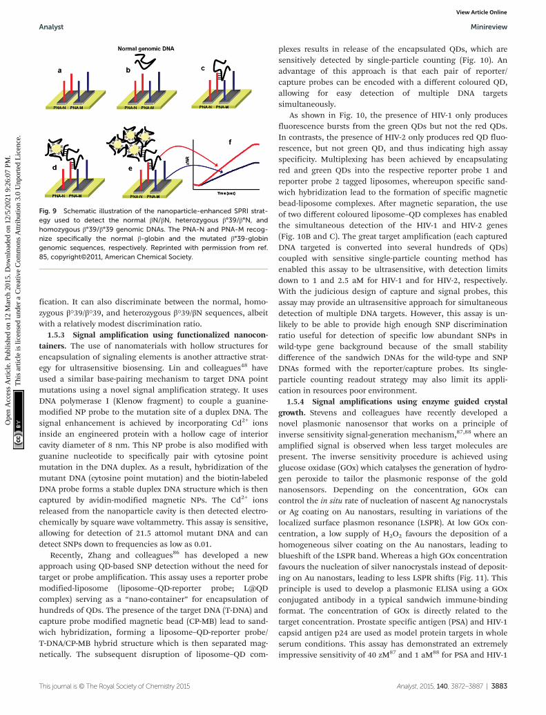
fication. It can also discriminate between the normal, homo-zygous β°39/β°39, and heterozygous β°39/βN sequences, albeitwith a relatively modest discrimination ratio.
1.5.3 Signal amplification using functionalized nanocon-tainers. The use of nanomaterials with hollow structures forencapsulation of signaling elements is another attractive strat-egy for ultrasensitive biosensing. Lin and colleagues48 haveused a similar base-pairing mechanism to target DNA pointmutations using a novel signal amplification strategy. It usesDNA polymerase I (Klenow fragment) to couple a guanine-modified NP probe to the mutation site of a duplex DNA. Thesignal enhancement is achieved by incorporating Cd2+ ionsinside an engineered protein with a hollow cage of interiorcavity diameter of 8 nm. This NP probe is also modified withguanine nucleotide to specifically pair with cytosine pointmutation in the DNA duplex. As a result, hybridization of themutant DNA (cytosine point mutation) and the biotin-labeledDNA probe forms a stable duplex DNA structure which is thencaptured by avidin-modified magnetic NPs. The Cd2+ ionsreleased from the nanoparticle cavity is then detected electro-chemically by square wave voltammetry. This assay is sensitive,allowing for detection of 21.5 attomol mutant DNA and candetect SNPs down to frequencies as low as 0.01.
Recently, Zhang and colleagues86 has developed a newapproach using QD-based SNP detection without the need fortarget or probe amplification. This assay uses a reporter probemodified-liposome (liposome–QD-reporter probe; L@QDcomplex) serving as a “nano-container” for encapsulation ofhundreds of QDs. The presence of the target DNA (T-DNA) andcapture probe modified magnetic bead (CP-MB) lead to sand-wich hybridization, forming a liposome–QD-reporter probe/T-DNA/CP-MB hybrid structure which is then separated mag-netically. The subsequent disruption of liposome–QD com-
plexes results in release of the encapsulated QDs, which aresensitively detected by single-particle counting (Fig. 10). Anadvantage of this approach is that each pair of reporter/capture probes can be encoded with a different coloured QD,allowing for easy detection of multiple DNA targetssimultaneously.
As shown in Fig. 10, the presence of HIV-1 only producesfluorescence bursts from the green QDs but not the red QDs.In contrasts, the presence of HIV-2 only produces red QD fluo-rescence, but not green QD, and thus indicating high assayspecificity. Multiplexing has been achieved by encapsulatingred and green QDs into the respective reporter probe 1 andreporter probe 2 tagged liposomes, whereupon specific sand-wich hybridization lead to the formation of specific magneticbead-liposome complexes. After magnetic separation, the useof two different coloured liposome–QD complexes has enabledthe simultaneous detection of the HIV-1 and HIV-2 genes(Fig. 10B and C). The great target amplification (each capturedDNA targeted is converted into several hundreds of QDs)coupled with sensitive single-particle counting method hasenabled this assay to be ultrasensitive, with detection limitsdown to 1 and 2.5 aM for HIV-1 and for HIV-2, respectively.With the judicious design of capture and signal probes, thisassay may provide an ultrasensitive approach for simultaneousdetection of multiple DNA targets. However, this assay is un-likely to be able to provide high enough SNP discriminationratio useful for detection of specific low abundant SNPs inwild-type gene background because of the small stabilitydifference of the sandwich DNAs for the wild-type and SNPDNAs formed with the reporter/capture probes. Its single-particle counting readout strategy may also limit its appli-cation in resources poor environment.
1.5.4 Signal amplifications using enzyme guided crystalgrowth. Stevens and colleagues have recently developed anovel plasmonic nanosensor that works on a principle ofinverse sensitivity signal-generation mechanism,87,88 where anamplified signal is observed when less target molecules arepresent. The inverse sensitivity procedure is achieved usingglucose oxidase (GOx) which catalyses the generation of hydro-gen peroxide to tailor the plasmonic response of the goldnanosensors. Depending on the concentration, GOx cancontrol the in situ rate of nucleation of nascent Ag nanocrystalsor Ag coating on Au nanostars, resulting in variations of thelocalized surface plasmon resonance (LSPR). At low GOx con-centration, a low supply of H2O2 favours the deposition of ahomogeneous silver coating on the Au nanostars, leading toblueshift of the LSPR band. Whereas a high GOx concentrationfavours the nucleation of silver nanocrystals instead of deposit-ing on Au nanostars, leading to less LSPR shifts (Fig. 11). Thisprinciple is used to develop a plasmonic ELISA using a GOxconjugated antibody in a typical sandwich immune-bindingformat. The concentration of GOx is directly related to thetarget concentration. Prostate specific antigen (PSA) and HIV-1capsid antigen p24 are used as model protein targets in wholeserum conditions. This assay has demonstrated an extremelyimpressive sensitivity of 40 zM87 and 1 aM88 for PSA and HIV-1
Fig. 9 Schematic illustration of the nanoparticle-enhanced SPRI strat-egy used to detect the normal βN/βN, heterozygous β°39/β°N, andhomozygous β°39/β°39 genomic DNAs. The PNA-N and PNA-M recog-nize specifically the normal β-globin and the mutated β°39-globingenomic sequences, respectively. Reprinted with permission from ref.85, copyright©2011, American Chemical Society.
Analyst Minireview
This journal is © The Royal Society of Chemistry 2015 Analyst, 2015, 140, 3872–3887 | 3883
Ope
n A
cces
s A
rtic
le. P
ublis
hed
on 1
2 M
arch
201
5. D
ownl
oade
d on
12/
5/20
21 9
:26:
07 P
M.
Thi
s ar
ticle
is li
cens
ed u
nder
a C
reat
ive
Com
mon
s A
ttrib
utio
n 3.
0 U
npor
ted
Lic
ence
.View Article Online

Fig. 10 (A) Schematic illustration for Liposome–QD complexes (L@QD complex) based ultrasensitive detection of attomolar DNA using single-par-ticle detection techniques. The assay involves the sandwich type hybridization of the target DNA to L@QD complex-tagged reporter probes andcapture probe modified-magnetic bead. Liposome disruption leads to the release of QD’s for single particle detection. (B) A plot of burst countsfrom the released QDs as a function of the concentrations of HIV-1 (green) and HIV-2 (red). There was no change in the burst counts in the controlgroups with non-complementary DNA (black and blue), and (C) Simultaneous detection of HIV-1 (green) and HIV-2 (red). The error bars correspondsto standard deviation of three replicates. Reprinted with permission from ref. 86, copyright©2013, American Chemical Society.
Fig. 11 (A) Schematic illustration of the proposed signal-generation mechanism by means of enzyme-guided crystal growth for the plasmonicELISA assay. (B) Immunoassay for the ultrasensitive detection of PSA with GOx-labelled antibodies and of PSA (red) and BSA (green) spiked intowhole serum. Reprinted with permission from ref. 87, copyright©2012, Nature Publishing group.
Minireview Analyst
3884 | Analyst, 2015, 140, 3872–3887 This journal is © The Royal Society of Chemistry 2015
Ope
n A
cces
s A
rtic
le. P
ublis
hed
on 1
2 M
arch
201
5. D
ownl
oade
d on
12/
5/20
21 9
:26:
07 P
M.
Thi
s ar
ticle
is li
cens
ed u
nder
a C
reat
ive
Com
mon
s A
ttrib
utio
n 3.
0 U
npor
ted
Lic
ence
.View Article Online

capsid antigen, respectively. Moreover, the assay results can bedirectly visualised by the naked eye, and therefore offers asimple, highly attractive alternative to the costly nucleic acid-based HIV infection diagnosis test. In principle, it can beadapted for the detection of any analyte with a suitable anti-body, making it a versatile tool for ultrasensitive diagnostics.Despite of great simplicity and ultrasensitivity, the plasmonicELISA however, has a rather small dynamic range of one orderof magnitude, making it difficult to quantify the exact targetconcentration.
2. Conclusion and outlook
Significant advances have been made over the past few years inthe development of PCR-free assays suitable for specific andultrasensitive detection of SNPs. The incorporation of variousbiofunctionalised nanomaterials coupled with novel amplifica-tion strategies have permitted the detection of extremely lowconcentrations of SNPs, down to the aM–zM range. Such levelsof sensitivity have already compared very favorably with manyPCR based methods. In general, the amplification strategiesmay be classified as one of the following categories:
(i) Nanoparticle-target assisted PCR amplification.(ii) Nanoparticle-target assisted probe amplification.(iii) Target recycling coupled with probe and/or signal
amplification.(iv) Tandem amplification schemes and signal catalytic
cascades.(v) Nanomaterial enhanced signal amplification.In general, the sensitivity of a nanomaterial-based SNP
assay can be greatly enhanced by combining target, probe andsignal amplification schemes. Most of the recent target andprobe amplification schemes have exploited the great catalyticpower and specificity of enzymes to achieve ultra-sensitivityand specificity. Of particular interest here is the use of DNA-zymes that can undergo the so-called enzymatic cascade reac-tions, where the activation of multiple enzymes by a targetDNA can result in ultra-sensitivity, comparable many PCRbased assays. Another widely used strategy is the use of restric-tion enzymes that specifically recognise the restriction sites todegrade the reporter probe, allowing for target recycling. Theseautocatalytic strategies have resulted in unmatched sensi-tivities while still maintaining high specificity. However, alimitation here is that restriction enzymes can only recognize aspecific sequence and therefore are not suitable for universalSNP detection. Several ingenious ways in the design of nano-materials for signal amplification strategies have also beendeveloped, including the use of multiple tagging and enrich-ment of nanoparticle probes with signal moieties to enableultra-sensitivity. Some of these strategies also hold great prom-ises for multiplexed detection, an important property for highdiagnostic accuracy.
Despite significant advances, several limitations still needto be resolved before they can be translated into clinical diag-nostic assays. For example, although a number of assays have
reported aM, even zM sensitivity, most of which were still atthe proof-of-concept stage and were carried out under cleanbuffers. They have not yet been tested in clinically relatedmedia, such as blood, serum, saliva and urine. Furthermore,most assays have reported a rather limited SNP discriminationratio (<10 fold), making them potentially unsuitable for detec-tion of low abundant disease related SNPs in the backgroundof wild-type gene/genomic DNA because of the strong interfer-ence from the background DNAs. Moreover, the lack of themultiplexing capability could pose a significant limiting factorfor the real clinical potential. The stability of the biofunctiona-lised nanomaterials is another important issue for such nano-enabled SNP biosensors. In this regard, the fundamentalunderstanding of the biomolecule-nanomaterial interactionsis imperative to alleviate problems of high background signalsdue to non-specific adsorptions in serum and/or othercomplex clinical samples.
The use of biofunctionalised nanomaterials together withnovel amplification strategies has transcended barriers to theattainment of extremely low detection limits of disease-relatedSNPs. This ultimately ushers the way for more practical con-cerns such as the realistic applications of the technology inclinical settings. Even more challenging is the development ofa robust, portable, point-of-care diagnostic system that canspecifically detect the ultralow level of disease-related SNPsrapidly and conveniently on the site of the patient and inplaces such as the doctor’s office, school clinic and in patients’residence.89
These also call for improving the assays’ amenabilitytowards automation and miniaturization because most currentassays still require the use of expensive and complex instru-mentation and complex procedures, limiting their potential inrapid diagnosis. Electro-chemical signal transduction canprovide an option for the miniaturization and automation butthese methods are prone to false positives.26 The recentadvances in microfluidics90,91 may be able to address samplethroughput and automation challenges in SNP assays. If all ofthese challenges were met, the automation of these ultrasensi-tive assays may lead to the integration of sample processing,quantification and signal measurement in an all-in-one devicein real clinical setting. This would greatly facilitate the rapid,accurate disease diagnosis and prognosis. In view of this,these are still crucial challenges that need to be addressed,and more efforts will be needed to improve the analyticalsensing performance and portability of SNP assays.
Acknowledgements
We would like to thank the University of Leeds for fundingthis project and also for funding L.DS.L with the Leeds Inter-national Research Scholarship (LIRS) to allow him pursuinghis PhD studies at University of Leeds. Y.G. would like tothank the Wellcome Trust (UK) for providing her a CareerRe-entry Fellowship (grant no: 097354/Z/11/Z).
Analyst Minireview
This journal is © The Royal Society of Chemistry 2015 Analyst, 2015, 140, 3872–3887 | 3885
Ope
n A
cces
s A
rtic
le. P
ublis
hed
on 1
2 M
arch
201
5. D
ownl
oade
d on
12/
5/20
21 9
:26:
07 P
M.
Thi
s ar
ticle
is li
cens
ed u
nder
a C
reat
ive
Com
mon
s A
ttrib
utio
n 3.
0 U
npor
ted
Lic
ence
.View Article Online

References
1 See the Human Genome Project website: http://www.genome.gov/10001772, Date retrieved: November 1, 2014.
2 D. G. Wang, et al., Science, 1998, 280, 1077–1082.3 H. C. Erichsen and S. J. Chanock, Br. J. Cancer, 2004, 90,
747–751.4 D. Botstein and N. Risch, Nat. Genet., 2003, 33(Suppl), 228–
237.5 C. S. Carlson, et al., Nat. Genet., 2003, 33, 518–521.6 M. Monot, et al., On the origin of leprosy, Science, 2005,
308, 1040–1042.7 D. Zhang, J. Ma, K. Brismar, S. Efendic and H. F. Gu, J. Dia-
betes Complications, 2009, 23, 265–272.8 Y. Suh and J. Vijg, Mutat. Res., 2005, 573, 41–53.9 B. N. Chorley, X. Wang, M. R. Campbell, G. S. Pitman,
M. A. Noureddine and D. A. Bell, Mutat. Res., 2008, 659,147–157.
10 J. Robert, V. Le Morvan, D. Smith, P. Pourquier andJ. Bonnet, Crit. Rev. Oncol. Hematol., 2005, 54, 171–196.
11 B. Rahim-Williams, J. L. Williams and R. B. Fillingim, PainMed., 2012, 13, 522–540.
12 J. Liu, C. He, C. Xing and Y. Yuan, Mutat. Res., 2014, 765,11–21.
13 A. Figl, et al., Mutat. Res., 2009, 661, 78–64.14 C. M. Oliveria, K. D. Hadfield, A. K. Siriwardena and
W. Newman, Pancreas, 2012, 41, 428–434.15 J. M. Ostrem, U. Peters, M. L. Sos, J. A. Wells and
K. M. Shokat, Nature, 2013, 503, 548–551.16 O. I. Sentina, T. V. Byzova, J. C. Adams, J. J. McCarthy,
E. J. Topol and E. F. Plow, Int. J. Biochem. Cell Biol., 2004,36, 1013–1030.
17 F. Coppedè, C. Armani, D. D. Bidia, L. Petrozzi, U. Bonuccelliand L. Migliore, Mutat. Res., 2005, 579, 107–114.
18 B. Roy, N. Maksemous, R. A. Smith, S. Menon, G. Daviesand L. R. Griffiths, Mutat. Res., 2012, 732, 3–8.
19 Z. Hu, J. Chen, T. Tian, X. Zhou, H. Gu, L. Xu, Y. Zeng,R. Miao, G. Jin, H. Ma, Y. Chen and H. Shen, J. Clin. Invest.,2008, 118, 2600–2608.
20 B. M. Ryan, A. I. Robles and C. C. Harris, Nat. Rev. Cancer,2010, 10, 389–402.
21 M. M. Mhlanga and L. Malmberg, Methods, 2001, 25, 463–471.
22 Y. V. Gerasimova and D. M. Kopaschchikov, Chem. Soc.Rev., 2014, 43, 6405–6438.
23 L. J. Chin, E. Ratner, S. Leng, R. Zhai, S. Nallur, I. Babar,R. U. Muller, E. Straka, L. Su, E. A. Burki, R. E. Crowell,R. Patel, T. Kulkarni, R. Homer, D. Zelterman, K. K. Kidd,Y. Zhu, D. C. Christiani, S. A. Belinsky, F. J. Slack andJ. B. Weidhaas, Cancer Res., 2008, 68, 8535.
24 J. M. Butler, Single nucleotide polymorphisms and appli-cations, in Advanced Topics in Forensic DNA Typing, Aca-demic Press, California, USA, 2012, pp. 347–369.
25 R. M. Twyman, Single nucleotide polymorphism analysis,in Encyclopedia of Nueroscience, Academic Press, California,USA, 2009, pp. 871–875.
26 M. L. Ermini, S. Mariani, S. Scarano and M. Minunni,Biosens. Bioelectron., 2014, 61, 28–35.
27 P. D. Howes, S. Rana and M. M. Stevens, Chem. Soc. Rev.,2014, 43, 3835–3853.
28 W. Shen, C. L. Lim and Z. Gao, Chem. Commun., 2013, 49,8114–8116.
29 X. Chen, A. Ying and Z. Gao, Biosens. Bioelectron., 2012, 36,89–94.
30 C. Bui, et al., Enzymatic and chemical cleavage methods toidentify genetic variation, in Molecular Diagnostics, AcademicPress, London, California, USA, 2010, pp. 29–44.
31 H. Zhang, X. Fu, L. Liu, Z. Zhu and K. Yang, Anal. Biochem.,2012, 426, 30–39.
32 F. Kakihara, Y. Kurebayashi, Y. Tojo, H. Tajima,S. Hasegawa and M. Yohda, Anal. Biochem., 2005, 341, 72–88.
33 H.-A. Ho, K. Dore’, M. Boissinot, M. G. Bergeron,R. M. Tanguay, D. Boudreau and M. Leclerc, J. Am. Chem.Soc., 2005, 127, 12673–12676.
34 H.-A. Ho, M. Boissinot, M. G. Bergeron, G. Corbeil,K. Dore’, D. Boudreau and M. Leclerc, Angew. Chem., Int.Ed., 2002, 41, 1548–1551.
35 S. Wang and G. C. Bazan, Adv. Mater., 2003, 15, 1425–1428.36 B. Liu, S. Baudrey, L. Jaeger and G. C. Bazan, J. Am. Chem.
Soc., 2004, 126, 4076–4077.37 B. Liu and G. C. Bazan, Chem. Mater., 2004, 16, 4467–4476.38 S. W. Thomas III, G. D. Joly and T. M. Swager, Chem. Rev.,
2007, 107, 1339–1386.39 J. Garcia, Y. Zhang, H. Taylor, O. Cespedes, M. E. Webb and
D. Zhou, Nanoscale, 2011, 3, 3721–3730.40 W. Shen, H. Deng and Z. Gao, J. Am. Chem. Soc., 2012, 134,
14678–14681.41 L. Qui, L. Qiu, H. Zhou, Z. Wu, G. Shen and R. Yu, New
J. Chem., 2014, 38, 4711–4715.42 W. Xu, X. Xie, D. Li, Z. Yang, T. Li and X. Liu, Small, 2012,
8, 1846–1850.43 L. Tang, I. S. Chun, Z. Wang, J. Li, X. Li and Y. Lu, Anal.
Chem., 2013, 85, 9522–9527.44 C. H. Liu, Z. P. Li, B. A. Du, X. R. Duan and Y. C. Wang,
Anal. Chem., 2006, 78, 3738–3744.45 C. Y. Zhang, H. C. Yeah, M. T. Kukori and T. H. Wang, Nat.
Mater., 2005, 24, 826–831.46 H. Ji, F. Yan, J. Lei and H. Ju, Anal. Chem., 2012, 84, 7166–
7171.47 G. D. Liu, T. M. H. Lee and J. J. Wang, J. Am. Chem. Soc.,
2005, 127, 38–39.48 G. Liu and Y. H. Lin, J. Am. Chem. Soc., 2007, 129, 10394–
10401.49 Y. Zhang, C. Pilapong, Y. Guo, Z. Ling, O. Cespedes,
P. Quirke and D. Zhou, Anal. Chem., 2013, 85, 9238–9244.
50 S. Bi, L. Li and S. Zhang, Anal. Chem., 2010, 88, 9447–9454.51 F. Patolsky, Y. Weizmann, E. Katz and I. Willner, Angew.
Chem., Int. Ed., 2003, 42, 2372–2376.52 S. Bi, Z. Zhang, Y. Dong and Z. Wang, Biosens. Bioelectron.,
2015, 65, 139–144.
Minireview Analyst
3886 | Analyst, 2015, 140, 3872–3887 This journal is © The Royal Society of Chemistry 2015
Ope
n A
cces
s A
rtic
le. P
ublis
hed
on 1
2 M
arch
201
5. D
ownl
oade
d on
12/
5/20
21 9
:26:
07 P
M.
Thi
s ar
ticle
is li
cens
ed u
nder
a C
reat
ive
Com
mon
s A
ttrib
utio
n 3.
0 U
npor
ted
Lic
ence
.View Article Online
53 Y. Zhang, Y. Guo, P. Quirke and D. Zhou, Nanoscale, 2013,5, 5027–5035.
54 G. Liu and Y. Lin, J. Am. Chem. Soc., 2007, 129, 10394–10401.55 W. Xu, X. J. Xue, T. H. Li, H. Q. Zeng and X. G. Liu, Angew.
Chem., Int. Ed., 2009, 48, 6849–6852.56 M. Luo, X. Chen, G. H. Zhou, X. Liang, L. Chen, H. Ji and
Z. K. He, Chem. Commun., 2012, 48, 1126–1128.57 D. Brouard, M. L. Viger, A. G. Bracamonte and
D. Boudreau, ACS Nano, 2011, 5, 1888–1896.58 D. Brouard, O. Ratelle, A. G. Bracamonte, M. St-Loius and
D. Boudreau, Anal. Methods, 2013, 5, 6896–6999.59 D. Brouard, O. Ratelle, J. Perreault, D. Boudreau and
M. St-Louis, Vox Sang., 2015, 108, 197–204.60 See the U.S.A. National Cancer Institute website, http://
www.cancer.gov/cancertopics/factsheet/detection/tumor-markers.
61 N. Li, J. Zhang, Y. Liang, J. Shao, F. Peng, M. Sun, N. Xu,X. Li, R. Wang, S. Liu and Y. Lu, J. Proteome Res., 2007, 6,3304–3312.
62 L. Yan, J. Zhou, Y. Zheng, A. S. Gamson, B. T. Roembke,S. N. Nakayama and H. O. Sintim, Mol. BioSyst., 2014, 10,970–1003.
63 X. Duan, L. Liu and S. Wang, Biosens. Bioelectron., 2009, 24,2095–2099.
64 X. Wu, L. Xu, L. Liu, W. Ma, H. Yin, H. Kuang, L. Wang,C. Xu and N. A. Kotov, J. Am. Chem. Soc., 2013, 135, 18629–18636.
65 W. Ma, H. Kuang, L. Xu, L. Ding, C. Xu, L. Wang andN. A. Kotov, Nat. Commun., 2013, 4, 2689.
66 Y. Zhao, L. Xu, W. Ma, L. Wang, H. Kuang, C. Xu andN. A. Kotov, Nano Lett., 2014, 14, 3908–3913.
67 Z. Zou, Z. Qing, X. He, K. Wang, D. He, H. Shi, X. Yang,T. Qing and X. Yang, Talanta, 2014, 125, 306–312.
68 S. Zhang, Z. Wu, G. Shen and R. Yu, Biosens. Bioelectron.,2009, 24, 3201–3207.
69 Y. Cheng, Z. Li, X. Zhang, B. Du and Y. Fan, Anal. Biochem.,2008, 378, 123–126.
70 Y. Chen, M. Yang, Y. Xiang, R. Yuan and Y. Chai, Anal.Chim. Acta, 2013, 796, 1–6.
71 C. Cheng, J. Wang, C. Yang, Y. Zhou, J. Chen, J. Zhang,N. Jia, H. Cao and G. Zhao, Anal. Biochem., 2013, 434, 34–38.
72 A. V. Demchinskaya, I. A. Shilov, A. S. Karyagina,V. G. Lunin, O. V. Sergienko, O. L. Voronina, M. Leiser andL. Plobner, J. Biochem. Biophys. Methods, 2001, 50, 79–89.
73 D. A. Giljohan, D. S. Seferos, W. L. Daniel, M. D. Massich,P. C. Patel and C. A. Mirkin, Angew. Chem., Int. Ed., 2010,49, 3280–3294.
74 Y. Sato, K. Sato, K. Hosokawa and M. Maeda, Anal.Biochem., 2006, 355, 125–131.
75 D. Kato and M. Oishi, ACS Nano, 2014, 8, 9988–9997.76 D. Wu, B.-C. Yin and B. C. Ye, Biosens. Bioelectron., 2011,
28, 232–238.77 Y. Cheng, J. Zhao, H. Jia, Z. Yuan and Z. Li, Analyst, 2013,
138, 2958–2963.78 Y. P. Zheng, G. Zhu, X. Y. Yang, J. Cao, Z.-L. Jing and
C. Y. Zhang, Chem. Commun., 2014, 50, 7160–7162.79 J. Lei and H. Ju, Chem. Soc. Rev., 2014, 41, 2122–2134.80 J. M. Nam, S. I. Stoeva and C. A. Mirkin, J. Am. Chem. Soc.,
2004, 124, 5932–5933.81 T. A. Taton, C. A. Mirkin and R. L. Letsinger, Science, 2000,
289, 1757–1760.82 J. Hu, X. Hou and P. Wu, J. Anal. At. Spectrom., 2015, DOI:
10.1039/c4ja00285g.83 C. Ding, Z. Wang, H. Zhong and S. Zhang, Biosens. Bioelec-
tron., 2010, 25, 1082–1087.84 F. Patolsky, Y. Weizmann, E. Katz and I. Willner, Angew.
Chem., Int. Ed., 2003, 42, 2372–2376.85 R. D’Agata, G. Breveglieri, L. M. Zanoli, M. Borgatti,
G. Spoto and R. Gambari, Anal. Chem., 2011, 83, 8711–8717.
86 J. Zhou, Q.-X. Wang and C.-Y. Zhang, J. Am. Chem. Soc.,2013, 135, 2056–2059.
87 L. Rodriguez-Lorenzo, R. de la Rica, R. A. Alvarez-Puebla,L. M. Liz-Marzan and M. M. Stevens, Nat. Mater., 2012, 11,604–607.
88 R. de la Rica and M. M. Stevens, Nat. Nanotechnol., 2012, 7,821–824.
89 D. A. Giljohann and C. A. Mirkin, Nature, 2009, 26, 461–464.
90 C. Zhang and D. Xing, Chem. Rev., 2010, 110, 4910–4947.91 J. Zhu, C. Qiu, M. Palla, T. H. Nguyen, J. J. Russo, J. Ju and
Q. Lin, RSC Adv., 2014, 4, 4269–4277.
Analyst Minireview
This journal is © The Royal Society of Chemistry 2015 Analyst, 2015, 140, 3872–3887 | 3887
Ope
n A
cces
s A
rtic
le. P
ublis
hed
on 1
2 M
arch
201
5. D
ownl
oade
d on
12/
5/20
21 9
:26:
07 P
M.
Thi
s ar
ticle
is li
cens
ed u
nder
a C
reat
ive
Com
mon
s A
ttrib
utio
n 3.
0 U
npor
ted
Lic
ence
.View Article Online


![Bioanalytical Methods I · Institute of Analytical and Bioanalytical Chemistry Faculty of Natural Sciences Ulm University. Modulinhalt 2 1 Modulinhalt Bioanalytical Methods [59]](https://img.dokumen.tips/doc/110x75/5f7e22e1de3c6028f1353020/bioanalytical-methods-i-institute-of-analytical-and-bioanalytical-chemistry-faculty.jpg)