Embed Size (px)
Citation preview

Coding Pitfalls 9/11/14
NAACCR 2013-2014 Webinar Series 1
Coding Pitfalls
2013‐2014 NAACCR Webinar Series
September 11, 2014
Q&A
Please submit all questions concerning webinar content through the Q&A panel.
Reminder:
If you have participants watching this webinar at your site, please collect their names and emails. We will be distributing a Q&A document in about one week. This document will fully answer questions asked during the webinar and will contain any corrections that we may discover after the webinar.
Fabulous Prizes
3

Coding Pitfalls 9/11/14
NAACCR 2013-2014 Webinar Series 2
Resources and Requirements
Collaborative Stage Data Collection System (CS)
V02.05 effective for cases diagnosed 1/1/2014 thru 12/31/2015
Required by CoC, CDC NPCR, and NCI SEER for cases diagnosed in 2014 and 2015
http://cancerstaging.org/cstage/index.html
AJCC TNM Stage AJCC Cancer Staging Manual 7th Edition CoC continues to require TNM stage
CDC NPCR 1/1/2014 Requires directly coded TNM as available from CoC providers
1/1/2015 Requires directly coded TNM from CoC providers and may be as available from small providers
1/1/2016 Requires directly coded TNM NCI SEER 1/1/2015 Requests directly coded TNM as available
1/1/2016 Requires directly coded TNM
Resources and Requirements
5
AJCC TNM Stage
AJCC Cancer Staging Manual 8th Edition
10/1/2016 Scheduled publication
1/1/2017 Scheduled implementation
Resources and Requirements
6
Coding Pitfalls 9/11/14
NAACCR 2013-2014 Webinar Series 3
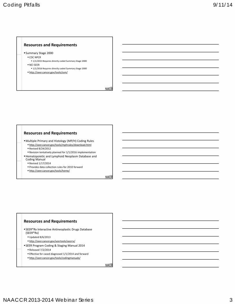
Summary Stage 2000
CDC NPCR 1/1/2015 Requires directly coded Summary Stage 2000
NCI SEER 1/1/2016 Requires directly coded Summary Stage 2000
http://seer.cancer.gov/tools/ssm/
Resources and Requirements
7
Resources and Requirements
Multiple Primary and Histology (MP/H) Coding Rules http://seer.cancer.gov/tools/mphrules/download.html
Revised 8/24/2012 Revision tentatively planned for 1/1/2016 implementation
Hematopoietic and Lymphoid Neoplasm Database and Coding Manual Revised 1/17/2014 Provides data collection rules for 2010 forward http://seer.cancer.gov/tools/heme/
Resources and Requirements
SEER*Rx Interactive Antineoplastic Drugs Database (SEER*Rx)
Updated 8/6/2013
http://seer.cancer.gov/seertools/seerrx/
SEER Program Coding & Staging Manual 2014
Released 7/2/2014
Effective for cased diagnosed 1/1/2014 and forward
http://seer.cancer.gov/tools/codingmanuals/

Coding Pitfalls 9/11/14
NAACCR 2013-2014 Webinar Series 4
FORDS 2013 http://www.facs.org/cancer/coc/fords/fords‐manual‐2013.pdf
Release of FORDS major revision tentatively scheduled for 1/1/2017
CoC Cancer Program Standards 2012: Ensuring Patient‐Centered Care
https://www.facs.org/~/media/files/quality%20programs/cancer/coc/programstandards2012.ashx
Resources and Requirements
10
Standards for Cancer Registries Volume II: Data Standards & Data Dictionary http://naaccr.org/StandardsandRegistryOperations/VolumeII.aspx#
Version 14: Implemented 1/1/2014
Version 15: Scheduled for implementation 1/1/2015
Version 16: Scheduled for implementation 1/1/2016
Version 17: Scheduled for implementation 1/1/2017
Version 18: Scheduled for implementation 1/1/2018
Resources and Requirements
11
Resources and Requirements
ICD‐O‐3 2014 and 2015 Guidelines for ICD‐O‐3 Update Implementation
http://www.naaccr.org/LinkClick.aspx?fileticket=u7d3sB71t5w%3d&tabid=126&mid=466
1/1/2016 ICD‐O‐3.1 tentatively scheduled for North American implementation
Online version http://codes.iarc.fr/usingicdo.php ICD‐O‐3 (2000) ICD‐O‐3.1 (2011)

Coding Pitfalls 9/11/14
NAACCR 2013-2014 Webinar Series 5
Multiple Primary & Histology Coding Rules
Hematopoietic & Lymphoid Neoplasm Database and Coding Manual
SEER*Rx Interactive Antineoplastic Drugs Database
ICD‐O‐3, ICD‐10‐CM, ICD‐9‐CM
SEER Coding & Staging Manuals
http://seer.cancer.gov/registrars/contact.html
Ask a SEER Registrar
AJCC TNM Staging
Collaborative Stage
FORDS/NCDB
2012 CoC Cancer Program Standards
http://cancerbulletin.facs.org/forums/content.php
CAnswer Forum
Where to Send Questions
13
Grade
14
Revised instructions http://seer.cancer.gov/tools/grade/
Are applicable for cases diagnosed 1/1/2014 and forward
Grade
15

Coding Pitfalls 9/11/14
NAACCR 2013-2014 Webinar Series 6
A: Gleason 7 will be coded as grade 2 beginning with 2014 cases.
Q: Is there going to be a change to Gleason 7, which is now = grade 3?
I've heard it will change to grade 2, MOD DIFF.
Grade
16
Gleason Score
CS Code Grade Code AJCC 7th SEER 2003-2013
2 002 1 G1 G13 003 1 G1 G14 004 1 G1 G15 005 1 G1 G26 006 1 G1 G27 007 2 G2 G38 008 3 G3 G39 009 3 G3 G310 010 3 G3 G3
Special Grade System Rules: Prostate
A:Yes. Per grade coding instruction #5 for solid tumors, code the highest grade if there is more than 1 grade.
Q: If there were multiple invasive tumors, would we code grade based on the higher grade even if it's the smaller tumor of the two?
Grade
18

Coding Pitfalls 9/11/14
NAACCR 2013-2014 Webinar Series 7
A: The WHO/ISUP grade for bladder is not defined as a special grade system in grade coding instruction #6 for solid tumors and should not be used to code the grade data item.
Q: What about grade for non‐invasive papillary urothelial carcinoma? Is there not a special grade system for bladder?
Grade
19
A:No. Brain is not a special grade system in reference to coding the grade data item. Under the table of special grade systems in instruction #6, it is documented that the tables are not used to code grade for WHO CNS tumors, WHO/ISUP for bladder & renal pelvis, or FIGO for female gynecologic sites.
Q: Should brain be on the list for a special grade system rule? We code the WHO grade as a SSF but don't code it in the grade field.
Grade
20
A: There is no special grade system for GE junction. That takes you to instruction 7, use 2, 3, or 4 grade system. The statement of grade 3/4 indicates a 4 grade system. 3/4 in 4 grade system is grade code 3.
Q: What is the grade code if you have a path report for a GE Junction, mucinous adenocarcinoma, histologic differentiation, poorly differentiated, grade 3/4, high grade?
Grade
21
Coding Pitfalls 9/11/14
NAACCR 2013-2014 Webinar Series 8
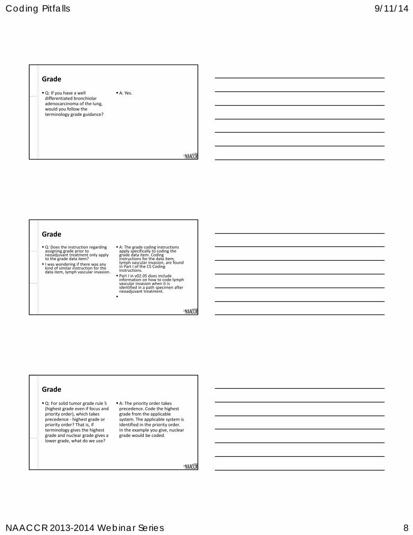
A: Yes. Q: If you have a well differentiated bronchiolar adenocarcinoma of the lung, would you follow the terminology grade guidance?
Grade
22
A: The grade coding instructions apply specifically to coding the grade data item. Coding instructions for the data item, lymph vascular invasion, are found in Part I of the CS Coding Instructions. Part I in v02.05 does include information on how to code lymph vascular invasion when it is identified in a path specimen after neoadjuvant treatment.
Q: Does the instruction regarding assigning grade prior to neoadjuvant treatment only apply to the grade data item? I was wondering if there was any kind of similar instruction for the data item, lymph vascular invasion.
Grade
23
A: The priority order takes precedence. Code the highest grade from the applicable system. The applicable system is identified in the priority order. In the example you give, nuclear grade would be coded.
Q: For solid tumor grade rule 5 (highest grade even if focus and priority order), which takes precedence ‐ highest grade or priority order? That is, if terminology gives the highest grade and nuclear grade gives a lower grade, what do we use?
Grade
24

Coding Pitfalls 9/11/14
NAACCR 2013-2014 Webinar Series 9
A: If the only description of grade for a gynecologic primary is FIGO grade 1, 2, or 3, assign code 9 in the grade data item. The FIGO grade system is different in that it describes the amount of non‐squamous or non‐modular solid growth pattern.
Q: If all we see is FIGO grade 1, 2, 3, do we assign grade code 1, 2, 3, or 9?
Grade
25
A:Instruction #4 says to code the grade for in situ tumor if it is documented. So if a grade was documented for an in situ tumor and it was documented using a 2‐grade system, not BR score or grade, then it should be coded. Remember that for breast BR ‐score/grade as coded in CS SSF7 is the first priority for coding the grade data item.
Q: Does the two‐grade system for breast apply to both invasive and in situ tumors?
Grade
26
A: For solid tumors, rules 1‐5 apply across the board. Rules 6‐9 are a hierarchy; you stop at the 1st rule that applies.
Q: It is my understanding that these new grade coding rules are somewhat "hierarchical". You stop at the first rule that applies. Is this true? Please clarify.
Grade
27
Coding Pitfalls 9/11/14
NAACCR 2013-2014 Webinar Series 10
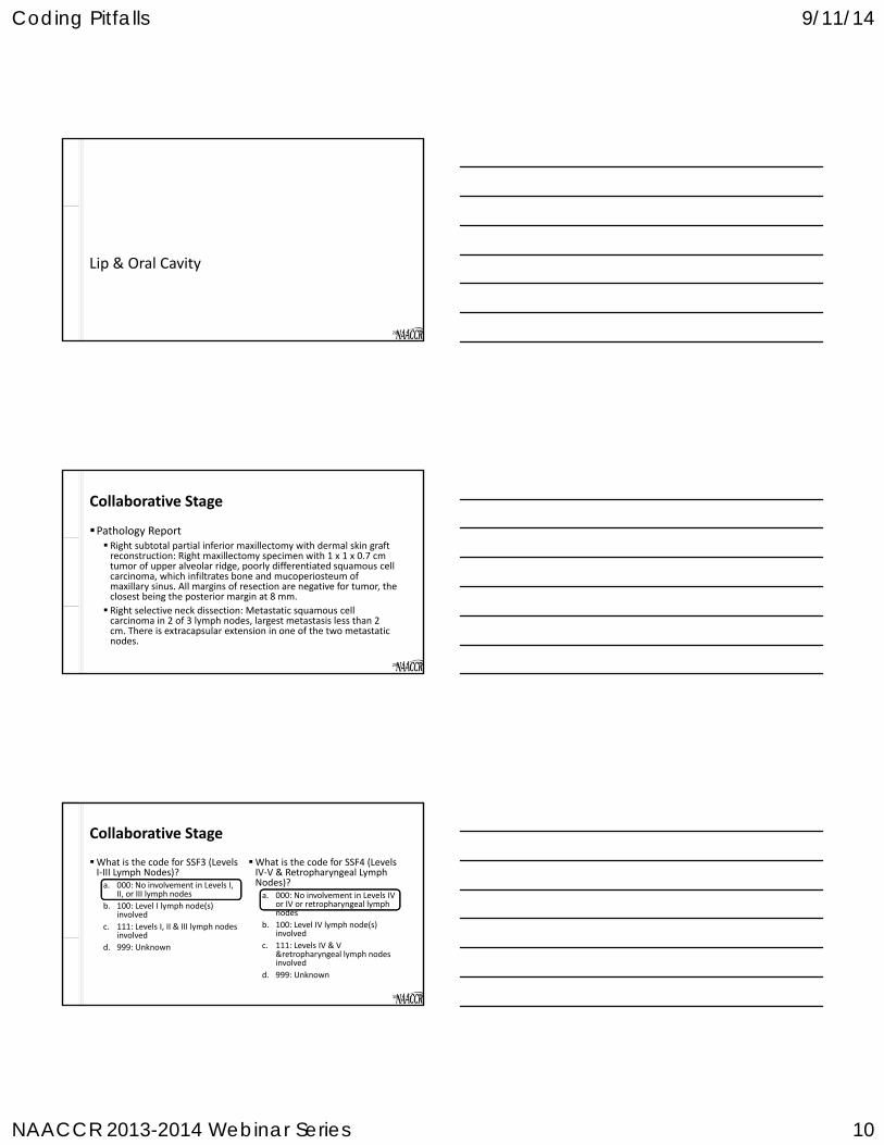
Lip & Oral Cavity
28
Pathology Report Right subtotal partial inferior maxillectomy with dermal skin graft reconstruction: Right maxillectomy specimen with 1 x 1 x 0.7 cm tumor of upper alveolar ridge, poorly differentiated squamous cell carcinoma, which infiltrates bone and mucoperiosteum of maxillary sinus. All margins of resection are negative for tumor, the closest being the posterior margin at 8 mm.
Right selective neck dissection: Metastatic squamous cell carcinoma in 2 of 3 lymph nodes, largest metastasis less than 2 cm. There is extracapsular extension in one of the two metastatic nodes.
Collaborative Stage
29
What is the code for SSF4 (Levels IV‐V & Retropharyngeal Lymph Nodes)?a. 000: No involvement in Levels IV
or IV or retropharyngeal lymph nodes
b. 100: Level IV lymph node(s) involved
c. 111: Levels IV & V &retropharyngeal lymph nodes involved
d. 999: Unknown
What is the code for SSF3 (Levels I‐III Lymph Nodes)?a. 000: No involvement in Levels I,
II, or III lymph nodes
b. 100: Level I lymph node(s) involved
c. 111: Levels I, II & III lymph nodes involved
d. 999: Unknown
Collaborative Stage
30
Coding Pitfalls 9/11/14
NAACCR 2013-2014 Webinar Series 11
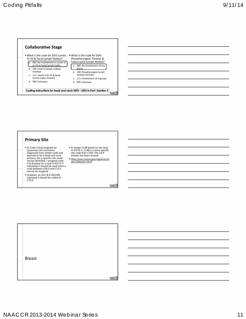
What is the code for SSF6 (Parapharyngeal, Parotid, & Suboccipital Lymph Nodes)?
a. 000: No involvement of any group
b. 100: Parapharyngeal lymph node(s) involved
c. 111: Involvement of 3 groups
d. 999: Unknown
What is the code for SSF5 (Levels VI‐VII & Facial Lymph Nodes)?
a. 000: No involvement in Levels VI or VII or facial lymph nodes
b. 100: Level VI lymph node(s) involved
c. 111: Levels VI & VII & facial lymph nodes involved
d. 999: Unknown
Collaborative Stage
31
A: Assign C148 based on the note in ICD‐O‐3. C148 is a more specific site code than C760. The I & R answer has been revised.
http://seer.cancer.gov/registrars/data‐collection.html
Q: Code C14.8 assigned for squamous cell carcinoma diagnosed from lymph node and deemed to be a head and neck primary, but a specific site could not be identified. I assigned code C14.8 based on a note in ICD‐O‐3 indicating it should be used when a code between C00.0 and C14.2 cannot be assigned.
However, an old I & R (46158) indicated it should be coded to C76.0.
Primary Site
32
Breast
33

Coding Pitfalls 9/11/14
NAACCR 2013-2014 Webinar Series 12
A: In most cases, the core biopsy would be coded as a diagnostic/staging procedure (02).
If the margins from the core biopsy were documented as negative on the pathology report, it could be coded as a surgical procedure.
The results of the lumpectomy are not a factor in how this procedure is coded.
Q: How do I code a core biopsy when there is no residual tumor on the subsequent lumpectomy?
Coding Breast Biopsies
34
http://cancerbulletin.facs.org/forums/showthread.php?2427-Breast-surgery
The Surveillance Epidemiology and End Results (SEER) program instructions for “Surgery of the Primary Site” are consistent:
“Code the most invasive, extensive, or definitive surgery if the patient has multiple surgical procedures of the primary site, even if there is no residual tumor found in the pathologic specimen from the more extensive surgery.”
SEER does not require “Surgical Diagnostic and Staging Procedure” to be coded.
SEER
Facility Oncology Registry Data Standards (FORDS) manual, Section One
“If surgery of the respective type was performed, the code that best describes the surgical procedure is recorded whether or not any cancer was found in the resected portion."
FORDS
FORDS and SEER
35http://newsmanager.commpartners.com/acscoc/issues/2014-09-02/4.html
Both FORDS and the SEER Coding Manual instructions say to code an incisional biopsy as excisional when the margins are microscopically or macroscopically free of tumor.
Neither SEER nor FORDS instructs registrars to use the pathologic examination from the subsequent surgery (for example, a lumpectomy) to determine whether a preceding biopsy was incisional or excisional. That coding decision depends only on marginal evaluation of the tissue removed in the biopsy.
Needle biopsies are not amenable to margin evaluation.
National Cancer Data Base News
36http://newsmanager.commpartners.com/acscoc/issues/2014-09-02/4.html

Coding Pitfalls 9/11/14
NAACCR 2013-2014 Webinar Series 13
Only record positive procedures
Do not code excisional biopsies with clear or microscopic margins
Diagnostic Staging Procedure
37
Lumpectomy followed by radiation
Mastectomy
Surgical Treatment
38http://beatthecancer.blogspot.com/2009/01/breast-cancer.html
Re‐excision Re‐excisions are performed when the patient has a lumpectomy and the entire tumor was not removed.
Coding a core needle biopsy as an excisional biopsy would artificially inflate the number of re‐excisions being done.
Standard Treatment
1. Diagnostic staging procedure (02)
2. Definitive surgical treatment
3. Adjuvant treatment (if necessary)
Big Picture
39
• 20 Partial mastectomy, NOS; less than total mastectomy, NOS
• 21 Partial mastectomy WITH nipple resection• 22 Lumpectomy or excisional biopsy• 23 Re-excision of the biopsy site for gross or
microscopic residual disease• 24 Segmental mastectomy (including wedge
resection, quadrantectomy, tylectomy
Coding Pitfalls 9/11/14
NAACCR 2013-2014 Webinar Series 14
Prostate
40
Patient has elevated PSA. Per physician note, DRE is benign. Needle biopsy of prostate: Adenocarcinoma right and left lobes. Per managing physician cT1c. MRI report states the result as cT2c prostate carcinoma.
What is the code for CS Extension – Clinical Extension?a. 150: Tumor identified by needle biopsy (clinically inapparent); Stated as
cT1c with no other information on clinical extension
b. 230: Clinically apparent tumor involves both lobes/sides; Stated as cT2c with no other info on clinical extension
c. 300: Localized NOS; Confined to prostate NOS; Intracapsular involvement only; Not stated if T1 or T2, clinically apparent or inapparent
d. 999: Unknown
Collaborative Stage
41
Patient has elevated PSA. Tumor involving about a third of the lobe palpated in left prostate lobe on DRE. Needle biopsy of prostate: Adenocarcinoma left lobe.
What is the code for CS Tumor Size/Ext Eval?
a. 0: Evaluation based on physical examination including DRE, imaging examination, or other non‐invasive clinical evidence
b. 1: Evaluation based on endoscopy, diagnostic biopsy (needle core biopsy or fine needle aspiration biopsy), TURP or other invasive techniques
Collaborative Stage
42

Coding Pitfalls 9/11/14
NAACCR 2013-2014 Webinar Series 15
Ovary
43
Debulking path report: High grade serous carcinoma, bilateral ovaries, with peritoneal metastasis beyond the pelvis, largest 2.5 cm; 2/2 mesenteric lymph nodes positive for metastasis.
How is the mesenteric lymph node involvement coded?
a. N1
b. M1
AJCC TNM Stage
44
Final diagnosis: Bilateral ovarian serous carcinoma with liver metastasis. How is the liver metastasis coded in CS?
A: Code liver parenchyma metastasis in CS Mets at DX. Code metastasis on liver surface in CS Extension.
Collaborative Stage
45

Coding Pitfalls 9/11/14
NAACCR 2013-2014 Webinar Series 16
GIST
46
A:GIST NOS has an ICD‐O‐3 behavior code of /1 (borderline). Malignant GIST has a behavior code of /3 (malignant). You should not code as /3 unless you have a statement of malignancy
Q: Please repeat the difference between GIST NOS and malignant GIST.
Histology
47
http://www.cancer.gov/cancertopics/pdq/treatment/gist/HealthProfessional/Table1

Coding Pitfalls 9/11/14
NAACCR 2013-2014 Webinar Series 17
Per SINQ 20091021 and 20021151, GIST cases are not reportable unless they are stated to be malignant. A pathologist or clinician must confirm the diagnosis of cancer. There are cases that are not stated to be malignant in the pathology report or confirmed as such by a clinician; however, these cases do have information that for other primary sites would typically be taken into consideration when determining reportability.(SEER SINQ 20100014)
Are there criteria other than a pathologist or clinician’s statement that a registrar can use to determine reportability of gastrointestinal stromal tumors (GIST)?
Reportability
Question
Pathologists have used tumor size and mitotic activity to determine whether GISTS were benign or malignant. The 7th Edition AJCC Manual uses criteria for Stage I GIST which would otherwise be considered benign.
Could you clarify if we are to go by staging criteria to determine if a GIST is reportable?
Answer
The CoC requires to report all sites malignancies with behavior 2 and 3, except skin cancers 8000‐8110, CIS, intraepithelial neoplasia grade III (8077/2) of the cervix (CIN III), prostate (PIN III), vulva (VIN III), vagina (VAIN III), and anus (AIN III). Benign GIST is not reportable since the behavior is 0. However, if your facility or your state requires to collect benign GIST, you should follow their requirements. When you submit the data, make sure you do not include benign GIST in your data file submitted to NCDB (CoC), otherwise it will be rejected.
Staging forms can be used to stage all GIST tumors /0/1 or /3. however, this does not mean that the GIST tumors with a /0 or /1 are reportable. Reportabitity is determined by the CoC, SEER, state and your cancer committee. You could however pick these up as a reportable by agreement case.
http://cancerbulletin.facs.org/forums/showthread.php?449-Is-benign-GIST-reportable-to-the-CoC&highlight=GIST
Coding Pitfalls 9/11/14
NAACCR 2013-2014 Webinar Series 18
A:The AJCC Cancer Staging Manual isn't based on the reportability rules that registrars use. I assume the authors felt there was sufficient clinical benefit to collect staging information on low and intermediate risk GISTs to provide staging criteria for clinicians that choose to collect these cases.
Q: If GIST is benign, why do pathologist stage them? This is confusing since we always assume if cancer is staged, it is malignant
Reportability
52
1. Would we create a new abstract with the date of diagnosis being the date the physician states the case is malignant and thus the patient would have two abstracts?
2. The first would have a sequence code of 60 and a behavior code of 1?
3. The second would have a sequence code of 00 (if it was the first malignancy) and behavior code of 3 and a different date of diagnosis?
Q: Our cancer committee has decided that we should collect ALL GIST tumors.
In the event that a GIST that we have abstracted becomes malignant and thus is now reportable to NCDB, how should be handle this case?
A:Yes, to your last three questions.
Question
http://cancerbulletin.facs.org/forums/showthread.php?3214-GIST-Tumors&highlight=GIST
A:Treatment cannot be used as a determination of malignancy because borderline GIST and malignant GIST receive the same types of treatments (surgery and imatinib).
Q: What if the patient is being treated as malignant even though there is no statement of malignancy (reportability)?
Reportability
54

Coding Pitfalls 9/11/14
NAACCR 2013-2014 Webinar Series 19
Bladder
55
TURB pathology: Bladder cancer in lateral wall; 1 cm urothelial carcinoma that invades the superficial muscularis propria.
Cystectomy: In situ urothelial carcinoma of bladder.
Stage
56
What is the code for CS Tumor Size/Ext Eval?
a. 0
b. 1
c. 3
d. 9
What is the code for CS Extension?
a. 060: Nonpapillary – Sessile CA in situ, CA in situ NOS, transitional cell CA in situ
b. 210: Muscle of bladder only; superficial muscle ‐ inner half
c. 220: Muscle of bladder only ‐deep muscle‐‐outer half
d. 240: Muscle invaded NOS of bladder only
Collaborative Stage
57

Coding Pitfalls 9/11/14
NAACCR 2013-2014 Webinar Series 20
What is the directly coded AJCC pathologic T category?
a. X: Primary tumor cannot be assessed
b. Tis: CA in situ – flat tumor
c. T2: Tumor invades muscularis propria
d. pT2a: Tumor invades superficial muscularis propria
What is the directly coded AJCC clinical T category?
a. X: Primary tumor cannot be assessed
b. Tis: CA in situ – flat tumor
c. T2: Tumor invades muscularis propria
d. pT2a: Tumor invades superficial muscularis propria
AJCC TNM Stage
58
Melanoma
59
Q: If patient has 1 lymph node positive for melanoma and primary skin site cannot be identified, how should the following fields be coded? CS Lymph Nodes: 100 (Regional nodes NOS)
Regional Nodes Positive: 01
Regional Nodes Examined: 01
Scope of Regional Lymph Node Surgery: Code surgery performed
Surgical Procedure/Other Site: 0 (None)
Collaborative Stage/Surgery Data Items
60
Coding Pitfalls 9/11/14
NAACCR 2013-2014 Webinar Series 21
Q1: What is the code for SSF4 [Serum Lactate Dehydrogenase (LDH)] if the 1st LDH test is negative and 2nd
test was unknown?
A1: 000 (Within normal limits)
Q2: What is the code for SSF5 (LDH Lab Value) if 1st test is positive and 2nd test is negative?
A2: Code lab value from negative test (2nd)
Collaborative Stage ‐ LDH
61
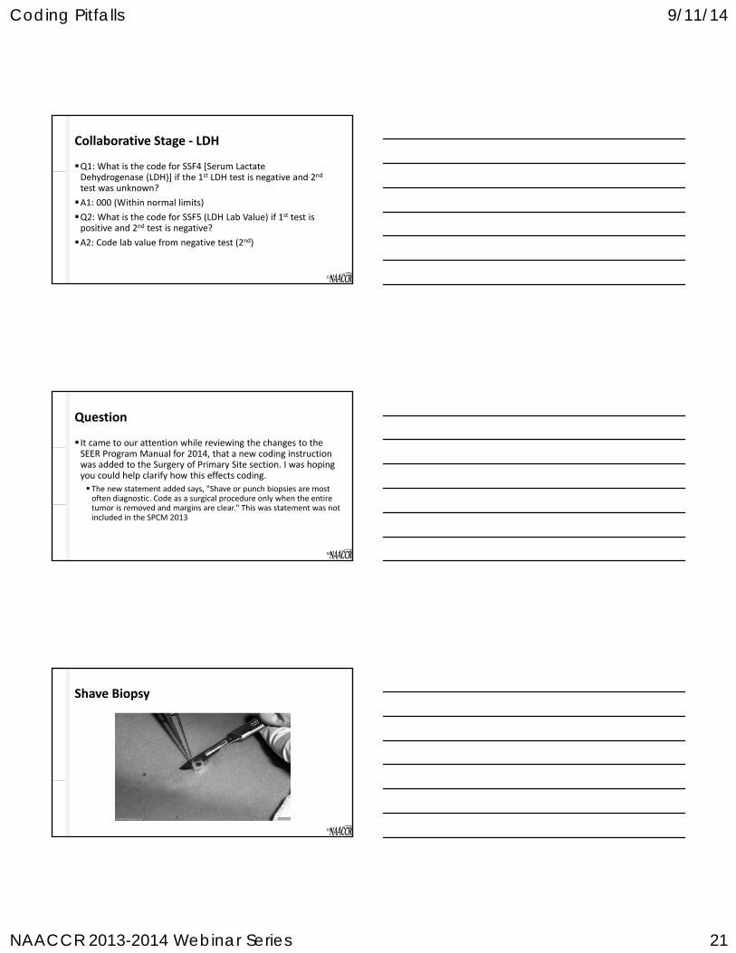
It came to our attention while reviewing the changes to the SEER Program Manual for 2014, that a new coding instruction was added to the Surgery of Primary Site section. I was hoping you could help clarify how this effects coding.
The new statement added says, "Shave or punch biopsies are most often diagnostic. Code as a surgical procedure only when the entire tumor is removed and margins are clear." This was statement was not included in the SPCM 2013
Question
62
Shave Biopsy
63

Coding Pitfalls 9/11/14
NAACCR 2013-2014 Webinar Series 22
If the tumor is very large or in a site that is difficult to biopsy, the physician may choose to take a small sample of the tumor rather than remove the entire tumor.
If this is done, the margins on the specimen sent to pathology will be grossly positive.
This would be coded as a Surgical Diagnostic Staging Procedure code 02.
Surgical Diagnostic Staging Procedure
64
If a physician suspects melanoma, they will probably try to remove the entire lesion. This may be done as a standard excisional biopsy, punch biopsy, or a shave biopsy.
Regardless of the approach, this procedure should be coded using the surgery code 27.
If the margins of the biopsy are microscopically positive or there is no information about the margins, assume it was an excisional biopsy.
The surgeon will attempt to take 3‐5mm of healthy tissue and will try to minimize damage to the lymphatics.
Excisional Biopsy
65
Following the excisional biopsy the patient will probably have a wide excision.
A wide excision removes a margin of healthy tissue from around the melanoma site.
If a sentinel lymph node biopsy is recommended, it will be done prior to the wide excision.
Wide Excision
66

Coding Pitfalls 9/11/14
NAACCR 2013-2014 Webinar Series 23
If the margin of healthy tissue is 1cm or less, code this procedure using codes 30‐33.
Codes 30‐33 would also be used if the margin of healthy tissue is not stated.
Even though these codes reflect two procedures, the date of surgery when assigning codes 30‐33 is the date of the wide excision.
Wide Excision
67
Code 30 is used if the original excisional biopsy was a standard excisional technique or if the technique was not indicated.
Code 31 is used if the original excisional biopsy was a shave biopsy.
Code 33 is used if the original biopsy was incisional and then a wide excision was done (the incisional biopsy was coded as a diagnostic staging procedure).
If your facility only codes one surgery for each abstract (i.e. hospital only reporting to the state cancer registry), use the code for the most definitive procedure.
Wide Excision
68
Code 45 is used if the patient has a wide excision and the margins are more than 1cm, but it is not documented if they are more or less than 2cm’s.
Code 46 is used if the patient has a wide excision and the margins are more than 1cm and it is documented that the margins are equal to or less than 2cm’s.
Code 47 is used if the patient has a wide excision and the margins are more than 2cm’s.
Wide Excision
69

Coding Pitfalls 9/11/14
NAACCR 2013-2014 Webinar Series 24
When a wide excision with 1‐2cm margins is performed (code 46), followed by re‐excision for wider margins:
If the total combined resection margins are >2cm, use code 47
If no information is available of the path report does not describe the distance from the margins to the previous spot, code the re‐excision as 46 (two entries with surgical code 46)
Wide Excision
70
If the margins are grossly positive code as dx staging procedure. If they are negative or only microscopically positive, code as surgery. The big question is what to do if the margins are unknown.
Answer
71
Hematopoietics
72

Coding Pitfalls 9/11/14
NAACCR 2013-2014 Webinar Series 25
Transformations to Acute or more severe neoplasm
Transformations from Chronic neoplasm
Examples Essential thrombocythemia (9962/3) Transformations to: Acute myeloid leukemia (9861/3)
Plasma cell myeloma (9732/3) Transformations from: Solitary plasmacytoma of bone (9731/3)
Hematopoietics‐Transformations
73
Colon & Rectum
74
A:Yes, the chemotherapy given prior to hemicolectomy would be considered neoadjuvant treatment.
Q: Please address staging polypectomies. Is polypectomy, then chemotherapy, lastly hemicolectomy considered neoadjuvant treatment?
Treatment
75

Coding Pitfalls 9/11/14
NAACCR 2013-2014 Webinar Series 26
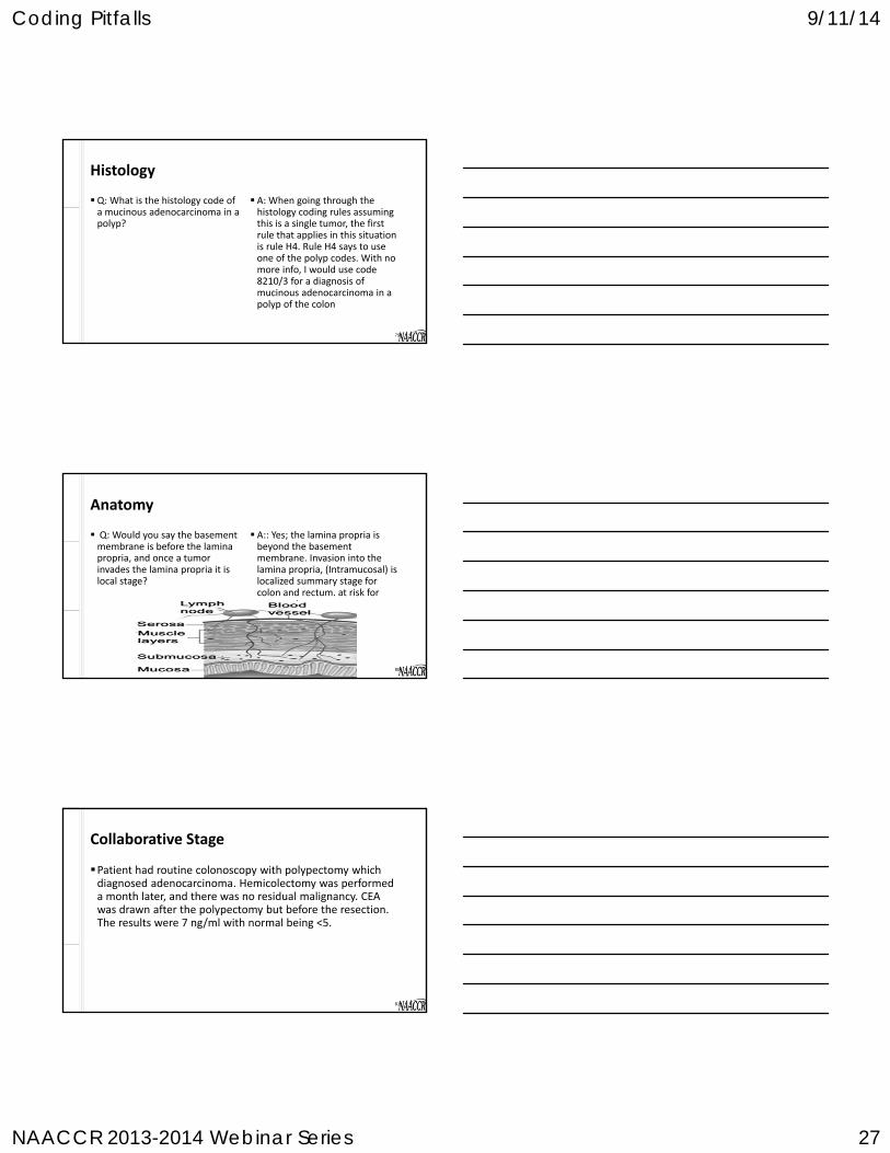
Q: When a tumor extends into the subserosa, isn't that considered pericolonic fat?
Anatomy
76
A: Subserosal fat indicates a portion of the colon covered by serosa.
If the colon is not covered by serosa, all of the fat outside of the colon is referred to as pericolic fat.
Anatomy
77
Subserosal Fat
Muscularis Propria
Lamina Propria
PericolicFat
77
Lumen
A:For cases diagnosed 2007‐2014, code 8480 (mucinous adenocarcinoma).
When the final diagnosis states "mucinous," code 8480.
When mucinous is stated in the final diagnosis, the percent does not need to be specified. See rule H5.
Q: If you had an adenocarcinoma with mucinous features how would you code the histology?
Histology
78
http://seer.cancer.gov/seerinquiry/index.php?page=search_results&records=n&search_results_show=first&search_within_results=0&search_type=quick_search&topic1=&topic2=&topic3=&start_year=2000&end_year=2014&quicksearch=20071122&question_id=&cat_andor=AND&cat0=+&cat1=+&cat2=+&last_update=&date_finalized=&free_andor=AND&free0=&free1=&free2=&free3=&asc_yr_text=&Question_1=1&Question_3=1&search_display_format=1
Coding Pitfalls 9/11/14
NAACCR 2013-2014 Webinar Series 27
A: When going through the histology coding rules assuming this is a single tumor, the first rule that applies in this situation is rule H4. Rule H4 says to use one of the polyp codes. With no more info, I would use code 8210/3 for a diagnosis of mucinous adenocarcinoma in a polyp of the colon
Q: What is the histology code of a mucinous adenocarcinoma in a polyp?
Histology
79
A:: Yes; the lamina propria is beyond the basement membrane. Invasion into the lamina propria, (Intramucosal) is localized summary stage for colon and rectum. at risk for metastasis.
Q: Would you say the basement membrane is before the lamina propria, and once a tumor invades the lamina propria it is local stage?
Anatomy
80
Patient had routine colonoscopy with polypectomy which diagnosed adenocarcinoma. Hemicolectomy was performed a month later, and there was no residual malignancy. CEA was drawn after the polypectomy but before the resection. The results were 7 ng/ml with normal being <5.
Collaborative Stage
81

Coding Pitfalls 9/11/14
NAACCR 2013-2014 Webinar Series 28
What is the code for SSF3 (CEA Lab Value)?
a. 050
b. 070
c. 998: Test not done
d. 999: Unknown
What is the code for SSF1 [Carcinoembryonic Antigen (CEA)]?
a. 010: Positive/elevated
b. 020: Negative/normal
c. 998: Test not done
d. 999: unknown
Collaborative Stage
82
Q: Patient has polypectomy. Path shows that the polyp is purely in situ, and there is no residual tumor. Can we pathologically stage this cancer?
A: Yes
AJCC TNM
83
Liver
84

Coding Pitfalls 9/11/14
NAACCR 2013-2014 Webinar Series 29
MRI: Hepatomegaly; 7 cm right liver lobe mass with intrahepatic metastases in both lobes and vascular invasion; small hepatic nodes; no other organomegaly. Liver is cirrhotic.
Hepatic biopsy: Hepatocellular carcinoma, grade 3.
AJCC TNM Stage
85
What is the pathologic T?a. TX: Primary tumor cannot be
assessed
b. T2: Solitary tumor with vascular invasion or multiple tumors none more than 5 cm
c. T3a: Multiple tumors more than 5 cm
d. T3b: Single tumor or multiple tumors of any size involving a major branch of the portal vein or hepatic vein
What is the clinical T?a. TX: Primary tumor cannot be
assessed
b. T2: Solitary tumor with vascular invasion or multiple tumors none more than 5 cm
c. T3a: Multiple tumors more than 5 cm
d. T3b: Single tumor or multiple tumors of any size involving a major branch of the portal vein or hepatic vein
AJCC TNM Stage
86
Lung
87

Coding Pitfalls 9/11/14
NAACCR 2013-2014 Webinar Series 30
A:Not according to the coding rules we currently use. This may change in the future, but for now consider it a /3 unless instructed otherwise by a physician or pathologist. The new edition of the lung WHO classification (blue books) is not expected until 2015.
Q: Is bronchioalveolar carcinoma considered in situ?
Histology & Behavior
88
A: Per “Ask a SEER Registrar” Assign code C349 when infrahilar refers to the infrahilar area of the lung and no further information is available.
See #12 on page 67 in the SEER manual, http://www.seer.cancer.gov/manuals/2014/SPCSM_2014_maindoc.pdf
Q: What is the ICD‐O site code for "infrahilar tumor"?
Topography
89
Q1: Please define pleural based mass.
A1: Lesion within lung that abuts visceral pleura on imaging
Q2: Is the CS Extension code for a pleural based mass with no other statement of invasion 410?
A2: No
Collaborative Stage
90

Coding Pitfalls 9/11/14
NAACCR 2013-2014 Webinar Series 31
Right lung cancer with right pleural effusion; single negative cytology of pleural effusion but fluid is exudative and bloody.
What is the code for CS Mets at DX?a. 00: No distant metastasis
b. 15: Malignant pleural effusion, ipsilateral or same lung
What is the code for CS Mets Eval?a. 0: Evaluation of distant metastasis based on non‐invasive clinical
evidence
b. 3: Specimen from metastatic site microscopically positive
Collaborative Stage
91
How would you code the T category for invasion of primary lung tumor into rib?
a. T3: Tumor more than 7cm or one that directly invades: parietal pleura chest wall, diaphragm, phrenic nerve, mediastinal pleura, parietal pericardium; or tumor in main bronchus; or associated atelectasis or obstructive pneumonitis of the entire lung or separate tumor nodules in same lobe
b. T4: Tumor of any size that invades: mediastinum, heart, great vessels, trachea, recurrent laryngeal nerve, esophagus, vertebral body, carina, separate tumor nodules in different ipsilateral lobe
AJCC TNM Stage
92
Questions?
93

Coding Pitfalls 9/11/14
NAACCR 2013-2014 Webinar Series 32
Coming Up…
Registration is open for 2014‐2015 Cancer Registry & Surveillance Webinar Series
http://www.naaccr.org/EducationandTraining/WebinarSeries.aspx
And the winners are……
95
CE Certificate Quiz/Survey
Phrase
Stand‐up
Link http://www.surveygizmo.com/s3/1800630/Coding‐Pitfalls‐2014

Coding Pitfalls 9/11/14
NAACCR 2013-2014 Webinar Series 33
Please send any questions to:
Jim Hofferkamp [email protected]
Shannon Vann [email protected]
Thank You!!!!


