Embed Size (px)
Citation preview

N-Vinylpyrrolidone - vinyl acetate block copolymers as drug delivery vehicles
Dissertation presented in partial fulfillment of the requirements for the degree of
PhD (Polymer Science)
by Nathalie Bailly
Promoter: Prof. Bert Klumperman
University of Stellenbosch- Faculty of Science
Department of Chemistry and Polymer Science
March 2012
Declaration By submitting this dissertation electronically, I declare that the entirety of the work contained
therein is my own, original work, that I am the owner of the copyright thereof (unless to the
extent explicitly otherwise stated) and that I have not previously in its entirety or in part
submitted it for obtaining any qualification
Nathalie Bailly Stellenbosch, March 2012
Copyright © 2012 Stellenbosch University All rights reserved
Stellenbosch University http://scholar.sun.ac.za
To my parents,
Chantal and
Claude
Stellenbosch University http://scholar.sun.ac.za

Abstract
The primary aim of this study was to investigate the feasibility of the amphiphilic block
copolymer poly((vinylpyrrolidone)-b-poly(vinyl acetate)) (PVP-b-PVAc) as a vehicle for
hydrophobic anti-cancer drugs.
PVP-b-PVAc block copolymers of constant hydrophilic PVP block length and varying
hydrophobic PVAc block lengths were synthesized via xanthate-mediated controlled radical
polymerization (CRP). The methodology consisted of growing the PVAc chain from a xanthate
end-functional PVP. In an aqueous environment the amphiphilic block copolymers self-
assembled into spherical vesicle-like structures consisting of a hydrophobic PVAc bilayer
membrane, a hydrophilic PVP corona and an aqueous core. The self-assembly behaviour and the
physicochemical properties of the self-assembled structures were investigated by 1H NMR
spectroscopy, fluorescence spectroscopy, transmission electron microscopy (TEM) and dynamic
and static light scattering.
Drug loading studies were performed using a model hydrophobic drug, clofazimine, and a
common anti-cancer drug paclitaxel (PTX) to evaluate the potential of the PVP-b-PVAc block
copolymers for drug delivery,. Clofazimine and PTX were physically entrapped into the
hydrophobic domain of the self-assembled PVP-b-PVAc block copolymers via the dialysis
method. The drug-loaded PVP-b-PVAc block copolymers were characterized regarding particle
size, morphology, stability and drug loading capacity in order to assess their feasibility as a drug
vehicle. The polymer vesicles had a relatively high drug loading capacity of 20 wt %. The effect
of the hydrophobic PVAc block length on the drug loading capacity and encapsulation efficiency
were also studied. Drug loading increased with increasing the hydrophobic PVAc block length.
The effect of the drug feed ratio of clofazimine and PTX on the drug loading capacity and
encapsulation efficiency were also investigated. The optimal formulation for the drug-loaded
PVP-b-PVAc was determined and further investigated in vitro. The size stability of the drug-
loaded PVP-b-PVAc block copolymers was also assessed under physiological conditions (PBS,
pH 7.4, 37 °C) and were stable in the absence and presence of serum.
Stellenbosch University http://scholar.sun.ac.za

PVP-b-PVAc block copolymers were tested in vitro on MDA-MB-231 multi-drug-resistant
human breast epithelial cancer cells and normal MCF12A breast epithelial cells to provide
evidence of their antitumor efficacy. In vitro cell culture studies revealed that the PVP-b-PVAc
drug carrier exhibited no cytotoxicity towards MDA-MB-231 and MCF12A cells, confirming the
biocompatibility of the PVP-b-PVAc carrier. In vitro cytotoxicity assays using clofazimine-PVP-
b-PVAc formulations showed that when MDA-MB-231 cells were exposed to the formulations,
an enhanced therapeutic effect was observed compared to the free drug. Cellular internalization
of the PVP-b-PVAc drug carrier was demonstrated by fluorescent labeling of the PVP-b-PVAc
carrier. Fluorescence microscopy results showed that the carrier was internalized by the MDA-
MB-231 cells after 3 hours and localized in the cytoplasm and the perinuclear region.
Overall, it was demonstrated that PVP-b-PVAc block copolymers appear to be promising
candidates for the delivery of hydrophobic anti-cancer drugs.
Stellenbosch University http://scholar.sun.ac.za

Opsomming
Die studie is gebaseer op die gebruik van amfifieliese blokkopolimere van poli((N-
vinielpirolidoon)-b-poli(vinielasetaat)) (PVP-b-PVAc) as potensiële geneesmiddeldraers.
PVP-b-PVAc blokkopolimere van konstante hydrofiliese bloklengte en verskillende
hydrofobiese bloklengte is voorberei via die RAFT/MADIX-proses. Blokkopolimere met
vinielasetaat is vanaf poli(N-vinielpirolidoon) met ‘n xantaatendfunksie voorberei. In ‘n
wateromgewing vorm die PVP-b-PVAc blokkopolimere vesikel strukture met ‘n
hydrofobiese membraan en ‘n hydrofiliese mantel.
Die fisies-chemiese eienskappe van die PVP-b-PVAc blokkopolimere is gekarakteriseerd
met gebruik van KMR spektroskopie, fluoresent spektroskopie, transmissie
elektronmikroskopie (TEM) en dinamiese en statiese lig verstrooiing.
Die potensiaal van PVP-b-PVAc as ‘n geneesmiddeldraer is ondersoek deur gebruik te
maak van die hydrofobiese geneesmiddel, clofazimine, en ‘n anti-kanker geneesmiddel,
paclitaxel. Clofazimine en paclitaxel is ge-inkapsuleer in die hydrofobiese gedeelte van
die blokkopolimere via die dialise-metode. Clofazimine-PVP-b-PVAc en paclitaxel-PVP-
b-PVAc blokkopolimere is gekarakteriseerd met betrekking tot die partikel grootte,
morfologie, stabiliteit en laai kapasitiet om die PVP-b-PVAc blokkopolimere as
geneesmiddeldraers te evalueer. Die PVP-b-PVAc geneesmiddeldraer het ‘n relatiewe
hoë laai kapsiteit van 20 gew % aangetoon. Die invloed van die bloklengte op die laai
kapasitiet is ook ondersoek en beskryf. ‘n Toename in die laai kapasitiet is gesien met ‘n
toename in die hydrofobiese bloklengte. Die invloed van die hoeveelheid geneesmiddel
op die laai kapasitiet en die inkapsuleer doeltreffendheid is ook ondersoek. Die optimale
formulasie is gevind en verder gebruik vir in vitro studies. Die stabiliteit van die
geneesmiddeldraer in fisiologiese omstandighede (pH 7.4, 37 °C) is ook beskryf.
Resultate toon aan dat die sisteem stabiel is onder hierdie omstandighede in die
afwesigheid en aanwesigheid van serum.
Stellenbosch University http://scholar.sun.ac.za
In vitro eksperimente is op MCF12A epiteel-borsselle en MDA-MB-231 epiteel-
borskankerselle getoets om die anti-tumoraktiwiteit te ondersoek. Resultate toon aan dat
die PVP-b-PVAc geen sitotoxiese effek op die selle het nie, wat aandui dat die polimere
bioverenigbaar is. Verder is dit bewys dat die PVP-b-PVAc geneesmiddel formualsie ’n
hoër sitotoxisiteit besit as die vry-geneesmiddel. Fluoresent studies het aangetoon dat die
geneesmiddeldraer na 3 uur opgeneen word deur MDA-MB231 selle en gelokaliseerd is
in die sitoplasma en in die omgewing van die kern van die selle.
In die algemeen is dit aangetoon dat PVP-b-PVAc blokkopolimere potensiële kandidate
vir die lewering van hydrofobiese geneesmiddels is.
Stellenbosch University http://scholar.sun.ac.za

i
Table of contents
List of acronyms ........................................................................................................... v
List of symbols ............................................................................................................ vi
List of schemes .......................................................................................................... vii
List of tables .............................................................................................................. viii
List of figures .............................................................................................................. ix
Chapter 1: Introduction and objectives
1 Introduction ................................................................................................................ 1
2. Amphiphilic block copolymers for drug delivery ..................................................... 2
3 Objective of dissertation ............................................................................................ 4
4. Outline of dissertation ............................................................................................... 4
5. References ................................................................................................................. 6
Chapter 2: Historical and theory
2.1 Introduction ............................................................................................................. 8
2.2 Controlled radical polymerization .......................................................................... 9
2.2.1 RAFT/MADIX-mediate polymerization ............................................................. 9
2.2.2 Xanthate-mediated synthesis of poly(N-vinylpyrrolidone) (PVP) and
poly(vinyl acetate) (PVAc) ......................................................................................... 10
2.3 Amphiphilic block copolymers in anti-cancer drug delivery ............................... 12
2.3.1 Amphiphilic block copolymers as drug delivery carriers .................................. 14
2.3.2 Amphiphilic block copolymer micelles and vesicles......................................... 14
2.3.3 Active and passive targeting .............................................................................. 17
2.3.4 Inherent size of polymer drug carriers and biodistribution of the drug ............. 19
2.3.5 Hydrophilic corona and hydrophobic core components of polymer micelles
or vesicles ................................................................................................................... 19
2.3.6 Stability of polymer drug carriers in aqueous and biological environment ....... 21
2.4 Preparation and drug loading into polymer drug carriers ..................................... 23
2.4.1 Drug loading methods for polymer micelles and vesicles ................................ .23
Stellenbosch University http://scholar.sun.ac.za

ii
2.4.2 Factors affecting drug loading ........................................................................... 25
2.5 Drug release and cellular internalization .............................................................. 27
2.5.1 Drug release ....................................................................................................... 27
2.5.2 Cellular internalization....................................................................................... 28
2.5.3 Polymer drug carriers and multiple drug resistance (MDR) .............................. 30
2.6 Conclusion ............................................................................................................ 30
2.7 References ............................................................................................................. 31
Chapter 3: Synthesis, characterization and self-assembly of PVP-b-PVAc
3.1 Introduction ........................................................................................................... 40
3.2 Materials and Methods .......................................................................................... 44
3.2.1 Materials ........................................................................................................... 44
3.2.2 Synthesis of xanthate chain-transfer agent ........................................................ 44
3.2.3 Synthesis of PVP macro-RAFT agent ............................................................... 44
3.2.4 Synthesis of PVP-b-PVAc block copolymers ................................................... 44
3.2.5 Polymer characterization ................................................................................... 45
3.2.6 Self-assembly of PVP-b-PVAc block copolymers ............................................ 46
3.2.7 Determination of the critical micelle concentration (CMC) of PVP-b-PVAc
block copolymers ........................................................................................................ 46
3.2.8 Size distribution and morphology of PVP-b-PVAc block copolymers ............. 47
3.2.9 Multiangle static light scattering (MASLS) ....................................................... 47
3.2.10 Stability of PVP-b-PVAc block copolymers in biological environment ......... 49
3.3 Results and discussion ......................................................................................... 50
3.3.1 Synthesis and characterization of PVP macro-RAFT-agent and PVP-b-PVAc
block copolymers ........................................................................................................ 50
3.3.2 Gradient HPLC characterization of PVP-b-PVAc block copolymers ............... 55
3.3.3 Self-assembly behavior of PVP-b-PVAc block copolymers ............................. 57
3.3.4 CMC of PVP-b-PVAc block copolymers .......................................................... 58
3.3.5 Size distribution and morphology of PVP-b-PVAc ........................................... 61
3.3.6 Stability of PVP-b-PVAc vesicles under physiological conditions ................... 66
3.4 Conclusion ............................................................................................................ 67
Stellenbosch University http://scholar.sun.ac.za

iii
3.5 References ............................................................................................................. 68
Chapter 4: PVP-b-PVAc: A potential drug carrier
4.1 Introduction ........................................................................................................... 72
4.2 Materials and methods .......................................................................................... 75
4.2.1 Chemicals ........................................................................................................... 75 4.2.2 Loading of clofazimine into PVP-b-PVAc block copolymers .......................... 75 4.2.3 1H NMR measurements of block copolymers ................................................... 75
4.2.4. Size distribution and morphology of clofazimine-loaded PVP-b-PVAc .......... 75
4.2.5 Evaluation of the drug loading capacity and encapsulation efficiency of
clofazimine-loaded PVP-b-PVAc ............................................................................... 76
4.2.6 Stability studies of clofazimine-loaded PVP-b-PVAc ....................................... 77
4.3. Results and discussion ......................................................................................... 77
4.3.1 Preparation and characterization of clofazimine-loaded PVP-b-PVAc
block copolymer.......................................................................................................... 77
4.3.2 Size distribution and morphology of clofazimine-loaded PVP-b-PVAc ........... 79
4.3.3 Drug loading capacity and encapsulation efficiency ......................................... 82
4.3.4 Stability studies of clofazimine-loaded PVP-b-PVAc ....................................... 86
4.4 Conclusion ............................................................................................................ 88
4.5 References ............................................................................................................. 89
Chapter 5: In vitro cytotoxicity and cellular uptake of PVP-b-PVAc
5.1 Introduction ........................................................................................................... 91
5.2 Materials and methods .......................................................................................... 95
5.2.1 Materials ............................................................................................................ 95
5.2.2 Cell culture and culture conditions .................................................................... 95
5.2.3 In vitro cytotoxicity assay .................................................................................. 95
5.2.4 Synthesis of fluorescently labeled PVP-b-PVAc ............................................... 96
5.2.5 Preparation of perylene red-loaded PVP-b-PVAc ............................................. 97
5.2.6 Cellular uptake .................................................................................................. 97
5.3 Results and discussion .......................................................................................... 98
Stellenbosch University http://scholar.sun.ac.za

iv
5.3.1 In vitro cytotoxicity studies ............................................................................... 98
5.3.2 Cellular uptake studies of fluorescently labeled PVP-b-PVAc ....................... 103
5.4 Conclusion ........................................................................................................ ̀ 109
5.5 References ........................................................................................................... 110
Chapter 6: Summary and Perspectives
6.1 Summary ............................................................................................................. 113
6.2 Perspectives......................................................................................................... 116
6.3 References ........................................................................................................... 119
Stellenbosch University http://scholar.sun.ac.za

v
List of acronyms
AIBN Azobisisobutyronitrile
ATRP Atom transfer radical polymerization
CI Combination Index Analysis
CMC Critical micelle concentration
CRP Controlled radical polymerization
DDI Distilled deionized water
DLS Dynamic light scattering
DMEM Dulbecco’s modification of Eagle’s Medium
ELSD Evaporative light scattering detector
EPR Enhanced permeation and retention
FRDC Fixed ratio drug combination
FRET Forster resonance emission transfer
FCS Fetal calf serum
GPEC Gradient polymer elution chromatography
MDR Multi-drug-resistance
MPS Mononuclear phagocytic system
MTT (4,5-dimethylthiazol-2-yl)-2,5-diphenyltetrazolium bromide
MADIX Macromolecular design via the interchange of xanthate
MWCO Molecular weight cut-off
NMP Nitroxide mediated polymerization
NVP N-vinyl pyrrolidone
NMR Nuclear magnetic resonance
PBS Phosphate buffered saline
PTX Paclitaxel
PVP poly(N-vinylpyrrolidone)
PVAc poly(vinyl acetate)
RAFT Reversible addition-fragmentation transfer
RES Reticulo-endothelial system
RI Refractive index
SFRP Stable free radical polymerization
Stellenbosch University http://scholar.sun.ac.za

vi
SLS Static light scattering
SEC Size exclusion chromatography
TCEP tris(2-carboxyethyl)phosphine
TEM Transmission electron microscopy
UV Ultraviolet
VAc Vinyl acetate
List of Symbols
A2 virial coefficient
Đ dispersity
I scattering intensity
Mn number average molar mass
Mw (particle)
weight average molecular weight of particle
NA Avagadro’s number
Rh Hydrodynamic radius
Rg Radius of gyration
R Rayleigh ratio
t time
T absolute temperature
ZAve Z-average particle size
α conversion
ρ density
θ angle of measurement
η viscosity of solution
χ Flory Huggins interaction parameter
λ wavelength of the light in vacuum
q scattering vector
Stellenbosch University http://scholar.sun.ac.za

vii
List of Schemes
Scheme 1: Amphiphilic block copolymers self-assemble into core-shell or vesicular
structures in aqueous environment. For spherical micelles the hydrophilic shell stabilizes
the micelle and protects the hydrophobic core which acts a as a depot for hydrophobic
guest molecules (top).Vesicle structures have a bilayer membrane and hydrophilic core.
The hydrophilic core acts as a depot for hydrophilic drugs and the hydrophobic bilayer
membrane, for hydrophobic drugs (enlarged region)
Scheme 2.1: Main equilibrium of the RAFT process
Scheme 3.1: Two-step reaction procedure for the synthesis of PVP-b-PVAc block
copolymers
Stellenbosch University http://scholar.sun.ac.za

viii
List of Tables
Table 2.1: Various drug delivery carriers for the solubilisation and delivery of
therapeutic agents
Table 2.2: Selection of hydrophilic and hydrophobic polymers often used for the preparation of micelles or vesicles as drug carriers
Table 3.1: Polymerization conditions, conversion and molecular weight of PVP macro-
RAFT-agent
Table 3.2: Synthesis of PVP and PVP-b-PVAc block copolymers with different PVAc
block length
Table 3.3: Particle size of PVP-b-PVAc block copolymers of varying PVAc block length
determined by DLS and TEM
Table 3.4: Physicochemical parameters of PVP-b-PVAc block copolymers obtained by SLS and DLS
Table 4.1: Particle sizes of unloaded and drug-loaded PVP-b-PVAc (20 % (w/w)
clofazimine/PVP-b-PVAc) measured by DLS analysis
Stellenbosch University http://scholar.sun.ac.za

ix
List of Figures
Figure 1: Four main divisions of “nanomedicine”
Figure 2.1: Particle sizes of various colloidal drug carrier systems
Figure 2.2: TEM images illustrating the effect of block length on the morphology of
micelles: a) vesicles from an aqueous solution of the diblock copolymer PS240-b-PEO15 b)
rodlike and spherical structures from an aqueous solution of the diblock copolymer PS240-
b-PEO80
Figure 2.3: Schematic representation of polymer micelles
Figure 2.4: TEM image (left) and schematic representation (right) of a polymer vesicle
Figure 2.5: Schematic representation of the EPR effect- the nonfunctionalised micelles or
vesicles extravastate in the leaky tumor vasculatures
Figure 2.6: Illustration of the dialysis method for drug encapsulation into resulting in
drug-loaded micelles or vesicles
Figure 2.7: Schematic representation of cellular uptake and release of the hydrophobic
drug from the polymer carrier
Figure 3.1: 1H NMR spectrum of PVP macro-RAFT agent in CDCl3
Figure 3.2: 1H NMR spectrum of PVP90-b-PVAc290 in CDCl3
Stellenbosch University http://scholar.sun.ac.za

x
Figure3.3: Normalized SEC chromatograms for the chain extension of starting
homopolymer chain-transfer agent (PVP macro-RAFT) with vinyl acetate
Figure 3.4: 1H NMR spectra of a) PVP-b-PVAc in CDCl3 not dialyzed (unpurified) b)
PVP-b-PVAc in CDCl3 dialyzed (purified)
Figure 3.5: Gradient polymer elution chromatogram of a) PVP90 prepared in bulk in the
presence of S-2-(cyano-2-propyl) )-(O-ethyl xanthate) b) PVAc200 prepared in bulk in the
presence of S-(2-ethylpropionate)-(O-ethyl xanthate)) c) PVP90-b-PVAc290 (-), PVP90-b-
PVAc210 (- -)block copolymers
Figure 3.6: Gradient polymer elution chromatogram of a) mixture of PVP90, PVAc200 and
PVP-b-PVAc290 b) PVP-b-PVAc290 (-) and PVP-b-PVAc210 (- -) of varying PVAc block
length
Figure 3.7: 1H NMR spectra of a) PVP-b-PVAc in CDCl3 b) PVP-b-PVAc in D2O
Figure 3.8: Fluorescence excitation spectra of pyrene (6.0 × 10 –7 M) containing PVP90-
b-PVAc290 at different concentrations (0.0001 – 1 mg/mL)
Figure 3.9: Fluorescence intensity ratio I338/I336 for pyrene as a function of logarithm of
concentration for PVP90-b-PVAc290. The CMC was calculated from the intersection of the
horizontal line at low polymer concentration and the tangent of the curve at high polymer
concentration
Figure 3.10: DLS size distributions of PVP-b-PVAc of varying PVAc block length
Stellenbosch University http://scholar.sun.ac.za

xi
Figure 3.11: TEM image of PVP90-b-PVAc290 block copolymer. Black corresponds to
hydrophobic region (bilayer) and grey to hydrophilic regions. The insert is a schematic
representation of the self-assembled, vesicular-like structure of the PVP-b-PVAc
Figure 3.12: Zimm plot for PVP90-b-PVAc210 extrapolated to zero angle and zero
concentration in water at 25 °C. Squares represent experimental data, measurements at
four different concentrations, from 50 ° – 120 °. Circles represent simulated data
Figure 3.13: Stability of PVP90-b-PVAc290 (left) and PVP-b-PVAc210 micelles (right) at
37 °C in PBS (pH 7.5) and PBS/FCS as determined by DLS
Figure 4.1: Chemical structure of parent compound clofazimine and its derivatives
(riminophenazines) R1 and R2 are chlorine substituted rings
Figure 4.2: Schematic representation of the encapsulation of clofazimine into PVP-b-
PVAc polymer vesicles
Figure 4.3: A) Clofazimine insoluble in aqueous media B) Clofazimine physically
encapsulated in PVP-b-PVAc in aqueous media
Figure 4.4: 1H NMR spectra of a) clofazimine-loaded PVP90-b-PVAc210 in DMSO-d6 b)
clofazimine-loaded PVP90-b-PVAc210 in D2O
Figure 4.5: Size distribution of clofazimine-loaded PVP-b-PVAc of varying PVAc block
length
Figure 4.6: The effect of different drug feed ratios (% (w/w) clofazimine/polymer) on the
particle size of clofazimine-loaded PVP-b-PVAc of varying PVAc block length. Error
bars represent the standard deviation (n = 3)
Stellenbosch University http://scholar.sun.ac.za

xii
Figure 4.7: TEM image of clofazimine-loaded PVP90-b-PVAc290 showing vesicular
structure
Figure 4.8: Drug loading capacity and encapsulation efficiency of clofazimine-loaded
PVP-b-PVAc of different hydrophobic PVAc block length. Feed ratios were 5, 10, 20, or
30 weight percentage (% w/w) of clofazimine relative to PVP-b-PVAc. Error bars
represent the standard deviation (n = 3)
Figure 4.9: Drug loading capacity and encapsulation efficiency of paclitaxel-loaded
PVP-b-PVAc of different hydrophobic PVAc block length. Feed ratios were 5, 10, or 20,
weight percentage (% w/w) of PTX relative to PVP-b-PVAc. Error bars present the
standard deviation (n = 3)
Figure 4.10: Particle size (Zave) of clofazimine-loaded (a) PVP90-b-PVAc290 (b) PVP90-b-
PVAc210 in(▲) PBS pH 7.4 and (●) PBS/FCS pH 7.4, as a function of time at 37 ºC
Figure 5.1: Cell viability (MTT) assays of (a) PVP-b-PVAc (b) clofazimine-loaded PVP-
b-PVAc against MCF12A breast cell line (Mean ±SD n=3) after 24 hours
Figure 5.2: Cell viability (MTT) assay of PVP-b-PVAc against MDR MDA-MB-231
breast cancer cell line (Mean ±SD n=3) after 24 hours
Figure 5.3: Cell viability (MTT) assays of free drug and clofazimine-loaded PVP-b-
PVAc against MDR MDA-MB-231 breast cancer cell line (Mean ±SD n=3 ) after 24
hours
Figure 5.4: Cell viability (MTT) assays of free drug and PTX-loaded PVP-b-PVAc
against MDR MDA-MB-231 breast cancer cell line (Mean ±SD n=3 ) after 24 hours
Figure 5.5: Reaction scheme for the attachment of fluorescein maleimide, a thiol-reactive
fluorescein dye, to PVP-b-PVAc
Stellenbosch University http://scholar.sun.ac.za

xiii
Figure 5.6: Fluorescence micrographs of MDA-MB-231 breast cancer cells incubated
with perylene red (control) for A) t = 0 hr B) t = 6 hr C) Fluorescein maleimide (control)
Figure 5.7: Fluorescence micrographs of MDA-MB-231 breast cancer cells incubated
with: (A) fluorescently labeled perylene red-loaded PVP-b-PVAc at 0 hr. (B) after 3h (C)
after 6h. For each panel, images from left to right show the cells with nuclear staining by
Hoechst 33342 and fluorescein-maleimide labeled PVP-b-PVAc, fluorescein-maleimide
labeled PVP-b-PVAc encapsulated with perylene red, and the overlays of both images.
Scale bars correspond to 20 µm
Figure 5.8: Slice viewer plots of fluorescently labeled PVP-b-PVAc loaded with
perylene red in MDA-MB-231 breast cancer cells (A) t = 0 (B) t = 6 hr
Figure 5.9: Iso-surface projection plots of fluorescently labeled PVP-b-PVAc loaded
with perylene red in MDA-MB-231 breast cancer cells (A) t = 0 (B) t = 3hr (C) t = 6 hr.
Stellenbosch University http://scholar.sun.ac.za

Chapter 1 Introduction and objectives
In vitro
diagnostics
Chapter 1 Introduction and o
1. Introduction
Over the past years, a large variety of synthetic polymeric materials
biomedical applications. With
polymers of controlled architecture such as diblock, triblock, star
have been prepared to accommodate the needs for
medicine.1 These polymer materials have been used in the vari
as illustrated in Figure 1
In the study presented, the application of polymers in the field of
interest. Currently, more than 40 % of novel
found to be hydrophobic in nature
formulation industry. Intravenou
precipitation and degradation
undesirable side effects. The poor solubility of these hydrophobic
problems and limits their possible application
drugs including the current formulations used
toxicity. This further limits them
beneficial drugs do not reach clinical trials due to
Figure 1: Four main divisions
bjectives
Nanomedicine
Biomaterials
Drug
Delivery
In vivo
imaging
In vitro
diagnostics
Chapter 1 Introduction and objectives
a large variety of synthetic polymeric materials
With the advent and development of controlled radical polymerization
olymers of controlled architecture such as diblock, triblock, star-shaped and branched structures
to accommodate the needs for applications in very specializ
These polymer materials have been used in the various sectors of “nanomedicine
application of polymers in the field of drug delivery
than 40 % of novel drugs for life-threatening and genetic diseases
found to be hydrophobic in nature.5,6 This is a major problem faced by the
Intravenous administration of these hydrophobic
tation and degradation in the bloodstream without reaching the target
The poor solubility of these hydrophobic molecules therefore
limits their possible application as drugs. In addition, most of the poor
current formulations used to solubilize the drugs have
them to their potential use. As a result, a large number of potentially
ot reach clinical trials due to their poor bioavailability.
of “nanomedicine”
1
a large variety of synthetic polymeric materials have been used for
radical polymerization,
shaped and branched structures
in very specialized fields of
ous sectors of “nanomedicine2-4”
drug delivery is of particular
threatening and genetic diseases are
the pharmaceutical drug
hydrophobic drugs often results in
the target site, resulting in
molecules therefore poses
most of the poorly soluble
have unacceptable levels of
a large number of potentially
their poor bioavailability.
Stellenbosch University http://scholar.sun.ac.za

Chapter 1 Introduction and objectives
2
To overcome this hurdle, various polymers such as polymer-drug conjugates,7,8 liposomes,9
polymer micelles10,11 and polymer vesicles,12,13 have been developed as carriers to administer
hydrophobic drugs. The above mentioned polymer systems are not solely used as solubilizing
agents for these hydrophobic pharmaceutical agents. Other existing challenges include the
stabilization and ability for controlled and sustained delivery of these pharmaceutical agents to
the desired biological sites, which will further improve the therapeutic efficacy of these active
pharmaceutical agents.14 A drug delivery system can therefore be defined as “one in which a
drug (one component of the system) is integrated with another chemical, or a drug
administration device, or a drug administration process to control the rate of drug release, the
tissue site of drug release, or both.15”
2. Amphiphilic block copolymers for drug delivery
Amphiphilic block copolymers are composed of a hydrophilic segment and a hydrophobic
segment. When the polymer is dissolved in selective solvents above their so-called critical
micelle concentration (CMC), they self-assemble into well-defined structures as illustrated in
Scheme 1. Depending on the molecular characteristics and molecular weight of the constituting
blocks, amphiphilic block copolymers can self-assemble into various ordered structures such as
spherical, worm-like or rod-like micelles, lamellar structures or vesicles.16 The hydrophobic or
electrostatic interaction is the driving force behind the segregation of the core from the
surrounding media resulting in core-shell structures.
Polymer micelles are composed of an outer hydrophilic shell and an inner hydrophobic core. The
hydrophilic shell shields the hydrophobic core and protects it from interactions with blood
components. This further allows for prolonged circulation times after intravenous administration,
which is a prerequisite for a drug delivery system. The hydrophobic core acts as a reservoir to
accommodate bioactive guest molecules such as hydrophobic drugs, which can either be
physically encapsulated or covalently bound to the core.17,18 On the other hand, polymer vesicles
(or polymersomes), are usually hollow spheres with a hydrophobic bilayer membrane and
hydrophilic internal and external coronas (Scheme 1). The hydrated hydrophilic coronas are
expressed on both the inside and outside of the hydrophobic membrane.
Stellenbosch University http://scholar.sun.ac.za

Chapter 1 Introduction and objectives
3
Therefore, vesicles have a hydrophilic core, which can accommodate hydrophilic guest
molecules and a hydrophobic bilayer membrane which can be used to solubilize and retain
hydrophobic guest molecules. Both the properties of the hydrophilic segment and the
hydrophobic segment influence the size, stability and the performance of a vesicle as drug
carrier.
The attractive properties of amphiphilic block polymers have found great interest and promise in
the delivery of anticancer agents, anti-inflammatory, antiviral, antibacterial and imaging agents.19
Scheme 1: Amphiphilic block copolymers self-assemble into core-shell or vesicular structures in aqueous
environment. For spherical micelles the hydrophilic shell stabilizes the micelle and protects the
hydrophobic core which acts a as a depot for hydrophobic guest molecules (top).Vesicle structures have a
bilayer membrane and hydrophilic core. The hydrophilic core acts as a depot for hydrophilic drugs and
the hydrophobic bilayer membrane, for hydrophobic drugs (enlarged region)4
The dissertation focuses on the development of an amphiphilic block copolymer comprised of a
hydrophilic poly(N-vinylpyrrolidone) (PVP) segment and a hydrophobic poly(vinyl acetate)
(PVAc) segment. PVP is highly hydrophilic, flexible, non-toxic and biocompatible making it an
attractive candidate for application in drug delivery. PVP has previously found application in
polymer-drug conjugates and polymeric micelles for the solubilization of hydrophobic drugs,20
20 – 200 nm
Amphiphilic block copolymer
Hydrophobic drug
> 200 nm
Stellenbosch University http://scholar.sun.ac.za
Chapter 1 Introduction and objectives
4
hydrogels,21 tissue engineering22,23 and in pharmaceutical formulations.24 Interestingly, its
application as a shell-forming block in polymeric micelles has shown superior properties to
poly(ethylene glycol) (PEG), the most commonly used hydrophilic polymer in drug delivery
systems.25 PVAc is a hydrophobic polymer used in applications ranging from adhesives, paints,
additives to pharmaceuticals. PVAc has found use within pharmaceutics as a precursor to
poly(vinyl alcohol) (PVA), a water-soluble, non-toxic polymer with bio-adhesive properties.
Block copolymers of PVP-b-PVAc have been previously reported in the literature.26,27,28 To our
knowledge no publications have been documented on the application and ability of PVP-b-PVAc
block copolymers as carriers for hydrophobic anti-cancer drugs.
3. Objective of dissertation
The purpose of the study was to investigate the potential of PVP-b-PVAc block copolymers as a
drug delivery vehicle for hydrophobic anti-cancer drugs.
The objectives of the study can be summarized as follows:
1. To synthesize amphiphilic block copolymers of PVP-b-PVAc via controlled radical
polymerization (CRP).
2. To study the self-assembly behaviour of the PVP-b-PVAc block copolymers in aqueous
media.
3. To establish the system as a suitable drug carrier by investigating the physiochemical and
biological properties of the amphiphilic block copolymers.
4. To demonstrate the potential of PVP-b-PVAc block copolymers as carriers for
hydrophobic drugs by the physical encapsulation of clofazimine (model drug) and a
widely used anti-cancer drug, paclitaxel, into the PVP-b-PVAc aggregates.
5. To conduct in vitro cytotoxicity and cellular uptake studies of the drug-loaded polymer
carrier in breast cancer cells to provide evidence of their antitumor efficacy.
Stellenbosch University http://scholar.sun.ac.za

Chapter 1 Introduction and objectives
5
4. Outline of dissertation
The dissertation comprises six chapters.
Chapter 1 Introduction and objectives
A brief introduction to the application of amphiphilic block copolymers in drug delivery is
described. The objectives of the research project are presented.
Chapter 2 Historical and theory
A literature review that provides a comprehensive overview of the critical features of polymer
drug carriers, including stability, drug loading, drug internalization and drug release of the
incorporated hydrophobic drugs is presented.
Chapter 3 Synthesis, characterization and self-assembly of poly(vinylpyrrolidone)-b-
poly(vinyl acetate)
The chapter addresses the synthesis and characterization of PVP-b-PVAc block copolymers. The
physicochemical properties relating to their potential as drug carriers for hydrophobic anti-cancer
drugs are described.
Chapter 4 Poly(vinylpyrrolidone)- b-poly(vinyl acetate): A potential drug carrier
The encapsulation of a model drug (clofazimine) and a common anti-cancer drug (paclitaxel)
into PVP-b-PVAc aggregates is reported. The drug-loaded PVP-b-PVAc are characterized
regarding particle size, morphology, stability and drug loading capacity in order to assess their
feasibility as a drug carrier.
Chapter 5 In vitro cytotoxicity and cellular uptake of PVP-b-PVAc
In vitro cytotoxicity and cellular uptake of the PVP-b-PVAc carrier are presented and discussed.
Chapter 6 Summary and Perspectives
The chapter provides a summary of the work described in this dissertation. Suggestions for
future research for the development of PVP-b-PVAc block copolymers for anti-cancer drug
delivery are also presented.
Stellenbosch University http://scholar.sun.ac.za

Chapter 1 Introduction and objectives
6
5. References
(1) Grodzinski, J. J. Polym. Adv. Technol. 2006, 17, 395 - 418.
(2) Duncan, R. Curr. Opin. Biotechnol. 2011, 22, 492 - 501.
(3) Farokhzad, O. C.; Langer, R. Adv. Drug Deliv. Rev. 2006, 58, 1456 - 1459.
(4) Tong, R.; Cheng, J. Polym. Rev. 2007, 47, 345 - 381.
(5) Lipinski, C. Amer. Pharm. Rev. 2002, 5, 82 - 85.
(6) Merisko-Liversidge, E. M.; Liversidge, G. G. Toxicol. Pathol. 2008, 36, 43 - 48.
(7) Duncan, R. Nat. Rev. Cancer 2006, 6, 688 - 701.
(8) Haag, R.; Kratz, F. Angew. Makromol. Chem. 2006, 45, 1198 - 1215.
(9) Barenholz, Y. Curr. Opin. Colloid Interface Sci. 2001, 6, 66 - 77.
(10) Mikhail, A. S.; Allen, C. J. Control. Rel. 2009, 138, 214 - 223.
(11) Nishiyama, N.; Kataoka, K. Pharmacol. Ther. 2006, 112, 630 - 648.
(12) Choucair, A.; Soo, P. L.; Eisenberg, A. Langmuir 2005, 21, 9308-9313.
(13) Sanson, C.; Schatz, C.; Meins, J. L.; Soum, A.; Thévenot, J.; Garanger, E.;
Lecommandoux, S. J. Control. Rel. 2010, 147, 428 - 435.
(14) Croy, S. R.; Kwon, G. S. Curr. Pharm. Des. 2006, 12, 4669 - 4684.
(15) Panchagnula, R. Int. J. Pharm. 1998, 172, 1 - 15.
(16) Kwon, G. S.; Kataoka, K. Adv. Drug Deliv. Rev. 1995, 295 - 309.
(17) Allen, C.; Maysinger, D.; Eisenberg, A. Colloids Surf., B 1999, 16, 3 - 27.
(18) Kataoka, K.; Harada, A.; Nagasaki, Y. Adv. Drug Deliv. Rev. 2001, 47, 113 - 131.
(19) Adams, M. L.; Lavasanifar, A.; Kwon, G. S. J Pharm. Sci. 2003, 92, 1343 - 1355.
(20) Garrec, D. L.; Gori, S.; Luo, L.; Lessard, D.; Smith, D. C.; Yessine, M.-A.; Ranger,
M.; Leroux, J.-C.; Ranger, M. J. Control. Rel. 2004, 99 83 - 101.
(21) Yu, H.; X . Xu; Chen, X.; Lu, T.; Zhang, P.; Jing, X. J. Appl. Poly. Sci. 2007, 103, 125
- 133.
(22) Krasovskaya, S. M.; Uzhinova, L. D.; Andrianova, M. Y.; Prischenko, M. Y.;
Livantsov, M. V. Biomaterials 1991, 12, 817 - 820.
(23) Tunney, M. M.; Gorman, S. P. Biomaterials 2002, 23, 4601 - 4608.
(24) Gaucher, G.; Asahina, K.; Wang, J.; Leroux, J. Biomacromolecules 2009, 10, 408 - 416.
Stellenbosch University http://scholar.sun.ac.za

Chapter 1 Introduction and objectives
7
(25) Kaneda, Y.; Tsutsumi, Y.; Yoshioka, Y.; Kamada, H.; Yamamoto, Y.; Kodaira, H.;
Tsunoda, S.; Okamoto, T.; Mukai, Y.; Shibata, H.; Nakagawa, S.; Mayumi, T.
Biomaterials 2004 25, 3259 - 3266.
(26) Nguyen, T. L. U.; Eagles, K.; Davies, T. P.; Barner-Kowollik, C.; Stenzel, M. H. J.
Polym. Sci., Part A: Polym. Chem. 2006, 44, 4372-4383
(27) Pound, G.; Aguesse, F.; McLeary, J. B.; Lange, R. F. M.; Klumperman, B.
Macromolecules 2007, 40, 8861 - 8871.
(28) Fandrich, N.; Falkenhagen, J.; Weidner, S. M.; Pfeifer, D.; Staal, B.; Thunemann, A.
F.; Laschewsky, A. Macromol. Chem. Phys. 2010, 211, 1678 - 1688.
Stellenbosch University http://scholar.sun.ac.za

Chapter 2 Historical and theory
8
Chapter 2 Historical and theory
2.1 Introduction
The application of natural and synthetic polymers for medical purposes has been an area of great
interest over the last decade in biomedical research.1 Synthetic polymers are ideal tools for
biomedical applications because they can be highly tailored in terms of composition and
architecture.
In the 1990s, the use of polymer-based drugs and drug delivery systems emerged as a potential
strategy in the treatment of various life-threatening diseases.2 As a result of the rapid
development in nanotechnology, the use of polymeric nanoparticle systems has shown potential
use in drug delivery systems, more specifically in drug solubilization, controlled drug release and
drug targeting. These “nanopharmaceuticals” are considered as first generation medicines and
contribute to the improvement in the treatment of genetic and life-threatening diseases.3,4
Polymers applied in drug delivery systems are divided into two main classes namely the
covalently linked polymer-drug conjugates and non-covalently, self-assembled conjugates.
Covalently linked conjugates are polymer-drug conjugates where the polymer is covalently
linked to the drug by a cleavable bond. Self-assembled, colloidal drug carrier systems include
polymeric nanoparticles, micelles, vesicles, liposomes, viruses (viral nanoparticles), and
organometallic compounds (nanotubes) in which the drug is physically entrapped (non-
covalently bound) to the polymer. These self-assembled structures differ in terms of their particle
size (10 nm – 1 µm) and morphology (Figure 2.1). The various drug delivery systems - polymer-
drug conjugates, polymer-protein conjugates, and colloidal drug delivery systems are collectively
termed as “polymer therapeutics”.2
1 nm 10 nm 100 nm 1 µm
dendrimers
micelles
vesicles
nanogels liposomes
Figure 2.1: Particle sizes of various colloidal drug carrier systems
Stellenbosch University http://scholar.sun.ac.za

Chapter 2 Historical and theory
9
2.2 Controlled radical polymerization
In the mid 1970s, Ringsdorf and coworkers proposed a model for drug delivery. However,
during those times the control of polymer architecture, molecular weight and molecular weight
distribution was an obstacle, which limited their ability to develop well-tuned drug delivery
vehicles.5,6 Controlled radical polymerization (CRP) was developed in the recent past as an
answer to the increasing demand for new materials with controlled properties. This concept is a
valuable approach to provide a large range of polymers with well-defined molecular
characteristics (length, composition and architecture) under not very demanding conditions. The
development of CRP therefore, now allows for control over several necessary design criteria for
well-tuned drug delivery systems. CRP techniques include Nitroxide-mediated Polymerization
(NMP), Atom Transfer Radical Polymerization (ATRP), Reversible Addition/Fragmentation
chain Transfer (RAFT), Macromolecular Design via the Interchange of Xanthate (MADIX),
Organotellurium-mediated radical polymerization (TERP) and Cobalt-mediated radical
polymerization (CMRP). These techniques are CRP techniques that are currently most popular
and are being used to control polymer molecular weight, polymer compositions, polymer
topologies, and functionalities.
2.2.1 RAFT/MADIX-mediated polymerization
In the late 1990s, the concept of RAFT/MADIX-mediated polymerization was first reported. The
CSIRO group reported on the use of thiocarbonyl thio compounds such as dithioesters,
trithiocarbonates and dithiocarbamates as chain transfer agents7,8 (CTAs) while Zard’s group
claimed the term MADIX, for the use of xanthates as CTAs.7,9,10
MADIX proceeds via an identical mechanism as the CSIRO-reported RAFT process. The
elementary steps of initiation, propagation and termination are present in the mechanism. The
important equilibrium step is illustrated in Scheme 2.1. Rapid equilibrium between the active
propagating radicals (Pm and Pn ) and the dormant species end-capped with the CTA ensures
equal probability for chains to grow. The mechanism is well documented in the literature and
will therefore not be discussed in detail.10,11
Stellenbosch University http://scholar.sun.ac.za

Chapter 2 Historical and theory
10
Pm+ Pn
S
Z
S
Pn
S SS
Pm
PmPn +
Z
S
ZM M
Scheme 2.1: Main equilibrium of the RAFT process
2.2.2 Xanthate-mediated synthesis of poly(N-vinylpyrrolidone) (PVP) and poly(vinyl acetate)
(PVAc)
PVP is an attractive water-soluble and biocompatible polymer and has been extensively used in
pharmaceuticals, cosmetics, foods, printing inks, textiles, and many more diverse applications.
PVAc is a water-insoluble polymer and it is used in applications ranging from adhesives, paints,
concrete additives to pharmaceuticals. CRP of most conjugated monomers has shown to be very
effective, however for non-conjugated monomers, such as VAc12,13 and NVP, CRP is more
challenging. The limited success to control these monomers is speculated to be due to the high
reactivity of the chain-end radicals which are prone to side-reactions resulting in dead polymer
chains.14
NVP can be polymerized by conventional radical polymerization to high molecular weight. CRP
techniques such as ATRP,15 NMP16 and organostibine-mediated polymerization17 have been
reported for the CRP of NVP. Xanthates have also been identified as being suitable for
polymerization of NVP resulting in narrow molar mass distributions. The group of Kamigaito
and Okamoto published the first paper on xanthate-mediated polymerization of NVP.18 Since
then, other xanthates have been reported. Pound et al.19 conducted a detailed study on NVP
polymerization using various O-ethyl xanthates with different R groups including PEG-based
chains. The results showed good control of the molecular weight with narrow molar mass
distributions.
The synthesis of PVAc with controlled molecular weight and functionality has become an
attractive goal. Although there has been limited success for the control of PVAc, several studies
in the literature have reported on the use of CRP of VAc. CRP techniques for VAc include
Stellenbosch University http://scholar.sun.ac.za
Chapter 2 Historical and theory
11
MADIX/RAFT, 20-22 iron-catalysed,23 cobalt-mediated,24,25 organotellurium and organostibine-
mediated polymerization.26 Successful control of VAc polymerization has only been reported
using xanthates as mediating agent under a RAFT mechanism.20
Many researchers have reported the use of CRP for the synthesis of well-defined block
copolymers and (end) functional polymers producing synthetic biomaterials and therapeutics.6
Amphiphilic block copolymers consisting of a hydrophilic monomer, (e.g. NVP) and a
hydrophobic monomer (e.g. VAc) have been reported previously. The first examples of block
copolymers containing a PVP block prepared via CRP were reported recently, with the syntheses
of poly(styrene)-b-poly(N-vinylpyrrolidone) and poly-(methyl methacrylate)-b-poly(N-
vinylpyrrolidone) via organostibine-mediated polymerization.27 Matyjaszewski et al.28 studied
CMRP for NVP and VAc. The results showed poor control for NVP compared to VAc, however,
statistical PVAc-co-PVP copolymers were synthesized in a controlled manner. Debuigne and
coworkers showed the polymerization of NVP using VAc macroinitiators of different chain
lengths.29 NVP was effectively initiated by various PVAc macroinitiators and well-defined
amphiphilic block copolymers (molecular weights 40 000 – 60 000 g/mol, Đ = 1.4 – 1.5) were
synthesized by CMRP. Recently Fandrich et al.30 synthesized amphiphilic block copolymers
consisting of PVP and PVAc via a xanthate-mediated polymerization system. PVP macroinitiator
was synthesized using S-2-propionic acid O-ethyl xanthate and further used for chain extension
with VAc. Random copolymers of P(VP-co-VAc) instead of well-defined PVP-b-PVAc block
copolymers were obtained. Their results indicated that side reactions during RAFT
polymerization have a strong influence over the control of the molar masses of the block
copolymers.
The study presented in this dissertation focuses on the use of PVP-b-PVAc block copolymers as
potential drug carriers. CRP is therefore favorable and advantageous as it allows one to tailor-
make polymers to fulfil the requirements for a suitable drug carrier system. However, in order to
design a drug carrier, it is necessary to understand the requirements for the polymers to be used.
In the section to follow, an overview of drug carriers mainly polymer micelles and vesicles will
be given.
Stellenbosch University http://scholar.sun.ac.za

Chapter 2 Historical and theory
12
2.3 Amphiphilic block copolymers in anti-cancer drug delivery
Chemotherapy has been a prominent and common way for treating cancer. However, there are
several obstacles that make chemotherapy challenging. Many therapeutic compounds are
available as drug candidates but one third of them are poorly water-soluble making delivery of
these agents to targeted sites challenging.31,32 Furthermore the inability to deliver adequate doses
of anti-cancer drugs to tumors in the body is a major obstacle in chemotherapy. The high toxicity
of these drugs limits their dose which is required in order for the treatment to be effective.33
Various drug delivery systems based on polymeric nanomaterials are currently under
development in biomedical research in order to reduce toxicity, to minimize drug degradation
and loss upon intravenous administration, to prevent harmful side effects and to increase the drug
bioavailability. Several methods are available to improve the solubility of these hydrophobic
agents and simultaneously act as drug carrier for drug delivery.34 One of the most widely used
drug delivery systems are amphiphilic block copolymers that have the ability to self-assemble in
aqueous environment to form polymer micelles or vesicles.35 A few examples have been briefly
described in Table 2.1 with each method having its own advantages and disadvantages.
Stellenbosch University http://scholar.sun.ac.za

Chapter 2 Historical and theory
13
Drug carrier Structure Advantages Disadvantages Polymer-drug conjugates
Drugs are conjugated to the side chain of a linear polymer via a labile bond
Administered by injection or infusion Multifunctionality Polymer backbone can be modified by adding targeting ligands or imaging agents
Chemical modification of drug could result in loss of bioactivity of the drug Only drugs with reactive side groups are potential candidates
Polymer micelles / Polymersomes (vesicles)
Amphiphilic block copolymers assemble and form micelles/vesicles in aqueous environment
Administered by injection or infusion
Well defined structures. Chemical composition, molecular weight and block length ratios can be tailored, allowing control of the size and morphology
Targeting potential -Active and passive targeting (EPR effect)
Suitable carrier for water-soluble/insoluble drug (or multiple drugs in the same carrier)
Variety of techniques can be used to encapsulate drug
Ease of functional modification => control and stimuli-response drug release
Often limited loading capacity and efficiency
Difficulty in transporting through cell membrane
Instability in aqueous environment and in the presence of blood components
Liposomes Self-assembled colloidal structures made of lipid bilayers
Administered by injection or infusion
Targeting potential
Ease of modification
Often limited loading capacity
Instability in the presence of blood components
Table 2.1: Various drug delivery carriers for the solubilisation and delivery of therapeutic agents
Stellenbosch University http://scholar.sun.ac.za

Chapter 2 Historical and theory
14
2.3.1 Amphiphilic block copolymers as drug delivery carriers
In the 1970s Ringsdorf was the first to report on the idea of the application of block copolymer
micelles for sustained release of drugs in drug delivery.36,37 In their approach, the drug was
covalently linked to one of the blocks of the copolymer via a cleavable linkage. In the late 1980s
the concept termed “micellar microcontainers” was introduced by the group of Kabanov.38 In
their approach, the drug was non-covalently fixed in the hydrophobic core of block copolymers
micelles. Thereafter, much research has been reported by the groups of Kataoka and Kabanov on
‘micellar microcontainers’. To date, it has become the preferred strategy in drug delivery.39
2.3.2 Amphiphilic block copolymer micelles and vesicles
In aqueous solution, AB block copolymers consisting of both a hydrophilic and a hydrophobic
block self-assemble into distinct nano-sized structures which range between 10 – 200 nm. The
micellar aggregates can adopt different morphologies, such as spherical, rod-like, core-corona,
vesicle, and worm-like micelles depending on the length of the hydrophilic and the hydrophobic
segments, the solvent system and the preparation method (Figure 2.2).40,41
Figure 2.2 TEM images illustrating the effect of block length on the morphology of micelles: a) vesicles
from an aqueous solution of the diblock copolymer PS240-b-PEO15 b) rodlike and spherical structures
from an aqueous solution of the diblock copolymer PS240-b-PEO8042
Spherical micelles are made up of a hydrophilic outer corona and an inner hydrophobic core as
illustrated in Figure 2.3. The micelles are structured in a way that the outer corona of the micelle
is made up of components that are unreactive towards the blood or tissue components. The
corona of the micelles acts as a protective shell that prevents hydrolysis and enzymatic
degradation of the drug during transport. It also prevents the drug from being
A B
Stellenbosch University http://scholar.sun.ac.za

Chapter 2 Historical and theory
15
recognized by the reticuloendothelial system (RES) (a class of cells responsible for clearing
foreign substances and pathogens from the bloodstream) thereby prolonging the circulation time
of the drug in the bloodstream. The hydrophobic core acts as a reservoir in which the
hydrophobic drug or multiple drugs43 can be loaded and carried to the target site. For example,
paclitaxel (PTX) is a poorly water-soluble anti-cancer drug. To improve the drug solubility,
surfactant or solvent (such as an ethanol/cremophor mixture for PTX) is normally used in
combination with these drugs.44 However, most surfactants and solvents are not fully
biocompatible. They are toxic to the human body and result in undesirable side-effects.
Therefore, the use of polymer micelles to improve the solubility of the drug (replacing the toxic
solubilising agent and thus allowing administration of higher doses), is an alternative approach.
Soga et al.45 showed that the water solubility of the anti-cancer drug PTX, increased from 0.0015
mg/mL to 2 mg/mL by the encapsulation into a polymer micelle. This is an important
improvement in drug delivery as it increases the availability of the drug for action within the
tumor. Furthermore it allows the drug to be administered, transported and delivered more
effectively to the desired area through the bloodstream which is mostly comprised of water.
Several anti-cancer drugs and polynucleotides have been effectively solubilised by polymeric
micelles and have demonstrated superior properties and lower toxicity compared to free drugs.
Polymer micelles can also be functionalized to improve the physicochemical and biological
properties of the self-assembled drug carriers. Substituents can be attached to both hydrophobic
and hydrophilic segments in order to start crosslinking in the core or corona region respectively.
The second use of substituents might be to enhance the functionality of the micelle surface.
Furthermore, the micelles can be chemically modified without changing the physicochemical
Hydrophobic inner core
Figure 2.3: Schematic representation of polymer micelles43
10 – 200 nm
Aqueous environment
[unimer] > CMC
Amphiphilic block copolymer
“unimers” and free drug
Hydrophilic outer shell
Hydrophobic drug
Stellenbosch University http://scholar.sun.ac.za

Chapter 2 Historical and theory
16
properties of the micelles, which makes them unique and “superior” over other drug carrier
systems.46
Polymer vesicles have attracted increased attention in recent years due to their similarity to
liposomes. Compared to liposomes that are composed of low molecular weight phospholipids or
surfactants, vesicles are based on high molecular weight amphiphilic block copolymers.34
Polymer vesicles are hollow, lamellar spherical structures with a hydrophilic surface a
hydrophobic membrane and an aqueous interior as illustrated in Figure 2.4. Like micelles,
vesicles also have a wide range of morphological variations (e.g. onion-like vesicles, elongated
tubular vesicles and large compound vesicles, flower-like vesicles).47 They range in diameter
from 0.1 – 1 µm and consist of a bilayer membrane having physical and chemical stability which
is advantageous for many applications such as drug delivery.48
Figure 2.4: TEM image (left) and schematic representation (right) of a polymer vesicle49
Unlike polymer micelles, which are mostly used for loading hydrophobic drugs (having only a
hydrophobic core), vesicles are capable of being loaded with both hydrophilic drugs (within the
aqueous interior) and hydrophobic drugs (in the bilayer membrane). The bilayer membrane
provides a physical barrier that isolates the encapsulated molecules from the external
environment. In recent years, combination therapy whereby a combination of multiple drugs as
opposed to a single drug are used for cancer treatment has gained significant interest. The
synergistic effects of cancer drugs has been extensively investigated and has found great
success.50 Ahmed et al.51 used PEG-b-PLA and PEG-b-polybutadiene (PEG-b-PBD) for the
delivery of two anticancer drugs. The relatively hydrophilic drug doxorubicin (DOX) was
located in the lumen, whereas the hydrophobic drug paclitaxel (PTX) was entrapped in the
bilayer. In vitro experiments showed that after one day, 80 % of DOX and 60 % of PTX were
Hydrophilic
Hydrophobic
Stellenbosch University http://scholar.sun.ac.za

Chapter 2 Historical and theory
17
released from the vesicles. In vivo studies on human breast tumors in nude mice showed that
higher concentrations of drug could be administered for the DOX and PTX-loaded vesicles
compared to the free drug cocktail. Eisenberg and co-workers synthesized DOX-loaded vesicles
that were able to release the drug at a slower rate (30 % in 5 minutes) and in a controlled manner
compared to free DOX (80 % in 5 minutes).52
2.3.3 Active and passive drug targeting
The application of amphilphilic block copolymers as drug carriers is advantageous in that they
are able to solubilise hydrophobic drugs and be used in passive and active targeting. Polymer
drug carriers such as micelles and vesicles have shown to target tumors through passive
accumulation through the enhanced permeation and retention (EPR) effect. This concept was
coined by Maeda et al. in 1989.53 The EPR effect (Figure 2.5) explains the mechanism through
which the drug carrier accumulates in the tumor tissue for prolonged times by taking advantage
of the leaky vasculature.54 Tumor cells have large leaky vasculature with poorly aligned
epithelial cells with wide openings. This enables the micellar drugs to become trapped and
accumulate within the tumor due to impaired lymphatic drainage at these areas. Simultaneously,
extravasation of the drug-loaded micelle or vesicles in normal healthy tissue is decreased,
compared to low molecular weight drugs, which extravasate in various tissues and are easily
removed from the body via renal clearance, resulting in toxicity to the kidneys. The size of the
tumor vasculature is dependent on the age and the type of tumor, having a pore size from 0.1 –
2µm.55 On the other hand, active targeting is achieved by the attachment of targeting ligands to
the micelles or vesicles so as to be recognized by cell receptors for binding. In these cases, the
likelihood that the drug-loaded micelle or vesicle will reach the tumor and be internalized by the
cancer cells is greater. Folate receptors are often used for active targeting of cancerous tumor
sites.56-58
Stellenbosch University http://scholar.sun.ac.za

Chapter 2 Historical and theory
18
2.3.4 Inherent size of polymer drug carriers and the biodistribution of the drug
The polymer drug carrier size and biodistribution of the drug is one of the determining factors of
the drug carrier efficacy. The size of the carrier is dependent on a number of variables including
the molecular weight of the copolymer and the length of the hydrophilic and hydrophobic
blocks.59 For passive drug targeting, the polymer carrier size should range from 10 – 200 nm.
These sizes are desirable in order to avoid renal excretion (< 10 nm), to prolong the circulation in
the bloodstream. This avoids RES elimination (> 200 nm)60 and allows for selective tumor
accumulation based on the EPR effect.33,61 The EPR effect is observed for macromolecules with
molecular weights greater than 20 kDa. In recent years, most research groups selected polymer
drug carriers with molecular weights in the range of 20 to 200 kDa. The renal excretion limit is
less than approximately 20 – 40 kDa.62 The molecular weight of the polymer micelles is usually
higher than this limit, making them difficult to be removed via renal clearance. It is, therefore,
assumed that only when the micelles fall apart through degradation or dilution, the copolymer
unimers could be eliminated via renal excretion. Opsonization is a process whereby proteins
adhere to the drug carrier when intravenously administered.63 This results in the drug being
released from the carrier and also in elimination of the carrier by the mononuclear phagocytic
system (MPS). These are phagocytic cells that recognize certain proteins on the surface of the
carrier and remove them from the bloodstream. The size and the surface properties of the
Bloodstream
Normal tissue
Drug loaded micelles or vesicles
Leaky tumor tissue
Tumor cells
Drug release from micelles
Figure 2.5: Schematic representation of the EPR effect- the nonfunctionalized micelles or vesicles
extravastate in the leaky tumor vasculatures
Stellenbosch University http://scholar.sun.ac.za

Chapter 2 Historical and theory
19
polymer drug carrier, therefore, influence the extent of opsonisation and further influence the
biodistribution and pharmacokinetics of the polymer drug carrier.
2.3.5 Hydrophilic corona and hydrophobic core components of polymer drug carriers
PEG is a water soluble, biocompatible polymer with low toxicity and immunogenicity. It is
undoubtedly the most frequently used hydrophilic polymer in amphiphilic block copolymers for
drug delivery application. Recently, it was shown that PVP is a viable alternative for PEG.64 PVP
is a well known water-soluble, biocompatible and relatively amphiphilic polymer. In selected
applications, PVP has been shown to have superior properties compared to PEG. Research has
shown that a tumor necrosis factor (TNF) conjugated to PVP shows a higher anti-cancer activity
compared to that of the corresponding PEG conjugate.65 Mayumi et al. also found a PVP-TNF-α-
conjugation was a more potent antitumor therapeutic agent than PEGylated TNF-α.65 PVP-based
drug carriers have also shown to improve the plasma half-life of drugs.66 Recently it has been
reported that PVP is able to prevent protein absorption.67 Several amphiphilic PVP-based block
copolymers have been reported, for example block copolymers with poly (ε-caprolactone)
(PCL), 68 poly(N-isopropyl acrylamide) (PNIPAM), 69 poly (D,L lactide) (PDLLA)67 and
poly(styrene) (PSty).70
Due to the large number of hydrophobic drugs available, several hydrophobic polymers as core
forming segments have been investigated. The hydrophobic core of micelles or bilayer
membrane of vesicles acts as a reservoir in which the hydrophobic drug is solubilized and the
affinity for the drug and the hydrophobic polymer determines the degree of solubilization. In
order for selective drug accumulation to take place, leakage of the drug from the micelles or
vesicles and early release of the drug need to be prevented. Therefore, the overall stability, the
drug loading capacity and the drug release profile are dependent on the hydrophobic block.
Examples of frequently used hydrophilic and hydrophobic polymers used are presented in Table
2.2. The encapsulation of commonly used anticancer drugs such as PTX,68,71 DOX,72-74 and
indomethacin75 into these hydrophobic core forming blocks has been well documented in the
literature.
Stellenbosch University http://scholar.sun.ac.za

Chapter 2 Historical and theory
20
Hydrophilic polymer Abbreviation Chemical structure Poly(ethylene glycol) PEG75,76
Poly(N-vinyl pyrrolidone) PVP77-79
Poly(acrylamide)
PAM49
O
NH2
n
Hydrophobic polymer Abbreviation Chemical structure Poly (lactic) acid PLA80,81
Poly(ε- caprolactone) PCL76,79,82
Poly(N-isopropyl acrylamide)
PNIPAM83,84
Poly(β-L-benzyl aspartate) Poly(benzyl-L- glutamate)
PBLA85,86 PBLG87
OHO
Hn
N O
n
O
O
O
n
O
O
O
n
O
NH
nO
R
O
O
NHn
R = CH2 or C2H4
Table 2.2: Selection of hydrophilic and hydrophobic polymers often used for the preparation of micelles or vesicles as drug carriers drug delivery
Stellenbosch University http://scholar.sun.ac.za

Chapter 2 Historical and theory
21
2.3.6 Stability of polymer drug carriers in aqueous and biological environment
In a selective solvent, block copolymers self-assemble into ordered structures (e.g. micelles or
vesicles) via a so-called closed association process, above the critical micelle concentration
(CMC). Below the CMC (i.e. at very low concentrations), the polymers only exist as single
chains (unimers).39 Above the CMC, micelles are in equilibrium with the unimers. The CMC of
polymer micelles are generally 10-6 – 10-7 M.39 Therefore, dissociation of the drug carrier
occurs at very low concentrations. This is essential for drug delivery, because the micelles are
subject to dilution upon intravenous administration and have to maintain the micellar form for
prolonged circulation in the bloodstream.88
Polymer micelles can be considered as thermodynamically stable (the potential of disassembly)
or kinetically stable (the rate of disassembly). The thermodynamic stability is dependent on the
length of the hydrophobic block, and inversely related to the CMC (for instance an increase in
the hydrophobic block length, decreases the CMC and increases the thermodynamic
stability).49,89 The kinetic stability refers to the rate at which dissociation of the micelles into
unimers occurs. The kinetic stability is dependent on both the hydrophilic and the hydrophobic
block, but the nature of the hydrophobic block has a more profound impact.38,90,91 Based on the
theory of micellization, other factors such as the nature of the hydrophobic block (being more or
less hydrophobic) and the hydrophilic block (neutral vs. charged), block length, polymer
concentration, and molecular weight may also affect the size, morphology and the stability of the
resulting carrier. It has been reported that the encapsulation of drugs can enhance the stability of
the micelles.92
Although the potential of block copolymer micelles or vesicles as carriers in drug delivery are
foreseen, the clinical application is limited until now. This is due to the large dilution effect,
which causes micelle or vesicle destabilization in the bloodstream. Polymer concentrations drop
below the CMC, which results in the collapse of the micelle or vesicle structures.93,94 In addition,
serum albumin, enzymes and other proteins present in the bloodstream have also shown to
interact with the carriers, affecting the stability.93,95-97 Garreau et al. reported evidence that
proteins possibly penetrate into the hydrophilic shell of PEG-b-PCL block micelles resulting in
their degradation.98
Stellenbosch University http://scholar.sun.ac.za

Chapter 2 Historical and theory
22
Förster resonance emission transfer (FRET) microscopy is often used to monitor the stability of
micelles in real time after intravenous injection. Chen et al.99 used this technique to show that
PEG-b-PDLLA micelles were unstable in the blood. Recently Lu et al.100 reported on the
stability of (D,L-lactide-co-2-methyl-2-carboxytrimethylene carbonate)-g-poly(ethylene glycol),
P(LA-co-TMCC)-g-PEG, in the presence of all the major proteins present in serum using FRET.
One way to increase the biological stability of the drug carrier (and subsequently prolong the
circulation time in the blood) is by the presence of a high surface coverage of hydrophilic chains
on the surface of the carrier. As mentioned previously, the hydrophilic corona is responsible for
the stability of the carrier. The extent to which the corona can stabilise the carrier depends on the
surface density and the thickness of the hydrophilic shell. Several publications have shown that a
high coverage of PEG on surfaces enhances the circulation longevity of carriers by reducing
interactions with plasma proteins and cell-surface proteins.101 Recently, PVP has come to be
recognized as effective in resisting non-specific protein adsorption.102,103 Numerous other
approaches are starting to emerge in aiming to improve the stabilization of drug-loaded polymer
carriers under physiological conditions. For example, in the case of micelles, strategies such as
the introduction of strong hydrophobic interactions or hydrogen bonds in the micelle cores, core-
crosslinking, shell-crosslinking and waist-crosslinking micelles have been undertaken to improve
the stability of micelles.104-108 However, cross-linked micelles do suffer from several drawbacks,
one being non-biodegradability and difficulty of the cross-linked micelles to be eliminated from
the body.109 Similar approaches have also been applied to vesicles in which crosslinking the
bilayer structure of the vesicle membrane further improves the stability.110The subject of cross-
linked micelles and vesicles shall not be addressed in this chapter and the reader is referred to the
above references for more information.
Stellenbosch University http://scholar.sun.ac.za

Chapter 2 Historical and theory
23
2.4 Preparation and drug loading into polymer drug carriers
2.4.1 Drug loading methods for polymer micelles and vesicles
Hydrophobic drugs can be incorporated into micelles or vesicles by chemical conjugation,
physical entrapment or polyionic complexation. Chemical conjugation implies the formation of a
covalent bond between a chemical group on the drug and one on the hydrophobic block
of the block copolymer via a biodegradable, pH- or enzyme-sensitive linker. Upon exposure to
the right trigger, release of the drug into the cells takes place. Despite the advantage that the drug
is stably retained in the hydrophobic domain, this technique suffers from several drawbacks.
Drug molecules and polymer do not always contain reactive functional groups. Specific block
copolymers, therefore, need to be synthesized for a specific drug in order to allow chemical
conjugation. In addition, chemical modification of a drug could result in alteration or loss of its
bioactivity. Polyionic complexation involves the use of charged therapeutic agents (e.g.
polynucleic acids) which are incorporated through electrostatic interaction with the oppositely
charged ionic segment of the block copolymer.111
In most cases, the preferred method for the incorporation of a drug into polymer is by physical
entrapment. A variety of drugs can be encapsulated in the hydropohobic core of micelles or the
hydrophobic bilayer membrane of vesicles, irrespective of the chemical structure of the drug and
the chemical structure of the block copolymer. The encapsulation of multiple drugs is
advantageous in that synergistic effects of cancer drugs have shown promising results from
multiple clinical trials.50 Commonly used preparation and drug loading methods via physical
entrapment are dialysis, oil-in-water (o/w) emulsion, solvent evaporation, solution casting and
freeze drying. These methods are briefly described below. It must be mentioned that depending
on the polymer system, each method can yield varying self-assembled structures (spherical
micelles, rod-like micelles, flower-like micelles, vesicles etc.). Factors such as the length of the
individual blocks, the nature of the solvent, the water content and the preparation method, all
provide control over the types of self-assemblies formed.112
• Dialysis method
In this study, the dialysis method was implemented. In the dialysis method, the polymer and the
hydrophobic drug are dissolved in a water-miscible organic solvent such as dimethylsulfoxide
Stellenbosch University http://scholar.sun.ac.za

Chapter 2 Historical and theory
24
(DMSO), dimethylformamide (DMF) or tetrahydrofuran (THF).61 The homogeneous solution is
then dialysed against distilled deionised water. The gradual replacement of the organic solvent
with water (non-solvent for the hydrophobic block) allows for the self-association of block
copolymers and the entrapment of the drug in the
hydrophobic core. Provided that the molecular weight of the polymer is high enough (i.e. the
molecular weight cut off (MWCO) of the dialysis tube is not too high), the dialysis membrane
keeps the micelles or vesicles inside the dialysis tube, but allows the removal of solvent and
unloaded free drug. Figure 2.6 illustrates this procedure
Literature studies have shown that the size of the self-assembled block copolymers is dependent
on the organic solvent chosen in this method.61,113 For example, La et al.85 used the dialysis
method in their micelle system of PEO-b-PBLA and studied the effect of the organic solvents
DMSO and N,N-dimethylacetamide (DMAc) on the particle size of the micelles. The results
obtained showed a difference in the particle size depending on the solvent used. Consequently,
the dialysis method is advantageous as it can be used to modify the properties of the micelles for
their specific application.
• Oil/water emulsion method
The polymer and the drug are dissolved in a water-immiscible organic solvent followed by the
addition of water. The mixture is then sonicated or stirred vigorously leading to the formation of
nano-sized droplets within the aqueous phase. The organic solvent is then removed by
evaporation resulting in drug-loaded micelles or vesicles.61,85,114
dialysis Amphiphilic block copolymer and drug in
solvent Polymer micelle or
vesicle
Figure 2.6: Illustration of the dialysis method for drug encapsulation into resulting in drug-loaded micelles or vesicles
Stellenbosch University http://scholar.sun.ac.za

Chapter 2 Historical and theory
25
• Solvent evaporation method
This method is based on dissolving the drug and the polymer in an organic solvent followed by
the removal of the solvent leading to the formation of a film. The film is then reconstituted in the
aqueous phase. This method is however limited to block copolymers having high hydrophilic
lipophilic balance (HLB) values such that the polymer film can easily dissolve in aqueous
media.61,82
• Freeze drying method
The freeze-drying method has been reported and appears to be effective for PVP-based block
copolymers. The polymer and the drug are dissolved in water and a solvent (e.g. tert-butyl
alcohol). The mixture is frozen and the aqueous phase removed via freeze-drying. The solid
polymer cake is then dissolved in a medium such as an aqueous sodium chloride or dextrose
solution, forming drug-loaded micelles.61,115 The freeze-drying method has been used for the
encapsulation of PTX and its derivatives in PVP-b-PDLLA block copolymers.91,116
2.4.2 Factors affecting the drug loading
In order to limit the toxicity and the undesirable side-effects that can arise from the drug carrier,
the loading efficiency is crucial. Furthermore, minimising the overall costs of the formulation is
also important. There are several factors that affect the drug encapsulation which will be
discussed briefly.
Drug loading is a process of solubilising drug in a polymer matrix. The mechanism of drug
loading can therefore be explained in terms of solubility parameters. An important factor that
controls the drug loading content is the drug-core compatibility. The Flory-Huggins interaction
parameter can be used to predict and to quantify the compatibility between the polymer and drug.
The Flory-Huggins interaction parameter, χdp, between the drug and the polymer is expressed
as:117
χdp = ( δd - δp )2 ��
��
Stellenbosch University http://scholar.sun.ac.za
Chapter 2 Historical and theory
26
where δd and δp are the Scatchard-Hildebrand solubility parameters of the drug (d) and the core
forming polymer (p) respectively, �� is the molecular volume of the drug , R is the ideal gas
constant and � the absolute temperature. It is expected that the lower the interaction parameter,
the greater the compatibility between the drug and the core forming polymer. The higher the
compatibility, the higher the loading capacity.
Interactions between the drug and hydrophilic block can also influence the loading capacity.
Benahmed et al.78 showed that block copolymers of PVP-b-PDLLA showed higher drug loading
efficiency than PEG-b-PDLLA micelles. This might be due to the binding of the drug (i.e.
indomethacin) to PVP probably via intermolecular hydrogen bonding between the drug hydroxyl
and PVP carbonyl groups. The length of both the hydrophobic and hydrophilic blocks also are an
important factor.118 In the first case, the longer the hydrophobic block, the larger the hydrophobic
domain and domain-space available, therefore, higher amounts of hydrophobic drug can be
entrapped resulting in higher drug loading. In the second case, increasing the length of the
corona hydrophilic block increased the CMC. Therefore, a smaller fraction of the polymer will
be present in micellar/vesicular form in aqueous solution. This results in a decrease in the
hydrophobic regions thus, the total amount of drug encapsulated. Letchford et al.119 studied the
effect of the hydrophobic block length of PEG-b-PCL micelles on the drug loading capacity.
Longer PCL blocks resulted in higher drug loading. An increase in the hydrophobic core
segment resulted in an increase in the partition coefficient resulting in an increase in loading
capacity. The entrapment efficiency can also depend on the initial amount of drug to be
solubilised. Incorporation of large amounts of drug into the micelles or vesicles can result in
precipitation of the drug and reduce the loading yield.120 The localization of the drug molecules
in the micelles also plays an important role. For example, in drug-loaded micelles it is often
assumed that the drug is only incorporated in the hydrophobic core. However, the drug can be
incorporated in either the corona, the hydrophobic core or at the interface of the corona-core,
which would influence the loading capacity.61
Stellenbosch University http://scholar.sun.ac.za
Chapter 2 Historical and theory
27
2.5 Drug release and cellular internalization of polymer drug carriers
2.5.1 Drug release
Drug release from the polymer drug carriers for drugs that have been encapsulated via physical
entrapment occurs by diffusion. One of the challenges in polymer-based drug delivery is the
ability to control the drug release kinetics. Polymeric self-assembled structures, for example, can
release more than 50 % of the encapsulated drugs within the first several hours due to the so-
called “burst effect.”121 This could be problematic as the carrier needs sufficient time to reach the
targeted tumor and deliver the drug (without any premature leakage of the drug from the carrier)
to the desired site. The release is controlled by the rate of diffusion of the drug from the drug
carrier, the stability of the drug carrier, and the rate of degradation of the block copolymer.
In order to achieve effective drug targeting, the drug must reach the target site intact and
selectively be released from polymer micelles or vesicles at the targeted site. Drug-loaded
polymer carriers should, therefore, have sufficient stability in the blood stream. Fast release of
drugs from polymer micelles or vesicles can result in precipitation of the hydrophobic drug in the
body and allow insufficient time for polymer micelles or vesicles to accumulate at the target site.
On the other hand, slow drug release allows for accumulation of the carrier at target sites with
minimal drug loss and localized drug release.
Interestingly, the release rate of a drug from the polymer drug carrier is dependent on several
factors similar to those affecting the loading capacity and micelle stability, however, having
opposite effects. The influence of the drug loading capacity, molecular weight and the
hydrophilic and the hydrophobic block length on drug release behaviour have been well
documented.122 Other factors affecting the release rate include the compatibility between the
drug and polymer.118 A strong interaction between the hydrophobic core- or bilayer membrane-
forming block and the hydrophobic drug results in high loading capacity and slow drug release.
This is due to the stronger interaction between the core or the bilayer and the drug molecules.
The location of the drug within the micelle or vesicle also determines the drug release.61 For
example, a hydrophobic drug that is present in the corona or at the interface of the corona and
core of the micelles will be released more rapidly than drug present inside the core. Hydrophobic
Stellenbosch University http://scholar.sun.ac.za
Chapter 2 Historical and theory
28
drug molecules located in the hydrophobic core of micelles will have to diffuse through a longer
path (larger core radius) resulting in slower release. Release kinetics often display an initial burst
associated to the fast release of drug molecules located in the shell or at the core-shell interface
followed by a slow release which corresponds to the diffusion of the drug from the core. The fast
release can also be attributed to premature micelle disassembly which was confirmed in a study
conducted by Chen et al.99
The physical state of both the hydrophobic block and the hydrophobic drug can also influence
the release characteristics. If the glass transition temperature (Tg) of the hydrophobic block is
above that of the physiological condition (37 °C), the polymer is in solid-like phase or glassy
state, therefore the diffusion of the drug is expected to be slower.123 Furthermore, crosslinking of
the hydrophobic core or vesicle membrane could also slow down the drug release from
micelles.76
2.5.2 Cellular internalization
After the extravasation into the tumor tissue, the polymer carrier (micelles or vesicles) are
internalized by the tumor cells (Figure 2.7). It has been observed that polymer carriers cannot
diffuse through the cell membrane and instead are taken up by cells via a process called
endocytosis (which is further divided into phagocytosis, macropinocytosis and/or (non) clathrin-
mediated endocytosis).124 Alternatively, in some cases the polymer carrier can release the drug
close to the region of the cells and the drug can then be taken up by diffusion.
During the endocytosis process, the polymer carrier is internalized by the endosomes, which later
fuse with lysosomes (Figure 2.7). During endocytosis the pH drops from physiological value (pH
7.4) to pH 6.5 – 5 in the endosomes and then to pH 4 in the lysosomes. In the acidic environment
several of the lysosomal enzymes become active (for example nucleases, proteases, lipases).125
Many studies have shown that the process is complex and details on the mechanism are not yet
fully understood. To further complicate matters, it has been reported that endocytosis could be
dependent on the molecular weight of the hydrophobic and hydrophilic blocks, concentration of
the polymer, particle size of the nanoparticle carrier and the charge of the polymer carrier (which
could interact with the cell membrane).126,127
Stellenbosch University http://scholar.sun.ac.za

Chapter 2 Historical and theory
29
Increasing efforts have focused on gaining insight into the pathway of cellular internalization.
Several examples of micelle internalization using fluorescence microscopy have been reported in
the literature. Yan et al. showed that fluorescently labelled PEG-b-PCL polymer micelles were
internalized by the cells and then localized in the cytoplasm.128 Musacchio and coworkers
prepared PEG-PE micelles loaded with paclitaxel.71 Fluorescence images showed that all the
Rhodamine-PE-labeled micelles were taken up by the living cells already after 3 hours.
The development of intelligent polymeric colloidal carriers for enhanced intracellular drug
delivery has also been investigated. Most polymer carriers are taken up by endocytosis and the
pH inside the endosomes are more acidic (pH 5 – 6.5) than that of physiological pH (pH 7.4).
The intracellular properties can be used for the development of polymer carriers which release
the drug specifically to the tumor cells. The insertion of acid-cleavable linkages such as
hydrazones,129 acetals,130 imines131 that hydrolyse easily in the acid environment of the
endosomes and therefore allow the release of the drug into the intracellular compartments.132
Figure 2.7: Schematic representation of cellular uptake and release of the hydrophobic drug from the polymer carrier
Cytoplasm
lysosome
Endosome pH~ 5 – 6
Endocytosis
EPR effect Blood ~ pH 7
Tumor tissue pH ~ 6.8
Cell membrane, enlarged tumor cell
Diffusion of drug into cytoplasm
nucleus
Stellenbosch University http://scholar.sun.ac.za

Chapter 2 Historical and theory
30
2.5.3 Polymer drug carriers and multiple drug resistance (MDR)
Cells are able to gain resistance to drugs through a variety of mechanisms such as increased drug
efflux, enzymatic deactivation and decreased cell permeability.133 However, the use of polymer
carriers has shown counteracting effects in multiple drug resistance (MDR). The MDR
mechanism is associated with a number of efflux pumps including P-glycoprotein (P-gp). P-gp is
a drug efflux transport protein that eliminates drugs from cancer cells and reduces the activity of
the anticancer drug. Kabanov was the first to demonstrate that PEO-b-PPO copolymers
(Pluronics) induce inhibition of the P-gp-mediated drug efflux system in cells.134 For example an
increase in DOX uptake by MDR cells with Pluronics was observed. This is believed to be
caused by a Pluronic-induced deactivation of drug efflux pumps.135
Currently, numerous studies on polymer micelles to overcome MDR are being conducted.134,136 137 The possible reasoning for polymer micelles to avoid recognition by the P-gp efflux pump is
by means of the drug being enclosed in an endosome when entering the cell. This further results
in higher intracellular drug concentrations inside the cells, compared to free drug which is easily
pumped out.138 The exact mechanism for polymer micelles in conjunction with MDR is not yet
fully understood.
2.6 Conclusion
Polymer micelles and vesicles hold promise for the delivery of therapeutic agents. An important
feature of these drug carriers is their amphiphilic character. Their core–shell or vesicular
structure and physical properties makes them desirable carriers for hydrophobic drugs to target
tumor sites in a controlled/triggered fashion. Currently, several polymer drug formulations have
entered clinical trials with promising results.139,140 However, researchers are faced with many
challenges and more progress must be achieved to control the stability of the polymer drug
carriers in vitro and in vivo. Perhaps future research in polymer chemistry and drug delivery will
eventually lead to the discovery of the “Magic Bullet”, the ideal carrier for targeted drug
delivery.141
Stellenbosch University http://scholar.sun.ac.za

Chapter 2 Historical and theory
31
2.7 References
(1) Tong, R.; Cheng, J. Polym. Rev. 2007, 47, 345 - 381.
(2) Duncan, R. Nat. Rev. Drug Discovery 2003, 2, 347 - 360
(3) Duncan, R. Nat. Rev. Cancer 2006, 6, 688 - 701.
(4) Duncan, R. Curr. Opin. Biotechnol. 2011, 22, 492 - 501.
(5) Ringsdorf, H. J. Polym. Sci: Polym. Symp. 1975, 51, 135 - 153.
(6) York, A. W.; Kirkland, S. E.; McCormick, C. L. Adv. Drug Deliv. Rev. 2008, 60, 1018 -
1036.
(7) Moad, G.; Rizzardo, E.; Thang, S. H. Aust. J. Chem. 2005, 58, 379 - 410.
(8) Chiefari, J.; Chong, Y. K.; Ercole, F.; Krstina, J.; Jeffery, J.; Le, T. P. T.; Mayadunne,
R. T. A.; Meijs, G. F.; Moad, C. L.; Moad, G.; Rizzardo, E.; Thang, S. H.
Macromolecules 1998, 31, 5559 - 5562.
(9) Yan, Y.; Zhang, W.; Qiu, Y.; Zhang, Z.; Zhu, J.; Cheng, Z.; Zhang, W.; Zhu, X. J.
Polym. Sci., Part A: Polym. Chem. 2009, 48, 5206 - 5214.
(10) Moad, G.; Rizzardo, E.; Thang, S. H. Aust. J. Chem. 2009, 62, 1402 - 1472.
(11) Moad, G.; Thang, S. H. Aust. J. Chem. 2009, 62, 1379 - 1381.
(12) Wakioka, M.; Baek, K.; Ando, T.; Kamigaito, M.; Sawamoto, M. Macromolecules
2002, 35, 330 - 333.
(13) Iovu, M. C.; Matyjaszewski, K. Macromolecules 2003, 36, 9346 - 9354.
(14) Wan, D. C.; Satoh, K.; Kamigaito, M.; Okamoto, Y. Macromolecules 2005, 38, 10397 -
10405.
(15) Kamigaito, M.; Ando, T.; Sawamoto, M. Chem. Rev. 2001, 101, 3689 - 3745.
(16) Hawker, C. J.; Bosman, A. W.; Harth, E. Chem. Rev. 2001, 101, 3661 - 3688.
(17) Ray, B.; Kotani, M.; Yamago, S. Macromolecules 2006, 39, 5259 - 5265.
(18) Wakioka, M.; Baek, K.; Ando, T.; Kamigaito, M.; Sawamoto, M. Macromolecules
2002, 35, 330 - 333.
(19) Pound, G.; Aguesse, F.; McLeary, J. B.; Lange, R. F. M.; Klumperman, B.
Macromolecules 2007, 40, 8861 - 8871.
(20) Stenzel, M. H.; Cummins, L.; Roberts, G. E.; Davis, T. P.; Vana, P.; Barner-Kowollik,
C. Macromol. Chem. Phys. 2003, 204, 1160 - 1168.
Stellenbosch University http://scholar.sun.ac.za

Chapter 2 Historical and theory
32
(21) Barner, L.; Davis, T. P.; Stenzel, M. H.; Barner-Kowollik, C. Macromol. Rapid
Commun. 2007, 28, 539 - 559.
(22) Russum, J. P.; Barbre, N. D.; Jones, C. W.; Schork, F. J. J. Polym. Sci., Part A: Polym.
Chem. 2005, 43, 2188 - 2193
(23) Wakioka, M.; Baek, K.; Ando, T.; Kamigaito, M.; Sawamoto, M. Macromolecules
2002, 35, 330 - 333.
(24) Debuigne, A.; Caille, J.; Willet, N.; Jérôme, R. Macromolecules 2005, 38, 9488 - 9496.
(25) Debuigne, A.; Warnant, J.; Jérôme, R.; Voets, I.; de Keizer, A.; Stuart, M. A. C.;
Detrembleur, C. Macromolecules 2008, 41, 2353 - 2360.
(26) Kwak, Y.; Goto, A.; Fukuda, T.; Kobayashi, Y.; Yamago, S. Macromolecules 2006, 39,
4671 - 4679.
(27) Ray, B.; Kotani, M.; Yamago, S. Macromolecules 2006, 39, 5259 - 5265.
(28) Kaneyoshi, H.; Matyjaszewski, K. Macromolecules 2006, 39, 2757 - 2763.
(29) Debuigne, A.; Willet, N.; Jérôme, R.; Detrembleur, C. Macromolecules 2007, 40, 7111 -
7118.
(30) Fandrich, N.; Falkenhagen, J.; Weidner, S. M.; Pfeifer, D.; Staal, B.; Thunemann, A.
F.; Laschewsky, A. Macromol. Chem. Phys. 2010, 211, 1678 - 1688.
(31) Lipinski, C. Amer. Pharm. Rev. 2002, 5, 82 - 85.
(32) Merisko-Liversidge, E. M.; Liversidge, G. G. Toxicol. Pathol. 2008, 36, 43 - 48.
(33) Kim, S.; Shi, Y.; Kim, J. Y.; Park, K.; Cheng, J. Expert Opin. Drug Deliv. 2010, 7, 49 -
62.
(34) Kim, K. T.; Meeuwissen, S. A.; Nolte, R. J. M.; van Hest, J. C. Nanoscale 2010, 2, 844
- 858.
(35) Torchilin, V. P. Expert Opin. Ther. Pat. 2005, 15, 63 - 74.
(36) Bader, H.; Ringsdorf, H.; Schmidt, B. Angew. Makromol. Chem. 1984, 123/124, 457 -
485.
(37) Kataoka, K.; Kwon, G. S.; Yokoyama, M.; Okano, T.; Sakurai, Y. J. Control. Rel.
1993, 24, 119 - 132.
(38) Kabanov, A. V.; Batrakova, E. V.; Alakhov, V. Y. J. Control. Rel. 2002, 82, 189 - 212.
(39) Riess, G. Prog. Polym. Sci. 2003, 28, 1107-1170.
(40) Choucair, A.; Eisenberg, A. J. Am. Chem. Soc. 2003, 125, 11993 - 12000.
Stellenbosch University http://scholar.sun.ac.za

Chapter 2 Historical and theory
33
(41) Geng, Y.; Dalhaimer, P.; Cai, S.; Tsai, R.; Tewari, M.; Minko, T.; Discher, D. E.
Nature Nanotechnology 2007, 2, 249 - 255.
(42) Yu, K.; Eisenberg, A. Macromolecules 1996, 29, 6359 - 6361.
(43) Shin, H.; Alani, A. W. G.; Rao, D. A.; Rockich, N. C.; Kwon, G. S. J. Control. Rel.
2009, 140, 294 - 300.
(44) Gelderblom, H.; Verweij, J.; Nooter, K.; Sparreboom, A. Eur. J. Cancer 2001, 37, 1590
- 1598.
(45) Soga, O.; van Nostrum, C. F.; Fens, M.; Rijcken, C. J. F.; Schiffelers, R. M.; Storm,
G.; Hennink, W. E. J. Control. Rel. 2005, 103, 341 - 353.
(46) Ponta, A.; Bae, Y. Pharm. Res. 2010, 27, 2330 - 2342.
(47) Dua, J.; O’Reilly, R. K. Soft Matter 2009, 5, 3544 - 3561.
(48) Meng, F.; Zhong, Z.; Feijen, J. Biomacromolecules 2009, 10, 197 - 209.
(49) Allen, C.; Maysinger, D.; Eisenberg, A. Colloids Surf., B 1999, 16, 3 - 27.
(50) Soundararajan, V.; Warnock, K.; Sasisekharan, R. Macromol. Rapid Commun. 2010, 31,
202 - 216.
(51) Ahmed, F.; Pakunlu, R. I.; Brannan, A.; Bates, F.; Minko, T.; Discher, D. E. J.
Control. Rel. 2006, 116, 150 - 158.
(52) Du, J.; Tang, Y.; Lewis, A. L.; Armes, S. P. J. Am. Chem. Soc. 2005, 127, 17982 -
17983.
(53) Maeda, H.; Wua, J.; Sawa, T.; Matsumura, Y.; Horic, K. J. Control. Rel. 2000, 65, 271
- 284.
(54) Torchilin, V. P. Pharm. Res. 2007, 24, 1 - 16.
(55) Nishiyama, N.; Kataoka, K. Pharmacol. Ther. 2006, 112, 630 - 648.
(56) Lee, E. S.; Na, K.; Bae, Y. H. J. Control. Rel. 2003, 91, 103 - 113.
(57) Lu, Y.; Low, P. S. Adv. Drug Deliv. Rev. 2002, 54, 675 - 693.
(58) Bae, Y.; Jang, W.; Nishiyama, N.; Fukushima, S.; Kataoka, K. Mol. BioSyst. 2005, 1,
242 - 250.
(59) Jones, M.; Ranger, M.; Leroux, J. Bioconjugate Chem. 2003, 14, 774 - 781.
(60) Li, S.; Huang, L. Mol. Pharm. 2008, 5, 496 - 504.
(61) Tyrrell, Z. L.; Shena, Y.; Radosza, M. Prog. Polym. Sci. 2010, 35, 1128-1143.
(62) Croy, S. R.; Kwon, G. S. Curr. Pharm. Des. 2006, 12, 4669 - 4684.
Stellenbosch University http://scholar.sun.ac.za

Chapter 2 Historical and theory
34
(63) Mikhail, A. S.; Allen, C. J. Control. Rel. 2009, 138, 214 - 223.
(64) Torchilin, V. P.; Levchenko, T. S.; Whiteman, K. R.; Yaroslavov, A. A.; Tsatsakis, A.
M.; Rizos, A. K.; Michailova, E. V.; Shtilman, M. I. Biomaterials 2001, 22, 3035 -
3044.
(65) Kamada, H.; Tsutsumi, Y.; Yamamoto, Y.; Kihira, T.; Kaneda, Y.; Mu, Y.; Kodaira,
H.; Tsunoda, S.; Nakagawa, S.; Mayumi, T. Cancer Res. 2000, 60, 6416 - 6420.
(66) Kaneda, Y.; Tsutsumi, Y.; Yoshioka, Y.; Kamada, H.; Yamamoto, Y.; Kodaira, H.;
Tsunoda, S.; Okamoto, T.; Mukai, Y.; Shibata, H.; Nakagawa, S.; Mayumi, T.
Biomaterials 2004 25, 3259 - 3266.
(67) Gaucher, G.; Asahina, K.; Wang, J.; Leroux, J. Biomacromolecules 2009, 10, 408 - 416.
(68) Zhu, Z.; Li, Y.; Li, X.; Li, R.; Jia, Z.; Liu, B.; Guo, W.; Wu, W.; Jiang, X. J. Control.
Rel. 2010, 142, 438 - 446.
(69) Chen, X.; Qi, Z.; Huang, Y.; Pleton, R.; Ghosh, R. Adv. Mat. Res. 2008, 47 - 50, 1311 -
1314.
(70) Hussain, H.; Tan, B. H.; Gudipati, C. S.; He, C. B.; Liu, Y.; Davis, T. P. Langmuir
2009, 25, 5557 - 5564.
(71) Musacchio, T.; Laquintana, V.; Latrofa, A.; Trapani, G.; Torchilina, V. P. Mol. Pharm.
2009, 6, 468 - 479.
(72) Batrakova, E. V.; Li, S.; Li, Y.; Alakhov, V. Y.; Elmquist, W. F.; Kabanov, A. V. J.
Control. Rel. 2004, 100, 389 - 397.
(73) Kataoka, K.; Matsumoto, T.; Yokoyama, M.; Okano, T.; Sakurai, Y.; Fukushima, S.;
Okamoto, K.; Kwon, G. S. J. Control. Rel. 2000, 64, 143 - 153.
(74) Tan, J. P. K.; Kim, S. H.; Nederberg, F.; Fukushima, K.; Coady, D. J.; Nelson, A.;
Yang, Y. Y.; Hedrick, J. L. Macromol. Rapid Commun. 2010 31, 1187 - 1192.
(75) Kim, S. Y.; Shin, I. G.; Lee, Y. M.; Cho, C. S.; Sung, Y. K. J. Control. Rel. 1998, 51,
13 - 22.
(76) Zhang, W.; Li, Y.; Liu, L.; Sun, Q.; Shuai, X.; Zhu, W.; Chen, Y. Biomacromolecules
2010, 11, 1331 - 1338.
(77) Chunga, T. W.; Cho, K. Y.; Lee, H.; Nah, J. W.; Yeo, J. H.; Akaike, T.; Cho, C. S.
Polymer 2004, 45, 1591 - 1597
(78) Benahmed, A.; Ranger, M.; Leroux, J. Pharm. Res. 2001, 18, 323 - 327.
Stellenbosch University http://scholar.sun.ac.za

Chapter 2 Historical and theory
35
(79) Devasia, R.; Borsali, R.; Lecommandoux, S.; Bindu, R. L.; Mougin, N.; Gnanou, Y.
Polym. Prepr. 2005, 46.
(80) Liang, H.-F.; Chen, S.-C.; Chen, M.-C.; Lee, P.-W.; Chen, C.; Sung, H.-W.
Bioconjugate Chem. 2006, 17, 291 - 299.
(81) Hans, M.; Shimoni, K.; Danino, D.; Siegel, S. J.; Lowman, A. Biomacromolecules
2005, 6, 2708 - 2717.
(82) Shuai, X.; Aia, H.; Nasongkla, N.; Kim, S.; Gao, J. J. Control. Rel. 2004, 98, 415 - 426.
(83) Schilli, C. M.; Zhang, M.; Rizzardo, E.; Thang, S. H.; Chong, Y. K.; Edwards, K.;
Karlsson, G.; Muller, A. H. E. Macromolecules 2004, 37, 7861 - 7866.
(84) Li, G.; Song, S.; Guo, L.; Ma, S. J. Polym. Sci., Part A: Polym. Chem. 2008, 46, 5028 -
5035.
(85) La, S. B.; Okano, T.; Kataoka, K. J. Pharm. Sci. 1996, 85, 86 - 90.
(86) Kwon, G. S.; Naito, M.; Yokoyama, M.; Okano, T.; Sakurai, Y.; Kataoka, K. Pharm.
Res. 1995, 12, 192 - 195.
(87) Oh, I.; Lee, K.; Kwon, H. Y.; Lee, Y. B.; Shin, S. C.; Cho, C. S.; Kim, C. K. Int. J.
Pharm. 1999, 181, 107 - 115
(88) Adams, M. L.; Lavasanifar, A.; Kwon, G. S. J Pharm. Sci. 2003, 92, 1343 - 1355.
(89) Rapoport, N. Prog. Polym. Sci. 2007, 32, 962-990.
(90) Kwon, G. S.; Yokoyama, M.; Okano, T.; Sakurai, Y.; Kataoka, K. Langmuir 1993, 970
- 974.
(91) Garrec, D. L.; Gori, S.; Luo, L.; Lessard, D.; Smith, D. C.; Yessine, M.-A.; Ranger,
M.; Leroux, J.-C.; Ranger, M. J. Control. Rel. 2004, 99 83 - 101.
(92) Yokoyama, M.; Fukushima, S.; Uehara, R.; Okamoto, K.; Kataoka, K.; Sakurai, Y.;
Okano, T. J. Control. Rel. 1998, 50, 79 - 92.
(93) Opanasopita, P.; Yokoyamab, M.; Watanabec, M.; Kawanoc, K.; Maitanic, Y.;
Okanoa, T. J. Control. Rel. 2005, 104, 313 - 321.
(94) Savic, R.; Azzam, T.; Eisenberg, A.; Maysinger, D. Langmuir 2006, 22, 3570 - 3578.
(95) Herbert, K. E.; Coppock, J. S.; Griffiths, A. M.; Williams, A.; Robinson, M. W.; Scott,
D. L. Ann. Rheum. Dis. 1987, 46, 734 - 740.
(96) Carstens, M. G.; Van Nostrum, C. F.; Verrijk, R.; Leede, L. G. J. D.; Crommelin, D. J.
A.; Hennink, W. E. J. Pharm. Sci. 2008, 97, 506 - 517.
Stellenbosch University http://scholar.sun.ac.za

Chapter 2 Historical and theory
36
(97) Diezi, T. A.; Bae, Y.; Kwon, G. S. Mol. Pharm. 2010, 7, 1355 - 1360.
(98) Li, S.; Garreau, H.; Pauvert, B.; McGrath, J.; Toniolo, A.; Vert, M. Biomacromolecules
2002, 3, 525 - 530.
(99) Chen, H.; Kim, S.; He, W.; Wang, H.; Low, P. S.; Park, K.; Cheng, J. Langmuir 2008,
24, 5213 - 5217.
(100) Lu, J.; Owen, S. C.; Shoichet, M. S. Macromolecules 2011, 44, 6002 - 6008.
(101) Stolnik, S.; Daudali, B.; Arien, A.; Whetstone, J.; Heald, C. R.; Garnett, M. C.; Davis,
S. S.; Iimun, L. Biochimica et Biophysica Acta 2001, 1514, 261 - 279.
(102) Liu, X.; Wu, Z.; Zhou, F.; Li, D.; Chen, H. Colloids Surf., B 2010, 79, 452 - 459.
(103) Wu, Z.; Chen, H.; Liu, X.; Zhang, Y.; Li, D.; Huang, H. Langmuir 2009, 25, 2900 -
2906.
(104) O'Reilly, R. K.; Hawker, C. J.; Wooley, K. L. Chem. Soc. Rev. 2006, 35, 1068 - 1083.
(105) Rijcken, C. J. F.; Schiffelers, R. M.; Van Nostrum, C. F.; Hennink, W. E. J. Control.
Rel. 2008, 132, 33 - 35.
(106) Wooley, K. L. J. Polym. Sci., Part A: Polym. Chem. 2000, 38, 1397 - 1407.
(107) Wooley, K. L. Chem.Eur. J. 1997, 3, 1397 - 1399.
(108) Yuan, C.; Xu, Y.; Deng, Y.; Chen, J.; Liu, Y.; Dai, L. Soft Matter 2009, 5, 4642 -
4646.
(109) Hales, M.; Barner-Kowollik, C.; Davis, T. P.; Stenzel, M. H. Langmuir 2004, 20, 10809
- 10817.
(110) Nardin, C.; Hirt, T.; Leukel, J.; Meier, W. Langmuir 2000, 16, 1035 - 1041.
(111) Harada, A.; Kataoka, K. Macromolecules 1996, 28, 5294 - 5299.
(112) Discher, D. E.; Eisenberg, A. Science 2002, 297, 967 - 973.
(113) Yu, Y.; Eisenberg, A. J. Am. Chem. Soc. 1997, 119, 8383 - 8384.
(114) Kwon, G.; Naito, M.; Yokoyama, M.; Okano, T.; Sakurai, Y.; Kataoka, K. J. Control.
Rel. 1997, 48, 195 - 201.
(115) Montazeri, H.; Afsane, A.; Lavasanifar, A. Expert Opin. Drug Deliv. 2006, 3, 139 - 162.
(116) Fournier, E.; Dufresne, M.; Smith, D. C.; Ranger, M.; Leroux, J. Pharm. Res. 2004, 21,
962 - 968.
(117) Nagarajan, R.; Barry, M.; Ruckensteinf, E. Langmuir 1986, 2, 210 - 215.
Stellenbosch University http://scholar.sun.ac.za

Chapter 2 Historical and theory
37
(118) Gaucher, G.; Dufresne, M.; Sant, V. P.; Kang, N.; Maysinger, D.; Leroux, J. J.
Control. Rel. 2005, 109, 169 - 188.
(119) Letchford, K.; Liggins, R.; Burt, H. J. Pharm. Sci. 2008, 97, 1179 - 1189.
(120) Yokoyama, M.; Satoh, A.; Sakurai, Y.; Okano, T.; Matsumur, Y.; Kakizoe, T.;
Kataoka, K. J. Control. Rel. 1998, 55, 219 - 229.
(121) Musumeci, T.; Ventura, C. A.; Giannone, I.; Ruozi, B.; Montenegro, L.; Pignatello,
R.; Puglisi, G. Int. J. Pharm. 2006, 325, 172 - 179.
(122) Kim, S. Y.; Shin, G.; Lee, Y. M.; Cho, C. S.; Sung, Y. K. J. Control. Rel. 1998, 51, 13 -
22.
(123) Teng, Y.; Morrison, M. E.; Munk, P.; Webber, S. E. Macromolecules 1998, 31, 3578 -
3587.
(124) Hillaireau, H.; Couvreur, P. Cell. Mol. Life Sci. 2009, 66, 2873 - 2896.
(125) Haag, R.; Kratz, F. Angew. Makromol. Chem. 2006, 45, 1198 - 1215.
(126) Mahmud, A.; Lavasanifar, A. Colloids Surf., B 2005, 45, 82 - 89.
(127) Savic, R.; Eisenberg, A.; Maysinger, D. J. Drug Targeting 2006, 14, 343 - 355.
(128) Yan, J.; Ye, Z.; Luo, H.; Chen, M.; .Zhou, Y.; Tan, W.; Xiao, Y.; Zhang, Y.; Lang,
M. Polym. Chem. 2011, 2, 1331 - 1340.
(129) Bae, Y.; Nishiyama, N.; Fukushima, S.; .Koyama, H.; Yasuhiro, M.; Kataoka, K.
Bioconjugate Chem. 2005, 16, 122 - 130.
(130) Chan, Y.; Bulmus, V.; Zareie, M. H.; Byrne, F. L.; Barner, L.; Kavallaris, M. J.
Control. Rel. 2006, 115, 197 - 207.
(131) Ding, C.; Gu, J.; Qu, X.; Yang, Z. Bioconjugate Chem. 2009, 20, 1163 - 1170.
(132) Rijcken, C. J. F.; Soga, O.; Hennink, W. E.; van Nostrum, C. F. J. Control. Rel. 2007,
120, 131 - 148.
(133) Jabr-Milane, L. S.; van Vlerken, L. E.; Yadav, S.; Amiji, M. M. Cancer Treat. Rev.
2008, 34, 592 - 602.
(134) Alakhov, V. Y.; Moskaleva, E. Y.; Batrakova, E. V.; Kabanov, A. V. Bioconjugate
Chem. 1996, 7, 209 - 216.
(135) Rapoport, N.; Pitt, W. G.; Sun, H.; Nelson, J. L. J. Control. Rel. 2003, 91, 85 - 95.
Stellenbosch University http://scholar.sun.ac.za

Chapter 2 Historical and theory
38
(136) Kabanov, A. V.; Batrakova, E. V.; Alakhov, V. Y. Adv. Drug Deliv. Rev. 2002, 54, 759 -
779.
(137) Lee, E. S.; Na, K.; Bae, Y. H. J. Control. Rel. 2005, 103, 405 - 418.
(138) Cho, K.; Wang, X.; Chen, S. N. Z.; Shin, D. M. Clin. Cancer Res. 2008, 14.
(139) Kim, T. Y.; Kim, D. W.; Chung, J. Y.; Shin, S. G.; Kim, S. C.; Heo, D. S.; Kim, N.
K.; Bang, Y. J. Clin. Cancer Res. 2004, 10, 3708 - 3716.
(140) Kim, D. W.; Kim, S. Y.; Kim, H. K.; Kim, S. W.; Shin, S. W.; Kim, J. S.; Park, K.;
Lee, M. Y.; Heo, D. S. Ann. Oncol. 2007, 18, 2009 - 2014.
(141) Kayser, O.; Lemke, A.; Hernández-Trejo, N. Curr. Pharm. Biotechnol. 2005, 6, 3 - 5.
Stellenbosch University http://scholar.sun.ac.za

Chapter 3 Synthesis, characterization and self-assembly of poly(vinylpyrrolidone)-b-poly(vinyl acetate)
39
Chapter 3 Synthesis, characterization and self-assembly of poly(N-vinylpyrrolidone)- b-
poly(vinyl acetate)
Abstract
Amphiphilic block copolymers of constant hydrophilic PVP block length and varying
hydrophobic PVAc block length were synthesized via xanthate-mediated radical polymerization.
In order to control the molecular weight of the hydrophilic PVP block, a xanthate chain transfer
agent, S-(2-cyano-2-propyl) O-ethyl xanthate, was used. The PVP-b-PVAc block copolymer is
composed of a hydrophilic and hydrophobic segment, which has the ability to self-assemble in
aqueous solution. The PVP-b-PVAc block copolymers were characterized by 1H NMR
spectroscopy to confirm self-assembly behavior of PVP-b-PVAc block copolymers in water. The
critical micelle concentration (CMC) was determined by a fluorescence technique. The CMC
decreased with increasing PVAc block length, which can be attributed to the greater
hydrophobicity of the PVAc segment of the PVP-b-PVAc block copolymer. A combination of
dynamic light scattering (DLS), transmission electron microscopy (TEM) and static light
scattering (SLS) was used to further characterize the block copolymers in water. DLS showed
that the average particle size of the self-assembled block copolymers was 180 – 200 nm. The
morphology of self-assembled PVP-b-PVAc block copolymer in water was examined by TEM,
which showed spherical vesicles (bilayer-type structures) which was further confirmed by SLS.
The stability of the PVP-b-PVAc block copolymers was evaluated using DLS under
physiological conditions (pH 7.4, 37 °C) in the absence and presence of serum. Results showed
that the block copolymers were stable in physiological conditions.
Stellenbosch University http://scholar.sun.ac.za

Chapter 3 Synthesis, characterization and self-assembly of poly(vinylpyrrolidone)-b-poly(vinyl acetate)
40
3.1 Introduction
In recent years, numerous novel drugs have been developed for the treatment of life-threatening
and genetic diseases. However, these drugs are generally poorly soluble in water, which limits
their application for intravenous administration.1 As mentioned previously, solubilizing agents
are often used to solubilize these drugs. However, these solubilizing agents have been associated
with a number of adverse effects. Furthermore, most of the therapeutic drugs in current clinical
treatments are small molecules, which generally spread throughout the body in the blood stream
affecting healthy tissues. In addition, the small molecule drugs can be rapidly cleared by the
kidneys, thus requiring high doses or continuous infusion for effective treatment. There is thus a
need for drug delivery systems to overcome the above-mentioned issues. Research has shown
that the use of drug delivery vehicles allows controlled and sustained delivery of drugs over a
period of time, which further improves the efficacy of the drug.
Amphiphilic block copolymers have drawn great attention and shown great progress over the
past years in the field of drug delivery.2-4 Amphiphilic block copolymers consist of hydrophilic
and hydrophobic segments that self-assemble in aqueous solution. The hydrophobic interactions
are generally the main driving force behind the micellization process. Dependent on the
molecular weight and hydrophilic/hydrophobic block ratios, these block copolymers can self-
assemble into various structures such as spherical micelles, cylindrical micelles, worm-like
micelles, or vesicles.5-7 Discher and Eisenberg presented several studies based on the relationship
between the block copolymer composition and the self-assembled morphologies.6 They found
that the self-assembled morphology was most simply a reflection of the hydrophilic-to-
hydrophobic ratio. In their theory, spherical micelles are expected for block polymers with
hydrophilic mass fraction (f) greater than 45 %, whereas block copolymers with lower
hydrophilic mass fraction (f) typically self-assemble into vesicles. Many interesting reports have
also been published on morphology transitions from spheres → cylinders → vesicles (also called
“polymersomes”), induced by decreasing the relative length of the hydrophilic block.8
Nevertheless, these self-assembled, nano-sized micelles or vesicles have applications in various
fields such as water purification,9 surface modification,10 pharmaceuticals and in drug
delivery.4,11,12
Stellenbosch University http://scholar.sun.ac.za
Chapter 3 Synthesis, characterization and self-assembly of poly(vinylpyrrolidone)-b-poly(vinyl acetate)
41
For their application in drug delivery, polymeric micelles have a hydrophobic core which can
provide a depot for hydrophobic guest molecules such as hydrophobic drugs. On the other hand,
vesicles can accomodate hydrophilic guest molecules in the hollow hydrophilic core and
hydrophobic guest molecules within the hydrophobic bilayer or membrane. Ideally, these drug
carriers should have long circulating properties after intravenous administration, without
premature loss of the encapsulated drug. This allows the drugs to reach the targeted sites
passively via the enhanced permeation and retention (EPR) effect. Therefore, an important
property of the amphiphilic block copolymers is the CMC. The CMC of block copolymers is
usually much lower than that of low molecular weight surfactants.4 This is crucial for application
in drug delivery as it ensures a good physical stability of the drug carrier against dilution after
intravenous injection into the blood stream.
The rapid development of controlled radical polymerization (CRP) is increasingly being utilized
to prepare well-defined homo-, gradient, di- and triblock copolymers for applications in drug,
protein, and gene delivery for the treatment of infectious and genetic diseases.13 The RAFT
technique has proven to be a versatile tool for the synthesis of polymers used for drug delivery
purposes. For the application of polymers in drug delivery, the chemical nature, chain
architecture, the control of the molecular weight, and hydrodynamic size are important properties
that contribute towards the stability, the drug loading, drug uptake and drug release of the drug
carrier system.4 RAFT is a versatile technique for a variety of functional monomers. Hydrophilic
and hydrophobic monomers with suitable biocompatibility and chain transfer agents with
reactive functionality further allow for bioconjugation of the polymers to drugs, peptides,
proteins, targeting moieties and fluorescent dyes.14 The evolution of CRP therefore allows for the
design and facile preparation of tailor-made polymers for drug delivery applications.14
To date, poly(ethylene glycol) (PEG) is the preferred and most common choice for the
hydrophilic segment of the amphiphilic block copolymers. In fact, most of the polymer drug
carriers that have been examined in clinical trials are PEG-based.15,16 However, PVP has shown
improved properties compared to PEG and is attracting attention as an alternative to PEG.17,18
PVP is an attractive biomaterial for drug delivery or tissue engineering since it is hydrophilic,
non-toxic and biocompatible with living tissue. Studies have already shown prolonged in vivo
Stellenbosch University http://scholar.sun.ac.za

Chapter 3 Synthesis, characterization and self-assembly of poly(vinylpyrrolidone)-b-poly(vinyl acetate)
42
circulation time after intravenous administration for PVP-coated liposomes.19 To date, PVP-
based block copolymers such as PVP-b-poly(D,L-lactide) (PVP-b-PDLLA), PVP-b-poly(ɛ-
caprolactone) (PVP-b-PCL) have been investigated for the self-assembly in micellar structures.
However, their evaluation for drug delivery both in vitro and in vivo still needs to be explored
further.
The purpose of the current study was to develop a PVP-b-PVAc drug carrier for hydrophobic
anticancer drugs. Herein, the synthesis, characterization and self-assembly of amphiphilic PVP-
b-PVAc block copolymers is presented. RAFT/MADIX polymerization was used for the
synthesis of the block copolymers. PVP-b-PVAc diblock copolymers were synthesized via
sequential monomer addition as illustrated in Scheme 1. The methodology used in this study
comprises a two step process. The first step involves the synthesis of a starting block in bulk
(referred to as the PVP macro-RAFT agent). Bulk polymerization was chosen as it only involves
initiator, RAFT agent and monomer. Therefore, it minimizes the possibility of contamination and
results in a high purity starting block. In order to control the molecular weight of the hydrophilic
PVP block, a xanthate chain transfer agent, S-(2-cyano-2-propyl) O-ethyl xanthate, was chosen.
In the second step, the starting block (macro-RAFT agent) is chain-extended upon further
addition of a hydrophobic monomer, vinyl acetate (VAc). After the first step, the majority of the
chains contain the active RAFT agent on the chain ends, making these potentially active for
chain extension. Finally, the PVP-b-PVAc block copolymer is composed of a hydrophilic and
hydrophobic segment, which has the ability to self-assemble in aqueous solution.
Stellenbosch University http://scholar.sun.ac.za

Chapter 3 Synthesis, characterization and self-assembly of poly(vinylpyrrolidone)-b-poly(vinyl acetate)
43
Scheme 3.1: Two-step reaction procedure for the synthesis of PVP-b-PVAc block copolymers
The self-assembly properties of the block copolymers were investigated using 1H NMR
spectroscopy, fluorescence spectroscopy, transmission electron microscopy (TEM) and light
scattering techniques. These results can provide useful insight regarding the drug loading
potential and hence the feasibility of PVP-b-PVAc block copolymers as a drug delivery vehicle
for hydrophobic anti-cancer drugs.
S
O S
CN
xanthate chain transfer agent
NVP
AIBNS
OS
N
NC
O
n
Chain extension
VAc
N
NC
OS
OS
O O
n m
Stellenbosch University http://scholar.sun.ac.za

Chapter 3 Synthesis, characterization and self-assembly of poly(vinylpyrrolidone)-b-poly(vinyl acetate)
44
3.2 Materials and methods
3.2.1 Materials
N-vinylpyrrolidone (Aldrich, 99 %) was dried over molecular sieves and distilled under reduced
pressure. Vinyl acetate (Protea Chemicals, 99 %) was distilled under reduced pressure. AIBN
(Riedel de Haen) was recrystallized from methanol. Dimethyl sulfoxide (Merck, 99 %) was used
as received. Distilled deionized water was obtained using a Millipore Milli-Q purification system
and used in all experiments. Phosphate buffer saline (PBS, pH 7.4, sterile) and fetal calf serum
(FCS, sterile) were obtained from Sigma-Aldrich.
3.2.2 Synthesis of xanthate chain-transfer agent S-(2-cyano-2-propyl) O-ethyl xanthate
The xanthate agent was synthesized according to a procedure reported in the literature.20 The
purity of the xanthate was determined by 1H NMR spectroscopy and was 98 %.
3.2.3 Synthesis of PVP macro-RAFT agent
The starting PVP blocks were synthesized via RAFT-mediated polymerization using a xanthate
RAFT agent. The polymerizations were carried out as follows:
The xanthate RAFT agent (0.09 g, 5 x10⁻⁴ mol), NVP (10 g, 9 x 10⁻² mol) and AIBN (0.017g,
1.03 x 10⁻⁴ mol) were placed in a dry, pear-shaped 50 mL Schlenk flask and sealed using a
rubber septum. The polymerization mixture was degassed via four freeze-pump-thaw cycles
followed by introduction of ultra-high purity argon gas. The flask was immersed in an oil bath
preheated at 60 °C. The polymerization reactions were stopped after 6 hours where the reaction
mixture became very viscous and stirring was impaired. The polymerization mixture was diluted
with dichloromethane (25 mL) and precipitate in diethyl ether (250 mL). The polymer was
recovered by filtration and immediately dried under vacuum at ambient temperature.
3.2.4 Synthesis of PVP-b-PVAc block copolymers
For the synthesis of the diblock copolymers, the PVP macro-RAFT agent (starting block) was
dissolved in methanol (2 mL) (a minimal amount of methanol was used in order to dissolve the
PVP macro-RAFT agent in the hydrophobic VAc monomer). Vinyl acetate (7 g, 8.3 x 10⁻² mol),
AIBN (0.005 g, 3 x 10⁻⁵ mol) and the starting block (1 g) were placed in a Schlenk flask and
Stellenbosch University http://scholar.sun.ac.za

Chapter 3 Synthesis, characterization and self-assembly of poly(vinylpyrrolidone)-b-poly(vinyl acetate)
45
degassed via freeze-pump-thaw. The flask was immersed in an oil bath at 60 °C for 4 hours. The
polymer was isolated by precipitation in diethyl ether and dried under vacuum at ambient
temperature.
3.2.5 Polymer characterization
The block copolymers were characterized by 1H NMR spectroscopy using a 600 MHz
spectrometer Varia Unity Inova instrument. Samples were dissolved in either deuterated dimethyl
sulfoxide (DMSO-d6), deuterated chloroform (CDCl3) or deuterium oxide (D2O).
The molecular weights were determined using size exclusion chromatography (SEC). The
instrument setup consisted of a Shimadzu, LC-10AD pump, a column system fitted with a 50 x 8
mm guard column in series with three 300x8 mm, 10 µm particle size GRAM columns (2 x 3000
Å and 100 Å) obtained from PSS, a Waters 2487 dual wavelength UV detector and a Waters
2414 differential refractive index (DRI) detector all in series. 100 µL injection volumes are
sampled individually with the oven temperature of the column and DRI detector kept at 40 °C.
The solvent, dimethylacetamide (DMAc) used was stabilized with 0.05 % BHT (w/v) and 0.03
% LiCl (w/v). Polymer samples were filtered through 0.45 µm GHP filters prior to analysis.
Calibration was done using poly(methyl methacrylate) PMMA standard sets (Polymer
Laboratories) ranging from 690 to 1.2 x 10⁶ g/mol.
HPLC was performed using a Waters 2690 separations Module (Alliance), a dual pump setup
comprising a Agilent 1100 series variable wavelength UV detector, and PL-ELS 1000 detector.
Data was recorded and processed using PSS WinGPC unity (Build 2019) software. A C18-
grafted silica column was used (Luna RP C18 3 µm, 150 mm, 4.60 mm, Phenomenex) at 30 °C.
The ELSD nebulizer temperature was 70 ⁰C. The flow rate was 0.5 mL min-1. For GPEC
analysis, the mobile phase was acetonitrile/ deionized water (with 0.1 % formic acid) 65:35
(v/v). Prior to HPLC and GPEC analyses, PVP-b-PVAc were dialysed for 24 h in distilled water
using SnakeSkin® pleated dialysis tubing (Pierce, 3.5 KDa MWCO). Samples were prepared in
the same solvent composition as the mobile phase at concentrations of 5 mg mL-1. The injection
Stellenbosch University http://scholar.sun.ac.za

Chapter 3 Synthesis, characterization and self-assembly of poly(vinylpyrrolidone)-b-poly(vinyl acetate)
46
volume was 20 µL. Homopolymers of PVP90 (Mn,NMR 8000 g.mol-1 (PMMA equivalents in
DMAc, Đ = 1.3) and PVAc200 (Mn,SEC 18 000 g.mol-1 (PS equivalents in THF, Đ = 1.2) were
prepared via xanthate-mediated polymerization and were used as a reference to estimate the
elution volume of the PVP and PVAc homopolymers. Homopolymers and block copolymers
were dissolved in 65:35 (v/v) acetonitrile/water. The gradient ranged from 65/35
acetonitrile/water to 100 % acetonitrile.
3.2.6 Self-assembly of PVP-b-PVAc block copolymers
Self-assembly of the PVP-b-PVAc block copolymers was achieved by the dialysis method. A
typical procedure is as follows: PVP-b-PVAc block copolymer (50 mg) was dissolved in DMSO
(5 mL). The solution was stirred for 15 minutes at room temperature. The homogeneous solution
was dialyzed against distilled deionized water for 24 hours using Snakeskin® dialysis tubing
(MWCO 3500 g/mol dextran equivalents). The water was replaced every 6 hours. The polymer
was recovered by freeze-drying for 24 hours.
3.2.7 Determination of the critical micelle concentration (CMC) of PVP-b-PVAc block
copolymers
The CMC of the block copolymers were determined using pyrene as probe. The fluorescence
excitation spectra of pyrene were measured at varying PVP-b-PVAc concentrations using a
Perkin Elmer Luminescence LS50B spectrometer using FL Winlab version 4.0 for data
processing. Aqueous solutions of PVP-b-PVAc of varying concentration (0.0001 – 1 mg/mL)
were prepared. 15 µL of pyrene dissolved in acetone (1.8 x 10⁻⁴ M) was added to 4.5 mL of the
PVP-b-PVAc aqueous solutions of varying concentration (pyrene concentration 6 x 10⁻⁷ M). The
polymer solutions containing pyrene were left for 24 hours at room temperature in the dark to
equilibrate the partitioning of pyrene into the hydrophobic domain of the polymer. The excitation
spectra were recorded from 300 nm to 360 nm with the emission wavelength at 390 nm. The
spectra were recorded with a scan rate of 250 nm min-1. The intensity ratio of I338
/I336
was plotted
against the logarithm of the PVP-b-PVAc concentration to determine the CMC.
Stellenbosch University http://scholar.sun.ac.za

Chapter 3 Synthesis, characterization and self-assembly of poly(vinylpyrrolidone)-b-poly(vinyl acetate)
47
3.2.8 Size distribution and morphology of PVP-b-PVAc block copolymers
The particle size and particle size distribution of the PVP-b-PVAc block copolymers were
determined by dynamic light scattering (DLS) using a Malvern Instrument ZetaSizer Nano ZS90
equipped with a 4 mW He-Ne laser, operating at a wavelength of 633.0 nm. The scattered light
was detected at a scattering angle of 90° at 25 °C or 37 °C. The final particle size and size
distribution were obtained from three measurements, each comprising 10 – 15 sub-runs, and
calculated via a CONTIN analysis.
TEM of the PVP-b-PVAc samples were carried out in order to observe the morphology of the
self-assembled PVP-b-PVAc block copolymers. TEM micrographs were obtained using a LEO
912 microscope operating at an acceleration voltage of 120 kV. A drop of the sample solution
was deposited onto a copper grid and dried at room temperature. The samples were negatively
stained with uranyl acetate.
3.2.9 Multiangle static light scattering (MASLS)
Multiangle static light scattering (MASLS) measurements were performed to determine the
radius of gyration of the PVP-b-PVAc block copolymers in water. Measurements were
performed on an ALV 7002 Correlator, ALV-SP/86 goniometer, RFIB263KF Photo Multiplier
Detector with ALV 200 µm Pinhole detection system and a Cobolt Samba-300 DPSS laser. The
wavelength was set to 532 nm, power to 300 mW and the temperature was controlled by a Haake
Phoenix II – C30P thermostatic bath. All measurements were carried out at 25 °C. Toluene was
used as a calibration standard.
Polymer solutions were prepared in Millipore water. All samples were passed through a 0.45 µm
nylon filter prior to analysis. Then, samples were subjected to MASLS measurements using 6
angles ranging from 50° to 120° in 10° increments for four polymer concentrations above the
CMC (0.25 to 2 mg/mL).
Stellenbosch University http://scholar.sun.ac.za

Chapter 3 Synthesis, characterization and self-assembly of poly(vinylpyrrolidone)-b-poly(vinyl acetate)
48
The Rh was determined by DLS which is based on the Stokes-Einstein equation
�� = ����
6��
in which kB is the Boltzmann constant, T is the absolute temperature, η is the solvent viscosity,
and Γ is the decay rate. q is the scattering vector and given by the equation
� = 4���� sin �
2
with θ being the measurement angle, nm the refractive index of the solvent and λ the wavelength
of the light in vacuum.
The weight-averaged molecular weight (Mw), the radius of gyration (Rg) and second virial
coefficient (A2) were calculated based on the equation
���(�) = 1
���1 + 1
3 ��! + 2"#
where R is the solute Rayleigh ratio expressed by the equation
� = �$%&,( � ��$%&
! ) − )+
)$%&sin �
Here Rtol,90 is the Rayleigh ratio of toluene at an angle of 90°, n is the refractive index of the
solvent, I, Io and Itol are the scattering intensities of the solution, solvent, and toluene,
respectively, and θ is the measurement angle. KR is the optical constant defined as
�� = 4�
,-�. �� �/�/#!
Stellenbosch University http://scholar.sun.ac.za

Chapter 3 Synthesis, characterization and self-assembly of poly(vinylpyrrolidone)-b-poly(vinyl acetate)
49
where dn/dc is the refractive index increment and NA is Avogadro’s constant. The dn/dc was
calculated for each polymer based on the composition of the block copolymer and the dn/dc of
PVP and PVAc in water at 25 °C.21
The data were analysed by the graphical method first reported by Zimm,22 which involves the
extrapolation of the scattering data to both zero angle and zero concentration, simultaneously.
This can be achieved by plotting KRc/Rq vs. q2 + (constant k)c, where k, an arbitrary
mathematical constant with no physical significance is chosen to give a convenient spacing of
the data points on the graph. The initial slope of the curve produced by points extrapolated to c =
0 is proportional to Rg2, whereas the slope of the curve of points extrapolated to θ = 0 is
proportional to A2 . Extrapolation of c = 0 and θ = 0 yields an intercept inversely proportional to
Mw. However, due to the well-known problem of static light scattering analysis of block
copolymers (which are polydisperse in composition and molecular weight) the quantities
obtained by Zimm have to be treated only as apparent values.
3.2.10 Stability of PVP-b-PVAc block copolymers in biological environment
The stability of the block copolymers was assessed by DLS. The stability of the block
copolymers was investigated in the systems mimicking biological fluids (PBS, pH 7.4) and PBS
containing 20 % fetal calf serum (FCS). The PVP-b-PVAc samples were prepared as described
in section 3.2.6. The solutions were incubated at 37 °C, and DLS measurements were recorded
out at specific time intervals, at 37 °C.
Stellenbosch University http://scholar.sun.ac.za

Chapter 3 Synthesis, characterization and self-assembly of poly(vinylpyrrolidone)-b-poly(vinyl acetate)
50
3.3 Results and discussion
3.3.1. Synthesis and characterization of PVP macro-RAFT-agent and PVP-b-PVAc block
copolymers
PVP macro-RAFT agent was successfully synthesized using S-(2-cyano-2-propyl) O-ethyl
xanthate. From Table 3.1 it is seen that the number average molecular weight (Mn) of the
homopolymerizations obtained by 1H NMR and SEC were closely in agreement to the theoretical
Mn.
aconversion bMn,SEC based on PMMA standards
cMn,NMR determined as the ratio of the xanthate signal to monomer backbone signal
A typical 1H NMR spectrum of the PVP macro-RAFT agent is shown in Figure 3.1. Peak
assignments are indicated in Figure 3.1 and were made based on the expected structure. The
peaks marked with an asteriks are diethyl ether. The signal at 4.60 ppm from the omega chain
end and the polymer peaks between 4.0-3.5 ppm was used to calculate the number average
molecular weight (Mn) of the PVP macro-RAFT agent.
Polymer αa (%)
Reaction time (hr)
Reaction temperature
Mn,theo, (g/mol)
Mn,SECb
(g/mol) Mn,NMR
c
(g/mol) Đ
PVP macro-RAFT
60 6 60 °C 10 000 6500 9500 1.3
Table 3.1: Polymerization conditions, conversion and molecular weight of PVP macro-RAFT-agent
Stellenbosch University http://scholar.sun.ac.za

Chapter 3 Synthesis, characterization and self-assembly of poly(vinylpyrrolidone)-b-poly(vinyl acetate)
51
In a second step VAc is added to the PVP-macro RAFT agent (starting hydrophilic block) to
obtain the corresponding PVP-b-PVAc block copolymer. The incorporation of the VAc units
into the PVP macro-RAFT agent was evidenced by 1H NMR spectroscopy. A typical 1H NMR
spectrum of PVP-b-PVAc is shown in Figure 3.2. Peak assignments are indicated in Figure 3.2
and were made based on the expected structure. The signals at 3 – 4 ppm are assigned to the
monomer repeat units of PVP (a, e). The signal at 4.9 ppm is attributed to the methine peak of
PVAc (g)
8 6 4 2 0
4.50 4.55 4.60 4.65 4.70 4.75
ppm
ppm
Figure 3.1: 1H NMR spectrum of PVP macro-RAFT agent in CDCl3
a
c
b d e
f g
NC
N
S O
S
O
n
a b
c d
e
f
* *
Stellenbosch University http://scholar.sun.ac.za

Chapter 3 Synthesis, characterization and self-assembly of poly(vinylpyrrolidone)-b-poly(vinyl acetate)
52
The various block copolymers of fixed hydrophilic PVP block length and varying hydrophobic
PVAc block length are tabulated in Table 3.2. It must be mentioned that SEC data is likely not to
reflect the true molar masses of the block copolymers due to the calibration based on PMMA
standards. Furthermore, SEC analysis of block copolymers suffers inherent difficulties
particularly when the blocks have a strongly differing polarity and interaction with the solvent
and the column. This could perhaps explain the high Mn values obtained from SEC.
8 6 4 2 0
4.0 3.5 3.0
ppm
ppm
NCO
NO
S O
S
h
O
nm
ab
c d
e
gf
Figure 3.2: 1H NMR spectrum of PVP-b-PVAc in CDCl3
a
e
h
d,f,b
g
c a e
Stellenbosch University http://scholar.sun.ac.za

Chapter 3 Synthesis, characterization and self-assembly of poly(vinylpyrrolidone)-b-poly(vinyl acetate)
53
Table 3.2: Synthesis of PVP and PVP-b-PVAc block copolymers with different PVAc block length
aNumber average molecular weight of vinyl acetate block based on initial molar ratios of the polymerization mixture
and conversion bMn,SEC based on PMMA standards
Figure 3.3 shows the SEC chromatogram for the chain extension of PVP-macro RAFT agent to
vinyl acetate. An obvious peak shift of the PVP macro-RAFT to PVP-b-PVAc (i.e. low
molecular weight to higher molecular weight) was seen. This indicated that propagation started
from the initial xanthate end-functional PVP, and generated the PVP-b-PVAc diblock
copolymer. The small tail seen at high elution time (i.e. low molecular weight) in the PVP-b-
PVAc SEC chromatogram is ascribed to PVP homopolymer. These chains are dead or inactive
chains that are unable to reinitiate in the second step. In the chain extension step, propagating
chains are initiated either by primary radicals that originate from the azo-initiator (in this case
AIBN) or by radicals from the leaving group of the RAFT agent. The polymeric radicals
terminate by either reacting with the RAFT agent or with the same initiating group (initiator or
leaving group of RAFT agent). Therefore, numerous different structures exist in these systems
which could contribute towards the high Mn and dispersity (Đ) obtained. Indeed, the chains that
contain the active RAFT agent are highly preferable since they can possibly be chain extended
upon addition of PVAc monomer, resulting in block copolymer formation. Inherent to the
mechanism of RAFT polymerization, PVAc homopolymer is inevitably formed.
Polymer Experimental description
Mn,theo,a
(g/mol) Theoretical (co)polymer composition
Mn,SECb
(g/mol) Đ
1 PVP90-xanthate-mediated diblock copolymerization of VAc
18000 PVP90-b-PVAc210 64100 1.6
2 PVP90-xanthate-mediated diblock copolymerization of VAc
22016 PVP90-b-PVAc256 74800 1.6
3 PVP90-xanthate-mediated diblock copolymerization of VAc
24900 PVP90-b-PVAc290 91000 1.7
Stellenbosch University http://scholar.sun.ac.za

Chapter 3 Synthesis, characterization and self-assembly of poly(vinylpyrrolidone)-b-poly(vinyl acetate)
54
The purification of the block copolymers is important for their application as drug delivery
carriers (Chapter 4). The dialysis method was implemented to purify the block copolymers. This
method allows for low molecular weight contaminants (e.g. solvent, residual monomer and
homopolymer) to be removed by diffusing through a dialysis membrane of a specific molecular
weight cut-off (MWCO). The purification of the PVP-b-PVAc block copolymer via the dialysis
method proved to be a very efficient purification technique. This is indicated in the 1H NMR
spectra (Figure 3.4) where unpurified and purified PVP-b-PVAc block polymers are compared.
Impurities are indicated by the arrows in the spectra.
Figure3.3: Normalized SEC chromatograms for the chain extension of starting homopolymer chain-
transfer agent (PVP macro-RAFT) with vinyl acetate
8 10 12 14 16 18
0.2
0.4
0.6
0.8
1.0
1.2
Nor
mal
ised
DR
I sig
nal
Elution time (min)
PVP starting homopolymerPVP-b-PVAc block copolymer
Stellenbosch University http://scholar.sun.ac.za

Chapter 3 Synthesis, characterization and self-assembly of poly(vinylpyrrolidone)-b-poly(vinyl acetate)
55
3.3.2 Gradient HPLC characterization of PVP-b-PVAc block copolymers
Previously in our group, Pound et al.23 successfully separated double hydrophilic block
copolymers of PEG-b-PVP and amphiphilic block copolymers of PEG-b-PVAc using gradient
HPLC, also referred to Gradient Polymer Elution Chromatography (GPEC). In our study, the
amphiphilic block copolymers of PVP-b-PVAc were therefore separated using the same method
and conditions already developed. Acetonitrile and water were chosen as the eluent mixture
which was able to dissolve all homo- and block copolymers. A dual detector system was used
(ELSD detector to identify the macromolecular species and UV detection for the xanthate
functionality). Homopolymers and block copolymers were dissolved in a 65/35 (v/v)
ACN/water. Homopolymers of PVP90 and PVAc200 and block copolymers (PVP90-b-PVAc210
and PVP90-b-PVAc290) were separated using GPEC with a gradient with increasing acetonitrile
content. By increasing the hydrophobicity of the mobile phase, PVP-b-PVAc block copolymers
eluted first follwed by PVAc homopolymer.
8 6 4 2 0
ppm
a
b
Figure 3.4: 1H NMR spectra of a) PVP-b-PVAc in CDCl3 not dialyzed (unpurified) b) PVP-b-
PVAc in CDCl3 dialyzed (purified)
Stellenbosch University http://scholar.sun.ac.za

Chapter 3 Synthesis, characterization and self-assembly of poly(vinylpyrrolidone)-b-poly(vinyl acetate)
56
0 2 4 6 8 10
0 2 4 6 8 10
0 2 4 6 8 10
a
Elution volume (mL)
b
c
ELS
D s
igna
l
Figure 3.5: Gradient polymer elution chromatogram of a) PVP90 prepared in bulk in the presence of
S-2-(cyano-2-propyl) )-(O-ethyl xanthate) b) PVAc200 prepared in bulk in the presence of S-(2-
ethylpropionate)-(O-ethyl xanthate)) c) PVP90-b-PVAc290 (-), PVP90-b-PVAc210 (- -)block copolymers
Figure 3.5 shows the chromatograms of the homopolymers (Figure 3.5 (a) and (b)) and of PVP-
b-PVAc with different PVAc length (Figure 3.5 (c), PVP90-b-PVAc290 and PVP90-b-PVAc210).
The PVP90-b-PVAc210 and PVP90-b-PVAc290 block copolymers present a broad peak with a
bimodal distribution (shoulder), eluting earlier than PVAc, as expected. The shoulder could
indicate the presence of PVAc homopolymer. PVP90-b-PVAc210 eluted slightly earlier than
PVP90-b-PVAc290 due to the shorter PVAc block length. No trace of PVP homopolymer was
detected (the presence of any short PVP chains was removed by dialysis). The chromatograms
also show a slight overlap of the PVP-b-PVAc with PVAc homopolymer. This could indicate
that most of the VAc was polymerized from the PVP starting block and that a small fraction of
the block copolymer also consists of PVAc homopolymer. This is not unexpected, and in
agreement with the theory for RAFT polymerization.
Stellenbosch University http://scholar.sun.ac.za

Chapter 3 Synthesis, characterization and self-assembly of poly(vinylpyrrolidone)-b-poly(vinyl acetate)
57
An additional experiment was carried out whereby PVP90, PVAc200 and PVP90-b-PVAc290 were
mixed together (ratio 1:1:1) in 65/35 (v/v) ACN/water. Figure 3.6 (a) shows the separation of the
mixture of homopolymers and the block copolymers. As expected, PVP90 eluted first folowed by
PVP90-b-PVAc256 and PVAc200. Figure 3.6 (b) represents the chromatogram of only the PVP90-b-
PVAc290 and PVP90-b-PVAc210 and confirms that the main structure was block copolymer.
Based on the GPEC measurements it is difficult to get quantitative information regarding the
presence of PVAc homopolymer contamination in the block copolymers. Further optimization
including different mobile and stationary phases for this separation is recommended as future
work.
3.3.3 Self-assembly behavior of PVP-b-PVAc block copolymers 1H NMR spectroscopy is a convenient characterization method to obtain a first indication of the
self-assembly process of amphiphilic block copolymers in various solvents.24 A comparison of
the 1H NMR spectra of PVP-b-PVAc in deuterated water (selective solvent for the PVP block)
and in deuterated chloroform (a common solvent for both blocks) allows for the identification of
the formation of aggregates.
Figure 3.7 (a and b) represents the 1H NMR spectra of PVP-b-PVAc in CDCl3 and PVP-b-PVAc
in D2O. Figure 3.7 (a) shows the 1H NMR spectrum of PVP-b-PVAc in CDCl3. Self-assembly of
PVP-b-PVAc is not expected in CDCl3 and all peaks of the hydrophilic PVP (3 – 4 ppm) and
hydrophobic PVAc (2 – 2.1 ppm) were detected. The self-assembly of PVP-b-PVAc into either
0 2 4 6 8 10
a0 2 4 6 8 10
Elution volume (mL)
b
ELS
D s
igna
l
Figure 3.6: Gradient polymer elution chromatogram of a) mixture of PVP90, PVAc200 and PVP-b-
PVAc290 b) PVP-b-PVAc290 (-) and PVP-b-PVAc210 (- -) of varying PVAc block length
Stellenbosch University http://scholar.sun.ac.za

Chapter 3 Synthesis, characterization and self-assembly of poly(vinylpyrrolidone)-b-poly(vinyl acetate)
58
NCO
NO
S O
S
O
nm
b
a
micelles or vesicles was confirmed by 1H NMR in D2O. The predominant peak is that of the PVP
(protons from the repeat units of polymer backbone, 3 – 4 ppm) - the corona of the micelle- or
vesicle-like structure - while the peaks of PVAc are suppressed (2 – 2.1 ppm) and not visible (5
ppm) due to the limited mobility of the PVAc-chains in the hydrophobic core or bilayer. 1H NMR spectroscopy confirmed that the PVP-b-PVAc block copolymers have the ability to
self-assemble in aqueous media.
3.3.4 CMC of PVP-b-PVAc block copolymers
The thermodynamic stability of the block copolymer micelle upon dilution is an important factor
for in vivo application. This is indicated by the CMC of the block copolymer. One of the
requirements of a drug delivery carrier is its stability to be maintained at a very high dilution.
This is crucial and therefore a low CMC is essential.
Figure 3.7: 1H NMR spectra of a) PVP90-b-PVAc290 in CDCl3 b) PVP-b-PVAc in D2O
8 6 4 2 0ppm
Stellenbosch University http://scholar.sun.ac.za

Chapter 3 Synthesis, characterization and self-assembly of poly(vinylpyrrolidone)-b-poly(vinyl acetate)
59
Several methods can be used to determine the CMC of amphiphilic block polymers. These
include surface tension,25 dye solubilisation,26,27 light scattering and fluorescence spectroscopy.28
The use of fluorescent probes, such a pyrene is the most common and accurate method used to
determine the CMC.29 Pyrene is a highly hydrophobic aromatic hydrocarbon and sensitive to the
polarity of the surrounding environment. The self-aggregation behavior of PVP-b-PVAc block
copolymers in aqueous environment were studied using pyrene as a fluorescent probe. The
transfer of pyrene from a hydrophilic to a more hydrophobic environment results in a red shift in
the excitation spectrum.30,31
The excitation spectra of pyrene is shown in Figure 3.8. A small change in the spectra is seen for
the polymeric solutions for which the copolymer concentration is lower than the CMC. When the
concentration increased above the CMC, an increase in the intensity and a red shift is seen. The
CMC of the copolymers was calculated by the crossover point of the intensity ratio of I338 nm/I336
nm from the pyrene excitation spectra versus the logarithm of the concentration for the various
PVP-b-PVAc copolymer solutions (Figure 3.9). The ratio I338 nm/I336 nm is almost constant at low
polymer concentrations, but from a certain concentration, it starts to increase steadily, indicative
of the incorporation of pyrene into the hydrophobic region of the vesicles. The CMC for PVP90-
b-PVAc290, PVP90-b-PVAc256 and PVP90-b-PVAc210 were 0.0012 mg/mL, 0.0014 mg/mL and
0.0085 mg/mL, respectively, which is in agreement with the copolymer composition.
300 320 340 360
Increasing concentration
Inte
nsity
(-)
Wavelength (nm)
Figure 3.8: Fluorescence excitation spectra of pyrene (6.0 × 10 –7 M) containing PVP90-b-PVAc290 at
different concentrations (0.0001 – 1 mg/mL)
Stellenbosch University http://scholar.sun.ac.za

Chapter 3 Synthesis, characterization and self-assembly of poly(vinylpyrrolidone)-b-poly(vinyl acetate)
60
An increase in the hydrophobic block length serves to lower the CMC.32 The results indicate that
an increase in the molecular weight of the hydrophobic block decreases the CMC or,
alternatively, the CMC of the block copolymer decreased with increasing PVAc block length, as
expected. This indicates that the hydrophobic block length plays a critical role in the formation
and the CMC of the polymer vesicles. Such extremely low CMC values obtained is an important
factor for the structural stability of the PVP-b-PVAc block copolymers, which is necessary for
long circulating polymer drug carriers in the body.33
Figure 3.9: Fluorescence intensity ratio I338/I336 for pyrene as a function of logarithm of concentration
for PVP90-b-PVAc290. The CMC was calculated from the intersection of the horizontal line at low
polymer concentration and the tangent of the curve at high polymer concentration
-4.5 -4.0 -3.5 -3.0 -2.5 -2.0 -1.5 -1.0 -0.5 0.0
I 338/
I 336
(-)
Log C (mg/mL)
Stellenbosch University http://scholar.sun.ac.za
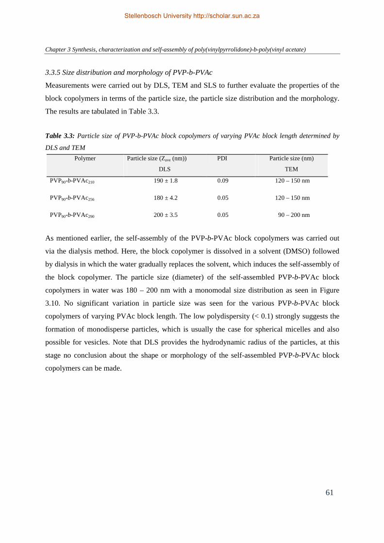
Chapter 3 Synthesis, characterization and self-assembly of poly(vinylpyrrolidone)-b-poly(vinyl acetate)
61
Polymer Particle size (Zave (nm))
DLS
PDI Particle size (nm)
TEM
PVP90-b-PVAc210 190 ± 1.8 0.09 120 – 150 nm
PVP90-b-PVAc256 180 ± 4.2 0.05 120 – 150 nm
PVP90-b-PVAc290 200 ± 3.5 0.05 90 – 200 nm
3.3.5 Size distribution and morphology of PVP-b-PVAc
Measurements were carried out by DLS, TEM and SLS to further evaluate the properties of the
block copolymers in terms of the particle size, the particle size distribution and the morphology.
The results are tabulated in Table 3.3.
Table 3.3: Particle size of PVP-b-PVAc block copolymers of varying PVAc block length determined by
DLS and TEM
As mentioned earlier, the self-assembly of the PVP-b-PVAc block copolymers was carried out
via the dialysis method. Here, the block copolymer is dissolved in a solvent (DMSO) followed
by dialysis in which the water gradually replaces the solvent, which induces the self-assembly of
the block copolymer. The particle size (diameter) of the self-assembled PVP-b-PVAc block
copolymers in water was 180 – 200 nm with a monomodal size distribution as seen in Figure
3.10. No significant variation in particle size was seen for the various PVP-b-PVAc block
copolymers of varying PVAc block length. The low polydispersity (< 0.1) strongly suggests the
formation of monodisperse particles, which is usually the case for spherical micelles and also
possible for vesicles. Note that DLS provides the hydrodynamic radius of the particles, at this
stage no conclusion about the shape or morphology of the self-assembled PVP-b-PVAc block
copolymers can be made.
Stellenbosch University http://scholar.sun.ac.za

Chapter 3 Synthesis, characterization and self-assembly of poly(vinylpyrrolidone)-b-poly(vinyl acetate)
62
It has been reported in literature that the size of the particles is dependent on the polymer
concentration. For example, Zhang and coworkers found that vesicles are preferentially formed
at higher polymer concentrations compared to concentrations at which micelles are formed.34
This is likely attributed to the higher probability of interpolymer aggregation at higher polymer
concentration. The effect of different initial concentration of the PVP-b-PVc block copolymer
(10 mg/mL – 1 mg/mL) on the particle size was investigated. In our case, it was found that the
particle size of the self-assembled PVP-b-PVAc remained between 160 – 200 nm at various
polymer concentrations (data not shown).
In another study, Vangeyte et al.35 investigated the influence of various preparation techniques
and solvents used, on the particle size of poly(ethylene oxide)-b-poly(ε-caprolactone) (PEO-b-
PCL) in water. The three preparation methods studied were the dialysis of the polymers in a
common organic solvent against water, rapid addition of water to the polymer organic solutions,
and rapid addition of the polymer organic solutions into water. Results indicated that direct
dialysis of the copolymer solutions in an organic solvent against water resulted in large and
polydisperse particles (> 1 µm). On the other hand, the two other methods that consist of the
rapid addition of the organic solution to water and vice versa resulted in well-defined,
monodisperse particles (30 – 60 nm). However, the particle size was dependent on the organic
Figure 3.10: DLS size distributions of PVP-b-PVAc of varying PVAc block length
0 100 200 300 400 500 600 700 800
0
5
10
15
20
25
Inte
nsity
(%
)
Zave
(nm)
PVP90
-b-PVAc210
PVP90
-b-PVAc256
PVP90
-b-PVAc290
Stellenbosch University http://scholar.sun.ac.za

Chapter 3 Synthesis, characterization and self-assembly of poly(vinylpyrrolidone)-b-poly(vinyl acetate)
63
solvent used. The research group of Eisenberg also reported on the relationship between the
morphology of poly(styrene)-b-(poly(acrylic acid) (PS-b-PAA) block copolymers and the nature
of the common solvent. It was found that the block copolymers yield only spherical micelle-like
aggregates in DMF, while vesicles were obtained in THF.36
In the present study, the effect of the preparation method and solvent on the self-assembly of the
PVP-b-PVAc block copolymers was not investigated. Self-assembly studies were only carried
out via the dialysis method using only DMSO as organic solvent. Importantly, the self-assembly
of PVP-b-PVAc block copolymers via the dialysis method was reproducible.
The morphology of the PVP-b-PVAc in water (5 mg/mL) was visualized by TEM. The self-
assembled PVP-b-PVAc block copolymers were clearly distinguished as uniform, spherical
structures as shown in Figure 3.11. Interestingly, we observed the characteristic feature for
vesicle structures: a dark corona surrounding a light core. Eisenberg and Dischner6 demonstrated
the influence of the fraction of the hydrophilic block and the hydrophobic block on the molecular
shape of the copolymer and particle morphology. The present observation is in line with their
findings. Since the hydrophobic PVAc block is the dominant segment in the PVP-b-PVAc block
copolymer, the formation of vesicles and bilayers is likely to be expected. The particle sizes
obtained by TEM ranged from 150 – 200 nm with a few slightly smaller particles of 100 nm. The
particle diameter derived from TEM was slightly smaller than that measured and compared to
DLS. This is attributed to the dehydration and shrinkage of the particles during TEM sample
preparation (drying process), as reported in previous studies.37
Figure 3.11: TEM image of PVP90-b-PVAc290 block copolymer. Black corresponds to hydrophobic
region (bilayer) and grey to hydrophilic regions. The insert is a schematic representation of the self-
assembled, vesicular-like structure of the PVP-b-PVAc
500 nm 200 nm
Stellenbosch University http://scholar.sun.ac.za

Chapter 3 Synthesis, characterization and self-assembly of poly(vinylpyrrolidone)-b-poly(vinyl acetate)
64
In addition to DLS and TEM, SLS experiments were performed in water to confirm the structure
of the PVP-b-PVAc block copolymers. From scattered light intensity measurements at various
PVP-b-PVAc concentrations, we could determine the radii of gyration (Rg) and the weight
average molecular weight (Mw) of the block copolymer aggregates. A typical Zimm plot of the
scattering intensity versus the square of the scattering vector (including the extrapolation to zero
concentration and zero angle) is given in Figure 3.12.
The ratio Rg/Rh is a characteristic value that depends on the morphology of the aggregates
formed. From the Rg and Rh values obtained by DLS and SLS, we calculated the Rg/Rh ratio,
which provided information on the structure of the PVP-b-PVAc in water. Table 3.4 summarizes
the results obtained by combining DLS and SLS analysis for PVP90-b-PVAc290 and PVP90-b-
PVAc210.
Figure 3.12: Zimm plot for PVP90-b-PVAc210 extrapolated to zero angle and zero concentration in
water at 25 °C. Squares represent experimental data, measurements at four different
concentrations, from 50° – 120°. Circles represent simulated data
(q²+kc) × µm² × 103
0.0 2.0 4.0 6.0 8.0
Kc/
R ×
g/m
ol
× 10-9
10.0
25.0
40.0
55.0
70.0
Stellenbosch University http://scholar.sun.ac.za

Chapter 3 Synthesis, characterization and self-assembly of poly(vinylpyrrolidone)-b-poly(vinyl acetate)
65
Table 3.4: Physicochemical parameters of PVP-b-PVAc block copolymers obtained by SLS and DLS PVP-b-PVAc Rh
a
(nm) Rg
b
(nm) Rg/Rh Mw(particle)
c
(g.mol-1) ρ
d
(g/cm3) Nagg
e S/Naggf
(nm2) PVP90-b-PVAc290 105 119 (± 3.5) 1.15 1.1 x10⁸ 0.3 1200
52
PVP90-b-PVAc210 96 98.7 (± 5.1) 1.02 6.3 x 10⁷ 0.1 1000 62 aHydrodynamic radius (Zave/2) determined by DLS. bRadius of gyration, extrapolated to zero
concentration. cMw(particle) weight average molecular weight of particles determined by SLS. dρ is the
density of the particles calculated by ρ = Mw(particle)/NAV, V is based on Rh. eAggregation number was
calculated by using the weight average molecular weight determined by SLS and number average
molecular weight determined by SEC. f Surface area per PVP chain
The hydrodynamic radius (Rh) was determined by DLS before the SLS measurements were
started. DLS measurements give a Rh in the range of 95 – 100 nm for both PVP90-b-PVAc290.and
PVP90-b-PVAc210. Theoretical values for the Rg/Rh ratio are 0.78 for hard spheres, 1 for vesicles
with a thin membrane, and values greater than 1.5 are for elongated structures or polymers with
extended conformations (e.g. rod-like micelles).6,38,39 For the reported block copolymers, the
ratios of Rg/Rh from light scattering (Table 3.4) correspond well to the theoretical value for
vesicles (~1). The results indicate that the PVP-b-PVAc block copolymers self-assembled
predominantly into vesicular structures, which is consistent with the TEM images which show
spherical bilayer structures. The density of the self-assembled structures were also calculated and
found to be 0.3 g/cm3 and 0.1 g/cm3 for PVP90-b-PVAc290 and PVP90-b-PVAc210. These values
are reasonable for water-swollen yet solid polymer self-assembled structures. The aggregation
number (Nagg) was calculated to be 1200 and 1000 for PVP90-b-PVAc290 and PVP90-b-PVAc210,
respectively. These values are typical in the range for vesicles. As expected, the aggregation
number increased with increasing hydrophobic PVAc block length and molecular weight.
It is necessary to point out that variables such as the preparation method, the nature and
composition of the solvent mixture, the water content, the relative hydrophilic/hydrophobic
block length and the molecular weight of the block copolymer, all provide control over the
resulting particle size, the aggregation number and morphology of self-assembled block
copolymers.8,40-43 Therefore, depending on the type of system and conditions used, an entire
range of morphologies can be obtained and only subtle changes in these variables can lead to
Stellenbosch University http://scholar.sun.ac.za

Chapter 3 Synthesis, characterization and self-assembly of poly(vinylpyrrolidone)-b-poly(vinyl acetate)
66
dramatic alterations in the physicochemical properties and morphologies of the self-assembled
block copolymers.
Based on the DLS, TEM and SLS results, PVP-b-PVAc block copolymers self-assembled into
spherical, vesicle structures of 180 – 200 nm in diameter. For the application of PVP-b-PVAc
block copolymers as drug carrier, these particle sizes obtained are suitable for passive targeting
via the EPR effect.
3.3.6 Stability of PVP-b-PVAc vesicles under physiological conditions
Upon intravenous administration, polymer drug carriers encounter blood cells and plasma
proteins before reaching the target cells. Serum is a complex fluid that is made up of serum
albumin, lipoproteins immuno- , γ- and macroglobulins. Serum proteins can bind to or penetrate
in the drug carriers which can result in changes in their physicochemical properties such as the
particle size.44-46 The stability of PVP-b-PVAc was therefore investigated in physiological
conditions (PBS, pH 7.4, 37 °C). Furthermore, in vitro studies are usually performed in the
presence of serum (10 – 20 % FCS) (Chapter 5, Section 5.2.3). For this reason, the particle size
stability of the PVP-b-PVAc carrier was also evaluated in PBS containing 20 % serum.
Figure 3.13: Stability of PVP90-b-PVAc290 (left) and PVP-b-PVAc210 (right) at 37 °C in PBS (pH
7.5) and PBS/FCS as determined by DLS
0 2 4 6 8 10 12 14 16 18 20150
160
170
180
190
200
210
220
230
240
250 PBS PBS/FCS
Zav
e (n
m)
Time(h)
0 2 4 6 8 10 12 14 16 18 20140
150
160
170
180
190
200
210
220
230
240
250PBS PBS/FCS
Zav
e (n
m)
Time (h)
Stellenbosch University http://scholar.sun.ac.za

Chapter 3 Synthesis, characterization and self-assembly of poly(vinylpyrrolidone)-b-poly(vinyl acetate)
67
Figure 3.13 shows the particle size stability of the PVP-b-PVAc block copolymers (0.5 mg/mL)
incubated with PBS (pH 7.4, 37 °C) or PBS conatining 20 % serum. Prior to incubation studies
in serum, a control experiment was carried out. As reported previously in the literature, DLS of
biological fluids such as serum result in significant background signal (~ 60 nm) due to their
heterogeneous nature.47 As a control experiment, the background scattering of PBS/FCS (20 %
serum) was measured alone and considered insignificant. PVP90-b-PVAc290 and PVP90-b-
PVAc210 samples incubated in PBS were stable for more than 18 hours. For the PVP90-b-
PVAc290 block copolymer incubated in the prescence of serum, the particle size remained
constant over 16 hours. PVP90-b-PVAc210 also showed stability over 18 hours in serum. The size
stability of the vesicles is likely due to the PVP hydrophilic surface, which protects them from
surface adsorption of serum components. High surface coverage of hydrophilic polymer chains is
a favorable property for a drug carrier. This allows for prolonged blood circulation and
protection of the carrier from protein adsorption.
3.4 Conclusion
PVP-b-PVAc block copolymers of constant hydrophilic PVP block length and varying
hydrophobic block length were synthesized via a CRP technique. The ability of the block
copolymers to self-assemble in aqueous solution was confirmed by 1H NMR, fluorescence
spectroscopy, DLS, SLS and TEM. PVP-b-PVAc self-assembled into spherical vesicle-like
structures ranging from 180 – 200 nm in aqueous solution. The low CMC values of the block
copolymers should further allow for the PVP-b-PVAc carrier to maintain its vesicular
morphology upon dilution. Furthermore, the stability of the PVP-b-PVAc carrier was maintained
under physiological conditions in the presence and absence of serum. With these above
mentioned properties, the PVP-b-PVAc block copolymer should be a versatile carrier for various
hydrophobic and hydrophilic drugs.
Stellenbosch University http://scholar.sun.ac.za

Chapter 3 Synthesis, characterization and self-assembly of poly(vinylpyrrolidone)-b-poly(vinyl acetate)
68
3.5 References
(1) Lipinski, C. Amer. Pharm. Rev. 2002, 5, 82 - 85.
(2) Zhang, H.; Bei, J.; Wang, S. Biomaterials 2009, 30, 100-107.
(3) Miguel, V. S.; Limer, A. J.; Haddleton, D. M.; Catalina, F.; Peinado, C. Eur. Polym. J.
2008, 44, 3853 - 3863.
(4) Riess, G. Prog. Polym. Sci. 2003, 28, 1107-1170.
(5) Wang, X. S.; Guerin, G.; Wang, H.; Wang, Y. S.; Manners, I.; Winnik, M. A. Science
2007, 317, 644 - 647.
(6) Discher, D. E.; Eisenberg, A. Science 2002, 297, 967 - 973.
(7) Yu, S. Y.; Azzam, T.; Rouiller, I. E.; Eisenberg, A. J. Am. Chem. Soc. 2009, 131, 10557
- 10566.
(8) Choucair, A.; Lavigueur, C.; Eisenberg, A. Langmuir 2004, 20, 3894 - 3900.
(9) Sidorov, S. N.; Bronstein, L. M.; Valetsky, P. M.; Hartmann, J.; Cölfen, H.;
Schnablegger, H.; Antonietti, M. J. Colloid Interface Sci. 1999, 212, 197 - 211.
(10) Spatz, J. P.; Sheiko, S.; Möller, M. Macromolecules 1996, 29, 3220 - 3226.
(11) Kataoka, K.; Harada, A.; Nagasaki, Y. Adv. Drug Deliv. Rev. 2001, 47, 113 - 131.
(12) Torchilin, V. P. Pharm. Res. 2007, 24, 1 - 16.
(13) Zhang, L.; Nguyen, T. L. U.; Bernard, J.; Davis, T. P.; Barner-Kowollik, C.; Stenzel,
M. H. Biomacromolecules 2007, 8, 2890 - 2901.
(14) York, A. W.; Kirkland, S. E.; McCormick, C. L. Adv. Drug Deliv. Rev. 2008, 1018 -
1036.
(15) Kim, D. W.; Kim, S. Y.; Kim, H. K.; Kim, S. W.; Shin, S. W.; Kim, J. S.; Park, K.;
Lee, M. Y.; Heo, D. S. Ann. Oncol. 2007, 18, 2009 - 2014.
(16) Sparreboom, A.; .Scripture, C. D.; Trieu, V. Clin. Cancer Res. 2005, 11, 4136 - 4143.
(17) Garrec, D. L.; Gori, S.; Luo, L.; Lessard, D.; Smith, D. C.; Yessine, M.-A.; Ranger,
M.; Leroux, J.-C.; Ranger, M. J. Control. Rel. 2004, 99 83 - 101.
(18) Zhu, Z.; Li, Y.; Li, X.; Li, R.; Jia, Z.; Liu, B.; Guo, W.; Wu, W.; Jiang, X. J. Control.
Rel. 2010, 142, 438 - 446.
(19) Torchilin, V. P.; Shtilman, M. I.; Trubetskoy, V. S.; Whiteman, K.; Milstein, A. M.
Biochimica et Biophysica Acta (BBA) - Biomembranes 1994, 1195, 181 - 184
(20) Shi, L.; Chapman, T. M.; Beckman, E. J. Macromolecules 2003, 36, 2563 - 2567.
Stellenbosch University http://scholar.sun.ac.za

Chapter 3 Synthesis, characterization and self-assembly of poly(vinylpyrrolidone)-b-poly(vinyl acetate)
69
(21) Brandrup, J.; Immergut, E. H.; Grulke, E. A. Polymer Handbook; John Wiley and Sons,
Inc, 1999.
(22) Zimm, B. J. J. Chem. Phys. 1948, 16, 1099 - 1116.
(23) Pound, G.; Aguesse, F.; McLeary, J. B.; Lange, R. F. M.; Klumperman, B.
Macromolecules 2007, 40, 8861 - 8871.
(24) Narrainen, A. P.; Pascual, S.; Haddleton, D. M. J. Polym. Sci. 2002, 40, 439.
(25) Kaewsaiha, P.; Matsumoto, K.; Matsuoka, H. Langmuir 2005, 21, 9938 - 9945.
(26) Patist, A.; Bhagwat, S. S.; Penfield, K. W.; Aikens, P.; Shah, D. O. J. Surfactants
Deterg. 2000, 3, 53 - 58.
(27) Chaibundit, C.; Ricardo, N. M. P. S.; Crothers, M.; Booth, C. Langmuir 2002, 18, 4277
- 4283.
(28) Winnik, F. M.; Regismond, S. T. A. Colloids Surf., A 1996, 118, 1 - 39
(29) Astafieva, I.; Zhong, X. F.; Eisenberg, A. Macromolecules 1993, 26, 7339 - 7352.
(30) Jones, M.; Leroux, J. Eur. J. Pharm. Biopharm. 1999, 48, 101 - 111.
(31) Kalyanasundaram, K.; Thomas, J. K. J. Am. Chem. Soc. 1977, 7, 2039 - 2044.
(32) Astafieva, I.; Zhong, X. F.; Eisenberg, A. Macromolecules 1993, 26, 7339 - 7352.
(33) Opanasopit, P.; Yokoyama, M.; Watanabe, M.; Kawano, K.; Maitani, Y.; Okano, T.
Pharm. Res. 2004, 11, 2001 - 2008.
(34) Zhang, L.; Eisenberg, A. Macromol. Symp. 1997, 113 221-232.
(35) Vangeyte, S.; Gautier, S.; Jérome, R. Colloids Surf. A: Physicochem. Eng. Aspects 2004,
242, 203 - 211.
(36) Yu, Y.; Eisenberg, A. J. Am. Chem. Soc. 1997, 119, 8383 - 8384.
(37) Matter, Y.; Enea, R.; Casse, O.; Lee, C. C.; Baryza, J.; Meier, W. Macromol. Chem.
Phys. 2011, 212, 937 - 949.
(38) Burchard, W. Adv. Polym. Sci 1986, 48, 1 - 124.
(39) Dou, H. J.; Jiang, M.; Peng, H. S.; Chen, D. Y.; Hong, Y. Angew. Chem., Int. Ed. 2003,
42, 1516 -1519.
(40) Stenzel, M. H.; Barner-Kowollik, C.; Davis, P.; Dalton, H. M. Macromol. Biosci. 2004,
4, 445.
(41) Yang, Z.; Yuan, J.; S . Cheng, J. Eur. Polym. J. 2005, 42, 267 - 274.
(42) Garnier, S.; Laschewsky, A. Macromolecules 2005, 38, 7580 - 7592.
Stellenbosch University http://scholar.sun.ac.za

Chapter 3 Synthesis, characterization and self-assembly of poly(vinylpyrrolidone)-b-poly(vinyl acetate)
70
(43) Hussain, H.; Tan, B. H.; Gudipati, C. S.; He, C. B.; Liu, Y.; Davis, T. P. Langmuir
2009, 25, 5557 - 5564.
(44) Diezi, T. A.; Bae, Y.; Kwon, G. S. Mol. Pharm. 2010, 7, 1355 - 1360.
(45) Kim, S.; Shi, Y.; Kim, J. Y.; Park, K.; Cheng, J. Expert Opin. Drug Deliv. 2010, 7, 49 -
62.
(46) Lu, J.; Owen, S. C.; Shoichet, M. S. Macromolecules 2011, 44, 6002 - 6008.
(47) van Gaal, E. V. B.; Spierenburg, G.; Hennink, W. E.; Crommelin, D. J. A.;
Mastrobattista, E. J. Control. Rel. 2010, 141, 328 - 338.
Stellenbosch University http://scholar.sun.ac.za

Chapter 4 Poly(vinylpyrrolidone)-b-poly(vinyl acetate): A potential drug carrier
71
Chapter 4
Poly(vinylpyrrolidone)- b-poly(vinyl acetate): A potential drug carrier
Abstract
Poly((N-vinylpyrrolidone)-b-poly(vinyl acetate)) (PVP-b-PVAc) block copolymers of varying
molecular weight and hydrophobic block lengths were synthesized via controlled radical
polymerization and investigated as carriers for the solubilization of highly hydrophobic
riminophenazine compounds. These compounds have recently been shown to exhibit a strong
activity against a variety of cancer types. PVP-b-PVAc self-assembles into polymer vesicles in
aqueous media, and the dialysis method was used to load the water-insoluble drug (clofazimine)
into these polymer vesicles. The polymer vesicles were characterized by 1H NMR spectroscopy
to confirm vesicle formation and the incorporation of the anti-cancer drugs into the polymer
vesicles. Dynamic light scattering was used to determine the particle size and particle size
distribution of the drug-loaded vesicles as well as the stability of the vesicles under physiological
conditions. The size of the polymer vesicles did not increase upon loading with clofazimine, and
the particle size of 180 – 200 nm and the narrow particle size distribution were maintained. The
morphology of the vesicles was examined by transmission electron microscopy. The drug
loading capacity and the encapsulation efficiency of the polymer vesicles were determined by
UV/Vis spectroscopy. The polymer vesicles had a relatively high drug loading capacity of 20
wt %. The results indicate that the present PVP-b-PVAc block copolymer could be a potential
candidate as a drug carrier for hydrophobic drugs.
Stellenbosch University http://scholar.sun.ac.za

Chapter 4 Poly(vinylpyrrolidone)-b-poly(vinyl acetate): A potential drug carrier
72
4.1 Introduction
To date, various drug delivery and drug targeting systems have been developed or are currently
under development for poorly water-soluble drugs. Many of the existing potent therapeutic
agents such as anti-cancer drugs are restricted to being used intravenously due to their poor water
solubility.1 Currently, conventional solubilizing agents (e.g. Cremophor EL®/ethanol) are used
for the formulation in order to improve the solubility. However, these solubilizing agents have
undesirable toxic side-effects such as hypersensitivity, neurotoxicity and nephrotoxicity.2,3
Research focuses on the development of formulations that avoid the use of Cremophor EL® to
allow an improved control of the toxicity and the pharmacological interactions caused by these
anti-cancer agents. As an alternative approach for the solubilization of poorly water-soluble drug
molecules, the use of amphiphilic block copolymers is currently the focus of interest in
nanopharmaceuticals.4
Polymer micelles used in drug delivery are formed from amphiphilic block copolymers
consisting of a hydrophilic and a hydrophobic biodegradable and/or biocompatible polymer
chain. In aqueous medium, amphiphilic block copolymers self-assemble into distinct structures
comprised of a hydrophilic outer shell and a hydrophobic inner core. The hydrophobicity of the
micellar core provides a host for the incorporation and solubilization of anti-cancer agents, which
are mostly hydrophobic.5 The hydrophilic shell stabilizes the micelle structure and protects it
from biological surroundings. Consequently, both the hydrophilic corona and the hydrophobic
core of the micelle carriers control the physicochemical and biological properties of the drug
delivery system. On the other hand, amphiphilic block copolymers, if made with suitable
amphiphilic proportions, can also self-assemble into vesicular structures. In this case, the
hydrophobic blocks self-assemble into bilayer structure whereas the hydrophilic block faces the
inner and outer aqueous phases. The hydrophobic bilayer membrane can incorporate
hydrophobic drugs while the hydrophilic core can incorporate hydrophilic drugs.
Stellenbosch University http://scholar.sun.ac.za

Chapter 4 Poly(vinylpyrrolidone)-b-poly(vinyl acetate): A potential drug carrier
73
Polymer micelles and vesicles have emerged as versatile drug carriers during the past decades
because of their attractive properties.6-8 As drug vehicles, they present numerous advantages,
such as the reduction of side effects of anti-cancer drugs, minimization of drug degradation and
of loss upon administration, prevention of harmful or undesirable side-effects, and enhancement
of the drug bioavailability. In addition, their nanoscale size (10 – 200 nm in diameter) prevents
them from uptake by the reticulo-endothelial system (RES) and allows for higher accumulation
at the target site through the enhanced permeation and retention (EPR) effect. 9,10 The
biocompatible hydrophilic corona provides these micelles or vesicles with “stealth” properties.
The low critical micelle concentration (CMC) makes them relatively stable, prevents them from
rapid dissociation, and further allows for the drug to be retained over a longer period of time in
the blood stream. This is crucial for the in vitro and in vivo application of polymer drug carriers.
Clofazimine [3–(4–chloroanilino)–10–(4–chlorophenyl)–2,10–dihydro–2 (isopropylimino)
phenazine] is a riminophenazine compound with anti-mycobacterial and anti-inflammatory
activity (Figure 4.1).11 It is one of the most active antimycobacterial agents in this class of
compounds (the riminophenazines). In 1957, Barry et al.12 investigated the antituberculosis
activity of clofazimine. To date, clofazimine has primarily been used in the treatment of
mycobacterial diseases such as leprosy and drug-resistant tuberculosis. It has also been used in
the treatment of Mycobacterium avium infections in AIDS patients.13 Recently clofazimine has
also been reported to inhibit the growth of various cancer cell types.14,15
N
N NH
N
Cl
Cl
NH
N
NH
N
N
R1
2
3
4 5
6
R2
6 2
3 5
Clofazimine Riminophenazine
Figure 4.1: Chemical structure of parent compound clofazimine and its derivatives (riminophenazines) R1 and R2 are chlorine substituted rings
Stellenbosch University http://scholar.sun.ac.za

Chapter 4 Poly(vinylpyrrolidone)-b-poly(vinyl acetate): A potential drug carrier
74
Like many anti-cancer drugs, clofazimine is a highly hydrophobic drug (water-solubility below
0.225 mg/L (virtually insoluble)), which limits its potential therapeutic value. O’Reilly et al.16
investigated systems of sodium cholate and sodium cholate/fatty acid mixtures to improve the
solubility of clofazimine. A 60-fold increase of clofazimine solubility was observed. Attempts of
modifying the chemical structure of clofazimine have been proven to be unsuccessful.17 To
enhance the solubility and bioavailability of clofazimine, the use of cyclodextrins, which are
water-soluble cyclic oligosaccharides composed of glucose units, has been reported by Salem et
al.17 The modified cyclodextrin could be used to solubilise riminophenazines. The solubility of
clofazimine increased by about 4000-fold. Hernandez-Valdepena and coworkers recently,
synthesized nanoaggregates formed by randomly hydrophobised polyacids derived from
poly(methyl vinyl ether-alt-maleic acid) (PMVEMAc), which were able to solubilize
clofazimine in neutral aqueous media.18 The encapsulation of clofazimine into liposomes has
also been reported to solubilise the drug and furthermore improve the overall therapeutic
efficacy. Mehta et al.19 showed that clofazimine can be effectively encapsulated in liposomes
with an efficiency of 95 to 100 %. In vitro and in vivo studies have demonstrated that liposome-
encapsulated clofazimine is much less toxic than free clofazimine. This is advantageous as the
encapsulated drug could be administered at higher doses than free drug due to the water-
insolubility of the drug.
In this study, PVP-b-PVAc block copolymers were evaluated as possible carriers for the
hydrophobic drug, clofazimine. Drug-loaded vesicles were prepared via physical encapsulation
as illustrated in Figure 4.2. The drug-loaded vesicles were characterized for drug loading
efficiency, morphology and stability as potential carriers for hydrophobic drugs.
PVP-b-PVAc block copolymer
Clofazimine-loaded PVP-b-PVAc vesicles
Clofazimine
(anticancer drug)
Figure 4.2 Schematic representation of the encapsulation of clofazimine into PVP-b-PVAc polymer vesicles
Stellenbosch University http://scholar.sun.ac.za

Chapter 4 Poly(vinylpyrrolidone)-b-poly(vinyl acetate): A potential drug carrier
75
4.2. Materials and methods
4.2.1 Chemicals
Dimethyl sulfoxide (Merck, 99 %) was used as received, clofazimine (B663) was obtained from
the University of Pretoria. Dulbecco’s modification of Eagles Medium (DMEM) (sterile) was
purchased from Sigma Aldrich. Distilled deionized water was obtained using a Millipore Milli-Q
purification system and used in all the experiments. Fetal calf serum (FCS) (sterile) and
phosphate buffer saline (PBS) (pH 7.5, sterile) were purchased from Sigma-Aldrich. Block
copolymers of PVP-b-PVAc were synthesized as described in Chapter 3.
4.2.2 Loading of clofazimine into PVP-b-PVAc block copolymers
Clofazimine was incorporated into PVP-b-PVAc block copolymers via the dialysis method. A
typical procedure for the preparation of the drug-loaded vesicles is as follows. PVP-b-PVAc (50
mg), and the hydrophobic drug, clofazimine (10 mg) were dissolved in DMSO (5 mL). The
solution was stirred for 10 minutes at room temperature. The homogeneous solution was
dialyzed against distilled water for 24 hours using a dialysis membrane (MWCO 3500 g/mol).
The water was replaced every 6 hours. The resultant solution was lyophilized to obtain dry drug-
loaded PVP-b-PVAc samples for long-term storage.
4.2.3 1H NMR measurements of block copolymers
1H NMR measurements were performed on PVP-b-PVAc block copolymers. The 1H NMR
spectra were recorded on a Varian 600 MHz Inova spectrometer (Varian Associates Inc. NMR
Instruments). The self-assembly of the block copolymers and drug incorporation were confirmed
using 1H NMR spectra in DMSO-d6 and in D2O.
4.2.4 Size distribution and morphology of clofazimine-loaded PVP-b-PVAc
The size and size distribution of the loaded and unloaded vesicles were determined by DLS using
a Malvern ZetaSizer Nano instrument (ZS90) fixed at an angle of 90 degrees, equipped with a 4
mW He-Ne laser operating at a wavelength of 633 nm. Time correlation functions were analysed
using the CONTIN analysis software provided by Malvern, to obtain the hydrodynamic diameter
of the particles (Zave) and the particle size distribution. TEM of the drug-loaded and unloaded
PVP-b-PVAc samples were carried out in order to observe the morphology of the vesicles. TEM
Stellenbosch University http://scholar.sun.ac.za

Chapter 4 Poly(vinylpyrrolidone)-b-poly(vinyl acetate): A potential drug carrier
76
micrographs were obtained using a LEO 912 microscope operating at an acceleration voltage of
120 kV. A drop of the sample solution was deposited onto a copper grid and dried at room
temperature. The sample solution was negatively stained with uranyl acetate.
4.2.5 Evaluation of the drug loading capacity and encapsulation efficiency of clofazimine-loaded
PVP-b-PVAc
The drug loading capacity and the encapsulation efficiency of clofazimine-loaded PVP-b-PVAc
of different vesicle formulations were determined spectrophotometrically. A Perkin Elmer
Lambda 20 photodiode array spectrophotometer was used to measure the UV-Vis spectra. It
consisted of a holographic monochromator, pre-aligned deuterium and halogen lamps and a
photodiode array detector. UV Winlab (version 2.0) software was used for data acquisition and
processing.
The drug encapsulation efficiency is defined as the ratio of the weight of the drug encapsulated
in the vesicles to the weight of the drug initially used, while the drug loading content is the
weight ratio of the drug encapsulated in the vesicles to that of the weight of the polymer vesicles.
The loading capacity and the encapsulation efficiency are calculated using the following
equations:
Drug loading capacity (DLC) % = mass of drug in vesicles
mass of polymer vesicles x 100%
Drug encapsulation efficiency (EE) % = mass of drug in vesicles
mass of drug initially used x 100%
The obtained vesicle solution was filtered to remove free drug and then lyophilized to obtain dry
clofazimine-loaded PVP-b-PVAc samples. The samples were dissolved in methanol to determine
the drug loading content and encapsulation efficiency of the vesicles. The clofazimine content
was determined by UV/Vis spectrometry. The concentration of the clofazimine was measured
using a standard calibration curve (correlation coefficient 0.998) experimentally obtained from
clofazimine/methanol solutions. Clofazimine has two UV absorbance peaks at 280 nm and 460
nm. The clofazimine content was determined by measuring the absorbance at 460 nm to
minimize the interference of the polymer with an absorbance at ~280 nm. The calibration curve
is linear over the range of 0.5 – 75 µg.mL-1.
Stellenbosch University http://scholar.sun.ac.za

Chapter 4 Poly(vinylpyrrolidone)-b-poly(vinyl acetate): A potential drug carrier
77
4.2.6 Stability studies of clofazimine-loaded PVP-b-PVAc
The particle size stability of drug-loaded vesicles was studied in PBS and in PBS/serum under
physiological conditions (pH 7.4, 37 °C). The clofazimine-loaded PVP-b-PVAc solution
(prepared as described in Section 4.2.2), was diluted in phosphate buffer solution (1:9 (v/v)
vesicle solution/PBS) at pH 7.4 (concentration of PVP-b-PVAc 0.5 mg/mL). For systems
mimicking biological fluids, the stability of the vesicles was assessed in PBS containing 20 %
(v/v) serum. Drug-loaded PVP-b-PVAc as prepared in Section 4.2.2 was diluted in buffer
solutions containing FCS. The particle sizes of the vesicles were measured as a function of time
at 37 °C by DLS.
Clofazimine-loaded vesicles were incubated at physiological conditions (pH 7.4, 37 °C) and the
concentration of the drug was monitored over time. 100 µL of a 20 % (w/w) clofazimine-loaded
vesicle solution was added to 900 µL of PBS buffer at pH 7.4 and incubated at 37 °C. 20 µL
aliquots were withdrawn at specific time intervals and the drug concentration analyzed by
UV/Vis spectroscopy.
4.3. Results and discussion
4.3.1 Preparation and characterization of clofazimine-loaded PVP-b-PVAc block copolymers
Clofazimine is a poorly water-soluble drug (water solubility 0.225 mg/L).20 This is illustrated in
Figure 4.3 (A) which clearly shows the flaky, undissolved drug particles in water. To overcome
the solubility issue, the drug was physically entrapped into PVP-b-PVAc via the dialysis method.
PVP-b-PVAc and clofazimine were dissolved in DMSO and dialyzed against distilled deionized
water (DMSO was chosen as solvent due to the solubility of clofazimine in this solvent). The
gradual replacement of DMSO by water triggered the micellization of the copolymer and the
incorporation of drug in the hydrophobic bilayer membrane and resulted in an orange,
homogeneous dispersion as seen in Figure 4.3 (B). Clofazimine was successfully loaded into
PVP-b-PVAc block copolymers and no drug precipitation was observed during the encapsulation
procedure. Drug-loaded PVP-b-PVAc could either be stored in solution or in freeze-dried form.
Stellenbosch University http://scholar.sun.ac.za

Chapter 4 Poly(vinylpyrrolidone)-b-poly(vinyl acetate): A potential drug carrier
78
The encapsulation of clofazimine into the hydrophobic PVAc bilayer membrane was confirmed
by 1H NMR. Figure 4.4 shows the 1H NMR spectra of clofazimine-loaded PVP90-b-PVAc210
block copolymers in different environments. Figure 4.4 (a) represents clofazimine-loaded PVP90-
b-PVAc210 block copolymer aggregates in DMSO-d6. Figure 4.4 (b) represents the 1H NMR
spectra of clofazimine-loaded PVP90-b-PVAc210- in deuterium oxide (D2O). For clofazimine-
loaded PVP90-b-PVAc210 in DMSO-d6 (Figure 4.4 (a)) vesicle formation is not expected and all
peaks of the hydrophilic (PVP) and hydrophobic (PVAc) segments were detected. Proton peaks
of clofazimine were also detected (6 – 7.8 ppm as indicated with a circle on the 1H NMR
spectrum). The formation of clofazimine-loaded PVP90-b-PVAc210 vesicles was confirmed by 1H
NMR spectroscopy in D2O (Figure 4.4 (b). The predominant peak is that of the PVP (3 – 4 ppm)
- the corona of the vesicle-like structure - while the peaks of PVAc are suppressed. This is due to
the limited mobility of the PVAc-chains in the hydrophobic bilayer membrane of the vesicle.
The intensity of the PVAc peak is reduced compared to that in Figure 4.4 (a) where the vesicles
are not expected to be present. No peaks of clofazimine are present. This is due to the drug being
incorporated within the vesicle bilayer membrane which restricts the mobility of the drug
molecules resulting in suppressed proton resonances. 1H NMR indicated that amphiphilic block
copolymer self-assembled into vesicular structures in water, comprised of a hydrophobic
membrane (PVAc) and a hydrophilic outer shell (PVP) and that clofazimine is contained in the
hydrophobic bilayer membrane of the amphiphilic block copolymer.
A B
Figure 4.3: A) Clofazimine insoluble in aqueous media B) Clofazimine physically encapsulated in
PVP-b-PVAc in aqueous media
Stellenbosch University http://scholar.sun.ac.za

Chapter 4 Poly(vinylpyrrolidone)-b-poly(vinyl acetate): A potential drug carrier
79
4.3.2 Size distribution and morphology of clofazimine-loaded PVP-b-PVAc block copolymers
The particle size and the size distribution of drug-loaded vesicles were measured by DLS. The
average particle size of the drug-loaded PVP-b-PVAc was 180 – 210 nm for the samples in water
(Table 4.1).
Reasonably narrow size distributions and monomodal pattern (PDI < 0.1) for the different block
copolymers were obtained as presented in Figure 4.5. After loading of clofazimine, the particle
size did not increase considerably for the different block copolymers of varying PVAc block
length. Furthermore, the size distribution of the drug-loaded PVP-b-PVAc block copolymers was
a
b
Table 4.1: Particle sizes of unloaded and drug-loaded PVP-b-PVAc (20 % (w/w)
clofazimine/PVP-b-PVAc) measured by DLS analysis
Polymer sample
Unloaded PVP-b-PVAc Zave (nm)
PDI Loaded PVP-b-PVAc Zave (nm)
PDI
PVP90-b-PVAc210 180 ± 3.1 0.09 190 ± 2.2 0.09 PVP90-b-PVAc256 PVP90-b-PVAc290
205 ± 2.1 200 ± 2.5
0.06 0.09
200 ± 3.2 210 ± 2.5
0.09 0.09
Figure 4.4: 1H NMR spectra of a) clofazimine-loaded PVP90-b-PVAc210 in DMSO-d6 b) clofazimine-
loaded PVP90-b-PVAc210 in D2O
8 7 6 5 4 3 2 1 0ppm
a
b
Stellenbosch University http://scholar.sun.ac.za

Chapter 4 Poly(vinylpyrrolidone)-b-poly(vinyl acetate): A potential drug carrier
80
approximately the same as that before drug loading. Since the optimal size of a drug carrier for
efficient delivery of an antitumor agent to a tumor is around 100 – 200 nm, the particle sizes for
the drug-loaded PVP-b-PVAc obtained are acceptable and meet the requirements for passive
targeting via the EPR effect.9
In the literature, the possibility that block copolymer vesicles can transform into micelles with
increasing amount of solubilized hydrophobic molecules has been theoretically predicted.21 In a
study conducted by Li et al.22, the effect of the amount of hydrophobic drug loaded into the
vesicles on the particle size was investigated. It was observed that with increasing amount of
hydrophobic drug the particle size of the vesicles decreased. They hypothesised that a
transformation from vesicles to micelles had occurred. It is likely that increasing amounts of
drug results in unfavourable stretching of the hydrophobic chains which eventually results into
micellization of the vesicles.
In order to determine whether the drug content has an effect on the particle size of the PVP-b-
PVAc carrier, different drug feed ratios were investigated. Figure 4.6 shows the relationship
between different drug feed ratios on the particle size of the PVP-b-PVAc carrier. All samples
were prepared using the same procedure (Section 4.2.2). It was observed that the PVP-b-PVAc
carrier sizes for the different drug formulations (5 – 20 (w/w) % drug/polymer) were in the range
Figure 4.5: Size distribution of clofazimine-loaded PVP-b-PVAc of varying PVAc block length
0 200 400 600 800 1000
0
5
10
15
20
25
30
Inte
nsity
(%
)
Zave
(nm)
PVP90-b -PVAc290 PVP90-b -PVAc210 PVP90-b -PVAc256
Stellenbosch University http://scholar.sun.ac.za

Chapter 4 Poly(vinylpyrrolidone)-b-poly(vinyl acetate): A potential drug carrier
81
of 190 – 210 nm. No significant change was observed. Therefore the increase in the drug content
had no major effect on the particle size of the carrier. In the case of 30 %, drug precipitation was
seen in the dialysis tubing and therefore samples were not analyzed.
The morphology of the drug-loaded PVP-b-PVAc vesicles was visualized by TEM. As depicted
in Figure 4.7, the average diameters of the drug-loaded PVP-b-PVAc vesicles were 150 – 200
nm. The characteristic dark outer layer (representing the membrane bilayer or vesicle wall) and
lighter inner core (representing the hydrated core) are clearly evident. TEM results are in fairly
good agreement with those determined by DLS. The presence of slightly smaller particles is as a
result of shrinkage of the particles which is consistent with similar reports studying polymer
vesicles via TEM in the dried state.23
Figure 4.6: The effect of different drug feed ratios (% (w/w) clofazimine/polymer) on the particle
size of clofazimine-loaded PVP-b-PVAc of varying PVAc block length. Error bars represent the
standard deviation (n = 3)
0
50
100
150
200
250
5 %
10 %
15 %
20 %
PVP90-b-PVAc210PVP90-b-PVAc256 PVP90-b-PVAc290
Par
ticle
siz
e (Z
ave
(nm
))
Stellenbosch University http://scholar.sun.ac.za

Chapter 4 Poly(vinylpyrrolidone)-b-poly(vinyl acetate): A potential drug carrier
82
4.3.3 Drug loading capacity and encapsulation efficiency
For any drug formulation, the drug loading capacity and efficiency are important parameters. As
outlined in Chapter 2, the loading capacity and the encapsulation efficiency of a polymer drug
carrier is influenced by several factors. The nature of the hydrophobic core (of micelles) or
bilayer membrane (of vesicles), the block length of the hydrophobic core or bilayer membrane,
the size or thickness of the core or the bilayer membrane and the degree of hydrophobicity of the
drug are all important factors to be considered.24,25
In order to find an optimal drug to polymer ratio for the PVP-b-PVAc drug carrier, a series of
clofazimine-loaded PVP-b-PVAc with different weight ratios of clofazimine to polymer (5 %, 10
%, 15 %, 20 % and 30 %) were prepared. The effect of the hydrophobic block length and the
drug/polymer weight ratio on the drug loading capacity and encapsulation efficiency were
investigated.
Figure 4.8 represents the drug loading capacity and the encapsulation efficiency of clofazimine-
loaded PVP-b-PVAc of different drug feed ratios (using a constant amount of polymer).
Figure 4.7: TEM image of clofazimine-loaded PVP90-b-PVAc290 showing the vesicular structure
200 nm
Stellenbosch University http://scholar.sun.ac.za

Chapter 4 Poly(vinylpyrrolidone)-b-poly(vinyl acetate): A potential drug carrier
83
Clofazimine-loaded PVP-b-PVAc of longer hydrophobic PVAc block length (PVP90-b-PVAc290
and PVP90-b-PVAc256) resulted in a higher drug loading capacity and encapsulation efficiency
(having the same amount of drug) compared to PVP90-b-PVAc210 of shorter PVAc block length.
The results follow the general trend: the loading capacity and the encapsulation efficiency of the
polymer carrier increase with increasing hydrophobic chain length. The loading capacity of
polymer vesicles is dependent on the molecular weight of the hydrophobic block, since
copolymers with larger hydrophobic molecular weight can form thicker hydrophobic bilayer
membranes. The thicker membrane can therefore accommodate more hydrophobic drug.22 It is
also possible that the longer the hydrophobic PVAc block, the stronger the hydrophobic
interaction between the drug and the hydrophobic bilayer membrane resulting in higher drug
loading capacities and higher encapsulation efficiencies.
The effect of the feed weight ratio on the drug loading capacity and encapsulation efficiency was
also studied. The drug loading capacity and encapsulation efficiency increased with increasing
initial drug feed (constant amount of polymer). As the feed amount of clofazimine increased
from 5 – 20 %, the drug loading capacity increased from 2.7 wt % to 20 wt %, for PVP90-b-
PVAc290 and 1.4 wt % to 20 wt % for PVP90-b-PVAc256. In the case of the more hydrophilic
5 % 10% 15% 20% 30%0
5
10
15
20
25
30
35
40
45 PVP90-b-PVAc210 PVP90-b-PVAc256 PVP90-b-PVAc290
Load
ing
capa
city
(%
)
Feed ratio (clofazimine/polymer) 5 % 10% 15% 20% 30%
0
10
20
30
40
50
60
70
80
90 PVP90-b-PVAc210 PVP90-b-PVAc256 PVP90-b-PVAc290
Enc
apsu
latio
n ef
ficie
ncy
(%)
Feed ratio (clofazimine/polymer)
Figure 4.8: Drug loading capacity and encapsulation efficiency of clofazimine-loaded PVP-b-PVAc
of different hydrophobic PVAc block length. Feed ratios were 5, 10, 20, or 30 weight percentage (%
w/w) of clofazimine relative to PVP-b-PVAc. Error bars represent the standard deviation (n = 3)
Stellenbosch University http://scholar.sun.ac.za

Chapter 4 Poly(vinylpyrrolidone)-b-poly(vinyl acetate): A potential drug carrier
84
block copolymer, clofazimine-loaded PVP90-b-PVAc210, the drug loading capacity increased
from 5 wt % to 42 wt %. The encapsulation efficiency also increased with increasing drug feed
amount. The encapsulation efficiency increased from 20 wt % to 60 wt % for clofazimine-loaded
PVP90-b-PVAc290, from 13 wt % to 40 wt % for clofazimine-loaded PVP90-b-PVAc210, and 14 wt
% to 60 wt % for clofazimine-loaded PVP90-b-PVAc256. A decrease in the encapsulation
efficiencies for clofazimine-PVP90-b-PVAc290 and PVP90-PVAc256 was seen when a 30 % (w/w)
drug/polymer ratio was used. This corresponded to drug precipitation that was observed in the
dialysis tubing during the loading process, which indicated that the vesicles had reached their
maximum loading capacity. Therefore the maximum drug/polymer feed ratio for clofazimine is
20 %, above this value clofazimine overloading of the system occurs. Exceeding the
solubilization capacity of the vesicles, results in release in the aqueous phase, followed by
crystallization or precipitation of the drug as reported previously in the literature.22,26
Interestingly, for the more hydrophilic block copolymer, the drug loading capacity reached a
maximum loading capacity of 40 wt % with an encapsulation efficiency of 70 wt % when a 30 %
(w/w) drug/polymer ratio was used. No drug precipitation was observed during dialysis,
however, the sample was unable to maintain its stability upon storage. The colloidal stability of
the drug-loaded carrier often becomes lower as the drug loading increases due to the enhanced
hydrophobicity of the carrier after loading with hydrophobic drug.27
The drug loading capacity and encapsulation efficiency is also dependent on the miscibility
between the polymers and the hydrophobic drugs.28 In a study conducted by Letchfold et al.28,
the solubilization of five model hydrophobic drugs by methoxy poly(ethylene glycol)-b-
poly(caprolactone) diblock copolymers (MePEG-b-PCL) was investigated. Indomethacin,
showed the best solubility in the MePEG-b-PCL micelles, having a higher loading capacity and
encapsulation efficiency than the poorly solubilized etoposide. Additionally, a higher PCL block
length also resulted in better loading capacity.
For comparative purposes, paclitaxel (PTX), a commonly used anti-cancer drug (water solubility
5.5 mg/L) was selected to evaluate the loading capacity and encapsulation efficiency of the
polymer vesicles. PVP-b-PVAc copolymers were loaded with PTX using the same procedure as
for clofazimine-loaded samples (Section 4.2.2).
Stellenbosch University http://scholar.sun.ac.za

Chapter 4 Poly(vinylpyrrolidone)-b-poly(vinyl acetate): A potential drug carrier
85
The loading capacity and encapsulation efficiency obtained for the PTX-loaded PVP90-b-
PVAc290 and PTX-loaded PVP90-b-PVAc210 is presented in Figure 4.9. Similarly to clofazimine-
loaded vesicles, the longer hydrophobic PVAc block provided a higher PTX loading capacity
and encapsulation efficiency as expected. PVP90-b-PVAc290 (10 % (w/w) PTX/polymer) showed
the highest drug loading levels of 15 wt % and encapsulation efficiency of 62 wt %. A further
increase in the initial drug feed to a 20 % (w/w) PTX/polymer did not result in a greater drug
loading level or encapsulation efficiency. Rather a decrease in encapsulation efficiency was seen
as a result of PTX precipitation that was found inside the dialysis membrane tube. At a feed ratio
of 20 % (w/w) PTX/polymer, no precipitation was observed for PVP90-b-PVAc210, however, the
sample was unstable upon storage.
The results clearly demonstrate that the loading capacity and encapsulation efficiency of the
PVP90-b-PVAcn vesicles is dependent on the hydrophobicity (PVAc block length) and the drug
feed ratio. In addition, differences in the hydrophobicity of the drugs and/or compatibility with
the PVAc domain are also important factors which could explain the differences in the loading
capacity and encapsulation efficiency for clofazimine and PTX. However, to prove the above
assumption solubility studies, polymer-drug compatibility parameters and partition coefficients
will need to be determined, which at this point is beyond the scope of the study.
Figure 4.9: Drug loading capacity and encapsulation efficiency of paclitaxel-loaded PVP-b-PVAc of
different hydrophobic PVAc block length. Feed ratios were 5, 10, or 20, weight percentage (% w/w) of
PTX relative to PVP-b-PVAc. Error bars present the standard deviation (n = 3)
5 % 10 % 20 %0
10
20
30
40
50
60
70
80
90
100
PVP90-b-PVAc210 PVP90-b-PVAc290
Feed ratio (PTX/polymer)
Enc
apsu
latio
n ef
ficie
ncy
(%)
5 % 10 % 20 %0
5
10
15
20
25
30
35
40
45
PVP90-b-PVAc210 PVP90-b-PVAc290
Load
ing
capa
city
(%
)
Feed ratio (PTX/polymer)
Stellenbosch University http://scholar.sun.ac.za

Chapter 4 Poly(vinylpyrrolidone)-b-poly(vinyl acetate): A potential drug carrier
86
Other factors such as the structural properties of the drugs can also have an impact on the loading
capacity and efficiency. For drugs with bulky groups and high molecular weight, it is more
difficult for high concentrations to become encapsulated in the core or the bilayer membrane due
to steric hindrance which prevents the close stacking of the drug molecules, resulting in lower
encapsulation efficiencies.
Our final optimized formulation of clofazimine and PTX-loaded PVP-b-PVAc had a
composition of 20 % (w/w) clofazimine/PVP-b-PVAc and 10 % PTX/PVP-b-PVAc. Due to the
longer hydrophobic block length, PVP90-b-PVAc290 showed the highest loading capacity and
encapsulation efficiency for both drugs. Furthermore, this polymer gave the lowest CMC (0.0012
mg/mL) which is important for its application. PVP90-b-PVAc290 showed to be the best candidate
for drug loading and was, therefore, further used for in vitro cytotoxicity studies (Chapter 5).
4.3.4 Stability studies of clofazimine-loaded PVP-b-PVAc block copolymers
The particle size stability of the polymer vesicles in biological fluids is important for in vitro and
in vivo applications. The particle size stability of 20 % (w/w) clofazimine-loaded PVP90-b-
PVAc290 vesicles (polymer concentration 0.5 mg/mL) in physiological conditions (PBS, pH 7.4,
37 ºC) was investigated using DLS. The particle sizes of clofazimine-loaded PVP90-b-PVAc290
vesicles in PBS/FCS were also investigated.
Figure 4.10 (a) shows that the average particle size of the PVP90-b-PVAc290 vesicles in PBS were
210 – 220 nm, and stable up to 16 hours. No precipitation was observed. The same sample was
incubated in PBS, pH 7.4 at 37 ºC. At designated time intervals, the concentration of the drug
was monitored by UV/Vis spectroscopy. The concentration of the drug in the drug-loaded PVP-
b-PVAc sample remained constant over 24 hours. After 30 hours the concentration decreased by
20 % due to leakage of drug from the vesicles (data not shown). For free drug incubated in PBS
(50/50 (v/v) ethanol/PBS, pH 7.4), the formation of fluffy-like precipitate was visible after 24
hours. This is not unexpected due to the low solubility of the drug in aqueous media. Similar
results have also been observed for other hydrophobic cancer drugs.29
Stellenbosch University http://scholar.sun.ac.za

Chapter 4 Poly(vinylpyrrolidone)-b-poly(vinyl acetate): A potential drug carrier
87
For clofazimine-loaded PVP90-b-PVAc290 in PBS/FCS, the average particle sizes were similar to
the vesicles in PBS. In addition, the vesicle sizes remained stable for 15 hours in PBS/FCS. No
drug precipitation was observed during the incubation time. Clofazimine-loaded PVP90-b-
PVAc210 also showed stability in PBS and PBS/FCS. Particle sizes of 180 – 190 nm (in PBS) and
170 nm (in PBS/FCS) were maintained over 16 hour incubation. No drug precipitation was
observed. This indicates that the hydrophilic PVP segment (the corona of the vesicles) was able
to stabilize and protect the vesicles from non-specific surface adsorption of serum components.
Figure 4.10: Particle size (Zave) of clofazimine-loaded (a) PVP90-b-PVAc290 (b) PVP90-b-PVAc210 in
(▲) PBS pH 7.4 and (●) PBS/FCS pH 7.4, as a function of time at 37 ºC
0 2 4 6 8 10 12 14
160
180
200
220
240
260
280
Par
ticle
siz
e (Z
ave(
nm))
Time (h)0 2 4 6 8 10 12 14 16
100
120
140
160
180
200
220
240
Par
ticle
siz
e (Z
ave(
nm))
Time (h)
(a) (b)
Stellenbosch University http://scholar.sun.ac.za

Chapter 4 Poly(vinylpyrrolidone)-b-poly(vinyl acetate): A potential drug carrier
88
4.4 Conclusion
PVP-b-PVAc block copolymers of constant hydrophilic block length and varying hydrophobic
block length were loaded with clofazimine, via the dialysis method. The polymer could
effectively solubilize the hydrophobic drug. 1H NMR spectroscopy confirmed the self-assembly
of the PVP-b-PVAc block copolymers and the localization of the drug within the hydrophobic
PVAc bilayer. The obtained drug-loaded vesicles had diameters from of 180 – 210 nm with
narrow size distributions. The optimal clofazimine-loaded PVP-b-PVAc vesicle formulation was
prepared using a 20 % drug/polymer feed ratio, having of a particle size of 200 nm which
remained stable in physiological conditions. Overall, PVP-b-PVAc vesicles could be a potential
candidate as a drug carrier for hydrophobic drugs.
Stellenbosch University http://scholar.sun.ac.za

Chapter 4 Poly(vinylpyrrolidone)-b-poly(vinyl acetate): A potential drug carrier
89
4.5 References
(1) Lipinski, C. A. J. Pharmacol. Toxicol. Methods 2000, 44, 235 - 249.
(2) Gelderblom, H.; Verweij, J.; Nooter, K.; Sparreboom, A. Eur. J. Cancer 2001, 37, 1590
- 1598.
(3) Singla, A. K.; Garg, A.; Aggarwal, D. Int. J. Pharm. 2002, 235, 179 - 192.
(4) Mikhail, A. S.; Allen, C. J. Control. Rel. 2009, 138, 214 - 223.
(5) Lasic, D. D. Nature 1992, 355, 279 - 280.
(6) Torchilin, V. P. J. Control. Rel. 2001, 73, 137 - 172.
(7) Kim, S.; Shi, Y.; Kim, J. Y.; Park, K.; Cheng, J. Expert Opin. Drug Deliv. 2010, 7, 49 -
62.
(8) Tyrrell, Z. L.; Shena, Y.; Radosza, M. Prog. Polym. Sci. 2010, 35, 1128-1143.
(9) Maeda, H.; Wua, J.; Sawa, T.; Matsumura, Y.; Horic, K. J. Control. Rel. 2000, 65, 271
- 284.
(10) Kwon, G. S.; Okano, T. Adv. Drug Deliv. Rev. 1996, 21, 107 - 116.
(11) Reddy, V. M.; O'Suvillan, J. F.; Gangadhram, P. R. J. J. Antimicrob. Chemother. 1999,
43, 615 - 623.
(12) Barry, V. C.; Belton, J. G.; Conalty, M. L.; Denneny, J. M.; Edward, D. W.;
O'Sullivan, J. F.; Twomey, D.; Winder, F. Nature 1957, 179, 1013 - 1015.
(13) Fournier, S.; Burguière, A. M.; Flahault, A.; Vincent, V.; Treilhou, M. P.; Eliaszewicz,
M. Eur. J. Clin. Microbiol. Infect. Dis. 1999, 18, 16 - 22.
(14) Pourgholami, M. H.; Lu, Y.; Wang, L.; Stephens, R. W.; .Morris, D. L. Cancer Lett.
2004, 207, 37 - 47.
(15) Van Rensburg, C. E. J.; Van Staden, A. M.; Anderson, R. Cancer Res. 1993, 53, 318 -
323.
(16) O’Reilly, J. R.; Corrigan, O. L.; .O’Driscoll, C. M. Int. J. Pharm. 1994, 109, 147 - 154.
(17) Salem, I. I.; Steffan, G.; Düzgünes, N. Int. J. Pharm. 2003, 260, 105 - 114.
(18) Hernandez-Valdepena, I.; Domurado, M.; Coudane, J.; Braud, C.; Baussard, J.;
Domurado, M. V. D. Eur. J. Pharm. Sci. 2009, 39, 345 - 351.
(19) Mehta, R. T.; Keyhani, A.; McQueen, T. J.; Rosenbaum, B.; Rolston, K. V.; Tarrand,
J. J. Antimicrob. Agents Chemother. 1993, 37, 2584 - 2587.
Stellenbosch University http://scholar.sun.ac.za

Chapter 4 Poly(vinylpyrrolidone)-b-poly(vinyl acetate): A potential drug carrier
90
(20) Feng, P. C.; Fenselau, C. C.; Jacobson, R. R. Drug Metab. Dispos. 1982, 10, 286 - 288.
(21) Nagarajan, R. Curr. Opin. Colloid In 1997, 2, 282 - 293.
(22) Li, S.; Byrne, B.; Welsh, J.; Palmer, A. F. Biotechnol. Prog. 2007, 23, 278 - 285.
(23) Fernyhough, C.; Ryan, A. J.; Battaglia, G. Soft Matter 2009, 5, 1674 - 1682.
(24) Lin, W. J.; Juang, L. W.; Lin, C. C. Pharm. Res. 2003, 668 - 673
(25) Carstens, M. G.; de Jong, P. H. J. L. F.; van Nostrum, F. C.; Kemmink, J.; Verrijk, R.;
de Leede, L. G. J.; Crommelin, D. J. A.; Hennink, W. E. Eur. J. Pharm. Biopharm. 2008,
68, 596 - 606.
(26) Torchilin, V. P. Pharm. Res. 2007, 24, 1 - 16.
(27) Burt, H. M.; Zhang, X.; Toleikis, P.; Embree, L.; Hunter, W. L. Colloids Surf., B 1999,
16, 161 -171.
(28) Letchford, K.; Liggins, R.; Burt, H. J. Pharm. Sci. 2008, 97, 1179 - 1189.
(29) Soga, O.; van Nostrum, C. F.; Fens, M.; Rijcken, C. J. F.; Schiffelers, R. M.; Storm,
G.; Hennink, W. E. J. Control. Rel. 2005, 103, 341 - 353.
Stellenbosch University http://scholar.sun.ac.za

Chapter 5 In vitro cytotoxicity and cellular uptake of PVP-b-PVAc
91
Chapter 5
In vitro cytotoxicity and cellular uptake of PVP-b-PVAc
Abstract
In vitro cytotoxicity studies of poly((N-vinylpyrrolidone)-b-poly(vinyl acetate)) (PVP-b-PVAc)
and drug-loaded PVP-b-PVAc were investigated against MDA-MB-231 multi-drug-resistant
breast epithelial cancer cells and MCF12A normal breast epithelial cells. In vitro experiments
demonstrated that the PVP-b-PVAc drug carrier showed no cytotoxicity, confirming the
biocompatibility of the PVP-b-PVAc drug carrier. In vitro cellular uptake of fluorescently
labeled PVP-b-PVAc was studied using fluorescence microscopy. The PVP-b-PVAc carrier was
taken up by the cancer cells after 6 hours and localized in both the cytoplasm and in the
perinuclear region. From these results, we could conclude that the present PVP-b-PVAc block
copolymer could potentially be useful as a drug carrier for hydrophobic drugs.
Stellenbosch University http://scholar.sun.ac.za

Chapter 5 In vitro cytotoxicity and cellular uptake of PVP-b-PVAc
92
5.1 Introduction
One of the essential requirements of a drug carrier or a drug delivery system is that they have no
inherent cytotoxicity.1 Prior to in vivo work, drug delivery systems are subjected to in vitro
studies in order to determine the biocompatibility of the polymer drug carrier and the efficacy of
the drug carrier. In vitro cytotoxicity assays and various screening assays are widely used in drug
development to study the effect of novel compounds/drugs, drug carriers or drug delivery
systems on healthy cells or cancer cells.2 Thus, in vitro investigation can provide preliminary
evidence to show the advantages of a drug carrier system in drug delivery application. However,
it must be mentioned that although in vitro studies are beneficial, they are not a true
representation of in vivo conditions.
A prerequisite for an effective polymer drug carrier, is the need for the drug to be delivered
intracellularly to exert its therapeutic action inside the cytoplasm or into the nucleus or other
specific organelles. In vitro cellular uptake and internalisation studies, therefore, provide
essential information regarding the intracellular pathway and cellular distribution of the drug
delivery carrier.
In recent years, fluorescence microscopy has become an important tool for studying the in vitro
cellular internalization and localization of polymer drug carriers.3,4 In order to confirm that the
drug carrier has successfully delivered the drug to the target site, it is necessary to track a
polymer drug carrier on its cellular journey. The attachment of a fluorescent label to the polymer
backbone is a convenient way to trace the polymer.5 Commonly used fluorescent labels include
rhodamines,6 and fluorescein dyes - sulfhydryl-reactive or amine-reactive fluorescein dyes e.g.
fluorescein isothiocyanate (FITC)7-9, which can be conjugated to polymers having appropriate
reactive functional groups For example, Eisenberg and coworkers showed internalization of
micelles through bioconjugation of a rhodamine fluorescent probe onto poly(ethylene glycol)-b-
poly(caprolactone) (PEG-b-PCL) micelles.10 In a subsequent study, Savic et al.11 used a triple-
labeling approach where the nucleus, the PCL-b-PEO micelles, and the plasma membrane were
individually labeled and selectively visualized. Fluorescence results indicated that the micelles
were predominantly localized in the cytoplasm and distributed within several cytoplasmic
organelles, but not in the nucleus.
Stellenbosch University http://scholar.sun.ac.za

Chapter 5 In vitro cytotoxicity and cellular uptake of PVP-b-PVAc
93
In some cases, the drug molecules themselves consist of aromatic ring systems exhibiting
inherent fluorescence (e.g. doxorubicin).12 Therefore, if fluorescently labeled micelles are
combined with a drug (which exhibits fluorescence) then the fate of both the micelles and the
drug can be detected independently. However, in order to distinguish between the drug delivery
carrier and the drug, it is imperative that they emit fluorescence at different wavelengths (i.e.
different colours).
The versatility of the RAFT technique has previously been addressed in Chapter 2. Not only does
it allow for the synthesis of well-defined end-functional polymers, but it also allows for
postmodification of otherwise inert polymers.13,14 Thiol-ene chemistry is the most widely used
and easiest accessible route to post-polymerization polymer modification on polymers
synthesized via the RAFT technique.15,16 Thiol groups lend themselves to thiol-ene processes for
polymer bioconjugation resulting in polymers that can be used for pharmaceutics, drug delivery
and biomedical imaging applications.17,18
Several procedures for the removal of the RAFT thiocarbonyl thio end-group, yielding thiol end-
group polymers have been thoroughly documented by Moad et al.19 There are several types of
thiol-reactive dyes reported in the literature, including iodoacetamides, disulfides, maleimides,
vinyl sulfones and various electron-deficient aryl halides and sulfonates. Iodoacetamides and
maleimides are by far the most popular thiol-reactive moeities. Thiol-based polymers are
therefore the preferred targets for these specific thiol-reactive fluorescent labels. The use of thiol-
maleimide coupling reactions has been documented in the literature.20-22 Li et al.23 described the
first example of sequential thiol-ene reactions, involving thiol-maleimide coupling. Poly(N-
isopropylacrylamide) (PNIPAm) prepared by a trithiocarbonate RAFT agent, when reacted with
2-ethanolamine in the presence of tributylphosphine in 1,4-dioxane, yielded the thiol-terminated
homopolymer, PNIPAm-SH. This further reacted with bismaleimidodiethyleneglycol to yield
maleimide end-functional PNIPAm. Similarly, Scales et al.24 synthesized fluorescently labeled
PNIPAM polymers. End groups were cleaved with sodium borohydride (NaBH4). The thiol-
terminated polymer reacted with N-(1-pyrene) maleimide in the presence of a catalytic amount of
ethylenediamine in DMF, which resulted in fluorescent ω-chain-end modified PNIPAm.
Stellenbosch University http://scholar.sun.ac.za

Chapter 5 In vitro cytotoxicity and cellular uptake of PVP-b-PVAc
94
With the advancement in polymer chemistry, the development of fluorescent polymers has also
found use in various applications in medicine, including in vitro and in vivo trafficking in drug
delivery. This is an alternative approach to track the cellular pathway and distribution of the
polymer drug carrier in vitro. The attachment of fluorescent labels to polymers is often restricted
due to the limited availability of fluorescent labels with specific functional groups for
conjugation to the polymer. In addition, the attachment of the fluorescent dyes to the polymers
can alter the physicochemical properties of the drug carrier, which could further affect the route
of internalisation and the intracellular distribution.11 To circumvent this, fluorescent amphiphilic
polymers have particularly gained interest. 14,25 For example, Perrier and coworkers25
synthesized a fluorescent, thermosensitive, amphiphilic poly(N-isopropyl-acrylamide-co-N-
vinylcarbazole) conjugated to pH responsive poly(2-(N,N-dimethylamino))ethyl acrylate,
(PNIPPAm-co-PNVC)-b-PDMAEA. These polymers could be useful to gain insight and
understand the in vivo fate of the drug delivery vehicle. In addition to fluorescent polymers, the
development of fluorogenic polymers (polymers bearing a fluorogenic label, whose fluorescence
is revealed only upon cellular uptake) for cellular internalization and trafficking studies have also
been investigated.26
In the present study, we evaluated the toxicity of the PVP-b-PVAc drug carrier. Furthermore, we
investigated the cytotoxicity of drug-loaded PVP-b-PVAc to evaluate its potential as a drug
delivery vehicle. The attachment of a fluorescent label to the PVP-b-PVAc carrier allowed for in
vitro cellular uptake studies using fluorescence microscopy. Clofazimine has no inherent
fluorescence, therefore, we considered perylene red (Exalite 613), a hydrophobic fluorescent
probe to mimic the hydrophobic drug, clofazimine. Perylene red was physically entrapped into
the fluorescently labeled PVP-b-PVAc using the same method as for the hydrophobic drug,
clofazimine. The cellular uptake of the resulting fluorescently labeled perylene red-loaded PVP-
b-PVAc vesicles was explored in vitro and compared with free perylene red.
Stellenbosch University http://scholar.sun.ac.za

Chapter 5 In vitro cytotoxicity and cellular uptake of PVP-b-PVAc
95
5.2 Materials and Methods
5.2.1 Materials
n-butyl amine, dichloromethane (DCM, 99 %), diethyl ether, dimethyl formamide (DMF, 98 %),
dimethyl sulfoxide (DMSO, 99 %), Dulbecco’s Modified Eagle’s Medium (DMEM, sterile),
tris(2-carboxyethyl)phosphine (TCEP.HCl) were purchased from Sigma Aldrich. Fluorescein
maleimide was obtained from Sigma-Aldrich. Perylene red laser dye (Exalite 613) was obtained
from Exciton (USA). Multi-drug-resistant (MDR) MDA-MB-231 and MCF12A cell lines were
obtained from the Department of Physiology, University of Stellenbosch. Amphiphilic block
copolymer, PVP90-b-PVAc290 was used in all cytotoxicity experiments.
5.2.2 Cell culture and culture conditions
MCF12A normal breast epithelial cells and MDR MDA-MB-231 breast epithelial cancer cells
were seeded in 96-well plates with 5x10⁴ cells/well and maintained in DMEM supplemented
with 1 % penicillin/streptomycin (PS), 10 % fetal calf serum and 1 % L-glutamine at 37 °C in a 5
% CO2 humidified atmosphere.
5.2.3 In vitro cytotoxicity assay
The MTT assay is a routine, quantitative assay used to assess the response of different cell lines
to drugs.2 This quantitative calorimetric assay is based on a yellow water-soluble tetrazolium
salt, (4,5-dimethylthiazol-2-yl)-2,5-diphenyltetrazolium bromide (MTT), which is reduced when
in contact with viable cells. The reduction reaction is facilitated by the mitochondrial reductase
enzymes, thus the conversion is directly related to the number of viable cells. In the MTT assay,
the yellow MTT is converted to purple, insoluble formazan crystals which are solubilised by
organic solvents (e.g. DMSO) and the resulting samples are measured by a spectrometer. The
absorbance is proportional to the concentration of the formazan solution which is proportional to
the number of live cells.27
To determine the cytotoxicity of clofazimine and clofazimine-loaded PVP-b-PVAc, in vitro
MTT assays were conducted on a MDA-MB-231 breast cancer epithelial cell line and a
MCF12A healthy breast epithelial cell line. Cells were seeded in 96-well plates and cultured
Stellenbosch University http://scholar.sun.ac.za

Chapter 5 In vitro cytotoxicity and cellular uptake of PVP-b-PVAc
96
overnight at 37 °C at a 5 % CO2 humidified atmosphere. All glassware was autoclaved (steam
sterilized at 121 °C for 20 minutes) prior to use. The clofazimine-loaded PVP-b-PVAc was
sterilized by filtration using 0.22 µm Millipore-Millex syringe filters. Clofazimine was dissolved
in DMSO (0.1 %) and diluted in DMEM. Serial dilutions were made from the stock solution to
yield final concentrations of 0.6 – 10 µg/mL of free drug. For clofazimine-loaded PVP-b-PVAc,
the clofazimine concentration for each formulation was prepared by serial dilutions in the cell
culture medium, to obtain final concentrations of 0.6 – 10 µg/mL. The cells were then incubated
in DMEM containing free clofazimine and clofazimine-loaded PVP-b-PVAc for 24 hours.
After treatment of the cells, 20 µL of MTT assay was added. The plates were incubated for a
further 4 hours after which the medium was aspirated and the precipitated formazan was
extracted with 200 mL of DMSO. The absorbance was measured at 570 nm using a microplate
reader. Experiments were done in triplicate. The relative cell viability of the unloaded and drug-
loaded polymer was compared to the control cell culture in complete DMEM.
Cell viability was calculated by the following equation
Cell viability (%) = ��� ����
��� ���� �� x 100 %
where Intsample is the fluorescence intensity the sample in the cells and Intcontrol is the fluorescence
intensity of the cells incubated in culture medium (positive control). The IC50 values of the drug
and drug-loaded PVP-b-PVAc was calculated from the cell viability data. The concentration of
drug required for 50 % inhibition of cell viability was determined from the dose-response curves
(logarithmic concentration of drug or drug-loaded PVP-b-PVAc) versus % viability).
5.2.4 Synthesis of fluorescently labeled PVP-b-PVAc
Aminolysis of PVP-b-PVAc
PVP-b-PVAc (1.2 g, 0.2 mmol) was dissolved in DCM (10 mL). After complete dissolution of
the polymer, n-butylamine (0.073 g, 1.0 mmol, 5-fold molar excess) was added to the reaction
mixture. The reaction was carried out for 2 hours at room temperature. The polymer was
recovered and purified by precipitation from diethyl ether.
Stellenbosch University http://scholar.sun.ac.za

Chapter 5 In vitro cytotoxicity and cellular uptake of PVP-b-PVAc
97
Conjugation of fluorescein maleimide to PVP-b-PVAc-SH
PVP-b-PVAc-SH was dissolved in dry DMF (3 mL) and flushed under argon for 10 minutes. A
solution of TCEP.HCl in DMF was added (150:1 mole ratio of TCEP to polymer). The reaction
mixture was stirred for 2 hours at room temperature under argon flow. 100 µL of fluorescein
maleimide (10 mM in DMF) was added to the reaction mixture after 2 hours. The reaction
mixture was then stirred at room temperature in the dark for 24 hours. The resulting reaction
mixture was dialyzed against water (MWCO 3.5 KDa) for 7 days at 4 °C and protected from
light, to remove residual or unreacted fluorescein maleimide. The water was changed three times
per day. The purified polymer was recovered by freeze-drying, stored at 4 °C and protected from
light.
5.2.5 Preparation of perylene red-loaded PVP-b-PVAc
Perylene red-loaded PVP-b-PVAc was prepared using the same method reported previously for
clofazimine-loaded PVP-b-PVAc. Briefly, 10 mg of fluorescently labeled PVP-b-PVAc was
dissolved in DMSO (1 mL) and perylene red (0.5 mg). The solution was stirred for 10 minutes
and then dialyzed against distilled deionized water for 24 hours. The water was changed every 6
hours. The micelle solution was filtered through a microsyringe filter (0.22 µm) to remove
aggregates and free (precipitated) dye. Dynamic light scattering (DLS) was used to determine the
particle size (ZAve) and the particle size distribution (PSD) of the perylene red-loaded PVP-b-
PVAc.
5.2.6 Cellular uptake
15 000 cells were seeded into 8-well chambered coverglasses (NuncTM) and utilized for
fluorescence microscopy upon reaching 80 % confluency. Nuclei were stained by using Hoechst
33342 in a final concentration of 50 µg/mL.
Perylene red-loaded PVP-b-PVAc (polymer concentration 5 mg/mL) was added to the cells and
images were acquired immediately thereafter. Samples were observed on an Olympus Cell®
system attached to an IX-81 inverted fluorescence microscope equipped with a F-view-II cooled
CCD camera (Soft Imaging Systems). Using a Xenon-Arc burner (Olympus Biosystems GMBH)
as light source, images were excited with the 360 nm, 472 nm or 572 nm excitation filter.
Emission was collected using a UBG triple-
Stellenbosch University http://scholar.sun.ac.za

Chapter 5 In vitro cytotoxicity and cellular uptake of PVP-b-PVAc
98
bandpass emission filter cube. For the z-stack image frame acquisition, a step width of 0.5 µM,
an Olympus Plan Apo N 100x/1.4 Oil objective and the Cell® imaging software have been used.
Images were processed and background-subtracted using the Cell® software. The time lapse
images were recorded with a cycle time of 10 s for a duration of 3 minutes. Images were
processed and background-subtracted, volume rendered and projected by using the Cell®
software.
5.3 Results and discussion
5.3.1 In vitro cytotoxicity studies
Many chemotherapeutic agents are limited in clinical application. This is not only ascribed to the
poor delivery of the therapeutic drugs to the targeted sites, but also its toxicity and undesirable
side effects on normal tissue or cells. As discussed previously, the purpose for polymer drug
carriers is to enhance the delivery and uptake of the drug to the target sites, and to reduce the
toxicity and side effects of the drug to normal cells. In order to achieve this effect, it is essential
that the drug carrier system itself is non-toxic to human cells.28
Prior to cell culture studies on MDA-MB-231 breast cancer epithelial cells, cytotoxicity studies
of PVP-b-PVAc were performed on MCF12A healthy breast epithelial cells. Figure 5.1
represents the cell viability results of various concentrations of PVP-b-PVAc on MCF12A cells.
PVP-b-PVAc block copolymers showed no cytotoxicity (cell viability greater than 80 %) for
concentrations 10 – 1000 µg/mL. Clofazimine-loaded PVP-b-PVAc and free drug (clofazimine)
of different concentrations are shown to be nontoxic to normal cells (Figure 5.1 (b)). These
results enhance the appeal of using PVP-b-PVAc block copolymers as drug carriers for
hydrophobic drugs.
Stellenbosch University http://scholar.sun.ac.za
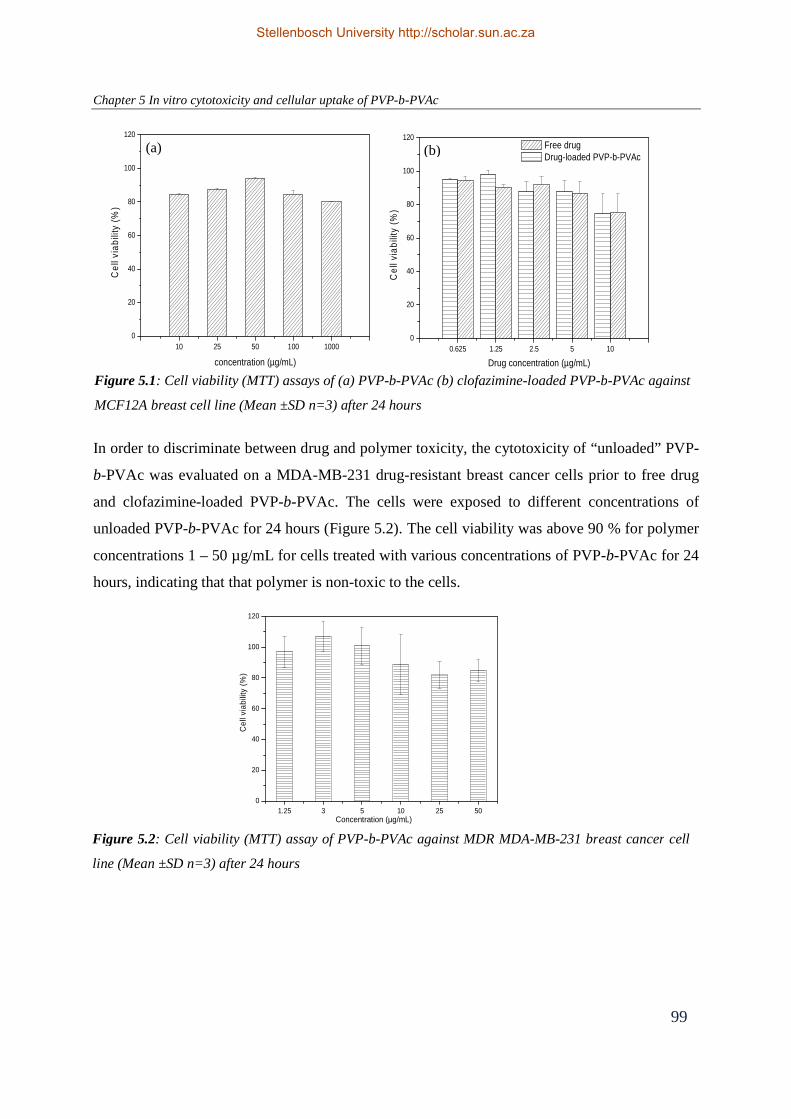
Chapter 5 In vitro cytotoxicity and cellular uptake of PVP-b-PVAc
99
In order to discriminate between drug and polymer toxicity, the cytotoxicity of “unloaded” PVP-
b-PVAc was evaluated on a MDA-MB-231 drug-resistant breast cancer cells prior to free drug
and clofazimine-loaded PVP-b-PVAc. The cells were exposed to different concentrations of
unloaded PVP-b-PVAc for 24 hours (Figure 5.2). The cell viability was above 90 % for polymer
concentrations 1 – 50 µg/mL for cells treated with various concentrations of PVP-b-PVAc for 24
hours, indicating that that polymer is non-toxic to the cells.
Figure 5.2: Cell viability (MTT) assay of PVP-b-PVAc against MDR MDA-MB-231 breast cancer cell
line (Mean ±SD n=3) after 24 hours
1.25 3 5 10 25 500
20
40
60
80
100
120
Cel
l via
bilit
y (%
)
Concentration (µg/mL)
10 25 50 100 10000
20
40
60
80
100
120
Cel
l via
bilit
y (%
)
concentration (µg/mL)0.625 1.25 2.5 5 10
0
20
40
60
80
100
120
Cel
l via
bilit
y (%
)
Drug concentration (µg/mL)
Free drug Drug-loaded PVP-b-PVAc
Figure 5.1: Cell viability (MTT) assays of (a) PVP-b-PVAc (b) clofazimine-loaded PVP-b-PVAc against
MCF12A breast cell line (Mean ±SD n=3) after 24 hours
(a) (b)
Stellenbosch University http://scholar.sun.ac.za

Chapter 5 In vitro cytotoxicity and cellular uptake of PVP-b-PVAc
100
The cytotoxicity of free drug and clofazimine-loaded PVP-b-PVAc of various drug
concentrations (1.25 – 20 µg/mL) are shown in Figure 5.3. It was found that increasing the
clofazimine concentration decreased the viability of the cells. At low concentration of
clofazimine, the cytotoxicity of free drug and drug-loaded PVP-b-PVAc is not obvious.
However, at higher concentrations of clofazimine, a difference was observed. Cells incubated for
24 hours with different clofazimine-loaded PVP-b-PVAc formulations showed higher
cytotoxicity compared to free clofazimine. It must be pointed out that at these drug
concentrations, the polymer is above the CMC therefore the drug is present inside the vesicles
rather than in free form. In addition, PVP-b-PVAc showed no obvious cytotoxicity (over 90 %
viability, Figure 5.2), which confirms that the enhanced cytotoxicity was not due to the presence
of the PVP-b-PVAc carrier. The IC50 value for the cytotoxicity experiment is the concentration
of drug required for 50 % inhibition of cell viability. If the PVP-b-PVAc carrier has a negligible
cytotoxic effect on cells, then any change in the IC50 value when treated with PVP-b-
PVAc/clofazimine formulations, is as a result of increased uptake of the drug or due to an
enhanced therapeutic effect. The IC50 value of drug-loaded PVP-b-PVAc (9 µg/mL) is much
lower if compared to the free drug (30 µg/mL) after 24 hours incubation. It has been reported
that polymer drug carriers often reduce the IC50 through improved drug solubility, more efficient
uptake and improved drug trafficking within the cell.29,30 The lower IC50 confirms the synergistic
action of the clofazimine-loaded PVP-b-PVAc formulation in vitro.
Figure 5.3: Cell viability (MTT) assays of free drug and clofazimine-loaded PVP-b-PVAc against
MDR MDA-MB-231 breast cancer cell line (Mean ±SD n=3 ) after 24 hours
1.25 3 5 10 200
20
40
60
80
100
120
Cel
l via
bilit
y (%
)
Drug concentration (µg/mL)
Free drug Clofazimine-loaded PVP-b-PVAc
Stellenbosch University http://scholar.sun.ac.za

Chapter 5 In vitro cytotoxicity and cellular uptake of PVP-b-PVAc
101
PTX is a widely used anticancer drug in the clinic and exhibits strong cytotoxic activity against
various cancer types, especially breast and ovarian cancer.31 The cytotoxicity of free paclitaxel
and paclitaxel-loaded PVP-b-PVAc formulations were evaluated using MDA-MB-231 cells, and
compared to clofazimine. The potency of PTX was clearly demonstrated by the low
concentration of the IC50 value of pure paclitaxel compared to pure clofazimine having the same
concentration in MDA-MB-231 cells. Figure 5.4 shows that PTX-loaded PVP-b-PVAc
demonstrated the same cytotoxicity (40 – 45 %) as free PTX when incubated at low
concentrations in MDA-MB-231 cells for 24 hours. At higher PTX concentrations, the
cytotoxicity was higher in PTX-loaded PVP-b-PVAc than free PTX. The calculated IC50 value of
PTX-loaded PVP-b-PVAc is 0.42 µg/mL, which is lower than that of the free drug, which has an
IC50 of 1.58 µg/mL.
When the cells were incubated for 24 hours with several PTX-loaded PVP-b-PVAc formulations,
the same trend (in which drug-loaded PVP-b-PVAc showed higher cytotoxicity than free drug)
was observed as for clofazimine-loaded PVP-b-PVAc formulations.
At this stage, the reason for the observed cytotoxicity is not exactly known but could possibly be
explained by the mechanism of cell entry (endocytosis for polymer micelles vs. drug permeation
for free drug).32 It is speculated that the PVP-b-PVAc drug carrier could facilitate the
Figure 5.4: Cell viability (MTT) assays of free drug and PTX-loaded PVP-b-PVAc against MDR
MDA-MB-231 breast cancer cell line (Mean ±SD n=3 ) after 24 hours
1 5 10 50 1000
10
20
30
40
50
60
Cel
l via
bilit
y (%
)
Drug concentration (µg/mL)
Free drug PTX-loaded PVP-b-PVAc
Stellenbosch University http://scholar.sun.ac.za

Chapter 5 In vitro cytotoxicity and cellular uptake of PVP-b-PVAc
102
intracellular uptake of the drug, resulting in a higher concentration of drug in the cytoplasm
which further improves the drug efficacy. Polymeric self-assemblies have previously been shown
to enhance the in vitro cellular uptake of anticancer drugs.33 For example, Danhier and
coworkers33 loaded poly(ethylene glycol)-b-poly(ɛ-caprolactone-co-trimethylenecarbonate) with
PTX, which was exposed to cervix epithelial carcinoma cells for 24 hours. Results indicated that
PTX-loaded formulations showed lower IC50 values than free PTX, which suggests that the
polymer carrier enhanced the cellular uptake of the PTX. Similarly, Burt et al.34 loaded PTX into
the hydrophobic bilayer of poly(butadiene)-poly(ethylene oxide) (PB-PEO) polymer vesicles.
PTX-loaded PB-PEO vesicles also showed higher cytotoxicity compared to free PTX against
human breast cells. In addition, the vesicles showed an increase in storage stability, beyond the 3
hour half-life of PDLLA-PEG micelles.
Polymer drug carriers have also been reported to overcome drug resistance in multidrug-resistant
cancer cells which are overexpressed with efflux transporters and numerous other multidrug-
resistant associated proteins in the plasma membranes.35 The efflux pumps play an important role
in protecting tissues and organs from toxic molecules.36 Among several mechanisms of drug
resistance, P-glycoprotein (P-gp) is the best known and most extensively investigated. P-gp, a
membrane associated glycoprotein preferentially effluxes hydrophobic compounds from the
inside of the cell back to the extracellular space. In tumor cells, the P-gp efflux pump is believed
to transport more than 50 % of anti-cancer drugs out of the cell membrane and contributes to the
multidrug resistance phenomenon (MDR). Of course, other efflux transporters also contribute to
this phenomenon and should not be ruled out.
The effectiveness of a drug is dependent on its intracellular concentration. Free drug diffuses
through the cell membrane and slowly gets transported out of the cell by efflux transporters (for
more precise details on drug binding, drug efflux and the P-gp action, the reader is referred to the
references)37,38. This results in a low concentration of drug in the cytoplasm, below the cell-
killing threshold which results in limited efficacy. In this way, efflux systems confer resistance to
drugs because they reduce the intracellular concentration of the drug. On the other hand, drug
encapsulated in a polymer carrier (being enclosed in an endosome when entering the cell) is not
Stellenbosch University http://scholar.sun.ac.za

Chapter 5 In vitro cytotoxicity and cellular uptake of PVP-b-PVAc
103
recognized by the efflux pumps. This results in higher intracellular drug concentrations, thereby
improving the efficacy of the drug. However, the mechanism of the effect of polymer carriers on
the P-gp drug efflux system still remains unclear. Further research is needed to elucidate the
exact mechanisms of the effect of drug carriers on the P-gp efflux system.
5.3.2 Cellular uptake studies of fluorescently labeled PVP-b-PVAc
PVP-b-PVAc-fluorescein maleimide was synthesised via a thiol-maleimide reaction as shown in
Figure 5.5. An important feature specific to RAFT polymerization is the preparation of polymers
bearing terminal thiocarbonyl thio groups, which can easily be aminolysed with primary or
secondary amines,39 producing terminal thiol groups. A general aminolysis procedure was
applied to the xanthate end-functional PVP-b-PVAc block copolymer to reduce the omega-
terminal thiocarbonyl thio chain-ends yielding thiol-terminal PVP-b-PVAc. The oxidative
coupling of the thiol-functionality (resulting in disulfide formation) was minimized by
conducting the conjugation reaction under an inert atmosphere in the presence of a reducing
agent, tris(2-carboxyethyl)phosphine (TCEP.HCl). The thiol end-group remained as a ‘‘chemical
handle’’ that was conjugated to the maleimide-functionalized dye, fluorescein maleimide.
In order to determine whether the fluorescein maleimide was attached to the polymer, the
fluorescently labeled PVP-b-PVAc polymer fraction that eluted at a specific retention time from
size exclusion chromatography (SEC) was collected. The fluorescence spectrum of the fraction
was recorded on a luminescence spectrometer (Perkin Elmer Model LS50B) at an excitation
n-butyl amine, DCM
ONO
O
NC
S O
S
nm O
NOO
NC
SHn
m
N OO
OO OH
S
O
ONO
NCn
m
DMF, TCEP
Figure 5.5: Reaction scheme for the attachment of fluorescein maleimide, a thiol-reactive fluorescein dye, to PVP-b-PVAc
fluorescein maleimide, 24 h
Stellenbosch University http://scholar.sun.ac.za

Chapter 5 In vitro cytotoxicity and cellular uptake of PVP-b-PVAc
104
wavelength of 480 nm. The polymer fraction showed an emission at 512 nm, which confirms the
presence of the fluorescent tag on the polymer. The efficiency of the conjugation reaction was
however not determined and the amount of free dye was not quantified. It was assumed that most
free dye was removed after extensive dialysis for 7 days followed by filtration. Perylene red
(Exciton 613), a hydrophobic laser dye was encapsulated into the fluorescently labeled PVP-b-
PVAc via the dialysis method. The cellular uptake of the resulting perylene-loaded fluorescently
labeled PVP-b-PVAc was explored in vitro and compared with free label (fluorescein maleimide
and perylene red).
The cellular uptake of fluorescently labeled PVP-b-PVAc encapsulated with perylene red was
visualized using fluorescence microscopy. Cell nuclei were labeled with Hoechst 33342.
Fluorescently labeled perylene red-loaded PVP-b-PVAc (Zave (DLS) 160 nm, PDI 0.09) were
incubated with MDA-MB-231 breast epithelial cancer cells grown on glass cover slips and
observed under fluorescence microscopy for 6 hours. At specific time intervals, images were
taken to study the time-dependent uptake of the PVP-b-PVAc carrier.
Incubation of breast epithelial cancer cells with “free” hydrophobic perylene red dye in PBS (0.1
% DMSO) at 37 °C, as control, showed almost no or minimal fluorescence signal at t = 0 (Figure
5.6 (A)). After 1 hour and 6 hours of incubation, no obvious fluorescence or particle signal was
detected in the intracellular or in the perinuclear region of the cells, strongly indicating that the
free perylene red was not taken up by the cells. After 6 hours of incubation, some aggregation of
the perylene dye far from the vicinity of the cells was observed (Figure 5.6 (B)). No fluorescent
image was observed for the free dye inside the cells, which could be ascribed to the
hydrophobicity of the dye and its limited solubility in aqueous media, resulting in no cellular
uptake. Control experiment for cells incubated with “free” hydrophilic fluorescein maleimide in
PBS (Figure 5.6 (C)) resulted in instantaneous uptake into the cells.
Stellenbosch University http://scholar.sun.ac.za

Chapter 5 In vitro cytotoxicity and cellular uptake of PVP-b-PVAc
105
Figure 5.7 displays the fluorescence micrographs of cells in contact with perylene red-loaded
vesicles at t = 0, t = 3 hours and t = 6 hours incubation time. Real-time imaging over the first five
minutes of incubation showed fluorescent nano-sized particles (which represent the vesicles)
throughout the outside region of the cells. Figure 5.7 (A), (overlay plot), clearly shows the
distinct, well-defined black region (representing the inside region of the cell) with no detectable
fluorescent signal indicating that no uptake has occurred. Strongly fluorescent particles were
detected outside the cell region. The overlay of the fluorescently labeled PVP-b-PVAc (green)
encapsulated with perylene red, results in yellow, indicating that the vesicles were intact. At this
stage no vesicles were taken up by the cells. The real-time imaging over the first few minutes
showed significant motion of the vesicles outside the cells with some vesicles aligning
themselves around the cell membrane and interacting with the cell membrane. This interaction is
said to be the initial step of endocytosis. Figure 5.7 (B) shows a low fluorescence signal observed
in the cytoplasm, indicating that a small fraction of vesicles were taken up by the cells after 3
hours. Strong fluorescence was still detected outside the cell wall, indicating that most of the
vesicles still remained outside the cell. With extended incubation time, 6 hours at 37 °C,
enhanced fluorescence was seen with a larger fraction of vesicles accumulating in the
intracellular region with some of the vesicles in very close proximity to the cell nucleus (Figure
5.7(C)).
Figure 5.6: Fluorescence micrographs of MDA-MB-231 breast cancer cells incubated with
perylene red (control) for A) t = 0 hr B) t = 6 hr C) Fluorescein maleimide (control)
A B C
Stellenbosch University http://scholar.sun.ac.za

Chapter 5 In vitro cytotoxicity and cellular uptake of PVP-b-PVAc
106
Figure 5.8 represents the slice viewer plot, where each slice corresponds to an X, Y or Z-axis
position inside the cell. By viewing the volume slices, a more complete three dimensional view
of the cell region is seen. Figure 5.8 (A), better illustrates the absence of the vesicles in the
cytoplasmic and the perinuclear region at incubation time t = 0 as seen in Figure 5.7 (A). In
Figure 5.8 (B) after 6 hours of incubation, the distinct cell membrane is less defined compared to
time t = 0, (Figure 5.8 (A)) indicating that vesicles were internalized and localized in the
cytoplasm.
Figure 5.7: Fluorescence micrographs of MDA-MB-231 breast cancer cells incubated with: (A)
fluorescently labeled perylene red-loaded PVP-b-PVAc at 0 hr. (B) after 3h (C) after 6h. For each
panel, images from left to right show the cells with nuclear staining by Hoechst 33342 and
fluorescein-maleimide labeled PVP-b-PVAc, fluorescein-maleimide labeled PVP-b-PVAc
encapsulated with perylene red, and the overlays of both images. Scale bars correspond to 20 µm
A
B
C
20 µm
Stellenbosch University http://scholar.sun.ac.za

Chapter 5 In vitro cytotoxicity and cellular uptake of PVP-b-PVAc
107
Figure 5.9 represents the iso-surface projection plots of the perylene red-loaded PVP-b-PVAc at
the different time intervals. These plots clearly show the intracellular distribution of the vesicles.
The iso-surface projection plot, Figure 5.9 (B) compared to Figure 5.9 (A), clearly shows the
distribution of the small fraction of vesicles which have been taken up by the cells and localized
in the cytoplasm and not in the nucleus. The iso-surface projection plot after 6 hours (Figure 5.9
(C)) strongly indicates a distinctly different fluorescence distribution showing that the vesicles
are more evenly distributed inside the cell cytoplasm and in the perinuclear region.
The results showed that the PVP-b-PVAc carrier was capable of entering the cells and carrying
the hydrophobic dye to the cells. Furthermore, during the incubation no obvious morphological
abnormalities of the cells or cytotoxicity of the PVP-b-PVAc carrier were observed. This
Figure 5.8: Slice viewer plots of fluorescently labeled PVP-b-PVAc loaded with perylene red in MDA-
MB-231 breast cancer cells (A) t = 0 (B) t = 6 hr
A B
A B C
Figure 5.9: Iso-surface projection plots of fluorescently labeled PVP-b-PVAc loaded with perylene red
in MDA-MB-231 breast cancer cells (A) t = 0 (B) t = 3hr (C) t = 6 hr.
Stellenbosch University http://scholar.sun.ac.za

Chapter 5 In vitro cytotoxicity and cellular uptake of PVP-b-PVAc
108
indicated that the PVP-b-PVAc carrier was well tolerated by the cells and is non-toxic. This
result is in agreement with our previous in vitro cytotoxicity experiments (Section 5.2) which
showed that the carrier is non-toxic.
Although the fluorescence results provided no information regarding the release of the
hydrophobic dye during the 6 hour incubation time, evidence that the PVP-b-PVAc carrier was
taken up by the cells and located in both the cytoplasm and the perinuclear region was clear. The
improved solubility and efficient cellular uptake of the dye, in the dye-loaded PVP-b-PVAc
(which mimics the drug-loaded PVP-b-PVAc to a certain extent), could further support the
reasoning for the enhanced cytotoxicity of the drug-loaded PVP-b-PVAc compared to free drug,
observed in the in vitro cytotoxicity experiments. Based on the observations for the dye-loaded
PVP-b-PVAc, similar behavior is anticipated for the drug-loaded PVP-b-PVAc. If this is the
case, then we hypothesize that for drug-loaded PVP-b-PVAc, subsequent degradation or
dissociation of the PVP-b-PVAc carrier (most likely as a result of interaction with proteins and
serum components) would result in the release of the hydrophobic drug in high concentrations
within the cytoplasmic region. This would further improve the efficacy of the drug, compared to
free drug, which is transported into the cells via a passive diffusion mechanism.40
It must be pointed out that there are numerous factors that contribute towards the cellular
internalization of polymer carriers. Cellular internalization of polymer carriers is a time, energy,
temperature and concentration dependent process.41 In addition, the chemical composition and
the physicochemical properties of polymeric carrier (e.g. the ratio of hydrophilic and
hydrophobic block lengths, charge, particle size) play an important role.42 Mahmud et al.43
demonstrated the effect of block length and molecular weight of the poly(ethylene glycol)-b-
poly(ɛ-caprolactone) (mPEG-b-PCL) carrier. Results showed that hydrophilic PEO block length
had a more profound effect on cellular internalization compared to PCL core forming block. The
effect of the core-forming block on the internalization of polymer carriers by different cells has
not been fully investigated in the literature. Furthermore, the internalization of carriers is also
dependent on the type of cell line in which the studies are carried out.44 As a result of all the
above mentioned factors that need to be taken into account, a comparison of the cellular uptake
results observed for the PVP-b-PVAc carrier with those of other carrier systems previously
Stellenbosch University http://scholar.sun.ac.za

Chapter 5 In vitro cytotoxicity and cellular uptake of PVP-b-PVAc
109
reported in the literature is rather difficult. Importantly, fluorescence data showed that the PVP-
b-PVAc carrier is capable of rapid entry into the cells, an important requirement for any drug
delivery system.
5.4 Conclusion
Cytotoxicity assays confirmed the biocompatibility of the PVP-b-PVAc carrier. The in vitro
MTT assays showed that PVP-b-PVAc had no toxic effect on healthy breast epithelial cells and
breast cancer epithelial cells. On the other hand, clofazimine-loaded PVP-b-PVAc showed
significant advantages in achieving higher cytotoxicity and smaller IC50 value over free drug
(clofazimine and PTX). Cellular uptake of the PVP-b-PVAc carrier was clearly evidenced by
fluorescence microscopy. PVP-b-PVAc vesicles were readily taken up by the breast epithelial
cancer cells and localized in the cytoplasmic region. From the in vitro studies it is anticipated
that PVP-b-PVAc is a promising drug carrier to improve the efficacy of hydrophobic drugs.
Stellenbosch University http://scholar.sun.ac.za

Chapter 5 In vitro cytotoxicity and cellular uptake of PVP-b-PVAc
110
5.5 References
(1) Choksakulnimitr, S.; Masuda, S.; Tokuda, H.; Takakura, Y.; Hashida, M. J. Control.
Rel. 1995, 34, 233 - 241.
(2) Li, A. P. Drug Discov. Today 2005, 2, 179 - 185.
(3) Torchilin, V. P. Adv. Drug Deliv. Rev. 2005, 95 - 109.
(4) Kim, Y.; Pourgholami, M. H.; Morris, D. L.; Stenzel, M. H. J. Mater. Chem. 2011, 21,
12777 - 12783.
(5) Maysinger, D.; Lovric, J.; Eisenberg, A.; Savic, R. Eur. J. Pharm Biopharm 2007, 65,
270 - 281.
(6) Rijcken, C. J. F.; Soga, O.; Hennink, W. E.; van Nostrum, C. F. J. Control. Rel. 2007,
120, 131 - 148.
(7) Zhang, W.; Li, Y.; Liu, L.; Sun, Q.; Shuai, X.; Zhu, W.; Chen, Y. Biomacromolecules
2010, 11, 1331 - 1338.
(8) Yan, J.; Ye, Z.; Luo, H.; Chen, M.; .Zhou, Y.; Tan, W.; Xiao, Y.; Zhang, Y.; Lang,
M. Polym. Chem. 2011, 2, 1331 - 1340.
(9) Duong, T. T.; Marquis, C. P.; Whittaker, M.; Davis, T. P.; Boyer, C. Macromolecules
2011, 44, 8008 - 8019.
(10) Luo, L. B.; .Tam, J.; Maysinger, D.; Eisenberg, A. Bioconjugate Chem. 2002, 13, 1259 -
1265.
(11) Savic, R.; Luo, L. B.; Eisenberg, A.; Maysinger, D. Science 2003, 300, 615 - 618.
(12) Ahmed, F.; Pakunlu, R. I.; Brannan, A.; Bates, F.; Minko, T.; Discher, D. E. J.
Control. Rel. 2006, 116, 150 - 158.
(13) Moad, G.; Rizzardo, E.; Thang, S. H. Aust. J. Chem. 2005, 58, 379 - 410.
(14) Beija, M.; Charreyre, M.; Martinho, J. M. G. Prog. Polym. Sci. 2010, 36, 568 - 602.
(15) Lowe, A. B. Polym. Chem. 2010, 1, 17 - 36.
(16) Roth, P. J.; Boyer, C.; Lowe, A. B.; Davis, T. P. Macromol. Rapid Commun. 2011, 32,
1123 - 1143.
(17) Zelikin, A. N.; Such, G. K.; Postma, A.; Caruso, F. Biomacromolecules 2007, 8, 2950 -
2953.
(18) Zelikin, A. N.; Li, Q.; Caruso, F. Chem. Mater. 2008, 20, 2655 - 2661.
Stellenbosch University http://scholar.sun.ac.za

Chapter 5 In vitro cytotoxicity and cellular uptake of PVP-b-PVAc
111
(19) Moad, G.; Rizzardo, E.; Thang, S. H. Polym. Int. 2011, 60, 9 - 25.
(20) Ghosh, S. S.; Kao, P. M.; McCue, A. W.; Chappelle, H. L. Bioconjugate Chem. 1990, 1,
71 - 76.
(21) Kakwere, H.; Perrier, S. J. Am. Chem. Soc. 2009, 131, 1889 – 1895.
(22) Pounder, R. J.; Stanford, M. J.; Brooks, P.; Richards, S. P.; Dove, A. P. Chem.
Commun. 2008, 5158 - 5160.
(23) Li, M.; De, P.; Gondi, S. R.; Sumerlin, B. S. J. Polym. Sci.,Part A: Polym. Chem. 2008,
46, 5093 - 5100
(24) Scales, C. W.; Convertine, A. J.; McCormick, C. L. Biomacromolecules 2006, 7, 1389 -
1392.
(25) Suchao-in, N.; Chirachanchai, S.; Perrier, S. Polymer 2009, 50, 4151 - 4158.
(26) Mangold, S. L.; Carpenter, R. T.; Kiessling, L. L. Org. Lett. 2008, 14, 2997 - 3000.
(27) Croy, A. H.; Owen, T. C.; Bartlop, J. A.; Cory, J. G. Cancer Commun. 1991, 3, 207 -
212.
(28) De Jong, W. H.; Borm, P. J. A. Int. J. Nanomedicine 2008, 3, 133 - 149.
(29) Lee, E. S.; Na, K.; Bae, Y. H. Nano Lett. 2005, 5, 325 - 329.
(30) Thanou, M.; Duncan, R. Curr. Opin. Invest. Drugs 2003, 4, 701 - 709.
(31) Panchagnula, R. Int. J. Pharm. 1998, 172, 1 - 15.
(32) Torchillan, V. P. Eur. J. Pharm. Biopharm. 2000, 11, S81 - S91.
(33) Danhier, F.; Magotteaux, N.; Ucakar, B.; Lecouturier, N.; Brewster, M.; Préat, V. Eur.
J. Pharm. Biopharm. 2009, 73, 230 - 238.
(34) Burt, H. M.; Zhang, X.; Toleikis, P.; Embree, L.; Hunter, W. L. Colloids Surf., B 1999,
16, 161 -171.
(35) Gottesman, M. M.; Fojo, T.; Bates, S. E. Nat. Rev. Cancer 2002, 2, 48 - 58.
(36) Leslie, E. M.; Deeley, R. G.; Cole, S. P. C. Toxicol. Appl. Pharmacol. 2005, 204, 216 -
237.
(37) Sharom, F. J. Pharmocogenomics 2009, 9, 105 - 127.
(38) Ambudkar, S. V.; Kim, I. W.; Sauna, Z. E. Eur. J. Pharm. Sci. 2006, 27, 392 - 400.
(39) Castro, E. A. Chem. Rev. 1999, 99, 3505 - 3524
(40) Lee, Y.; Park, S. Y.; Mok, H.; Park, T. G. Bioconjugate Chem. 2008, 19, 525 - 531.
Stellenbosch University http://scholar.sun.ac.za

Chapter 5 In vitro cytotoxicity and cellular uptake of PVP-b-PVAc
112
(41) Liaw, J.; Aoyagi, T.; Kataoka, K.; Sakurai, Y.; Okano, T. Pharm. Res. 1999, 16, 213 -
220.
(42) Savic, R.; Eisenberg, A.; Maysinger, D. J. Drug Targeting 2006, 14, 343 - 355.
(43) Mahmud, A.; Lavasanifar, A. Colloids Surf., B 2005, 45, 82 - 89.
(44) Zauner, W.; Farrow, N. A.; Haines, A. M. R. J. Control. Rel. 2001, 71, 39 - 51.
Stellenbosch University http://scholar.sun.ac.za

Chapter 6 Summary and Perspectives
113
Chapter 6 Summary and Perspectives
6.1 Summary
Amphiphilic block copolymers have been used extensively in drug and gene delivery research.
Their applications in drug delivery include drug solubilization, controlled drug release and drug
targeting. A feature unique to amphiphilic block copolymers is their self-assembly behaviour in
aqueous environment. This attractive feature allows for the encapsulation of highly hydrophobic
anti-cancer drugs to increase the effective solubility of these therapeutic agents. Furthermore, it
permits drugs to be more effectively administered, transported, and eventually delivered to the
desired targeted site through the blood stream, which consists mostly of water.
The results presented in this dissertation describe the potential application of the amphiphilic
block copolymer, poly((vinylpyrrolidone)-b-poly(vinyl acetate)) (PVP-b-PVAc) as an effective
drug carrier for hydrophobic anti-cancer drugs. In this chapter the results of the dissertation are
summarized and future perspectives are given.
In Chapter 1, a general introduction, the aim of the study and structure of the dissertation is
given. The primary objective of this study was to develop a PVP-b-PVAc polymer drug carrier
and explore its potential as a drug carrier. The physicochemical properties, formulation stability
and anti-cancer activity of the carrier were examined.
A general overview of amphiphilic block copolymers in drug delivery is presented in Chapter 2.
The chapter discusses the properties of drug carriers (e.g. particle size, size distribution, drug
loading capacity, drug release, stability) which determine their ability to deliver drugs. The
chapter also highlights the various key factors that primarily influence these properties.
The synthesis, characterization and self-assembly of PVP-b-PVAc is described in Chapter 3.
The amphiphilic block copolymers were synthesized via the RAFT/MADIX technique. PVP-b-
PVAc block copolymers may be synthesized by a two-step sequential monomer addition
polymerization procedure. The methodology consisted of growing the PVAc chain from a
xanthate end-functional PVP. Block copolymers of constant hydrophilic PVP block length (Mn
~10 000 g/mol) and varying hydrophobic block length were synthesized. In aqueous medium, the
hydrophilic PVP segment forms the shell/corona region and hydrophobic PVAc segment forms
Stellenbosch University http://scholar.sun.ac.za

Chapter 6 Summary and Perspectives
114
the bilayer membrane region where the hydrophobic molecules can be entrapped. In this
dissertation the PVP-b-PVAc self-assembled system were formed via the dialysis method.
The self-assembly was confirmed by 1H NMR spectroscopy in D2O. Both the morphology and
the average size of the self-assembled PVP-b-PVAc were investigated by dynamic light
scattering (DLS), transmission electron microscopy (TEM) and static light scattering (SLS).
Results revealed the formation of spherical, vesicular-like particles of 180 – 200 nm in diameter.
Considering their size, these particles are expected to be suitable for the delivery of drugs to
tumors.1 The self-assembly behaviour of the block copolymer in aqueous solution was further
investigated by fluorescence spectroscopy. The critical micelle concentration (CMC) of the PVP-
b-PVAc block copolymers was determined using pyrene as a fluorescent probe. Increasing the
PVAc block length resulted in a lower CMC, which can be attributed to the increased
hydrophobicity of the PVP-b-PVAc block copolymer. The CMC values were low (~ 0.001
mg/mL – 0.01 mg/mL), which is important in order for the self-assembled structures to remain
intact after dilution upon intravenous administration. Furthermore, the stability of the block
copolymers was also assessed under physiological conditions (PBS, pH 7.4, 37 °C) and were
stable in the absence and presence of serum.
The feasibility of PVP-b-PVAc as a drug carrier for the solubilization and delivery of
hydrophobic anti-cancer drugs was addressed in Chapter 4. To assess the potential of PVP-b-
PVAc as a drug delivery vehicle, the loading of a model drug, clofazimine, and a commonly
used anti-cancer drug, paclitaxel (PTX), into PVP-b-PVAc was investigated. A simple loading
method, namely the dialysis method was used to entrap the drugs in the hydrophobic PVAc
domain. It was found that PVP-b-PVAc was able to successfully solubilize the chemotherapeutic
agents. 1H NMR spectroscopy confirmed the formation of phase-separated structures and the
localization of the drug within the hydrophobic domain.
The method used allowed for reproducible formation of drug-loaded PVP-b-PVAc vesicles, in
the size range of 180 – 200 nm. These particle sizes should be suitable for passive targeting via
the enhanced permeation and retention (EPR) effect.
Stellenbosch University http://scholar.sun.ac.za

Chapter 6 Summary and Perspectives
115
In order to find an optimal formulation for the drug-loaded PVP-b-PVAc, various factors that
influence the loading capacity namely the length of the hydrophobic PVAc segment and the
amount of feed drug were also investigated. Different ratios of drug and polymer were explored
and it was found that the composition of 20 % (w/w) clofazimine: PVP90-b-PVAc290 (w/w) and
10 % (w/w) for PTX: PVP90-b-PVAc290 were the optimum formulations. Lower ratios led to
lower efficiency, whereas at higher ratios the PVP-b-PVAc carrier exceeded its solubilization
capacity resulting in drug precipitation. Generally, the different PVP-b-PVAc block copolymers
of varying PVAc block length had different loading capacities, which indicated that the
hydrophobic block length affects the ability to load hydrophobic drug within the bilayer
membrane. Increasing the amount of feed drug also increased the drug loading capacity. DLS
and TEM of the drug-loaded PVP-b-PVAc showed particle sizes of 200 nm which is suitable for
passive targeting via the enhanced permeation and retention (EPR) effect. The effect of the drug
loading on the particle size was also investigated and found to have no significance on the
particle size of the PVP-b-PVAc carrier. The stability of the drug-loaded PVP-b-PVAc was
investigated in physiological conditions (PBS, pH 7.4, 37 °C) in the presence and absence of
serum. Drug-loaded PVP-b-PVAc was stable in the presence of serum indicating that the
hydrophilic PVP corona was able to stabilize the vesicles.
Next, in vitro studies of PVP-b-PVAc were conducted and presented in Chapter 5. These studies
are an essential first step to assess the drug carriers potential. Cytotoxicity assays confirmed the
biocompatibility of the PVP-b-PVAc carrier. PVP-b-PVAc was shown to be non-toxic to healthy
MCF12A breast cells and MDA-MB-231 multi-drug-resistant breast epithelial cancer cells. The
MTT assays show that the PVP-b-PVAc carrier can deliver clofazimine and PTX and prohibit
growth of cancer cells in vitro. In addition, the drug-loaded polymers showed increased
cytotoxicity compared to free drug against breast cancer epithelial cells, indicating the enhanced
therapeutic effect. To determine whether the PVP-b-PVAc carrier was taken up by the cells,
fluorescently labeled PVP-b-PVAc was loaded with hydrophobic dye (perylene red) which
mimicked the drug. In doing so, one is able to track whether the carrier is taken up by the cells
and determine the fate of both the vesicles and the dye upon cellular internalization.
Fluorescence indicated that the PVP-b-PVAc carrier was taken up by the cells within 3 hours
Stellenbosch University http://scholar.sun.ac.za
Chapter 6 Summary and Perspectives
116
incubation and localized in the cytoplasm and perinuclear region, but not in the nucleus after 6
hours.
6.2 Perspectives
The results presented in the thesis indicate the potential of the amphiphilic PVP-b-PVAc block
copolymer as a carrier for hydrophobic anti-cancer drugs. However, further optimization and
development of the system need to be considered.
In terms of the PVP-b-PVAc drug carrier system developed so far in this study, the following
aspects require further research:
• In vitro studies
In Chapter 5, the cellular internalization and localization and the time taken for cellular
uptake of the PVP-b-PVAc carrier was investigated. From the results, it was anticipated
that the cellular uptake of the carrier is via endocytic pathways, which is suggested for
most polymer carriers. However, the specific mechanism of endocytic uptake of the drug
carrier, the intracellular trafficking pathway the carrier follows upon cell entry and the
subcellular distribution still remains unknown. Understanding of the cellular uptake
routes and trafficking is essential for the development of efficient drug delivery carriers.
These questions can be answered by staining the organelles in the cell e.g. cell membrane
or mitochondria by using, membrane- or organelle-selective dyes.2 The answers to these
essential questions could provide valuable information, which could further be used to
improve the PVP-b-PVAc drug carrier system.
• In vivo studies
In Chapter 4 and 5, data on the stability of the PVP-b-PVAc carrier in physiological
conditions as well as data on the cytotoxic effect of the PVP-b-PVAc carrier and
clofazimine and PTX-loaded PVP-b-PVAc on MDA-MB-231 multi-drug-resistant breast
epithelial cancer cells are presented. For many polymer drug delivery systems, promising
in vitro stability and efficacy data on culture cells has proven to be poorly representative
of the performance or effectiveness of the drug carriers in vivo. To date we could only
Stellenbosch University http://scholar.sun.ac.za

Chapter 6 Summary and Perspectives
117
evaluate and demonstrate the effectiveness of PVP-b-PVAc in vitro, however, in vivo
evaluation needs to be considered. What remains to be achieved is to test whether drug-
loaded PVP-b-PVAc can alter the pharmacokinetics of free drug in vivo.
In addition, the in vivo biodistribution and pharmacokinetic studies need to be performed
to confirm the therapeutic efficacy of the drug-loaded PVP-b-PVAc carrier.
• Towards a more controlled drug delivery system
The dissertation describes the first steps towards the development of a PVP-b-PVAc drug
carrier. However, further development for a more effective drug release system should be
considered. Firstly, an important requirement of a drug carrier is to maintain its integrity
during circulation time upon intravenous administration. Because of the relatively high
hydrophobic PVAc block length, the CMCs of the PVP-b-PVAc block copolymers
developed and described in the thesis were low and the assemblies that were formed were
correspondingly stable. This should maintain their integrity upon dilution. However,
interaction with serum proteins can also destabilize the carrier. This results in premature
release of the drug or alternatively accelerates the release of the drug from the carrier.
One approach would be to covalently link the drug to the hydrophobic block via a
hydrolytically sensitive linker (e.g. hydrazone linker). In this way, the drug remains
stably linked to the polymer and only upon entering the acidic tumor environment or the
acidic organelles is the drug released. To further improve the efficacy of the carrier,
selective targeting groups can also be attached to the carrier. Alternatively, an
amphiphilic copolypeptide drug carrier can also be considered whereby the hydrophobic
drug is covalently linked to the carrier. Peptides exhibit high biological specificity which
could further enhance the drug targeting property of the carrier.
• Combination therapy - Multiple drug delivery
Over the past years, research has focused on single drug administration, while not much
attention has been placed on combination administration. Recent cancer research is based
on combination therapy – the use of synergistic anticancer drug “cocktails” whereby two
different drugs are delivered to tumors simultaneously.
Stellenbosch University http://scholar.sun.ac.za

Chapter 6 Summary and Perspectives
118
Synergistic drug combinations produce an even greater response rate than is possible with
each drug used alone at its optimum dose.3 Generally, anti-cancer drugs are typically
administered at their individual maximum tolerated doses.4 This is termed the “more-is-
better” approach. However, this approach ignores the possibility of subtle concentration
and ratio dependent dynamic interactions capable of causing a synergistic response.5
Recently, a more rational approach for the combination of anticancer drugs has been
proposed – the fixed ratio drug combinations (FRDC) approach – which holds the
promise of capturing the maximum pharmacodynamic cytotoxic specificity for cancer
cells.
Riminophenazines possess a potent anticancer multi-mechanism that is thought to be best
utilised in combination with standard chemotherapeutics. Interestingly, these
riminophenazines have been shown to inhibit P-glycoprotein (P-gp), the transmembrane
efflux pump that is largely responsible for acquired resistance.6,7 Recently, FRDC
comprising clofazimine in combination with paclitaxel has demonstrated synergistic
antiproliferative activity in vitro against a multi-drug resistant (MDR) cell line. Recently,
the in vitro optimal synergistic fixed drug ratio combination of clofazimine-PTX has been
determined (Combination Index Analysis (CI) < 1).8 The work presented in this study has
shown that PVP-b-PVAc may serve as a suitable carrier for single delivery of
hydrophobic drugs namely clofazimine and PTX. Therefore, current work focuses on the
encapsulation of the FRDC of clofazimine and paclitaxel into PVP-b-PVAc. Lastly,
owing to the vesicular morphology of the self-assembled PVP-b-PVAc, its potential for
the simultaneous delivery of a combination of hydrophobic (e.g. PTX) and hydrophilic
drugs (e.g. doxorubicin) can also be explored.
Stellenbosch University http://scholar.sun.ac.za

Chapter 6 Summary and Perspectives
119
6.3 References
(1) Maeda, H.; Wua, J.; Sawa, T.; Matsumura, Y.; Horic, K. J. Control. Rel. 2000, 65, 271
- 284.
(2) Savic, R.; Luo, L. B.; Eisenberg, A.; Maysinger, D. Science 2003, 300, 615 - 618.
(3) Shin, H.; Alani, A. W. G.; Rao, D. A.; Rockich, N. C.; Kwon, G. S. J. Control. Rel.
2009, 140, 294 - 300.
(4) Decker, S.; Sausville, E. A. Ann. N.Y. Acad. Sci. 2005, 1059, 61 - 69.
(5) Mayer, L. D.; Harasym, T. O.; Tardi, P. G.; Harasym, N. L.; Shew, C. R. Mol Cancer
Ther 2006, 5, 1854 - 1863.
(6) Van Rensburg, C. E. J.; Van Staden, A. M.; Anderson, R. Cancer Res. 1993, 53, 318 -
323.
(7) Van Rensburg, C. E. J.; Joone, G. K.; O’Sullivan, J. F. Oncol. Rep. 2000, 7, 193 - 195.
(8) Koot, D.; Strategic pre-clinical development of Riminophenazines as resistance
circumventing anticancer agent Univeristy of Pretoria, 2012.
Stellenbosch University http://scholar.sun.ac.za

Acknowledgements First and foremost, I would like to acknowledge my promoter, Bert Klumperman for
giving me the opportunity to study under his supervision. I consider it an honor to work
within your research group. Thank you for your support, motivation and encouragement
throughout my PhD study. Above of all, you have always shown continuous confidence
in my capabilities and for that I am very grateful.
I would like to acknowledge Prof. R. Sanderson who my inspired me throughout my
Honours, Masters and PhD years at the University of Stellenbosch. You were my mentor.
Some parts of the work presented in this dissertation could not have been accomplished
without the contributions of my collaborators. Firstly, I would like to thank Dwayne Koot
from the Department of Pharmacology (University of Pretoria) for answering my stream
of endless “pharmaceutical” questions! All our discussions and your thoughtful
suggestions were gratefully appreciated. Mark Thomas and Ben Loos from the
Department of Physiology (University of Stellenbosch) - thank you for your guidance,
your patience, your time and sharing of your knowledge. Mark, a very special thank you
for your guidance and help in the cell culture laboratory. I have learnt mostly everything
involving cell work from you and I am very grateful for your time and help. Mohammed
Jaffer from the University of Cape Town – thank you for all your time and help with the
TEM analyses.
I would like to thank Prof. W. Hennink and Prof B. Klumperman for giving me the
opportunity to spend a few months at the Department of Pharmaceutics, Utrecht
University, The Netherlands. A special thank you to Yang Shi for his assistance in the
research labs during my visit.
Too many people to mention individually have assisted in so many ways during my work
in the Department of Polymer Science. Om nooit te vergeten – Dr Eric van den Dungen -
bedankt voor de raad, de leerzame besprekingen en bemoedigende woorden. Jouw kennis
en toewijding op dit onderwerp is een inspiratie voor mij.
Stellenbosch University http://scholar.sun.ac.za

I would like to thank Dr Gwenaelle Pound for her guidance during the initial stages of my
PhD. To all the research students in the Free Radical Lab - you all have my sincere
gratitude. All our laughs and enjoyable moments in the lab kept me smiling during good
times and difficult times. I also thank the support staff who aided me in so many aspects
during my PhD – Erinda Cooper, Aneli Fourie, Margie Hurndell, Deon Koen, Calvin
Maart and Jim Motshweni.
Of course no acknowledgments would be complete without giving thanks to my parents,
to Chantal and to Claude. Thank you for your unconditional love, support,
encouragement and believing in me. You have been my tower of strength.
Nathalie
March 2012
Stellenbosch University http://scholar.sun.ac.za