Embed Size (px)
Citation preview

8/13/2019 Mycotic Infections OF ORAL CAVITY
http://slidepdf.com/reader/full/mycotic-infections-of-oral-cavity 1/45
o o d m o r n i n g

8/13/2019 Mycotic Infections OF ORAL CAVITY
http://slidepdf.com/reader/full/mycotic-infections-of-oral-cavity 2/45
P R E S E N T E D B Y : P U S H K A R S . D A H I W A L
G U I D E D B Y : D R . S O N I A S O D H I M A M
D R . L A T A D A B H A D E M A M
M y c o t i c i n f e c t i o n s
o f t h e o r a l
c a v i t y

8/13/2019 Mycotic Infections OF ORAL CAVITY
http://slidepdf.com/reader/full/mycotic-infections-of-oral-cavity 3/45
FUNGAL / MYCOTIC INFECTIONS
1. CANDIDIASIS2. HISTOPLASMOSIS3. BLASTOMYCOSIS4. PARACOCCIDIOIDOMYCOSIS5. MUCORMYCOSIS6. CRYPTOCOCCOSIS7. COCCIDIODOMYCOSIS8. GEOTRICHOSIS9. SPOROTRICHOSIS10. RHINOSPORIDIOSIS11. ASPERGILLOSIS
8/13/2019 Mycotic Infections OF ORAL CAVITY
http://slidepdf.com/reader/full/mycotic-infections-of-oral-cavity 4/45
CANDIDIASIS
It is also called as candidosis. Refers to infection with yeast like fungal organism candida
albicans.. Most common oral fungal infection. It is a component of normal oral flora in the gastrointestinal
and genitourinary tracts of humans. Can occur in persons who are debilitated by other diseases or
in otherwise healthy individuals also.
Candida species- albican non-albican: C. glabrata, C. krusei, C. parapsilosis, C.
tropicalis, C. parapsilosis

8/13/2019 Mycotic Infections OF ORAL CAVITY
http://slidepdf.com/reader/full/mycotic-infections-of-oral-cavity 5/45

8/13/2019 Mycotic Infections OF ORAL CAVITY
http://slidepdf.com/reader/full/mycotic-infections-of-oral-cavity 6/45
Biology of Candida albicans Commensal Pathogen
A thin-walled dimorphic fungus
MorphogenesisUnicellular yeast (harmeless)Filamentous (pathogenic)
Principal Cell Wall PolymersGluccan
Mannan
Strict aerobe, favors moist surfacesCommensally found in gut, genitals, and lungsBody Temp 37º C, neutral pH
Figure 1. Yeast in Oral Scraping A sample of an oral scraping
contains yeast cells andpseudohyphae
Rapid Multiplication & Spread
8/13/2019 Mycotic Infections OF ORAL CAVITY
http://slidepdf.com/reader/full/mycotic-infections-of-oral-cavity 7/45
PREDISPOSING FACTORS:
a) Local Factors :
- Mucosal trauma (local irritant)
- Denture wearers
- Denture hygiene- Tobacco smoking
- Drugs (Broad spectrum antibiotics,steroids,immunosuppressant / cytotoxic agents)
- Xerostomia

8/13/2019 Mycotic Infections OF ORAL CAVITY
http://slidepdf.com/reader/full/mycotic-infections-of-oral-cavity 8/45
b) S ystemic factors : - Iron deficiency anaemia
- Megaloblastic anaemia- Acute leukaemia
- Diabetes mellitus
- HIV infection- Other immunodeficiency states

8/13/2019 Mycotic Infections OF ORAL CAVITY
http://slidepdf.com/reader/full/mycotic-infections-of-oral-cavity 9/45
CLASSIFICATION OF CANDIDIASIS: -
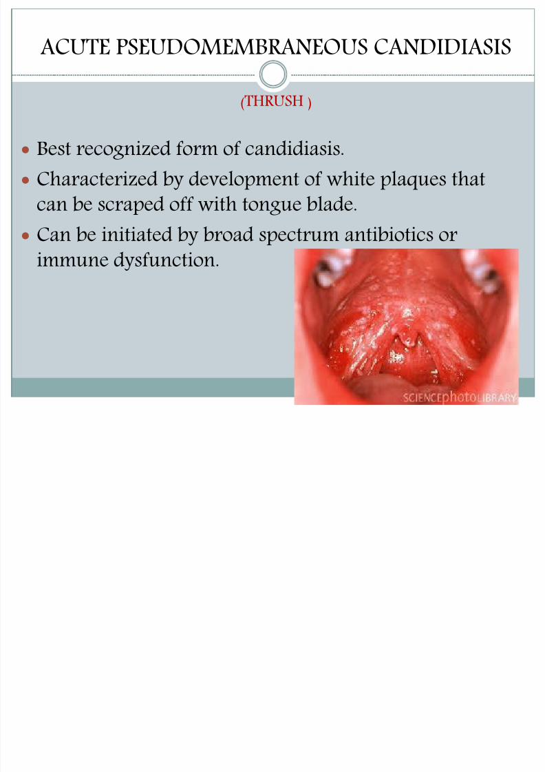
Acute -- Acute pseudomembranous candidiasis (thrush)
- Acute atrophic candidiasis
(antibiotic sore-mouth)
Chronic –
1. Chronic hyperplastic candidiasis
2.chronic mucocutaneous candidiasis
3. Chronic atrophic candidiasisDENTURE STOMATITIS(DENTURE SORE MOUTH)
ANGULAR CHEILITIS
MEDIAN RHOMBOID GLOSSITIS
( adapted from lanhar t .classification & clinicopathological features of candida infections in the mouth)

8/13/2019 Mycotic Infections OF ORAL CAVITY
http://slidepdf.com/reader/full/mycotic-infections-of-oral-cavity 10/45
A ) primary candidiasis1)Acute form Pseudomembranous. Erythematous2) Chronic form Hyperplastic form Erythematous
Pseudo-membranous3)Candida associated lesion denture stomatitis Angular stomatitis
Median rhomboid glossitis4) Keratinised primary lesion superinfected with candida Leukoplakia,lichen planus, lupus erythematousB) Secondory candidiasisOral manifestations of systemic mucocutaneous candidosis
8/13/2019 Mycotic Infections OF ORAL CAVITY
http://slidepdf.com/reader/full/mycotic-infections-of-oral-cavity 11/45

8/13/2019 Mycotic Infections OF ORAL CAVITY
http://slidepdf.com/reader/full/mycotic-infections-of-oral-cavity 12/45
Occurs characteristicallyon buccal mucosa, palateand dorsal tongue.
Usually asymptomatic orpatients may c/o burningsensation of mucosa orunpleasant taste inmouth.
Can occur in infants also.

8/13/2019 Mycotic Infections OF ORAL CAVITY
http://slidepdf.com/reader/full/mycotic-infections-of-oral-cavity 13/45
ACUTE ATROPHICCANDIDIASIS:
Also called “antibiotic sore
mouth” as it follows courseof broad spectrum antibiotics.
Patients c/o burning sensationof mucosae.
Seen as diffuse loss offilliform papillae resulting ina bald appearance of tongue.

8/13/2019 Mycotic Infections OF ORAL CAVITY
http://slidepdf.com/reader/full/mycotic-infections-of-oral-cavity 14/45
CHRONIC HYPERPLASTIC CANDIDIASIS:
Least common of all types.
Appears as non scrapable white patchresembling leukoplakia candidal
leukoplakia)
Believed that it represents candidiasissuperimposed on pre-existingleukoplakia.
Diagnosis confirmed bydemonstration of candidal hyphaewithin the lesion and resolution oflesion after antifungal therapy.

8/13/2019 Mycotic Infections OF ORAL CAVITY
http://slidepdf.com/reader/full/mycotic-infections-of-oral-cavity 15/45
CHRONIC MUCOCUTANEOUSCANDIDIASIS:
Severe oral candidiasis can also occuras a component of a rareimmunological disorder calledmucocutaneous candidiasis.
Autosomal recessive disorder.
Immune dysfunction becomes evidentin early life – patient develops
candidiasis of mouth, nails, skin andother mucosae.
Oral lesions appear as thick, white nonscrapable patches.

8/13/2019 Mycotic Infections OF ORAL CAVITY
http://slidepdf.com/reader/full/mycotic-infections-of-oral-cavity 16/45
3. CHRONIC ATROPHICCANDIDIASIS:
Denture stomatitis Characterized by varying
degrees of erythema indenture bearing areas ofusually maxillary prostheses.
Usually asymptomatic. Patients give h/o wearing
denture continuously.
8/13/2019 Mycotic Infections OF ORAL CAVITY
http://slidepdf.com/reader/full/mycotic-infections-of-oral-cavity 17/45
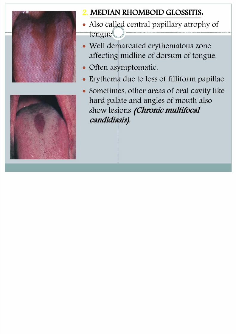
2. MEDIAN RHOMBOID GLOSSITIS: Also called central papillary atrophy of
tongue.
Well demarcated erythematous zoneaffecting midline of dorsum of tongue.
Often asymptomatic.
Erythema due to loss of filliform papillae. Sometimes, other areas of oral cavity like
hard palate and angles of mouth alsoshow lesions Chronic multifocal
candidiasis).

8/13/2019 Mycotic Infections OF ORAL CAVITY
http://slidepdf.com/reader/full/mycotic-infections-of-oral-cavity 18/45
ANGULAR CHEILITIS: Also called perleche.
Characterized by erythema,fissuring and scaling of corners ofmouth.
Typically occurs either along with
multifocal candidiasis or in oldpatients with reduced verticaldimension.
Saliva pools in these areas, keeping
them moist and thus favoringfungal infection

8/13/2019 Mycotic Infections OF ORAL CAVITY
http://slidepdf.com/reader/full/mycotic-infections-of-oral-cavity 19/45
HISTOLOGICAL FEATURES:
Biopsy specimen showhyperparakeratinization,elongation of rete ridges, chronic
inflammatory cell infiltration ofunderlying CT and smallmicroabscesses collection ofPMNL’s) within parakeratin
layer. Candidal hyphae can be seen
embedded in parakeratin layerand superficial spinous layer.
8/13/2019 Mycotic Infections OF ORAL CAVITY
http://slidepdf.com/reader/full/mycotic-infections-of-oral-cavity 20/45
Amphotericin B 0.7 mg/kg per day
Caspofungin is 50 mg/day after a loading dose of 70 mg
Voriconazole is 3 mg/kg twice daily after a loading doseof 6 mg/kg twice daily for one day.
Duration of therapy for candidemia : A minimum of two weeks of therapy after blood cultures become
negative
Treatment

8/13/2019 Mycotic Infections OF ORAL CAVITY
http://slidepdf.com/reader/full/mycotic-infections-of-oral-cavity 21/45
Topical treatment:
Limit the systemic absorption
Clotrimazole -1 troche 10 mg tablet five times daily
Nystatin preprations.
It includes suspension,vaginal &an oral pastile
Vaginal tab.- 1 tab, 100,000 units dissolved in mouth 3times a daily
Nystatin oral pastile 200,000 units
Five times a daily
Nystatin suspension 1 teaspoon mixed wiyh water & rinse
Topical creams & oinements of nystatin,ketoconazole,orclotrimazole

8/13/2019 Mycotic Infections OF ORAL CAVITY
http://slidepdf.com/reader/full/mycotic-infections-of-oral-cavity 22/45
Amphotericin B 0.08 mg/ kg 2 to 3 hourly inoinetment, cream based , suspension
Rinses-
Nystatin for 7 -10 days 3- 4 times daily
0.2 % chlorhexidine

8/13/2019 Mycotic Infections OF ORAL CAVITY
http://slidepdf.com/reader/full/mycotic-infections-of-oral-cavity 23/45
Systemic treatment
Nystatin 250 mg TDS for 2 weeks followed by 1troche / day for 3rd week
Ketoconazole 200 mg with food daily
( careful monitoring required of liver function) Fluconazole 100 mg tab once daily for two weeks
Itraconazole 100 mg cap. For 2 weeks

8/13/2019 Mycotic Infections OF ORAL CAVITY
http://slidepdf.com/reader/full/mycotic-infections-of-oral-cavity 24/45
HISTOPLASMOSIS
It is also called as DARLING’S DISEASE.
It is caused by histoplasma capsulatum, a dimorphicfungus that grows in yeast form in infected tissue.
Infection results from inhalation of dustcontaminated with droppings , particularly frominfected birds.
8/13/2019 Mycotic Infections OF ORAL CAVITY
http://slidepdf.com/reader/full/mycotic-infections-of-oral-cavity 25/45
TYPES OF HISTOPLASMOSIS
1. Acute primary histoplasmosis
Primary infection is mild ,manifesting as self limitedpulmonary disease that heals to leave fibrisis and
calcification.2. Progressive disseminated histoplasmosis
-manifested by hepatosplenomegaly &lymphodenopathy .
-patients show evidence of bone marrowinvolvement by anemia & leucopenia.

8/13/2019 Mycotic Infections OF ORAL CAVITY
http://slidepdf.com/reader/full/mycotic-infections-of-oral-cavity 26/45
3. chronic cavitary histoplasmosislesions mimics the chronic cavitary tuberculosis.
Histoplasmosis is an intracellular mycoticinfection of the reticuloendothelial system
caused by the inhalation of conidia fromthe fungus Histoplasma capsulatum.Histoplasmosis has a world widedistribution, however, the Mississippi-Ohio
River Valley in the U.S.A. is recognized as amajor endemic region. Africa, Australia andparts of East Asia, in particular India andMalaysia are also endemic regions

8/13/2019 Mycotic Infections OF ORAL CAVITY
http://slidepdf.com/reader/full/mycotic-infections-of-oral-cavity 27/45
Approximately 95% of cases ofHistoplasmosis are in apparent,
subclinical or benign. Five percent ofthe cases have chronic progressivelung disease, chronic cutaneous or
systemic disease or an acute
fulminating fatal systemic disease. All stages of this disease may mimic
tuberculosis

8/13/2019 Mycotic Infections OF ORAL CAVITY
http://slidepdf.com/reader/full/mycotic-infections-of-oral-cavity 28/45

8/13/2019 Mycotic Infections OF ORAL CAVITY
http://slidepdf.com/reader/full/mycotic-infections-of-oral-cavity 29/45
DIAGNOSIS
Made by culture on sabouraud’s media Biopsy will show small oval yeasts within the macrophages &
reticoendothelial cells
As well as chronic grannulomas , epitheloid cells, giant cells &
occaisionally caseation necrosis .
Diffrential diagnosis:
Tuberculosis-sputum examination, tuberculin test
Blastomycosis- biopsy &culturing the organism from tissue.
Mucormycosis-biopsy
Cryptococcosis-organism cultured on saboraud’s glucose agar

8/13/2019 Mycotic Infections OF ORAL CAVITY
http://slidepdf.com/reader/full/mycotic-infections-of-oral-cavity 30/45
Treatment of histoplasmosis:
Mild to moderate cases treated with ketoconazole for6 – 12 months
Immunesuppresed patients /severe patients require
Amphotericin B to 10 weeks

8/13/2019 Mycotic Infections OF ORAL CAVITY
http://slidepdf.com/reader/full/mycotic-infections-of-oral-cavity 31/45
African histoplasmosis
It is caused by fungus histoplasma dubosii
It is larger than h.capsulatum
Disease has clinically distinct form & geographical
distribution so called african histoplasmosis

8/13/2019 Mycotic Infections OF ORAL CAVITY
http://slidepdf.com/reader/full/mycotic-infections-of-oral-cavity 32/45
BLASTOMYCOSIS
It is cause by blastomycosis dermatitidis.(northamerican blastomycosis) also called as gilchrist’s disease.
Organism is a normal inhabitat of soil & so that iscommom in agricultural worker .
It is transmitted by respiratory tract.

8/13/2019 Mycotic Infections OF ORAL CAVITY
http://slidepdf.com/reader/full/mycotic-infections-of-oral-cavity 33/45
Types of the blastomycosis
Primary pulmonary blastomycosis Cutaneous blastomycosis Disseminated or systemic blastomycosis
Blast mycosis is a chronic granulomatous andsuppurative disease having a primary
pulmonary stage that is frequently followed by dissemination to other body sites,chiefly the skin and bone. Although the
disease was long thought to be restrictedto the North American continent, in recent years autochthonous cases have beendiagnosed in Africa, Asia and Europe.
8/13/2019 Mycotic Infections OF ORAL CAVITY
http://slidepdf.com/reader/full/mycotic-infections-of-oral-cavity 34/45

8/13/2019 Mycotic Infections OF ORAL CAVITY
http://slidepdf.com/reader/full/mycotic-infections-of-oral-cavity 35/45
Oral manifestations
It may be primary or secondary to some infectionselsewhere in the body.
Symptoms & signs-
Orophayrngeal pain with enlargement of cervicallymph nodes.

8/13/2019 Mycotic Infections OF ORAL CAVITY
http://slidepdf.com/reader/full/mycotic-infections-of-oral-cavity 36/45
Diagnosis
When chronic, painless,oral ulcer appears in anagricultural worker or when review of system revealspulmonary symptoms.
Diagnosis is made on the basis of biopsy & on culturing
the organism from the body.
Differential diagnosis-
1.Squamous cell carcinoma- older patient
2.Tuberculosis3.Histoplasmosis –culture & biopsy
4. mucormycosis-biopsy

8/13/2019 Mycotic Infections OF ORAL CAVITY
http://slidepdf.com/reader/full/mycotic-infections-of-oral-cavity 37/45
TREATMENT
I ntrvaenous
Amphotericin B
Itraconazole Ketoconazole
For 8- 10 weeks
8/13/2019 Mycotic Infections OF ORAL CAVITY
http://slidepdf.com/reader/full/mycotic-infections-of-oral-cavity 38/45
PARACOCCIDIOIDOMYCOSIS
Paracoccidioidomycosis is a chronicgranulomatous disease that characteristically produces aprimary pulmonary infection, often in apparent, and thendisseminates to form ulcerative granulomata of the buccal,
nasal andoccasionally the gastrointestinal mucosa.
. The only etiological agent,
Paracoccidioides brasiliensis is
geographically restricted to areas of Southand Central America also called as south american
blastomycosis/ lutz’ disease

8/13/2019 Mycotic Infections OF ORAL CAVITY
http://slidepdf.com/reader/full/mycotic-infections-of-oral-cavity 39/45
Chronic granulomatus infectionParacoccodioides brasilensis,
Lungs- Mucosa – Skin – Lymphatic vessels
Enter through the lungs Saprophytic in nature,
Humid forests of South and
Central
Common in 20 – 40 years,
8/13/2019 Mycotic Infections OF ORAL CAVITY
http://slidepdf.com/reader/full/mycotic-infections-of-oral-cavity 40/45
MUCORMYCOSIS
It is also called as phycomycosis.
Etiology & predisposing factors-
-caused by saprophyte fungus
-more common in patients with decreased resistancedue to diseases like diabetes , tuberculosis , renalfailure ,leukemia , cirrhosis of liver & in severe burncases.

8/13/2019 Mycotic Infections OF ORAL CAVITY
http://slidepdf.com/reader/full/mycotic-infections-of-oral-cavity 41/45
Types of mucormycosis:
1. superficial
2.Visceral
- Pulmonary- Gastrointestinal
- Rhinocerebral/ rhinomaxilary

8/13/2019 Mycotic Infections OF ORAL CAVITY
http://slidepdf.com/reader/full/mycotic-infections-of-oral-cavity 42/45
Rhinomaxillary form begins with inhalation offungus by susectible individual
Infection usually arises in the lateral wall of nose &maxillary sinus,may rapidly spread by arterialinvasion to involve orbit,palate,maxillaryalveolus,then cavernous sinus & then brain through
hematogenous spread & may cause death

8/13/2019 Mycotic Infections OF ORAL CAVITY
http://slidepdf.com/reader/full/mycotic-infections-of-oral-cavity 43/45
Differential diagnosis:
Squamous cell carcinoma.
Necrotising sialometaplasia…..usually painless
Management1. Surgical debridement treatment of choice
2. Systemis amphtericin
3. Control of systemic disease

8/13/2019 Mycotic Infections OF ORAL CAVITY
http://slidepdf.com/reader/full/mycotic-infections-of-oral-cavity 44/45
CRYPTOCOCCOSIS
It is also called as torulosis It is chronic fungal infection caused by cryptococcus
neoformans & cryptococcus bacillispora.
Infection usually occurs in lungs
Oral manifestations-
locations –lesions of hard palate ,soft palate ,gingiva ,
Extraction socket, tongue& tonsillar pillar
TREATMENT:Ketoconazole for 6 -12 weeks for mild cases
Amphotericin –B for 10 weeks for severe cases.

8/13/2019 Mycotic Infections OF ORAL CAVITY
http://slidepdf.com/reader/full/mycotic-infections-of-oral-cavity 45/45
THANKS FOR YOUR PATIENCE