Embed Size (px)
Citation preview

Proc. Natl. Acad. Sci. USAVol. 90, pp. 2532-2536, March 1993Immunology
Mutual regulation of the transcriptional activator NF-cB and itsinhibitor, I1cB-a
(gene regulation/cell activation/human immunodeficiency virus/transcription factor/nuclear translocation)
KEITH BROWN, SUN PARK, TOMOHIKO KANNO, GuIDo FRANZOSO*, AND ULRICH SIEBENLIST
Laboratory of Immunoregulation, National Institute of Allergy and Infectious Diseases, National Institutes of Health, Bethesda, MD 20892
Communicated by Anthony S. Fauci, December 29, 1992 (received for review December 11, 1992)
ABSTRACT The NF-#cB transcription factor complex issequestered in the cytoplasm by the inhibitory protein I#cB-a(MAD-3). Various cellular stimuli relieve this inhibition bymechanisms largely unknown, leading to NF-KB nuclear lo-calization and transactivation of its target genes. It is demon-strated here with human T lymphocytes and monocytes thatdifferent stimuli, including tumor necrosis factor a and phor-bol 12-myristate 13-acetate, cause rapid degradation ofIOcB-a,with concomitant activation of NF-cB, followed by a dramaticincrease in IKB-a mRNA and protein synthesis. Transfectionstudies reveal that the IKB-a mRNA and the encoded proteinare potently induced by NF-cB and by homodimers of p65 andof c-Rel. We propose a model in which NF-.cB and IKB-amutually regulate each other in a cycle: saturating amounts ofthe inhibitory IKB-a protein are destroyed upon stimulation,allowing rapid activation of NF-KB. Subsequently, IcB-amRNA and protein levels are qulickly induced by the activatedNF-cB. This resurgence of IKB-a protein acts to restore anequilibrium in which NF-KB is again inhibited.
NF-KB is a dimeric transcription factor that binds and regu-lates gene expression through decameric cis-acting KB DNAmotifs (reviewed in refs. 1 and 2). Although a p50/p65heterodimer has traditionally been referred to as NF-KB andremains the prototypical and most abundant form, it has beenrecognized recently that several distinct but closely relatedhomo- and heterodimeric factors are responsible for KBsite-dependent DNA binding activity and regulation. Thevarious dimeric factors are composed of members of thefamily of Rel-related polypeptides. One subclass of thisfamily, distinguished by its proteolytic processing from pre-cursor forms and lack of recognized activation domains,includes p50 (NFKB1) (3-6) and pSOB (NFKB2, pS2) (7-10),whereas the second subclass contains recognized activationdomains and includes p65 (RelA) (11-13), RelB (14, 15), c-Rel(16), v-Rel (17, 18), and the Drosophila protein Dorsal (19).All Rel-related members share a 300-amino acid region ofhomology, responsible for DNA binding and dimerization,called the Rel homology domain.
Activation of the NF-KB transcription factor and variousrelated forms can be initiated by a variety of agents, includingtumor necrosis factor a (TNF-a) and phorbol 12-myristate13-acetate (PMA) (1, 2). Activation proceeds through apost-translational event in which preformed cytoplasmicNF-KB is released from a cytoplasmic inhibitory protein,IKB-a (MAD-3) (20-23). IKB-a inhibits transactivation of thep50/p65 heterodimer, by binding to the p65 component,blocking the dimer's translocation to the nucleus (20, 21, 23).IKB-a also inhibits complexes containing c-Rel or RelB (24,25). IKB-a blocks binding in vitro of various NF-KB dimers toKB binding sites in DNA (11, 12, 15, 22, 26, 27). Because the
latter effect requires nuclear IKB-a, its relevance, in vivo, isunknown. Although lKB-a is generally a cytoplasmic protein(21, 23), it and its chicken homolog (pp4O) have also beendetected in the nucleus (refs. 28-30 and K.B., G.F., andU.S., unpublished results). In addition to the well-characterized and cloned IKB-a and its chicken and rathomologs (24, 31), another biochemically defined form,IKB-/3, has been reported, but this remains to be fullycharacterized (26, 31, 32).
Phosphorylation of IKB-a in vitro by protein kinases A andC and heme-activated kinase abolished IKB-a's inhibition ofDNA binding by NF-KB (24, 32, 33). Although activation ofNF-KB has been conjectured to proceed through phosphor-ylation, no modification of IKB-a has yet been reported invivo. It is also unknown how the inhibited state is maintainedin unstimulated cells-that is, how the ratio of IKB-a proteinto target complexes is regulated. We show here that a mutualcycle of regulation of IKB-a and NF-KB exists. Stimulationresults in a rapid loss of IKB-a from cells (probably followingits modification) and the rapid nuclear translocation of NF-KB. Transactivation by NF-KB, in turn, induces high levels ofIKB-a synthesis, which can then restore the unstimulatedinhibited state.
MATERIALS AND METHODSExtract Preparations and Transfections. Cell extracts were
prepared by centrifugation after freeze-thawing. Nuclearextracts were prepared by the method of Dignam et al. (34).Transfections were performed in NTera-2 cells using thePMT2T vector plasmid as described (9). The PMT2T-IKB-aplasmid was constructed by insertion of an EcoRI fragmentof 1550 nucleotide pairs containing full-length IKB-a/MAD-3cDNA (22) into the EcoRI site of PMT2T. PMT2T-p65 andp50 plasmids were described (9). PMT2T c-Rel plasmid wasconstructed by cloning the EcoRI fragment of full-lengthhuman c-Rel cDNA (16) into the EcoRI site of PMT2T.Western Blotting. Cell extracts were subjected to SDS/
PAGE and blotted onto nitrocellulose (Schleicher & SchuellBA85), and 1KB-a was detected with polyclonal rabbit anti-body directed against either N- or C-terminal domains (aminoacids 1-129 and 230-315, respectively) or both, followed bya second incubation with 125I-labeled protein A (New En-gland Nuclear). Both antibodies detected the same protein inWestern blots. [In addition, a peptide antibody directedagainst an N-terminal peptide reacted with the same proteinand this could be blocked by competition with added peptide(K.B. and U.S., unpublished results).]
Northern Blotting. Total RNA was prepared (35), fraction-ated on an agarose formaldehyde gel, blotted, and hybridized
Abbreviations: TNF-a, tumor necrosis factor a; PMA, phorbol12-myristate 13-acetate; PHA, phytohemagglutinin; EMSA, electro-phoretic mobility shift assay; PD, palindromic.*On leave from: Servizio di Microbiologica del Complesso Conven-zionato Universita-U.L.S.S. 21, Via Ospedale 2, 35100-Padua, Italy.
The publication costs of this article were defrayed in part by page chargepayment. This article must therefore be hereby marked "advertisement"in accordance with 18 U.S.C. §1734 solely to indicate this fact.
2532
Proc. Natl. Acad. Sci. USA 90 (1993) 2533
APMA + IONOMYCIN
0 5' 10' 20' 40 1h 2h
TNF+ CHX
0 2.5' 5 10' 20' 40' 1 h 2h 10' 2h
fluorescein-labeled goat anti-rabbit antibody (Tago) diluted1:50 in PBS was added and incubated for 1 h at roomtemperature. Slides were washed five times in PBS, air dried,and viewed under UV fluorescence (Zeiss Axioplan) with oilimmersion.
RESULTS
BPMA + IONOMYCIN TNF0 5' 10' 20' 0 2.5' 5' 10' 20'
p50/p65 2--- 1 1
" §" ~~p50/p5O i
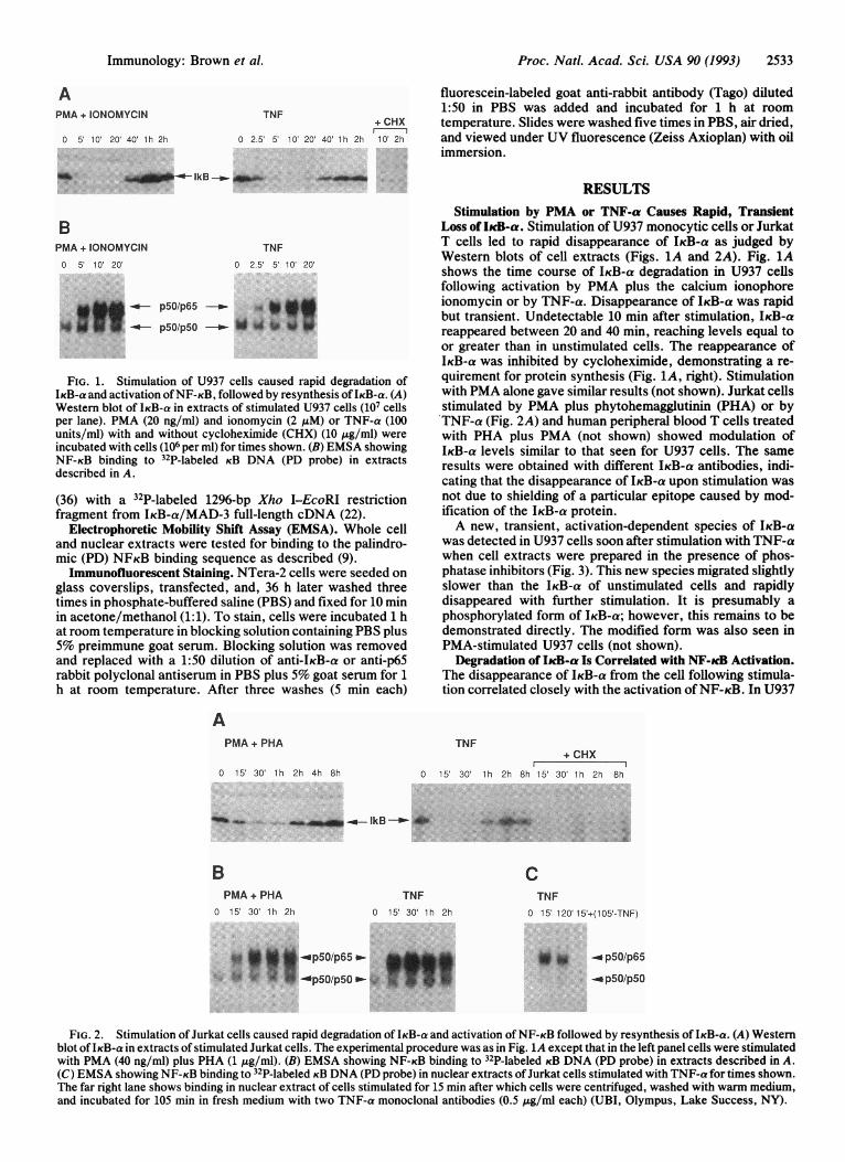
FIG. 1. Stimulation of U937 cells caused rapid degradation ofIKB-a and activation ofNF-KB, followed by resynthesis of IKB-a. (A)Western blot of IKB-a in extracts of stimulated U937 cells (107 cellsper lane). PMA (20 ng/ml) and ionomycin (2 uM) or TNF-a (100units/ml) with and without cycloheximide (CHX) (10 ,g/ml) wereincubated with cells (106 per ml) for times shown. (B) EMSA showingNF-KB binding to 32P-labeled KB DNA (PD probe) in extractsdescribed in A.
(36) with a 32P-labeled 1296-bp Xho I-EcoRI restrictionfragment from IKB-a/MAD-3 full-length cDNA (22).
Electrophoretic Mobility Shift Assay (EMSA). Whole celland nuclear extracts were tested for binding to the palindro-mic (PD) NFKB binding sequence as described (9).Immunofluorescent Staining. NTera-2 cells were seeded on
glass coverslips, transfected, and, 36 h later washed threetimes in phosphate-buffered saline (PBS) and fixed for 10 minin acetone/methanol (1:1). To stain, cells were incubated 1 hat room temperature in blocking solution containing PBS plus5% preimmune goat serum. Blocking solution was removedand replaced with a 1:50 dilution of anti-IKB-a or anti-p65rabbit polyclonal antiserum in PBS plus 5% goat serum for 1h at room temperature. After three washes (5 min each)
APMA + PHA
0 15' 30' 1 h 2h 4h 8h
Stimulation by PMA or TNF-a Causes Rapid, TransientLoss of IkcB-a. Stimulation of U937 monocytic cells or JurkatT cells led to rapid disappearance of IKB-a as judged byWestern blots of cell extracts (Figs. 1A and 2A). Fig. 1Ashows the time course of IKB-a degradation in U937 cellsfollowing activation by PMA plus the calcium ionophoreionomycin or by TNF-a. Disappearance of IKB-a was rapidbut transient. Undetectable 10 min after stimulation, IKB-areappeared between 20 and 40 min, reaching levels equal toor greater than in unstimulated cells. The reappearance ofIKB-a was inhibited by cycloheximide, demonstrating a re-quirement for protein synthesis (Fig. 1A, right). Stimulationwith PMA alone gave similar results (not shown). Jurkat cellsstimulated by PMA plus phytohemagglutinin (PHA) or byTNF-a (Fig. 2A) and human peripheral blood T cells treatedwith PHA plus PMA (not shown) showed modulation ofIKB-a levels similar to that seen for U937 cells. The sameresults were obtained with different IKB-a antibodies, indi-cating that the disappearance of IKB-a upon stimulation wasnot due to shielding of a particular epitope caused by mod-ification of the IKB-a protein.A new, transient, activation-dependent species of IKB-a
was detected in U937 cells soon after stimulation with TNF-awhen cell extracts were prepared in the presence of phos-phatase inhibitors (Fig. 3). This new species migrated slightlyslower than the IKB-a of unstimulated cells and rapidlydisappeared with further stimulation. It is presumably aphosphorylated form of IKB-a; however, this remains to bedemonstrated directly. The modified form was also seen inPMA-stimulated U937 cells (not shown).
Degradation of I#cB-a Is Correlated with NF-cB Activation.The disappearance of IKB-a from the cell following stimula-tion correlated closely with the activation of NF-KB. In U937
TNF+ CHX
10 15' 30 1h 2h 8h 15' 30' 1h 2h 8h
__ IkB - ::
BPMA + PHA
0 15' 30' lh 2h
TNF0 15' 30' 1h 2h
op5O/p65 .-.i.3iB. |p50/p50 *
CTNF
0 15' 120' 15'+(105'-TNF)
to a* .* p5O/p65.:.. _.p..50/p50
FIG. 2. Stimulation of Jurkat cells caused rapid degradation of IKB-a and activation of NF-KB followed by resynthesis of IKB-a. (A) Westernblot ofIKB-a in extracts of stimulated Jurkat cells. The experimental procedure was as in Fig. 1A except that in the left panel cells were stimulatedwith PMA (40 ng/ml) plus PHA (1 jig/ml). (B) EMSA showing NF-KB binding to 32P-labeled KB DNA (PD probe) in extracts described in A.(C) EMSA showing NF-KB binding to 32P-labeled KB DNA (PD probe) in nuclear extracts ofJurkat cells stimulated with TNF-a for times shown.The far right lane shows binding in nuclear extract of cells stimulated for 15 min after which cells were centrifuged, washed with warm medium,and incubated for 105 min in fresh medium with two TNF-a monoclonal antibodies (0.5 ,ug/ml each) (UBI, Olympus, Lake Success, NY).
Immunology: Brown et al.
IkB --am.

Proc. Natl. Acad. Sci. USA 90 (1993)
TNF0 2' 5' 10'
FIG. 3. Stimulated U937 cells produced a modified, short-livedform of IKB-a. Westem blot of IKB-a in cell extracts of U937stimulated with TNF-a for the times shown. The modified form ofIKB-a is indicated by a dot. The experimental procedure was as inFig. 1A except that following stimulation, cells were washed in PBScontaining 1 mM sodium orthovanadate and extracts were preparedby lysis on ice in Tris HCI buffer (50 mM, pH 7.4) containing NaCI(100 mM), sodium pyrophosphate (30 mM), NaF (50 mM), sodiumorthovanadate (1 mM), 0.5% Nonidet P-40, phenylmethylsulfonylfluoride (0.5 mM), leupeptin (10 ,ug/ml), and aprotinin (10 ,ug/ml).
cells NF-KB binding to KB sites (i.e., p50/p65) was easilydetected 2.5-5 min after stimulation with TNF-a (Fig. 1B).The induced binding activity was composed largely of p50/p65 heterodimers as determined by supershifting with spe-cific antibodies. These experiments also confirmed that thefaster migrating species present in unstimulated and stimu-lated cells was composed ofp50 homodimers [not shown; theband assignments concur with earlier reports (22, 37-39)].NF-KB binding activity continued to increase in concert withthe loss of IKB-a for up to 10 min of stimulation with PMAplus ionomycin or with TNF-a. Similar results were obtainedwith stimulated Jurkat T cells (Fig. 2B).
Analysis of nuclear and cytosolic extracts of resting andstimulated Jurkat and U937 cells revealed that the loss ofIKB-a correlated with translocation of NF-KB from the cy-toplasm into the nucleus (Fig. 2C shows nuclear extracts ofJurkat cells; U937 data not shown). Provided that the exter-nal stimulus was maintained, nuclear p50/p65 levels re-mained high despite the appearance of newly synthesizedIKB-a (a 2-h stimulation is shown in Fig. 2C). The mechanismby which NF-KB persists in the nucleus with continuedstimulation is unclear. When TNF-a was removed after 15min-by washing and addition of TNF-a antibodies, nuclearp50/p65 binding activity declined rapidly, in accord with aprior report (40) (a 105-min time point following removal ofTNF-a is shown in Fig. 2C). Thus, brief stimulation byTNF-a produced a transient loss ofIKB-a and only a transientincrease of nuclear NF-KB (p50/p65).
Kinetics of I#cB-a mRNA Induction Following Stimulation.Stimulation of U937 by PMA or TNF-a for short periods, upto 10 min, had little effect on the level of IKB-a mRNA (Fig.4). By this time IKB-a protein had disappeared and NF-KB
PMA TNF0 5' 1 O 20 140 20 5' 10' 20' 40 0 5' 10t' 20' 40'
FIG. 4. Stimulation of U937 cells caused induction of IKB-amRNA. Northern blot of IKB-a RNA extracted from U937 cells (106per ml) stimulated with PMA (20 ng/ml) or TNF-a (100 units/ml) fortimes shown. Each lane contained 20 jig of total RNA and the blotwas probed with 32P-labeled IKB-a cDNA. As a control the blotshown was reprobed with glyceraldehyde-3-phosphate dehydroge-nase (GAPDH) cDNA (Clontech). No change in the level ofGAPDHmRNA with stimulation was seen (not shown).
was activated. IKB-a mRNA levels surged, however, by 20min. Again, Jurkat cells behaved similarly (not shown). Thus,although the loss of IKB-a that follows quickly after stimu-lation was not associated with a loss ofmRNA, the new IKB-aprotein synthesis observed subsequently was associated witha massive increase in IKB-a mRNA, which accords with thecycloheximide sensitivity of this IKB-a protein induction.
Induction ofIcB-a by p65 and c-Rel. The induction ofIKB-amRNA and protein shortly after activation of NF-KB sug-gested that NF-KB was directly responsible. UndifferentiatedNTera-2 human embryonic carcinoma cells were transfectedwith various plasmid constructs directing the synthesis ofcomponents of NF-KB. These cells are devoid of NF-KBactivity (9) and contained no detectable IKB-a protein ormRNA (Fig. 5 A and B, respectively). Transfection ofNTera-2 cells with p65, known to form potently transacti-vating homodimers (7, 13, 41), generated high levels ofendogenous IKB-a mRNA and protein expression (Fig. 5 Band A, respectively). The simplest explanation for the in-crease in IKB-a mRNA is direct transactivation of this gene,
A BLO
Co mL 0CDLO i) (0
> CL CL CL > 0.
IkB Ab IkB Ab
vector alone p65
IkB Ab _ p65 Ab
p50/p65 p65
FIG. 5. p65, p50/p65, and c-Rel induced IKB-a in transfectedNTera-2 cells. (A) Western blot of IKB-a protein produced in cellextracts of NTera-2 transiently transfected with PMT2T (vector)-bearing cDNAs as indicated. The Western blotting procedure was asin Fig. 1A. Cells (5 x 10⁶) in 100-mm Petri dishes were transfectedwith 4 ,ug ofplasmidDNA and harvested 48 h after transfection. Eachlane was loaded with the extract of 1.5 x 10⁶ cells. (B) Northern blotof IKB-a mRNA produced in NTera-2 cells transfected with 4 pg ofPMT2T (vector) or PMT2T-p65. Each lane was loaded with 20 pg oftotalRNA and the blot was probed with 32P-labeled IKB-a cDNA. (C)Immunofluorescent stained transfected NTera-2 cells. The upper leftpanel shows cells transiently transfected with the PMT2T vectorplasmid (4 ,ug) stained for IKB-a. The upper right and lower leftpanels show cells transfected with p65 (4 pg) and p65 plus p50 (2 mgeach) cDNAs, respectively, and stained for IKB-a. The lower rightpanel shows cells transfected with p65 stained for p65. Ab, antibody.
2534 Immunology: Brown et al.
r. -- . .-
Proc. Natl. Acad. Sci. USA 90 (1993) 2535
presumably through a KB element. It is formally possible,however, that the increase is mediated indirectly or is due tomRNA stabilization. In contrast to p65, transfection withp50, which generates non-transactivating p50 homodimers (7,9, 13, 14, 41), did not induce IKB-a synthesis. It is curious thattransfection with the IKB-a expression construct did notresult in detectable levels of the encoded protein (Fig. 5A),despite the fact that high, functional levels of IKB-a can beexpressed from this plasmid when cotransfected with poten-tial targets for inhibition and binding, such as p65 (39).Binding to target proteins may therefore stabilize IKB-a,allowing its detection in Western blots. We speculate furtherthat the inherent instability of free IKB-a is the basis for itsrapid removal from the cell upon activation-induced disso-ciation from NF-KB (see Discussion). Consistent with thisnotion, the transfection of p50 plus p65 and of c-Rel alsoinduced considerable levels ofIKB-a (Fig. 5A), the respectivedimers being capable of transactivation and being targets forbinding and inhibition by IKB-a (11, 20-24, 31, 42-45).
Immunofluorescent staining ofIKB-a confirmed the induc-tion and cytoplasmic location of this protein in NTera-2 cellstransfected with p65 or with p50/p65 (Fig. SC). Staining forp65 revealed high levels of this protein in p65 transfectedcells. The distribution of p65 in nuclear and cytoplasmiccompartments suggested that the highly abundant exoge-nously introduced p65 is partially retained in the cytoplasmby the induced endogenous IKB-a.
DISCUSSIONWe show here thatIKB-a is physiologically regulated by rapidremoval from the cell in response to stimulation. In three celltypes tested-the monocytic cell line U937, Jurkat T lym-phocytes, and peripheral blood T cells-IKB-a was present inunstimulated cells but disappeared within a few minutes ofstimulation by PMA or TNF-a. The removal of IKB-a iscoupled to a concomitant translocation of NF-KB (predom-inantly p50/p65) from the cytoplasm into the nucleus.How does activation increase the lability ofIKB-a protein?
There are at least two possible explanations: (i) modificationofIKB-a and/or its bound partners (e.g., p50/p65), renderingit susceptible to hydrolysis by a cytoplasmic protease; thisincreased susceptibility could result simply from the releaseof the modifiedIKB-a as a free, labile molecule; (ii) activationof a cytoplasmic protease with specificity for IKB-a. Thoughnot excluding the latter explanation, our data support theformer. The observation of a short-lived, more slowly mi-grating speciesofIKB-a in stimulated U937 cells suggests thatmodification of IKB-a occurs and may precede dissociationfrom NF-KB and its degradation. Secondly, because noIKB-aprotein could be demonstrated even upon transfection withIKB-a expression vectors in NTera-2 cells that lack NF-KB,IKB-a appears to be inherently unstable in the absence ofNF-KB binding partners. IKB-a is stabilized and readilydetected in NTera-2 cells transfected with p65, p50/p65, orc-Rel. Our observations are consistent with the hypothesisthat activation-induced modification of IKB-a and/or itsbound NF-KB partner leads to dissociation ofIKB-a, render-ing it susceptible to immediate protease attack.The loss of IKB-a and the activation of NF-KB upon
stimulation are transient phenomena. IKB-a mRNA tran-scription is quickly induced, leading to replenishment of theIKB-a protein level. The induction of endogenous IKB-amRNA upon transfection of NTera-2 cells with transactiva-tors such as p65 suggests direct transactivation of the IKB-agene by NF-KB, possibly through a KB element. Thus, p65positively regulates IKB-a, probably by transcriptional acti-vation, and may also regulate IKB-a turnover by post-translational stabilization. In addition to p65 homodimers,p5O/p65 and c-Rel have similar effects on the induction of
Cellular Activation Signal
I
Replenishment ' Modification-DegradationkicB-a - ,- O- NF-KcB - 1cB-a w I<
f/ Cytoplasm
Nucleus
""II --- Activated NF-icB
IxB-a mRNA --
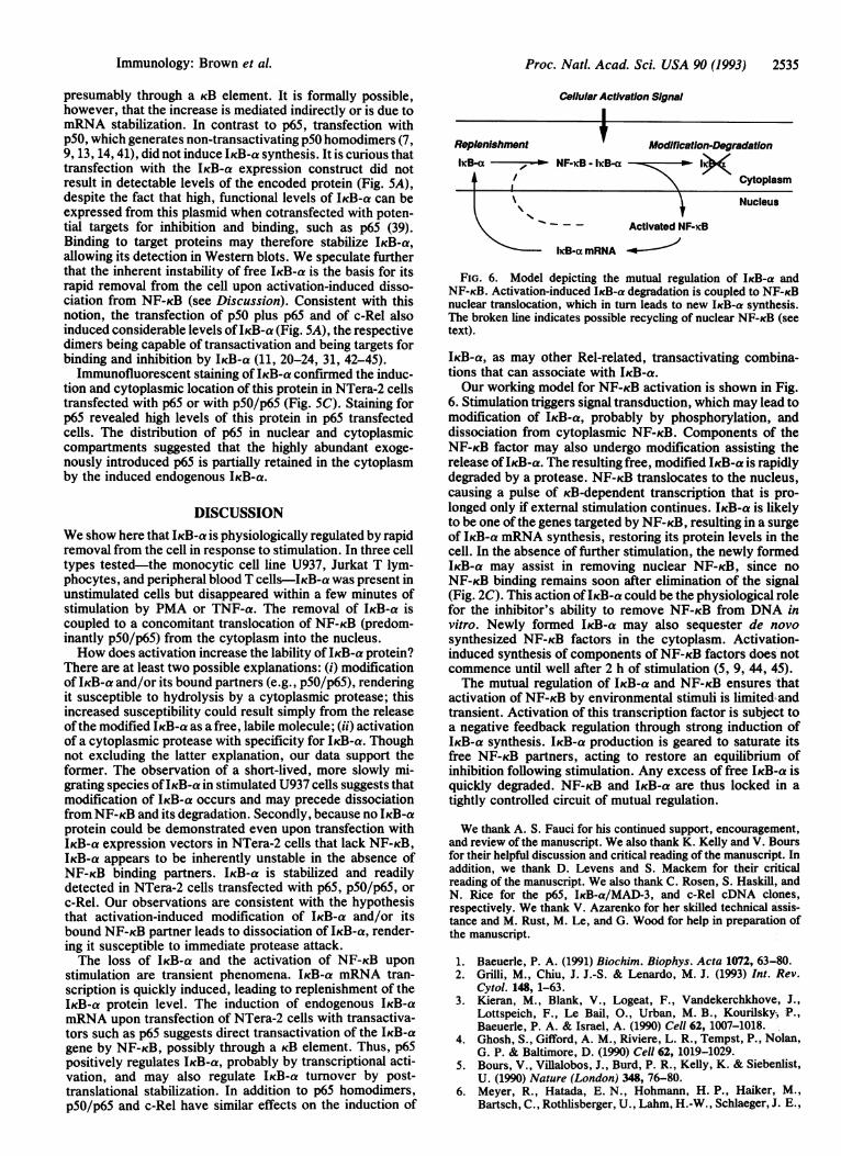
FIG. 6. Model depicting the mutual regulation of IKB-a andNF-KB. Activation-induced IKB-a degradation is coupled to NF-KBnuclear translocation, which in turn leads to new IKB-a synthesis.The broken line indicates possible recycling of nuclear NF-KB (seetext).
IKB-a, as may other Rel-related, transactivating combina-tions that can associate with IKB-a.Our working model for NF-KB activation is shown in Fig.
6. Stimulation triggers signal transduction, which may lead tomodification of IKB-a, probably by phosphorylation, anddissociation from cytoplasmic NF-KB. Components of theNF-KB factor may also undergo modification assisting therelease ofIKB-a. The resulting free, modified IKB-a is rapidlydegraded by a protease. NF-KB translocates to the nucleus,causing a pulse of KB-dependent transcription that is pro-longed only if external stimulation continues. IKB-a is likelyto be one of the genes targeted by NF-KB, resulting in a surgeof IKB-a mRNA synthesis, restoring its protein levels in thecell. In the absence of further stimulation, the newly formedIKB-a may assist in removing nuclear NF-KB, since noNF-KB binding remains soon after elimination of the signal(Fig. 2C). This action ofIKB-a could be the physiological rolefor the inhibitor's ability to remove NF-KB from DNA invitro. Newly formed IKB-a may also sequester de novosynthesized NF-KB factors in the cytoplasm. Activation-induced synthesis of components of NF-KB factors does notcommence until well after 2 h of stimulation (5, 9, 44, 45).The mutual regulation of IKB-a and NF-KB ensures that
activation of NF-KB by environmental stimuli is limited andtransient. Activation of this transcription factor is subject toa negative feedback regulation through strong induction ofIKB-a synthesis. IKB-a production is geared to saturate itsfree NF-KB partners, acting to restore an equilibrium ofinhibition following stimulation. Any excess of free IKB-a isquickly degraded. NF-KB and IKB-a are thus locked in atightly controlled circuit of mutual regulation.
We thank A. S. Fauci for his continued support, encouragement,and review of the manuscript. We also thank K. Kelly and V. Boursfor their helpful discussion and critical reading of the manuscript. Inaddition, we thank D. Levens and S. Mackem for their criticalreading of the manuscript. We also thank C. Rosen, S. Haskill, andN. Rice for the p65, IKB-a/MAD-3, and c-Rel cDNA clones,respectively. We thank V. Azarenko for her skilled technical assis-tance and M. Rust, M. Le, and G. Wood for help in preparation ofthe manuscript.
1. Baeuerle, P. A. (1991) Biochim. Biophys. Acta 1072, 63-80.2. Grilli, M., Chiu, J. J.-S. & Lenardo, M. J. (1993) Int. Rev.
Cytol. 148, 1-63.3. Kieran, M., Blank, V., Logeat, F., Vandekerchkhove, J.,
Lottspeich, F., Le Bail, O., Urban, M. B., Kourilsky, P.,Baeuerle, P. A. & Israel, A. (1990) Cell 62, 1007-1018.
4. Ghosh, S., Gifford, A. M., Riviere, L. R., Tempst, P., Nolan,G. P. & Baltimore, D. (1990) Cell 62, 1019-1029.
5. Bours, V., Villalobos, J., Burd, P. R., Kelly, K. & Siebenlist,U. (1990) Nature (London) 348, 76-80.
6. Meyer, R., Hatada, E. N., Hohmann, H. P., Haiker, M.,Bartsch, C., Rothlisberger, U., Lahm, H.-W., Schlaeger, J. E.,
Immunology: Brown et al.

Proc. Natl. Acad. Sci. USA 90 (1993)
Van Loon, A. P. G. M. & Scheidereit, C. (1991) Proc. Natl.Acad. Sci. USA 88, 966-970.
7. Schmid, R. M., Perkins, N. D., Duckett, C. S., Andrews,P. C. & Nabel, G. J. (1991) Nature (London) 352, 733-736.
8. Neri, A., Chang, C.-C., Lombardi, L., Salina, M., Corradini,P., Maiolo, A. T., Chaganti, R. S. K. & Dalla-Favera, R. S. K.(1991) Cell 67, 1075-1087.
9. Bours, V., Burd, P. R., Brown, K., Villalobos, J., Park, S.,Ryseck, R.-P., Bravo, R., Kelly, K. & Siebenlist, U. (1992)Mol. Cell. Biol. 12, 685-695.
10. Mercurio, F., Didonato, J., Rosette, C. & Karin, M. (1992)DNA Cell Biol. 11, 523-537.
11. Ruben, S. M., Dillon, P. J., Schreck, R., Henkel, T., Chen,C.-H., Maher, M., Baeuerle, P. A. & Rosen, C. A. (1991)Science 251, 1490-1493.
12. Nolan, G. P., Ghosh, S., Liou, H.-C., Tempst, P. & Baltimore,D. (1991) Cell 64, 1-10.
13. Ballard, D. W., Dixon, E. P., Peffer, N. J., Bogerd, H., Doerre,S., Stein, B. & Greene, W. C. (1992) Proc. Natl. Acad. Sci. USA89, 1875-1879.
14. Ryseck, R.-P., Bull, P., Takamiya, M., Bours, V., Siebenlist,U., Dobrzanski, P. & Bravo, R. (1992) Mol. Cell. Biol. 12,674-684.
15. Ruben, S. M., Klement, J. F., Coleman, T. A., Maher, M.,Chen, C.-H. & Rosen, C. A. (1992) Genes Dev. 6, 745-760.
16. Brownell, E., Mittereder, N. & Rice, N. R. (1989) Oncogene 1,935-942.
17. Stephens, R. M., Rice, N. R., Hiebsch, R. R., Bose, H. R. &Gilden, R. V. (1983) Proc. Natl. Acad. Sci. USA 80, 6229-6232.
18. Wilhelmsen, K. C., Eggleton, K. & Temin, H. M. (1984) J.Virol. 52, 172-182.
19. Steward, R. (1987) Science 238, 692-694.20. Baeuerle, P. A. & Baltimore, D. (1988) Cell 53, 211-217.21. Baeuerle, P. A. & Baltimore, D. (1988) Science 242, 540-546.22. Haskill, S., Beg, A. A., Tompkins, S. M., Morris, J. S., Yu-
rochko, A. D., Sampson-Johannes, A., Mondal, K., Ralph, P.& Baldwin, A. S., Jr. (1991) Cell 65, 1281-1289.
23. Beg, A. A., Ruben, S. M., Scheinman, R. I., Haskill, S.,Rosen, C. A. & Baldwin, A. S., Jr. (1992) Genes Dev. 6,1899-1913.
24. Kerr, L. D., Inoue, J. I., Davis, N., Link, E., Baeuerle, P. A.,Bose, H. R., Jr., & Verma, I. (1991) Genes Dev. 5, 1464-1476.
25. Tewari, M., Dobrzanski, P., Mohn, K. L., Cressman, D. E.,
Hsu, J.-C., Bravo, R. & Taub, R. (1992) Mol. Cell. Biol. 12,2898-2908.
26. Zabel, U. & Baeuerle, P. A. (1990) Cell 61, 255-265.27. Urban, M. B. & Baeuerle, P. A. (1990) Genes Dev. 4, 1975-
1984.28. Zabel, U., Henkel, T., dos Santos Silva, M. & Baeuerle, P. A.
(1993) EMBO J. 12, 201-211.29. Davis, N., Bargmann, W., Lim, M.-Y. & Bose, H. (1990) J.
Virol. 64, 584-591.30. Morin, P. J. & Gilmore, T. D. (1992) Nucleic Acids Res. 20,
2453-2458.31. Davis, N., Ghosh, S., Simmons, D. L., Tempst, P., Liou,
H.-C., Baltimore, D. & Bose, H. R., Jr. (1991) Science 253,1268-1271.
32. Link, E., Kerr, L. D., Schreck, R., Zabel, U., Verma, I. &Baeuerle, P. A. (1992) J. Biol. Chem. 267, 239-246.
33. Ghosh, S. & Baltimore, D. (1990) Nature (London) 344, 678-682.
34. Dignam, J. P., Lebovitz, R. M. & Roeder, R. G. (1983) NucleicAcids Res. 11, 1475-1489.
35. Chomczynski, P. & Sacchi, N. (1987) Anal. Biochem. 162,156-159.
36. Sambrook, J., Fritsch, E. F. & Maniatis, T. (1989) MolecularCloning: A Laboratory Manual (Cold Spring Harbor Lab.,Plainview, NY), 2nd Ed.
37. Molitor, J. A., Walker, W. H., Doerre, S., Ballard, D. W. &Greene, W. C. (1990) Proc. Natl. Acad. Sci. USA 87, 10028-10032.
38. Kaufman, P. A., Weinberg, J. B. & Greene, W. C. (1992) J.Clin. Invest. 90, 121-129.
39. Franzoso, G., Bours, V., Park, S., Tomita-Yamaguchi, M.,Kelly, K. & Siebenlist, U. (1992) Nature (London) 359, 339-342.
40. Hohmann, H.-P., Remy, R., Scheidereit, C. & van Loon,A. P. G. M. (1991) Mol. Cell. Biol. 11, 259-266.
41. Schmitz, M. L. & Baeuerle, P. A. (1991) EMBO J. 10, 3805-3817.
42. Baeuerle, P. A. & Baltimore, D. (1989) Genes Dev. 3, 1689-1698.
43. Nolan, G. P., Ghosh, S., Liou, H.-C., Tempst, P. & Baltimore,D. (1991) Cell 64, 961-969.
44. Urban, M. B., Schreck, R. & Baeuerle, P. A. (1991) EMBO J.10, 1817-1825.
45. Inoue, J.-I., Kerr, L. D., Rashid, D., David, N., Bose, H. R.,Jr., & Verma, I. (1992) Proc. Natl. Acad. Sci. USA 89,4333-4337.
2536 Immunology: Brown et al.