Embed Size (px)
Citation preview

Received 07/13/2014 Review began 07/14/2014 Review ended 09/10/2014 Published 09/15/2014
© Copyright 2014Kusuzaki et al. This is an openaccess article distributed under theterms of the Creative CommonsAttribution License CC-BY 3.0.,which permits unrestricted use,distribution, and reproduction in anymedium, provided the originalauthor and source are credited.
Intraoperative Photodynamic Surgery(iPDS) with Acridine Orange forMusculoskeletal SarcomasKatsuyuki Kusuzaki , Takao Matsubara , Haruhiko Satonaka , Akihiko Matsumine , TomokiNakamura , Akihiro Sudo , Hiroaki Murata , Shigekuni Hosogi , Nicola Baldini
1. Department of Orthopaedic Surgery, Kyoto Kujo Hospital 2. Department of Orthopaedic Surgery, MieUniversity Graduate School of Medicine 3. Department of Orthopaedic Surgery, Mie University GraduateSchool of Medicine 4. Department of Orthopaedic Surgery, Matsushita Memorial Hospital 5. Departmentof Biomedical and Neuromotor Sciences, University of Bologna 6. Department of Biomedical andNeuromotor Sciences, University of Bologna
Corresponding author: Katsuyuki Kusuzaki, [email protected] Disclosures can be found in Additional Information at the end of the article
AbstractWe recently established the new limb salvage modality of acridine orange (AO) therapy (AOT),in an attempt to develop minimally invasive limb salvage surgery with minimal damage ofnormal tissues and a low risk of local recurrence. The treatment modality consists ofintraoperative photodynamic surgery (iPDS) and photodynamic therapy (iPDT), followed bypostoperative radiodynamic therapy (RDT) using AO for patients with high-grade malignantmusculoskeletal sarcomas. Clinical results have shown that the treatment is associated with alow risk of local recurrence, the risk being almost the same as that following conventional wideresection, and yields superior limb function as compared to that obtained after wide resection.
In this review, we present the detailed mechanism of selective accumulation of AO in sarcomas,which is related to the acidic environment and lysosomal acidity of the tumor cells induced bycancer-specific glycolysis not involving the define tricarboxylic acid (TCA) cycle (Warburg'seffect). We also describe the clinical uses of AOT and the procedure for intraoperativephotodynamic surgery (iPDS) using local administration of AO.
Categories: General Surgery, Oncology, OrthopedicsKeywords: acridine orange, photodynamic therapy, photodynamic surgery, musculoskeletal sarcomas,cancer acidity, lysosomes, photodynamic diagnosis, radiodynamic therapy
Introduction And BackgroundFor all tumor surgeons in every medical area, it would be a dream to accurately visualize thetumor during surgical resection. It would enable easy and complete removal of the tumor masswithout excessive damage of normal tissues. It would result in good functioning of organs ortissues. However, no such ideal method has been devised yet, although there are scatteredreports of the use of various kinds of fluorescence dyes towards achieving this purpose [1-5].The present review paper introduces one such ideal agent that could aid surgeons in realizingtheir dream: acridine orange (AO).
AO is not a newly synthesized product. It was first extracted from coal tar in the late 19thcentury, as a weak basic dye for dyeing clothes or staining microorganisms. AO has manyunique biological activities, as previously reported, such as antitumor activity, photosensitizing
1 2 2 3
2 3 4 5 6
Open Access ReviewArticle DOI: 10.7759/cureus.204
How to cite this articleKusuzaki K, Matsubara T, Satonaka H, et al. (September 15, 2014) Intraoperative Photodynamic Surgery(iPDS) with Acridine Orange for Musculoskeletal Sarcomas . Cureus 6(9): e204. DOI 10.7759/cureus.204

activity, pH-detecting activity, fluorescence emission, staining activity in sperm, bacteria,viruses, parasites, and fungi [6-14]. AO also has the unique feature of metachromasia, emittinggreen fluorescence from the monomer form and orange fluorescence from the dimer formfollowing blue light excitation [7].
Since AO has a simple chemical structure and a very low molecular weight (M.W. 265) (Figure 1),it has the ability to rapidly flow into the cytoplasm of living cells through the plasma membraneby passive diffusion and to bind to a variety of RNAs and lysosomes [15].
FIGURE 1: Chemical structure of acridine orange (AO)
AO accumulates especially under acidic conditions, because it is a weak basic dye. Recently, itwas reported that most of cancers have an acidic microenvironment (acidic extracellular fluid:pH 6.5-7.0) and numerous large lysosomes with strongly acidic vesicular fluid (pH 3.0-5.0) [16].We demonstrated that high-grade malignant sarcomas have more acidic microenvironmentsthan benign tumors and normal soft tissues [17]. Therefore, we could easily expect AO toselectively accumulate in sarcomas and be useful for detecting tumor localization inphotodynamic diagnosis (PDD) during surgery with fluorescence [18]. If it were clinicallyapplicable, we could detect fluorescing tumor tissue by fluorescence surgical microscopy(fluorovisualization effect), and precisely resect only tumor tissue. It causes minimal damage tonormal tissues like major nerves, vessels, muscles, bones and joints, etc. [4]. It may be better tocall such treatment photodynamic surgery (PDS). We also demonstrated that AO exerts selectivecytocidal effects against sarcoma cells both in vitro and in vivo after illumination with visiblelight or irradiation of low-dose X-rays. It is available clinically for photodynamic therapy (PDT)[16-21] or radiodynamic therapy (RDT) [22-23].
Based on the data of basic research, we employed reduction surgery supported by AO therapy(AOT) following local administration of AO for patients with musculoskeletal sarcomas. Thisphototherapeutic modality consists of three main procedures: 1) intraoperative PDS (iPDS)immediately after intraoperative PDD (iPDD), 2) intraoperative PDT (iPDT) after intra-lesionalor extra-capsular marginal tumor resection, and 3) RDT immediately following surgery(postoperative RDT). We have obtained good local control rates and markedly better limbfunctions with this approach as compared to the results of conventional wide resectionsurgery which commonly causes serious limb dysfunction [24-31].
2014 Kusuzaki et al. Cureus 6(9): e204. DOI 10.7759/cureus.204 2 of 21

In the present review, we focus on iPDD and iPDS with local administration of AO using afluorescence surgical microscope, demonstrating the results of basic research and details of theprocedure of AOT for clinical application.
ReviewBasic researchStainability of AO in Living Cells Vs. Apoptotic or Fixed Cells
AO stains nuclear DNAas green fluorescence (533 nm), and cytoplasmic and nucleolar RNA, asred fluorescence (656 nm), after blue light excitation (492 nm) of cells fixed with ethanolfollowed by treatment with a two-step procedure using special phosphate buffer solutions [32-33] (Figures 2, 3). On the other hand, AO stains cytoplasmic and nucleolar RNA green andvesicles red in living cells in culture medium (Figure 4) [15].
FIGURE 2: Excitation and emission (metachromasia) of AOAO is excited by blue light of 492 nm (max.) and emits green fluorescence (533 nm max.) frommonomer type and red fluorescence (565 nm max.) from dimmer type.
AO is excited by blue light of 492 nm (max.) and emits green fluorescence (533 nm max.) fromthe monomer type and red fluorescence (565 nm max.) from the dimer type.
2014 Kusuzaki et al. Cureus 6(9): e204. DOI 10.7759/cureus.204 3 of 21

FIGURE 3: Fluorescence view of mouse osteosarcoma cells(MOS) stained with AO after ethanol fixation usingfluorescence microscopeFrom cytoplasm and nucleolus, AO densely binding to RNAs emits red fluorescence, while AOsparsely binding DNA from nucleus emits green fluorescence. Binding mechanism of AO tonucleic acid is intercalation to their strands.
From cytoplasm and nucleolus, AO densely binding to RNAs emits red fluorescence, while AOsparsely binding DNA from nucleus emits green fluorescence. The binding mechanism of AO tonucleic acid is intercalation to their strands.
2014 Kusuzaki et al. Cureus 6(9): e204. DOI 10.7759/cureus.204 4 of 21

FIGURE 4: Fluorescence view of living mouse osteosarcomacells (MOS) exposed to AO after excitation using fluorescencemicroscopeAO binding to cytoplasmic and nucleolar RNAs emits green fluorescence, while acidic vesicleslike lysosomes, emits red fluorescence.
AO binding to cytoplasmic and nucleolar RNAs emits green fluorescence, while acidic vesicleslike lysosomes emits red fluorescence. Nuclear DNA of intact living cells is not stained by AO,whereas that of apoptotic cells is stained (Figures 5, 6).
2014 Kusuzaki et al. Cureus 6(9): e204. DOI 10.7759/cureus.204 5 of 21

FIGURE 5: Scanning fluorescence microscopic view of humangastric cancer cells (MK28) exposed to AO after blue lightexcitation using a confocal laser fluorescence microscopeA: intact living cells, B: early and late apoptotic cells, C: blebs and microparticles of apoptoticcells (arrows)
FIGURE 6: Sequential changes of AO stainability from intact
2014 Kusuzaki et al. Cureus 6(9): e204. DOI 10.7759/cureus.204 6 of 21

living cells to apoptotic cells (see text)
Thus, AO stainability differs completely between fixed cells and cultured cells. The different AOstainability of fixed and cultured cells has confused many for a long time. Since AO is a weakbasic dye, it is ionized by protonation. AO is more soluble in acidic solutions, like HCl, than inalkaline solutions (AO powder is not soluble in concentrated NaOH solution). AO more easilyaccumulates in acidic environments of tissues than in neutral or alkaline environments. Infixed cells, AO binds more densely by intercalation to the acidic portions of single strands ofRNA than to the double strands of DNA [6-7, 32-35]. Therefore, the dimer type of AO (dense AO)in the RNA emits red fluorescence, while the monomer type of AO (sparse AO) in the DNA emitsgreen fluorescence. This phenimenon is the so called “metachromasia”. On the other hand, inintact living cells, AO binds sparsely by intercalation to the acidic portions of transfer, micro,ribosomal, and messenger RNAs in the cytoplasm, emitting green fluorescence after blue lightexcitation. But AO does not bind to the double strands of nuclear DNA. AO shows denseaccumulation in acidic vesicles, of which most are lysosomes, emitting red fluorescence. Intactliving cells have intact bio-membranes, including cytoplasmic, nuclear and lysosomalmembranes. This is the most important reason for the different stainability of AO between fixedcells, apoptotic cells and intact cells. Ionized AO cannot pass through such biologicalmembranes because of the lack of special transporters or ion channels, whereas non-ionized AOcan diffuse passively across these membranes [36]. Fixed cells, or apoptotic and dead cells havelost the membrane barrier system; therefore, AO binds to all acidic portions, independent of thebiological proton distribution. The lysosomes of these cells have also lost their acidic fluidthrough the damaged membrane; therefore, AO does not accumulate in the lysosomes of thesecells. In living cells cultured in neutral pH (7.4) medium, non-ionized AO quickly and passivelyenters the cytoplasm by diffusion through the cytoplasmic membrane containing numerousfatty acids and cholesterols for which AO shows strong binding affinity. AO rapidly binds totransfer RNAs and micro RNAs in the cytoplasm, as well as to the ribosomal RNAs andmessenger RNAs in the nucleolus by intercalation. AO also accumulates in the acidic luminalfluid of lysosomes based on its affinity for protons. Since AO-binding RNAs emits greenfluorescence, AO sparsely binds to the RNA in monomer form. AO in lysosomes is ionized wellby protonation due to its strong acidity. Ionized AO cannot pass across the lysosomalmembrane into cytoplasm for the same reason as that mentioned above. The concentration ofionized AO increases and ionized AO forms dimers under the high concentration of protons,which results in the emission by AO of red fluorescence following blue light excitation [36]. Wehave already morphologically confirmed that AO does not accumulate in other organelles, suchas the mitochondria, Golgi apparatus, or endoplasmic reticulum (ER) (Figure 7) [15].
2014 Kusuzaki et al. Cureus 6(9): e204. DOI 10.7759/cureus.204 7 of 21

FIGURE 7:Scanning fluorescence microscopic view of human gastric cancer cells (MK28): (A) exposed toAO, (B) Golgi (BODIPY® FL C5-Ceramide), (C) mitochondria (JC-1), and (D) ER (ER tracker red)
AO does not bind to the nuclear DNAs in intact living cells, whereas it does bind to the nuclearDNA in apoptotic or dead cells (Figures 5, 6). This is the reason why AO is commonly used todetect apoptosis [37-38]. It is concluded that the binding targets of AO in intact living cells arethe various RNAs and acidic vesicles, most of which are lysosomes.
Selective Accumulation of AO in Mouse Osteosarcoma Cells In Vitro and In Vivo
The results of our basic studies using mouse osteosarcoma cells (MOS cell line) have revealedthat AO binds densely in dimer form to lysosomes and other acidic vesicles, includingendosomes, phagosomes, secretion granules, etc., emitting orange fluorescence after blue lightexcitation. AO also binds only sparsely in the monomer form to the RNAs in the cytoplasm(transfer RNAs and micro RNAs) and nucleolus (ribosomal RNAs messenger RNAs), emittinggreen fluorescence (Figure 4).
In an in-vivo study conducted using a mouse osteosarcoma model, the tumors emitted stronggreen fluorescence at two hours after intraperitoneal (10 mg/kg) or intravenous injection (1mg/kg) of AO followed by blue light excitation, while normal muscle and adipose tissue cells didnot emit any such fluorescence. Therefore, the tumors could be clearly visualized byfluorescence under the fluorescence surgical microscope equipped with a high resolution CCDcamera (fluorovisualization effect) (Figure 8) [18, 20-21].
2014 Kusuzaki et al. Cureus 6(9): e204. DOI 10.7759/cureus.204 8 of 21

FIGURE 8: Macroscopic fluorescence view of mouseosteosarcoma subcutaneously inoculated in the back of nudemouse after blue light excitation at two hours after AOinjection through the tail vein (see text)Tumors emit green fluorescence (arrows). Injection site of the tail vein also emits greenfluorescence.
Even small lesions, such as multiple pulmonary metastases from mouse osteosarcoma,measuring less than 1 mm of diameter can be easily detected by the emitted fluorescence(Figure 9).
2014 Kusuzaki et al. Cureus 6(9): e204. DOI 10.7759/cureus.204 9 of 21

FIGURE 9: Macroscopic fluorescence view of pulmonarymetastatic lesions of mouse osteosarcoma excised from samenude mouse as that shown in Fig. 8 (see text)Multiple metastatic lesions, even small lesions, emit brilliant green fluorescence (arrows).
We investigated the sequential changes of the AO fluorescence intensity from both mouseosteosarcomas and muscles after AO injection. The AO flowed rapidly into both the tumor andmuscles; however, the muscles excluded the AO quickly within two hours. On the other hand,the tumor excluded AO more slowly, the dye remaining in the tumor, even after two hours.Therefore, AO is retained for longer periods of time in the tumors than in the muscles [18, 21].
Selective Accumulation of AO in Human Musculoskeletal Sarcomas Following Local Administration
The surgically resected and cut tumor specimens without a tumor capsule emit intense greenfluorescence from only the tumor tissue, and not from the surrounding normal tissues, afterexposure to AO solution followed by blue light excitation. We confirmed that most humanmalignant bone and soft tissue tumors are sensitive to AO [17].
Using fresh and cut sarcoma specimens, we investigated the relationship between tumor acidityand the AO fluorescence intensity. The average pH of resected tissues measured using a needle-type pH meter was 6.78 in 35 sarcomas, 7.16 in 27 benign tumors, 7.26 in normal muscles, and7.43 in normal adipose tissues. The fluorescence intensity of AO increased in a mannerdependent on the acidity of these tissues. The acidic malignant tumors showed strong AOfluorescence. These results suggest that selective AO binding to musculoskeletal sarcomas isdue to the acidic extracellular fluid and acidic lysosomes of the sarcoma cells [17]. Staining withAO is therefore useful to visually detect residual tumor tissues during surgery after tumorcurettage under a fluorescence surgical microscope. It makes easy for the surgeons toadditionally and completely excise only tumor tissue while causing minimal damage to normaltissues, as iPDS [17-18].
Mechanism of Selective AO Accumulation in Sarcomas
Although AO has been shown to stain all living cells, including not only sarcoma cells, but also
2014 Kusuzaki et al. Cureus 6(9): e204. DOI 10.7759/cureus.204 10 of 21

normal cells in an in-vitro study, AO accumulates only in sarcomas and emits green fluorescencewith blue light excitation at two hours after systemic AO administration. We also showed in anin vivo research that AO accumulated in all cells, but normal cells excluded AO more rapidlythan sarcoma cells. However, we did not have any evidence to suggest that normal cells(fibroblast cell line) excluded AO more rapidly in vitro, although the AO fluorescence intensityand cytocidal effect of AO-PDT was weaker in fibroblasts than in sarcoma cells. Thisphenomenon suggests the existence of some difference in AO accumulation in normal andsarcoma cells between in vitro and in vivo conditions.
Many recent studies have revealed that 1) cancer cells produce numerous protons (H +) by activeglycolysis under hypoxia, or the Warburg effect [39-40]; 2) the protons are stored in lysosomes
and other acidic vesicles by vacuolar-type H+-ATPase (V-ATPase), resulting in the lysosomesand other acidic vesicles becoming even more acidic in the cancer cells than in normal cells[41-42]; 3) the extracellular fluid around cancer cells is also more acidic due to the larger
number of protons excluded from the cytosolic space by NHE (Na- H+ exchanger), V-ATPase andMCT (monocarboxylate transporter) than that of normal cells, or the larger number of protonsproduced by carbonic anhydrase 9 [43]. Since AO accumulates in acidic environments, sarcomacells with a large number of acidic vesicles, are not able to exclude AO easily, whereas normalcells with non-acidic environments and weak acidic lysosomes can quickly exclude AO. It hasbeen clarified that inhibition of V-ATPase activity by bafilomycin causes a decrease in AOaccumulation in the lysosomes [44]. It suggests that AO accumulates in lysosomes in anacidity-dependent manner.
On the other hand, the pH partition theory [45] hypothesizes that weak basic agents like AO ormany anticancer drugs, such as doxorubicin, accumulate in acidic fluids of the human body andare ionized by protonation (Figure 10).
FIGURE 10: AO accumulation mechanism speculated by the pHpartition theory in normal and cancer cells in vivoNon-ionized AO (yellow) first accumulates in acidic extracellular fluid, with some becomingionized (red) by protonation. Ionized AO cannot enter the cytoplasm, while non-ionized AO caneasily enter the cell by diffusion. Non-ionized AO in the cytoplasm can also accumulate in the
2014 Kusuzaki et al. Cureus 6(9): e204. DOI 10.7759/cureus.204 11 of 21

lysosomes by diffusion, depending on the acidity and ionization level in the lysosomes; ionizedAO cannot diffuse back into cytoplasm because of the membrane barrier.
Non-ionized AO (yellow) first accumulates in acidic extracellular fluid, with some becomingionized (red) by protonation. Ionized AO cannot enter the cytoplasm, while non-ionized AO caneasily enter the cell by diffusion. Non-ionized AO in the cytoplasm can also accumulate in thelysosomes by diffusion, depending on the acidity and ionization level in the lysosomes. IonizedAO cannot diffuse back into cytoplasm because of the membrane barrier.
Therefore, our hypothesis to explain the AO accumulation mechanism in sarcoma cells is asfollows. Active glycolysis through lactate produces a large number of hydrogen ions (protons)that are stored in the lysosomes or acidic vesicles by V-ATPase. The extracellular fluid aroundthe cancer cells is also acidic due to the extrusion of protons by various proton transporters.Locally or systemically administered AO first accumulates in the acidic extracellular fluid andpassively diffuses into the cytosolic space followed by accumulation in lysosomes based on thelow pH (high acidity). Cytoplasmic AO also binds sparsely to RNAs by intercalation, which is adifferent type of binding to that in the lysosomes (Figure 11).
FIGURE 11: AO accumulation in the extracellular space,cytoplasm, RNAs and lysosomes (see text)
AO-PDT and AO-RDT
We also found that AO exerted a strong cytocidal effect on mouse osteosarcoma cells after bluelight illumination (AO-PDT), both in vitro and in vivo. In vitro, the osteosarcoma cells diedquickly within 24 hours after exposure to 1 μg/ml of AO followed by light illumination for 10minutes with 10,000 luminescence (same as lux units), and all of the cells died within 72 hours.This cytocidal effect was even noted in multi-drug resistant osteosarcoma cells. In an in-vivostudy carried out using a mouse osteosarcoma model, tumor growth was significantly inhibited
2014 Kusuzaki et al. Cureus 6(9): e204. DOI 10.7759/cureus.204 12 of 21
by AO injection at 10 mg/kg into the peritoneum followed by light illumination of the tumor.This result suggested the potential usefulness of AO for photodynamic therapy in patients withmusculoskeletal sarcomas [20-22].
Furthermore, we found that low-dose X-ray irradiation at 5 Gy after exposure to AO of a mouseosteosarcoma yielded the same strong cytocidal effect in vitro and in vivo as that of by AO-PDT[22]. This radiation effect with AO was independently demonstrated by an Iowa Universitygroup in the USA in 2006 [23]. They showed that AO exposure followed by X-ray irradiation at 3Gy significantly enhanced the cell death of radio-resistant chondrosarcoma cells, but not of theradio-sensitive cells. Therefore, this effect might be mediated through a different mechanismfrom that of AO-PDT. However, AO definitely enhanced the radiation-induced killing of cancercells. We have named the treatment method based on this phenomenon radiodynamic therapy(RDT). X-ray irradiation has the advantage of reaching deeper areas of the human body than alight beam, even though it is more injurious to normal tissues. AO invades deeper tissuesquickly at the rate of 5 mm per 30 minutes.
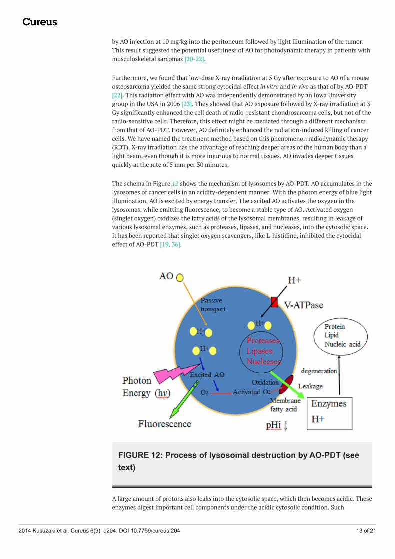
The schema in Figure 12 shows the mechanism of lysosomes by AO-PDT. AO accumulates in thelysosomes of cancer cells in an acidity-dependent manner. With the photon energy of blue lightillumination, AO is excited by energy transfer. The excited AO activates the oxygen in thelysosomes, while emitting fluorescence, to become a stable type of AO. Activated oxygen(singlet oxygen) oxidizes the fatty acids of the lysosomal membranes, resulting in leakage ofvarious lysosomal enzymes, such as proteases, lipases, and nucleases, into the cytosolic space.It has been reported that singlet oxygen scavengers, like L-histidine, inhibited the cytocidaleffect of AO-PDT [19, 36].
FIGURE 12: Process of lysosomal destruction by AO-PDT (seetext)
A large amount of protons also leaks into the cytosolic space, which then becomes acidic. Theseenzymes digest important cell components under the acidic cytosolic condition. Such
2014 Kusuzaki et al. Cureus 6(9): e204. DOI 10.7759/cureus.204 13 of 21

lysosomal stress activates caspase, which induces cellular apoptosis with cell swelling, blebformation, and release of microparticles, including exosomes, eventually followed by cellularfragmentation (Figures 5, 6).
Clinical application of AOTProcedure of AOT for Patients with Musculoskeletal Sarcomas (Figure 13A-13E).
FIGURE 13: Procedure of AO therapy for clinical application(see text)A) Sarcoma in the muscle close to major vessels and nerves; B) Tumor resection with marginal /intralesional margin; C) Local administration of AO solution D) Additional tumor resection undera fluorescence microscope; (Photodynamic Surgery: AO-PDS) E) Photodynamic Therapy (AO-PDT); F) Radiodynamic Therapy (AO-RDT)
1) Macroscopic curettage of tumor (Figure 13A, 13B)
If a sarcoma is localized in the muscle or bone close to major nerves, vessels, muscles, or bonesand joints, intralesional tumor excision or marginal resection (extra-capsular resection) of thetumor with partial curettage is performed. These procedures are performed to minimize thedamage to intact muscles and bones or to the major nerves and vessels in close contact with thetumor, in order to obtain good limb function after surgery.
2) Intraoperative Photodynamic Surgery (iPDS) with Local Administration of AO (Figure 13C)
Microscopic curettage with an ultrasonic surgical knife (Olympus Co. Ltd., Tokyo, Japan) isadditionally performed using a fluorescence surgical microscope. After local administration(soaking) of 1 μg/ml of AO solution (SIGMA-ALDRICH CO, St. Louis, MO, USA) for five minutes,washing out of the excess AO solution with saline, and excitation with blue light is followed forfluorovisualization of the tumor. The surgical microscope manufactured by Carl Zeiss Co., Ltd,Oberkochen, Germany is specially equipped with an interference filter (466.5 nm) for selectionof the blue beam from a xenon lamp and an absorption filter (>520 nm) for observation of the
2014 Kusuzaki et al. Cureus 6(9): e204. DOI 10.7759/cureus.204 14 of 21

green fluorescence of AO (Figure 14A). The iPDS was repeated until complete disappearance ofthe green fluorescence from the remnant tumor tissue (Figure 14B, 14C).
FIGURE 14: Fluorescence surgical microscope (A) and scenesof PDS during surgery (B and C)
In this procedure, we apply AO locally by soaking to expose residual tumor fragment aftermacroscopic tumor excision. If the same procedure was applied to the encapsulated tumorbefore excision, AO does not effectively accumulate in the tumor tissue, including in theinterstitial stroma or cells, because the tumor capsule serves as a barrier between the non-acidic normal and acidic cancer extracellular fluid (Figure 15).
2014 Kusuzaki et al. Cureus 6(9): e204. DOI 10.7759/cureus.204 15 of 21

FIGURE 15: Difference in AO accumulation following localadministration between encapsulated and fragmented (non-encapsulated) tumor tissues (see text)
Therefore, for local administration of AO, macroscopic tumor curettage to remove capsularbarrier is a prerequisite. However, if AO is administered systemically, such as by intravenousinjection two hours before the surgery, AO effectively and homogeneously accumulates even inthe encapsulated tumor because it diffuses into the acidic stroma and tumor cells via thecapillaries (Figure 16).
2014 Kusuzaki et al. Cureus 6(9): e204. DOI 10.7759/cureus.204 16 of 21

FIGURE 16: AO accumulation in encapsulated tumor tissue bysystemic administration (see text)
Although we have never tried intravenous administration of AO in humans, no serious adverseeffects were observed following administration of AO at the dose of 1 μg/ml to mice and dogs inan experimental study. In the near future, we propose to investigate the safety of intravenousadministration of AO to humans by carrying out a clinical study with the approval of the IRB,since systemic administration of AO is much better in PDD to detect multiple lesions, such asmetastasis or dissemination than local administration.
3) Intraoperative Photodynamic Therapy (iPDT) (Figure 13E)
After iPDS, intraoperative AO-iPDT is sequentially applied to the tumor curettage area for 10minutes using a surgical microscope under illumination with unfiltered light (more than 5000lx) from a xenon lamp.
4) Postoperative Radiodynamic Therapy (RDT) (Figure 13F)
After closure of the surgical wound without washing out the AO solution, AO-RDT is applied tothe resected area immediately by X-ray irradiation of 5 Gy in a single session in theradiotherapy room for AO-RDT [24, 29-31].
Patients
The 67 patients with high-grade malignant musculoskeletal sarcomas received AOT underapproval from the IRB of our hospitals. The treatment was undertaken with the consent of thepatients and/or their close families obtained after a full explanation was provide about themethod/purpose of the clinical study. The 51 soft tissue sarcoma patients included patientswith high-grade malignant sarcomas, such as synovial sarcoma, rhabdomyosarcoma, MHFs,leiomyosarcoma, etc., and the 15 bone sarcoma patients also included patients with high-grademalignant sarcomas, such as osteosarcoma and Ewing’s sarcoma.
Clinical Outcome
Analysis of the clinical outcomes in our study showed that the five year survival rate (SR) was67.8% and the five year local recurrence-free rate (LRFR) was 71.2% in the 57 patients with soft-tissue sarcoma. According to the AJCC classification, the SR was 100% for Stage II cases, 86.7%for Stage III cases and 0% for Stage IV cases, and the LRFR was 92.3% for Stage II cases, 64.3%for Stage III cases, and 60.2% for Stage IV cases. In regard to the results classified according tothe tumor size, the LRFR was 78% for tumors than 10 cm in diameter, while it was only 46.2%for larger tumors (Figure 2). On the other hand, the SR was 85.7% and the LRFR was 93.8% inthe 15 cases with high-grade malignant bone sarcoma (Figure 3). Thus, while the local controlrate after AO-PDT may not be superior to that after conventional wide resection surgery, theresidual limb function is far superior as compared to that after wide resection. Most of thepatients enrolled in our study showed excellent limb function for running fast, jumping,swimming, and throwing a ball [31].
We believe that all of our patients enrolled in this study would spend the rest of their lives asnearly normal or at least not handicapped people. Maintenance of good limb function is veryimportant for sarcoma survivors, especially children, in order to ensure a long life with a high
2014 Kusuzaki et al. Cureus 6(9): e204. DOI 10.7759/cureus.204 17 of 21

quality of life after surgery.
Toxicity and carcinogeniety of AOIt is generally assumed that because AO is mutagenic in bacteria [46-47], it would becarcinogenic in humans. However, that is not true. Several studies have been carried out toinvestigate the carcinogenicity of AO [48-49]; however, none has provided any evidence tosuggest the carcinogenicity of AO. Therefore, the International Agency of Research on Cancer(IARC) of the WHO and other official reports do not classify AO as a carcinogen [50]. There aresome reports in the literature, in addition to ours, on the application of AO to human clinicalstudies. Two Japanese papers have reported using local/oral administration of AO for thediagnosis of cervical cancer or gastric cancer [51, another in Japanese]. Recently, a studyreported by an Italian group applied our method of AO-RDT to a patient with synovial sarcoma[52]. In 2009, a group from USA also applied AO to some patients with ovarian disease in aclinical study of confocal laser laparoscopic biopsy under FDA approval [53]. The FDA in theUSA has provided approval for the application of AO to particular clinical studies afterinvestigation of the acute and chronic toxicity and carcinogenicity of AO using mice [54]. Ourstudy using mice revealed that the LD50 of AO following intravenous administration was 28-30
mg/kg [18]. The toxicity of 0.1 μg/ml AO administered by intravenous injection was investigatedin dogs and no significant adverse effects were recognized [55]. None of the above clinical orexperimental reports have shown evidence of any serious complications caused by AOadministration. Since the concentration of AO solution used by us in our present clinical studywas very low, and the substance was administered only locally, we believe that the risk ofcarcinogenesis induced by AO in our patients was significantly lower than that associated withmost other known anticancer agents.
ConclusionsIn conclusion, based on the results of basic research and clinical data, we strongly believe thatAOT, consisting of AO-iPDS, AO-iPDT, and AO-RDT, may be a promising new limb salvagemodality for excellent preservation of limb function with a low risk of local tumor recurrence inmusculoskeletal sarcoma patients. This therapeutic modality may also be applicable to manyother solid cancers, although studies on a larger number of patients with longer durations offollow-up are required to verify this notion.
In the AOT procedure, iPDS is useful to detect the residual tumor tissue after intra-lesionaltumor excision and makes it easy to precisely carry out additional excision of residual tumortissue under fluorovisualization using a fluorescence surgical microscope.
Additional InformationDisclosuresThis study did not involve human participants or tissue. This study did not involve animalsubjects or tissue. Conflicts of interest: The authors have declared that no conflicts of interestexist except for the following: Payment/services info: Animal studies were supported andverified by Grants-in-Aid 08877235, 12470312, 11877256 for Scientific Research from theMinistry of Education, Science, Sports and Culture of Japan.
References1. Ishizuka M, Abe F, Sano Y, Takahashi K, Inoue K, Nakajima M, Kohda T, Komatsu N, Ogura S,
Tanaka T: Novel development of 5-aminolevurinic acid (ALA) in cancer diagnoses andtherapy. Int Immunopharmacol. 2011, 11:358-65. 10.1016/j.intimp.2010.11.029
2. Satou S, Ishizawa T, Masuda K, Kaneko J, Aoki T, Sakamoto Y, Hasegawa K, Sugawara Y,
2014 Kusuzaki et al. Cureus 6(9): e204. DOI 10.7759/cureus.204 18 of 21

Kokudo N: Indocyanine green fluorescent imaging for detecting extrahepatic metastasis ofhepatocellular carcinoma. J Gastroenterol. 2013, 48:1136-43. 10.1007/s00535-012-0709-6
3. Schaafsma BE, Mieog JS, Hutteman M, van der Vorst JR, Kuppen PJ, Löwik CW, Frangioni JV,van de Velde CJ, Vahrmeijer AL: The clinical use of indocyanine green as a near-infraredfluorescent contrast agent for image-guided oncologic surgery. J Surg Oncol. 2011, 104:323-32. 10.1002/jso.21943
4. Mito JK, Ferrer JM, Brigman BE, Lee CL, Dodd RD, Eward WC, Marshall LF, Cuneo KC, CarterJE, Ramasunder S, Kim Y, Lee WD, Griffith LG, Bawendi MG, Kirsch DG: Intraoperativedetection and removal of microscopic residual sarcoma using wide-field imaging. Cancer.2012, 118:5320-30. 10.1002/cncr.27458
5. Sampath L, Wang W, Sevick-Muraca EM: Near infrared fluorescent optical imaging for nodalstaging. J Biomed Opt. 2008, 13:041312. 10.1117/1.2953498
6. Kapuscinski J, Darzynkiewicz Z, Melamed MR: Interactions of acridine orange with nucleicacids. Properties of complexes of acridine orange with single stranded ribonucleic acid.Biochem Pharmacol. 1983, 32:3679-94.
7. Amagasa J: Mechanisms of photodynamic inactivation of acridine orange-sensitized transferRNA: Participation of singlet oxygen and base damage leading to inactivation. J Radiat Res.1986, 27:339-51.
8. Cools AA, Janssen LH: Fluorescence response of acridine orange to changes in pH gradientsacross liposome membranes. Experientia. 1986, 42:954-6.
9. Zdolsek JM, Olsson GM, Brunk UT: Photooxidative damage to lysosomes of culturedmacrophages by acridine orange. Photochem Photobiol. 1990, 51:67-76.
10. Sastry KS, Gordon MP: The photodynamic inactivation of tobacco mosaic virus and itsribonucleic acid by acridine orange. Biochim Biophys Acta. 1966, 129:32-41.
11. Lewis RM, Goland PP: In vivo staining and retardation of tumors in mice by acridinecompounds. Am J Med Sci. 1948, 215:282-9.
12. Korgaonkar K, Sukhatankara J: Anti-tumor activity of the fluorescent dye, acridine orange, onYoshida sarcoma (ascites). Br J Cancer. 1963, 17:471-3.
13. Giorgio A, Rambaldi M, Maccario P, Ambrosone L, Moles DA: Detection of microorganisms inclinical specimens using slides prestained with acridine orange (AOS). Microbiologica. 1989,12:97-100.
14. Rickman LS, Long GW, Oberst R, Cabanban A, Sangalang R, Smith JI, Chulay JD, Hoffman SL:Rapid diagnosis of malaria by acridine orange staining of centrifuged parasites . Lancet. 1989,1:68-71.
15. Kusuzaki K, Murata H, Takeshita H, Hashiguchi S, Nozaki T, Emoto K, Ashihara T, Hirasawa Y:Intracellular binding sites of acridine orange in living osteosarcoma cells . Anticancer Res.2000, 20:971-5.
16. Parks SK, Chiche J, Pouysségur J: Disrupting proton dynamics and energy metabolism forcancer therapy. Nat Rev Cancer. 2013, 13:611-23. 10.1038/nrc3579
17. Matsubara T, Kusuzaki K, Matsumine A, Shintani K, Satonaka H, Uchida A: Acridine orangeused for photodynamic therapy accumulates in malignant musculoskeletal tumors dependingon pH gradient. Anticancer Res. 2006, 26:187-93.
18. Satonaka H, Kusuzaki K, Matsubara T, Shintani K, Wakabayashi T, Matsumine A, Uchida A:Extracorporeal photodynamic image detection of mouse osteosarcoma in soft tissues utilizingfluorovisualization effect of acridine orange. Oncology. 2006, 70:465-73.
19. Kusuzaki K, Minami G, Takeshita H, Murata H, Hashiguchi S, Nozaki T, Ashihara T, HirasawaY: Photodynamic inactivation with acridine orange on a multidrug-resistant mouseosteosarcoma cell line. Jpn J Cancer Res. 2000, 91:439-45.
20. Kusuzaki K, Aomori K, Suginoshita T, Minami G, Takeshita H, Murata H, Hashiguchi S,Ashihara T, Hirasawa Y: Total tumor cell elimination with minimum damage to normaltissues in musculoskeletal sarcomas following photodynamic therapy with acridine orange.Oncology. 2000, 59:174-80.
21. Kusuzaki K, Suginoshita T, Minami G, Aomori K, Takeshita H, Murata H, Hashiguchi S,Ashihara T, Hirasawa Y: Fluorovisualization effect of acridine orange on mouse osteosarcoma .Anticancer Res. 2000, 20:3019-24.
22. Hashiguchi S, Kusuzaki K, Murata H, Takeshita H, Hashiba M, Nishimura T, Ashihara T,Hirasawa Y: Acridine orange excited by low-dose radiation has a strong cytocidal effect onmouse osteosarcoma. Oncology. 2002, 62:85-93.
2014 Kusuzaki et al. Cureus 6(9): e204. DOI 10.7759/cureus.204 19 of 21

23. Moussavi-Harami F, Mollano A, Martin JA, Ayoob A, Domann FE, Gitelis S, Buckwalter JA:Intrinsic radiation resistance in human chondrosarcoma cells . Biochem Biophys Res Commun.2006, 346:379-85.
24. Kusuzaki K, Murata H, Matsubara T, Miyazaki S, Shintani K, Seto M, Matsumine A, Hosoi H,Sugimoto T, Uchida A: Clinical outcome of a novel photodynamic therapy technique usingacridine orange for synovial sarcomas. Photochem Photobiol. 2005, 81:705-9.
25. Kusuzaki K, Murata H, Matsubara T, Miyazaki S, Okamura A, Seto M, Matsumine A, Hosoi H,Sugimoto T, Uchida A: Clinical trial of photodynamic therapy using acridine orangewith/without low dose radiation as new limb salvage modality in musculoskeletal sarcomas.Anticancer Res. 2005, 25:1225-35.
26. Nakamura T, Kusuzaki K, Matsubara T, Matsumine A, Murata H, Uchida A: A new limbsalvage surgery in cases of high-grade soft tissue sarcoma using photodynamic surgery,followed by photo- and radiodynamic therapy with acridine orange. J Surg Oncol. 2008,97:523-8. 10.1002/jso.21025
27. Matsubara T, Kusuzaki K, Matsumine A, Murata H, Satonaka H, Shintani K, Nakamura T,Hosoi H, Iehara T, Sugimoto T, Uchida A: A new therapeutic modality involving acridineorange excitation by photon energy used during reduction surgery for rhabdomyosarcomas.Oncol Rep. 2009, 21:89-94.
28. Matsubara T, Kusuzaki K, Matsumine A, Murata H, Nakamura T, Uchida A, Sudo A: Clinicaloutcomes of minimally invasive surgery using acridine orange for musculoskeletal sarcomasaround the forearm, compared with conventional limb salvage surgery after wide resection. JSurg Oncol. 2010, 102:271-5. 10.1002/jso.21602
29. Matsubara T, Kusuzaki K, Matsumine A, Murata H, Marunaka Y, Hosogi S, Uchida A, Sudo A:Photodynamic therapy with acridine orange in musculoskeletal sarcomas . J Bone Joint SurgBr. 2010, 92:760-2. 10.1302/0301-620X.92B6.23788
30. Kusuzaki K, Hosogi S, Ashihara E, Matsubara T, Satonaka H, Nakamura T, Matsumine A, SudoA, Uchida A, Murata H, Baldini N, Fais S, Marunaka Y: Translational research ofphotodynamic therapy with acridine orange which targets cancer acidity. Curr Pharm Des.2012, 18:1414-20.
31. Kusuzaki K, Ashihara E, Hosogi S, Matsubara T, Satonaka H, Nanamura T, Matsumine A,Murata H, Sudo A, Uchida A, Murata H, Baldini N, Fais S, Marunaka Y: New concept of limbsalvage surgery in musculoskeletal sarcomas with acridine orange therapy. Sarcoma. Butler EJ(ed): Nova Science Publisher Inc, New York; 2012. 123-137.
32. Darzynkiewicz Z, Traganos F, Sharpless T, Melamed MR: Lymphocyte stimulation: a rapidmultiparameter analysis. Proc Natl Acad Sci USA. 1976, 73:2881-4.
33. Takeshita H, Kusuzaki K, Kuzuhara A, Tsuji Y, Ashihara T, Gebhardt MC, Mankin HJ,Springfield DS, Hirasawa Y: Relationship between histologic grade and cytofluorometriccellular DNA and RNA content in primary bone tumors. Anticancer Res. 2001, 21:1271-7.
34. Frankfurt OS: Flow cytometric analysis of double-stranded RNA content distributions . JHistochem Cytochem. 1980, 28:663-9.
35. Wang AH, Quigley GJ, Rich A: Atomic resolution analysis of a 2:1 complex of CpG andacridine orange. Nucleic Acids Res. 1979, 6:3879-90.
36. Lovelace MD, Cahill DM: A rapid cell counting method utilising acridine orange as a noveldiscriminating marker for both cultured astrocytes and microglia. J Neurosci Methods. 2007,165:223-9.
37. Darzynkiewicz Z, Bruno S, Del Bino G, Gorczyca W, Hotz MA, Lassota P, Traganos F: Featuresof apoptotic cells measured by flow cytometry. Cytometry. 1992, 13:795-808.
38. Bertho AL, Santiago MA, Coutinho SG: Flow cytometry in the study of cell death . Mem InstOswaldo Cruz. 2000, 95:429-33.
39. Gatenby RA, Gillies RJ: Why do cancers have high aerobic glycolysis? . Nat Rev Cancer. 2004,4:891-9.
40. Yamagata M, Hasuda K, Stamato T, Tannock IF: The contribution of lactic acid to acidificationof tumours: studies of variant cells lacking lactate dehydrogenase. Br J Cancer. 1998, 77:1726-31.
41. De Milito A, Canese R, Marino ML, Borghi M, Iero M, Villa A, Venturi G, Lozupone F, Iessi E,Logozzi M, Della Mina P, Santinami M, Rodolfo M, Podo F, Rivoltini L, Fais S: pH-dependentantitumor activity of proton pump inhibitors against human melanoma is mediated byinhibition of tumor acidity. Int J Cancer. 2010, 127:207-19. 10.1002/ijc.25009
2014 Kusuzaki et al. Cureus 6(9): e204. DOI 10.7759/cureus.204 20 of 21

42. Fais S, De Milito A, You H, Qin W: Targeting vacuolar H+-ATPases as a new strategy againstcancer. Cancer Res. 2007, 67:10627-30.
43. Swietach P, Vaughan-Jones RD, Harris AL: Regulation of tumor pH and the role of carbonicanhydrase 9. Cancer Metastasis Rev. 2007, 26:299-310.
44. Hiruma H, Katakura T, Takenami T, Igawa S, Kanoh M, Fujimura T, Kawakami T: Vesicledisruption, plasma membrane bleb formation, and acute cell death caused by illuminationwith blue light in acridine orange-loaded malignant melanoma cells. J Photochem PhotobiolB. 2007, 86:1-8.
45. Gerweck LE, Kozin SV, Stocks SJ: The pH partition theory predicts the accumulation andtoxicity of doxorubicin in normal and low-pH-adapted cells. Br J Cancer. 1999, 79:838-42.
46. Zampieri A, Greenberg J: Mutagenesis by acridine orange and proflavine in Escherichia colistrain S. Mutat Res. 1965, 2:552-6.
47. McCann J, Choi E, Yamasaki E, Ames BN: Detection of carcinogens as mutagens in theSalmonella/microsome test: assay of 300 chemicals. Proc Natl Acad Sci USA. 1975, 72:5135-9.
48. Van Duuren BL, Sivak A, Katz C, Melchionne S: Tumorigenicity of acridine orange . Br JCancer. 1969, 23:587-90.
49. Beeken WL, Roessner KD: In vivo labeling of hepatic lysosomes by intragastric administrationof acridine orange. Lab Invest. 1972, 26:173-7.
50. International Agency for Research on Cancer: Acridine Orange. IARC Monographs Program onthe Evaluation of Carcinogenic Risks to Human. IARC (ed): IARC Press, Lyon; 1978. 16:145.
51. Kato A: Gastrofiberscopic diagnosis with acridine orange fluorescence . Gastroenterol Endosc.1970, 12:351-362.
52. Coli A, Bigotti G, Parente R, Massi G: Myxoid monophasic synovial sarcoma: Case report of anunusual histological variant. J Exp Clin Cancer Res. 2006, 25:287-91.
53. Tanbakuchi AA, Rouse AR, Udovich JA, Hatch KD, Gmitro AF: Clinical confocalmicrolaparoscope for real-time in vivo optical biopsies. J Biomed Opt. 2009, 14:044030.10.1117/1.3207139
54. Udovich JA, Besselsen DG, Gmitro AF: Assessment of acridine orange and SYTO 16 for in vivoimaging of the peritoneal tissues in mice. J Microsc. 2009, 234:124-9. 10.1111/j.1365-2818.2009.03153.x
55. Maruo T, Shibuya K, Takahashi M, Nakayama T, Fukunaga K, Orito K: Safety of intravenousadministration of acridine orange in dogs. Intern. J. Appl. Res. Vet. Med. 2012, 10:164-168.
2014 Kusuzaki et al. Cureus 6(9): e204. DOI 10.7759/cureus.204 21 of 21


![SOME N- AND S-HETEROCYCLIC POLYCYCLIC AROMATIC … · ]acridine, benz[c] acridine, dibenz[a, j]acridine, dibenzo[c, h]acri dine and carbazole by gas chromatography from tobacco-smoke](https://img.dokumen.tips/doc/110x75/5e15aaf1fc75030377117681/some-n-and-s-heterocyclic-polycyclic-aromatic-acridine-benzc-acridine-dibenza.jpg)


![SARCOMAS Corregido[1]](https://img.dokumen.tips/doc/110x75/55721128497959fc0b8e7930/sarcomas-corregido1.jpg)