Embed Size (px)
Citation preview

15
Musculo-Skeletal System(Trunk, Limbs, and Head)
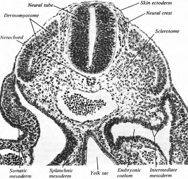
General Statements: Bilaterally, paraxial mesoderm become somites and somitomeres. (Somitomeres develop ros-tral to the notochord in the head. They are like somites, but smaller and less distinctly organized.) The mesoderm comprising each somite differentiates into three regions: — dermatome (lateral) which migrates to form dermis of the skin — sclerotome (medial) forms most of the axial skeleton (vertebrae, ribs, and base of the skull). — myotome (middle) forms skeletal mus-culature. Individual adult muscles are produced by merger of adjacent myotomes.
Note: Nerves make early connections with adjacent myotomes and dermatomes, establishing a segmental innervation pattern. As myotome/dermatome cells migrate to assume adult positions, the segmental nerve supply must follow along to maintain its connection to the innervation target. (Recurrent laryngeal & phrenic nerves travel long distances because their targets migrated far away.)
Skin. Consists of dermis and epidermis. Epidermis, including hair follicles & glands, is derived from ectoderm. Neural crest cells migrate into epidermis and become melanocytes. (Other neural crest cells become tactile disc receptors.) Dermis arises from mesoderm (dermatomes of somites). Each dermatome forms a continu-ous area of skin innervated by one spinal nerve. Because adjacent dermatomes overlap, a locus of adult skin is formed by 2 or 3 dermatomes, and innervated by 2 or 3 spinal nerves.
Muscle. Muscles develop from mesoderm, except for muscles of the iris which arise from optic cup ectoderm. Cardiac and smooth muscles originate from splanchnic mesoderm. All skeletal muscle is derived from paraxial meso-derm that forms somites and, in the rostral region of the head, somitomeres. Mesodermal cells of the myotome region of each so-mite/somitomere differentiate into myoblasts which fuse to form multinucleate muscle cells that synthesize myosin & actin and appear striated. Developing muscles and tendons must be under tension (stretched by growing bone) in order to grow to proper lengths. Muscle development requires innervation. Muscles release trophic molecules that determine muscle cell type (I, IIa, IIb). Also, muscles release trophic molecules that affect nerve growth.
Note: Each anatomical muscle is genetically allocated a specific number of myoblasts that is deter-mined by the time of birth. Thereafter, muscle cell growth is due solely to cellular hypertro-phy. Regeneration (hyperplasia) of adult muscle cells does not occur.
Mesoderm Regions
ectoderm
endoderm
neural tube
neural crest
aorta
coelom
somite:dermatome
sclerotomemyotome
intermediatemesoderm
somaticmesoderm
splanchnic mesoderm
notochord
head
heart limbbudeye
somitomeresin
pharyngeal arches
somites
Somites & Somitomeres


The INNNERVATION PROBLEM: How to establish connections between nerves and their targets during embryonic development? Would you . . .
A] First, have targets migrate to their locations, then
have nerves find them. B] First, connect nerves to targets, then have nerves
follow targets as they migrate.


15
Musculo-Skeletal System(Trunk, Limbs, and Head)
General Statements: Bilaterally, paraxial mesoderm become somites and somitomeres. (Somitomeres develop ros-tral to the notochord in the head. They are like somites, but smaller and less distinctly organized.) The mesoderm comprising each somite differentiates into three regions: — dermatome (lateral) which migrates to form dermis of the skin — sclerotome (medial) forms most of the axial skeleton (vertebrae, ribs, and base of the skull). — myotome (middle) forms skeletal mus-culature. Individual adult muscles are produced by merger of adjacent myotomes.
Note: Nerves make early connections with adjacent myotomes and dermatomes, establishing a segmental innervation pattern. As myotome/dermatome cells migrate to assume adult positions, the segmental nerve supply must follow along to maintain its connection to the innervation target. (Recurrent laryngeal & phrenic nerves travel long distances because their targets migrated far away.)
Skin. Consists of dermis and epidermis. Epidermis, including hair follicles & glands, is derived from ectoderm. Neural crest cells migrate into epidermis and become melanocytes. (Other neural crest cells become tactile disc receptors.) Dermis arises from mesoderm (dermatomes of somites). Each dermatome forms a continu-ous area of skin innervated by one spinal nerve. Because adjacent dermatomes overlap, a locus of adult skin is formed by 2 or 3 dermatomes, and innervated by 2 or 3 spinal nerves.
Muscle. Muscles develop from mesoderm, except for muscles of the iris which arise from optic cup ectoderm. Cardiac and smooth muscles originate from splanchnic mesoderm. All skeletal muscle is derived from paraxial meso-derm that forms somites and, in the rostral region of the head, somitomeres. Mesodermal cells of the myotome region of each so-mite/somitomere differentiate into myoblasts which fuse to form multinucleate muscle cells that synthesize myosin & actin and appear striated. Developing muscles and tendons must be under tension (stretched by growing bone) in order to grow to proper lengths. Muscle development requires innervation. Muscles release trophic molecules that determine muscle cell type (I, IIa, IIb). Also, muscles release trophic molecules that affect nerve growth.
Note: Each anatomical muscle is genetically allocated a specific number of myoblasts that is deter-mined by the time of birth. Thereafter, muscle cell growth is due solely to cellular hypertro-phy. Regeneration (hyperplasia) of adult muscle cells does not occur.
Mesoderm Regions
ectoderm
endoderm
neural tube
neural crest
aorta
coelom
somite:dermatome
sclerotomemyotome
intermediatemesoderm
somaticmesoderm
splanchnic mesoderm
notochord
head
heart limbbudeye
somitomeresin
pharyngeal arches
somites
Somites & Somitomeres


16
Bone.Bonesoriginatefromparaxial mesoderm(endochondralaxialskeletonfromsclero-tomes),somatic mesoderm (endochondralappendicularskeleton),orectomesenchyme (intramem-branous bonesofthecalvariaandfacefromneuralcrest).Ligaments,tendons,andmuscle-relatedconnectivetissueoriginatefromlocalmesenchymeorectomesenchyme. Thus, most bones are formed endochondrally (ossification of a cartilage model), but bones ofthecalvaria(topoftheskull)andthefaceareformedintramembranously(ectomesenchymecellsbecomeosteoblastsdirectlyratherthanbecomingchondroblasts).
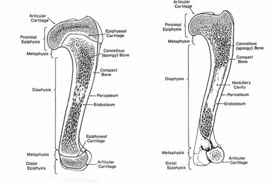
Endochondral bone formation: —localmesenchymeundergoescondensation;somecellsdifferentiateintochondroblasts —chondroblastssecretematrixtoproduceacartilagemodelofthefuturebone;themodelissurrounded by perichondral fibrous tissue — the diaphysis of the cartilage model undergoes ossification first; epiphyseal ossification occurs later; physis ossification is postponed until bones stop growing in length. —overallboneshapeisgeneticallydetermined;surfaceirregularitiesofboneareacquiredduetolocalizedtension(stress)producedbyligamentsandtendons.
Joints.Condensationofmesenchymeproducesaninterzoneregionwithinperichondraltis-sueconnectingadjacentcartilagemodelsofbones.Accordingtothenatureofthefuturejoint,theinterzone becomes fibrous connective tissue, or fibrocartilage, or a synovial cavity.
Synovial jointsformasfollows: —mesenchymeatthecenteroftheinterzoneundergoescavitationandthetissueborderingthecavitybecomesyno-vial membrane;unevenexpansionofthesynovialcavitycreatessynovialfolds(theinterzonemesenchymealsoformsintra-articularligamentsandtendonswherethesearepresent); —perichondraltissuesurroundingtheinterzonebecomesjoint capsuleandlocalizedthick-eningsofthejointcapsuleformsligaments.
Note: Muscleactivityisessentialforpropersynovialjointdevelopmentafterthejointcavitydevelops.Jointsmustmoveduringin uteroandpostnataldevelopment to prevent ankylosis (fixed/frozen joint).
Regional Specifics
Trunk Region: Skeletalmusculatureisformedbysomitemyotomeswhichfusetoformbroadmusclesthataresegmentallyinnervated(eachmyotomebringsitsowninnervationasitmergeswithadjacentmyotomes).Myotomeaccumulationssegregateintoadorsalmass(epimere)innervatedbydorsalbranchesofspinalnervesandaventralmass(hypomere)innervatedbyventralbranchesofspinalnerves.Theepimerebecomesepaxial musclesandthehypomerebecomeshypaxial muscles. Sclerotomesgiverisetovertebraeandribs. The sternum develops differently, from chondrification/os-sification of somatic mesenchyme of the ventral thorax.
Synovial Joint Development
perichondral layer
cartilage (bone)
interzone ligament
cavitation
fibrouscapsule
synovialmembrane
synovialcavity


16
Bone.Bonesoriginatefromparaxial mesoderm(endochondralaxialskeletonfromsclero-tomes),somatic mesoderm (endochondralappendicularskeleton),orectomesenchyme (intramem-branous bonesofthecalvariaandfacefromneuralcrest).Ligaments,tendons,andmuscle-relatedconnectivetissueoriginatefromlocalmesenchymeorectomesenchyme. Thus, most bones are formed endochondrally (ossification of a cartilage model), but bones ofthecalvaria(topoftheskull)andthefaceareformedintramembranously(ectomesenchymecellsbecomeosteoblastsdirectlyratherthanbecomingchondroblasts).
Endochondral bone formation: —localmesenchymeundergoescondensation;somecellsdifferentiateintochondroblasts —chondroblastssecretematrixtoproduceacartilagemodelofthefuturebone;themodelissurrounded by perichondral fibrous tissue — the diaphysis of the cartilage model undergoes ossification first; epiphyseal ossification occurs later; physis ossification is postponed until bones stop growing in length. —overallboneshapeisgeneticallydetermined;surfaceirregularitiesofboneareacquiredduetolocalizedtension(stress)producedbyligamentsandtendons.
Joints.Condensationofmesenchymeproducesaninterzoneregionwithinperichondraltis-sueconnectingadjacentcartilagemodelsofbones.Accordingtothenatureofthefuturejoint,theinterzone becomes fibrous connective tissue, or fibrocartilage, or a synovial cavity.
Synovial jointsformasfollows: —mesenchymeatthecenteroftheinterzoneundergoescavitationandthetissueborderingthecavitybecomesyno-vial membrane;unevenexpansionofthesynovialcavitycreatessynovialfolds(theinterzonemesenchymealsoformsintra-articularligamentsandtendonswherethesearepresent); —perichondraltissuesurroundingtheinterzonebecomesjoint capsuleandlocalizedthick-eningsofthejointcapsuleformsligaments.
Note: Muscleactivityisessentialforpropersynovialjointdevelopmentafterthejointcavitydevelops.Jointsmustmoveduringin uteroandpostnataldevelopment to prevent ankylosis (fixed/frozen joint).
Regional Specifics
Trunk Region: Skeletalmusculatureisformedbysomitemyotomeswhichfusetoformbroadmusclesthataresegmentallyinnervated(eachmyotomebringsitsowninnervationasitmergeswithadjacentmyotomes).Myotomeaccumulationssegregateintoadorsalmass(epimere)innervatedbydorsalbranchesofspinalnervesandaventralmass(hypomere)innervatedbyventralbranchesofspinalnerves.Theepimerebecomesepaxial musclesandthehypomerebecomeshypaxial muscles. Sclerotomesgiverisetovertebraeandribs. The sternum develops differently, from chondrification/os-sification of somatic mesenchyme of the ventral thorax.
Synovial Joint Development
perichondral layer
cartilage (bone)
interzone ligament
cavitation
fibrouscapsule
synovialmembrane
synovialcavity


17
Formation of Vertebrae and Ribs: —sclerotomeregionsofsomitesmigrate&becomeacon-tinuousmasssurroundingthenotochordandneuraltube.Thustheoriginalsomitesegmentationislost —thecontinuousmassdifferentiatesintodiffuse&denseregionsperoriginalsclerotome.Thediffuseregionfromonesomitecombineswiththedenseregionofanadjacentsomitetoproduceacartilagemodelofonevertebra. —betweennewlyformedvertebrae(intervertebraldiscregions)sclerotomemesenchymeformsannulus fibrousandnotochordformsnucleus pulposus(notochorddegeneratesintheregionofthevertebralbody) —ribsdevelopasprocessesofthoracicvertebrae.
Note: Asaresultoftheabovere-segmentation,vertebraeareshiftedrelativetoothersegmentalstructures(seenextpage).Consequently,musclesspanadjacentvertebrae;spinalnervestraverseintervertebralforamina(locateddorsaltointervertebraldiscs);andembryonicintersegmentalarteriesbecomespinalarteriesthatrunalongsidevertebralbodies.
Vertebralanomaliesinclude:stenosisofthevertebralcanal;mal-articu-lation; hemivertebra; and spinal bifida (absent vertebral arch). The notochord, neuraltube,andneuralcrestallplayaroledirectingsomitedifferentiationandvertebralsegmentation(formation).
Note:ThedensoriginatesasthebodyofvertebraC1(atlas),butitfuseswithvertebraC2(axis).
Limbs:
Limbsgrowoutwardfrombodywallsomatopleureaslimbbuds.Bone,cartilage,andconnectivetissueofthelimbarisefromsomaticmesodermofthelimbbud.Dermisandskeletalmusclecomefromdermatomeandmyotomemigrationsintothelimb.
Limb Morphogenesis: — a limb begins as a limb field (an area of somatopleure committedtoformingalimb) —next,alimb budisproducedbylocalizedproliferation&condensationofmesenchyme,coveredbyectoderm —regionsofthelimbdevelopinproximodistalorderasthelimb bud elongates (the shoulder/hip appears first, the manus/pes is thelasttobeadded) — the distal end of the limb bud (footplate) is flattened like apaddleandectodermalongitsoutermarginthickenstoformanapicalridge(theridgeisinducedtoformbyunderlyingmesodermanditinducesthemesodermtocontinuegrowinganddifferentiatingintoalimb)
dorsalbranch
ventralbranch
epaxialmuscles
hypaxialmuscles
coelom
spinalnerve
gut
MyotomeSegregation
L-2
L-3
Canine Dermatomes

14
intervertebralforamen
Features of Vertebrae
nucleus pulposusanulus fibrosus
Intervertebral Disc:
lamina
pedicle
Vertebral pcesses:
spinous
transverse
articular
Vertebral body
Vertebral arch:
Transverse Section Through A Vertebra
vertebralforamen
Lateral View of Vertebrae
caudalarticular process
spinousprocess
cranialarticularprocess
transverse process
intervertebral discsVertebra
vertebralforamen
transverseprocess
cranial articularprocess
costal fovea(rib articulation)
spinaous process
Craniolateral Viewof a Thoracic
Vertebra
A Laminectomy Exposes Spinal Cord Within Vertebral Canal
vertebral canal
intervertebralforamen
four primarybranches of a spinal nerve
spinal nerve
Transverse Sectionthrough an Intervertebral Disc
cut vertebral arch (pedicle) spinal cordwing ofatlas
spinal ganglion (T2)
vertebra T5
processes:

18
caudal
sclerotome
spinal n.
neural segmentstube
cranial
myotome
continuous mass
dense diffuse
vertebra
intervertebral discmuscle
neural tube dorsal rootectoderm
notochord
somite
sclerotome myotome
dermatome
ossificationspinous process
ribtubercle
ribhead
vertebralcanal
transverse process
vertebralbody notochord
Sclerotomes to Vertebrae


LIMB GROWTH: Limbs arise from the lateral wall of the body. Thus limbs are products mainly of . . .
A] paraxial mesoderm B] intermediate mesoderm C] somatic lateral mesoderm D] splanchnic lateral mesoderm

LIMB GROWTH
How do limbs grow? A] proximal to distal order B] distal to proximal order
How do digits grow? A] digits grow outward from manus/pes B] digits appear by selective degeneration of manus/

17
Formation of Vertebrae and Ribs: —sclerotomeregionsofsomitesmigrate&becomeacon-tinuousmasssurroundingthenotochordandneuraltube.Thustheoriginalsomitesegmentationislost —thecontinuousmassdifferentiatesintodiffuse&denseregionsperoriginalsclerotome.Thediffuseregionfromonesomitecombineswiththedenseregionofanadjacentsomitetoproduceacartilagemodelofonevertebra. —betweennewlyformedvertebrae(intervertebraldiscregions)sclerotomemesenchymeformsannulus fibrousandnotochordformsnucleus pulposus(notochorddegeneratesintheregionofthevertebralbody) —ribsdevelopasprocessesofthoracicvertebrae.
Note: Asaresultoftheabovere-segmentation,vertebraeareshiftedrelativetoothersegmentalstructures(seenextpage).Consequently,musclesspanadjacentvertebrae;spinalnervestraverseintervertebralforamina(locateddorsaltointervertebraldiscs);andembryonicintersegmentalarteriesbecomespinalarteriesthatrunalongsidevertebralbodies.
Vertebralanomaliesinclude:stenosisofthevertebralcanal;mal-articu-lation; hemivertebra; and spinal bifida (absent vertebral arch). The notochord, neuraltube,andneuralcrestallplayaroledirectingsomitedifferentiationandvertebralsegmentation(formation).
Note:ThedensoriginatesasthebodyofvertebraC1(atlas),butitfuseswithvertebraC2(axis).
Limbs:
Limbsgrowoutwardfrombodywallsomatopleureaslimbbuds.Bone,cartilage,andconnectivetissueofthelimbarisefromsomaticmesodermofthelimbbud.Dermisandskeletalmusclecomefromdermatomeandmyotomemigrationsintothelimb.
Limb Morphogenesis: — a limb begins as a limb field (an area of somatopleure committedtoformingalimb) —next,alimb budisproducedbylocalizedproliferation&condensationofmesenchyme,coveredbyectoderm —regionsofthelimbdevelopinproximodistalorderasthelimb bud elongates (the shoulder/hip appears first, the manus/pes is thelasttobeadded) — the distal end of the limb bud (footplate) is flattened like apaddleandectodermalongitsoutermarginthickenstoformanapicalridge(theridgeisinducedtoformbyunderlyingmesodermanditinducesthemesodermtocontinuegrowinganddifferentiatingintoalimb)
dorsalbranch
ventralbranch
epaxialmuscles
hypaxialmuscles
coelom
spinalnerve
gut
MyotomeSegregation
L-2
L-3
Canine Dermatomes

19
—mechanically,limbgrowthconsistsof: - elongation of a dorsoventrally flattened limb bud - ventroflexion of the distal half of the limb (ventral now faces medially) -pronationofthedistalhalf(previousmedialsurfacenowbecomescaudal) —separatedigitsareproducedbyinterdigitalnecroticzones(species withfewerdigitsundergofurtherdegenerationand/orfusionofdigits); —localmesenchymecondensestoformcartilagemodelsoflimbbones —myotomecellsmigrateintothebaseofthelimbformingextensor& flexor muscle masses that subsequently segregate into the individualmusclesofthelimb; —vesselsandnervesgrowintothelimb.
Clinical considerations: Achondroplasia (dwarfism; Dachshund) — inherited, systemic prema-ture ossification of physes of extremities.
Arthrogryposis[Gr.gryposis=crooked]canresultfrommalformedjoints,denervation,abnormalmuscletension,orimpairedmobilityin utero. Polydactyly(extradigits);syndactyly(fuseddigits);brachydactyly(stumpydigitsGr.dactylos=digit] Amelia(nolimb);meromelia(absenceofpartoflimb);micromelia(smalllimbGr.melos=limb] Note:phocomelia(seallimb)=absenceofproximalsegment(s)oflimbwasaconsequenceofpregnantwomentakingthalidomideinthelate1950s.
Head Region: Theheadconsistsofacranium,whichcontainsthebrainwithinacranialcavity,andaface.
The face develops separately from the frontonasal process and first pharyngeal arch. Since the face and cranium have different embryonic origins they can be independently influenced genetically (e.g., inthecaseofbrachycephalicbreeds)orbyteratogens.
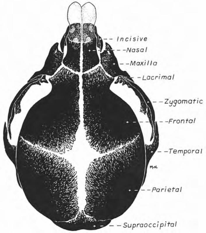
Skull. Bones of the base of the cranium develop endochondrally; the relatively flat bones that comprisethecalvaria(roofofthecranium)andthefacedevelopintramembranously.(Themandiblehasacomplexorigininvolvingbothendochondralandintramembranousdevelopment.)
Theendochondralbonesareformedfromsclerotomesofsomitomersandsclerotomesofthefirst four somites (occipitalsomites).
Theintramembranousbonesarisefromectomesenchyme(mesenchymederivedfromneuralcrest),whichgivesrisetocartilage,bone,andconnectivetissueofthefaceanddorsalhead.Intramem-branous bones articulate by means of fibrous joints called sutures. Widened suture areas, at the corners of growing bones,arecalledfontanels.Suturesandfontanelsallowbonyplatestooverlaponeanotherduringparturition.
Manus/PesDevelopment
foot plate
cell death
digit
face(intramembranous)
base of cranium (endochondral )
Regions of the Skullcalvaria of cranium(intramembranous )


19
—mechanically,limbgrowthconsistsof: - elongation of a dorsoventrally flattened limb bud - ventroflexion of the distal half of the limb (ventral now faces medially) -pronationofthedistalhalf(previousmedialsurfacenowbecomescaudal) —separatedigitsareproducedbyinterdigitalnecroticzones(species withfewerdigitsundergofurtherdegenerationand/orfusionofdigits); —localmesenchymecondensestoformcartilagemodelsoflimbbones —myotomecellsmigrateintothebaseofthelimbformingextensor& flexor muscle masses that subsequently segregate into the individualmusclesofthelimb; —vesselsandnervesgrowintothelimb.
Clinical considerations: Achondroplasia (dwarfism; Dachshund) — inherited, systemic prema-ture ossification of physes of extremities.
Arthrogryposis[Gr.gryposis=crooked]canresultfrommalformedjoints,denervation,abnormalmuscletension,orimpairedmobilityin utero. Polydactyly(extradigits);syndactyly(fuseddigits);brachydactyly(stumpydigitsGr.dactylos=digit] Amelia(nolimb);meromelia(absenceofpartoflimb);micromelia(smalllimbGr.melos=limb] Note:phocomelia(seallimb)=absenceofproximalsegment(s)oflimbwasaconsequenceofpregnantwomentakingthalidomideinthelate1950s.
Head Region: Theheadconsistsofacranium,whichcontainsthebrainwithinacranialcavity,andaface.
The face develops separately from the frontonasal process and first pharyngeal arch. Since the face and cranium have different embryonic origins they can be independently influenced genetically (e.g., inthecaseofbrachycephalicbreeds)orbyteratogens.
Skull. Bones of the base of the cranium develop endochondrally; the relatively flat bones that comprisethecalvaria(roofofthecranium)andthefacedevelopintramembranously.(Themandiblehasacomplexorigininvolvingbothendochondralandintramembranousdevelopment.)
Theendochondralbonesareformedfromsclerotomesofsomitomersandsclerotomesofthefirst four somites (occipitalsomites).
Theintramembranousbonesarisefromectomesenchyme(mesenchymederivedfromneuralcrest),whichgivesrisetocartilage,bone,andconnectivetissueofthefaceanddorsalhead.Intramem-branous bones articulate by means of fibrous joints called sutures. Widened suture areas, at the corners of growing bones,arecalledfontanels.Suturesandfontanelsallowbonyplatestooverlaponeanotherduringparturition.
Manus/PesDevelopment
foot plate
cell death
digit
face(intramembranous)
base of cranium (endochondral )
Regions of the Skullcalvaria of cranium(intramembranous )


20
Note:AuditoryossiclesariseendochondrallyfrompharyngealarchesI(malleus&incus)andII(stapes).
Muscles.Musclesoftheheadarisefrommyotomesderivedfromsomitomeres(seven)orsomites(fouroccipitalsomites: Somitomeremyotomesmigratetotheorbit(twogivingriseeyemuscles)ortheymigratetopharyngealarches(becomingmusclesofmastication,facialexpressionmuscles). Somitemyotomesbecometongueandneckmusclesandtheymigratetopharyngealarches(IV-VI),becomingpharyngeal,laryngeal&esophagealmuscles. Cranialnervesestablishearlyconnectionswithadjacentsomitomeres&somitesandac-company them to definitive muscle sites. Pharyngeal arches are each innervated by specific cranial nerves(I=trigeminal;II=facial;III=glossopharyngeal;IV-VI=vagus).
Pharyngeal (Branchial) Arch Summary: Ectomesenchymemigratestopharyngealarchestoformconnectivetissue,cartilageandbone.Somitomere/somitemyotomesmigrateintothearchesandgiverisetoskeletalmuscle.Eacharchisinnervatedbyaparticularcranialnerve.
Firstarch.(innervatedbycranialnerveV) —jawbones(mandible&maxilla);also,ossiclesofthemiddleear(incus&stapes) —musclesofmastication,plusrostraldigastricus,mylohyoid,&tensortympanimm.
Secondarch:(innervatedbycranialnerveVII) —hyoidbones&stapes(ossicleofthemiddleear) —musclesoffacialexpression,includingcaudaldigastricus&stapediusmm.
Thirdarch:(innervatedbycranialnerveIX) —hyoidbones —onepharyngealmuscle(stylopharyngeusmm.)
ArchesIVthroughVI:(innervatedbycranialnerveX) —laryngealcartilages —pharyngealmm&cricothyroidm—innervatedbycranialbranchofX —intrinsiclaryngealmm—innervatedbyrecurrentlaryngealn.ofX