Embed Size (px)
Citation preview
Understanding cancer biology and assessment of complex biological function
requires simultaneous interrogation of multiple pathways combined with context provided by tissue images. Standard methods provide an incomplete picture. Flow cytometry excels at multiplex analysis, but destroys the context provided by tissue. Analysis of individual markers in serial sections is unable to promote understanding of single cell coexpression, particularly for three or more markers at a time.
Development of robust research methods for simultaneous measurement of three or more biomarkers in FFPE tissue or cryosections remains challenging due to antibody species similarity and cross reactivity, stability of labels through multiple rounds of processing, balancing high and low signals and measurement of weakly expressed markers.
The Solution: Opal Multiplex Tissue Staining
With Opal™, you can overcome these challenges and achieve robust, specific multiplex tissue biomarker measurement. Select antibodies based on performance rather than species. The typical 4-plex Opal staining procedure may be completed within one day. The method is compatible with the standard research IHC workflow in your lab and is amenable to automation.
Get More Insight from Tissue with Opal Multiplex Staining
MULTIPLEX TISSUE BIOMARKERSIN CONTEXT
Opal will enable you to:
• Stain and image three or more tissue biomarkers at once
• Use multiple unlabelled primary antibodies raised in the same species, with no cross talk
• Visualize context that is lost in flow cytometry
• Confirm single cell co-expression for many biomarkers in one tissue section
• Get more information while conserving precious tissue
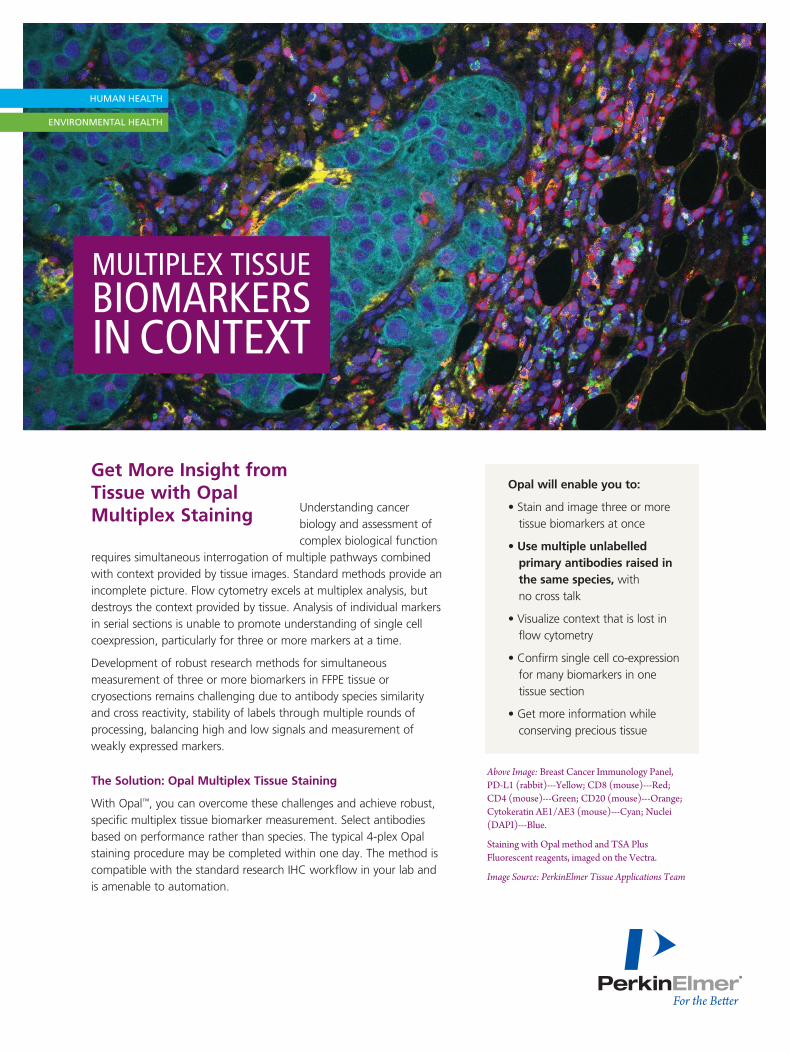
Above Image: Breast Cancer Immunology Panel, PD-L1 (rabbit)---Yellow; CD8 (mouse)---Red; CD4 (mouse)---Green; CD20 (mouse)---Orange; Cytokeratin AE1/AE3 (mouse)---Cyan; Nuclei (DAPI)---Blue.
Staining with Opal method and TSA Plus Fluorescent reagents, imaged on the Vectra.
Image Source: PerkinElmer Tissue Applications Team
For more information please contact your local sales representative or visit www.perkinelmer.com/Opal
For a complete listing of our global offices, visit www.perkinelmer.com/ContactUs
Copyright ©2014, PerkinElmer, Inc. All rights reserved. PerkinElmer® is a registered trademark of PerkinElmer, Inc. All other trademarks are the property of their respective owners. 011660A_01
PerkinElmer, Inc. 940 Winter Street Waltham, MA 02451 USA P: (800) 762-4000 or (+1) 203-925-4602www.perkinelmer.com
What can you achieve with Opal?
PerkinElmer’s applications team has developed a range of research multiplex biomarker panels in FFPE tissue, and more are coming.
• Breast cancer: ER, PR, Her2, Ki-67
• Breast / Ovarian tumor invading lymphocytes: CD4, CD8, CD20
• B-cell lymphoma: CD3, CD10, CD20, CD21, BcL2, BcL6
• Cancer immunology: PD-L1, Foxp1, CD8, CD34
More Opal research multiplex biomarker assays are under development. We can offer assistance in assay development, and for fastest results PerkinElmer offers Opal multiplex IHC services.
How Does Opal Work?
Opal is an iterative process that uses covalent labelling with tyramide signal amplification (TSA™) followed by removal of antibodies. Removal of antibodies has minimal impact on the TSA signal and clears the tissue to be probed with another primary antibody without fear of cross reactivity.
Opal and Multispectral Imaging: More and Better
Multispectral imaging from PerkinElmer enables researchers to obtain quantitative results from up to eight fluorophores simultaneously, making it the perfect system for visualizing Opal tissue. When combined with inForm® image analysis software, the system delivers accurate per cell quantification of multiple biomarkers.
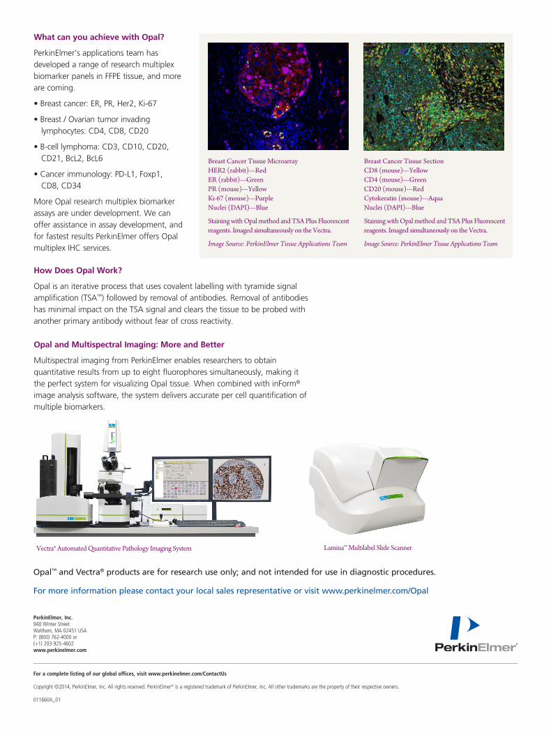
Breast Cancer Tissue Microarray HER2 (rabbit)---Red ER (rabbit)---Green PR (mouse)---Yellow Ki-67 (mouse)---Purple Nuclei (DAPI)---Blue
Staining with Opal method and TSA Plus Fluorescent reagents. Imaged simultaneously on the Vectra.
Image Source: PerkinElmer Tissue Applications Team
Breast Cancer Tissue Section CD8 (mouse)---Yellow CD4 (mouse)---Green CD20 (mouse)---Red Cytokeratin (mouse)---Aqua Nuclei (DAPI)---Blue
Staining with Opal method and TSA Plus Fluorescent reagents. Imaged simultaneously on the Vectra.
Image Source: PerkinElmer Tissue Applications Team
Lamina™ Multilabel Slide ScannerVectra® Automated Quantitative Pathology Imaging System
Opal™ and Vectra® products are for research use only; and not intended for use in diagnostic procedures.