Embed Size (px)
Citation preview

Multiple Plastids Collected by the Dinoflagellate Dinophysis mitrathrough Kleptoplastidy
Goh Nishitani,a,b Satoshi Nagai,a Shiho Hayakawa,c Yuki Kosaka,d Kiyonari Sakurada,e Takashi Kamiyama,a and Takashi Gojoboric
Harmful Algal Bloom Division, National Research Institute of Fisheries and Environment of Inland Sea, Hatsukaichi, Hiroshima, Japana; Graduate School of AgriculturalScience, Tohoku University, Amamiya-machi, Aoba-ku, Sendai, Japanb; Center for Information Biology and DNA Data Bank of Japan, National Institute of Genetics,Mishima, Shizuoka, Japanc; Aomori Prefectural Fisheries Research Center, Aquaculture Institute, Moura, Hiranai-machi, Aomori, Japand; and Kumamoto PrefecturalFisheries Research Center, Oyano-machi, Kamiamakusa, Kumamoto, Japane
Kleptoplastidy is the retention of plastids obtained from ingested algal prey, which may remain temporarily functional andbe used for photosynthesis by the predator. We showed that the marine dinoflagellate Dinophysis mitra has great klepto-plastid diversity. We obtained 308 plastid rbcL sequences by gene cloning from 14 D. mitra cells and 102 operational taxo-nomic units (OTUs). Most sequences were new in the genetic database and positioned within Haptophyceae (227 sequences[73.7%], 80 OTUs [78.4%]), particularly within the genus Chrysochromulina. Others were closely related to Prasinophy-ceae (16 sequences [5.2%], 5 OTUs [4.9%]), Dictyochophyceae (14 sequences [4.5%], 5 OTUs [4.9%]), Pelagophyceae (14sequences [4.5%], 1 OTU [1.0%]), Bolidophyceae (3 sequences [1.0%], 1 OTU [1.0%]), and Bacillariophyceae (1 sequence[0.3%], 1 OTU [1.0%]); however, 33 sequences (10.8%) as 9 OTUs (8.8%) were not closely clustered with any particulargroup. Only six sequences were identical to those of Chrysochromulina simplex, Chrysochromulina hirta, Chrysochro-mulina sp. TKB8936, Micromonas pusilla NEPCC29, Micromonas pusilla CCMP491, and an unidentified diatom. Thus, wedetected >100 different plastid sequences from 14 D. mitra cells, strongly suggesting kleptoplastidy and the need for mix-otrophic prey such as Laboea, Tontonia, and Strombidium-like ciliates, which retain numerous symbiotic plastids fromdifferent origins, for propagation and plastid sequestration.
Dinoflagellates are a diverse group of unicellular flagellate eu-karyotic algae which widely inhabit fresh and marine waters
of the world and serve as significant primary producers. Manydinoflagellate species are photosynthetic. The diversity of plastids(chloroplasts) in dinoflagellates results from the acquisition ofdifferent types of photosynthetic endosymbionts (3, 10, 23, 49, 51,54), heterotrophic nutrition (4, 36, 46), or both, i.e., mixotrophy(50, 60). The types of plastids found in dinoflagellates to date areclassified into five categories: (i) a type containing chlorophyll aplus c (a/c) and fucoxanthin derived from a diatom, (ii) a chloro-phyll a/b type derived from a chlorophyte, (iii) a chlorophyll a/cand fucoxanthin derivative type derived from a haptophyte, (iv) achlorophyll a/c and phycobilin type derived from a cryptophyte(51), and (v) kleptoplastids (12, 49, 51). In addition to dinoflagel-lates with permanent plastids (permanent endosymbioses), thereare several taxonomic groups of dinoflagellates having kleptoplas-tids (stolen plastids) (49), which are temporary but functionalplastids captured from prey (6, 13, 20, 22, 29, 37, 40, 41, 44, 52,57). Amphidinium poecilochroum (22), Amphidinium latum (13),Amylax buxus and Amylax triacantha (21), Gymnodinium aerugi-nosum (52), Gymnodinium acidotum (6, 62), Gymnodinium graci-lentum (53), Cryptoperidiniopsis sp. (5), Pfiesteria piscicida (29),and mixotrophic Dinophysis species (35, 37, 40, 41, 44, 57) robplastids from cryptophyte prey directly or indirectly.
Diarrhetic shellfish poisoning (DSP) is a severe gastrointestinaldisease caused by the consumption of shellfish that are contami-nated with DSP toxins (64). DSP toxins are derived from dinofla-gellates belonging to the genera Dinophysis and Prorocentrum (28,63). At present, 11 species of Dinophysis are known to be toxic (28,30). Despite extensive studies in the last 2 decades, little is knownabout the ecophysiology, toxicology, and bloom mechanisms ofDinophysis species, primarily because of the inability to culture
them (15, 30, 38, 48). Recently, feeding by myzocytosis was re-vealed in Dinophysis acuminata, Dinophysis caudata, Dinophysisfortii, and Dinophysis infundibulus, with successful culturing of thespecies at high cell densities (0.8 � 103 to 11.0 � 103 cells ml�1)(37, 40, 41, 44). The four Dinophysis species feed on the marineciliate Myrionecta rubra (Mesodinium rubrum) (Mesodiniidae:Litostomatea) grown with the cryptophyte Teleaulax. The exper-imental data clearly showed that these species could not grow byingestion of only the Teleaulax sp. Instead, the Dinophysis speciesrequired M. rubra as prey for enabling their vegetative growth andsequestered the ciliate plastids in order to utilize them as klepto-plastids (37).
Dinophysis mitra is known to be one of the toxic Dinophysisspecies (28) and a kleptoplastidic species, and on the basis of trans-mission electron microscopy (TEM) and molecular analysis, it isspeculated that the kleptoplastids have originated from hapto-phytes (20). Usually, D. mitra retains a number of plastids in a cell(Fig. 1); these plastids are robbed from other microalgae, and theyretain their photosynthetic activity in D. mitra cells (20). How-ever, in this regard, only one study has been conducted thus far;therefore, plastid diversity in D. mitra and precise origins fromprey organisms remain unknown. Naturally, no D. mitra culturehas been established to date because of lack of prey information. Inthe present study, we analyzed the plastid rbcL (encoding the large
Received 15 August 2011 Accepted 4 November 2011
Published ahead of print 18 November 2011
Address correspondence to Satoshi Nagai, [email protected].
Copyright © 2012, American Society for Microbiology. All Rights Reserved.
doi:10.1128/AEM.06544-11
0099-2240/12/$12.00 Applied and Environmental Microbiology p. 813–821 aem.asm.org 813
Dow
nloa
ded
from
http
s://j
ourn
als.
asm
.org
/jour
nal/a
em o
n 19
Oct
ober
202
1 by
79.
131.
201.
112.

subunit of ribulose-1,5-bisphosphate carboxylase/oxygenase[RuBisCO]) sequences in natural D. mitra cells isolated from Jap-anese coastal waters to reveal the origins of kleptoplastids in D.mitra and reported a previously unknown type of kleptoplastidy asthe cause of the diversity of acquired plastids.
MATERIALS AND METHODSIsolation of D. mitra and DNA extraction. A total of 14 natural D. mitracells were isolated by micropipetting from seawater samples collected inthe Mutsu Bay,(40°56=N, 140°51=E) in August 2009, Seto Inland Sea(33°53=N, 132°10=E) in July 2009, and Yatsushiro Sea (32°27=N, 130°29=E)in July 2009, in Japan (Fig. 2). Species identification was based on themorphological characteristics observed by light microscopy (1) and mo-lecular analysis. Each cell of D. mitra was washed by several transfers infiltered (pore size, 0.1 �m) seawater, separately inoculated into 0.2-mltubes containing 15 �l of 10% Chelex suspension (Bio-Rad Laboratories
Inc., Richmond, CA), and frozen at �30°C until use. DNA was extractedby heating at 95°C for 20 min, according to Richlen and Barber (47).
PCR and cloning of nuclear 18S rRNA and plastid rbcL genes in D.mitra cells. For amplifying the nuclear 18S rRNA gene of D. mitra, threeprimer pairs (18S-1F1/18S-1R632, 18S-2F576/18S-2R1209, and 18S-3F1129/18S-R1772) were used (Table 1), and the gene sequences weredetermined according to the method of Nishitani et al. (39, 41). In orderto check for any contamination by symbiotic microorganisms within theD. mitra cells or any ingested prey species in food vacuoles, partial nuclear18S rRNA gene sequences were also amplified from the DNA extracts ofthe D. mitra cells by using a universal primer pair (18S-F1289/18S-R1772)(Table 1), and 5 to 11 sequences (6.9 � 1.8, average � standard deviation[SD]) were determined from each D. mitra cell (total number of se-quences, 96).
For amplifying the plastid rbcL (encoding the RuBisCO large subunit)gene of D. mitra, two primer pairs (rbcL-F118/rbcL-R876 and rbcL-F184/rbcL-R704) were designed as universal primers to cover chromophytemicroalgae (Bolidophyceae, Chrysophyceae, Cryptophyceae, Bacillario-phyceae, Dictyochophyceae, Eustigmatophyceae, Haptophyceae, Pelago-phyceae, Pinguiophyceae, and Raphidophyceae) and chlorophyte mi-croalgae (Euglenophyceae, Chlorarachniophyceae, Prasinophyceae,Pedinophyceae, Chlorophyceae, and Trebouxiophyceae), respectively(Table 1). PCR was performed on a thermal cycler in a reaction mixture(50.0 �l) containing 2.0 �l of template DNA, 0.2 mM each deoxynucleo-side triphosphate (dNTP), 1� PCR buffer, 1.5 mM Mg2�, 1.0 U of KOD-Plus, version 2 (Toyobo), that has intensive 3= ¡ 5= exonuclease activity,and 1.0 �M each primer. The PCR cycling conditions were as follows:initial denaturation at 94°C for 2 min, followed by 30 cycles at 94°C for 15s, at 50°C for 30 s, and at 68°C for 1 min. Results of PCR amplification werechecked on 1.5% agarose gels, with ethidium staining.
PCR products were purified and concentrated using Suprec-PCR (Ta-kara Bio, Shiga, Japan). Following an additional A-overhang elongationstep, the purified PCR products were ligated to a pGEM-T Easy Vector(Promega, Madison, WI) and then transformed into ECOS competent
FIG 1 Micrographs of a vegetative cell of Dinophysis mitra obtained from anatural sample in bright field (left) and with fluorescence under blue lightexcitation (right). Plastids in the D. mitra cell are emitting red autofluores-cence. Scale bar, 30 �m.
FIG 2 Sampling locations of natural D. mitra cells. The numbers in parentheses refer to the numbers of isolated cells.
Nishitani et al.
814 aem.asm.org Applied and Environmental Microbiology
Dow
nloa
ded
from
http
s://j
ourn
als.
asm
.org
/jour
nal/a
em o
n 19
Oct
ober
202
1 by
79.
131.
201.
112.

Escherichia coli DH5� (Nippon Gene, Tokyo, Japan). Twenty-four whitecolonies were randomly chosen from each library and picked for sequenc-ing. To check whether 24 colonies from a single D. mitra cell were enoughto investigate the plastid diversity within the D. mitra cells, more than 100white colonies were picked from a D. mitra cell (0908MUT01) and se-quenced. The presence of the desired gene insert in the plasmid was ver-ified by colony PCR with a U19 forward primer (5=-GGTTTTCCCAGTCACGACG-3=) and pUC/M13 reverse primer (5=-TCACACAGGAAACAGCTATGAC-3=). DNA sequences were determined using a DYEnamicET Terminator Cycle Sequencing Kit (GE Healthcare, Little Chalfont,Buckinghamshire, United Kingdom) and analyzed on a DNA sequencer(ABI 3100; Applied Biosystems). The forward and reverse sequences werealigned using GENETYX software (Genetyx Corporation), and thealigned sequences were checked against the GenBank database by usingthe nucleotide Basic Local Alignment Search Tool (BLASTN).
Phylogenetic analysis. Partial sequences of the nuclear 18S rRNA andplastid rbcL genes were aligned with the sequences of other related speciesobtained from GenBank using the Clustal W algorithm (59) by carefulchecking manually, and neighbor-joining (NJ) trees were generated usingthe Molecular Evolutionary Genetics Analysis (MEGA) software, version4.0 (58), with the default setting. Bootstrapping with 1,000 replicationswas used to estimate the reliability of the phylogenetic trees. Sequences
were regarded as the same operational taxonomic units (OTUs) accordingto McDonald et al. (31) if they had more than 99% similarity.
Nucleotide sequence accession numbers. Sequences obtained in thisstudy for the nuclear 18S rRNA genes and plastid rbcL genes have beendeposited in the DDBJ under the accession numbers shown in Fig. 3 to 5for the respective genes.
RESULTS AND DISCUSSIONAnalysis of the nuclear 18S rRNA gene in D. mitra. We deter-mined the nuclear 18S rRNA gene sequences (1,740 bp) of the 14D. mitra isolates to identify the exact species although there is noavailable data for this species in the GenBank at present. All thesequences matched completely and were closest to the sequence ofPhalacroma rapa (Fig. 3), which is the most morphologically sim-ilar species to D. mitra, and 29 bp (including insertions/deletionsof 4 bp) were different from those in the sequence of Phalacromarapa EU780655. Phylogenetic analysis and observations of cellmorphology indicated that the 14 isolated cells were certainlythose of D. mitra. Using the universal primers for eukaryotic phy-toplanktons, we also determined the partial nuclear 18S rRNA
TABLE 1 Details of the primers used in this studya
Target region Target organism Primer name Sequence (5=–3=) Annealing site (nt)b
Nuclear 18S rRNA Marine dinoflagellate 18S-1F1 AACCTGGTTGATYCTGCCAG 1–20c
18S-1R632 ACTACGAGCTTTTTAACYGCARC 610–632c
18S-2F576 GGTAATTCCAGCTCYAATRG 576–595c
18S-2R1209 AAGTTTYCCCGTGTTGARTC 1190–1209c
18S-3F1129 GCTGAAACTTAAAGRAATTGACGGA 1129–1153c
Marine eukaryotic microalga 18S-F1289 TGGAGTGATTTGTCTGGTTRATTCCG 1289–1314d
18S-R1772 TCACCTACGGAAACCTTGTTACG 1749–1772d
Plastid rbcL Marine chromophyte microalga rbcL-F118 ACWTGGACWGTWGTWTGGAC 118–137e
rbcL-R876 CATCCAYTTACARATWACACGG 855–876e
Marine chlorophyte microalga rbcL-F184 TCWACWGGWACWTGGACWAC 184–203f
rbcL-R704 AYYTCRCCWGTYTCWGHTTG 685–704f
a All primers were designed in the course of the present study.b nt, nucleotides.c Dinophysis acuta (AJ506973).d Chlamydomonas reinhardtii (M32703).e Imantonia rotunda (AB043696).f Ostreococcus tauri (CR954199).
FIG 3 A neighbor-joining tree inferred from the analysis of nuclear 18S rRNA genes. There were 1,667 informative sites in the final data set. A sequence of D.mitra obtained in this study is highlighted in gray. Bootstrap values of �50% are given as percentage of 1,000 bootstrap replicates at the respective nodes.Prorocentrum micans was used as the outgroup. The scale bar represents the number of substitutions per site.
Diversity of Kleptoplastids in Dinophysis mitra
February 2012 Volume 78 Number 3 aem.asm.org 815
Dow
nloa
ded
from
http
s://j
ourn
als.
asm
.org
/jour
nal/a
em o
n 19
Oct
ober
202
1 by
79.
131.
201.
112.

816 aem.asm.org Applied and Environmental Microbiology
Dow
nloa
ded
from
http
s://j
ourn
als.
asm
.org
/jour
nal/a
em o
n 19
Oct
ober
202
1 by
79.
131.
201.
112.

gene sequences in the DNA extracts of the D. mitra cells to checkfor any contamination by other microorganisms. No other speciesexcept D. mitra were detected in the 96 sequences detected fromthe 14 D. mitra cells, indicating no contamination in the DNAextracts of the D. mitra cells and no symbiotic microorganismswithin the cells. In addition, these data also clearly showed thatany nuclear DNAs, originated from microorganisms ingested inthe food vacuoles, were not detected in the D. mitra cells.
Kleptoplastid diversity in D. mitra cells. When we used theprimer pair rbcL-F118/rbcL-R876 targeting chromophyte mi-croalgae, a total of 292 plastid rbcL sequences (717 bp) were de-termined by gene cloning from the 14 cells of D. mitra, and 97operational taxonomic units were obtained (Fig. 4 and Table 2).Namely, 76, 17, and 21 OTUs were detected from the clone librar-ies in Mutsu Bay (10 cells of D. mitra), Seto Inland Sea (2 cells),and Yatsushiro Sea (2 cells), respectively. Most of the plastid rbcLsequences detected from the 14 D. mitra cells were novel ones inthe GenBank database and positioned within the lineage of Hap-tophyceae (227 sequences, 80 OTUs), particularly within the ge-nus Chrysochromulina. Other sequences were closely related toDictyochophyceae (14 sequences, 5 OTUs), Pelagophyceae (14sequences, 1 OTU), Bolidophyceae (3 sequences, 1 OTU), andBacillariophyceae (1 sequence, 1 OTU); however, 33 sequences as9 OTUs were not closely clustered with any particular group, andtherefore these OTUs were provisionally named as unknown(clade 1, clade 2, and clade 3) (Fig. 4).
About 500 species are now recognized in Haptophyceae; how-ever, the total number of species has been estimated at ca. 2,000,which is four times those currently known (2). This implies that
haptophyte diversity remains to be fully described. Recent geneticstudies have suggested the existence of hidden diversity in Hapto-phyceae (7, 31), detecting a number of novel sequences from en-vironmental clone libraries. Therefore, it is always possible thatenvironmental samples contain previously undescribed species.However, we obtained four plastid rbcL sequences identical tothose of Chrysochromulina simplex (Haptophyceae: Prymnesia-les), Chrysochromulina hirta (Haptophyceae: Prymnesiales),Chrysochromulina sp. TKB8936 (Haptophyceae: Prymnesiales),and an unidentified diatom (Bacillariophyceae). The unidentifieddiatom is reported to be an endosymbiont in the marine dinofla-gellate Peridinium quinquecorne (14). Nuclear 18S rRNA geneanalysis indicated that the diatom species was closely related toChaetoceros sp. (14), and plastid rbcL gene analysis showed a closephylogenetic affiliation with Chaetoceros socialis (Fig. 4).
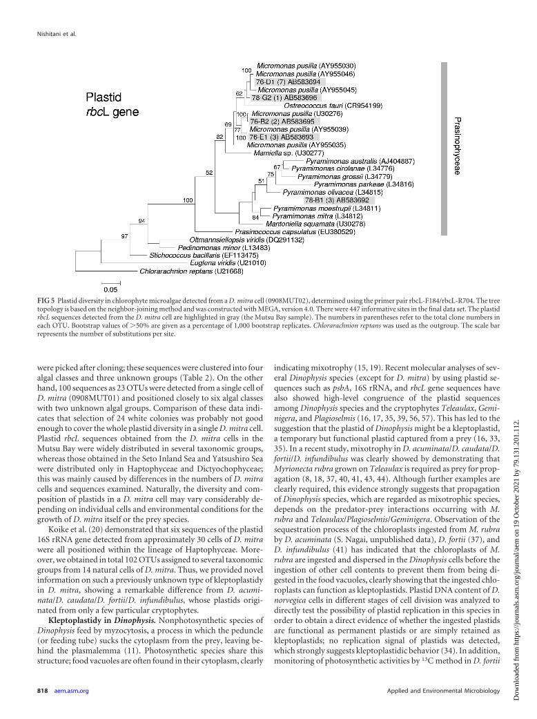
When the primer pair rbcL-F184/rbcL-R704 was used for chlo-rophyte microalgae, a very thin PCR band was obtained only forthe cell of D. mitra 0908MUT02. Five different OTUs from 16plastid rbcL sequences (481 bp) were obtained (Fig. 5 and Table 2).All the OTUs were positioned within the lineage of Prasinophy-ceae; four of the five OTUs were positioned in the Micromonaspusilla clade, and the remaining one OTU was positioned in thePyramimonas clade. Chlorophyte plastids appear to be markedlyless abundant than chromophyte plastids in the natural D. mitracells.
In the plastid rbcL gene analysis, 10 to 40 sequences (16.0 � 4.9,average � standard deviation [SD]) as 6 to 26 OTUs (13.4 � 6.1,average � SD) were successfully determined in 13 D. mitra cellswhen 24 or 48 (in the case of D. mitra 0908MUT02) white colonies
FIG 4 Plastid diversity in chromophyte microalgae detected from natural D. mitra cells, determined using the primer pair rbcL-F118/rbcL-R876. The treetopology is based on the neighbor-joining method and was constructed with MEGA, version 4.0. There were 627 informative sites in the final data set. The plastidrbcL sequences detected from the D. mitra cells are indicated in colored letters. The numbers in parentheses refer to the total number of clones in each OTU.Bootstrap values of �50% are given as a percentage of 1,000 bootstrap replicates. Palmaria palmata was used as the outgroup. The scale bar represents the numberof substitutions per site. �, sequence of a diatom endosymbiont in the marine dinoflagellate Peridinium quinquecorne (14).
TABLE 2 Number of plastid rbcL clones sequenced in each cell
Isolation area Cell no.
No. of clones assigned to:aTotal no. ofclonessequenced
No. ofOTUsBolido Bacillario Dictyocho Hapto Pelago Prasino
Unknown,clade 1
Unknown,clade 2
Unknown,clade 3
Mutsu Bay 0908MUT01 3 1 4 62 9 4 17 100 230908MUT02 22 16 1 1 40 260908MUT03 5 3 1 1 10 60908MUT04 13 1 14 130908MUT05 14 2 16 130908MUT06 4 9 13 100908MUT07 1 20 2 1 24 230908MUT08 13 13 110908MUT09 1 10 1 12 120908MUT10 1 7 1 1 1 11 10
Seto Inland Sea 0907SET01 13 13 70907SET02 2 12 14 11
Yatsushiro Sea 0907YAT01 15 15 120907YAT02 1 12 13 11
Total 3 1 14 227 14 16 7 5 21 308 102a Bolido, Bolidophyceae; Bacillario, Bacillariophyceae; Dictyocho, Dictyochophyceae; Hapto, Haptophyceae; Pelago, Pelagophyceae; Prasino, Prasinophyceae; Unknown, unknownalgae.
Diversity of Kleptoplastids in Dinophysis mitra
February 2012 Volume 78 Number 3 aem.asm.org 817
Dow
nloa
ded
from
http
s://j
ourn
als.
asm
.org
/jour
nal/a
em o
n 19
Oct
ober
202
1 by
79.
131.
201.
112.
were picked after cloning; these sequences were clustered into fouralgal classes and three unknown groups (Table 2). On the otherhand, 100 sequences as 23 OTUs were detected from a single cell ofD. mitra (0908MUT01) and positioned closely to six algal classeswith two unknown algal groups. Comparison of these data indi-cates that selection of 24 white colonies was probably not goodenough to cover the whole plastid diversity in a single D. mitra cell.Plastid rbcL sequences obtained from the D. mitra cells in theMutsu Bay were widely distributed in several taxonomic groups,whereas those obtained in the Seto Inland Sea and Yatsushiro Seawere distributed only in Haptophyceae and Dictyochophyceae;this was mainly caused by differences in the numbers of D. mitracells and sequences examined. Naturally, the diversity and com-position of plastids in a D. mitra cell may vary considerably de-pending on individual cells and environmental conditions for thegrowth of D. mitra itself or the prey species.
Koike et al. (20) demonstrated that six sequences of the plastid16S rRNA gene detected from approximately 30 cells of D. mitrawere all positioned within the lineage of Haptophyceae. More-over, we obtained in total 102 OTUs assigned to several taxonomicgroups from 14 natural cells of D. mitra. Thus, we provided novelinformation on such a previously unknown type of kleptoplastidyin D. mitra, showing a remarkable difference from D. acumi-nata/D. caudata/D. fortii/D. infundibulus, whose plastids origi-nated from only a few particular cryptophytes.
Kleptoplastidy in Dinophysis. Nonphotosynthetic species ofDinophysis feed by myzocytosis, a process in which the peduncle(or feeding tube) sucks the cytoplasm from the prey, leaving be-hind the plasmalemma (11). Photosynthetic species share thisstructure; food vacuoles are often found in their cytoplasm, clearly
indicating mixotrophy (15, 19). Recent molecular analyses of sev-eral Dinophysis species (except for D. mitra) by using plastid se-quences such as psbA, 16S rRNA, and rbcL gene sequences havealso showed high-level congruence of the plastid sequencesamong Dinophysis species and the cryptophytes Teleaulax, Gemi-nigera, and Plagioselmis (16, 17, 35, 39, 56, 57). This has led to thesuggestion that the plastid of Dinophysis might be a kleptoplastid,a temporary but functional plastid captured from a prey (16, 33,35). In a recent study, mixotrophy in D. acuminata/D. caudata/D.fortii/D. infundibulus was clearly showed by demonstrating thatMyrionecta rubra grown on Teleaulax is required as prey for prop-agation (8, 18, 37, 40, 41, 43, 44). Although further examples areclearly required, this evidence strongly suggests that propagationof Dinophysis species, which are regarded as mixotrophic species,depends on the predator-prey interactions occurring with M.rubra and Teleaulax/Plagioselmis/Geminigera. Observation of thesequestration process of the chloroplasts ingested from M. rubraby D. acuminata (S. Nagai, unpublished data), D. fortii (37), andD. infundibulus (41) has indicated that the chloroplasts of M.rubra are ingested and dispersed in the Dinophysis cells before theingestion of other cell contents to prevent them from being di-gested in the food vacuoles, clearly showing that the ingested chlo-roplasts can function as kleptoplastids. Plastid DNA content of D.norvegica cells in different stages of cell division was analyzed todirectly test the possibility of plastid replication in this species inorder to obtain a direct evidence of whether the ingested plastidsare functional as permanent plastids or are simply retained askleptoplastids; no replication signal of plastids was detected,which strongly suggests kleptoplastidic behavior (34). In addition,monitoring of photosynthetic activities by 13C method in D. fortii
FIG 5 Plastid diversity in chlorophyte microalgae detected from a D. mitra cell (0908MUT02), determined using the primer pair rbcL-F184/rbcL-R704. The treetopology is based on the neighbor-joining method and was constructed with MEGA, version 4.0. There were 447 informative sites in the final data set. The plastidrbcL sequences detected from the D. mitra cell are highlighted in gray (the Mutsu Bay sample). The numbers in parentheses refer to the total clone numbers ineach OTU. Bootstrap values of �50% are given as a percentage of 1,000 bootstrap replicates. Chlorarachnion reptans was used as the outgroup. The scale barrepresents the number of substitutions per site.
Nishitani et al.
818 aem.asm.org Applied and Environmental Microbiology
Dow
nloa
ded
from
http
s://j
ourn
als.
asm
.org
/jour
nal/a
em o
n 19
Oct
ober
202
1 by
79.
131.
201.
112.
without the prey ciliate after feeding heavily on M. rubra clearlyindicated that the activities decreased rapidly within 1 week (S.Nagai, unpublished data). However, on the basis of TEM andlaboratory culture experiments, Garcia-Cuetos et al. (8) foundthat several plastid features separated D. acuminata from both thecryptophyte Teleaulax amphioxeia and the ciliate M. rubra. Theirinterpretation of the data was that D. acuminata harbors perma-nent plastids of cryptophyte origin, not kleptoplastids. Therefore,it remains debatable whether the plastids of the dinoflagellate Di-nophysis are temporarily sequestered (kleptoplastids) or perma-nently established (8, 43, 45). In this study, however, we detectedmore than 100 different plastid sequences from 14 D. mitra cells,which strongly suggested kleptoplastidic behavior. This wouldalso be supported by the fact that natural D. mitra cells withoutplastids have occasionally been observed (20).
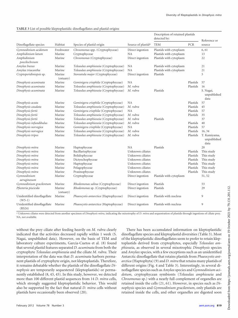
There has been accumulated information on kleptoplastidicdinoflagellate species and kleptoplastid diversities (Table 3). Mostof the kleptoplastidic dinoflagellates seem to prefer to retain klep-toplastids derived from cryptophytes, especially Teleaulax am-phioxeia, as observed in several mixotrophic Dinophysis speciesand Amylax species, with a few exceptions such as an unidentifiedAntarctic dinoflagellate that retains plastids from Phaeocystis ant-arctica (Haptophyta) (9) and D. mitra that retains many plastids ofdifferent origins (Fig. 4 and Table 3). Interestingly, in several di-noflagellate species such as Amylax species and Gymnodinium aci-dotum, cryptophycean symbionts (Teleaulax amphioxeia andChroomonas sp.) with a nearly full complement of organelles areretained inside the cells (21, 61). However, in species such as Di-nophysis species and Gymnodinium gracilentum, only plastids areretained inside the cells, and other organelles are digested soon
TABLE 3 List of possible kleptoplastidic dinoflagellates and plastid origins
Dinoflagellate species Habitat Species of plastid origin Source of plastida
Description of retained plastidsdetected by:
Reference orsourceTEM PCR
Gymnodinium acidotum Freshwater Chroomonas spp. (Cryptophyceae) Direct ingestion Plastids with cytoplasm 6, 61Amphidinium latum Marine Cryptophyceae NA Plastids with cytoplasm 13Amphidinium
poecilochroumMarine Chroomonas (Cryptophyceae) Direct ingestion Plastids with cytoplasm 22
Amylax buxus Marine Teleaulax amphioxeia (Cryptophyceae) NA Plastids with cytoplasm 21Amylax triacantha Marine Teleaulax amphioxeia (Cryptophyceae) NA Plastids with cytoplasm 21Cryptoperidiniopsis sp. Marine
(estuary)Storeatula major (Cryptophyceae) Direct ingestion Plastids 5
Dinophysis acuminata Marine Geminigera criophila (Cryptophyceae) NA Plastids 57Dinophysis acuminata Marine Teleaulax amphioxeia (Cryptophyceae) M. rubra Plastids 16Dinophysis acuminata Marine Teleaulax amphioxeia (Cryptophyceae) M. rubra Plastids S. Nagai,
unpublisheddata
Dinophysis acuta Marine Geminigera criophila (Cryptophyceae) NA Plastids 57Dinophysis caudata Marine Teleaulax amphioxeia (Cryptophyceae) M. rubra Plastids 45Dinophysis fortii Marine Geminigera criophila (Cryptophyceae) NA Plastids 57Dinophysis fortii Marine Teleaulax amphioxeia (Cryptophyceae) M. rubra Plastids 35Dinophysis fortii Marine Teleaulax amphioxeia (Cryptophyceae) M. rubra Plastids 37Dinophysis infundibulus Marine Teleaulax amphioxeia (Cryptophyceae) M. rubra Plastids 40Dinophysis norvegica Marine Geminigera criophila (Cryptophyceae) NA Plastids 57Dinophysis norvegica Marine Teleaulax amphioxeia (Cryptophyceae) M. rubra Plastids 16, 34Dinophysis tripos Marine Teleaulax amphioxeia (Cryptophyceae) M. rubra Plastids T. Kamiyama,
unpublisheddata
Dinophysis mitra Marine Haptophyceae NA Plastids 20Dinophysis mitra Marine Bacillariophyceae Unknown ciliates Plastids This studyDinophysis mitra Marine Bolidophyceae Unknown ciliates Plastids This studyDinophysis mitra Marine Dictyochophyceae Unknown ciliates Plastids This studyDinophysis mitra Marine Haptophyceae Unknown ciliates Plastids This studyDinophysis mitra Marine Pelagophyceae Unknown ciliates Plastids This studyDinophysis mitra Marine Prasinophyceae Unknown ciliates Plastids This studyGymnodinium
aeruginosumMarine Cryptophyceae Direct ingestion Plastids with cytoplasm 51, 52
Gymnodinium gracilentum Marine Rhodomonas salina (Cryptophyceae) Direct ingestion Plastids 53Pfiesteria piscicida Marine
(estuary)Rhodomonas sp. (Cryptophyceae) Direct ingestion Plastids 29
Unidentified dinoflagellate(W5-1)
Marine Phaeocystis antarctica (Haptophyceae) Direct ingestion Plastids with nucleus 9
Unidentified dinoflagellate(RS24)
Marine Phaeocystis antarctica (Haptophyceae) Direct ingestion Plastids with nucleus 9
a Unknown ciliates were detected from another specimen of Dinophysis mitra, indicating the mixotrophy of D. mitra and sequestration of plastids through ingestions of ciliate prey.NA, not available.
Diversity of Kleptoplastids in Dinophysis mitra
February 2012 Volume 78 Number 3 aem.asm.org 819
Dow
nloa
ded
from
http
s://j
ourn
als.
asm
.org
/jour
nal/a
em o
n 19
Oct
ober
202
1 by
79.
131.
201.
112.

after they are ingested, possibly showing the evolutionary processof trial and selection to utilize kleptoplastids more functionally.Members of the genus Dinophysis are perhaps in the earliest stagesof plastid acquisition (10). What “benefit” do some dinoflagellatespecies derive from kleptoplastids? The presence of starch indi-cates that photosynthate is produced (29). It has been assumedthat the starch produced by cryptophyte kleptoplastids is an avail-able carbon source for the host (23, 50), implying that photosyn-thesis is a critical mechanism for supplementing an energy sourceto survive starvation periods when food is scarce. In any case, werevealed that D. mitra has the novel type of kleptoplastidy.
Origin of plastids in D. mitra. Photosynthetic Dinophysis spe-cies generally contain plastids of cryptophyte origin; however, D.mitra cells show red fluorescent, non-phycoerythrin-containingplastids, indicating that the plastids are not of cryptophyte origin(Fig. 1). The molecular phylogenetic position of D. mitra, inferredfrom the plastid sequences, indicates that the D. mitra plastid isoriginated mainly of haptophytes. This is also supported by themorphological features of the plastids as observed using TEM;however, no other organelles such as nuclei and Golgi bodies wereobserved (20). Measurements of photosynthetic activity by 14Cincorporation method clearly showed the activity in the retainedplastids, and analysis of the plastid 16S rRNA gene revealed het-erogeneity in the sequences of six clones obtained from differentD. mitra cells, which strongly suggests that the kleptoplastidy orig-inated from several haptophyte species (20).
The presence of food vacuoles has been confirmed in naturalD. mitra cells (20, 42), indicating predation of some unknownprey organisms by myzocytosis. In this study, we obtained morethan 100 different plastid sequences from 14 cells of D. mitra;these sequences were assigned to several microalgal classes such asBolidophyceae, Bacillariophyceae, Dictyochophyceae, Haptophy-ceae, Pelagophyceae, and Prasinophyceae. This suggests that D.mitra is a unique dinoflagellate, which can easily acquire func-tional plastids from many and different microalgal origins byfeeding on several ciliate prey species, in comparison with otherDinophysis species possessing kleptoplastids, originating fromonly one or two particular cryptophytes obtained by ingestion of aciliate prey, M. rubra.
It is well known that the ciliates Laboea, Strombidium, andTontonia retain hundreds of plastids (symbiotic plastids) (24,25, 26, 27, 55) from different origins, i.e., Chrysophyceae,Bacillariophyceae, Dinophyceae, Euglenophyceae, Haptophy-ceae, and Ulvophyceae, as inferred on the basis of cytologicalcharacteristics observed by TEM (25, 27, 55) and PCR (32).Plastids are always situated at the periphery of ciliate cells andnot surrounded by the dense endoplasmic reticulum (i.e., pres-ent outside the digestive system) to be retained and utilized asfunctional plastids (27). Our data strongly suggest mixotrophyin D. mitra, which requires prey such as Laboea, Tontonia, andStrombidium-like ciliates for its propagation and carries outthe sequestration of robbed plastids, in a manner similar to thatof other Dinophysis species (37, 41). Establishing laboratorycultures would also further understanding of the ecophysiol-ogy and toxicology of toxic dinoflagellate species.
ACKNOWLEDGMENTS
This work was supported by a Grant-in-Aid for Scientific Research (B)from the Japan Society for the Promotion of Science (KAKENHI, number20380116) and a Grant-in-Aid for Young Scientists (B) from the Ministry
of Education, Culture, Sports, Science and Technology (KAKENHI, num-ber 21780191).
REFERENCES1. Abé TH. 1967. The armored Dinoflagellata: II. Prorocentridae and dino-
physidae (B)—Dinophysis and its allied genera. Publ. Seto Mar. Biol. Lab.15:37–78.
2. Andersen RA. 1992. Diversity of eukaryotic algae. Biodivers. Conserv.1:267–292.
3. Archibald JM, Keeling PJ. 2004. The evolutionary history of plastids: amolecular phylogenetic perspective, p 55–74. In Hirt RP, Horner DS (ed),Organelles, genomes, and eukaryote phylogeny: an evolutionary synthesisin the age of genomics. CRC Press, Boca Raton, FL.
4. Elbrächter M. 1991. Food uptake mechanisms in phagotrophic dinofla-gellates and classification, p 303–312. In Patterson DJ, Larsen J (ed), Thebiology of free-living heterotrophic flagellates. Clarendon Press, Oxford,United Kingdom.
5. Eriksen NT, Hayes KC, Lewitus AJ. 2002. Growth responses of themixotrophic dinoflagellates, Cryptoperidiniopsis sp. and Pfiesteria pisci-cida, to light under prey-saturated conditions. Harmful Algae 1:191–203.
6. Fields SD, Rhodes RG. 1991. Ingestion and retention of Chroomonas spp.(Cryptophyceae) by Gymnodinium acidotum (Dinophyceae). J. Phycol.27:525–529.
7. Fuller NJ, et al. 2006. Analysis of photosynthetic picoeukaryote diversityat open ocean sites in the Arabian Sea using a PCR biased towards marinealgal plastids. Aquat. Microb. Ecol. 43:79 –93.
8. Garcia-Cuetos L, Moestrup Ø, Hansen PJ, Daugbjerg N. 2010. The toxicdinoflagellate Dinophysis acuminata harbors permanent chloroplasts ofcryptomonad origin, not kleptochloroplasts. Harmful Algae 9:25–38.
9. Gast RJ, Moran DM, Dennett MR, Caron DA. 2007. Kleptoplasty in anAntarctic dinoflagellate: caught in evolutionary transition? Environ. Mi-crobiol. 9:39 – 45.
10. Hackett JD, Anderson DM, Erdner DL, Bhattacharya D. 2004.Dinoflagellates: a remarkable evolutionary experiment. Am. J. Bot. 91:1523–1534.
11. Hansen PJ. 1991. Dinophysis: a planktonic dinoflagellate genus which canact both as a prey and a predator of a ciliate. Mar. Ecol. Prog. Ser. 69:201–204.
12. Horiguchi T. 2006. Algae and their chloroplasts with particular referenceto the dinoflagellates. Paleontol. Res. 10:299 –309.
13. Horiguchi T, Pienaar RN. 1992. Amphidinium latum Lebour (Dinophy-ceae), a sand-dwelling dinoflagellate feeding on cryptomonads. Jpn. J.Phycol. 40:353–363.
14. Horiguchi T, Takano Y. 2006. Serial replacement of a diatom endosym-biont in the marine dinoflagellate Peridinium quinquecorne (Peridiniales,Dinophyceae). Phycol. Res. 54:193–200.
15. Jacobson DM, Andersen RA. 1994. The discovery of mixotrophy inphotosynthetic species of Dinophysis (Dinophyceae): light and electronmicroscopical observations of food vacuoles in Dinophysis acuminata, D.norvegica and two heterotrophic dinophysoid dinoflagellates. Phycologia33:97–110.
16. Janson S. 2004. Molecular evidence that plastids in the toxin-producingdinoflagellate genus Dinophysis originate from the free-living cryptophyteTeleaulax amphioxeia. Environ. Microbiol. 6:1102–1106.
17. Janson S, Granéli E. 2003. Genetic analysis of the psbA gene from singlecells indicates a cryptomonad origin of the plastid in Dinophysis (Dino-phyceae). Phycologia 42:473– 477.
18. Kamiyama T, Suzuki T. 2009. Production of dinophysistoxin-1 andpectenotoxin-2 by a culture of Dinophysis acuminata (Dinophyceae).Harmful Algae 8:312–317.
19. Koike K, Koike K, Takagi M, Ogata T, Ishimaru T. 2000. Evidence ofphagotrophy in Dinophysis fortii (Dinophysiales, Dinophyceae), a dino-flagellate that causes diarrhetic shellfish poisoning (DSP). Phycol. Res.48:121–124.
20. Koike K, et al. 2005. A novel type of kleptoplastidy in Dinophysis(Dinophyceae): presence of haptophyte-type plastid in Dinophysis mitra.Protist 156:225–237.
21. Koike K, Takishita K. 2008. Anucleated cryptophyte vestiges in thegonyaulacalean dinoflagellates Amylax buxus and Amylax triacantha (Di-nophyceae). Phycol. Res. 56:301–311.
22. Larsen J. 1988. An ultrastructural study of Amphidinium poecilochroum
Nishitani et al.
820 aem.asm.org Applied and Environmental Microbiology
Dow
nloa
ded
from
http
s://j
ourn
als.
asm
.org
/jour
nal/a
em o
n 19
Oct
ober
202
1 by
79.
131.
201.
112.

(Dinophyceae), a phagotrophic dinoflagellate feeding on small species ofcryptophytes. Phycologia 27:366 –377.
23. Larsen J. 1992. Endocytobiotic consortia with dinoflagellate hosts, p427– 442. In Reisser W (ed), Algae and symbioses: plants, animals, fungi,viruses, interactions explored. Biopress, Ltd., Bristol, United Kingdom.
24. Laval-Peuto M. 1992. Plastidic protozoa, p 471– 499. In Reisser W (ed),Algae and symbioses: plants, animals, fungi, viruses, interactions ex-plored. Biopress, Ltd., Bristol, United Kingdom.
25. Laval-Peuto M, Febvre M. 1986. On plastid symbiosis in Tontonia ap-pendiculariformis (Ciliophora, Oligotrichina). Biosystems 19:137–158.
26. Laval-Peuto M, Rassoulzadegan F. 1988. Autofluorescence of marineplanktonic Oligotrichina and other ciliates. Hydrobiologia 159:99 –110.
27. Laval-Peuto M, Salvano P, Gayol P, Greuet C. 1986. Mixotrophy inmarine planktonic ciliates: ultrastructural study of Tontonia appendicu-lariformis (Ciliophora, Oligotrichina). Mar. Microb. Food Webs1:81–104.
28. Lee JS, et al. 1989. Determination of diarrhetic shellfish toxins in variousdinoflagellate species. J. Appl. Phycol. 1:147–152.
29. Lewitus AJ, Glasgow HB, Jr, Burkholder JM. 1999. Kleptoplastidy in thetoxic dinoflagellate Pfiesteria piscicida (Dinophyceae). J. Phycol. 35:303–312.
30. Maestrini SY. 1998. Bloom dynamics and ecophysiology of Dinophysisspp., p 243–265. In Anderson DM, Cembella AD, and Hallegraeff GM(ed), Physiological ecology of harmful algal blooms. Springer-Verlag, Ber-lin, Germany.
31. McDonald SM, Sarno D, Scanlan DJ, Zingone A. 2007. Genetic diversityof eukaryotic ultraphytoplankton in the Gulf of Naples during an annualcycle. Aquat. Microb. Ecol. 50:75– 89.
32. McManus GB, Zhang H, Lin S. 2004. Marine planktonic ciliates that preyon macroalgae and enslave their chloroplasts. Limnol. Oceanogr. 49:308 –313.
33. Melkonian M. 1996. Phylogeny of photosynthetic protists and their plas-tids. Verh. Dtsch. Zool. Ges. 89:71–96.
34. Minnhagen S, Carvalho WF, Salomon PS, Janson S. 2008. ChloroplastDNA content in Dinophysis (Dinophyceae) from different cell cycle stagesis consistent with kleptoplasty. Environ. Microbiol. 10:2411–2417.
35. Minnhagen S, Janson S. 2006. Genetic analyses of Dinophysis spp. sup-port kleptoplastidy. FEMS Microbiol. Ecol. 57:47–54.
36. Nagai S, Matsuyama Y, Takayama H, Kotani Y. 2002. Morphology ofPolykrikos kofoidii and P. schwartzii (Dinophyceae, Polykrikaceae) cystsobtained in culture. Phycologia 41:319 –327.
37. Nagai S, Nishitani G, Tomaru Y, Sakiyama S, Kamiyama T. 2008.Predation by the toxic dinoflagellate Dinophysis fortii on the ciliate Myri-onecta rubra and observation of sequestration of ciliate chloroplasts. J.Phycol. 44:909 –922.
38. Nishitani G, Miyamura K, Imai I. 2003. Trying to cultivation of Dinophy-sis caudata (Dinophyceae) and the appearance of small cells. PlanktonBiol. Ecol. 50:31–36.
39. Nishitani G, et al. 2010. High-level congruence of Myrionecta rubra preyand Dinophysis species plastid identities as revealed by genetic analyses ofisolates from Japanese coastal waters. Appl. Environ. Microbiol. 76:2791–2798.
40. Nishitani G, Nagai S, Sakiyama S, Kamiyama T. 2008. Successful culti-vation of the toxic dinoflagellate Dinophysis caudata (Dinophyceae).Plankton Benthos Res. 3:78 – 85.
41. Nishitani G, et al. 2008. Growth characteristics and phylogenetic analysisof the marine dinoflagellate Dinophysis infundibulus (Dinophyceae).Aquat. Microb. Ecol. 52:209 –221.
42. Nishitani G, Sugioka H, Imai I. 2002. Seasonal distribution of species ofthe toxic dinoflagellate genus Dinophysis in Maizuru Bay (Japan), withcomments on their autofluorescence and attachment of picophytoplank-ton. Harmful Algae 1:253–264.
43. Park MG, Kim M, Kim S, Yih W. 2010. Does Dinophysis caudata (Di-nophyceae) have permanent plastids? J. Phycol. 46:236 –242.
44. Park MG, et al. 2006. First successful culture of the marine dinoflagellateDinophysis acuminata. Aquat. Microb. Ecol. 45:101–106.
45. Park MG, Park JS, Kim M, Yih W. 2008. Plastid dynamics duringsurvival of Dinophysis caudata without its ciliate prey. J. Phycol. 44:1154 –1163.
46. Raven JA. 1997. Phagotrophy in phototrophs. Limnol. Oceanogr. 42:198 –205.
47. Richlen ML, Barber PH. 2005. A technique for the rapid extraction ofmicroalgal DNA from single live and preserved cells. Mol. Ecol. Notes5:688 – 691.
48. Sampayo MAde M. 1993. Trying to cultivate Dinophysis spp., p 807– 810.In Smayda TJ, Shimizu Y (ed), Toxic phytoplankton blooms in the sea.Elsevier, Amsterdam, Netherlands.
49. Schnepf E. 1993. From prey via endosymbiont to plastid: comparativestudies in dinoflagellates, p 53–76. In Lewin RA (ed), Origins of plastids:symbiogenesis, prochlorophytes, and the origins of chloroplasts. Chap-man and Hall, New York, NY.
50. Schnepf E, Elbrächter M. 1992. Nutritional strategies in dinoflagellates. Areview with emphasis on cell biological aspects. Eur. J. Protistol. 28:3–24.
51. Schnepf E, Elbrächter M. 1999. Dinophyte chloroplasts and phylo-geny—a review. Grana 38:81–97.
52. Schnepf E, Winter S, Mollenhauer D. 1989. Gymnodinium aeruginosum(Dinophyta): a blue-green dinoflagellate with a vestigial, anucleate, cryp-tophycean endosymbiont. Plant Syst. Evol. 164:75–91.
53. Skovgaard A. 1998. Role of chloroplast retention in a marine dinoflagel-late. Aquat. Microb. Ecol. 15:293–301.
54. Stoebe B, Maier U-G. 2002. One, two, three: nature’s tool box for build-ing plastids. Protoplasma 219:123–130.
55. Stoecker DK, Silver MW, Michaels AE, Davis LH. 1988. Obligate mix-otrophy in Laboea strobila, a ciliate which retains chloroplasts. Mar. Biol.99:415– 423.
56. Takahashi Y, et al. 2005. Development of molecular probes for Dinophy-sis (Dinophyceae) plastid: a tool to predict blooming and explore plastidorigin. Mar. Biotechnol. 7:95–103.
57. Takishita K, Koike K, Maruyama T, Ogata T. 2002. Molecular evidencefor plastid robbery (kleptoplastidy) in Dinophysis, a dinoflagellate causingdiarrhetic shellfish poisoning. Protist 153:293–302.
58. Tamura K, Dudley J, Nei M, Kumar S. 2007. MEGA4: molecular evo-lutionary genetics analysis (MEGA) software version 4.0. Mol. Biol. Evol.24:1596 –1599.
59. Thompson JD, Higgins DG, Gibson TJ. 1994. CLUSTAL W: improvingthe sensitivity of progressive multiple sequence alignment through se-quence weighting, position-specific gap penalties and weight matrixchoice. Nucleic Acids Res. 22:4673– 4680.
60. Uchida T, Kamiyama T, Matsuyama Y. 1997. Predation by a photosyn-thetic dinoflagellate Gyrodinium instriatum on loricated ciliates. J. Plank-ton Res. 19:603– 608.
61. Wilcox LW, Wedemayer GJ. 1984. Gymnodinium acidotum Nygaard(Pyrrophyta), a dinoflagellate with an endosymbiotic cryptomonad. J.Phycol. 20:236 –242.
62. Wilcox LW, Wedemayer GJ. 1985. Dinoflagellate with blue-green chlo-roplasts derived from an endosymbiotic eukaryote. Science 227:192–194.
63. Yasumoto T, et al. 1980. Identification of Dinophysis fortii as the causativeorganism of diarrhetic shellfish poisoning. Bull. Jpn. Soc. Sci. Fish. 46:1405–1411.
64. Yasumoto T, Oshima Y, Yamaguchi M. 1978. Occurrence of a new typeof shellfish poisoning in the Tohoku district. Bull. Jpn. Soc. Sci. Fish.44:1249 –1255.
Diversity of Kleptoplastids in Dinophysis mitra
February 2012 Volume 78 Number 3 aem.asm.org 821
Dow
nloa
ded
from
http
s://j
ourn
als.
asm
.org
/jour
nal/a
em o
n 19
Oct
ober
202
1 by
79.
131.
201.
112.