Embed Size (px)
Citation preview

Citation: Pessach I, Bartzis V, Tzenou T, Roumelioti M, Palaiologou D, et al. Multiple Myeloma and Chronic Myelogenous Leukemia; an Uncommon Coexistence in 2 Patients, with Literature Review. Ann Hematol Oncol. 2015;2(3): 1030.
Ann Hematol Oncol - Volume 2 Issue 3 - 2015ISSN : 2375-7965 | www.austinpublishinggroup.com Pessach et al. © All rights are reserved
Annals of Hematology & OncologyOpen Access
Abstract
The coexistence of Multiple Myeloma (MM) and Chronic Myelogenous Leukemia (CML) is an extremely uncommon event that has only been reported in very few cases. Here, we present 2 new cases of concurrent MM and CML. The case of 63 year old woman with CML, who was treated with imatinib mesylate and subsequently developed MM 6 years after the diagnosis of CML, and the case of a 68 year old man with MM, who was treated with radiation therapy and chemotherapy and subsequently developed CML 4 years and 7 months after the diagnosis of MM. The relationship between CML and MM, is also discussed.
Keywords: Multiple myeloma; Chronic myeloid leukemia; Cell transformation
IntroductionMultiple Myeloma (MM) is a malignancy of lymphoid origin,
characterized by monoclonal proliferation of malignant plasma cells in the bone marrow microenvironment, monoclonal protein in serum and/or urine and associated organ dysfunction [1]. The diagnosis is based on the presence of at least 10 % bone marrow infiltration by clonal plasma cells that are usually secreting a monoclonal immunoglobulin in addition with or without end organ damage manifestations [1-3]. Chronic Myeloid Leukemia (CML) is a clonal disorder of myeloid origin characterized by a chromosomal reciprocal t(9;22) (q34;q11) translocation, the Philadelphia (Ph) chromosome [4]. The crucial pathogenetic consequence of this translocation is the creation of a chimeric BCR/ABL fusion protein, resulting in a constitutively active tyrosine kinase with high proliferative potential [5,6].
Therefore, the respective malignant cells in MM and CML are completely different, being lymphoplasmacytic and myeloid cells, accordingly. The occurrence of MM and CML in the same patient, either at diagnosis or with one or the other disease pre-existing, is a rare event that has previously been reported in a limited number of case reports in the literature [7–25], and the question of the origin of these 2 malignancies in such patients, remains unanswered.
In this report, we reviewed previously published cases, and presented 2 more patients with concurrent MM and CML.
Patient 1A 63 year old woman was referred to our hospital in October 2002
because of leukocytosis. She had a history of hepatits A, hysterectomy for benign fibroids (1996), and colon cancer (1999) in complete remission after surgical resection and chemotherapy. She had no symptoms and no clinical findings.
Peripheral blood analysis revealed white blood cell count of 17.8x10⁹/l, (64% segmented neutrophils, 7% basophils, 4% myelocytes, 2% metamyelocytes, 3% band forms, 18% lymphocytes,
Case Report
Multiple Myeloma and Chronic Myelogenous Leukemia; an Uncommon Coexistence in 2 Patients, with Literature ReviewPessach I, Bartzis V, Tzenou T, Roumelioti M, Palaiologou D, Nikolaou E, Konstantis S, Bitsani K, Panayiotidis P and Kyrtsonis MC*Haematology Section of the First Department of Propaedeutic Internal Medicine, Laikon University Hospital, Greece
*Corresponding author: Marie-Christine Kyrtsonis, Haematology Section of the First of Department Propaedeutic Internal Medicine, Laikon University Hospital, Agiou Thoma 17, Athens 11527, Greece, Email: [email protected]
Received: December 11, 2014; Accepted: February 24, 2015; Published: February 26, 2015
and 2% monocytes). Haemoglobin was 12.8gr/dl, and platelets were 339x109/l. Neutrophil Alkaline Phosphatase (NAP) was 128, and Lactate Dehydrogonase (LDH) elevated (765U/L). Bone Marrow (BM) aspirate and biopsy were typical of CML; BM karyotype showed Philadelphia (Ph) chromosome in 18 out of 20 metaphases. Molecular analysis by quantitave reverse transcription-polymerase chain-reaction (qRT-PCR) detected the chimeric BCR-ABL messenger RNA, confirming the diagnosis of CML.
The patient was started on imatinib mesylate at standard dose (400mg/day) and achieved complete hematologic and cytogenetic response at 3 months and complete molecular response at 9 months after treatment initiation. Two years after, and although she was still under imatinib treatment, qRT-PCR became positive for BCR-ABL transcripts, and BM karyotype revealed Ph chromosome in 1 out of 20 metaphases; BM biopsy showed additionally 5-8% κ light chain restricted plasma cell infiltration, while a small M-spike was present on serum protein electrophoresis, serum immunofixation showed an IgGκ monoclonality and quantitative Immunoglobulin (Ig) measurements were within normal range; Monoclonal Gammopathy of Undetermined Significance (MGUS) was diagnosed. Imatinib mesylate dosage was increased to 600mg/day, resulting again in a complete molecular response.
While under follow-up and regular CML evaluation with BM karyotypes and BCR-ABL quantification, and although she remained in complete molecular CML remission, her karyotype presented in 2005 an additional abnormality del (7)(q32) and two more in 2006 [t(1;7)(q32;q21) and t(2;7)(q33-35;q21)]. At that time BM examination showed 12% infiltration by clonal plasma cells (Table 1). As she had no MM-related symptoms, the diagnosis of asymptomatic MM (AMM) was made.
In October 2012, BM biopsy revealed 80% monoclonal plasma cell infiltration, IgG and serum FLC-κ quantification were steadily increasing (Table 1) and 6 months later she presented severe bone
Ann Hematol Oncol 2(3): id1030 (2015) - Page - 02
Pessach I Austin Publishing Group
Submit your Manuscript | www.austinpublishinggroup.com
pains with spontaneous lumbar fractures. Her MM was Symptomatic (SyMM) and she received treatment with Velcade (bortezomib) and Dexamethazone, while imatinib continued. After 4 cycles, MM was in very good partial response but treatment had to be stopped due to severe peripheral neuropathy. The patient deteriorated 3 months after, and 2nd line treatment with Revlimid (lenalidomide) and Dexamethazone was started, additional radiation therapy was also performed for a new fracture. The patient is stable since then. With regard to combined treatment toxicities, it should be reported that after six months of treatment with lenalidomide 25 mg per day and imatinib, the patient presented neutropenia and lenalidomide dosage was reduced to 15 mg per day, while she still sporadically needs granulocyte colony stimulating factor injections in order to keep safe her neutrophils counts.
Patient 2In April 2008, a 68 year old man with history of hypertension
(2003), diabetes (2003), hypothyroidism (2007), and diverticulosis (2005), presented with mild anaemia and spinal bone pains and an elevated monoclonal spike on serum protein electrophoresis. Serum immunofixation showed IgGκ monoclonality, Urine total protein was 1005mg/24h. IgG and serum FLC-λ levels were increased (2400mg/dl and 97mg/L respectively). BM biopsy revealed 40-45% plasma cell infiltration, with λ light chain monoclonality. Fluorescence In Situ Hybridization (FISH) analysis was negative for IgH rearrangements, chromosome 13q and 17p deletion. Lytic lesions and a L5 bone plasmacytoma were present in lumbar spine. The patient had IgGλ
SyMM, He was treated with radiation therapy followed by VAD (vincristine, doxorubicin and dexamethazone) resulting in very good partial response.
From September 2008 until October 2012, the disease remained in plateau. He was re-evaluated in October 2012 because of a slight paraprotein increase. BM trephine biopsy and smears revealed limited 10% plasma cell infiltration; however BM karyotype detected the presence of Philadelphia chromosome in 24 of 28 metaphases. BCR-ABL transcripts were also detected confirming the diagnosis of CML. At that time, blood analysis exhibited white blood cell count of 11810x109/l (78% neutrophils, 2% myelocytes, 12% lymphocytes, 5% monocytes, 3% basophils). Haemoglobin level was 11.3gr/dl, and platelet count 417x109/l.
The patient was placed on imatinib mesylate at standard dose, and achieved major molecular response at 9 months after treatment initiation. He is thereafter asymptomatic and clinically stable as concerning both diseases. It should however be mentioned that at the time of imatinib mesylate administration, the patient was still under zoledronic acid bi-monthly adjuvant administration for the prevention of bony manifestations but as serum creatinine increased, we were obliged to discontinue zoledronic acid and renal function returned to normal thereafter.
Literature ReviewThere are only 19 cases in the literature, in which coexisting MM
Date Karyotype % BCR-ABL BM PC infiltration IgG (mg/dl) FLCκ (g/L) FLCR Diagnosis
10/15/2002 46,XX [2] 46,XX, t(9;22)(q34;q11) yes 0% CML Imatinib 400mg
2/20/2003 46,XX [20] yes CML Imatinib 400mg
7/15/2003 46,XX [20] yes CML Imatinib 400mg
10/7/2003 0.0005 no clonal plasmacytes CML Imatinib
400mg
11/30/2004 46,XX [19] 46,XX, t(9;22)(q34;q11) yes 5% 980 50 7 CML+MGUS Imatinib 600mg
4/5/2005 46,XX [19] 46,XX del(7)(q22) [1] CML+MGUS Imatinib 600mg
6/6/2006 46,XX [16] 46,XXt(1;7)(q32;q21) 46, XX t(2;7)(q33-35;q21) yes 12% CML+AMM Imatinib
600mg
3/29/2007 46,XX [17] 46,XX t (1;7)(q32;q21) [3] not detected 10% 1310 296 21.9 CML+AMM Imatinib 600mg
9/30/2008 46,XX [19] 46,XX t (1;7)(q32;q21) [1] not detected 50% 1570 349 24.5 CML+AMM Imatinib 600mg
10/20/2009 46,XX [20] not detected 15% 1520 427 33.8 CML+AMM Imatinib 600mg
4/15/2011 46,XX [20] 0.02% 30% CML+AMM Imatinib 600mg
6/20/2011 0.03% 1910 337 33.7 CML+AMM Imatinib 600mg
2/24/2012 not detected 2100 422 40 CML+AMM Imatinib 600mg
6/26/2012 not detected 1880 889 330.48 Imatinib 600mg
10/24/2012 46,XX [19] 46,XX t (1;7)(q32;q21) [1] not detected 80% 1760 1130 634.83 CML+SyMM Vel-Dex
5/3/2013 not detected 2090 1180 1296 CML+SyMM
10/9/2013 46,XX [23] 46,XX del(7)(q11.2q32) [2] not detected 40% 1620 686 264 CML+SyMM Rev-Dex
10/10/2014 not detected 2% 1050 20 1.05 CML+SyMM Rev-Dex
Table 1: Laboratory follow up tests (of patient 1), concerning CML & MM.
BM PC: Bone Marrow Plasma Cells; FLCκ: Free Light Chains κ; FLCR: Free Light Chains Ratio; CML: Chronic Myeloid Leukemia; MGUS: Monoclonal Gammopathy of Undetermined Significance; AMM: Asymptomatic Multiple Myeloma; SyMM: Symptomatic Multiple Myeloma; Vel-Dex: Velcade-Dexamethazone; Rev-Dex: Revlimid-Dexamethazone
Ann Hematol Oncol 2(3): id1030 (2015) - Page - 03
Pessach I Austin Publishing Group
Submit your Manuscript | www.austinpublishinggroup.com
and CML have been reported (Table 2) [7-25]. In 7 of the 20 cases, MM and CML were diagnosed simultaneously [7-13]. In 5 of the 20 cases, MM diagnosis preceded that of CML [14-18], while in the rest 7, CML diagnosis preceded that of MM [19-25]. Regarding the interval between the diagnosis of each disease, MM preceded CML from 17 to 33 months, while CML preceded MM from 3 to 113 months. As for treatment after diagnosis of the first disease, anti-neoplastic therapy or radiotherapy was performed in 3 out of 5 and in 7 out of 7 cases, in the MM preceding CML group and in the CML preceding MM group, respectively. No particular disease characteristics were found in literature that could be connected somehow with increased or decreased possibility of these 2 diseases coexisting.
DiscussionThe coexistence of MM and CML is an extremely uncommon
event. However, there are reports of MM coexisting with a variety of myeloproliferative disorders, including polycythemia vera [26-28], myelofibrosis [28,29], essential thrombocytosis [30,31], and chronic neutrophilic leukemia (CNL) [32-35]. In addition MGUS, a pre-neoplastic plasma cell disorder, has also been reported to develop in pre-existing myeloproliferative neoplasms, while myeloid malignancies have been reported to develop in MGUS patients with accumulative incidence risk of <2% [36-38]. As for CML, in most cases the patient’s chronic disease eventually changes to a more aggressive disease, usually towards acute myelogenous leukemia
or acute lymphoblastic leukemia [39]. In addition, lymphoma [40-42], monoclonal gammopathy [34,43], or waldenstrom macroglobulinemia [44] could also occur in association with CML.
All the above suggest the existence of a common malignant pluripotent progenitor stem cell, capable of differentiating into both myeloid and lymphoid cell lineages that could lead to the development of CML and MM in the same patient [15,18,33,34]. In favour of this theory, it has been shown that Ph+ B lymphoblastoid cells may be observed in patients with CML even during the chronic phase, and that they may arise from the CML stem cell alone [45]. Furthermore, despite the fact that MM unlike leukemias and lymphomas, typically has no specific chromosomal abnormalities and is characterized by genomic instability [46,47], there have been cases of MM patients with a Philadelphia chromosome [48-50].
Another potential theory is that when CML develops before MM and vice versa, the secondary disease may be caused by the cytotoxic drugs or irradiation used to treat the first disease. In the literature several clinical observations suggest that Imatinib mesylate treatment for CML, could promote the development of MM [18,21-24]. This is further supported by a study in which Imatinib was shown to stimulate MM cell proliferation through activation of the Erk1 and Erk2 Mitogen-Activated Protein Kinases (MAPKs) [51]. Neverthelsess, Imatinib has also been shown to inhibit proliferation of MM cells in vitro by arresting cell-cycle progression [51]. Therefore, development
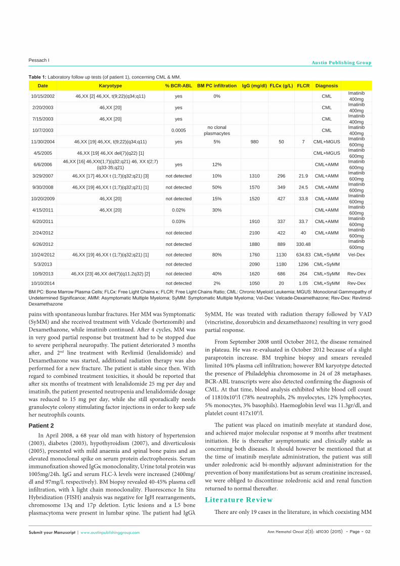
MM CML
Patient Reference Year Age/Sex Diagnosis 1st disease Type Marrow
PC Bone WBC count Spleno- Ph+
Interval treatment lesions (x109/L ) megalyDiagnosed
simultaneously 1 Boots MA et al. [7] 1982 58/M IgG-k numerous yes 140 yes yes
2 Tanaka M et al. [8] 1998 72/F IgG-k 3% yes 162.4 yes yes
3 Alvarez-Larran A et al. [9] 2001 81/M IgA-k 25% yes 28.7 NS yes
4 Schwarzmeier JD et al. [10] 2003 66/M IgG-k 3% yes 171 yes yes
5 Wakayama T et al. [11] 2005 85/F IgG-λ 32.8% NS 8.1 no yes
6 Offiah C et al. [12] 2012 71/F IgG-k 30% no 12.7 no yes
7 Romanenko NA et al. [13] 2013 64/F IgA-k 12.4% no 27.2 NS yes
MM/+CML 8 Macsween JM et al. [14]. 1972 77/M 33 months no BJP 9-12% no 145 yes yes
9 Klenn PJ et al. [15] 1993 71/M 24 months yes IgG-k cluster yes 40.8 NS yes
10 Nitta M et al. [16] 1999 70/M 33 months no IgG-k 25% no 25.2 yes yes
11 Nakagawa M et al. [17] 2003 47/M 33 months yes BJP-k 9.8% yes 23.9 NS yes
12. Ragupathi L et al. [18] 2013 62/F 17 months yes IgG-k 70% no yes
CML/+MM 13 Derghazarian C et al. [19] 1974 65/F 113 months yes IgG-k 9% yes 43. NS yes
14 Zoumbos NC et al. [20] 1987 57/M 60 months yes light-k NS NS NS NS no
15 Yokota A et al.[21] 2005 71/M 38 months yes BJP-λ 74.4% NS NS NS yes
16 Garipidou V et al. [22] 2005 68/M 20 months yes IgG-λ 25% no NS NS yes
17 Galanopoulos A et al. [23] 2009 76/M 14 months yes IgA-k 60% no NS NS yes
18 Michael M et al. [24] 2009 57/F 65 months yes IgA-k 95% NS 52.38 NS yes
19 Ide M et al. [25] 2010 72/F 3 months yes IgG-k 21.6% no 31.3 no yes
Table 2: Coexisting MM and CML cases.
MM: Multiple Myeloma; CML: Chronic Myeloid Leukemia; Marrow PC: Marrow Plasma Cells; Ph+: Philadelphia Positive; WBC: White Blood Cells

Ann Hematol Oncol 2(3): id1030 (2015) - Page - 04
Pessach I Austin Publishing Group
Submit your Manuscript | www.austinpublishinggroup.com
of MM in Imatinib treated CML backround can neither be ruled out, nor can be taken for granted, and since there are only a few reports on this issue, long-term studies are needed. Furthermore, exposure to radiation has been reported to induce t(9;22) translocation [52]. Therefore, CML development as a secondary disease in MM patients having received radiation therapy in the past also cannot be excluded.
Regarding MM patients, the chronic immunological deficiency of the disease, could lead to the formation of secondary malignancies including CML [53].
Lastly, it is notable that despite allogeneic stem cell transplantation is the only potentially curative treatment both for CML and MM separately, no report could be found in the literature in the setting of these two entities coexisting. This is probably due to the fact that most coexisting CML and MM cases seen in the literature are patients older than 65 years old.
In conclusion, coexistence of MM and CML suggests either a different clonal evolution from a common pluripotent malignant stem cell, or exposure to previous chemotherapy and radiation, or existence of a pro-carcinogenic environment, or a coincidence, or every each one of the above possible factors. Further investigation of associated possible causes is needed to get to definitive conclusions.
References1. Palumbo A, Anderson K. Multiple myeloma. N Engl J Med. 2011; 364: 1046-
1060.
2. McKenna RW, Kyle RA, Kuehl WM, Grogan TM, Harris NL, Coupland RW, et al. WHO Classification of Tumors of Hematopoietic and Lymphoid Tissues. 4th edition. In: WHO press. 2008; 200–212.
3. Rajkumar SV, Dimopoulos MA, Palumbo A, Blade J, Merlini G, Mateos MV, Kumar S. International Myeloma Working Group updated criteria for the diagnosis of multiple myeloma. Lancet Oncol. 2014; 15: e538-e548.
4. Al Achkar W, Wafa A, Mkrtchyan H, Moassass F, Liehr T. A rare case of chronic myeloid leukemia with secondary chromosomal changes including partial trisomy 17q21 to 17qter and partial monosomy of 16p13.3. Mol Cytogenet. 2010; 3: 6.
5. La Starza R, Testoni N, Lafage-Pochitaloff M, Ruggeri D, Ottaviani E, Perla G, et al. Complex variant Philadelphia translocations involving the short arm of chromosome 6 in chronic myeloid leukemia. Haematologica. 2002; 87: 143-147.
6. Lugo TG, Pendergast AM, Muller AJ, Witte ON. Tyrosine kinase activity and transformation potency of bcr-abl oncogene products. Science. 1990; 247: 1079-1082.
7. Boots MA, Pegrum GD. Simultaneous presentation of chronic granulocytic leukaemia and multiple myeloma. J Clin Pathol. 1982; 35: 364-365.
8. Tanaka M, Kimura R, Matsutani A, Zaitsu K, Oka Y, Oizumi K. Coexistence of chronic myelogenous leukemia and multiple myeloma. Case report and review of the literature. Acta Haematol. 1998; 99: 221-223.
9. Alvarez-Larrán A, Rozman M, Cervantes F. Simultaneous occurrence of multiple myeloma and chronic myeloid leukemia. Haematologica. 2001; 86: 894.
10. Schwarzmeier JD, Shehata M, Ackermann J, Hilgarth M, Kaufmann H, Drach J. Simultaneous occurrence of chronic myeloid leukemia and multiple myeloma: evaluation by FISH analysis and in vitro expansion of bone marrow cells. Leukemia. 2003; 17: 1426–1428.
11. Wakayama T, Fujita S, Ago H. An elder patient coexisted with multiple myeloma and chronic myeloid leukemia before treatments. Med J Shimane Pref Cent Hosp. 2005; 29: 63–67.
12. Offiah C, Quinn JP, Thornton P, Murphy PT. Co-existing chronic myeloid
leukaemia and multiple myeloma: rapid response to lenalidomide during imatinib treatment. Int J Hematol. 2012; 95: 451-452.
13. Romanenko NA, Bessmel’tsev SS, Udal’eva VIu, Zenina MN, Martynkevich IS, Rugal’ VI, et al. The combination of chronic myeloid leukemia and multiple myeloma in one patient. Vopr Onkol. 2013; 59: 103-110.
14. MacSween JM, Langley GR. Light-chain disease (hypogammaglobulinemia and Bence Jones proteinuria) and sideroblastic anemia-preleukemic chronic granulocytic leukemia. Can Med Assoc J. 1972; 106: 995–998.
15. Klenn PJ, Hyun BH, Lee YH, Zheng WY. Multiple myeloma and chronic myelogenous leukemia--a case report with literature review. Yonsei Med J. 1993; 34: 293-300.
16. Nitta M, Tsuboi K, Yamashita S, Kato M, Hayami Y, Harada S, et al. Multiple myeloma preceding the development of chronic myelogenous leukemia. Int J Hematol. 1999; 69: 170-173.
17. Nakagawa M, Noto S, Kobayashi H, Hayashi M. A case of a 47 year old man who developed chronic myelogenous leukemia after therapy for multiple myeloma. J Obihiro Kosei Gen Hosp. 2003; 6: 101–106.
18. Ragupathi L, Najfeld V, Chari A, Petersen B, Jagannath S, Mascarenhas J. A case report of chronic myelogenous leukemia in a patient with multiple myeloma and a review of the literature. Clin Lymphoma Myeloma Leuk. 2013; 13: 175-179.
19. Derghazarian C, Whittemore NB. Multiple myeloma superimposed on chronic myelocytic leukemia. Can Med Assoc J. 1974; 110: 1047-1050.
20. Zoumbos NC, Chrysanthopoulos C, Starakis J, Kapatais-Zoumbos K. Kappa light chain myeloma developing in a patient with chronic neutrophilic leukaemia. Br J Haematol. 1987; 65: 504-505.
21. Yokota A, Onoda M, Uehara T, Terano T. Coexistence of chronic myelogenous leukemia and multiple myeloma. Rinsho Ketsueki. 2005; 46: 919.
22. Garipidou V, Vakalopoulou S, Tziomalos K. Development of multiple myeloma in a patient with chronic myeloid leukemia after treatment with imatinib mesylate. Oncologist. 2005; 10: 457-458.
23. Galanopoulos A, Papadhimitriou SI, Kritikou-Griva E, Georgiakaki M, Anagnostopoulos NI. Multiple myeloma developing after imatinib mesylate therapy for chronic myeloid leukemia. Ann Hematol. 2009; 88: 281-282.
24. Michael M, Antoniades M, Lemesiou E, Papaminas N, Melanthiou F. Development of multiple myeloma in a patient with chronic myeloid leukemia while on treatment with imatinib mesylate for 65 months. Oncologist. 2009; 14: 1198-1200.
25. Ide M, Kuwahara N, Matsuishi E, Kimura S, Gondo H. Uncommon case of chronic myeloid leukemia with multiple myeloma. Int J Hematol. 2010; 91: 699-704.
26. Maeda K, Abraham J. Polycythemia associated with myeloma. Am J Clin Pathol. 1984; 82: 501-505.
27. Heinle EW, Sarasti HO, Garcia D, Kenny JJ, Westerman MP. Polycythemia vera associated with lymphomatous diseases and myeloma. Arch Intern Med. 1966; 118: 351-355.
28. Brody Ji, Beizer Lh, Schwartz S. Multiple Myeloma And The Myeloproliferative Syndromes. Am J Med. 1964; 36: 315-319.
29. Kasimis BS, Yen-Lin R, Neiman RS. Multiple myeloma associated with myelofibrosis. Report of a case and literature review. Oncology. 1981; 38: 369-372.
30. Cobo F, Cervantes F, Martinez C, Salgado C, Blade J, Montserrat E, Rozman C. Multiple myeloma following essential thrombocythemia. Leuk Lymphoma. 1995; 20: 177-179.
31. Selroos O, van Assendelft A. Thrombocythaemia and multiple myeloma. A report on two cases. Acta Med Scand. 1977; 201: 243-247.
32. Standen GR, Jasani B, Wagstaff M, Wardrop CA. Chronic neutrophilic leukemia and multiple myeloma. An association with lambda light chain expression. Cancer. 1990; 66: 162-166.
33. Lewis MJ, Oelbaum MH, Coleman M, Allen S. An association between chronic

Ann Hematol Oncol 2(3): id1030 (2015) - Page - 05
Pessach I Austin Publishing Group
Submit your Manuscript | www.austinpublishinggroup.com
neutrophilic leukaemia and multiple myeloma with a study of cobalamin-binding proteins. Br J Haematol. 1986; 63: 173-180.
34. Naparstek Y, Zlotnick A, Polliack A. Coexistent chronic myeloid leukemia and IgA monoclonal gammopathy: report of a case and review of the literature. Am J Med Sci. 1980; 279: 111-115.
35. Vorobiof DA, Benjamin J, Kaplan H, Dvilansky A. Chronic granulocytic leukemia, neutrophilic type, with paraproteinemia (IgA type K). Acta Haematol. 1978; 60: 316-320.
36. Anelli L, Zagaria A, Minervini A, Casieri P, Coccaro N, Tota G, et al. IgG-lymphoplasmacytic lymphoma following polycythemia vera: JAK2 V617F and MYD88 L265P mutations separated in the same house. Ann Hematol. 2014; 93: 1605-1607.
37. ValkoviÄ T. The possible association of monoclonal gammopathy of undetermined significance and JAK2-V617F positive chronic myeloproliferative neoplasm. Acta Clin Croat. 2014; 53: 256-257.
38. Turesson I, Kovalchik SA, Pfeiffer RM, Kristinsson SY, Goldin LR, Drayson MT, et al. Monoclonal gammopathy of undetermined significance and risk of lymphoid and myeloid malignancies: 728 cases followed up to 30 years in Sweden. Blood. 2014; 123: 338-345.
39. Hornsten P, Nordenson I, Wahlin A. Philadelphia chromosome negative acute lymphoblastic leukemia preceding Philadelphia positive chronic myelogenous leukemia. Cancer genet cytogenet. 1989; 39: 147-152.
40. Vannier JP, Bizet M, Bastard C, Bernard A, Ducastelle T, Tron P. Simultaneous occurrence of a T-cell lymphoma and a chronic myelogenous leukemia with an unusual karyotype. Leuk Res. 1984; 8: 647-657.
41. Djulbegovic B, Hadley T, Yen F. Occurrence of high-grade T-cell lymphoma in a patient with Philadelphia chromosome-negative chronic myelogenous leukemia with breakpoint cluster region rearrangement: case report and review of the literature. Am J Hematol. 1991; 36: 63-64.
42. Tittley P, Trempe JM, van der Jagt R, Drouin J, Huebsch L, McLeish B, et al. Occurrence of T-cell lymphoma in a patient with Philadelphia chromosome-positive chronic myelogenous leukemia with rearrangements of BCR and TCR-beta genes in the lymph nodes. Am J Hematol. 1993; 42: 229-230.
43. Shoenfeld Y, Berliner S, Ayalone A, Shaklai M, Djaldetti M, Pick AI, et
al. Monoclonal gammopathy in patients with chronic and acute myeloid leukemia. Cancer. 1984; 54: 280-283.
44. Vitali C, Bombardieri S, Spremolla G. Chronic myeloid leukemia in Waldenström’s macroglobulinemia. Arch Intern Med. 1981; 141: 1349-1351.
45. Martin PJ, Najfeld V, Hansen JA, Penfold GK, Jacobson RJ, Fialkow PJ. Involvement of the B-lymphoid system in chronic myelogenous leukaemia. Nature. 1980; 287: 49-50.
46. Shaughnessy JD, Barlogie B. Integrating cytogenetics and gene expression profiling in the molecular analysis of multiple myeloma. Int J Hematol. 2002; 76: 59-64.
47. Sawyer JR. The prognostic significance of cytogenetics and molecular profiling in multiple myeloma. Cancer Genet. 2011; 204: 3-12.
48. Van Den Berghe H, Louwagie A, Broeckaert-Van Orshoven A, David G, Verwilghen R, Michaux JL, et al. Philadelphia chromosome in human multiple myeloma. J Natl Cancer Inst. 1979; 63: 11-16.
49. Martiat P, Mecucci C, Nizet Y, Stul M, Philippe M, Cassiman JJ, et al. P190 BCR/ABL transcript in a case of Philadelphia-positive multiple myeloma. Leukemia. 1990; 4: 751-754.
50. Roper N, DeAngelo DJ, Kuo F, Dal Cin P, Ghobrial I, Aster JC. An asymptomatic 61-year-old man with BCR-ABL-positive bone marrow following autologous transplantation for multiple myeloma. Am J Hematol. 2010; 85: 944-946.
51. Pandiella A, Carvajal-Vergara X, Tabera S, Mateo G, Gutiérrez N, San Miguel JF. Imatinib mesylate (STI571) inhibits multiple myeloma cell proliferation and potentiates the effect of common antimyeloma agents. Br J Haematol. 2003; 123: 858-868.
52. Holmberg M. Is the primary event in radiation-induced chronic myelogenous leukemia the induction of the t(9;22) translocation? Leuk Res. 1992; 16: 333-336.
53. Thomas A, Mailankody S, Korde N, Kristinsson SY, Turesson I, Landgren O. Second malignancies after multiple myeloma: from 1960s to 2010s. Blood. 2012; 119: 2731-2737.
Citation: Pessach I, Bartzis V, Tzenou T, Roumelioti M, Palaiologou D, et al. Multiple Myeloma and Chronic Myelogenous Leukemia; an Uncommon Coexistence in 2 Patients, with Literature Review. Ann Hematol Oncol. 2015;2(3): 1030.
Ann Hematol Oncol - Volume 2 Issue 3 - 2015ISSN : 2375-7965 | www.austinpublishinggroup.com Pessach et al. © All rights are reserved