Embed Size (px)
Citation preview

JOURNAL OF VIROLOGY, Aug. 2010, p. 8007–8020 Vol. 84, No. 160022-538X/10/$12.00 doi:10.1128/JVI.00334-10Copyright © 2010, American Society for Microbiology. All Rights Reserved.
Multiple DNA Damage Signaling and Repair Pathways Deregulated bySimian Virus 40 Large T Antigen�
Sergei Boichuk, Liang Hu, Jennifer Hein, and Ole V. Gjoerup*Cancer Virology Program, University of Pittsburgh Cancer Institute, Pittsburgh, Pennsylvania 15213
Received 12 February 2010/Accepted 27 May 2010
We demonstrated previously that expression of simian virus 40 (SV40) large T antigen (LT), without a viralorigin, is sufficient to induce the hallmarks of a cellular DNA damage response (DDR), such as focalaccumulation of �-H2AX and 53BP1, via Bub1 binding. Here we expand our characterization of LT effects onthe DDR. Using comet assays, we demonstrate that LT induces overt DNA damage. The Fanconi anemiapathway, associated with replication stress, becomes activated, since FancD2 accumulates in foci, and mono-ubiquitinated FancD2 is detected on chromatin. LT also induces a distinct set of foci of the homologousrecombination repair protein Rad51 that are colocalized with Nbs1 and PML. The FancD2 and Rad51 focirequire neither Bub1 nor retinoblastoma protein binding. Strikingly, wild-type LT is localized on chromatin at,or near, the Rad51/PML foci, but the LT mutant in Bub1 binding is not localized there. SV40 infection waspreviously shown to trigger ATM activation, which facilitates viral replication. We demonstrate that productiveinfection also triggers ATR-dependent Chk1 activation and that Rad51 and FancD2 colocalize with LT in viralreplication centers. Using small interfering RNA (siRNA)-mediated knockdown, we demonstrate that Rad51and, to a lesser extent, FancD2 are required for efficient viral replication in vivo, suggesting that homologousrecombination is important for high-level extrachromosomal replication. Taken together, the interplay of LTwith the DDR is more complex than anticipated, with individual domains of LT being connected to differentsubcomponents of the DDR and repair machinery.
Simian virus 40 (SV40) carries genes encoding three earlyproteins: large T antigen (LT), small t antigen, and 17k. LT hasserved as a powerful model system for understanding cellularprocesses, such as DNA replication and malignant transforma-tion (1, 16). An in vitro SV40 replication system based onpurified cellular factors was established long ago, which inmany ways recapitulates in vivo replication (33, 71). However,although very insightful, certain aspects of DNA replicationcannot be reconstructed in a test tube and remain incompletelyunderstood. Since SV40 relies extensively on cellular replica-tion factors, it must reprogram the cellular environment tosupport viral DNA replication. A key component is cell cyclereprogramming, perhaps most importantly the potent induc-tion of S phase in quiescent cells (19).
The ability of LT to induce aberrant cellular proliferationdepends on binding to and inactivating key tumor suppressorslike p53 and the retinoblastoma protein (pRB) family (re-viewed in references 1 and 16). These interactions are alsocritical for oncogenic transformation and induction of tumorsin a wide variety of cell types and tissues. Additional functionscontribute to transformation. A functional DnaJ domain re-sides within the first 70 amino acids, which directs binding ofthe Hsc70 molecular chaperone and contributes to both onco-genic transformation and viral replication proficiency (10, 59).Distinct binding sites for a Cul7 ubiquitin ligase complex andthe Bub1 mitotic checkpoint kinase are found immediately
downstream of the DnaJ domain, and both binding proteinscontribute to malignant transformation induced by LT (2, 17).
Efficient viral replication in vivo depends not only on LT-induced S-phase entry. Recent studies have illuminated howviruses target the DNA damage response (DDR) (reviewed inreferences 14 and 35). The DDR can be divided into two majorbranches according to the lesions that are sensed. Double-strand breaks (DSBs) are known to activate the ATM (ataxia-telangiectasia mutated) kinase, which in turn triggers cell cyclecheckpoints to halt the cell cycle and promote DNA repair.Conversely, lesions with single-stranded DNA (ssDNA), forexample arising from replication stress, trigger ATR (ataxia-telangiectasia and Rad3-related) kinase activation.
DSBs are a serious threat to genomic stability. When DSBsarise, they are sensed by the MRN (Mre11-Rad50-Nbs1) com-plex that triggers ATM-mediated phosphorylation of histoneH2AX (referred to as �-H2AX) in the flanking chromatin (32,50). Strikingly, �-H2AX accumulates in characteristic nuclearfoci that are often considered a surrogate marker of DSBs (52).These foci are believed to be fundamentally important forretention of repair proteins at the break site and possiblycheckpoint signaling (5, 63). DSBs can be repaired by eitherone of two distinct repair pathways, nonhomologous end join-ing (NHEJ) or homologous recombination (HR). A key com-ponent of the HR machinery is the Rad51 protein, which isalso recruited to subnuclear foci to orchestrate repair (5, 26,41, 42, 51). A subset of foci is composed of ssDNA, arisingfrom resection of a DSB or from stalled replication forks. ThessDNA is coated by replication protein A (RPA), which sub-sequently recruits ATR and its obligate partner ATRIP (ATR-interacting protein). The Fanconi anemia pathway is involvedin recognition and repair of stalled replication forks or other
* Corresponding author. Mailing address: Hillman Cancer Center,Research Pavilion Suite 1.8, 5117 Centre Avenue, Pittsburgh, PA15213. Phone: (412) 623-7717. Fax: (412) 623-7715. E-mail: [email protected].
� Published ahead of print on 2 June 2010.
8007
at University of P
ittsburgh HS
LS July 19, 2010
jvi.AS
M.O
RG
- D
OW
NLO
AD
ED
FR
OM

kinds of replication stress (26, 29, 44). Fanconi anemia, to-gether with other genetic disorders, such as Nijmegen break-age syndrome (Nbs1) and breast cancer 1 (BRCA1), are strik-ing examples that loss of genomic stability throughcompromise of DNA repair pathways can directly contribute tohuman malignancies.
Some viruses, like adenovirus, dismantle the DDR via ex-pression of specific viral proteins (14, 35, 62). In contrast, SV40and mouse polyomavirus exploit the DDR by activating theDDR and benefiting from it (18, 23, 54, 76). SV40 infectionleads to activation of the ATM kinase and downstream com-ponents, such as �-H2AX, p53, and Chk2 (54, 76). The MRNcomplex is recruited to viral replication centers together withLT, RPA, and in close proximity, the PML protein. PML, atumor suppressor, nucleates the formation of PML oncogenicdomains known to participate in a diverse array of cellularfunctions, such as antiviral defenses, DNA damage repair, apop-tosis, and transcriptional regulation (6). Late in infection, theMRN complex is, at least partially, degraded via Cul7 (76).Genetic loss of ATM or inhibition of ATM kinase substantiallycompromises viral replication and virion production, concom-itant with the disappearance of replication centers and lack ofMRN degradation. It was suggested that SV40 might havecommandeered an intrinsic cellular DNA repair pathway tocarry out its replication, since it appears to have usurped manyof the factors involved (76). Nevertheless, it remains poorlyunderstood mechanistically exactly how DDR activation canpromote viral replication.
Our recent observations have indicated that expression ofLT alone, in the absence of a viral replication origin, is suffi-cient to induce hallmarks of the ATM/ATR-mediated DDR,such as large �-H2AX/53BP1 foci, p53 phosphorylation/stabi-lization, and Chk1/2 kinase activation in human BJ/tert fibro-blasts (23). This response depends primarily on Bub1 bindingand can be efficiently induced by an pRB binding-deficient 17kprotein, which shares the first 131 amino acids with LT butends in four unique residues.
In this study, we have significantly extended the analysisof mechanisms and consequences of DDR perturbations byLT. Comet assays demonstrate that LT induces overt DNAdamage. We discovered further complexities in the DDRsignaling and DNA repair elements influenced by LT. Thus,LT induces activation of the Fanconi anemia proteinFancD2 by relocalizing it into foci on chromatin, a processnormally linked to replication stress, for example, fromstalled replication forks (26, 44). LT also induces distinctfoci of the HR repair protein Rad51, which are colocalizedwith Nbs1 and PML. Both FancD2 and Rad51 foci areinduced by LT independent of Bub1 and pRB binding. LT,but not the Bub1-binding mutant, is localized in foci juxta-posed with Rad51 and PML. Complementing previous stud-ies that showed ATM activation following SV40 infection(54, 76), we find that Chk1 is activated through the ATRpathway. During viral infection, both FancD2 and Rad51colocalize with LT in replication centers, and small inter-fering RNA (siRNA)-mediated downregulation of FancD2or Rad51 causes an impairment of SV40 origin-dependentreplication in vivo.
MATERIALS AND METHODS
Cell culture. BJ/tert fibroblasts stably expressing empty vector, LT, or itsmutants were grown in 80% Dulbecco’s modified Eagle’s medium (Lonza) and20% medium 199 (Invitrogen) supplemented with 10% fetal calf serum (FCS)(HyClone) and 1% penicillin-streptomycin (Invitrogen). GM07166 tert cells (74)(Nbs1 deficient, kindly provided by X. Wu, The Scripps Research Institute) aswell as monkey kidney epithelial BSC40 cells (kindly provided by J. Pipas,University of Pittsburgh) and COS-1 cells were cultured in DMEM with 10%FCS and 1% penicillin-streptomycin. All cells were cultured at 37°C in a humid-ified incubator containing 5% CO2.
Plasmids and transfection. Retroviral vectors (pLNBCX) expressing wild-typeLT or the dl89-97 mutant were previously described (23). The pRB-bindingmutant of LT (K1) was generated by site-directed mutagenesis of pLBNCX LT.
siRNAs for ATM, ATR, FancD2, or Rad51 (SMARTpool; Dharmacon), or anontargeting control siRNA, were transfected into COS-1 or BSC40 cells usingLipofectamine RNAiMAX reagent according to the manufacturer’s protocol(Invitrogen). Although SMARTpool siRNAs were designed against the humansequences, we found that they efficiently silenced expression of the homologousmonkey proteins. Cy3-labeled siRNA indicated that �90% of cells were trans-fected. Plasmid DNA was transfected by Fugene-6 (Roche).
Viral infections. Retroviral vectors were transiently transfected into Phoenixamphotropic packaging cell line using the calcium phosphate precipitationmethod (15, 23). At 48 h posttransfection, the virus-containing supernatant washarvested, filtered through a 0.45-�m membrane (Millipore), aliquoted, andstored at �80°C. Cells were infected with 1 ml of virus supernatant in thepresence of Polybrene (8 �g/ml). After 48 h, the infected cells were passed andcontinued to grow under blasticidin (5 �g/ml) selection for 5 to 7 days.
SV40 infections were performed essentially as previously outlined (76).Antibodies and inhibitors. Primary mouse monoclonal antibodies were pur-
chased from BD Biosciences (Orc2 clone 920-4-41), Millipore (Rad51 clone3C10), Oncogene (BRCA1 clone Ab-1), Rockland (pATM S1981 200-301-400),Santa Cruz (PML clone PG-M3, cyclin A clone 6E6, BRCA1 D-9), and Sigma(�-tubulin clone B-5-1-2). Monoclonal antibodies against LT (PAb416, PAb419,and PAb423) and rabbit polyclonal antibody against ATRIP were previouslydescribed (23). Antibody to polymerase � (SJK132-20) was kindly provided byEllen Fanning. Primary rabbit polyclonal antibodies were purchased from BethylLaboratories (RPA32 A300-244A and pRPA S4/8 A300-245A), Calbiochem(53BP1 Ab-1), Chemicon (PML AB1370), Genetex (FancD2 GTX30142), NovusBiologicals (Nbs1 NB100-143, Mre11 NB100-142, and ATR NB100-323), R&DSystems (pChk1 S317, pChk2 T68), and Santa Cruz (SV40 LT [v-300], p53[FL-393], and Rad51 [H-92]). ATM and pATM S1981 rabbit monoclonal anti-bodies were from Epitomics. Secondary antibodies for immunofluorescencestaining were obtained from Invitrogen (Alexa Fluor 488-labeled antibodies)and Jackson ImmunoResearch (Cy3-labeled antibodies). ATM inhibitor(KU55933) was purchased from Tocris Bioscience and used at a final con-centration of 10 �M.
Cellular fractionation and Western blotting. Whole-cell extracts were pre-pared as previously described (23). In brief, the cells were washed in ice-coldphosphate-buffered saline (PBS) and lysed with TEB buffer (20 mM Tris [pH7.5], 137 mM NaCl, 10% glycerol, 1% Nonidet P-40) supplemented with pro-tease and phosphatase inhibitors. Some samples were extracted in radioimmu-noprecipitation assay (RIPA) buffer (50 mM Tris [pH 8.0], 150 mM NaCl, 1%Nonidet P-40, 0.5% sodium deoxycholate, 0.1% sodium dodecyl sulfate [SDS]).The samples were resolved on 4 to 12% Bis-Tris or 3 to 8% Tris-acetateNuPAGE gels (Invitrogen), transferred to a nitrocellulose membrane (Bio-Rad), probed with specific antibody, and visualized by enhanced chemilumines-cence (Western Lightning Plus-ECL reagent). Western blot images were pro-cessed using Adobe Photoshop 7.0, and bands were quantitated using ImageJsoftware.
Chromatin fractionation was performed essentially by the method of Mendezand Stillman (40). Briefly, the cells were resuspended in buffer A (10 mMHEPES [pH 7.9], 10 mM KCl, 1.5 mM MgCl2, 10 mM NaF, 340 mM sucrose,10% glycerol, 0.1% Triton X-100) supplemented with protease/phosphatase in-hibitors and 1 mM dithiothreitol (DTT) and incubated on ice for 5 min. Thenuclear pellet was collected by low-speed centrifugation, washed in buffer A, andsubsequently lysed in buffer B (3 mM EDTA, 0.2 mM EGTA) while incubatingon ice for 10 min. The insoluble chromatin was collected by centrifugation,resuspended in SDS sample buffer, sonicated, and boiled.
Immunofluorescence. BJ/tert cell lines were seeded on glass coverslips(Fisher) coated with poly-L-lysine (Sigma) and cultured for the next 48 h. Thecells were washed twice with ice-cold PBS and fixed at 4°C for 15 min with 4%paraformaldehyde (Electron Microscopy Sciences, PA). In some cases, before
8008 BOICHUK ET AL. J. VIROL.
at University of P
ittsburgh HS
LS July 19, 2010
jvi.AS
M.O
RG
- D
OW
NLO
AD
ED
FR
OM
the cells were fixed, the slides were preextracted with CSK buffer [10 mMpiperazine-N,N�-bis(2-ethanesulfonic acid) (PIPES) (pH 7), 100 mM NaCl, 300mM sucrose, 3 mM MgCl2, 1 mM EGTA] containing 0.5% Triton X-100 for 10min on ice (41). The fixed cells were washed twice with PBS, permeabilized for5 min with 0.5% Triton X-100 in PBS, and blocked for 30 min in 10% normalgoat serum in PBS. Staining with primary antibody was performed overnight at4°C in PBS with 0.5% goat serum. The slides were washed three times in PBS at5-min intervals and incubated with fluorochrome-conjugated (Cy3 or AlexaFluor 488) secondary antibodies for 30 min at room temperature in the dark.Finally, the slides were washed with PBS three times at 5-min intervals, stainedwith 4�,6�-diamidino-2-phenylindole (DAPI) (Molecular Probes), mounted, andexamined by fluorescence microscopy. Images were visualized using an Olympusmicroscope and captured with a Spot Advanced imaging system.
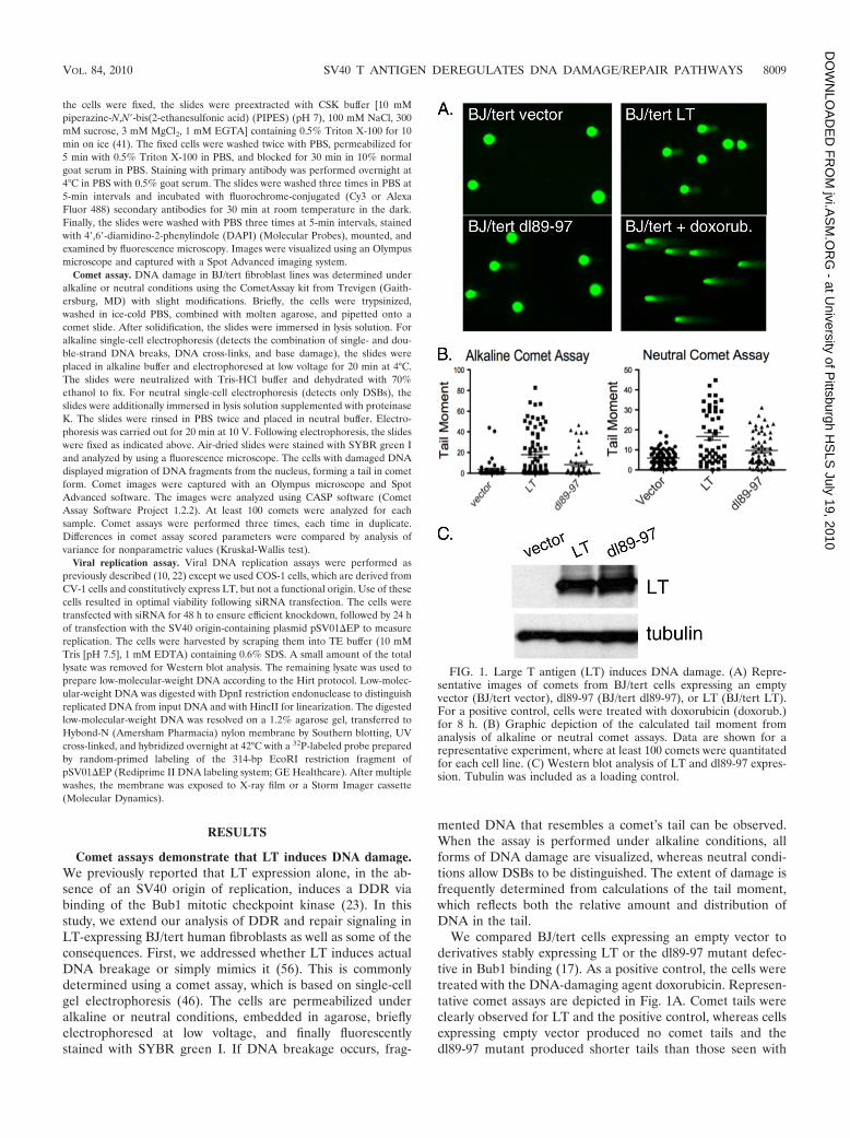
Comet assay. DNA damage in BJ/tert fibroblast lines was determined underalkaline or neutral conditions using the CometAssay kit from Trevigen (Gaith-ersburg, MD) with slight modifications. Briefly, the cells were trypsinized,washed in ice-cold PBS, combined with molten agarose, and pipetted onto acomet slide. After solidification, the slides were immersed in lysis solution. Foralkaline single-cell electrophoresis (detects the combination of single- and dou-ble-strand DNA breaks, DNA cross-links, and base damage), the slides wereplaced in alkaline buffer and electrophoresed at low voltage for 20 min at 4°C.The slides were neutralized with Tris-HCl buffer and dehydrated with 70%ethanol to fix. For neutral single-cell electrophoresis (detects only DSBs), theslides were additionally immersed in lysis solution supplemented with proteinaseK. The slides were rinsed in PBS twice and placed in neutral buffer. Electro-phoresis was carried out for 20 min at 10 V. Following electrophoresis, the slideswere fixed as indicated above. Air-dried slides were stained with SYBR green Iand analyzed by using a fluorescence microscope. The cells with damaged DNAdisplayed migration of DNA fragments from the nucleus, forming a tail in cometform. Comet images were captured with an Olympus microscope and SpotAdvanced software. The images were analyzed using CASP software (CometAssay Software Project 1.2.2). At least 100 comets were analyzed for eachsample. Comet assays were performed three times, each time in duplicate.Differences in comet assay scored parameters were compared by analysis ofvariance for nonparametric values (Kruskal-Wallis test).
Viral replication assay. Viral DNA replication assays were performed aspreviously described (10, 22) except we used COS-1 cells, which are derived fromCV-1 cells and constitutively express LT, but not a functional origin. Use of thesecells resulted in optimal viability following siRNA transfection. The cells weretransfected with siRNA for 48 h to ensure efficient knockdown, followed by 24 hof transfection with the SV40 origin-containing plasmid pSV01�EP to measurereplication. The cells were harvested by scraping them into TE buffer (10 mMTris [pH 7.5], 1 mM EDTA) containing 0.6% SDS. A small amount of the totallysate was removed for Western blot analysis. The remaining lysate was used toprepare low-molecular-weight DNA according to the Hirt protocol. Low-molec-ular-weight DNA was digested with DpnI restriction endonuclease to distinguishreplicated DNA from input DNA and with HincII for linearization. The digestedlow-molecular-weight DNA was resolved on a 1.2% agarose gel, transferred toHybond-N (Amersham Pharmacia) nylon membrane by Southern blotting, UVcross-linked, and hybridized overnight at 42°C with a 32P-labeled probe preparedby random-primed labeling of the 314-bp EcoRI restriction fragment ofpSV01�EP (Rediprime II DNA labeling system; GE Healthcare). After multiplewashes, the membrane was exposed to X-ray film or a Storm Imager cassette(Molecular Dynamics).
RESULTS
Comet assays demonstrate that LT induces DNA damage.We previously reported that LT expression alone, in the ab-sence of an SV40 origin of replication, induces a DDR viabinding of the Bub1 mitotic checkpoint kinase (23). In thisstudy, we extend our analysis of DDR and repair signaling inLT-expressing BJ/tert human fibroblasts as well as some of theconsequences. First, we addressed whether LT induces actualDNA breakage or simply mimics it (56). This is commonlydetermined using a comet assay, which is based on single-cellgel electrophoresis (46). The cells are permeabilized underalkaline or neutral conditions, embedded in agarose, brieflyelectrophoresed at low voltage, and finally fluorescentlystained with SYBR green I. If DNA breakage occurs, frag-
mented DNA that resembles a comet’s tail can be observed.When the assay is performed under alkaline conditions, allforms of DNA damage are visualized, whereas neutral condi-tions allow DSBs to be distinguished. The extent of damage isfrequently determined from calculations of the tail moment,which reflects both the relative amount and distribution ofDNA in the tail.
We compared BJ/tert cells expressing an empty vector toderivatives stably expressing LT or the dl89-97 mutant defec-tive in Bub1 binding (17). As a positive control, the cells weretreated with the DNA-damaging agent doxorubicin. Represen-tative comet assays are depicted in Fig. 1A. Comet tails wereclearly observed for LT and the positive control, whereas cellsexpressing empty vector produced no comet tails and thedl89-97 mutant produced shorter tails than those seen with
FIG. 1. Large T antigen (LT) induces DNA damage. (A) Repre-sentative images of comets from BJ/tert cells expressing an emptyvector (BJ/tert vector), dl89-97 (BJ/tert dl89-97), or LT (BJ/tert LT).For a positive control, cells were treated with doxorubicin (doxorub.)for 8 h. (B) Graphic depiction of the calculated tail moment fromanalysis of alkaline or neutral comet assays. Data are shown for arepresentative experiment, where at least 100 comets were quantitatedfor each cell line. (C) Western blot analysis of LT and dl89-97 expres-sion. Tubulin was included as a loading control.
VOL. 84, 2010 SV40 T ANTIGEN DEREGULATES DNA DAMAGE/REPAIR PATHWAYS 8009
at University of P
ittsburgh HS
LS July 19, 2010
jvi.AS
M.O
RG
- D
OW
NLO
AD
ED
FR
OM

wild-type LT. Quantitation of these observations is depicted inthe graphs shown in Fig. 1B. The median tail moments were17.87 (LT), 8.29 (dl89-97), and 3.58 (vector) for alkaline cometassay versus 16.71(LT), 9.71 (dl89-97), and 5.93 (vector) for theneutral comet assay. Statistical analysis was performed accord-ing to the Kruskal-Wallis test. For both alkaline and neutralconditions, the LT tail moment was significantly higher thanfor cells expressing empty vector and the dl89-97 mutant. Al-though the dl89-97 mutant was attenuated in its ability toinduce DNA breakage, it consistently retained the ability toinduce some degree of DNA damage. The levels of expressionof LT and dl89-97 were equivalent (Fig. 1C). Since LT expres-sion causes the appearance of comet tails also under neutralconditions, it means that DSBs are induced, which is consistentwith our previous observation of ATM activation based onincreased pATM S1981 signal (23). Taken together, LT doesnot just mimic DNA damage but induces overt DNA breakagepartly in the form of DSBs, and this is partially dependent onBub1 binding.
LT affects recruitment or retention of activated ATM atfocal sites of DNA damage. We wanted to investigate theresponse to external damage at the single-cell level. To probethe response to DSBs, we analyzed pATM S1981 as a markerof ATM activation. As shown in Fig. 2A, BJ/tert cells express-ing LT (BJ/tert LT) had significantly increased pannuclearstaining for pATM S1981, whereas BJ/tert cells expressing anempty vector (BJ/tert vector) exhibited negligible backgroundstaining. While the LT-mediated increase in pannuclear stain-ing correlated with Western blotting results for pATM S1981(23), it was intriguing that accumulation was not focal. Asexpected, in response to ionizing radiation (IR), pATM S1981in BJ/tert vector relocalized into foci that are colocalized with53BP1 (4, 55) (Fig. 2A and B). In contrast, pATM S1981remained pannuclear following IR treatment of BJ/tert LTcells, although 53BP1 was mobilized into foci (Fig. 2A and B).These results suggest that ATM when activated fails to beretained at DNA damage sites, which could result in an im-pairment of ATM signaling.
To test that hypothesis, we looked at the ATM response at0, 2, or 24 h following 10 Gy of IR (Fig. 2C). As we previouslynoted, LT-expressing cells have elevated levels of pATM S1981and pChk2 T68, whereas in cells expressing dl89-97, the levelsare lower. We observed that ATM is still capable of beingfurther activated when BJ/tert LT cells are irradiated, becausepATM S1981 and pChk2 T68 increase beyond their baselinelevel (Fig. 2C). Interestingly, pChk2 T68 levels in LT cellscontinue to increase with time, peaking at 24 h, in contrast tocells expressing empty vector that have already returned tobaseline levels by then. Although the ATM response appearsto be intact, we cannot exclude the possibility that some sub-strates are affected due to the pannuclear rather than focallocalization of pATM S1981.
LT activates the Fanconi anemia and HR pathways. Tofurther delineate all the DDR and repair pathways perturbedby LT, we also investigated markers of HR (Rad51) and rep-lication stress (FancD2) (44, 68). The FancD2 protein, a keymember of the Fanconi anemia family, which responds tointerstrand DNA cross-links, is activated on chromatin by mono-ubiquitination, thus converting it from the short (S) form to thelong (L) form (44). This is often used as a surrogate marker of
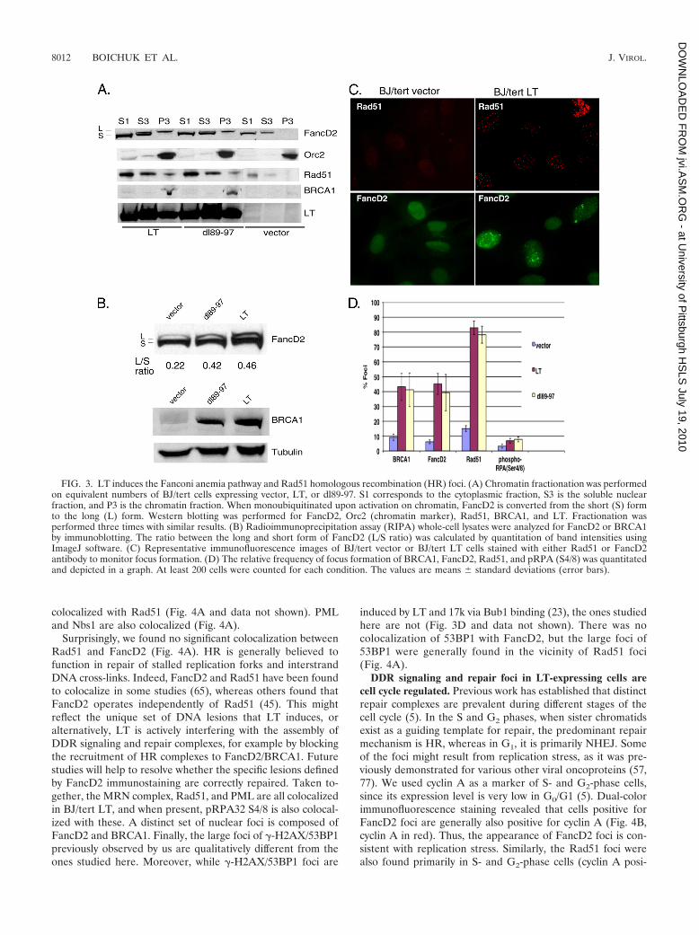
Fanconi anemia pathway activation (57). To assay the activa-tion state of FancD2, we performed chromatin fractionationon BJ/tert cells expressing empty vector, BJ/tert cells express-ing LT, and dl89-97 cells (Fig. 3A). Strikingly, Western blottingshows that both BJ/tert LT and dl89-97 induce the monoubiq-uitinated FancD2 species in the chromatin fraction (P3),whereas an empty vector does not. A perceptible increase inFancD2 levels is also observed when LT or dl89-97 is ex-pressed; this might result from E2F-mediated transcriptionalupregulation by LT (25). Similarly, both BRCA1 and Rad51are increased (total level) and present in the chromatin frac-tions for LT and dl89-97, not in the vector control (Fig. 3A andB). LT and dl89-97 are expressed at similar levels, and both arepresent in the chromatin fraction. Orc2 was used as a markerfor the chromatin fraction. To quantitate the activation state ofFancD2, we determined the ratio between the long and shortforms of the protein by Western blotting of RIPA whole-celllysates (Fig. 3B). Densitometry revealed that cells expressingboth LT and dl89-97 exhibit significant activation of FancD2compared with the empty vector control.
To confirm and extend the results obtained by Westernblotting, we looked by immunofluorescence for nuclear foci,another hallmark of DDR activation (5). Strikingly, LT-ex-pressing BJ/tert cells exhibit an increased frequency of FancD2and Rad51 foci compared to the control (Fig. 3C). The for-mation of Rad51 and FancD2 foci is known to occur followingionizing radiation or replication stress (5, 26, 41, 42, 51). Therelative frequency of focus formation was quantitated and de-picted in Fig. 3D. Approximately 80% of the cells expressingLT exhibit Rad51 foci, and this occurs independently of Bub1binding. Similarly, LT induces foci of both FancD2 andBRCA1 in approximately 40% of the cells, again indepen-dently of Bub1 binding. LT and dl89-97 also induced a slightincrease in foci of pRPA32 S4/8, believed to reflect ssDNAlesions, but the frequency was low (Fig. 3D). This RPA phos-phorylation is sometimes dependent on ATR (12, 47) and mayshift RPA action from cellular replication toward DNA repair(7). In all cases, the background frequency of foci was muchlower. Experiments with an pRB binding-deficient mutant ofLT (K1) demonstrated that binding of pocket proteins doesnot contribute to Rad51, FancD2, or BRCA1 focus formation(data not shown).
Colocalization studies on DDR signaling proteins in LT-expressing BJ/tert cells. In the previous section, we demon-strated that LT induces striking increases in focus formation ofthe Rad51, FancD2, and BRCA1 proteins, a modus operandi ofDDR signaling molecules upon DNA damage or replicationstress (5). Next, we addressed which of the relevant DDRsignaling proteins are colocalized in BJ/tert LT. Notably,FancD2 and BRCA1 are highly colocalized (Fig. 4A), as pre-viously reported for cells following genotoxic damage (21, 65).LT-expressing cells have a minor population of cells withpRPA32 S4/8 foci (Fig. 3D). When present, these were per-fectly colocalized with the Rad51 recombinase (Fig. 4A). SinceRPA and Rad51 have previously been reported to colocalizewith PML (8), we incorporated it into our colocalization anal-ysis. Indeed, Rad51 is colocalized with PML (Fig. 4A). TheMRN complex is involved both in sensing of DSBs and in theirresection to create ssDNA (5, 32). Mre11, as well as Nbs1, are
8010 BOICHUK ET AL. J. VIROL.
at University of P
ittsburgh HS
LS July 19, 2010
jvi.AS
M.O
RG
- D
OW
NLO
AD
ED
FR
OM
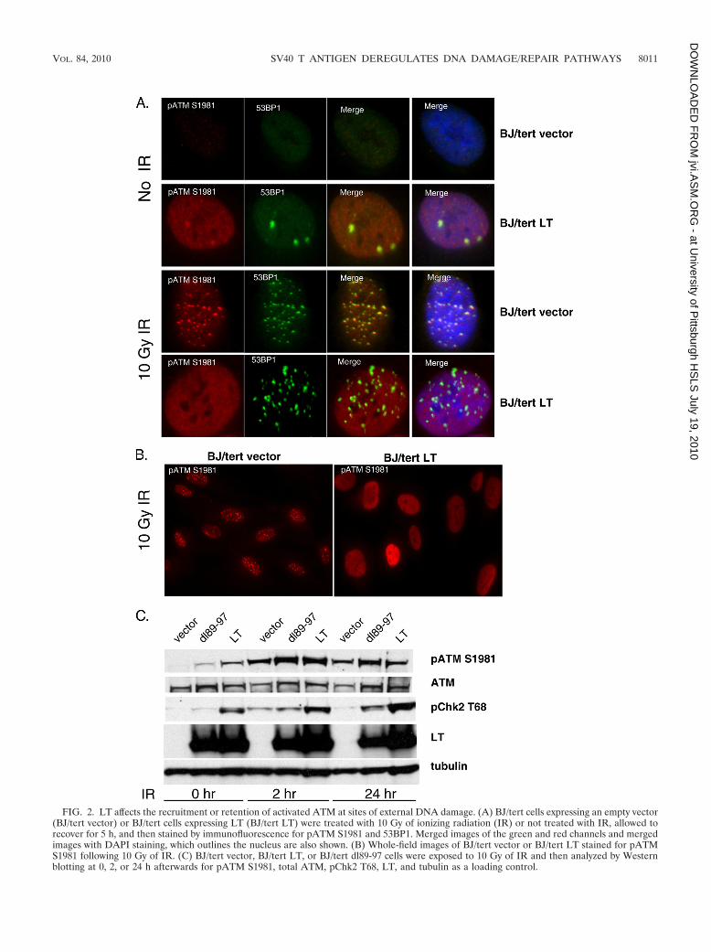
FIG. 2. LT affects the recruitment or retention of activated ATM at sites of external DNA damage. (A) BJ/tert cells expressing an empty vector(BJ/tert vector) or BJ/tert cells expressing LT (BJ/tert LT) were treated with 10 Gy of ionizing radiation (IR) or not treated with IR, allowed torecover for 5 h, and then stained by immunofluorescence for pATM S1981 and 53BP1. Merged images of the green and red channels and mergedimages with DAPI staining, which outlines the nucleus are also shown. (B) Whole-field images of BJ/tert vector or BJ/tert LT stained for pATMS1981 following 10 Gy of IR. (C) BJ/tert vector, BJ/tert LT, or BJ/tert dl89-97 cells were exposed to 10 Gy of IR and then analyzed by Westernblotting at 0, 2, or 24 h afterwards for pATM S1981, total ATM, pChk2 T68, LT, and tubulin as a loading control.
VOL. 84, 2010 SV40 T ANTIGEN DEREGULATES DNA DAMAGE/REPAIR PATHWAYS 8011
at University of P
ittsburgh HS
LS July 19, 2010
jvi.AS
M.O
RG
- D
OW
NLO
AD
ED
FR
OM
colocalized with Rad51 (Fig. 4A and data not shown). PMLand Nbs1 are also colocalized (Fig. 4A).
Surprisingly, we found no significant colocalization betweenRad51 and FancD2 (Fig. 4A). HR is generally believed tofunction in repair of stalled replication forks and interstrandDNA cross-links. Indeed, FancD2 and Rad51 have been foundto colocalize in some studies (65), whereas others found thatFancD2 operates independently of Rad51 (45). This mightreflect the unique set of DNA lesions that LT induces, oralternatively, LT is actively interfering with the assembly ofDDR signaling and repair complexes, for example by blockingthe recruitment of HR complexes to FancD2/BRCA1. Futurestudies will help to resolve whether the specific lesions definedby FancD2 immunostaining are correctly repaired. Taken to-gether, the MRN complex, Rad51, and PML are all colocalizedin BJ/tert LT, and when present, pRPA32 S4/8 is also colocal-ized with these. A distinct set of nuclear foci is composed ofFancD2 and BRCA1. Finally, the large foci of �-H2AX/53BP1previously observed by us are qualitatively different from theones studied here. Moreover, while �-H2AX/53BP1 foci are
induced by LT and 17k via Bub1 binding (23), the ones studiedhere are not (Fig. 3D and data not shown). There was nocolocalization of 53BP1 with FancD2, but the large foci of53BP1 were generally found in the vicinity of Rad51 foci(Fig. 4A).
DDR signaling and repair foci in LT-expressing cells arecell cycle regulated. Previous work has established that distinctrepair complexes are prevalent during different stages of thecell cycle (5). In the S and G2 phases, when sister chromatidsexist as a guiding template for repair, the predominant repairmechanism is HR, whereas in G1, it is primarily NHEJ. Someof the foci might result from replication stress, as it was pre-viously demonstrated for various other viral oncoproteins (57,77). We used cyclin A as a marker of S- and G2-phase cells,since its expression level is very low in G0/G1 (5). Dual-colorimmunofluorescence staining revealed that cells positive forFancD2 foci are generally also positive for cyclin A (Fig. 4B,cyclin A in red). Thus, the appearance of FancD2 foci is con-sistent with replication stress. Similarly, the Rad51 foci werealso found primarily in S- and G2-phase cells (cyclin A posi-
FIG. 3. LT induces the Fanconi anemia pathway and Rad51 homologous recombination (HR) foci. (A) Chromatin fractionation was performedon equivalent numbers of BJ/tert cells expressing vector, LT, or dl89-97. S1 corresponds to the cytoplasmic fraction, S3 is the soluble nuclearfraction, and P3 is the chromatin fraction. When monoubiquitinated upon activation on chromatin, FancD2 is converted from the short (S) formto the long (L) form. Western blotting was performed for FancD2, Orc2 (chromatin marker), Rad51, BRCA1, and LT. Fractionation wasperformed three times with similar results. (B) Radioimmunoprecipitation assay (RIPA) whole-cell lysates were analyzed for FancD2 or BRCA1by immunoblotting. The ratio between the long and short form of FancD2 (L/S ratio) was calculated by quantitation of band intensities usingImageJ software. (C) Representative immunofluorescence images of BJ/tert vector or BJ/tert LT cells stained with either Rad51 or FancD2antibody to monitor focus formation. (D) The relative frequency of focus formation of BRCA1, FancD2, Rad51, and pRPA (S4/8) was quantitatedand depicted in a graph. At least 200 cells were counted for each condition. The values are means � standard deviations (error bars).
8012 BOICHUK ET AL. J. VIROL.
at University of P
ittsburgh HS
LS July 19, 2010
jvi.AS
M.O
RG
- D
OW
NLO
AD
ED
FR
OM
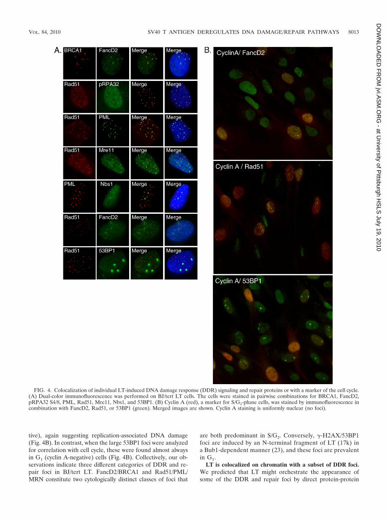
tive), again suggesting replication-associated DNA damage(Fig. 4B). In contrast, when the large 53BP1 foci were analyzedfor correlation with cell cycle, these were found almost alwaysin G1 (cyclin A-negative) cells (Fig. 4B). Collectively, our ob-servations indicate three different categories of DDR and re-pair foci in BJ/tert LT. FancD2/BRCA1 and Rad51/PML/MRN constitute two cytologically distinct classes of foci that
are both predominant in S/G2. Conversely, �-H2AX/53BP1foci are induced by an N-terminal fragment of LT (17k) ina Bub1-dependent manner (23), and these foci are prevalentin G1.
LT is colocalized on chromatin with a subset of DDR foci.We predicted that LT might orchestrate the appearance ofsome of the DDR and repair foci by direct protein-protein
FIG. 4. Colocalization of individual LT-induced DNA damage response (DDR) signaling and repair proteins or with a marker of the cell cycle.(A) Dual-color immunofluorescence was performed on BJ/tert LT cells. The cells were stained in pairwise combinations for BRCA1, FancD2,pRPA32 S4/8, PML, Rad51, Mre11, Nbs1, and 53BP1. (B) Cyclin A (red), a marker for S/G2-phase cells, was stained by immunofluorescence incombination with FancD2, Rad51, or 53BP1 (green). Merged images are shown. Cyclin A staining is uniformly nuclear (no foci).
VOL. 84, 2010 SV40 T ANTIGEN DEREGULATES DNA DAMAGE/REPAIR PATHWAYS 8013
at University of P
ittsburgh HS
LS July 19, 2010
jvi.AS
M.O
RG
- D
OW
NLO
AD
ED
FR
OM
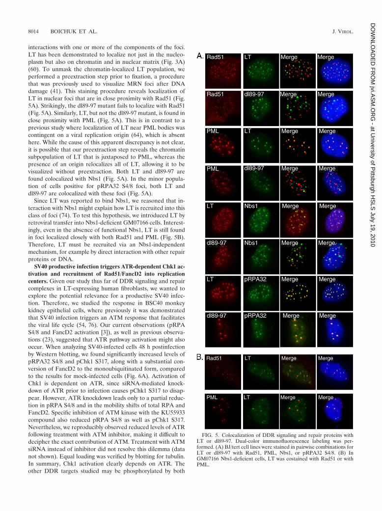
interactions with one or more of the components of the foci.LT has been demonstrated to localize not just in the nucleo-plasm but also on chromatin and in nuclear matrix (Fig. 3A)(60). To unmask the chromatin-localized LT population, weperformed a preextraction step prior to fixation, a procedurethat was previously used to visualize MRN foci after DNAdamage (41). This staining procedure reveals localization ofLT in nuclear foci that are in close proximity with Rad51 (Fig.5A). Strikingly, the dl89-97 mutant fails to localize with Rad51(Fig. 5A). Similarly, LT, but not the dl89-97 mutant, is found inclose proximity with PML (Fig. 5A). This is in contrast to aprevious study where localization of LT near PML bodies wascontingent on a viral replication origin (64), which is absenthere. While the cause of this apparent discrepancy is not clear,it is possible that our preextraction step reveals the chromatinsubpopulation of LT that is juxtaposed to PML, whereas thepresence of an origin relocalizes all of LT, allowing it to bevisualized without preextraction. Both LT and dl89-97 arefound colocalized with Nbs1 (Fig. 5A). In the minor popula-tion of cells positive for pRPA32 S4/8 foci, both LT anddl89-97 are colocalized with these foci (Fig. 5A).
Since LT was reported to bind Nbs1, we reasoned that in-teraction with Nbs1 might explain how LT is recruited into thisclass of foci (74). To test this hypothesis, we introduced LT byretroviral transfer into Nbs1-deficient GM07166 cells. Interest-ingly, even in the absence of functional Nbs1, LT is still foundin foci localized closely with both Rad51 and PML (Fig. 5B).Therefore, LT must be recruited via an Nbs1-independentmechanism, for example by direct interaction with other repairproteins or DNA.
SV40 productive infection triggers ATR-dependent Chk1 ac-tivation and recruitment of Rad51/FancD2 into replicationcenters. Given our study thus far of DDR signaling and repaircomplexes in LT-expressing human fibroblasts, we wanted toexplore the potential relevance for a productive SV40 infec-tion. Therefore, we studied the response in BSC40 monkeykidney epithelial cells, where previously it was demonstratedthat SV40 infection triggers an ATM response that facilitatesthe viral life cycle (54, 76). Our current observations (pRPAS4/8 and FancD2 activation [3]), as well as previous observa-tions (23), suggested that ATR pathway activation might alsooccur. When analyzing SV40-infected cells 48 h postinfectionby Western blotting, we found significantly increased levels ofpRPA32 S4/8 and pChk1 S317, along with a substantial con-version of FancD2 to the monoubiquitinated form, comparedto the results for mock-infected cells (Fig. 6A). Activation ofChk1 is dependent on ATR, since siRNA-mediated knock-down of ATR prior to infection causes pChk1 S317 to disap-pear. However, ATR knockdown leads only to a partial reduc-tion in pRPA S4/8 and in the mobility shifts of total RPA andFancD2. Specific inhibition of ATM kinase with the KU55933compound also reduced pRPA S4/8 as well as pChk1 S317.Nevertheless, we reproducibly observed reduced levels of ATRfollowing treatment with ATM inhibitor, making it difficult todecipher the exact contribution of ATM. Treatment with ATMsiRNA instead of inhibitor did not resolve this dilemma (datanot shown). Equal loading was verified by blotting for tubulin.In summary, Chk1 activation clearly depends on ATR. Theother DDR targets studied may be phosphorylated by both
FIG. 5. Colocalization of DDR signaling and repair proteins withLT or dl89-97. Dual-color immunofluorescence labeling was per-formed. (A) BJ/tert cell lines were stained in pairwise combinations forLT or dl89-97 with Rad51, PML, Nbs1, or pRPA32 S4/8. (B) InGM07166 Nbs1-deficient cells, LT was costained with Rad51 or withPML.
8014 BOICHUK ET AL. J. VIROL.
at University of P
ittsburgh HS
LS July 19, 2010
jvi.AS
M.O
RG
- D
OW
NLO
AD
ED
FR
OM

FIG. 6. SV40 infection of permissive cells induces both ATM and ATR signaling, and LT colocalizes in replication centers with multiple DDRfactors. (A) BSC40 cells were transfected with control or ATR siRNA for 48 h followed by mock infection or SV40 infection for another 48 h. Aspecific ATM inhibitor (KU55933) was applied the last 48 h where indicated. Cells were analyzed by Western blotting for ATR, FancD2, LT, pRPAS4/8, RPA, tubulin, and pChk1 S317. (B) Expression of LT was assessed by immunofluorescence in either mock-infected or SV40-infected cells.Images were merged with DAPI staining. Nearly all cells were infected. (C) At 48 h after SV40 infection, BSC40 cells were stained by dual-colorimmunofluorescence. LT was examined for colocalization with RPA32, pATM S1981, Nbs1, Mre11, pRPA S4/8, ATRIP, FancD2, Rad51, 53BP1,p53, and polymerase � (Pol alpha). Identical patterns of localization were observed when LT costaining was omitted.
VOL. 84, 2010 SV40 T ANTIGEN DEREGULATES DNA DAMAGE/REPAIR PATHWAYS 8015
at University of P
ittsburgh HS
LS July 19, 2010
jvi.AS
M.O
RG
- D
OW
NLO
AD
ED
FR
OM
ATM and ATR or possibly by DNA-dependent protein kinase(DNA-PK) as well.
We also investigated whether various DDR and repair fac-tors are recruited into viral replication centers by performingimmunofluorescence on SV40-infected BSC40 cells. Single im-munostaining of cells with LT antibody revealed that virtuallyall cells were infected (Fig. 6B). Subsequent cell staining wasperformed by dual-color immunofluorescence to assess colo-calization with LT. As expected, on the basis of a previousstudy, 48 h postinfection, LT was colocalized with RPA32,pATM S1981, Nbs1, and Mre11 in large nuclear foci (Fig. 6C)(76). Consistent with the Western blotting results, we findabundant phosphorylation of RPA32 (S4/8), and the immuno-staining coincides with replication centers. Repeated attemptsto probe ATR localization were unsuccessful probably due tothe unsuitability of antibodies for cell staining; however,ATRIP was found partially colocalized with LT. Furthermore,LT was found to be colocalized with FancD2, Rad51, 53BP1,and p53 (Fig. 6C). The observed colocalization of FancD2 andRad51 in infected cells clearly contrasts with what we observedin BJ/tert cells expressing LT alone. The difference may beexplained by the large amounts of unusual replication struc-tures (theta-type intermediates, for example) that are pro-duced during an SV40 infection and that are predicted toinitiate DDR and repair signaling. Finally, consistent with theprevious report that LT foci correspond to viral replicationcenters (76), we find LT colocalized with polymerase � ininfected cells (Fig. 6C), whereas in noninfected cells, it wasdiffusively nuclear (data not shown). Whether the recruitmentof a wide array of DDR and repair signaling proteins to rep-lication centers reflects ongoing repair of SV40 genomes orwhether these molecules contribute in an active way to SV40replication, for example through HR, are important questionsto be addressed.
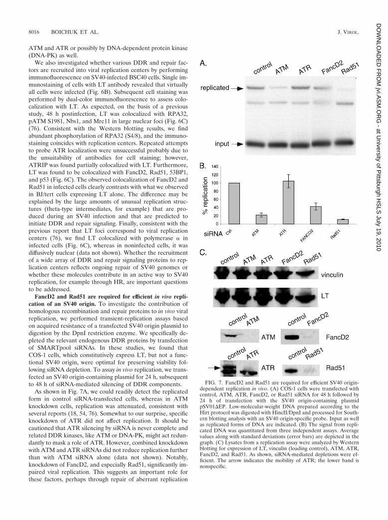
FancD2 and Rad51 are required for efficient in vivo repli-cation of an SV40 origin. To investigate the contribution ofhomologous recombination and repair proteins to in vivo viralreplication, we performed transient-replication assays basedon acquired resistance of a transfected SV40 origin plasmid todigestion by the DpnI restriction enzyme. We specifically de-pleted the relevant endogenous DDR proteins by transfectionof SMARTpool siRNAs. In these studies, we found thatCOS-1 cells, which constitutively express LT, but not a func-tional SV40 origin, were optimal for preserving viability fol-lowing siRNA depletion. To assay in vivo replication, we trans-fected an SV40 origin-containing plasmid for 24 h, subsequentto 48 h of siRNA-mediated silencing of DDR components.
As shown in Fig. 7A, we could readily detect the replicatedform in control siRNA-transfected cells, whereas in ATMknockdown cells, replication was attenuated, consistent withseveral reports (18, 54, 76). Somewhat to our surprise, specificknockdown of ATR did not affect replication. It should becautioned that ATR silencing by siRNA is never complete andrelated DDR kinases, like ATM or DNA-PK, might act redun-dantly to mask a role of ATR. However, combined knockdownwith ATM and ATR siRNAs did not reduce replication furtherthan with ATM siRNA alone (data not shown). Notably,knockdown of FancD2, and especially Rad51, significantly im-paired viral replication. This suggests an important role forthese factors, perhaps through repair of aberrant replication
FIG. 7. FancD2 and Rad51 are required for efficient SV40 origin-dependent replication in vivo. (A) COS-1 cells were transfected withcontrol, ATM, ATR, FancD2, or Rad51 siRNA for 48 h followed by24 h of transfection with the SV40 origin-containing plasmidpSV01�EP. Low-molecular-weight DNA prepared according to theHirt protocol was digested with HincII/DpnI and processed for South-ern blotting analysis with an SV40 origin-specific probe. Input as wellas replicated forms of DNA are indicated. (B) The signal from repli-cated DNA was quantitated from three independent assays. Averagevalues along with standard deviations (error bars) are depicted in thegraph. (C) Lysates from a replication assay were analyzed by Westernblotting for expression of LT, vinculin (loading control), ATM, ATR,FancD2, and Rad51. As shown, siRNA-mediated depletions were ef-ficient. The arrow indicates the mobility of ATR; the lower band isnonspecific.
8016 BOICHUK ET AL. J. VIROL.
at University of P
ittsburgh HS
LS July 19, 2010
jvi.AS
M.O
RG
- D
OW
NLO
AD
ED
FR
OM

forms like stalled or collapsed forks. Relative replication pro-ficiency compared with control siRNA-transfected cells wascalculated by quantitation of three independent assays, and theaverage values with standard deviations are depicted in thegraph of Fig. 7B.
To confirm the knockdown of individual DDR factors, wealso performed Western blotting on the same lysates used forreplication assays. As shown in Fig. 7C, all of the DDR factorswere substantially depleted following siRNA transfection.Blotting with LT and tubulin antibodies further confirmedequal loading between samples. Taken together, we find that inaddition to ATM, FancD2 and Rad51 also participate in SV40origin-dependent replication in vivo.
DISCUSSION
Our current study has uncovered further complexities inDDR induction and regulation by LT that are important formechanistic understanding of how LT has coopted the DDRfor enhancement of viral replication (18, 23, 54, 76). Recentwork has established that cellular DNA damage is not neces-sarily a prerequisite for a DDR (56). Importantly, our cometassay results indicate that LT is intrinsically capable of induc-ing DNA damage, partly in the form of DSBs. It is possible thatthe induction of DSBs by LT facilitates viral integration eventsand contributes to the previously observed promotion of geneamplification (49).
At least three distinct classes of DDR signaling and repairfoci are induced by LT expression in BJ/tert cells, apparentlyvia distinct LT domains. Our model is depicted schematicallyin Fig. 8. Large foci of �-H2AX/53BP1, probably correspond-ing to DSBs, are induced by LT in a Bub1 binding-dependentmanner (23). An pRB binding-deficient N-terminal LT frag-ment (17k) suffices to induce these foci (23). Since these fociare prominent in G1 phase, they might result from errors inpreceding mitotic segregation and checkpoint control, whereBub1 is primarily known to act (70). Interestingly, LT-express-ing BJ/tert cells frequently exhibit chromatin bridge formation
in anaphase (data not shown), which might trigger a DDRupon breakage of the DNA following cell division.
In contrast, foci of FancD2 and BRCA1 are mainly foundin S/G2 and likely connected with a replication stress re-sponse, for example from stalled or collapsed replicationforks (26, 29, 44). Chromatin fractionation also indicates LTactivation of FancD2. It was recently demonstrated that thehuman papillomavirus E7 protein, in part via pRB familybinding, induces FancD2 foci and its accumulation on chro-matin (57). It was further shown that E7 via a replicationstress response causes alternative lengthening of telomeres(ALT)-associated promyelocytic leukemia bodies (APBs) inprimary keratinocytes (58). The APBs contain FancD2,PML, and telomeric DNA. It was proposed that this induc-tion of APBs might underlie the observed telomere main-tenance, independent of telomerase, in E7-expressing cells(58). LT might similarly promote ALT via replication stressand FancD2 activation, but in our cell system (BJ/tert), thismay not be relevant, since telomerase is expressed. We didnot observe colocalization of the telomeric factor Trf2 witheither FancD2 or PML (data not shown), suggesting thatthese foci do not represent APBs.
The third class of foci that LT induces is composed of therecombinase Rad51, PML, the MRN complex, and LT itself.These proteins have often been found colocalized with eachother during the DDR (8, 11, 37, 73), although in some con-texts it was reported that MRN and Rad51 were present indistinct types of repair foci (39). Appearance of these foci maybe a result of replication-associated DNA damage that the cellis attempting to repair by HR. In the absence of functional p53,Rad51 was previously found upregulated (38, 75). Indeed, p53can also suppress HR by direct binding to Rad51 (36). Theincrease in total Rad51 together with focus formation suggestsan overall increase in HR, which might be correlated with thepreviously observed prorecombinogenic activities of LT (61,75). However, JC polyomavirus LT modulates Rad51 in wayssimilar to those of SV40 but is believed to attenuate HR repair
FIG. 8. Model of DDR signaling and repair pathways induced by LT. Focal accumulation of �-H2AX/53BP1 in the G1 phase is induced by LT,as well as 17k, via Bub1 binding to the 89-97 region of LT. In contrast, distinct foci of either FancD2/BRCA1 or Rad51/MRN/PML are prominentin S/G2 phase. Induction of these two main classes of foci appears independent of pRB family or Bub1 binding. Only Rad51/MRN/PML arelocalized at, or near, LT foci. The different sized black dots for the three different categories of DDR foci reflect both the relative sizes of the fociby microscopy and their differences in spatial localization.
VOL. 84, 2010 SV40 T ANTIGEN DEREGULATES DNA DAMAGE/REPAIR PATHWAYS 8017
at University of P
ittsburgh HS
LS July 19, 2010
jvi.AS
M.O
RG
- D
OW
NLO
AD
ED
FR
OM

by forcing an interaction between Rad51 and insulin receptorsubstrate 1 (67).
Our results from SV40 productive infection of BSC40 cellsdemonstrate ATR-dependent activation of Chk1. LT is colo-calized during infection with phospho-RPA, ATRIP, FancD2,and Rad51. Phosphorylation of RPA is thought to act as aswitch from cellular replication to DNA repair activity, consis-tent with the proposed concept of SV40 replication proceedingthrough DNA repair pathways (7, 76). Phosphorylation ofRPA at S4/8 following SV40 infection is apparently mediatedby both ATM and ATR. Recruitment of various repair pro-teins might simply reflect that the cell is detecting viral ge-nomes as damaged DNA, but it could also be interpreted tomean that at least a subset of these repair proteins plays anactive role in SV40 replication.
While we did not observe a role for ATR in SV40 origin-dependent replication in vivo, we conversely demonstrated thatFancD2 and Rad51 play important roles. Although we do notknow the precise mechanisms involved, we speculate that high-copy episomal replication with high fidelity might require theHR machinery to repair damage to the viral genome, for ex-ample because of stalled replication forks. For both herpessimplex virus type 1 (HSV-1) and Epstein-Barr virus (EBV),lytic replication also leads to recruitment of Rad51 to replica-tion compartments; for EBV, it was demonstrated that Rad51knockdown causes a reduction in viral genome synthesis, sug-gesting that HR is necessary for efficient replication (30, 69).
Notably, we found that the dl89-97 mutant fails to localizeat, or near, Rad51 and PML foci, while maintaining colocal-ization with the MRN complex. This could be significant, sincethe dl89-97 mutant exhibits a defect in driving viral replicationin vivo (data not shown). PML oncogenic domains are commontargets of viruses, and polyomaviruses localize to and replicatetheir genomes near these structures (20, 27, 28, 64). Viralreplication might be more efficient in the vicinity of thesesubnuclear bodies, but the underlying mechanisms have notbeen delineated. An intriguing possibility is that PML onco-genic domains act as master nuclear organizers for variousDDR and repair factors, thereby allowing LT to target orrecruit the HR machinery and facilitating repair on the viralgenome (8, 28).
Why does dl89-97 fail to localize near Rad51 and PML?While we do not yet know the answer, possibilities include lackof a Bub1-mediated phosphorylation event, for example onLT, or failure to directly interact with Rad51, PML, or anothercomponent of PML oncogenic domains. The temporal assem-bly of DDR factors following ionizing radiation is criticallydependent on phosphorylation events, for example histoneH2AX phosphorylation to generate �-H2AX generates a markon chromatin to recruit Mdc1 and the MRN complex (63). LTmight have adapted a similar mechanism for its recruitment toDDR foci. Phosphorylation of LT on S120, a proposed ATMsite, is required for replication in vivo, thus providing a possiblecandidate (53, 54). Indeed, we know that specific ATM inhi-bition causes at least partial loss of LT localization at replica-tion centers (76). The BRCT-related region present within theSV40 helicase domain suggests another potential mode of re-cruitment (31). BRCT domains, present in Mdc1 and BRCA1,are phosphoserine/threonine-dependent interaction modulesimportant for DDR signaling (43).
What are the underlying lesions that trigger these distinctDDR and repair responses, and how are they generated?While we do not know the answer, it may be useful to firstidentify features or binding sites on LT that are responsible forinduction of each response. The induction of both FancD2 andRad51 foci is S/G2 phase specific and independent of both pRBand Bub1 binding. Known LT binding partners that might berelevant for inducing these responses are Nbs1, RPA, and p53.A recent study demonstrated that JC polyomavirus LT inducesan ATM/ATR-mediated G2 checkpoint arrest by binding tocellular DNA (48). These studies make it important to inves-tigate whether SV40 LT binding to cellular DNA, mediated byits origin-binding domain, is responsible for the observed rep-lication stress response and either FancD2 or Rad51 focusformation. Importantly, we do not see evidence of significantG2 accumulation when LT alone is stably expressed (data notshown), despite activation of multiple DDR and repair signal-ing pathways. The apparent uncoupling of DDR signaling fromproper checkpoint responses may require LT interaction withadditional checkpoint proteins besides p53.
Here we further show that LT induces constitutive pan-nuclear pATM S1981, which fails to be relocalized to focifollowing external DNA damage, as occurs in healthy BJ/tertcells. This suggests that LT causes a failure for ATM to beretained at sites of DNA damage, which is believed to beimportant for proper DDR signaling (55). Nevertheless, we didnot see any significant differences in the downstream signalingevents that were analyzed. It remains possible that some sub-strates are affected. Our observations share some similaritywith the recent demonstration that HSV-1 ICP0 (infected cellpolypeptide 0) by targeting cellular histone ubiquitin ligasesalso causes diffuse nuclear staining of pATM S1981 and itsfailure to be retained at repair foci (34).
Does activation of DDR and repair pathways impact LT-mediated oncogenic transformation? Multiple genetic disor-ders caused by DNA repair deficiency confer an increasedsusceptibility to cancer. Importantly, it is known that fibro-blasts from individuals with Fanconi anemia are hypersensitiveto SV40 transformation (66). While this observation could berelated to an increased SV40 integration frequency, an alter-native view is that DDR activation potentially limits SV40oncogenic transformation, consistent with the general notionthat the DDR acts as a barrier to malignancy. However, thestabilization of p53 by LT, presumably resulting from an acti-vated DDR, can contribute to a gain of function in neoplastictransformation (9, 23, 24). The enhanced transforming abilityof LT in wild-type p53 cells compared to p53-deficient coun-terparts is likely due to altered transcriptional patterns, per-haps because the stabilized form of p53 can recruit p300/CBPcoactivators (24). Taken together, the DDR might act as adouble-edged sword when modulating the LT transformationoutcome. Perhaps different subsets of DDR pathways are in-volved in FancD2 activation versus p53 stabilization.
LT has long been known to cause structural as well as nu-merical chromosome instability, such as sister chromatid ex-changes, dicentric chromosomes, chromosome breaks, and tet-raploidy (13, 23, 72). It is possible that a links exists betweenperturbations of the DDR and chromosomal aberrations, but acause-effect relationship has not been established. Future stud-ies will be aimed at resolving these potential links between
8018 BOICHUK ET AL. J. VIROL.
at University of P
ittsburgh HS
LS July 19, 2010
jvi.AS
M.O
RG
- D
OW
NLO
AD
ED
FR
OM
DDR, repair, and chromosomal aberrations and illuminatehow they relate to oncogenic transformation or an enhancedviral replication program.
ACKNOWLEDGMENTS
We thank Jim Pipas for kindly providing BSC40 cells, Xiaohua Wufor GM07166 cells, and Ellen Fanning for polymerase � antibody.
We gratefully acknowledge financial support from the NIH (R01AI078926 to O.V.G.).
REFERENCES
1. Ahuja, D., M. T. Saenz-Robles, and J. M. Pipas. 2005. SV40 large T antigentargets multiple cellular pathways to elicit cellular transformation. Oncogene24:7729–7745.
2. Ali, S. H., J. S. Kasper, T. Arai, and J. A. DeCaprio. 2004. Cul7/p185/p193binding to simian virus 40 large T antigen has a role in cellular transforma-tion. J. Virol. 78:2749–2757.
3. Andreassen, P. R., A. D. D’Andrea, and T. Taniguchi. 2004. ATR couplesFANCD2 monoubiquitination to the DNA-damage response. Genes Dev.18:1958–1963.
4. Bakkenist, C. J., and M. B. Kastan. 2003. DNA damage activates ATMthrough intermolecular autophosphorylation and dimer dissociation. Nature421:499–506.
5. Bekker-Jensen, S., C. Lukas, R. Kitagawa, F. Melander, M. B. Kastan, J.Bartek, and J. Lukas. 2006. Spatial organization of the mammalian genomesurveillance machinery in response to DNA strand breaks. J. Cell Biol.173:195–206.
6. Bernardi, R., and P. P. Pandolfi. 2007. Structure, dynamics and functions ofpromyelocytic leukaemia nuclear bodies. Nat. Rev. Mol. Cell Biol. 8:1006–1016.
7. Binz, S. K., A. M. Sheehan, and M. S. Wold. 2004. Replication protein Aphosphorylation and the cellular response to DNA damage. DNA Repair(Amst.) 3:1015–1024.
8. Bischof, O., S. H. Kim, J. Irving, S. Beresten, N. A. Ellis, and J. Campisi.2001. Regulation and localization of the Bloom syndrome protein in re-sponse to DNA damage. J. Cell Biol. 153:367–380.
9. Bocchetta, M., S. Eliasz, M. A. De Marco, J. Rudzinski, L. Zhang, and M.Carbone. 2008. The SV40 large T antigen-p53 complexes bind and activatethe insulin-like growth factor-I promoter stimulating cell growth. CancerRes. 68:1022–1029.
10. Campbell, K. S., K. P. Mullane, I. A. Aksoy, H. Stubdal, J. Zalvide, J. M.Pipas, P. A. Silver, T. M. Roberts, B. S. Schaffhausen, and J. A. DeCaprio.1997. DnaJ/hsp40 chaperone domain of SV40 large T antigen promotesefficient viral DNA replication. Genes Dev. 11:1098–1110.
11. Carbone, R., M. Pearson, S. Minucci, and P. G. Pelicci. 2002. PML NBsassociate with the hMre11 complex and p53 at sites of irradiation inducedDNA damage. Oncogene 21:1633–1640.
12. Carson, C. T., N. I. Orazio, D. V. Lee, J. Suh, S. Bekker-Jensen, F. D. Araujo,S. S. Lakdawala, C. E. Lilley, J. Bartek, J. Lukas, and M. D. Weitzman. 2009.Mislocalization of the MRN complex prevents ATR signaling during ade-novirus infection. EMBO J. 28:652–662.
13. Chang, T. H., F. A. Ray, D. A. Thompson, and R. Schlegel. 1997. Disregu-lation of mitotic checkpoints and regulatory proteins following acute expres-sion of SV40 large T antigen in diploid human cells. Oncogene 14:2383–2393.
14. Chaurushiya, M. S., and M. D. Weitzman. 2009. Viral manipulation of DNArepair and cell cycle checkpoints. DNA Repair (Amst.) 8:1166–1176.
15. Chen, C., and H. Okayama. 1987. High-efficiency transformation of mam-malian cells by plasmid DNA. Mol. Cell. Biol. 7:2745–2752.
16. Cheng, J., J. A. DeCaprio, M. M. Fluck, and B. S. Schaffhausen. 2009.Cellular transformation by simian virus 40 and murine polyoma virus Tantigens. Semin. Cancer Biol. 19:218–228.
17. Cotsiki, M., R. L. Lock, Y. Cheng, G. L. Williams, J. Zhao, D. Perera, R.Freire, A. Entwistle, E. A. Golemis, T. M. Roberts, P. S. Jat, and O. V.Gjoerup. 2004. Simian virus 40 large T antigen targets the spindle assemblycheckpoint protein Bub1. Proc. Natl. Acad. Sci. U. S. A. 101:947–952.
18. Dahl, J., J. You, and T. L. Benjamin. 2005. Induction and utilization of anATM signaling pathway by polyomavirus. J. Virol. 79:13007–13017.
19. Dickmanns, A., A. Zeitvogel, F. Simmersbach, R. Weber, A. K. Arthur, S.Dehde, A. G. Wildeman, and E. Fanning. 1994. The kinetics of simian virus40-induced progression of quiescent cells into S phase depend on fourindependent functions of large T antigen. J. Virol. 68:5496–5508.
20. Everett, R. D. 2006. Interactions between DNA viruses, ND10 and the DNAdamage response. Cell. Microbiol. 8:365–374.
21. Garcia-Higuera, I., T. Taniguchi, S. Ganesan, M. S. Meyn, C. Timmers,J. Hejna, M. Grompe, and A. D. D’Andrea. 2001. Interaction of the Fanconianemia proteins and BRCA1 in a common pathway. Mol. Cell 7:249–262.
22. Gjorup, O. V., P. E. Rose, P. S. Holman, B. J. Bockus, and B. S. Schaff-hausen. 1994. Protein domains connect cell cycle stimulation directly to
initiation of DNA replication. Proc. Natl. Acad. Sci. U. S. A. 91:12125–12129.
23. Hein, J., S. Boichuk, J. Wu, Y. Cheng, R. Freire, P. S. Jat, T. M. Roberts, andO. V. Gjoerup. 2009. Simian virus 40 large T antigen disrupts genomeintegrity and activates a DNA damage response via Bub1 binding. J. Virol.83:117–127.
24. Hermannstadter, A., C. Ziegler, M. Kuhl, W. Deppert, and G. V. Tolstonog.2009. Wild-type p53 enhances efficiency of simian virus 40 large-T-antigen-induced cellular transformation. J. Virol. 83:10106–10118.
25. Hoskins, E. E., R. W. Gunawardena, K. B. Habash, T. M. Wise-Draper, M.Jansen, E. S. Knudsen, and S. I. Wells. 2008. Coordinate regulation ofFanconi anemia gene expression occurs through the Rb/E2F pathway. On-cogene 27:4798–4808.
26. Howlett, N. G., T. Taniguchi, S. G. Durkin, A. D. D’Andrea, and T. W.Glover. 2005. The Fanconi anemia pathway is required for the DNA repli-cation stress response and for the regulation of common fragile site stability.Hum. Mol. Genet. 14:693–701.
27. Ishov, A. M., and G. G. Maul. 1996. The periphery of nuclear domain 10(ND10) as site of DNA virus deposition. J. Cell Biol. 134:815–826.
28. Jul-Larsen, A., T. Visted, B. O. Karlsen, C. H. Rinaldo, R. Bjerkvig, P. E.Lonning, and S. O. Boe. 2004. PML-nuclear bodies accumulate DNA inresponse to polyomavirus BK and simian virus 40 replication. Exp. Cell Res.298:58–73.
29. Knipscheer, P., M. Raschle, A. Smogorzewska, M. Enoiu, T. V. Ho, O. D.Scharer, S. J. Elledge, and J. C. Walter. 2009. The Fanconi anemia pathwaypromotes replication-dependent DNA interstrand cross-link repair. Science326:1698–1701.
30. Kudoh, A., S. Iwahori, Y. Sato, S. Nakayama, H. Isomura, T. Murata, and T.Tsurumi. 2009. Homologous recombinational repair factors are recruitedand loaded onto the viral DNA genome in Epstein-Barr virus replicationcompartments. J. Virol. 83:6641–6651.
31. Kumar, A., W. S. Joo, G. Meinke, S. Moine, E. N. Naumova, and P. A.Bullock. 2008. Evidence for a structural relationship between BRCT do-mains and the helicase domains of the replication initiators encoded by thePolyomaviridae and Papillomaviridae families of DNA tumor viruses. J. Vi-rol. 82:8849–8862.
32. Lee, J. H., and T. T. Paull. 2005. ATM activation by DNA double-strandbreaks through the Mre11-Rad50-Nbs1 complex. Science 308:551–554.
33. Li, J. J., and T. J. Kelly. 1984. Simian virus 40 DNA replication in vitro. Proc.Natl. Acad. Sci. U. S. A. 81:6973–6977.
34. Lilley, C. E., M. S. Chaurushiya, C. Boutell, S. Landry, J. Suh, S. Panier,R. D. Everett, G. S. Stewart, D. Durocher, and M. D. Weitzman. 2010. A viralE3 ligase targets RNF8 and RNF168 to control histone ubiquitination andDNA damage responses. EMBO J. 29:943–955.
35. Lilley, C. E., R. A. Schwartz, and M. D. Weitzman. 2007. Using or abusing:viruses and the cellular DNA damage response. Trends Microbiol. 15:119–126.
36. Linke, S. P., S. Sengupta, N. Khabie, B. A. Jeffries, S. Buchhop, S. Miska, W.Henning, R. Pedeux, X. W. Wang, L. J. Hofseth, Q. Yang, S. H. Garfield,H. W. Sturzbecher, and C. C. Harris. 2003. p53 interacts with hRAD51 andhRAD54, and directly modulates homologous recombination. Cancer Res.63:2596–2605.
37. Lombard, D. B., and L. Guarente. 2000. Nijmegen breakage syndrome dis-ease protein and MRE11 at PML nuclear bodies and meiotic telomeres.Cancer Res. 60:2331–2334.
38. Marusyk, A., L. J. Wheeler, C. K. Mathews, and J. DeGregori. 2007. p53mediates senescence-like arrest induced by chronic replicational stress. Mol.Cell. Biol. 27:5336–5351.
39. Maser, R. S., K. J. Monsen, B. E. Nelms, and J. H. Petrini. 1997. hMre11 andhRad50 nuclear foci are induced during the normal cellular response toDNA double-strand breaks. Mol. Cell. Biol. 17:6087–6096.
40. Mendez, J., and B. Stillman. 2000. Chromatin association of human originrecognition complex, cdc6, and minichromosome maintenance proteins dur-ing the cell cycle: assembly of prereplication complexes in late mitosis. Mol.Cell. Biol. 20:8602–8612.
41. Mirzoeva, O. K., and J. H. Petrini. 2001. DNA damage-dependent nucleardynamics of the Mre11 complex. Mol. Cell. Biol. 21:281–288.
42. Mladenov, E., B. Anachkova, and I. Tsaneva. 2006. Sub-nuclear localizationof Rad51 in response to DNA damage. Genes Cells 11:513–524.
43. Mohammad, D. H., and M. B. Yaffe. 2009. 14-3-3 proteins, FHA domainsand BRCT domains in the DNA damage response. DNA Repair (Amst.)8:1009–1017.
44. Moldovan, G. L., and A. D. D’Andrea. 2009. How the Fanconi anemiapathway guards the genome. Annu. Rev. Genet. 43:223–249.
45. Ohashi, A., M. Z. Zdzienicka, J. Chen, and F. J. Couch. 2005. Fanconianemia complementation group D2 (FANCD2) functions independently ofBRCA2- and RAD51-associated homologous recombination in response toDNA damage. J. Biol. Chem. 280:14877–14883.
46. Olive, P. L., and J. P. Banath. 2006. The comet assay: a method to measureDNA damage in individual cells. Nat. Protoc. 1:23–29.
47. Olson, E., C. J. Nievera, V. Klimovich, E. Fanning, and X. Wu. 2006. RPA2
VOL. 84, 2010 SV40 T ANTIGEN DEREGULATES DNA DAMAGE/REPAIR PATHWAYS 8019
at University of P
ittsburgh HS
LS July 19, 2010
jvi.AS
M.O
RG
- D
OW
NLO
AD
ED
FR
OM
is a direct downstream target for ATR to regulate the S-phase checkpoint.J. Biol. Chem. 281:39517–39533.
48. Orba, Y., T. Suzuki, Y. Makino, K. Kubota, S. Tanaka, T. Kimura, and H.Sawa. 2010. Large T antigen promotes JC virus replication in G2-arrestedcells by inducing ATM- and ATR-mediated G2 checkpoint signaling. J. Biol.Chem. 285:1544–1554.
49. Perry, M. E., M. Commane, and G. R. Stark. 1992. Simian virus 40 largetumor antigen alone or two cooperating oncogenes convert REF52 cells to astate permissive for gene amplification. Proc. Natl. Acad. Sci. U. S. A.89:8112–8116.
50. Petrini, J. H., and T. H. Stracker. 2003. The cellular response to DNAdouble-strand breaks: defining the sensors and mediators. Trends Cell Biol.13:458–462.
51. Raderschall, E., E. I. Golub, and T. Haaf. 1999. Nuclear foci of mammalianrecombination proteins are located at single-stranded DNA regions formedafter DNA damage. Proc. Natl. Acad. Sci. U. S. A. 96:1921–1926.
52. Rogakou, E. P., D. R. Pilch, A. H. Orr, V. S. Ivanova, and W. M. Bonner.1998. DNA double-stranded breaks induce histone H2AX phosphorylationon serine 139. J. Biol. Chem. 273:5858–5868.
53. Schneider, J., and E. Fanning. 1988. Mutations in the phosphorylation sitesof simian virus 40 (SV40) T antigen alter its origin DNA-binding specificityfor sites I or II and affect SV40 DNA replication activity. J. Virol. 62:1598–1605.
54. Shi, Y., G. E. Dodson, S. Shaikh, K. Rundell, and R. S. Tibbetts. 2005.Ataxia-telangiectasia-mutated (ATM) is a T-antigen kinase that controlsSV40 viral replication in vivo. J. Biol. Chem. 280:40195–40200.
55. So, S., A. J. Davis, and D. J. Chen. 2009. Autophosphorylation at serine 1981stabilizes ATM at DNA damage sites. J. Cell Biol. 187:977–990.
56. Soutoglou, E., and T. Misteli. 2008. Activation of the cellular DNA damageresponse in the absence of DNA lesions. Science 320:1507–1510.
57. Spardy, N., A. Duensing, D. Charles, N. Haines, T. Nakahara, P. F. Lambert,and S. Duensing. 2007. The human papillomavirus type 16 E7 oncoproteinactivates the Fanconi anemia (FA) pathway and causes accelerated chromo-somal instability in FA cells. J. Virol. 81:13265–13270.
58. Spardy, N., A. Duensing, E. E. Hoskins, S. I. Wells, and S. Duensing. 2008.HPV-16 E7 reveals a link between DNA replication stress, Fanconi anemiaD2 protein, and alternative lengthening of telomere-associated promyelo-cytic leukemia bodies. Cancer Res. 68:9954–9963.
59. Srinivasan, A., A. J. McClellan, J. Vartikar, I. Marks, P. Cantalupo, Y. Li,P. Whyte, K. Rundell, J. L. Brodsky, and J. M. Pipas. 1997. The amino-terminal transforming region of simian virus 40 large T and small t antigensfunctions as a J domain. Mol. Cell. Biol. 17:4761–4773.
60. Staufenbiel, M., and W. Deppert. 1983. Different structural systems of thenucleus are targets for SV40 large T antigen. Cell 33:173–181.
61. St-Onge, L., L. Bouchard, S. Laurent, and M. Bastin. 1990. Intrachromo-somal recombination mediated by papovavirus large T antigens. J. Virol.64:2958–2966.
62. Stracker, T. H., C. T. Carson, and M. D. Weitzman. 2002. Adenovirusoncoproteins inactivate the Mre11-Rad50-NBS1 DNA repair complex. Na-ture 418:348–352.
63. Stucki, M., and S. P. Jackson. 2006. �H2AX and MDC1: anchoring the
DNA-damage-response machinery to broken chromosomes. DNA Repair(Amst.) 5:534–543.
64. Tang, Q., P. Bell, P. Tegtmeyer, and G. G. Maul. 2000. Replication but nottranscription of simian virus 40 DNA is dependent on nuclear domain 10.J. Virol. 74:9694–9700.
65. Taniguchi, T., I. Garcia-Higuera, P. R. Andreassen, R. C. Gregory, M.Grompe, and A. D. D’Andrea. 2002. S-phase-specific interaction of the Fan-coni anemia protein, FANCD2, with BRCA1 and RAD51. Blood 100:2414–2420.
66. Todaro, G. J., H. Green, and M. R. Swift. 1966. Susceptibility of humandiploid fibroblast strains to transformation by SV40 virus. Science 153:1252–1254.
67. Trojanek, J., S. Croul, T. Ho, J. Y. Wang, A. Darbinyan, M. Nowicki, L. DelValle, T. Skorski, K. Khalili, and K. Reiss. 2006. T-antigen of the humanpolyomavirus JC attenuates faithful DNA repair by forcing nuclear interac-tion between IRS-1 and Rad51. J. Cell. Physiol. 206:35–46.
68. West, S. C. 2003. Molecular views of recombination proteins and theircontrol. Nat. Rev. Mol. Cell Biol. 4:435–445.
69. Wilkinson, D. E., and S. K. Weller. 2004. Recruitment of cellular recombi-nation and repair proteins to sites of herpes simplex virus type 1 DNAreplication is dependent on the composition of viral proteins within prerep-licative sites and correlates with the induction of the DNA damage response.J. Virol. 78:4783–4796.
70. Williams, G. L., T. M. Roberts, and O. V. Gjoerup. 2007. Bub1: escapades ina cellular world. Cell Cycle 6:1699–1704.
71. Wobbe, C. R., F. Dean, L. Weissbach, and J. Hurwitz. 1985. In vitro repli-cation of duplex circular DNA containing the simian virus 40 DNA originsite. Proc. Natl. Acad. Sci. U. S. A. 82:5710–5714.
72. Woods, C., C. LeFeuvre, N. Stewart, and S. Bacchetti. 1994. Induction ofgenomic instability in SV40 transformed human cells: sufficiency of theN-terminal 147 amino acids of large T antigen and role of pRB and p53.Oncogene 9:2943–2950.
73. Wu, G., X. Jiang, W. H. Lee, and P. L. Chen. 2003. Assembly of functionalALT-associated promyelocytic leukemia bodies requires Nijmegen breakagesyndrome 1. Cancer Res. 63:2589–2595.
74. Wu, X., D. Avni, T. Chiba, F. Yan, Q. Zhao, Y. Lin, H. Heng, and D.Livingston. 2004. SV40 T antigen interacts with Nbs1 to disrupt DNA rep-lication control. Genes Dev. 18:1305–1316.
75. Xia, S. J., M. A. Shammas, and R. J. Shmookler Reis. 1997. Elevatedrecombination in immortal human cells is mediated by HsRAD51 recombi-nase. Mol. Cell. Biol. 17:7151–7158.
76. Zhao, X., R. J. Madden-Fuentes, B. X. Lou, J. M. Pipas, J. Gerhardt, C. J.Rigell, and E. Fanning. 2008. Ataxia telangiectasia-mutated damage-signal-ing kinase- and proteasome-dependent destruction of Mre11-Rad50-Nbs1subunits in simian virus 40-infected primate cells. J. Virol. 82:5316–5328.
77. Zimmerman, E. S., M. P. Sherman, J. L. Blackett, J. A. Neidleman, C. Kreis,P. Mundt, S. A. Williams, M. Warmerdam, J. Kahn, F. M. Hecht, R. M.Grant, C. M. de Noronha, A. S. Weyrich, W. C. Greene, and V. Planelles.2006. Human immunodeficiency virus type 1 Vpr induces DNA replicationstress in vitro and in vivo. J. Virol. 80:10407–10418.
8020 BOICHUK ET AL. J. VIROL.
at University of P
ittsburgh HS
LS July 19, 2010
jvi.AS
M.O
RG
- D
OW
NLO
AD
ED
FR
OM