Embed Size (px)
Citation preview
Biochem. J. (1994) 301, 693-702 (Printed in Great Britain)
Multi-site phosphorylation of the inhibitory guanine nucleotide regulatoryprotein Gi-2 occurs in intact rat hepatocytesNicholas J. MORRIS,* Mark BUSHFIELD,t Brian E. LAVAN* and Miles D. HOUSLAYtMolecular Pharmacology Group, Department of Biochemistry, University of Glasgow, Glasgow G12 800, Scotland, U.K.
A phosphorylated form of cx-Gi-2 (the ca-subunit of Gi-2),immunoprecipitated from hepatocytes under basal conditions,migrated as a single species of pl - 5.7, the labelling of whichincreased - 2-fold in cells challenged with either vasopressin orphorbol 12-myristate 13-acetate (PMA); agents which activateprotein kinase C. In contrast, treatment of hepatocytes with8-bromo-cyclic AMP produced a more acidic species ofphosphorylated cc-Gi-2 having a pl of - 5.4 and whose labellingwas increased - 3-fold. Trypsin digestion of labelled a-Gi-2isolated from hepatocytes under basal conditions identified, ontwo-dimensional peptide analyses, three positively chargedphosphoserine-containing peptides (Cl, C2 and C3), with onlypeptides Cl and C2 being evident upon less extensive digestionwith trypsin. These are suggested to reflect a single site ofphosphorylation, with proteolysis by trypsin being incomplete,and where C2 is larger than C1, which is larger than C3. An
INTRODUCTION
Many cell-surface receptors control the activity of their effectorsignal-generating systems through specific members of a familyof G-proteins (Birnbaumer et al., 1990). The receptor-dependentproduction of cyclic AMP (cAMP) is effected by adenylatecyclases whose activities are regulated by two distinct hetero-trimeric G-proteins termed, generically, G5 (stimulatory) and G.(inhibitory). These G-proteins are characterized by unique a-subunits which are the products of different genes. A family of'Gi-like' proteins has been identified which comprises threehighly related pertussis-toxin-sensitive G-proteins termed Gi-1,Gi-2 and Gi-3 (Birnbaumer et al., 1990). Although the specificityof these 'Gi-like' proteins is not yet fully characterized, a numberof groups have provided independent evidence that G1-2 can actas an inhibitory ('G1') G-protein controlling adenylate cyclaseactivity (Simonds et al., 1989; Senogles et al., 1990; McKenzieand Milligan, 1990; Bushfield et al., 1990a; Remaury et al.,1993).
In various cell types, the activation of protein kinase C (PKC)has been shown to result in alterations in the regulation ofadenylate cyclase activity (reviewed by Houslay, 1991a). In somesystems enhanced basal and agonist-stimulated actions havebeen observed, whereas, in others, a loss of receptor-mediatedstimulation was recorded. The underlying molecular basis forthese differences may lie in the cell-specific expression of par-ticular control systems, where changes may be dependent on the
identical pattern of tryptic phosphopeptides was seen in hepato-cytes treated with either vasopressin or PMA, although labellingof this group of peptides was increased by - 2-fold comparedwith the basal state. In contrast, treatment of hepatocytes withglucagon, 8-bromo-cyclic AMP or forskolin not only resulted inincreased labelling of the 'basal' sites - 3-fold, but identified anovel positively charged tryptic phosphoserine-containing pep-tide (AN). All four tryptic peptides were susceptible to proteolysisby V8 protease. Treatment of labelled a-Gi-2 from basal andPMA-treated cells produced a pattern of peptides which wasidentical with those found when the tryptic phosphopeptide wastreated with V8 protease. We tentatively suggest that, on a-Gi-2,Ser144 is phosphorylated through the action of protein kinase Cand Ser207 is phosphorylated upon elevation of the intracellularconcentrations of cyclic AMP.
expression of particular isoforms of PKC, adenylate cyclase andcAMP phosphodiesterase, as well as the susceptibility of par-ticular receptors to be phosphorylated. However, evidence froma number of studies has indicated that the inhibitory regulationof adenylate cyclase, mediated through Gi, can be prevented bythe action ofPKC (see Houslay, 1991a; Bushfield et al., 1991). Inthis regard, we (Pyne et al., 1989; Bushfield et al., 1990a, 1991)and others (Rothenberg and Kahn, 1988; Daniel-Issakani et al.,1989) have shown that PKC activation can lead to thephosphorylation of the a-subunit of the G1-2 isoform (a-Gi-2)and to the loss of GTP-elicited Gi functioning in hepatocytes,U-973 cells and platelets. Such phosphorylation, however, onlyappears to occur in certain cell types (Houslay, 1991a), and anadditional mechanism for the PKC-mediated loss ofGi inhibitionneeds to be identified. This may be due to the phosphorylation ofthe catalytic unit of adenylate cyclase (Chen and Iyengar, 1993).However, even this mechanism may itself be cell-specific, as suchan action is apparently restricted to the type-II isoform ofadenylate cyclase (Chen and Iyengar, 1993).
In intact hepatocytes, treatment with a range of ligands capableof activating protein kinase C, including phorbol 12-myristate13-acetate (PMA), vasopressin and angiotensin II, resulted in theselective serine-specific phosphorylation of a-Gi-2, but did notaffect the labelling of either x-Gi-3 or a-G5 (Bushfield et al.,1990a). Furthermore, exposure of hepatocytes to the phospho-protein phosphatase (1 and 2A) inhibitor okadaic acid alsocaused the phosphorylation of Gi-2 (Bushfield et al., 1991). On
Abbreviations used: Gi, inhibitory G-protein controlling adenylate cyclase activity; Gs, stimulatory G-protein controlling adenylate cyclase activity;PMA, phorbol 12-myristate 13-acetate ('TPA'); TPCK, tosylphenylalanylchloromethane; PKA, protein kinase A; PKC, protein kinase C; cAMP, cyclic AMP;DNP-, dinitrophenyl.
* Present address: Department of Biochemistry, Dartmouth Medical School, Hanover, NH 03756, U.S.A.t Present address: Pfizer Central Research, Sandwich, Kent CT13 9NJ, U.K.I To whom all correspondence and reprint requests should be addressed.
693

694 N. J. Morris and others
this basis we have suggested (Bushfield et al., 1991; Houslay,1991b, 1993) that the activity of this key G-protein may becontrolled, in hepatocytes, by a phosphorylation/dephosphoryl-ation cycle. In addition, however, ligands capable of activatingcAMP-dependent protein kinase (protein kinase A; PKA), suchas glucagon and 8-bromo-cAMP, caused further phosphorylation(Bushfield et al., 1990a), which suggested to us that this G-protein may be susceptible to multi-site phosphorylation. Herewe have used two-dimensional peptide-mapping techniques toaddress this issue.
EXPERIMENTALMaterials[32P]P1 was obtained from Amersham International. PMA wasfrom Cambridge Bioscience. Hormones and Protein A-agarosewere from Sigma. Cellulose t.l.c. plates were from EastmanKodak Co., Rochester, NY, U.S.A. Tosylphenylalanylchloro-methane (TPCK)-treated trypsin, a-chymotrypsin and Staphylo-coccus aureus V8 protease were obtained from Lorne Labora-tories, Reading, Berks., U.K. Okadaic acid was from MonaBioproducts, Hawaii, U.S.A. All other biochemicals were fromBoehringer, U.K., and all other chemicals were of A.R. grade,from BDH.
Hepatocyte preparation and labelling conditionsHepatocytes were prepared as previously described (Berry andFriend, 1969; Heyworth and Houslay, 1983) from 220-250 g fedmale Sprague-Dawley rats. Cells (4106-107/ml) were preincubatedfor 50 min at 37 °C in Krebs-Henseleit buffer (50,M potassiumphosphate, 1 mCi of [32P]Pi supplemented with 2.5% (w/v) BSA,2.5 mM CaCl2 and 10 mM glucose. Cells were gassed with02/C02 (19: 1) for 30 s every 10 min. Ligands were added in lessthan 1 % of the total incubation volume and, after an appropriatetime, the reactions were stopped by addition of 10 vol. of ice-coldKrebs-Henseleit buffer. The cells were harvested by centri-fugation (100 g, 2 min).
Immunoprecipitation of oc-G,-2This was performed as described previously (Bushfield et al.,1990a). Briefly, a pellet of cells (106/ml) was extracted by theaddition of 1 ml of a buffer containing 1 % Triton X-100, 0.1 0%SDS, 10 mM EDTA, 100 mM NaH2PO4,l 100 ,M Na3VO4,2 mMphenylmethanesulphonyl fluoride, 10 ,ug/ml leupeptin, 10 ,ug/mlaprotinin, 10 nM okadaic acid and 50 mM Hepes, pH 7.2. After1 h at 4 °C, non-solubilized material was removed by centri-fugation (14000 g; 10 min; 4 °C). Labelled a-Gi-2 was immuno-precipitated by using either SG1 or 1867 antiserum. Both theantisera AS7 and SGI were raised in rabbits against the C-terminal decapeptide of the a-subunit of transducin (conjugatedto keyhole-limpet haemocyanin), and each is capable of recog-nizing both a-Gi-I and a-Gi-2, in addition to transducin, butdoes not recognize either a-Gi-3 or a-G, (Bushfield et al., 1990a).Antiserum 1867 was raised in rabbits against the C-terminaldecapeptide of the a-subunit of G,-2 (conjugated to keyhole-limpet haemocyanin) and, because of absolute identity overthis sequence (see Kaziro, 1990), would be expected to recognizea-G,-1 as well as a-Gi-2. However, since transducin is expressedspecifically in the visual system, and a-G,-1 cannot be detected
analysis ofmRNA (Griffiths et al., 1990; Bushfield et al., 1990a),then the antisera SGl and 1867 can both be used as specific toolsto immunoprecipitate a-Gi-2 in these cells. Identical results, asregards phosphopeptide profiles, were obtained by using thesetwo antisera in this study, as we had indeed noted previouslywhen using both AS7 and SGI to analyse hormone- and PMA-induced alterations in the level of a-G1-2 phosphorylation (Pyneet al., 1989; Bushfield et al., 1990a, 1991). Antiserum (50 pcl) wasadded to 1 ml of this cell extract and samples were incubated for12 h at 4 'C. After this period, 50 ,ul of Protein A-agarose (25 ,ulof packed gel in PBS) was added and the incubation continuedfor a further 2 h at 4 'C. Immune complexes were collected asProtein A-agarose pellets by centrifugation (4 'C, 100 g, 1 min),and the pellets were washed twice in a buffer containing 1%Triton X-100, 0.1 % SDS, 100 mM NaCl, 50 mM NaH2PO4 and50 mM Hepes, pH 7.2, and once with the above buffer butlacking SDS. Immunoprecipitation of a-G,-2, detected by sub-sequent immunoblotting or the presence of an - 40 kDaphosphorylated species, only occurred in the presence of a specificantiserum in the incubation mixture. It was not apparent ifimmunoglobulins from pre-immune sera or sera raised against adecapeptide to a-G2 were used, nor if Protein A-agarose alonewas employed. Furthermore, it could be competed out with theC-terminal decapeptide from either a-transducin or a-G,-2, butnot the C-terminal decapeptide from a-Gz (see Bushfield et al.,1990a, and the present paper). Analyses done with competingpeptides were performed as described previously by us (Bushfieldet al., 1990a).
Gel electrophoresis and autoradiographyBefore SDS/PAGE, Protein A-agarose pellets were resuspendedin Laemmli (1970) sample buffer and placed in a boiling-waterbath for 3 min. Samples were then centrifuged (14000 g, 2 min)and the supernatants taken for SDS/PAGE. This was performedat 60 mA for 2 h in 10% acrylamide gels. After electrophoresis,gels were dried and subjected to autoradiography. Gels werescanned and analysed quantitatively by using a Shimadzu C5-9000 dual-wavelength flying-spot scanning densitometer. Label-led bands of interest were excised and radioactivity was de-termined by Cerenkov counting. Two-dimensional gel electro-phoresis of immunoprecipitated oc-G,-2 was carried out asdescribed by O'Farrell (1975), with the pH gradient beingdetermined in each instance by the use of commercial markerproteins (Sigma) of established pl values (namely IEF MIX3.6-9.3). In addition, pH gradients of two or three control tubegels run in parallel to the duplicate (or triplicate) experimentswere also checked by slicing up the gels and determining the pHvalue of the slices. These served to corroborate findings observedwith the marker proteins.
Immunoblotting a-G,-2 and a-G,-3 in SG1 immunoprecipltatesProteins were transferred electrophoretically from polyacryl-amide gels on to nitrocellulose, and this was blocked for 2 h with5% (w/v) low-fat milk protein in PBS. Primary antiserum(1:200 dilution) in 1 % low-fat milk protein in PBS was thenadded and incubated for 16 h at room temperature. Blots werethen washed extensively in PBS containing 0.2% (v/v) NonidetP-40 before incubation for a further 2 h with secondary anti-serum, donkey anti-rabbit IgG coupled to horseradish peroxidase(Scottish Antibody Production Unit, Wishaw, Scotland, U.K.),in 1% (w/v) low-fat milk protein in PBS. The blots were thenwashed as before in PBS containing 0.2% Nonidet P-40. Theantibody complex was detected by using o-dianisidine hydro-in hepatocytes by immunoblotting with specific antisera or by

Multi-site phosphorylation of a-Gr-2 695
chloride (Sigma) as substrate. As described above, the antiseraSG1 and 1867 were used to detect a-G.-2 and, as describedpreviously (Mitchell et al., 1989), the antiserum 13B was used asspecific reagent to detect a-Gi-3.
Elution of 32P-labelled ac-G,-2 from polyacrylamide gels and theseparation of 32P-labelled enzymic cleavage productsAs a routine, the method of Boyle et al. (1991) was used.Polyacrylamide gel chips containing the samples were rehydratedin 50 mM NH4HCO3, 0.1 % SDS and 0.5 %0 (v/v) 2-mercapto-ethanol for 5 min at room temperature before being homo-genized. Elution was carried out in two steps, each of 3 h, at 37 °Cto give a final volume of 1.2 ml. The eluate was cleared bycentrifugation (O min, 14000 g), and 20,tg/ml carrier protein(BSA) and trichloroacetic acid [final concn. 20%, (w/v)] wereadded. The samples were then left on ice for 1 h to allow theprotein to precipitate. The protein was collected by centrifugation(10 min, 14000 g, 4 °C), washed with ice-cold acetone andallowed to air-dry. The pellets were then dissolved in 50 #l ofcold performic acid [98% formic acid/30% H202 (9:1, v/v),incubated for 1 h at room temperature] and incubated on ice for60 min before 400,ul of water (4 °C) was added and the samplewas freeze-dried. In some instances an alternative method ofextraction of material from gel chips was employed (Tavare andDenton, 1988), with similar results. The protein was resuspendedin 50 ,ul of 50 mM NH4HCO3 (pH 8) and either 10 ,l of trypsin(1 mg/ml) or 10 #l of V8 protease (1 mg/ml) was added. Thesamples were digested for 8 h at 37 °C before addition of afurther 10 ,ul of protease, and incubation was continued for 16 h.In some instances, where stated, a milder digestion with trypsinwas done at a lower temperature (30 °C) for periods of 6 h and16 h. Again, where stated, in some instances, after treatment withtrypsin samples were exposed to chymotrypsin (10 jtg for 6 h,then 10 #g for 16 h). After such periods of incubation, 400 1l ofwater was added to the reaction mixtures and samples werefreeze-dried before being resuspended in water and again freeze-dried. This sequence of freeze-drying and resuspension wasrepeated at least four times in order to remove residualNH4HC03. Before the final freeze-drying, the samples werecentrifuged (10 min, 14000 g) and the supernatant was collectedfor freeze-drying. The material accruing from this final freeze-drying was resuspended in 10 ,ul of pH 1.9 electrophoresis buffer[formic acid (88 %)/acetic acid/deionized water (25:28:897, byvol.)] and applied to 20 cm x 20 cm t.l.c. plates (Eastman Kodak).Peptides were first separated by electrophoresis at pH 1.9 for 2 hat 400 V in the presence of marker dyes e-dinitrophenyl (DNP)-lysine (positive charge), DNP-DL-glutamic acid (neutral charge)and xylene cyanol FF (negative charge) and then by ascendingchromatography for 3-4 hours [butanol/pyridine/acetic acid/water (15: 10:3: 12, by vol.)]. The labelled peptides were identifiedby autoradiography.For secondary digests, tryptic phosphopeptides were located
by autoradiography and the spots scraped from the t.l.c. plates.The resulting powder was subjected to Cerenkov counting beforebeing resuspended in 300 ,u of water and vortex-mixed for 1 min.The suspension was then centrifuged at 14000 gav for 2 min atroom temperature. The supernatant was collected and the pelletre-extracted, as above, but with 200,u of water at least once oruntil the combined supernatants from these extractions wereshown to contain > 800% of the original radioactivity. Thisextract was then freeze-dried and resuspended in 50,l ofNH4HC03 buffer, pH 8, before treatment with V8 protease and
Phosphoamino acid analysis of tryptic peptides
Phosphoamino acid analysis was carried out as outlined by Boyleet al. (1991). Briefly, autoradiography of t.l.c. plates served toidentify phosphopeptides which were then isolated by excising(scraping) the silica from the plate, with the labelled peptidebeing subsequently recovered by extraction in water. The effi-ciency of recovery was ascertained by Cerenkov counting. Thesamples were then freeze-dried, dissolved in 500 ,ul of 6 M HCIand transferred to tubes which were then sealed under vacuum.The samples were heated to 110 °C for 90 min, and the resultingsolutions were collected and freeze-dried again before beingdissolved in pH 1.9 buffer and applied to a 20 cm x 20 cm t.l.c.plate (Eastman Kodak). They were then subjected to electro-phoresis at 400 V for 30 min at pH 1.9 in the first dimension, air-dried, rotated through 90° and run at 400 V for 30 min at pH 3.5(in acetic acid/pyridine/deionized water, 10: 1: 189, by vol.) inthe second dimension. Markers of phosphoserine, phospho-threonine and phosphotyrosine were included with the samples,and these were detected by staining with 0.25% (w/v) ninhydrinin acetone.
Prediction of enzyme cleavage products and mass-charge ratiosThe prediction of peptide fragments produced by enzymiccleavage and estimations of their mass-charge ratios were basedon the methods outlined in Boyle et al. (1991). All possiblepeptides produced by the enzymes trypsin (cleaves at arginine andlysine) and V8 protease (cleaves at glutamate and aspartate) were
predicted on the basis of the known sequence of rat a-Gi-2 (seeKaziro, 1990). The mass-charge ratio of these was calculatedfrom mr = keMA, where mr is the mass-charge ratio, k is aconstant, e is the charge on the peptide and M is the molecularmass of the peptide (Offord, 1966). The charge of the peptide was
calculated from the sum of the charges, at pH 1.9, of itsconstituent amino acids, where the N-terminal amino acid as
well as arginine, histidine and lysine all possess a charge of + 1,cysteine a charge of approximately -1 and phosphoserine a
charge of -1. In predicting possible serine containing peptidesarising from cleavage by, for example, trypsin action, we con-sidered all possible partial cleavage products that may arise, forexample, due to the occurrence of multiple arginine and lysineresidues, adjacent lysine and arginine residues, adjacent aspartateand glutamate residues, and putative adjacent phosphoserineresidues. In a-Gi-2, there are 20 serine residues together with 18arginine residues and 27 lysine residues (see Kaziro, 1990).
RESULTS AND DISCUSSIONImmunoprecipitatlon of phosphorylated at-G,-2 from 32P-labelledrat hepatocytes
In the experiments described in this study, the antisera SG1 and1867 were both used to immunoprecipitate a-Gi-2 selectively,with similar results. These antisera were produced againstdecapeptides representing the C-terminal ends of transducin anda-Gi-2, respectively. The high homology (9 residues out of 10)between these two G-proteins over this region (see Kaziro, 1990)allows for cross-reactivity in terms of both immunoblotting andimmunoprecipitation, as shown with the antiserum AS7 (seeMilligan, 1990), made against a transducin C-terminal decapep-tide, and which has also been used by us (Pyne et al., 1989) andothers (Rothenberg and Kahn, 1988) to immunoprecipitatephosphorylated a-G,-2 from hepatocytes. In Figure 1 we dem-onstrate that a 41 kDa phosphoprotein was immunoprecipitatedfrom 32P-labelled rat hepatocytes by both of these antisera. This
analysis as described above. __.j_pnosphopeptide co-migrated with immunoreactive a-G,-2 pres-

696 N. J. Morris and others
(a) Antiserum SG1 immunoprecipitatesphosphorylated Gi-2, but not Gi-3,
from rat hepatocytes
Immunoprecipitation ...Immunoblot ...
SG1 (a-G,-2)13B
(a-G -3)s.
13B(a-Gi-3)
41 kDa-+ '
2 3 4
(b) 1 2 3 4 lkDa)s~~~~~~~~~~~~~~~~~~~~~I
- 200
97.4
68
(b) 98684545 0;.
------- 0 ,1 ,_18- Lm -18Basic 6.6 5.9 5 1 Acidic Basic 6 6 5.9
5.4 4.3 454 4.3 AcdicId. ~~~~~(kDal (ka5.4~~~~~~ ~(dl98 -9868 -68-45 -45
-25 25
18 18
Basic 6;6 5.9k 5.1 Acidic Basic 6.6 5.9d 45.1 Acidic
5.4 4.3 5.4 4.3
Figure 2 Two-dimensional PAGE analysis of phosphorylated a-G,-2Hepatocytes were labelled with 32P and then challenged with (a) vehicle, (b) TPMA (10 ng/ml)for 15 min, and (c) 3,8-bromo-cAMP (300 #M) for 15 min. After harvesting the hepatocytes,they were subjected to detergent extraction, immunoprecipitation with antiserum SG1 and thentwo-dimensional PAGE and autoradiography as described in the Experimental section. In (d) amixture of the solubilized immunoprecipitates from the experiments shown in (b) and (c) wereanalysed together, showing the presence of two distinct spots. The autoradiographs shown arefrom a typical experiment which was performed three times with similar results.
_ 43
r 29
18
Figure 1 Antisera SG1 and 1867 immunoprecipitate phosphorylated-G,-2, but not a-G,-3, from 32P-labelled hepatocytes
In (a), hepatocytes were labelled with [32p]p; for 65 min and then subjected to detergentextraction, immunoprecipitation with antiserum SG1 and subsequent SDS/PAGE. Tracks 2 and4 show immunoblots done on the immunoprecipitates with either antiserum 13B (track 2), whichrecognizes a-Gi-3, or antiserum SG1 (track 4), which recognizes a-Gr-2. Tracks 1 and 3 showthe corresponding autoradiographs for the tracks which were immunoblotted with these twoantisera. Note the presence of a 41 kDa phosphoprotein which co-migrated with immunoreactiveax-Gi-2 and the absence of immunoreactive a-G1-3 in the material immunoprecipitated by SG1.The diffuse band occurring at around 60 kDa in the immunoblots represents antibody heavychains in the immunoprecipitate analysed. The 13B immunoblot (track 2) was 'over-exposed',as made obvious from the 'Ig' band, to maximize sensitivity in order to try to detect thepresence of trace amounts of a-Gr-3 in the immunoprecipitate; however, none was apparent.Under the same conditions, this serum did recognize a-Gr-3 in hepatocyte membranes whichhad not been subjected to prior immunoprecipitation before immunoblotting. In (b), antiserum1867 was used to immunoprecipitate 32P-labelled a-G*-2 from hepatocytes. Lane 1 showslabelled material which was immunoprecipitated with antiserum 1867 added alone. In lanes 2and 3 antiserum 1867 was used in the presence of two different concentrations of the competingC-terminal a-Gr-2 decapeptide at concentrations of 1 and 10 u,tg/ml respectively. Lane 4 showsthe labelled material that was found if antiserum 1867 was preincubated with the C-terminaldecapeptide from a-Gr-3 (10 ,ug/ml). Only use of the c-G,-2 C-terminal decapeptide competesout the immunoprecipitation of the 41 kDa band (tracks 2 and 3), demonstrating therefore thatantiserum 1867 specifically immunoprecipitates a-Gr-2. The results shown are typical ofexperiments performed three times.
ent in the immunoprecipitates (Figure la). Furthermore, we
could find no evidence for the presence of immunoreactive a-Gi-3 in immunoprecipitates done with SG1 (Figure la), indicatingthat, as with the antiserum AS7 (Bushfield et al., 1990a, 1991),
the antiserum SG1 specifically immunoprecipitated a-G.-2 andnot the only other G, protein, a-G,-3, that can be detected inhepatocytes by immunoblotting (Figure la) and by transcriptanalysis (Griffiths et al., 1990). Immunoprecipitation of a-G1-2by SG1 could be inhibited by using the C-terminal decapeptidefrom transducin (results not shown), but not the equivalentpeptide from a-GZ, as shown previously by us (Bushfield et al,.1990a). Here we show (Figure lb) that the immunoprecipitationof a phosphorylated protein of - 41 kDa by antiserum 1867could be specifically inhibited by using the C-terminal decapep-tide from a-Gi-2, but not with the equivalent peptide from a-G,-3, indicating that this labelled species is indeed a-Gi-2. The othermember of the Gi family, a-Gi-1, is apparently not present inhepatocytes as determined by both immunoblotting, using aspecific antiserum (Bushfield et al., 1990b), or by the presence oftranscripts as determined by using specific oligonucleotide probesthat we have developed (Griffiths et al., 1990). We thus believethat the phosphorylated species that we (Pyne et al., 1989;Bushfield et al., 1990a,b, 1991) and others (Rothenberg andKahn, 1988) have immunoprecipitated from hepatocytes usingthree different antisera is indeed a-Gi-2.We observed in this study that the level of phosphorylation of
a-Gi-2, as detected by resolution of the immunoprecipitatedspecies on SDS/PAGE, was increased in response to treatmentof intact hepatocytes with vasopressin (10 nM, 5 min)(2.38+0.25-fold), angiotensin 11(10 nM, 5 min) (2.35+0.25-fold), glucagon (10 nM, 5 min) (3.19+0.20-fold), 8-bromo-cAMP (300 #M, 15 min) (3.97 + 0.54-fold) or the phorbol esterPMA (10 nM, 15 min) (2.03+0.21-fold) (values shown are foldincreases over the labelling seen under control conditions in theabsence of any added ligand; means+ S.D., n = 6). Such datawere similar to those that we have reported previously (Bushfieldet al., 1990a).
Immunoprecipitated 32P-labelled a-G,-2 from untreated cellsmigrated as a single spot on two-dimensional gel electrophoresis,with a pl of 5.71 + 0.08 (n = 3; Figure 2). This value was identicalwith that previously reported for phosphorylated a-G,-2 from
.... X ....y. l . i.. BS..... .. .
.:.;. .: :...o.. >6
.:;::..::........... . .....~~~~~~~.... ....:..
.. .:
~~~~~~~~~~~~~~~~~~.:::.

Multi-site phosphorylation of a-Gi-2 697
Chromatoqraphv
Solvent front (a) Basal
o DNP-lysineC1* C2
A Origin
Electrophoresis
omatographyI I---------Solvent front (d} 1 nM glucagon
DNP-lysine
t C1
Origin
Electrophoresis
Chio-
e
Chroi
Ct
matography
(b) PMASolvent front
DNP-lysine
0
C2
a Origin
Electrophoresis
matography
-(e) 1 PM glucagon-Solvent front
DNP-lysine0
o C2
9 AN
I o Origin
Electrophoresis
Chronnatography
Solvent front (c) Vasopressin
DNP-lysine0
C2
EOrigin
Electrophoresis
Chromatography
p-
-Solvent frontf 8-Bromo-cAMP
I DNP-lysir0
* Cl
C2
AN
Origin
Electrophoresis
e
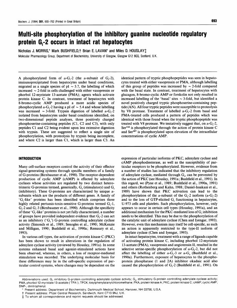
Figure 3 x-G,-2 tryptic (lower-temperature digestion) phosphopeptides from agonist-stimulated hepatocytes resolved by two-dimensional thin-layer analysis
Hepatocytes were labelled with 32P and then challenged with: vehicle (a); 10 ng/ml PMA, 15 min (b); 10 nM vasopressin, 5 min (c); 1 nM glucagon, 5 min (d); 1 uM glucagon, 5 min (e); 300 ,M8-bromo-cAMP, 15 min (f). After harvesting the hepatocytes, they were subjected to detergent extraction, immunoprecipitation with antiserum SG1, SDS/PAGE and identitication by autoradiography.The phosphorylated x-Gr-2 was electo-eluted and then digested with TPCK-treated trypsin as described in the Experimental section for the 'mild' procedure. 32P-labelled tryptic phosphopeptideswere then separated on thin-layer cellulose plates by electrophoresis at pH 1.9 and ascending chromatography, with DNP-lysine as an internal marker dye as described in Experimental section.The autoradiographs shown are from typical experiments, and each condition was performed at least three times with similar results.
U-937 cells (Daniel-Issakani et al., 1989). Treatment of cells withPMA indicated the labelling of this species (1.95 + 0.05-fold;n = 3), but did not change the pl (5.72+0.07, n = 3; Figure 2).However, 8-bromo-cAMP treatment, which caused a furtherincrease in labelling of a-Gi-2 (3.75 + 0.33-fold, n = 3; Figure 2),induced a shift in the mobility of a-Gi-2 which was consistentwith the production of a more acidic form, with pl of- 5.38 +0.07 (n = 3; Figure 2).a-Gi-2 is partially phosphorylated in resting hepatocytes
(Rothenberg and Kahn, 1988; Pyne et al., 1989; Bushfield et al.,1990a,b, 1991; the present work) and thus the increased labellingof a-Gi-2 seen upon treating cells with PMA, in the absence ofany change in pl, suggests that the activation of PKC by PMAserves to phosphorylate unmodified cx-G1-2 rather than to modifyalready labelled G-protein further. This would be consistent withphorbol ester treatment allowing levels of incorporation to attain_ 1 mol of 32P/mol of a-G,-2 (Bushfield et al, 1990a, 1991). Sucha conclusion would also be warranted from our observation(Bushfield et al., 1991) that treatment of hepatocytes with theprotein phosphatase inhibitor okadaic acid rapidly increased thelabelling of a-Gi-2, which attained a stoichiometry of near unityand became insensitive to any action of PMA, implying the
active action of PKC and a phosphatase under basal conditions.The inactivation of the latter, by okadaic acid, leading to theincreased labelling.
In contrast with this, treatment of hepatocytes with 8-bromo-cAMP, which elicits the activation of PKA, caused a decrease inthe pl of a-G1-2 (Figure 2). This occurred concomitantly with a
marked increase in the labelling of a-G1-2 over and above thatseen with PMA (Bushfield et al., 1990a, 1991; see above). Sucha change in the p1 for immunoprecipitated a-G1-2 is consistentwith the labelling of an additional site on this protein, as
suggested previously by us to occur on the basis of dose-dependency studies done with glucagon (Bushfield et al., 1990a)and with phosphorylation studies done using mixtures of ligands(Pyne et al., 1989). As the major fraction of the phosphorylateda-Gi-2 migrates as a pl - 5.4 component after treatment with 8-bromo-cAMP, it would seem that most of the ct-Gi-2 was
phosphorylated at this putative additional site after treatment ofcells with this ligand. This would be consistent with our previousstudies (Bushfield et al, 1990b, 1991) showing that treatment ofintact hepatocytes either with the phorbol ester PMA or withvasopressin yielded a stoichiometry of up to 1 mol of 32P/molof a-G1-2, whereas treatment of hepatocytes with glucagon or
I0T
Chr
I9
0l

N. J. Morris and others
- 0 +
- 0 +
(d)
SF SF
ie
(-H (+)
-SF
O DNP-lysine
(-
Origin
-0+
(g)
1SF'ODNP-Iysine
Origin
(+)
- 0 +
(b)fbi~~~~~~~~~S
I-O DNP-lysin
*
- O +
f(e)
IIO DNP-lysine.*,
0 HiZe,ro
Zero
51 -------
,F
ae
)rigin
SF
DNP-lysine
0 -~~~~~~Origin
SF
_9-a
m (+o ACD
+04_m
El
._I0)
Uc-C
a)U,cn
Electri
Ze ro
(c)Lys0
C3 C1C2 *AN
1-0
Trypsin
ophoresis, pH 1.9
Ze roII
(f)SF-
Q-
° (+) C30gA
010o 0
0)
r: ElectrophoTrypsin anda)
Electrophoresis, pH 1.9
(+)
V8
(-
Zero
(ii
0DNP-Iysine
..1 ..0~~~~~~:: .
Zero
DNP-lysine.. ..: ..........-.......-:: : -SF -
(-) f+)
Zero
(k)
DNP-lysine0
0
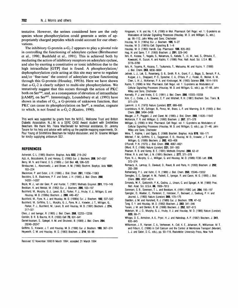
Figure 4 a-G,-2 tryptic (higher-temperature digestion) phosphopeptides fromanalysis and the action of V8 protease
agonist-stimulated hepatocytes resolved by two-dimensional thin-layer
Hepatocytes were labelled with 32P and then challenged with 10 ng/ml PMA for 15 min (a, d) or 1 piM glucagon for 5 min (b, e). After harvesting the hepatocytes, they were subjected to detergentextraction, immunoprecipitation with antiserum SGf1 or 1867, and SDS/PAGE, and phosphorylated t-Gr-2 was extracted. In (a) and (b) samples were digested with TPCK-treated trypsin only under
the 'higher-temperature conditions' described in the Experimental section. A schematic of the various phosphopeptides produced is shown in (c). The AN phosphopeptide was found uniquely with
glucagon challenge at the higher concentration ot 1 liM (b), but not at 1 nM (results not shown, but see Figure 3d). In (d) and (e) trypsin digestion was followed by digestion with V8 proteaseas described in the Experimental section. The AN peptide and all the C peptides were susceptible to proteolysis by V8. The schematic (f) shows the novel peptides produced, namely C2', C2",
(a)
0 DNP-Iysi
t 0
SF -
SF-
(S)
SF _
(+)
-SF
(-H
-SFLys0
IeC2
N QAN
C2'C2"
-SF
H-)
(+)
- SF
(-)
698
)

Multi-site phosphorylation of a-Gi-2 699
8-bromo-cAMP causes the level of labelling to rise to up to- 2 mol of 32P/mol of a-Gi-2.
Two-dimensional thin-layer analysis of o-G,-2 tryptic peptidesTo gauge whether a-G1-2 was subjected to multi-site phosphoryl-ation, we tried to identify 32P-labelled phosphopeptides by two-dimensional mapping analyses. Trypsin digestion of a-G.-2, doneat 30 °C, on resting (basal-state) hepatocytes led to the resolutionof two closely migrating phosphopeptides (Cl, C2), which wereresolved by chromatography, but not by electrophoresis (Figure3a). The ratio of these two peptides varied between experimentsand was influenced by alterations in the time of digestion withtrypsin (results not shown), indicating that they reflected a singlesite of phosphorylation, but with partial digestion of a-G.-2 withtrypsin. Similar examination of a-Gi-2 from hepatoeytes thathad been treated with PMA, in order to activate PKC, identifiedan identical pattern of phosphopeptides (Figure 3b), albeit withincreasing labelling. The variability in the ratio of the Cl and C2peptides precluded any meaningful analysis of the PMA-inducedincrease in labelling of these individual peptides. However, if thetotal labelling of the Cl and C2 peptides was determined, then anincrease of 2.2 + 0.3-fold was noted (n = 3 different cell prepar-ations). Such data are consistent with the formulation madeabove, namely that PMA treatment caused the phosphorylationof native a-Gi-2 at the same site where a fraction of this G-protein was phosphorylated under basal conditions. This alsoconcurs with our previous stoichiometry studies (Bushfield et al.,1991), which implied a single site for the phosphorylation ofac-Gi-2 by PKC.As well as using the tumour-promoting phorbol ester PMA to
activate PKC, we also investigated the effect of the hormonevasopressin, which stimulates phosphoinositide hydrolysis anddiacylglycerol production in hepatocytes (Kirk et al., 1981) andactivates protein kinase C (Tang and Houslay, 1992). Vasopressinincreased labelling of the C-group of peptides (1.8+ 0.2-fold;n = 3), with no evidence for the appearance of any other labelledspecies (Figure 3c). This supports our suggestion (Bushfield etal., 1991) that activation of PKC, either directly by PMA or as aresult of vasopressin-induced diacylglycerol production, resultsin the phosphorylation of unmodified a-Gi-2 at the same time asthat found to be phosphorylated in a small fraction of a-Gi-2 inresting cells.
There is considerable evidence to support the notion thatglucagon can exert actions in hepatocytes which are independentof cAMP (for reviews, see Houslay, 1991a, 1993). This isundoubtedly attributable to the fact that glucagon can activatePKC (Pittner and Fain, 1991; Tang and Houslay, 1992) and canalso increase intracellular free [Ca2+] in two phases, one of whichis cAMP-independent (Mine et al., 1988). The mechanismthrough which these effects occur may involve the very smallstimulation of phosphoinositide metabolism that has been ob-served by us and others (Wakelam et al., 1986; Blackmore andExton, 1986; Williamson et al., 1986; Whipps et al., 1987), anda robust stimulation of phosphatidylcholine metabolism (Pittnerand Fain, 1991), which leads to the noted glucagon-stimulatedproduction of diacylglycerol in hepatocytes (Bocckino et al.,
1985; Pittner and Fain, 1991). Such actions of glucagon appear,however, to occur at lower glucagon concentrations than thoserequired to activate adenylate cyclase. The stimulation ofmultiplesignalling pathways by glucagon may then result from a singlereceptor able to stimulate at least two G-proteins, one of whichis coupled to adenylate cyclase and the other to a phospholipase.Alternatively, multiple splice variants ofthe receptor may activatedifferent G-proteins selectively, as has been noted for othermembers of the G-protein-linked receptor sub-family to whichthe glucagon receptor appears to belong (Jelinek et al, 1993;Spengler et al., 1993). We have shown previously (Bushfield etal., 1990a) that the glucagon-stimulated phosphorylation of a-G1-2 is biphasic, with low concentrations of glucagon (- 1 nM)causing a cAMP-independent phosphorylation which parallelsloss of GTP-driven G1 inhibition, as is also seen with treatmentof cells with either vasopressin or PMA. Here we see (Figue 3d)that challenge of hepatocytes with 1 nM glucagon increasedlabelling of the C-group of peptides (2.1 + 0.2-fold; n = 3) in afashion identical with that found with both PMA and vaso-pressin. This is consistent with glucagon, at such concentrations,being able to activate PKC in hepatocytes (Tang and Houslay,1992). In contrast with this, using higher concentrations ofglucagon (1 ,uM), where maximal phosphorylation of a-Gi-2 andproduction of cAMP occurs (Bushfield et al., 1990a), we then seethe appearance of a novel phosphopeptide 'AN', having distinctchromatographic and electrophoretic properties (Figure 3e).Under such conditions, the increase in labelling of the C-peptidegroup, over that seen under basal conditions, was identical withthat found with low concentrations of glucagon (2.0 ± 0.3-fold;n = 3), indicating that all of the increased labelling of a-Gi-2,occurring at high concentrations of glucagon, was at the new siterepresented by the 'AN' peptide. The stoichiometry of labellingof a-G1-2 at high glucagon concentrations has been shown to be- 2 mol of 32P/mol of a-Gi-2 (Bushfield et al., 1990a, 1991),again consistent with there being two sites for phosphorylationon a-Gi-2: one for PKC and one resulting from elevation of[cAMP]. Indeed, the ability of glucagon to increase intracellularfree [Ca2+] has two components, a cAMP-independent one notedat low hormone levels and a cAMP-dependent one seen at higherglucagon concentrations (Mauger et al., 1985; Mine et al., 1988).Furthermore, 8-bromo-cAMP can also increase hepatocyte intra-cellular free [Ca2+] (Staddon and Hansford, 1989), and thuscan activate hepatocyte PKC (Tang and Houslay, 1992) directly(Ashendel, 1985). As with glucagon, challenge of cells with 8-bromo-cAMP caused the production of the novel 'AN' peptide(Figure 3f), as well as increasing levels of labelling of the C-peptides (2.3 + 0.2-fold; n = 3) to an extent which was similar tothat seen with PMA. Such data show that a-G1-2 is subject tomulti-site phosphorylation in intact hepatocytes and that thiscan be achieved by glucagon in a biphasic fashion. It alsodemonstrates that the site which is phosphorylated as a result ofan increase in intracellular [cAMP] is distinct from that which ispartially labelled under basal conditions, and it thus represents anovel site of modification.To explore the possibility that Cl and C2 were partial digestion
products, we tried changing the digestion conditions. In doingso, we found that if digestion was performed at 37 °C then a
C3' and AN'. In (g) are shown data for bepatocytes treated with PMA, as above, with subsequent treatment of the immunoprecipitate with V8 alone. Secondary digests, with V8 protease,are shown for the individually isolated and treated tryptic phosphopeptides Cl (h), C2 (i), C3 j) and AN (k). All of the 32P-labelled tryptic phosphopeptides were separated on thin-layer celluloseplates by electrophoresis at pH 1.9 and ascending chromatography. The final position of DNP-lysine is marked on each plate, 'SF' represents the position of the solvent front, 'Origin' is the pointof application of the sample, (+) and (-) indicate the orientation of the electric field, and the bar at the top of the plate indicates the separation of the negative, neutral and positive markers.The autoradiographs show the results from typical experiments, where vehicle and PMA produced spots Cl, C2, and C3 (also C2', C2" and C3' on V8 digestion), and 8-bromo-cAMP, glucagonand forskolin stimulation produced spots Cl, C2, C3 and AN upon trypsin digestion and peptides C2', C2", C3' and AN' after V8. Peptides Cl and AN showed most resistance to V8 digestion.Each condition was performed at least six times with similar results.

700 N. J. Morris and others
further positively charged tryptic phosphopeptide (C3) could beresolved by electrophoresis at pH 1.9, using a-Gi-2 immuno-precipitated from both basal (results not shown) and PMA-treated cells (Figure 4a; schematic Figure 4c). These threepeptides were consistently identified by using cell preparationsfrom 12 different animals. As with digestion at 30 °C, in therange of experiments done, there was considerable variation inthe ratio of these three peptides, which militated against anyuseful comparison of changes in their individual labelling oc-curring on PMA treatment of the cells. However, analysis oftotal labelling of the C-phosphopeptide pool showed an increaseof 2.0+ 0.3-fold (n = 12 different cell preparations). This suggeststhat Cl and C2 are capable of being further processed bytrypsin, and support the contention that they represent a singlephosphorylation site. When a similar digestion strategy was used,performed on cells which had been treated with 1 ,uM glucagon,the 'AN' peptide was evident, as was the C3 peptide (Figure 4b;schematic Figure 4c). However, no other peptide was evidentwhich might have signified further processing of the 'AN'peptide. Identical results (not shown) were obtained for cellstreated with either 8-bromo-cAMP or the adenylate cyclaseactivator forskolin (100lM) (Birnbaumer et al., 1990).
All three of the 'C'-group peptides (Cl, C2 and C3) wereshown to be susceptible to cleavage by V8 protease (Figure 4d;schematic Figure 4f). In one series of experiments, immuno-precipitates were digested first with trypsin at 37 °C and directlydigested with V8 protease (Figures 4d and 4e) before beingresolved by two-dimensional chromatography. In the secondinstance, immunoprecipitates were digested with trypsin,phosphopeptides were resolved by two-dimensional chroma-tography, and individual phosphopeptides eluted for subsequentcleavage with V8 protease (Figures 4h-j). These experimentsshowed that all of the C-peptides produced small amounts ofC3', but in addition Cl also produced the phosphopeptide C2',and C2 produced both C2' and C2". All such peptides werepositively charged under the resolution conditions. Such analyseswith V8 protease confirm the relatedness of these peptides toeach other, indicating that they are partial digestion products. Cland C2 are thus clearly related, but the fact that C2 producedC2" indicates that it is larger than Cl and contains a further sitefor action ofV8 protease. That C3 produced only C3' is consistentwith C3 being the smallest of the peptides, resulting from themost complete digestion of a-G1-2 that we were able to achieve.Digestion of phosphorylated a-Gi-2 with V8 protease aloneproduced peptides corresponding to the peptides C2', C2" andC3' (Figure 4g). Thus C1, C2 and C3 must all contain V8cleavage points on either side of the phosphorylation site. The'AN' peptide was also subjected to V8-protease cleavage, pro-ducing the peptide AN' (Figure 4e and 4k; schematic Figure 4g),although increased incubation times of up to 48 h were requiredto achieve this. With all of such peptides, phosphoamino acidanalyses showed that the labelled amino acid was serine (Figure5).As the amino acid sequence of rat a-Gi-2 is known (Itoh et al.,
1986), this has allowed us to attempt to predict the peptides thatmight be produced as a result of protease cleavage (see Boyle etal, 1991). Although trypsin will cleave at the C-terminal side ofboth arginine and lysine, it can also produce partial digestionproducts as a result of the occurrence of adjacent arginine andlysine, proximity to acidic residues and also to proline, as well asthe occurrence of phosphorylated residues near to the point ofcleavage (see Boyle et al., 1991; Sorenson et al., 1991; Breddamand Meldal, 1992). Such occurrences are common in a-G1-2 (Itohet al, 1986, 1988), which, together with the fact that there are 20serine residues in this protein, 18 arginine residues and 27 lysine
Figure 5 Phosphoamino acid analysis of tryptic peptides Cl, C2, C3 andAN
Peptides Cl, C2, C3 and AN were recovered from t.l.c. plates and subjected to phosphoaminoacid analysis as outlined in the Experimental section. Peptide Cl was applied to position 1, C2to position 2, C3 to position 3 and AN to position 4. The plate was run at pH 1.9 in the firstdimension and rotated anticlockwise through 900 before being run in the second dimension.P; represents phosphate liberated from the peptide as a result of hydrolysis. P-Ser, P-Thr, andP-Tyr represent the final position of the applied standards phosphoserine, phosphothreonineand phosphotyrosine. All the peptides produced phosphoserine, therefore confirming that thephosphorylation sites are serines.
residues, severely complicates analyses. Furthermore, features ofsecondary structure can also affect susceptibility to proteolysis,as has been ably demonstrated for the related G-protein trans-ducin (Hingorami and Ho, 1990). In contrast with trypsin, V8-protease cleavage occurs predominantly at glutamate residues,but can also occur at aspartate residues with actions affected bynearby positively charged groups and changes in secondarystructure (see Breddam and Meldal, 1992; Sorensen et al., 1991).Indeed, we experienced this when ax-Gi-2 became resistant todigestion with V8 protease after isolation from cells treated with8-bromo-cAMP.Our data indicate the presence of a single phosphorylation site
for protein kinase C within a-Gi-2 where various partial digestionproducts were formed upon trypsin cleavage. We have attemptedto analyse such data using two strategies. Firstly, we determinedthe charge of all possible serine-containing peptides that mightbe produced through the action of trypsin and V8, so as to assess
their relative mass-charge ratios and the changes that wouldoccur upon V8-protease cleavage of tryptic phosphopeptides.Secondly, we considered whether particular serine residues mightfall within a motif which would make them potential substratesfor phosphorylation by PKC. Although there is some diversity as
regards such motifs (Kennelly and Krebs, 1991; Pearsonand Kemp, 1991; Azzi et al., 1992; Hug and Sarre, 1993), we
have identified seven potential sites in x-G,-2, namely Ser'6,Ser44, Ser47, Ser44, Ser207, Ser247 and Ser306. These are identi-fiable as: AERS16KMID; GES44GKS47TIV; FGRS144REY;GQRS207ERK; RMHES247MKL; YIQS306KFED.
P-Ser (N2SerP-Thr PThrP-Tyr P-Tyr
Pikm/) +3 Pi +4P-Ser P-Ser
P-Thr P-gePrTh r
P-Tyr * P-Tyr
+2 +1

Multi-site phosphorylation of a-Gi-2 701
Potential candidates would be those serines which containedsites for V8-protease cleavage on either side and which werecontained within tryptic peptides able to produce V8 peptides ofelevated mass-charge ratio. Such parent tryptic phosphopeptideswould also have to produce sufficient V8 peptides to account forthe occurrence of C2', C2" and C3'. On this basis, if the putativeparent tryptic peptide was deduced to produce a V8 peptideproduct with zero or a net negative charge, then these peptides,and their parent tryptic peptides, were eliminated, as we notedthat all the V8 phosphopeptides produced were positively charged(Figures 4d-4g) This approach allowed us to eliminate all serineresidues except for Ser47 and Ser'44, with only Ser'" generatingsets of V8 peptides with appropriate relative mass-charge ratios.Such data indicates that Ser'44 is the site for phosphorylation byPKC on a-G1-2. This residue is also proposed to have a surfacelocalization, where it should be available for PKC to modify,based on predictions concerning the secondary structure ofa-G1-2 (Kaziro, 1990; Conklin and Bourne, 1993) and is believedto lie within a domain involved in coupling to the effector,adenylate cyclase.Although elevations in intracellular [cAMP] elicit phosphoryl-
ation of a-Gi-2 at a second site, it is not known whether or notthis results from the direct action of PKA, as there is evidencewhich suggests that, when isolated membranes are used, Gi-2 isnot phosphorylated by purified PKA (see Houslay, 199 la). Thusone cannot make any a priori decisions on the possible motifsurrounding the target serine in AN. We have therefore con-sidered all potential serine-containing tryptic peptides in order toidentify species where, after cleavage with V8, a peptide would beproduced whose mass-charge ratio was decreased, as observed inthe production ofAN' from AN. Such an analysis points only toSer'07 as the target residue. Indeed, this residue has an argininegroup to the N-terminal side, which, of course, might allow it toserve as a substrate for PKA, albeit a weak one. Predictions ofsecondary structure (Kaziro, 1990; Conklin and Bourne, 1993)would also place this group near the surface of the molecule, but,in this instance, near the GTPase domain.
Using the above strategy to eliminate peptides ofinappropriatecharge, then, from those remaining, we can produce a tentativescheme for the production of a series of digestion products whichare compatible with the data observed. On this basis we suggestthat C2, the largest peptide we observed, might have an identityas:
Q106LFALSCAAEEQGMLPEDLSGVIRR"10LWADHGVQA-CFGRS144REYQLNDSAAYYLNDLERIAQSDYIPTQQD-VLR178
This could undergo further digestion by trypsin to yield Cl,as:
L13'WADHGVQACFGRS144REYQLNDSAAYYLNDLER'62-IAQSDYIPTQQDVLR178and C3 as:
L13'WADHGVQACFGRS144REYQLNDSAAYYLNDLER162We would suggest that trypsin might be quite resistant to
acting at either R143 or R145, due to the phosphoserine which issuggested to be found between these two residues. A similarsituation may also hold true for trypsin action at R162, as thisresidue has an acidic glutamate on its N-terminal side, thusexplaining our observation that it is difficult to achieve fullbreakdown to C3, and hence the occurrence of partial digestionproducts. As discussed above, although we routinely observedthe formation ofCl and C2, we noted considerable differences in
experiments. Cleavage of C2, in this instance, to give Cl isproposed to occur at R"0, for which there is an adjacentarginine. We have to presume that the large peptide formed byC2 has sufficient secondary structure such that there is a degreeof hindrance to the access of trypsin to this region, yieldingpartial digests of Cl and C2.
All three peptides, as well as native a-Gi-2, produce C3' upon
treatment with V8 protease (Figure 4). This indicates that thephosphorylated serine can, in all instances, be excised by V8treatment. It is possible to envisage such a scenario with thepeptides indicated above, where the sequence of C3' is suggestedto be:
H13"GVQACFGRS144REYQLND"'5
We suggest that C3' has the above sequence, rather thanterminating at E'46. This is because such a foreshortened peptidewould have a considerably higher mass-charge ratio than thosefor either Cl or C2. Indeed, it is possible that the arginineresidue, which is C-terminal to E'46, might attenuate the actionof V8 protease at such a site. The other possible site for action ofV8 on C3 would be at E161. However, this too lies adjacent to anarginine residue, which might be expected to attenuate its activity.Action at such a residue would also generate a peptide ofinappropriate charge, which eliminates it from consideration.V8 treatment of both Cl and C2 generates a peptide which we
have called C2' (Figure 4). We suggest that this might be:
H"35GVQACFGRS144REYQLNDSAAYYLNDLERIAQSD167
The alternative candidate would be for termination at D159.However, this would give a peptide of inappropriate charge.C2 also produces a further peptide, C2", upon treatment with
V8 protease (Figure 4). This is a partial digestion product whichis unique to C2. Thus the position where V8 cleavage forms theN-terminal end of this peptide would have to occur at one of fourpossible positions, namely El6, El7, E'22 or D23. There are thusa large number of possible V8 peptides that may be generated.The strategy that we have adopted can be used to exclude certainof these on the basis of inappropriate charge, leaving the mostlikely candidate for C2" as:
L124SGVIRRLWADHGVQACFGRS144REYQLND"51This assumes V8 cleavage after D'23, generating an appro-
priately charged peptide, and also at D"5', as in C3', leavingC2" as a partial digestion product which contains C3'.We have suggested that Ser207 may provide the site for
phosphorylation in the AN peptide. Various tryptic peptidescontaining this residue can be seen to be nested within a pair ofresidues where V8 might act. As seen below, tryptic cleavageafter K198 could generate a family of peptides with ambiguity as
to the residue at the C-terminus:
M'99FDVGGQRS207ERKK
However, of these only
M'99FDVGGQRS207ERK210
would generate a peptide of appropriate charge. Presumably,cleavage after R209 is not favoured, due to the adjacent glutamateresidue. We thus suggest that this peptide might reflect thesequence of AN. Treatment with V8 would be expected to cleaveat D201 and, in order for an appropriately charged peptide to beproduced, also at E208 to yield AN', suggested to be:
VGGQRS207E
The assignations made above, as regards the identity of thethe proportion of these labelled phosphopeptides in various peptides generated by both V8 cleavage and trypsin cleavage, are
702 N. J. Morris and others
tentative. However, the serines considered here are the onlyspecies whose phosphorylation could generate a series of ap-propriately charged peptides which could account for our obser-vations.The inhibitory G-protein a-Gi-2 appears to play a ivotal role
in controlling the functioning of adenylate cyclase ( irnbaumeret al., 1990; Bushfield et al., 1991). This is achiev d both bymediating the action of inhibitory receptors on adeny ate cyclase,and also by exerting a constitutive or tonic inhibitio due to thehigh intracellular [GTP] that is found. A phospl orylation/dephosphorylation cycle acting at this site may serve to regulateand/or 'fine-tune' the control of adenylate cyclase Functioningthrough this G-protein (Houslay, 1991b). Here we Ilave shownthat a-G1-2 is clearly subject to multi-site phosphor lation. Wetentatively suggest that this occurs through the actian of PKCboth on Ser144, and, as a consequence of elevation of i itracellular[cAMP], on Ser207. Interestingly, Loundsbury et al. 1993) haveshown in studies of GZ, a G-protein of unknown fu iction, thatPKC can cause its phosphorylation on Ser27, a resid ue, cognateto which, is not found in a-Gi-2 (Kaziro, 1990).
This work was supported by grants from the M.R.C., Wellcome TrDiabetic Association. N.J.M. is a SERC CASE Award student uBeecham. We thank Ms. Fiona MacDonald for technical assistantTavare for his help and advice with setting up the peptide-mapping ePaul Young of SmithKline Beecham for helpful discussion, and Dr. Gfor kindly supplying antiserum SG1.
REFERENCES
ist and Britishith SmithKlinee, Dr. Jeremyperiments, Dr.raeme Milligan
Ashendel, C. L. (1985) Biochim. Biophys. Acta 822, 219-242Azzi, A., Boscoboinik, D. and Hensey, C. (1992) Eur. J. Biochem. 208, 547-557Berry, M. N. and Friend, D. S. (1969) J. Cell Biol. 43, 506-520Birnbaumer, L., Abramowitz, J. and Brown, A. M. (1990) Biochim. Biophys. Acta 1031,
163-224Blackmore, P. and Exton, J. H. (1986) J. Biol. Chem. 261, 11056-11063Bocckino, S. B., Blackmore, P. F. and Exton, J. H. (1985) J. Biol. Chem. 21i,
14201-1 4207Boyle, W. J., van der Geer, P. and Hunter, T. (1991) Methods Enzymol. 201, 110-149Breddam, K. and Meldal, M. (1992) Eur. J. Biochem. 206, 103-107Bushfield, M., Murphy, G. J., Lavan, B. E., Parker, P. J., Hruby, V. J., Milli lan, G. and
Houslay, M. D. (1990a) Biochem. J. 268, 449-457Bushfield, M., Pyne, N. J. and Houslay, M. D. (1990b) Eur. J. Biochem. 1 12, 537-542Bushfield, M., Griffiths, S. L., Murphy, G. J., Pyne, N. J., Knowler, J. T., M lligan, G.,
Parker, P. J., Bushfield, M., Lavan, B. and Houslay, M. D. (1991) Bioch m. J. 274,31 7-321
Chen, J. and lyengar, R. (1993) J. Biol. Chem. 268, 12253-12256Conklin, B. R. & Bourne, H. R. (1993) Cell 73, 631-641Daniel-Issakani, S., Spiegel, A. M. and Strulovici, B. (1989) J. Biol. Chem. 264,
20240-20247Griffiths, S., Knowler, J. T. and Houslay, M. D. (1990) Eur. J. Biochem. 193, 367-374Heyworth, C. M. and Houslay, M. D. (1983) Biochem. J. 214, 93-98
Hingorami, V. N. and Ho, Y. K. (1990) in Mol. Pharmacol. Cell Regul. vol. 1: G-proteins asModulators of Cellular Signalling Processes (Houslay, M. D. and Milligan, G., eds.),pp. 95-112, John Wiley and Sons, Chichester
Houslay, M. D. (1991a) Eur. J. Biochem. 195, 9-27Houslay, M. D. (1991b) Cell. Signalling 3,1-9Houslay, M. D. (1993) Handb. Exp. Pharmacol. 108, 835-853Hug, H. and Sarre, T. F. (1993) Biochem. J. 291, 329-343Itoh, H., Kozasa, T., Nagata, S., Nakamura, S., Katada, T., Ui, M., Iwai, S., Ohtsuka, E.,
Kawasaki, H., Suzuki, K. and Kaziro, Y. (1986) Proc. Natl. Acad. Sci. U.S.A. 83,3776-3780
Itoh, H., Toyama, R., Kozasa, T., Tsukamoto, T., Matsuoka, M. and Kaziro, Y. (1988)J. Biol. Chem. 263, 6656-6664
Jelinek, L. J., Lok, S., Rosenberg, G. B., Smith, R. A., Grant, F. J., Biggs, S., Bensch, P. A.,Kuijper, J. L., Sheppard, P. O., Sprecher, C. A., O'Hara, P. J., Foster, D., Walker, K. M.,Chen, L. H. J., McKernan, P. A. and Kindsvogel, W. (1993) Science 259, 1614-1616
Kaziro, Y. (1990) in Mol. Pharmacol. Cell Regul. vol. 1: G-proteins as Modulators ofCellular Signalling Processes (Houslay, M. D. and Milligan, G., eds.), pp. 47-66, JohnWiley and Sons, Chichester
Kennelly, P. J. and Krebs, E. G. (1991) J. Biol. Chem. 266, 15555-15558Kirk, C. J., Creba, J. A., Downes, C. P. and Mitchell, R. H. (1981) Biochem. Soc. Trans. 9,
377-379Laemmli, U. K. (1970) Nature (London) 227, 680-685Loundsbury, K. M., Schleger, B., Poncz, M., Brass, L. F. and Manning, D. R. (1993) J. Biol.
Chem. 268, 3494-3498Mauger, J. P., Poggioli, J. and Claret, M. (1985) J. Biol. Chem. 260, 11635-11642McKenzie, F. R. and Milligan, G. (1990) Biochem. J. 267, 371-378Milligan, G. (1990) in Mol. Pharmacol. Cell Regul. vol. 1: G-proteins as Modulators of
Cellular Signalling Processes (Houslay, M. D. and Milligan, G. eds.), pp. 31-46, JohnWiley and Sons, Chichester
Mine, T., Kojime, I. and Ogata, E. (1988) Biochim. Biophys. Acta 970, 166-171Mitchell, F. M., Griffiths, S. L., Saggerson, E. D., Houslay, M. D., Knowler, J. T. and
Milligan, G. (1989) Biochem. J. 262, 403-408O'Farrell, P. H. (1975) J. Biol. Chem. 250, 4007-4021Offord, R. E. (1966) Nature (London) 221, 591-593Pearson, R. B. and Kemp, B. E. (1991) Methods Enzymol. 200, 62-81Pittner, R. A. and Fain, J. N. (1991) Biochem. J. 277, 371-378Pyne, N. J., Murphy, G. J., Milligan, G. and Houslay, M. D. (1989) FEBS Left. 236,
372-374Remaury, A., Larrouy, D., Daviaud, D., Rouot, B. and Paris, H. (1993) Biochem. J. 292,
283-288Rothenberg, P. L. and Kahn, C. R. (1988) J. Biol. Chem. 263,15546-15552Senogles, S. E., Speigel, A. M., Padrell, E., lyengar, R. and Caron, M. G. (1990) J. Biol.
Chem. 265, 4507-4514Simonds, W. F., Goldsmith, P. K., Codina, J., Unson, C. and Spiegel, A. M. (1989) Proc.
Natl. Acad. Sci. U.S.A. 86, 7809-7813Sorensen, S. B., Sorensen, T. L. and Breddam, K. (1991) FEBS Left. 294,195-197Spengler, D., Waeber, C., Pantaloni, C., Holsboer, F., Bockaert, J., Seeburg, P. H. and
Journot, L. (1993) Nature (London) 365,170-175Staddon, J. M. and Hansford, R. J. (1989) Eur. J. Biochem. 179, 47-52Tang, E. Y. and Houslay, M. D. (1992) Biochem. J. 283, 341-346Tavare, J. M. and Denton, R. M. (1988) Biochem. J. 252, 607-615Wakelam, M. J. O., Murphy, G. J., Hruby, V. J. and Houslay, M. D. (1986) Nature (London)
323, 68-71Whipps, D. E., Armstron, A. E., Pryor, H. J. and Halestrap, A. P. (1987) Biochem. J. 241,
835-845Williamson, J. R., Hansen, C. A., Verhoeven, A., Coll, K. E., Johanson, R., Williamson, M. T.
and Filburn, C. (1986) in Cell Calcium and the Control of Membrane Transport (Mandell,L. J. and Eaton, D. C., eds.), pp. 93-116, Rockefeller University Press, New York
Received 12 November 1993/9 March 1994; accepted 21 March 1994