Embed Size (px)
Citation preview
1
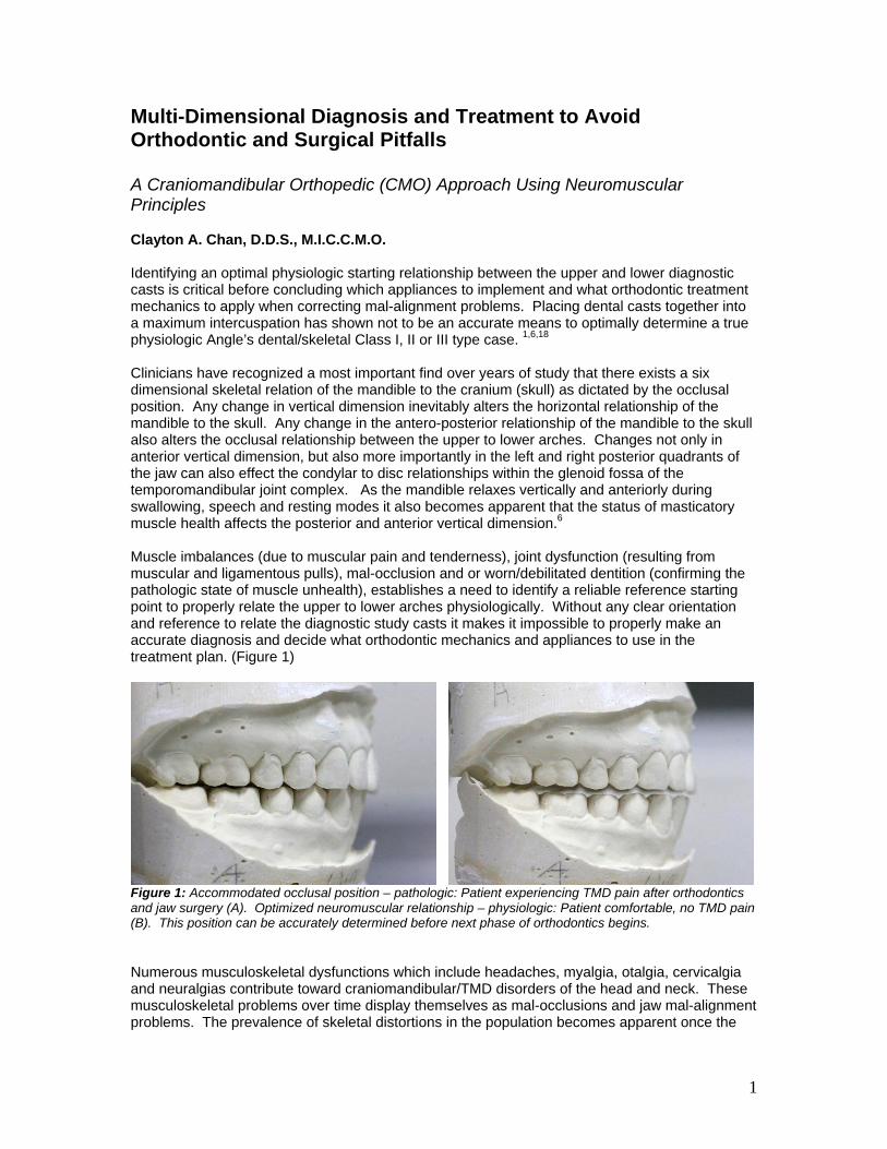
Multi-Dimensional Diagnosis and Treatment to Avoid Orthodontic and Surgical Pitfalls A Craniomandibular Orthopedic (CMO) Approach Using Neuromuscular Principles Clayton A. Chan, D.D.S., M.I.C.C.M.O. Identifying an optimal physiologic starting relationship between the upper and lower diagnostic casts is critical before concluding which appliances to implement and what orthodontic treatment mechanics to apply when correcting mal-alignment problems. Placing dental casts together into a maximum intercuspation has shown not to be an accurate means to optimally determine a true physiologic Angle’s dental/skeletal Class I, II or III type case. 1,6,18 Clinicians have recognized a most important find over years of study that there exists a six dimensional skeletal relation of the mandible to the cranium (skull) as dictated by the occlusal position. Any change in vertical dimension inevitably alters the horizontal relationship of the mandible to the skull. Any change in the antero-posterior relationship of the mandible to the skull also alters the occlusal relationship between the upper to lower arches. Changes not only in anterior vertical dimension, but also more importantly in the left and right posterior quadrants of the jaw can also effect the condylar to disc relationships within the glenoid fossa of the temporomandibular joint complex. As the mandible relaxes vertically and anteriorly during swallowing, speech and resting modes it also becomes apparent that the status of masticatory muscle health affects the posterior and anterior vertical dimension.6 Muscle imbalances (due to muscular pain and tenderness), joint dysfunction (resulting from muscular and ligamentous pulls), mal-occlusion and or worn/debilitated dentition (confirming the pathologic state of muscle unhealth), establishes a need to identify a reliable reference starting point to properly relate the upper to lower arches physiologically. Without any clear orientation and reference to relate the diagnostic study casts it makes it impossible to properly make an accurate diagnosis and decide what orthodontic mechanics and appliances to use in the treatment plan. (Figure 1)
Figure 1: Accommodated occlusal position – pathologic: Patient experiencing TMD pain after orthodontics and jaw surgery (A). Optimized neuromuscular relationship – physiologic: Patient comfortable, no TMD pain (B). This position can be accurately determined before next phase of orthodontics begins. Numerous musculoskeletal dysfunctions which include headaches, myalgia, otalgia, cervicalgia and neuralgias contribute toward craniomandibular/TMD disorders of the head and neck. These musculoskeletal problems over time display themselves as mal-occlusions and jaw mal-alignment problems. The prevalence of skeletal distortions in the population becomes apparent once the

2
musculature is deconditioned and the neuromuscular cervical neck relationships are relaxed to allow a more accurate assessment of the occlusal relationships when establishing a centric occlusion. Before beginning treatment, the orthodontic clinician should consider muscle dysfunction as an underlying cause of these symptoms or as a presymptomatic potential for future dysfunctions contributing to mal-alignment of the jaw to the skull when establishing an occlusion. A Need for Objective Assessment in Determining A Starting Point In Orthodontic/TMD Treatment Objective data clearly establishes a new precedence in any orthodontic practice dealing with muscle accommodated occlusions that result in muscle stress, twisting, torquing, and skewing of the mandible in all directions. Varying degrees of muscular dysfunction, pain and joint derangement resulting from sustained muscle contracture and chronic shortening of muscles as they pull the mandible to an existing occlusion are often overlooked and undiagnosed. Due to hidden subtleties of muscle accommodation, the orthodontic clinician cannot rely on intuitive subjective assessment to take a starting bite relationship and related the dental casts back to the pathologic bite relationship. Using the pathologic accommodated bite relationship and placing the dental cast together to that relation to decide what appliances to use and orthodontic mechanics to implement can lead to misdiagnosis of: Cross bites (unilateral or bilateral), midline discrepancies, Angle’s dental and or skeletal relationships (antero-posterior), deep bites, anterior open bites (as they relate to anterior and posterior occlusal vertical issues), bicuspid drop offs, deep curve of Spee, and steep or low mandibular plane angled cases to name a few. Clinicians nationally and abroad have discovered that capturing a more accurate bite relationship in six dimensions between the upper and lower arches physiologically using objective measured instrumentation has enhanced the accuracy of their diagnosis and visualization of study cast orientation for effective treatment.14, 15, 16 Essentials for Multi-Dimensional Diagnosis The use of electronically derived objective measurements and quantifiable data to diagnose the functional status of the musculoskeletal system of the head and cervical neck region is a significant advancement of orthodontics within the orthopedic specialty. Bio-medical technology that tracks mandibular movement clearly shows the proprioceptive dominance of occlusion over musculature. No matter how badly malpositioned the occlusion or how much torquing and twisting are required to bring the teeth into an occlusion, the muscles and joints will proprioceptively and instantaneously accommodate to that mandibular occlusal position and maintain it there.1 With the advent of measuring technology clinicians are now realizing that there are varying degrees of muscle accommodation as well as sustained tension generated to accomplish an intercuspal position. Computerized Mandibular Scanning (CMS) and Electromyography (EMG) The computerized mandibular scanning (CMS) or jaw tracking and electromyography (EMG) (K7 Myotronics-Noromed, Inc., Tukwila, WA) respectfully measure mandibular to cranium relationships and muscle activity.2 This data are essential for comprehensive diagnosis, to enable the clinician to visualize jaw positioning combined with muscular responses which cannot be seen with the human eye on routine evaluation. Monitoring treatment progress and objective verification of an optimal neuromuscular environment can be achieved – which is the goal and objective of any finished orthodontic case! (Figure 2)

3
CMS displays three dimensional spatial data on a computer monitor. A small light weight magnet placed in the lower anterior vestibule behind the lower lip can be tracked with a sensor array. EMG measures the action potential levels of muscle pairs such as the left and right temporalis anterior, masseter, digastric and cervical neck groups via electrodes. These action potential levels can be graphically displayed as pre and post treatment records to document degrees of muscle force. When combining these diagnostic tools together, contracture and torque as they relate to proprioceptive directional pull on the mandible that contribute to craniomandibular mal-alignment and occlusal dysfunction, can be visualized.3
A B Figure 2: A) Three dimensional subjective assessment of how dental casts relate to one another before and after orthodontics and orthognathic jaw advancement surgery is not enough to keep this 22 year old female TMD paining patient from a life of debilitating pain and dysfunction. B) Multi-Dimensional objective analysis can be used to better determine an optimal starting relationship of the upper and lower dental casts non- invasively. Even though the patient can voluntarily bring the teeth consistently to a fully intercuspated position with seeming ease should not lull the treating clinician into believing that the existing occlusion is correct, rather he/she should recognize the underlying contributory musculoskeletal cause of this dysfunction. Once the musculoskeletal dysfunction is dynamically measured and diagnosed additional complimentary static records should be gathered such as cephalometrics, radiographs, photographs (intra and extra oral) as well as diagnostic study casts. (Figure 3-5)
Figure 3: Restricted jaw pain exists with limited mouth opening and abnormal jaw opening and closing patterns. Diagnostic jaw scanning assists in visualizing dyskinetic movements.

4
Figure 4: Improved mouth opening after protruding the mandible forward. Note improved opening/closing velocity cycles. Diagnostic jaw scanning validates smoothness of jaw function.
A B
Figure 5: Pre and post electromyographic data documents the direction and degree to which muscles are being forced into sustained contracture as they are proprioceptively directed to pull and hold the mandible in a skeletal mal-relation of a malpositioned occlusion. EMG before (A) and after (B) low frequency TENS are used to objectively determine quality of muscle relaxation. Case No. 1– Neuromuscular Diagnostics Used to Resolve Conventional Orthodontic Jaw Mal-Alignment and Surgical Problem A 22 year old female presents with severe headaches in the temporal region, forehead and back of the head. She complains of difficulty chewing, pain in left jaw joint, grinds her teeth, clicking and popping of right joint, limited opening of jaw, pressure in the right ear, awakens with dry mouth. Previous conventional orthodontics as well as mandibular jaw advancement surgery was performed with resulting debilitating unresolved TMD pain and mandibular dysfunction. (Figure 6-9)

5
Figure 6: Accommodated centric position after orthodontics and jaw surgery. Note: Condylar beaking, hyperplastic changes in the neck of the condyle, flattening of the anterior surface, minimal posterior and superior joint space. This existing occlusal position proprioceptively over rides the isotonic path of jaw closure contributing to hypertonic muscles activity and TMD pain.
When evaluating and talking with the patient, it became apparent that the patient’s mandible, during functioning and resting modes, postured more anterior than the established bite position. Numerous pain sights were identified and confirmed subjectively and objectively. (Figure 7)
Figure 7: Note the unposed functional jaw position during speech after conventional orthodontics and jaw surgery. Muscles will pull, torque and skew the mandible into a centric occlusion contributing to TMD pain.
6
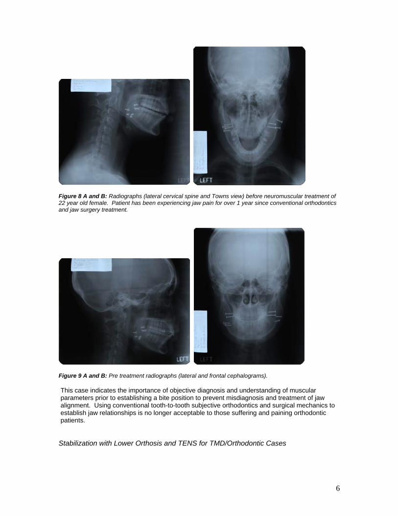
Figure 8 A and B: Radiographs (lateral cervical spine and Towns view) before neuromuscular treatment of 22 year old female. Patient has been experiencing jaw pain for over 1 year since conventional orthodontics and jaw surgery treatment.
Figure 9 A and B: Pre treatment radiographs (lateral and frontal cephalograms). This case indicates the importance of objective diagnosis and understanding of muscular parameters prior to establishing a bite position to prevent misdiagnosis and treatment of jaw alignment. Using conventional tooth-to-tooth subjective orthodontics and surgical mechanics to establish jaw relationships is no longer acceptable to those suffering and paining orthodontic patients.
Stabilization with Lower Orthosis and TENS for TMD/Orthodontic Cases

7
An anatomical lower orthosis is essential in muscular stabilization prior to proceeding onward with orthodontic treatment mechanics. 8,9 (Figure 10) Without reduction of hyperactive muscle activity first and removing the mandibular skews and torques of jaw mal-alignment in six dimensions, orthodontic treatment will only become more difficult and frustrating leading to unresolved musculoskeletal occlusal issues down the read. An orthosis was fabricated based on objective measured jaw tracking data to identify an optimal “physiologic” starting relation for her mandible to cranium using the Myotronics K7 kinesiograph, (Figure 11) after Myomonitor TENS (Transcutaneous electro-neural stimulation, J5 Myotronics-Noromed, Inc., Tukwila, WA) muscle relaxation. 11-13
Figure 10: Mandibular anatomical orthosis – Is directed toward orthopedic realignment of the mandible to the cranium. Stabilizes the temporomandibular joints, restores them to normal physiologic function while concomitantly reducing contracted (spastic) craniofacial and cervical musculature and develops functional and resting modes within normal physiological parameters.
The Myomonitor low frequency TENS reconditions the various hyperactive musculature simultaneously via the fifth and seventh cranial nerves to anaerobic resting levels of health. 4, 5 In most cases, one appointment is all it takes to reduce significant spastic musculature. Subsequent visits to adjust and fine tune the orthosis combined with TENS is also required to optimize jaw function and further muscle relaxation.
A B C
8
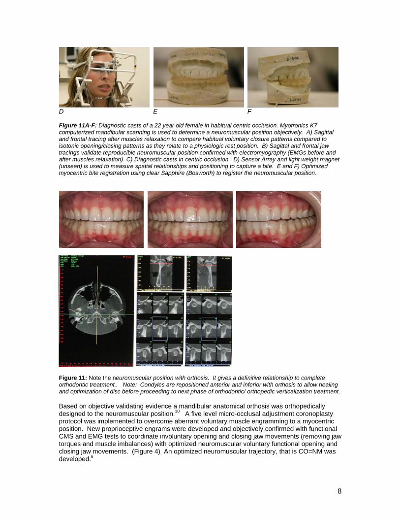
D E F Figure 11A-F: Diagnostic casts of a 22 year old female in habitual centric occlusion. Myotronics K7 computerized mandibular scanning is used to determine a neuromuscular position objectively. A) Sagittal and frontal tracing after muscles relaxation to compare habitual voluntary closure patterns compared to isotonic opening/closing patterns as they relate to a physiologic rest position. B) Sagittal and frontal jaw tracings validate reproducible neuromuscular position confirmed with electromyography (EMGs before and after muscles relaxation). C) Diagnostic casts in centric occlusion. D) Sensor Array and light weight magnet (unseen) is used to measure spatial relationships and positioning to capture a bite. E and F) Optimized myocentric bite registration using clear Sapphire (Bosworth) to register the neuromuscular position.
Figure 11: Note the neuromuscular position with orthosis. It gives a definitive relationship to complete orthodontic treatment.. Note: Condyles are repositioned anterior and inferior with orthosis to allow healing and optimization of disc before proceeding to next phase of orthodontic/ orthopedic verticalization treatment. Based on objective validating evidence a mandibular anatomical orthosis was orthopedically designed to the neuromuscular position.10 A five level micro-occlusal adjustment coronoplasty protocol was implemented to overcome aberrant voluntary muscle engramming to a myocentric position. New proprioceptive engrams were developed and objectively confirmed with functional CMS and EMG tests to coordinate involuntary opening and closing jaw movements (removing jaw torques and muscle imbalances) with optimized neuromuscular voluntary functional opening and closing jaw movements. (Figure 4) An optimized neuromuscular trajectory, that is CO=NM was developed.6

9
The orthosis was worn 24/7 as recommended. The patient faithfully wore the orthotic appliance for 12 months. Resolution of musculoskeletal and joint dysfunction was immediate. The patient no longer experiences any of the previous listed complaints that she had for 7 years. She is pleased and satisfied with treatment. A finishing phase of orthopedic orthodontics is indicated to correct the mal-alignment and vertical deficiency to the proven neuromuscular position applying CMO verticalizing mechanics.16 Diagnosing from a Physiologic Relationship Prior to Orthodontics and Orthognathic Surgery Makes a Difference Conventional orthodontic diagnosis commonly focuses on horizontal tooth-to-tooth relationships with the teeth in existing occlusion. In order for teeth to move primarily in the horizontal directions, there must be available space in which teeth can move. If teeth are crowded, the extraction of bicuspids or second molars often becomes necessary. However, moving teeth horizontally into spaces provided by extraction does not address the functional six dimensional problem (vertical, antero-posterior, lateral, pitch, yaw and roll) and etiology of these dental/skeletal problems which may involve crowding of dentition, lingually tipped teeth, abnormal occlusal alignment, with accompany abnormal swallowing problems, lack of tongue space, deep bites and over-jet problems with insufficient lower third facial development, to name a few. The six dimensional neuromuscular approach to treatment expedites the orthodontic/orthopedic treatment. The relationship of the mandible to the cranium/skull is altered six dimensionally when the muscles are relaxed to their resting lengths. As the muscles are relaxed, the hypertonic tensions are released from physical restrictions of the existing mal-position and occlusion and Angle’s classification if often changed. When the masticatory and cervical neck muscles are relaxed an increased vertical dimension results, often in the posterior molar regions with an accompanying change in the antero-posterior relationship of the mandible to the maxilla. This increased space automatically provides space for tooth movement, more optimal tongue positioning, improved oral pharyngeal air way space and reduction in need for tooth extractions and surgery. The value of diagnosis from a physiologic position versus an accommodated occlusal position makes an often seemingly complex Class II type case a simpler Class I. This phenomenon is illustrated in many cases that have been recommended for surgery or have been surgerized with no prior objective quantifiable diagnostic data to validate where to relate the mandible to the cranium.1 To emphasize, relaxation of musculature and establishing homeostasis of the complete cranio-mandibular cervical system is critical before making a definitive diagnosis and treatment plan. Without first establishing muscle relaxation, the clinician will easily assume the patient is asymptomatic and a subjective intuitive analysis can result in a misdiagnosis and treatment. Neuromuscular Diagnosis for Retrognathic Class II and Prognathic Class III Cases In Class II type cases, which are often over-closed, release from the restrictions of existing occlusion is significant to the muscle engramming action of the antero-posterior jaw relationship. The significance of overlooking the need to properly optimize the hypertonic muscles prior to diagnosis and treatment of the mal-occlusion may eliminate the need for extractions and simplify treatment while correcting any musculoskeletal dysfunction of the head and neck. It has been often observed after myomonitor TENS relaxation that the assumed skeletal and dental Class II type cases will physiologically appear Class I skeletal/ dental, resulting in easier orthodontic mechanics. Class III type case identification of muscle tension and consequent over-closure at the existing occlusal level often simplifies the therapeutic approach and improves the prognosis. Moreover the retention phase of treatment is often shorter and less need for mechanical retentive appliances are required due to quieter relaxed musculature.

10
Case No. 2 – A Conservative Nonsurgical Resolution of Class III Prognathism with Neuromuscular Principles
The diagnostic importance of validating vertical dimension increase and its accompanying horizontal change in mandibular positioning confirms the need for quantitative data of existing muscle tension and skeletal mal-relation in evaluating the indications for surgical correction of pragnathism. Since the mandible moves posterior an average of 1 mm for every 2 mm of increased vertical opening (1:2 A/V ratio), the mere act of vertically repositioning the mandible 5 mm to a more open relaxed position concomitantly moves the mandible 2.5 mm posteriorly, altering the Angle classification (Figure 13-15). Once a physiologic bite relationship was determined, a diagnosis and treatment plan could be made to apply the proper orthodontic mechanics and appliances to avoid jaw surgery. Previous specialist and doctors were planning to do jaw surgery to correct a pseudo class III relationship in CO. The neuromuscular position indicates a skeletal class I relationship with maxillary arch deficiency antero-posteriorly, not a skeletal class III prognathic jaw as was formerly diagnosed. Arch expansion was implemented using the Nitanium Palatal Expander 2 (Ortho Organizers, Inc., Carlsbad, CA) combined with arch leveling wires and open coiled springs (Thermal Kinetic, Ortho Organizers, Inc.) to develop the maxillary incisors and jump the anterior cross bite tendency. Interproximal slimming of the lower arch was used to help bring the upper and lower occlusion together.
Figure 13: An initial .016 x.016 Thermal Kinetic arch wire (Ortho Organizers, Carlsbad, CA) was used to begin leveling and aligning of this 17 year old male. Round .014 NiTi arch wire and bracket placed on left lateral incisor is used to further align arch with maxillary Nitanium Palatal Expander (NPE2).

11
Figure 14: Computerized mandibular jaw tracking indicates improve muscle activity as the mandible moves vertically open and posterior.
Figure 15: 9 months wear with NP2 (Nitanium Palatal Expander 2-Ortho Organizers, Carlsbad, CA) to assist in orthopedic arch development and spacing for upper left canine.
Case No. 3 – Neuromuscular Orthodontics Used to Treat Unresolved Conventional Orthodontics and Prevent Mandibular Advancement Surgery
A 15 year old female was in the middle of conventional orthodontic treatment for the past 2 years. A surgeon and two orthodontist specialist recommended orthognathic surgery to finish this Class II mal-aligned case. (Figure 16-17) An aberrant tongue posture habit as well as accompany abnormal jaw closure patterns were identified using neuromuscular protocols (Figure 18). A neuromuscular position was objectively determined and established using jaw tracking data to locate a physiologic relation for her mandible to cranium using the Myotronics K7 kinesiograph, after Myomonitor TENS muscle relaxation. Patient was treated non-surgically using cranio-mandibular orthopedic (CMO) occlusal principles to verticalize the teeth to the neuromuscular stable position. (Figure 19-22)

12
Figure 16: Controlled craniomandibular orthopedic verticalization mechanics are used to maintain the neuromuscular position and improve facial form and profile.
Figure 17: Maxillo-mandibular relation – Initial centric occlusion(A), Neuromuscular position (B) and Molar blocks at neuromuscular position (C).
A B Figure 18: CMS recordings confirmed an aberrant tongue swallowing pattern contributing to Class II malocclusion with anterior open bite tendency (A-B). Normal Swallow pattern (C).
Protruding Unposed Position
Neuromuscular/ Comfortable
MANDIBULAR STABILIZATION – Non Surgical Approach to Class II Retrognathic Type Cases
Before NM Treatment
(After Ortho)
After NM Treatment

13
A B Figure 19: Functional EMG test indicate imbalanced muscle recruitment during voluntary clenching in centric occlusion versus cottonroll controls (A). An abnormal opening and closing jaw pattern is recorded 4 mm posterior to CO compared to a physiologic jaw trajectory after muscle relaxation (B).
A Figure 20: Before - Unresolved orthodontic position ready to have mandibular advancement surgery (A). After - Neuromuscular position after Myomonitor TENS relaxation, stabilized and ready to proceed to completion using neuromuscular orthopedic verticalization principles non-surgically (B).
Figure 21: Orthopedic verticalization of the teeth and surrounding bone are developed to a neuromuscular position using tripoded occlusal stops to segmentally verticalize the teeth. This is a result of first establishing calm musculature and occlusal contacts on an optimal neuromuscular trajectory which override the abnormal posteriorizing jaw closure patterns that contribute to musculoskeletal occlusal dysfunction.

14
Figure 22: Note improved facial form and profile. Orthognathic surgery was avoided using neuromuscular principles and establishing a stable neuromuscular bite on trajectory. Summary Clinicians are now realizing that vertical orthopedic eruption of teeth is active throughout life, and with the development of clinical techniques to help in vertical erupting of teeth and increasing the vertical dimension, there is a growing demand to learn how to effectively verticalize and control proprioceptive occlusal inputs from the teeth orthopedically within the neuromuscular parameters of occlusion. 7 As long as mal-occlusion dominates the musculoskeletal system, mandibular jaw open and closing patterns will be posterior to an isotonic path of physiologic closure. Muscles lengths will foreshorten, resulting in muscular pains and pathologic dysfunction. Optimizing muscular health and identifying muscular disease objectively will increase case stability and improve long term retention. Addressing these orthopedic/functional orthodontic musculoskeletal problems through the eyes of neuromuscular principles will prevent misdiagnosing and mistreatment of the abnormal posterior Class II and abnormal anterior Class III jaw relationships. The significance of the increase in occlusal vertical dimension and its accompanying horizontal change in mandibular position confirms the need to quantitatively measure existing muscle tension and relaxation modes of the masticatory system. These diagnostic tests are clinically useful to assess skeletal mal-relations in evaluating the indications for surgical correction of prognathism or retrognathism. In cases in which the cuspal anatomy has not yet been unduly defaced by wear or extensive restorative treatment, orthodontic vertical eruption alone is the treatment of choice, especially for musculoskeletal dysfunction and temporomandibular joint derangement problems. Rather than adding to tooth height by prosthodontic/ reconstruction techniques, it is imperative that the clinician fairly presents a non-invasive orthodontic approach first rather than only consider a convenient restorative approach to treatment. The clinician’s lack of understanding and awareness of these neuromuscular principles is no excuse to have our patient’s experience the devasting and ravaging pains of craniomandibular dysfunction. Dentist have a professional obligation to their patient’s and profession to make a proper diagnosis to help bring a quality of dental health to patients of all ages. Meeting the next level of comprehensive orthodontic care is the clinician’s responsibility. Dr. Clayton A. Chan is dedicated to sharing his passion and teaches the neuromuscular principles that have worked for him. He is an educator to thousands of dentist all around the world, inspiring them to take their practices to another level. He is considered by many an authority on Neuromuscular Dentistry and Occlusion. He practice’s in Las Vegas, Nevada where he focuses on Aesthetic Orthopedic Rehabilitation and Craniomandibular Facial Pain implementing the gnathological and neuromuscular principles. Email: [email protected] Web: www.claytonchandds.com References:
15
1. Jankelson B: Three –Dimensional Orthodontic Diagnosis and Treatment: A
Neuromuscular Approach, J Clin Orthod 1984; 18:9:627. 2. Myotronics-Noromed, Inc., K7 Kinesiograph K6-i/K7 and the Myomonitor J4/J5, Tukwila,
Washington. 3. Cooper B: The Role of Bioelectrical Instrumentation in the Documentation and
Management of Temporomandibular Disorders. Oral Surg Oral Med Oral Endod 1997;83:91-100.
4. Fuji and Mitani (Fuji, H and Mitani, H. : Reflex Response of the Masseter and Temporal Muscles in Man, J. Dent. Res. 52:1046, 1050, 1973).
5. Choi BB, Mitani H: On the Mandibular Position Regulated by Myo-monitor stimulation. J Japan Prosth Soc 1973:17:79-96.
6. Jankelson RR: Neuromuscular Diagnosis and Treatment. Ishiyaku Euro America, Inc, St Louis, Missouri.
7. Butterworth, JC: Passive Eruption in the Treatment of Craniomandibular Dysfunction: A Post treatment Study of 151 Patients. Journal of Prosthetic Dentistry, April 1992; 67: 4: 525-535.
8. Roth, RH: Functional Occlusion for the Orthodontist. J Clin Orthod 1981;15:45-6. 9. Jankelson B.: Neuromuscular Aspects of Occlusion. Dent Clin North Am 1979;23:157-68. 10. Clark GT: A Critical Evaluation of Orthopedic Interocclusal Appliance Therapy. Part I.
Theory Design, and Overall Effectiveness. J Am Dent Assoc 1984;108:358-64. 11. Wessberg GA, Epker BN, Elliot AC. Comparison of mandibular rest positions induced by
phonetics, transcutaneous and electrical stimulation and masticatory electromyography. J Prosthet Dent 1983;49:100-5.
12. George JP, Boone ME. A clinical study of rest position using the kinesiograph and myo-monitor. J Prosthet Dent 1979;41:456-62.
13. Nielsen IL. Marcel T, chun d, Miller AJ. Patterns of mandibular movements in subjects with craniomandibular disorders. J Prosthet Dent 1990;63:202-117.
14. Bruzzone, GL: Orthodontic Finishing for Two TMD Patients. Anthology of ICCMO, VII, 2005, 67-90.
15. Kadooka, H: Transferring of neuromuscular dental cases from a general practitioner to an orthodontist during TMD therapy. Anthology of ICCMO (Japan), July 2, No. 1, June 2005.
16. Chan, CA.: Applying neuromuscular principles in TMD and orthodontics. Journal American Orthodontics Society. Spring 2004.
17. Woodside, DG.: A consideration of some aspects of vertical dimension in orthodontics treatment planning. Parts 1 and 2. Am. Assoc. of Orthodontists, St. Louis.
18. Gerber, JW.: TMD Stabilization: Orthodontic Finishing Success. Anthology of ICCMO. Volume VI. 2003.