Embed Size (px)
Citation preview

Mrs. Paulgaard Biology 20
Notes & Diagrams
Organic Chemicals, Enzymes, Cells, Cellular Respiration, Photosynthesis, Muscles, Digestion, Kidney, & Pulmonary
Systems

Unit 1: Energy Flow and Cellular Matter Cellular Chemistry:
Metabolism: sum of all chemical reactions that occur within the cell.
Catabolism: chemical reactions in which complex molecules are broken down into smaller compounds.
Anabolism: chemical reactions where simple molecules are combined together to form more complex compounds.
Organic Chemistry:
Chemistry of carbon ~ hydrocarbons (HC’s)
Simplest compounds that are classified based on the number of carbons.
Consist of carbohydrates, lipids, and proteins Carbohydrates: sugars (CHO) ending of OSE
Role: Structure: cell wall in plant cells ~ cellulose. Function: energy ~ rapidly available energy.
Monosaccharides: simple single sugars ~ C3 to C10 Glucose, Fructose, and Galactose.
Disaccharides: double sugars that are combinations of single sugars. Sucrose, Maltose
o Example: C6H12O6 + C6H12O6 C12H22O11 + H2O Glucose + Glucose Maltose + Water
Polysaccharides: many sugars. General formula (C6H10O5). o Polymers: molecules composed of 3 to several million sub-units. o Examples:
Cellulose ~ 1000 to 2000 glucose units. (dietary fibre) Starch ~ plant storage carbohydrate. Glycogen: animal starch ~ animal storage carbohydrate that is stored in the liver
and muscles. Chemical Tests:
Starch Test: drops of iodine solution; blue or black color indicates presence of starch. Reducing (mono & diasaccharide) Sugar: drops of Benedict’s solution. Heat to boiling point but
don’t boil. Color indicates the % of sugar. Lipids: (CHOP) ending of OL
Role: Structural: phospholipids in cell membranes and fat deposits for physical protection.
Functional: fat deposits for heat insulation and long term storage of energy.
Triglycerides: union of glycerol and three fatty acids. o Fats: animal lipids composed of glycerol and saturated fatty acids. o Oils: plant lipids composed of glycerol and unsaturated fatty acids.
Phospholipids: phosphate molecule is attached to the glycerol molecule making the molecule polar. Major components to cell membranes in animals and plants.
Chemical Test:
Lipids Test: 1. Grease spot test on brown paper, translucent result indicates fat or oil. 2. Add a trace of Sudan IV stain that is only soluble in lipids: color will show.

Proteins: structural components of cells. (CHONS) ending of IN
Roles: Structural: cell membrane and determine the shape of cells. Functional: enzymes control all chemical reactions, hormones, and energy.
Very large macromolecules composed of Amino Acids. o 20 types of amino acids that are joined together by peptide bonds, as a result proteins
are often called polypeptides. Most proteins have up to 3000 amino acids. Amino Acid ~~~ peptide bond ~~~~ Amino Acid
Primary Structure: polypeptide chain O-O-O-O
Secondary Structure: coil made of weak hydrogen bonds
Tertiary Structure: unique 3-dimensional structure allows proteins to become very specific.
o As a result of its structure, each protein has a unique configuration (surface shape) which determines largely the properties of that protein.
o If environmental factors change this shape then the protein cannot carry out its proper role. This process is called Denaturation ~ uncoiling the protein shape.
Caused by environmental agents:
Heavy metals ~ Pb and Hg.
Electricity ~ electrocution
Heat ~ cooking eggs or fever
pH Sometimes if the agent is removed the protein can recapture its shape, if not the
permanent change is referred to Coagulation.
Nucleic Acids: hereditary material found within the genes of chromosomes. o Deoxyribonucleic Acid ~ DNA. o Nucleotides: the functional units of nucleic acids.
A and T together G and C together
Chemical Test:
Protein Test: drops of Biuret Reagent (test for the peptide bond), a violet color indicates the presence of protein.

Chemical Processes:
Dehydration Synthesis: combination of simple molecules to form larger, macro-molecules which yields a water molecule.
Hydrolysis: breaking down of a macromolecule to form simpler, micro-molecules through the addition of water.
Review:
Organic Compound Elements Types Examples Processes Chemical Test
Carbohydrates (sugars)
Carbon Hydrogen Oxygen
Monosaccharides Disaccharides
Polysaccharides
Glucose Sucrose Cellulose
Dehydration synthesis & Hydrolysis
Benedict’s Solution Iodine
Lipids Carbon Hydrogen Oxygen
Phosphate
Triglycerides:
Fat
Oils Glycerol and Fatty Acids
Phosopholipids: Glycerol and phosphate
Fats, Cholesterol, Steroids, Wax
Dehydration synthesis &
Hydrolysis
Paper Test Sudan IV
Proteins Carbon Hydrogen Oxygen Nitrogen Sulphur
Amino Acids Polypeptides Chains
Coils 3-D Proteins
Enzymes, Haemoglobin,
Antibodies
Dehydration synthesis & Hydrolysis
Denaturation Coagulation
Biuret Reagent
Nucleic Acid Carbon Hydrogen Oxygen Nitrogen
Phosphate
Nucleotides DNA and RNA
DNA
Enzymes: “lock and key” hypothesis. Suffix ~ ase
Induced fit hypothesis. Substrate and enzyme must stretch and strain to fit. The strain breaks the bonds.

Characteristics of Enzymes:
Proteins
Needed in small quantities (reuseable)
Substrate specific
Action is reversible
Organic catalysts Great energy Less energy of activation Of activation
No Enzyme Enzyme
Allow chemical reactions to proceed under “milder” conditions in terms of temperature and pH.
Have optimum pH and temperature where they are most effective or work the best. Temperature pH Reaction Opt. Reaction Rate Rate
Temp. pH
Rate is regulated by the relative amounts of enzyme and substrate.
High (150) Reaction Rate Enzyme (100) Low Concentration of Substrate
Co-Enzyme:
Chemical molecules, such as a vitamin, which are needed to alter the active site of enzymes to correctly fit it with the substrate.

Competitive Inhibition:
Interference, caused by a non-substrate, with the active site of an enzyme or a specific site on a substrate.
This chemical interference prevents the normal chemical reaction that involves that enzyme. Non-Competitive Inhibition:
A process where a substance that doesn’t resemble the substrate at all attaches to the enzyme (not on its active site) which rearranges the enzyme rendering it useless.
Negative Feedback ~ Homeostasis:
The ability of the body to keep the normal internal body environment in a stable state even as the external environment is changing.
Stops a process Positive Feedback ~ Precursor Activity:
Activation of the last enzyme in a pathway due to a build-up of the initial substrate.
Starts a process

Biology 20: Unit 2 Cellular Respiration and Photosynthesis Cell Organelles:
Cell membrane: controls what comes in and out of the cell.
Cytoplasm: solution within the cell.
Endoplasmic reticulum (ER): contain ribosomes which are responsible for protein synthesis.
Golgi Apparatus: prepare materials for secretion (proteins or enzymes).
Lysosomes: Digestive enzymes
Mitochondria: produces most of the cell’s energy, has its own DNA.
Plastids: plants only, sites of photosynthesis, has its own DNA.
Vacuoles: storage deposits
Cilia and Flagella: movement of the cell.
Microfilaments and Microtubules: movement of materials within the cell and movement of the cell itself.
Cell Wall: plants only, supports the cell.
Nucleus: determines the shape, metabolism, and heredity of the cell (DNA).
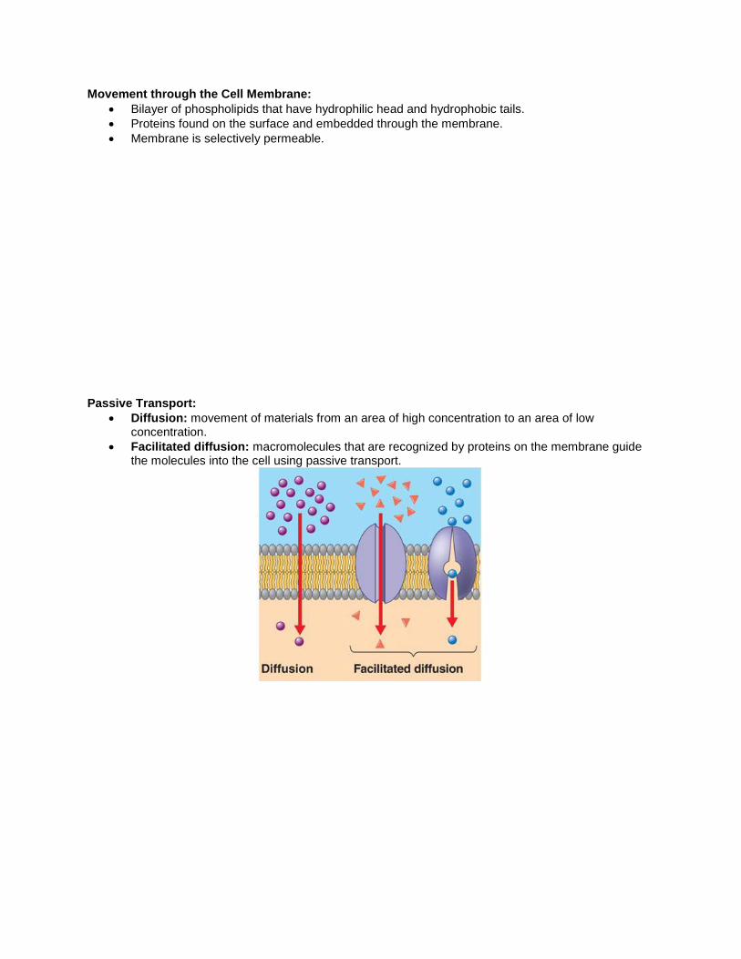
Movement through the Cell Membrane:
Bilayer of phospholipids that have hydrophilic head and hydrophobic tails.
Proteins found on the surface and embedded through the membrane.
Membrane is selectively permeable.
Passive Transport:
Diffusion: movement of materials from an area of high concentration to an area of low concentration.
Facilitated diffusion: macromolecules that are recognized by proteins on the membrane guide the molecules into the cell using passive transport.

Osmosis: movement of water across a semi-permeable membrane from an area of low solute concentration to high solute concentration.
o Isotonic – solution on both sides have the same concentration of water.
Hypotonic solution: solution with less solute concentration and more water.
Hypertonic: solution with more solute and less water
Active Transport: similar to facilitated diffusion but the movement of the molecules requires the cell to use energy.
o Endocytosis: large particle is engulfed by the cell Pinocytosis: small particles engulfed by the cell Phagocytosis: large particles engulfed by the cell.
o Exocytosis: vacuole moves toward the cell membrane and dumps the contents out.
o Ion Pump: small ions move against the gradient

ATP:
Adenosine Triphosphate (ATP) is the storage form of energy for cellular activity.
High energy bond between the second and third phosphate group. Once broken the energy released is transformed and used within the cell.
Anaerobic Metabolism: ~ absence of oxygen Glucose 2 lactic acid + 6 ATP ~ animals Glucose Alcohol + CO2 + 6 ATP ~ plants = fermentation Aerobic Metabolism: ~ presence of oxygen Glucose + 6 O2 6 CO2 + 6 H2O + 36 ATP * more energy and excrete wastes. (C6H12O6)
Phosphorylation: the process of adding a phosphate to a molecule. FOOD + P FOOD – P FOOD ~ P Food Fragments Phosphorylation internal (into the bond) (lactic acid) arrangement
ADP ATP
Oxidation and Reduction:
Oxidation: release energy when a compound loses electrons.
Reduction: absorb energy when a compound gains electrons.
Hydrogen or Electron Transport: o Hydrogen electrons in hydrogen gas or in organic molecule have a great deal of energy. o Hydrogen electrons in water have low energy.
H2 + ½ O2 Explosive release of energy H2O *if our bodies used this pathway we would burn up.
Electron transport releases the energy step by step so that the cells can convert the energy into ATP.

H2 ~ hydrogen is separated Advantage: lots of energy and easily excrete wastes (water and carbon dioxide). Disadvantage: needs oxygen. Electron Transport Inhibitors:
Certain chemicals can block the chain so that the complete reduction cannot occur.
Ex: carbon monoxide, cyanide, and hydrogen sulfide. Cellular Respiration: Glycolysis: Outside of the mitochrondria ATP ADP Glucose Glucose – Phosphate Fructose - Phosphate 6C no P add P 6 C 1 P 6 C 1 P add p ATP ADP Fructose - Diphoshphate 6 C 2 P 2P + 2ADP 2ATP 2 Phosphoglyceric Acid 2 diphosphoglyceric acid 2 Glyceraldehyde – 2 P (PGA) (PGA) add 2 P (PGAL) 2 P 4 P 3 C 2 P 2ATP 2 ADP ETC 6 ATP 2 phosphopyruvic acid 2ADP 2 ATP 2 P 2 pyruvic acid (0 P 3 C) Used 4 ATP Gained 10 ATP Net gain of 6 ATP

Fate of Pyruvic Acid:
Without Oxygen: Anaerobic Staircase is incomplete 0 ATP Lactic Acid animals Ethanol plants ~ fermentation Amount of ATP = 6 ATP due to glycolysis
With Oxygen: Aerobic Staircase is complete 36 ATP Water as a waste Citric Acid/Krebs Cycle: within mitochondria 2 pyruvic acid 2O2 6 ATP and 2H2O and 2CO2
2 Acetyl coA 1O2 3 ATP and 1H2O and 1CO2
6 ATP
2 X 1O2 3 ATP and 1H2O and 1CO2
The cycle is repeated 2 times. Total ATP produced = 6 + (2 x 12) + 6 (from glycolysis) = 36

Photosynthesis:
The process whereby plants store solar energy into organic compounds.
6 CO2 + 12 H2O + solar energy C6H12O6 + 6 O2 + 6 H2O carbon water sunlight glucose oxygen water dioxide (Leaf) (Roots) (Root/Leaf) (Leaf) (Leaf)
Takes place in the chloroplasts of plant cells. o Contains cellulose and chlorophyll which is the pigment that traps sunlight. o Photosynthesis takes place within specialized membranes called thylakiod membranes. o These membranes are stacked one upon another to form stacks known as grana o The fluid surrounding the grana is called stroma.
thylakoid grana
Chemiosmosis: o Different pigments absorb different wavelengths of light that provide the right amount of
energy to the electrons within them. Ex: chlorophyll a, chlorophyll b, carotenoid See colours not absorbed by the object (Chloroplasts absorb red and blue)
o The trapped energy excites the electrons and boosts them to a higher energy level. The reactions of photosynthesis occurs in two phases:
o Photosystems o Carbon-Fixing Cycle or Calvin-Benson Cycle

Photosystems:
Energy capturing phase ~ light dependent
The thylakoid membrane appears to have two systems that operate at the same time. Photolysis: splitting of water by light energy
Calvin-Benson Cycle:
Carbon fixing phase ~ light independent
Occurs in the stroma
Light Thylakiod Membrane 6 O2 12 H2O (Photosystems)
6ADP, NADP+ 6ATP, NADPH2
6 CO2 6 H2O
Glucose Sucrose Lipids Proteins
Light Independent Reactions
3 cycles

Comparison between Cellular Respiration and Photosynthesis:
Cellular Respiration Photosynthesis
Energy produced (36 ATP) Energy required (6 ATP)
Oxidation Reduction
High energy reactants (glucose) Low energy reactants (CO2 and H2O)
Oxygen required (aerobic) Oxygen released
CO2 and H2O produced Glucose produced
Processes: Phosphorylation Kreb’s Cycle (2)
Processes: Photosystems Calvin-Benson Cycle (3)
Mitochondria Chloroplasts
Unit 3: Energy and Matter Flow in the Human Body The human body is made up of a various systems responsible for specific functions:
Muscular System: movement and heat production
Digestive system: nutrients
Respiratory system: gas exchange
Circulatory system: blood transport
Urinary system: excretion
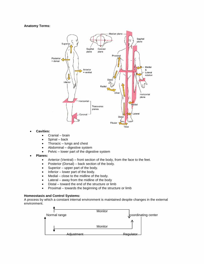
Anatomy Terms:
Cavities:
Cranial – brain
Spinal – back
Thoracic – lungs and chest
Abdominal – digestive system
Pelvic – lower part of the digestive system
Planes:
Anterior (Ventral) – front section of the body, from the face to the feet.
Posterior (Dorsal) – back section of the body.
Superior – upper part of the body.
Inferior – lower part of the body.
Medial – close to the midline of the body.
Lateral – away from the midline of the body
Distal – toward the end of the structure or limb
Proximal – towards the beginning of the structure or limb Homeostasis and Control Systems: A process by which a constant internal environment is maintained despite changes in the external environment.
Monitor Normal range coordinating center
Monitor
Adjustment Regulator

Muscles:
Tissue designed to convert chemical energy (ATP) into kinetic energy (movement & heat).
Supports body functions
Responsible for locomotion (bones), heat production, peristalsis, breathing etc. Types of Muscle Tissue:
Smooth Muscle: non-striated, one nucleus, contracts involuntarily, slow and long contracts, don’t fatigue easily, and found along the wall of internal organs.
Cardiac Muscle: striated, tubular and branched, one nucleus, contracts involuntarily, found in the walls of the heart.
Skeletal Muscle: striated and tubular, contain many nuclei, contracts voluntarily, attached to bones of the skeleton.
Functions of Skeletal Muscle:
Opposes the force of gravity and enables standing
Constant temperature by releasing of metabolic heat is distributed to the body (shivering)
Protects internal organs and stabilizes joints: Ligaments hold bones (cartilage in between) together at the joints. Tendons attach muscle to bones
Cooperation of Skeletal Muscle:

All muscle tissue contracts (shortens) and relaxes (lengthens).
Muscles can only pull on a bone when they contract but there must be a force that stretches the muscle after it has stopped contracting and relaxes
Flexing causes the bone or limb to move away from its original position. Extension is when the bone or limb moves towards its original position.
Muscles are allows in pairs: antagonistic o Bicep causes the arm to flex as the muscle shortens o Triceps causes the arm to extend as the muscle shortens.
Hierarchy of Muscle Structure:
Muscle (Tendon is heavy tissue that attaches to bone)
Muscle-Fibre Bundle (connective tissue surrounds each muscle fibre with nerve and blood vessel running between each bundle of fibres)
Muscle Fibre (single muscle cell) o Myoglobin (stores oxygen) o Sarcoplasm (cytoplasm of the muscle fibre, contains myoglobin & glycerine) o Sarcolemma (membrane of the muscle fibre that regulates movement of material) o Sarcoplasmic Reticulum (stores calcium ions) o Myofibrils (cylindrical sub-units that make up a muscle fibre)
Myofilaments (protein structures responsible for muscle contractions)
Thick Filaments: composed of myosin(heads)
Thin Filaments: composed of actin
Muscle Fibre Bundle

Mechanisms of Muscle Contractions: 1. Myosin head attaches to actin 2. Myosin head flexes, advancing the actin filament 3. Myosin head releases and unflexes, powered by ATP. 4. Myosin reattaches to actin farther along the fibre.
Sliding Filament Model of the Sarcomere
1. The heads of the two ends of myosin filament are oriented in opposite directions. When the heads attach to the actin, they bend towards the centre of the myosin.
2. As one end of the myosin filament draws the actin filament and its attached Z line towards the centre, the other end of the myosin filament does the same.
3. Both Z lines move towards the centre, and contraction occurs.
Striations

Role of Calcium Ions in Muscle Contraction: 1. Muscle is at rest: A long filament, composed of the protein molecule tropomyosin, blocks the
myosin binding sites of the actin molecule. Without these sites exposure, muscle contraction will not occur.
2. Calcium ions bond with a molecule called troponin, which results in exposing the myosin binding
sites of actin so now muscle contraction can occur.
Sequence in Muscle Contraction:
1. Nerve impulse travels to the muscle fibre bundle (stimulus) 2. Ca ions are released from the sarcoplasmic reticulum into the sarcoplasm. 3. Ca ions attach to the troponin (Ca receptor site) thereby causing the tropomyosin to release from
the actin. 4. Myosin heads can now attach, release, and reattach using ATP thereby causing muscle
contraction (z-lines move together). Sequence in Muscle Relaxation:
1. Nerve impulse stops 2. Ca ions reabsorbed from sarcoplasm into the sarcoplasmic reticulum. 3. Absence of the Ca ions on the troponin allows tropomyosin to reattach to the actin preventing the
binding of myosin. 4. Myosin and actin just slide away from each other (z-lines move away).
Rigor Mortis After death, calcium levels inside the muscle cells rise and the body's level of ATP drops. Inside the muscles, myosin binds to actin and the muscles contract. However, with no ATP to reset the crossbridges and release the myosin, all of the muscles remain contracted and stiff -- this state is called rigor mortis.

Energy for Muscle Contractions:
Stored Energy in a Resting Muscle: 1. Creatine Phosphate is built up and stored in a resting muscle. 2. Glucose and Glycogen is stored in muscle to be used during cellular respiration.
Release Energy (make ATP) and Contract the Muscle: 1. Breaks down creatine phosphate, adding the phosphate to ADP to create ATP for
immediate use. 2. Carries out anaerobic respiration, by which glucose is broken down to lactic acid and
ATP is formed. Lead to fermentation (another way of providing ATP without oxygen which
causes cramping and muscle fatigue) Oxygen Debt (replenish creatine phosphate and remove lactate)
More in shape a person is the more mitochondria he or she has the less oxygen debt.
3. Carries out aerobic respiration, by which glucose, glycogen, fats and amino acids are broken down in the presence of oxygen to produce ATP.
Muscle Contractions or Twitches:
Muscles require a stimulus (nerve) to contract, latent period, contraction period (muscle shortens), and a relaxation period (when the muscle returns to its former length).
All or none response (one muscle fibre will contract).
When there is a short relaxation period, the muscle will fatigue due to a lack of glycogen and excess lactic acid.
More stimulus is received, more 100% fibres bundles contract
1. Latent 2. Contract 3. Relax 4. Summation 5. Tetanus 6. Fatigue
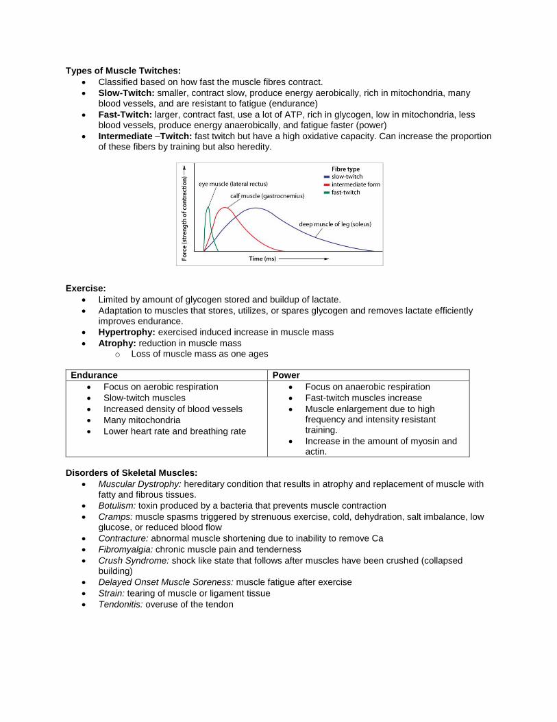
Types of Muscle Twitches:
Classified based on how fast the muscle fibres contract.
Slow-Twitch: smaller, contract slow, produce energy aerobically, rich in mitochondria, many blood vessels, and are resistant to fatigue (endurance)
Fast-Twitch: larger, contract fast, use a lot of ATP, rich in glycogen, low in mitochondria, less blood vessels, produce energy anaerobically, and fatigue faster (power)
Intermediate –Twitch: fast twitch but have a high oxidative capacity. Can increase the proportion of these fibers by training but also heredity.
Exercise:
Limited by amount of glycogen stored and buildup of lactate.
Adaptation to muscles that stores, utilizes, or spares glycogen and removes lactate efficiently improves endurance.
Hypertrophy: exercised induced increase in muscle mass
Atrophy: reduction in muscle mass o Loss of muscle mass as one ages
Endurance Power
Focus on aerobic respiration
Slow-twitch muscles
Increased density of blood vessels
Many mitochondria
Lower heart rate and breathing rate
Focus on anaerobic respiration
Fast-twitch muscles increase
Muscle enlargement due to high frequency and intensity resistant training.
Increase in the amount of myosin and actin.
Disorders of Skeletal Muscles:
Muscular Dystrophy: hereditary condition that results in atrophy and replacement of muscle with fatty and fibrous tissues.
Botulism: toxin produced by a bacteria that prevents muscle contraction
Cramps: muscle spasms triggered by strenuous exercise, cold, dehydration, salt imbalance, low glucose, or reduced blood flow
Contracture: abnormal muscle shortening due to inability to remove Ca
Fibromyalgia: chronic muscle pain and tenderness
Crush Syndrome: shock like state that follows after muscles have been crushed (collapsed building)
Delayed Onset Muscle Soreness: muscle fatigue after exercise
Strain: tearing of muscle or ligament tissue
Tendonitis: overuse of the tendon

Technologies to treat muscle condition:
Cold: reduces swelling after a tearing
Heat: encourages blood flow to healing area (reduces pain and muscle stiffness)
Ultrasound: sound waves heat up the tissue (speeds healing) and increases blood flow
Massage: increased blood flow and breaks down lactic acid
Strengthening & Stretching Exercises Drugs:
Anabolic steroids
Creatine Phosphate
Athletic Drinks Unit 4 Digestion Digestive System:
The organs which collectively perform the task of breaking down nutrients or organic molecules for the use of cell (digestion).
Digestive Processes:
1. Ingestion: taking food into the body (eating). 2. Movement: propels food through the digestive system. 3. Secretion: release of digestive juices in response to a specific stimulus. 4. Digestion: breakdown of food into molecular components through the use of chemical and
mechanical means. 5. Absorption: passage of the molecules into the body’s blood stream and movement into the cells. 6. Egestion: removal of undigested food and wastes.
Mechanical Digestion verses Chemical Digestion: Mechanical Digestion: molecules stay the same size and the physical motions break big pieces into smaller pieces. Ex: chewing. Chemical Digestion: molecules change and different molecules are produced. Ex: enzyme action. Factors that stimulate ingestion:
Habit
Hunger caused by low blood glucose levels.
Brain stimulation

Organs of Digestion:
Organs are classified into two groups:
Gastrointestinal (GI) Tract: Tube Oral cavity, pharynx, epiglottis, esophagus, stomach, the small and large
intestines, appendix, and the rectum/anus.
Accessory Structures: Teeth, tongue, salivary glands, liver, gallbladder, and the pancreas. Digestive secretions.
Attached by the mesentery ~ tissue that attaches various organs to the body cavity

Organs of Digestion: Mouth: moistens food with secretions of saliva, grids food which increase the surface area for chemical digestion, and directs the food down the esophagus. Chewing or mastication food creates bolus. Salivary Glands: secrets saliva into food, contains amylase (enzyme) that begins the digestion of starch. Epiglottis: a flap of skin in the pharynx region that closes off the trachea when swallowing food. Esophagus: a large muscular tube that carries food to the stomach. It is made of smooth muscle that contracts in a peristaltic wave motion, pushing the bolus of food along. Peristalsis – squeezing, pushing down Vomiting – reverse wave pushing up Stomach:
1. Provides storage for 1 to 2 litres of material for 3 to 5 hours. 2. Mixes organic juices with a muscular wave-like motion. 3. Starts protein digestion. 4. Sets the rate of digestion between 4 to 24 hours.
Cardiac sphincter chyme Pyloric sphincter
Liver: Chemistry lab of the body and is the largest gland Produces bile (breaks down fats and neutralizes strong acids) which is stored in the gall bladder. Duel blood supply (poison smasher).
Hepatic Vein Hepatic Artery (oxygenated) (deoxygenated) from the heart toward the heart Hepatic Portal Vein (deoxygenated) From intestine
Pancreas:

Endocrine gland that produces and secretes hormones into the blood stream. (Insulin and Glucagon)
Exocrine gland that releases chemicals into the small intestine. Small Intestine: Six meters long. Majority of digestion and absorption occurs in this area. Movement through active transport. Three sections: duodenum (shortest), jejunum, and the ileum (longest)
Absorption:
Villius Structure: Villi Microvilli Arterial Venous Blood To the liver (HPV) Lymph Fluid
Uses active transport so the cells contain a large number of mitochondria.
A capillary net supplies the mircovilli with oxygenated blood (arterial) and removes carbon dioxide and organic molecules (amino acids, glucose, fatty acids) through the venous vessels (deoxygenated) toward the liver.
Glycerol and more fatty acids are removed via the lacteal vessel that transports the materials to the lymphatic system.
Adaptations for Absorption: 1. Villi and Microvilli – increased surface area 2. Length – buys time and provides more opportunity for digestion. 3. Water is secreted – moistens the contents 4. Peristalsis – digested food contacts the absorptive surface faster. 5. Narrow diameter – molecules are closer to the absorptive surface.

Appendix: Stores beneficial bacteria which assists in the digestion of organic material. Large Intestine: Colon that is three meters long and has three sections: ascending colon, transverse colon, descending colon.
1. Absorbs water, minerals, and salts. 2. Decomposes left-over organic material with the help of resident bacteria (e-coli) which
produces vitamin B, K, and Folic acid. Rectum & Anus: Stores feces (undigested cellulose and matter) until it is appropriate to eliminate.
Digestion of Carbohydrates

Digestion of Lipids
Digestion of Proteins

Enzyme Summary: Organ Digestive
Secretion Active Digestive Agent Action on Food
Salivary Glands Saliva Amylase Breaks down starch into maltose
Stomach Gastric Juice Pepsinogen (+HCL) into Pepsin
Rennin
Lipase
Mucin
Protein to peptide chains
Clots milk
lipids into 3 fatty acids and 1 glycerol
protective mucus secretion
Liver Bile (gall bladder)
Bile Salts Emulsifies fats
Neutralizes acids
Pancreas Pancreatic Juice Sodium Bicarbonate
Lipase
Amylase
Peptidase Trypsin (active)
Chymotrypsin
(active)
Neutralizes acids
Breaks down fats to fatty acids and glycerol
Breaks down starch to maltose
Continues the protein breakdown of amino acids.
Small Intestine Intestinal Juice (intestinal glands)
Carbohydrase o Maltase o Sucrase
o Lactase
Mucin
Erepsin
Completes digestion of sugars to glucose.
protective mucus secretion
Continues the protein breakdown of amino acids.

Absorption:
Stomach: alcohol and drugs
Small Intestine: organic compounds
Large Intestine: water, minerals, salts, vitamins How the liver handles excessive material:
Glucose is stored in the liver and muscle in the form of glycogen controlled by insulin. Insulin is produced by the pancreas.
Liver Glucose HPV HV (lots) (balanced) Glycogen
Glycogen is released by the liver and muscles in the form of glucose controlled by glucagon. Glucagon is produced by the pancreas.
Liver Glucose HPV HV (none) (balanced) Glycogen
Glycerol and fatty acids are converted into lipids and is stored as fat.
Amino acids broken down (deamination) into fatty acids that is stored in the liver or in fatty tissue and urea which is excreted via the kidney.
Water and minerals are stored in the blood and excess goes through kidneys.
Vitamins that are water soluble goes through kidneys and vitamins that are fat soluble are stored in fatty tissue.
Glands are stimulated by:
Neural control: senses
Hormonal control: hormones in the blood
Mechanical control or Movement: peristalsis and other movement

The hormonal control of digestion: Gastrin: Trigger: presence of undigested food in the stomach Produced by: cells lining the stomach Released into: blood Travels to: gastric glands Cause: release of HCl, pepsinogen, and lipase Effect: digests proteins and lipids
Cholecystokinin (CCK) and Secretin: Trigger: food in the small intestine Produced by: cells lining the duodenum Released into: blood Travels to: gall bladder and pancreas Cause: release of bile to emulsify fat and the release of pancreatic juice (protease, amylase, and lipase) Effect: neutralizes acids and digests fats, starch, and proteins
Technologies:
Endoscope: tube-shaped camera that is inserted into the abdominal cavity.

Unit 5: Excretion & Pulmonary Systems Excretion: Location of the Kidney: Renal
Abdominal cavity towards the back
Right kidney is slightly higher than the left kidney. Diagram of the Urinary System:
Role of the Kidney:
Remove “waste”
Remove any chemical that is present in the blood in amounts greater than the body needs. (water, salt, glucose, etc.)
o I.e., the kidney is an organ of homeostasis Structure of the Kidney:

Nephron: Deamination: Removal of amino group from an organic compound which forms one molecule of urea and one fatty acid.
Urea ~ two molecules of ammonia and one molecule of carbon dioxide (less toxic)
Uric Acid ~ waste from breakdown of nucleic acid

Processes of the Kidney: 1. Force or Glomerular Filtration:
Movement of fluids (exception of proteins) from the glomerulus (blood) into the Bowman’s capsule due to blood pressure to become apart of the nephric filtrate.
2. Tubular Reabsorption:
Selective transfer of essential solutes back into the blood through active transport. Molecules the body still needs.
Proximal tubule, ascending loop of Henle, and distal tubule
3. Tubular Secretion:
Movement of wastes from the blood into the nephron through diffusion.
15% of the molecules stay within the nephric filtrate and are removed via the bladder.
Proximal tubule, distal tubule
4. Water Reabsorption:
Removes water from the filtrate and returns it to the blood.
Proximal tubule, descending loop of Henle, distal tubule, and collecting tube. Kidney Reabsorption:
Glucose, amino acids, vitamins, and minerals are absorbed by active transport.
Change in solute concentration as Cl- is attracted to the + blood causes the tubule to become hypotonic and the blood hypertonic.

Hormonal Control of Water Reabsorption: Urine Formation.
Hormone: Aldosterone Source: Adrenal Cortex Trigger: Decreased solute concentration in the blood and increased osmotic pressure. Released: blood Target: Proximal tubule, ascending loop of Henle, and distal tubule Effect: Reabsorbs Na ions (Na pump) into the blood
Adrenal Cortex
Nephron
Proximal, Distal, & Loop
of Henle
Na+ ion pump Increase solute concentration in the blood
Aldosterone
Osmoreceptors
Hypertonic Blood
Hypotonic Blood

Hormone: Anti Diuretic Hormone (ADH) Source: Posterior end of the Pituitary Gland Trigger: increased solute concentration in the blood and decreased osmotic pressure. Released: blood Target: Proximal tubule, descending loop of Henle, distal tubule, and collecting tube. Effect: increased water reabsorption.
Kidney Diseases:
Diabetes Mellitus: sugar diabetes, body is not producing enough insulin, sugar is not reabsorbed, causing a greater loss of water.
Diabetes Insipidus: cannot produce ADH to regulate water reabsorption.
Kidney stones: precipitation of mineral solutes in the blood break the kidney tissue.
Bright’s disease: protein found in the urine
Posterior Pituitary
Nephron
All tubules
Water Reabsorption
ADH
Hypothalamus Osmoreceptors
More Fluid Decrease in Osmotic Pressure Hypotonic
Less Fluid Increase in Osmotic Pressure Hypertonic Blood

Case Study: Comparing Solutes in Plasma, Nephron, and Urine Micropipettes were used to draw fluids from the Bowman’s capsule, the glomerulus, the loop of Henle, and the collecting duct. Solutes in the fluids were measured. The resulting data are displayed in Table 1. Table 1 Solute Concentrations in Various Parts of the Kidney
Solute Bowman’s Capsule
Glomerulus Loop of Henle Collecting Duct
Protein 0 0.8 0 0
Urea 0.05 0.05 1.50 2.00
Glucose 0.10 no data 0 0
Chloride Ions 0.37 no data no data 0.6
Ammonia 0.0001 0.0001 0.0001 0.04
Substance X 0 9.15 0 0
Quantities are in g/100ml Application Questions:
1. Which of the solutes was not filtered into the nephron? Explain your answer.
2. The test for glucose was not completed for the sample taken from the glomerulus. Predict whether glucose would be found in the glomerulus. Provide reasons for your prediction.
3. Why do urea and ammonia levels increase after filtration occurs?
4. Chloride ions, Cl-, follow actively transported sodium ions, Na+, from the nephron into the blood. Therefore, you would expect the Cl-, concentration to decrease as fluids are extracted along the nephron. What causes this discrepancy?
5. Is it correct to say that veins carry blood with high concentrations of waste products and arteries carry blood with high concentrations of nutrients? Explain.
6. Compare the blood found in a renal artery and a renal vein with respect to urea and glucose.

Technologies:
Dialysis: From Body To Body Dialysis Fluid
o Solution is maintained at body temperature, contains no urea, and has a balanced concentration of glucose, amino acids, minerals, vitamins, and fatty acids.
Breathing and Gas Exchange: Pulmonary System:
Bronchiole: Cilia Beating cilia move a river of mucus up so we can clear our throat and swallow. Cleaning of our bronchus of dust particles. Mucus Alveoli

Bronchiole Alveolus: Artery Vein * surface area Alveolus (Alveoli)
Composition of Air:
Compound Inhaled Air Exhaled Air
N2 78% 78%
O2 21% 16%
CO2 0.04% 5%
High pressure to Low pressure Forces air into the lungs

Breathing Centre:
Acidity (low pH) & High Temperature.
Breathing Muscles High blood CO2 Relax No Nerve Impulse Low blood O2 Breathing Centre Elastic Recoil Medulla Of Cartilage, Alveoli, Breathing impulse & Lung Muscles Contraction Nerve Impulse Inhibiting Medulla Decrease in Lung/Chest Volume Increase in Lung Volume Stretch Receptors Decrease Lung Pressure
Inhalation Inflated Alveoli
Breathing Centre Stimulated Increase in Lung Pressure Exhalation Deflated Alveoli Stops Impulse Removes Inhibition of Medulla
Chemoreceptors: send an impulse to the medulla initiating the breathing process. Stretch Receptors: (inflated) inhibit the nerve impulse to the ribs. (deflated) enables chemoreceptors to signal the medulla
Breathing Volumes Residual (Dead) Air: air needed to maintain the inflation of the lung Tidal Volume: normal breathing (10%) Inspiratory Reserve Volume: inhale deeply as much as one can Expiratory Reserve Volume: exhale deeply as much as one can Vital Lung Capacity: Tidal, inspiratory, and expiratory reserve volumes. Total Lung Capacity: All capacities together.

Case Study: Measuring Respiratory Volumes
Patient Tidal Volume (mL)
Vital capacity (mL)
Respiratory rate of patients at rest (breaths/min)
1 (normal) 500 5 000 18
2 500 4 000 20
3 400 3 000 38
4 550 5 000 17
5 550 6 000 17
1. Distinguish between tidal volume and vital capacity.
2. What is the relationship between vital capacity and respiratory rate?
3. Which patient is likely a long distance runner? How do you know?
4. Which patient is likely suffering from pneumonia? How do you know?
5. A ventilator is a piece of medical equipment that maintains respiratory movements in a person who is unable to breathe. Consider a young, otherwise healthy person who is paralyzed as a result of a car crash. Would it be a good idea to adjust a ventilator to maximize the volume of air inhaled and exhaled? Explain.
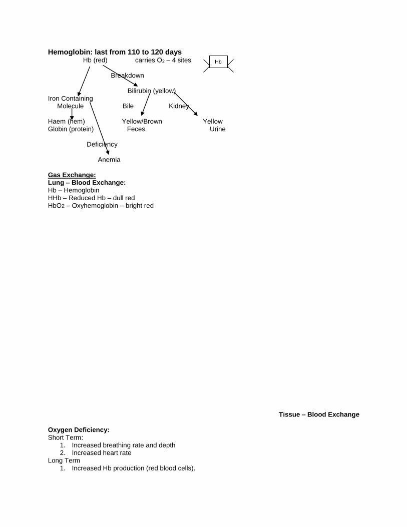
Hemoglobin: last from 110 to 120 days Hb (red) carries O2 – 4 sites Breakdown Bilirubin (yellow) Iron Containing Molecule Bile Kidney Haem (hem) Yellow/Brown Yellow Globin (protein) Feces Urine Deficiency Anemia Gas Exchange: Lung – Blood Exchange: Hb – Hemoglobin HHb – Reduced Hb – dull red HbO2 – Oxyhemoglobin – bright red
Tissue – Blood Exchange
Oxygen Deficiency: Short Term:
1. Increased breathing rate and depth 2. Increased heart rate
Long Term 1. Increased Hb production (red blood cells).
Hb

Carbon Monoxide Poisoning: Hb picks up CO easier than O2 and has a stronger bond. One dies of a lack of oxygen. O2 CO CO H CO CO Hb Hb Hb O2 O2 H H O2 O2 Bends: Gases in the body are dissolved into the blood. At a low altitude (underwater) the blood has more gas can dissolve. However, once one rises to the top (higher altitude) the gases become insoluble and bubbles form.
Diseases of the Pulmonary System:
1. Tonsillitis: infection of tonsils 2. Laryngitis: inflammation of the larynx 3. Bronchitis: bronchi become inflamed and filled with mucus 4. Pneumonia: alveoli fills with liquid due to inflammation 5. Pleurisy: swelling an irritation of the pleura 6. Emphysema: over-inflation of the alveoli. Continued over-inflation can lead to the rupture of the
alveoli. 7. Cystic Fibrosis: genetic condition that causes the mucus in the lung to become sticky 8. Asthma: reduced air flow due to inflammation. 9. Lung Cancer: most common death of Canadians. Uncontrolled growth of cells that decreases
the surface area for diffusion. Technologies:
CT scan- specialized X-ray can be used to detect lung cancer.