Embed Size (px)
Citation preview

Don A. Wilson 1
John R. Prince
This article appears in the March/April 1989 issue of AJNR and the May 1989 issue of AJR.
Recipient of the John Caffey Award for Best Scientific Paper presented at the annual meeting of the Society for Pediatric Radiology, San Diego, 1988.
'Both authors: Magnetic Resonance Center, Oklahoma Medical Center, 940 NE 13th St., P.O. Box 26307 , Oklahoma City, OK 73126. Address reprint requests to D. A. Wilson .
AJNR 10:259-262, March/ April 1989 0195-6108/89/1002-0259 © American Society of Neuroradiology
MR Imaging Determination of the Location of the Normal Conus Medullaris Throughout Childhood
259
This retrospective study was designed to determine the location of the conus medullaris in normal children by reviewing a series of MR images of the lumbar spine. The study group consisted of 184 children ranging in age from newborn to 20 years who had a normal conus level as reported by the radiologist of record. The range of conus levels for the entire group of normal children was T12 to L3. The range for the 0-2-year-old group was T12 to L2-L3 with an average of L 1-L2. The range of conus levels for the 19-20-year-old group was L 1 to L2 with an average of L 1-L2.
We conclude that the conus medullaris does not ascend throughout childhood as stated by previous authors but attains the adult level sometime during the first few months of life. A conus level at L2-L3 or above should be considered normal at any age. A conus level at L3 is indeterminate, since it is possible for a normal or a tethered conus to be located at this level.
In order to diagnose a low-lying or tethered cord in children, it is necessary to know the level at which the normal conus medullaris terminates. This is a topic that has not received a great deal of attention in the literature. Commonly used reference texts for pediatric neuroradiology and pediatric imaging [1 , 2] state that the lower level of the conus is normally at the third lumbar vertebral body (L3) at birth and that it reaches L2-L3 by age 5, L2 by age 12, and eventually arrives at about L 1 by adulthood. This series of age-related levels clearly suggests that the conus ascends throughout childhood and that different criteria of normality should be used for different ages. The purpose of this study was to develop· a better statistical basis for determining the location of the normal conus medullaris throughout childhood by reviewing a large series of MR images. Comparisons were also made with a group of children who had surgically proved tethered cord syndrome.
Materials and Methods
MR images of the lumbar spine were retrospectively reviewed in 184 children ranging in age from newborn to 20 years . The subjects were divided into two groups. Group I consisted of 85 infants and children (42 girls and 43 boys) who were studied for reasons other than spinal dysraphism. These patients were referred for MR imaging because of low back pain or back injury (37 , or 43%), possible infection or tumor of the spine or cord (21 , or 25%), brain tumor to exclude dropped metastasis (11 , or 13%), leukemia (11 , or 13%), and abdominal mass (5, or 6%). The average age of this group was 1 0.8 years.
Group II contained 99 subjects (50 females and 49 males) referred with possible symptoms of tethered cord syndrome but reported to be normal by the radiologist of record . The clinical indications for these studies were as follows: poor bowel and/or bladder control (39, or 39%), scoliosis (27 , or 27%), abnormal muscle tone or gait (25, or 25%), spina bifidia (13, or 13%), foot deformities (13 , or 13%), and sacral dimple or hairy patch (8 , or 8%). The clinical indications exceed the total number of patients in the group because many of the patients had multiple symptoms. The age range for this group was 5 days to 20 years, with an average age of 7.9 years.
Over the same time span in which the normal subjects were being studied (approximately

260 WILSON AND PRINCE AJNR :10, March/April1989
3 years), 40 infants and children (25 girls and 15 boys) from our institution were diagnosed and surgically confirmed to have the tethered cord syndrome. These children ranged in age from birth to 19 years, with an average age of 6.7 years . These cases were reviewed to determine the range of pathologic conus levels in our population.
MR scans of the lumbar spine were also reviewed in 1 00 young adults who were referred for lumbar disk disease but who did not have symptoms or signs of myelodysplasia. This group was used to establish the MR criteria for the normal adult conus level. There were 37 women and 63 men ranging in age from 21 to 40 years. The average age of this group was 32 years.
All the imaging studies for both groups were obtained on superconducting magnets operating at 0.5 T* or 1.5 T.t To be included in this retrospective investigation, an MR study had to consist of a good-quality sagittal T1-weighted image of the lumbar spine that included visualization of the distal spinal cord and conus medullaris . The pulse sequences used were 350-600/20-30/2-4 (TR range/TE range/number of excitations). The slice thickness was 3 mm for 62/ 85 (73%) of the subjects in group I and 68/99 (69%) of the subjects in group II. The remainder of the examinations were done at 5-mm slice thickness. The matrix was 128 x 256 or 256 x 256, with a 20-40-cm field of view. The level of the conus medullaris was determined in each instance by first locating the lumbosacral junction on the sagittal MR image and assuming that there were five lumbar vertebral bodies. The intervertebral disk space or midpoint of the vertebral body closest to the tip of the conus was recorded as the conus level. Coronal T1-weighted images were also obtained by using the same pulse sequences cited above for the sagittal images in 52/85 (61 %) of group-1 subjects and in 92/99 (93%) of group-11 subjects . The coronal images were correlated with the sagittal images to confirm the level of conus termination (Fig . 1 ). Some of the subjects also had balanced and T2-weighted sagittal images with parameters of 2000-21 00/20-30,80-100/2. These images were used for additional confirmation of the findings on the T1-weighted images. In the total study
• S5 Gyroscan (Philips, Shelton, CT). t S15 Gyroscan (Philips, Shelton , CT) or Signa G. E., Milwaukee, WI).
A 8
group of 184 subjects , only 11 (6%) who were imaged at a 5-mm slice thickness did not have coronal images. All of the lumbar spine MR images were obtained by using rectangular surface coils with dimensions of 10 em x 20 em or 10 em x 40 em (Philips) or 13 em X 28 em (GE).
Radiographs of the lumbar spine were available for review in 46/ 85 (54%) of the group-1 subjects and in 50/99 (51 %) of the group-II subjects. The true number of lumbar vertebrae and the occurrence of transitional vertebrae were noted in each case and adjustments were made to the designated level of conus termination as necessary.
For statistical analysis , the anatomic level determined for each conus was converted to ordinal data using the following schema. The 84 through 81 vertebral bodies were assigned a score of 1 through 4, respectively. The L5/S1 disk space was assigned a score of 5, L5 a score of 6, the L4-L5 disk space a score of 7, and so on through T11 , which was assigned a score of 18. The means and standard deviations were determined by age intervals for both groups. These ranking scores were further analyzed for statistical differences between groups as a whole and by age intervals using the MannWhitney test.
Results
The range of conus levels, in brackets, and the mean score ± 1 standard deviation, in parentheses, by age intervals rounded to the nearest year tor group-1 subjects (normal controls) were: 0-2 years (T12.5*-L 1.5](13 .8§ ± 0.57), 3-4 years (T12.5-L3](13.6 ± 1.58), 5-6 years (L 1- L2](13.4 ± 0.89), 7-8 years [T12.5-L 1 ](14.1 ± 0.38), 9-10 years [L 1-L2](13.3 ± 1.15), 11-12 years [L 1.5](13), 13-14 years [L 1-L 1.5](13.2 ± 0.44), 15-16 years (L 1-L 1.5](13.6 ± 0.53), 17-18 years [T12 .5-L 1.5](13.8 ± 0.75), 19-20 years [L 1-L2] (13.1 ± 0.78).
; T12.5 indicates the T12-L 1 disk space, etc. § 13 indicates the L 1-L2 disk space, 14 indicates the L 1 vertebral body.
Fig. 1.-Sagittal (A) and coronal (8) MR images (600/20) show good correlation of conus location at L 1 in this normal 2-year-old girl.
The range of conus levels , in brackets, and the mean ± 1 standard deviation, in parentheses, by age intervals rounded off to the nearest year for group-11 subjects (patients with symptoms but whose MR findings were normal) were: 0-2 years [T12-L2.5](13 ± 1.45), 3-4 years [T12.5-L2.5](13.5 ± 1.30), 5-6 years [T12.5-L2](13.2 ± 0.97), 7-8 years [T12-L2](14.1 ± 1.46), 9-10 years [T12-L2.5](13.2 ± 1 .48), 11-12 years [L1-L2](12.9 ± 0.78), 13-14 years [T12-L2 .5](12.8 ± 1.48), 15-16 years [L1-L2.5](12.0 ± 1.73), 17-18 years [T12.5-L2](13.0 ± 1.22), 19-20 years [L 1-L 1.5](13.7 ± 0.58).
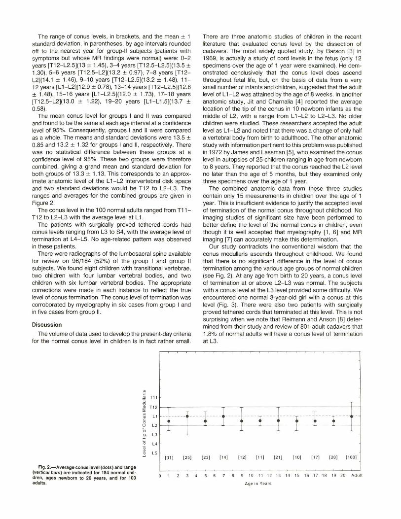
The mean conus level for groups I and II was compared and found to be the same at each age interval at a confidence level of 95%. Consequently, groups I and II were compared as a whole. The means and standard deviations were 13.5 ± 0.85 and 13.2 ± 1.32 for groups I and II , respectively. There was no statistical difference between these groups at a confidence level of 95%. These two groups were therefore combined, giving a grand mean and standard deviation for both groups of 13.3 ± 1.13. This corresponds to an approximate anatomic level of the L 1-L2 intervertebral disk space and two standard deviations would be T12 to L2-L3. The ranges and averages for the combined groups are given in Figure 2.
The conus level in the 1 00 normal adults ranged from T11-T12 to L2-L3 with the average level at L 1.
The patients with surgically proved tethered cords had conus levels ranging from L3 to S4, with the average level of termination at L4-L5. No age-related pattern was observed in these patients.
There were radiographs of the lumbosacral spine available for review on 96/184 (52%) of the group I and group II subjects. We found eight children with transitional vertebrae, two children with four lumbar vertebral bodies, and two children with six lumbar vertebral bodies. The appropriate corrections were made in each instance to reflect the true level of conus termination. The conus level of termination was corroborated by myelography in six cases from group I and in five cases from group II.
Discussion
The volume of data used to develop the present-day criteria for the normal conus level in children is in fact rather small.
V> ·.::
"" T11 ::l '0
There are three anatomic studies of children in the recent literature that evaluated conus level by the dissection of cadavers. The most widely quoted study, by Barson [3] in 1969, is actually a study of cord levels in the fetus (only 12 specimens over the age of 1 year were examined). He demonstrated conclusively that the conus level does ascend throughout fetal life, but, on the basis of data from a very small number of infants and children, suggested that the adult level of L 1-L2 was attained by the age of 8 weeks. In another anatomic study, Jit and Charnalia [ 4] reported the average location of the tip of the conus in 1 0 newborn infants as the middle of L2 , with a range from L 1-L2 to L2-L3. No older children were studied . These researchers accepted the adult level as L 1-L2 and noted that there was a change of only half a vertebral body from birth to adulthood. The other anatomic study with information pertinent to this problem was published in 1972 by James and Lassman [5], who examined the conus level in autopsies of 25 children ranging in age from newborn to 8 years. They reported that the conus reached the L2 level no later than the age of 5 months, but they examined only three specimens over the age of 1 year.
The combined anatomic data from these three studies contain or.ly 15 measurements in children over the age of 1 year. This is insufficient evidence to justify the accepted level of termination of the normal conus throughout childhood . No imaging studies of significant size have been performed to better define the level of the normal conus in children , even though it is well accepted that myelography [1 , 6] and MR imaging [7] can accurately make this determination.
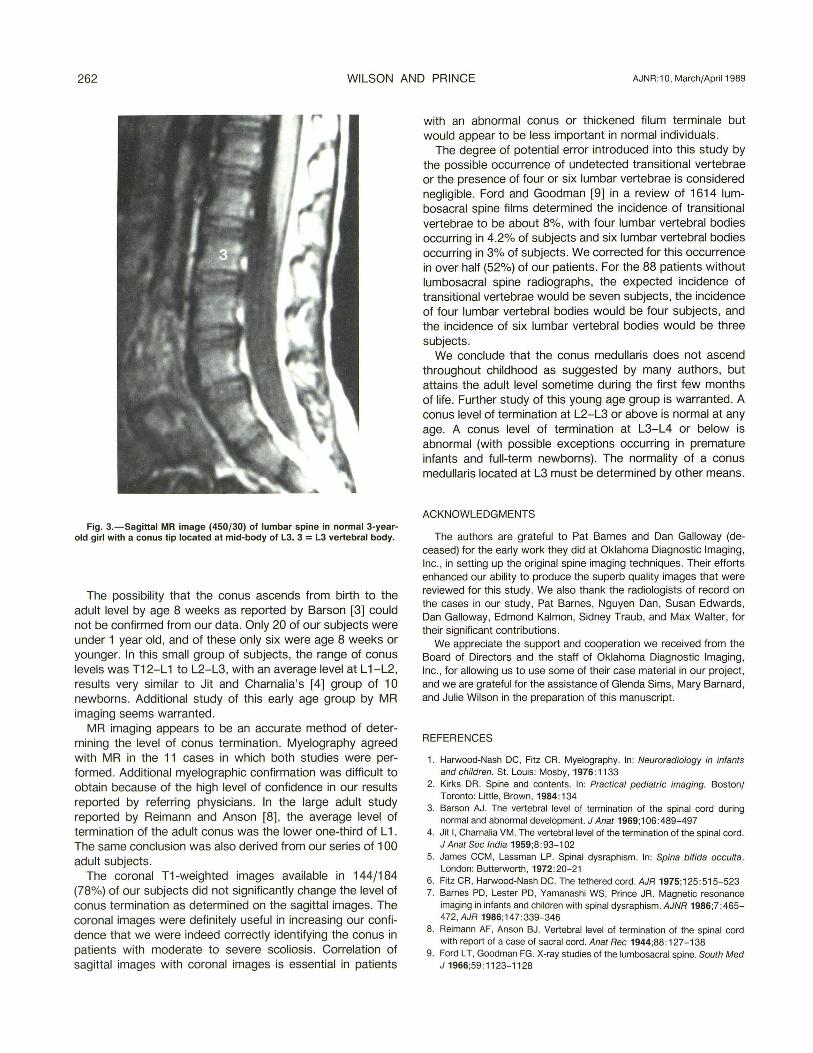
Our study contradicts the conventional wisdom that the conus medullaris ascends throughout childhood. We found that there is no significant difference in the level of conus termination among the various age groups of normal children (see Fig. 2). At any age from birth to 20 years, a conus level of termination at or above L2-L3 was normal. The subjects with a conus level at the L3 level provided some difficulty. We encountered one normal 3-year-old girl with a conus at this level (Fig . 3). There were also two patients with surgically proved tethered cords that terminated at this level. This is not surprising when we note that Reimann and Anson [8] determined from their study and review of 801 adult cadavers that 1.8% of normal adults will have a conus level of termination at L3.
--~ ---- -t ----l --- -t----- -----r--- -----------±------ --- -f ! - -
"' T1 2 :2 V>
L1 1! 0 u L2
Fig. 2.-Average conus level (dots) and range (vertical bars) are indicated for 184 normal children, ages newborn to 20 years , and for 100 adults.
0 .9-
0 a; > "' -'
1 L3
L4
L5 [31]
0
-
[25] [23] [14] [12] [11] [21] [10] [17] [20] ( 100]
2 3 4 5 6 7 8 9 10 11 12 13 14 15 16 17 18 19 20 Adul1
Age in Years
262 WILSON AND PRINCE AJNR:10, March/April1989
Fig. 3.-Sagittal MR image (450/30) of lumbar spine in normal 3-yearold girl with a conus tip located at mid-body of L3. 3 = L3 vertebral body.
The possibility that the conus ascends from birth to the adult level by age 8 weeks as reported by Barson [3] could not be confirmed from our data. Only 20 of our subjects were under 1 year old, and of these only six were age 8 weeks or younger. In this small group of subjects, the range of conus levels was T12-L 1 to L2-L3, with an average level at L 1-L2, results very similar to Jit and Charnalia's [4] group of 10 newborns. Additional study of this early age group by MR imaging seems warranted .
MR imaging appears to be an accurate method of determining the level of conus termination. Myelography agreed with MR in the 11 cases in which both studies were performed . Additional myelographic confirmation was difficult to obtain because of the high level of confidence in our results reported by referring physicians. In the large adult study reported by Reimann and Anson [8] , the average level of termination of the adult conus was the lower one-third of L 1. The same conclusion was also derived from our series of 1 00 adult subjects.
The coronal T1-weighted images available in 144/184 (78%) of our subjects did not significantly change the level of conus termination as determined on the sagittal images. The coronal images were definitely useful in increasing our confidence that we were indeed correctly identifying the conus in patients with moderate to severe scoliosis. Correlation of sagittal images with coronal images is essential in patients
with an abnormal conus or thickened filum terminale but would appear to be less important in normal individuals.
The degree of potential error introduced into this study by the possible occurrence of undetected transitional vertebrae or the presence of four or six lumbar vertebrae is considered negligible. Ford and Goodman [9] in a review of 1614 lumbosacral spine films determined the incidence of transitional vertebrae to be about 8%, with four lumbar vertebral bodies occurring in 4.2% of subjects and six lumbar vertebral bodies occurring in 3% of subjects. We corrected for this occurrence in over half (52%) of our patients. For the 88 patients without lumbosacral spine radiographs, the expected incidence of transitional vertebrae would be seven subjects, the incidence of four lumbar vertebral bodies would be four subjects, and the incidence of six lumbar vertebral bodies would be three subjects.
We conclude that the conus medullaris does not ascend throughout chi ldhood as suggested by many authors, but attains the adult level sometime during the first few months of life. Further study of this young age group is warranted . A conus level of termination at L2-L3 or above is normal at any age. A conus level of termination at L3-L4 or below is abnormal (with possible exceptions occurring in premature infants and full-term newborns). The normality of a conus medullaris located at L3 must be determined by other means.
ACKNOWLEDGMENTS
The authors are grateful to Pat Barnes and Dan Galloway (deceased) for the early work they did at Oklahoma Diagnostic Imaging, Inc. , in setting up the original spine imaging techniques. Their efforts enhanced our ability to produce the superb quality images that were reviewed for this study. We also thank the radiologists of record on the cases in our study, Pat Barnes, Nguyen Dan, Susan Edwards, Dan Galloway, Edmond Kalmon , Sidney Traub, and Max Walter, for their significant contributions.
We appreciate the support and cooperation we received from the Board of Directors and the staff of Oklahoma Diagnostic Imaging, Inc. , for allowing us to use some of their case material in our project, and we are grateful for the assistance of Glenda Sims, Mary Barnard, and Julie Wilson in the preparation of this manuscript.
REFERENCES
1. Harwood-Nash DC, Fitz CR . Myelography. In : Neuroradiology in infants and children. St. Louis: Mosby, 1976 :1133
2. Kirks DR . Spine and contents. In: Practical pediatric imaging. Boston/ Toronto: Little, Brown, 1984 :134
3. Barson AJ. The vertebral level of termination of the spinal cord during normal and abnormal development. J Anat 1969;1 06 :489-497
4. Jit I, Charnalia VM . The vertebral level of the termination of the spinal cord. J Anat Soc India 1959;8:93-102
5. James CCM, Lassman LP. Spinal dysraphism. In: Spina bifida occulta . London: Butterworth , 1972:20-21
6. Fitz CR , Harwood-Nash DC. The tethered cord. AJR 1975;125:515-523 7. Barnes PO , Lester PO, Yamanashi WS, Prince JR. Magnetic resonance
imaging in infants and children with spinal dysraphism. AJNR 1986;7:465-472, AJR 1986;147:339-346
8. Reimann AF, Anson BJ. Vertebral level of termination of the spinal cord with report of a case of sacral cord. Anat Rec 1944;88 : 127-138
9. Ford L T, Goodman FG. X-ray studies of the lumbosacral spine. South Med J 1966;59 : 11 23-11 28