Embed Size (px)
Citation preview

Mouse Piwi interactome identifies binding mechanismof Tdrkh Tudor domain to arginine methylated MiwiChen Chena, Jing Jina, D. Andrew Jamesa, Melanie A. Adams-Cioabab, Jin Gyoon Parka, Yahong Guob, Enrico Tenagliaa,c,Chao Xub, Gerald Gisha, Jinrong Minb,d, and Tony Pawsona,c,1
aSamuel Lunenfeld Research Institute, Mount Sinai Hospital, Toronto, ON, Canada M5G 1X5; bStructural Genomics Consortium, University of Toronto,Toronto, ON, Canada M5G 1L6; and cDepartment of Molecular Genetics and dDepartment of Physiology, University of Toronto, Toronto, ON,Canada M5S 1A8
Contributed by Tony Pawson, October 8, 2009 (sent for review September 15, 2009)
Tudor domains are protein modules that mediate protein–proteininteractions, potentially by binding to methylated ligands. A group ofgermline specific single and multiTudor domain containing proteins(TDRDs) represented by drosophila Tudor and its mammalian or-thologs Tdrd1, Tdrd4/RNF17, and Tdrd6 play evolutionarily conservedroles in germinal granule/nuage formation and germ cell specificationand differentiation. However, their physiological ligands, and thebiochemical and structural basis for ligand recognition, are largelyunclear. Here, by immunoprecipitation of endogenous murine Piwiproteins (Miwi and Mili) and proteomic analysis of complexes relatedto the piRNA pathway, we show that the TDRD group of Tudorproteins are physiological binding partners of Piwi family proteins. Inaddition, mass spectrometry indicates that arginine residues in RGrepeats at the N-termini of Miwi and Mili are methylated in vivo.Notably, we found that Tdrkh/Tdrd2, a novel single Tudor domaincontaining protein identified in the Miwi complex, is expressed in thecytoplasm of male germ cells and directly associates with Miwi.Mutagenesis studies mapped the Miwi–Tdrkh interaction to the veryN-terminal RG/RA repeats of Miwi and showed that the Tdrkh Tudordomain is critical for binding. Furthermore, we have solved the crystalstructure of the Tdrkh Tudor domain, which revealed an aromaticbinding pocket and negatively charged binding surface appropriatefor accommodating methylated arginine. Our findings identify amethylation-directed protein interaction mechanism in germ cellsmediated by germline Tudor domains and methylated Piwi familyproteins, and suggest a complex mode of regulating the organizationand function of Piwi proteins in piRNA silencing pathways.
Tudor domains, together with Chromo, MBT, PWWP, andAgenet-like domains, comprise the ‘‘Tudor Royal Family’’ of
domains (1). The core structure of this protein domain superfamilyis characterized by an antiparallel �-barrel-like topology and me-diates protein–protein interactions, in some cases by recognizingmethylated lysine/arginine-containing ligands with a binding sitecomposed of aromatic residues (2, 3). Their methylated targetproteins are implicated in diverse biological processes such aschromatin remodeling and RNA splicing. For example, the Tudordomain of Smn binds to methylated arginine-glycine (RG) motifson Sm proteins essential for spliceosome assembly (4), while theTudor domains of Jmjd2a bind to methylated lysines in histoneH4K20 (5).
Drosophila Tudor, the founding member of the Tudor domainfamily, is a germ cell-specific protein with multiple Tudor domainsand is involved in germ plasm formation and germ cell specification(6). By analyzing the expression pattern of mammalian genesencoding Tudor domain proteins, we identified a group whoseexpression is highly enriched in germ cells, which we therefore termgermline Tudor proteins (Tdrd1, Tdrkh/Tdrd2, RNF17/Tdrd4,Tdrd5, Tdrd6, Tdrd7, Stk31/Tdrd8, Tdrd9, Tdrd10, Akap1) (sup-porting information (SI) Fig. S1). While the physiological functionsof germline proteins with a single Tudor domain (Tdrkh, Tdrd5,Stk31, and Tdrd9) are largely unknown, mouse knockout studies ofTdrd1, Tdrd4, and Tdrd6 have revealed crucial roles for thesemultiTudor domain proteins in nuage/chromatoid body formation,
spermatogenesis, and small RNA pathways (7–9). However, thebinding properties of these germline Tudor proteins are poorlyunderstood.
Piwi proteins are conserved germline-specific Argonaute fam-ily members that are associated with Piwi-interacting RNAs(piRNAs), and thereby function in piRNA-mediated posttranscrip-tional silencing (10). Three murine Piwi paralogs, Miwi, Mili, andMiwi2, play pivotal roles in germ cell development, transposonsilencing and spermatogenesis (11–13). The presence of multiplearginine-glycine and arginine-alanine (RG/RA)-rich clusters at theN-termini of these proteins prompted us to question whether theseRG/RA motifs can be methylated in vivo and thereby serve asdocking sites for the binding of various germline Tudor proteins.
To test this hypothesis, we performed a comprehensive pro-teomic analysis of Miwi and Mili complexes in adult male germ cellsand determined the methylation status of these Piwi proteins. Weshow that several germline Tudor proteins are physiological bindingpartners of the Piwi family. In particular, we identify Tdrkh as anovel Miwi-interacting protein that binds Miwi through its singleTudor domain, likely via arginine methylation, as suggested by acombination of mass spectrometry, mutagenesis, and structuralanalysis.
ResultsTudor Domain-Containing Proteins Are Major Physiological BindingPartners of Piwi Family Proteins. To test whether Tudor domainfamily proteins comprise the in vivo binding partners of the Piwiproteins, we immunoprecipitated endogenous Miwi and Mili fromlysates of adult testes and purified the complexes by acid elution. Toobtain a comprehensive survey of the components of the Piwicomplexes, we used a gel-free liquid chromatography coupledtandem mass spectrometry (LC-MS/MS) approach employing solidphase tryptic digestion. This technique allowed us to unambigu-ously identify an extensive list of candidate proteins that specificallyassociated with Piwi proteins (Fig. 1). Hierarchical clustering of 2independent repeats of Miwi and Mili immunoprecipitations (IP)with their respective IgG control IPs reproducibly revealed distinctprotein complex profiles for Miwi and Mili (Fig. 1). We foundproteins that were specifically associated with either Miwi (Fig. 1A,blue box and Fig. S2) or Mili (Fig. 1A, red box), and proteins sharedby both Miwi and Mili complexes (Fig. 1A, cyan box). All of theseproteins were absent from the IgG control IPs, which containnonspecific binding proteins (Fig. S2). In this analysis, we observedseveral previously known Piwi-interacting proteins or piRNA path-
Author contributions: C.C., J.J., and T.P. designed research; C.C., J.J., D.A.J., M.A.A.-C., Y.G.,E.T., C.X., and G.G. performed research; C.C., J.G.P., and J.M. analyzed data; and C.C. andT.P. wrote the paper.
The authors declare no conflict of interest.
Data deposition: The atomic coordinates have been deposited in the Protein Data Bank,www.pdb.org (PDB ID code 3fdr).
1To whom correspondence should be addressed. E-mail: [email protected].
This article contains supporting information online at www.pnas.org/cgi/content/full/0911640106/DCSupplemental.
20336–20341 � PNAS � December 1, 2009 � vol. 106 � no. 48 www.pnas.org�cgi�doi�10.1073�pnas.0911640106
Dow
nloa
ded
by g
uest
on
July
9, 2
020

way components such as Ddx4, Kif17, Fxr1, and Mov10l1 (12,14–16).
Notably, several germline Tudor domain proteins (Tdrd1, Tdrkh,Tdrd6, Tdrd7, and Stk31) were present in Miwi immunoprecipitates(Fig. 1B). Among them, Tdrd1, Tdrkh, and Tdrd6 had highsequence coverage, suggesting a relatively higher abundance in thecomplex as compared with Tdrd7 and Stk31, which showed lowerpeptide numbers. Although Tdrd1 and Tdrd6 have very recentlybeen implicated in binding Miwi (9), Tdrkh, Tdrd7, and Stk31 havenot been characterized in association with Miwi. Only one Tudorprotein, Tdrd1, was identified as one of the top hits in the Milicomplex (Fig. 1B), consistent with recent findings characterizing aMili-Tdrd1 interaction (17, 18). Due to the sensitivity and detectionlimit of the method, we cannot exclude the possibility that othergermline Tudor proteins might also associate with Miwi or Milicomplexes. In this report, we focus primarily on the cellular,biochemical, and structural characterization of the Tdrkh proteinand its interaction with Miwi.
Identification of in Vivo Arginine Methylation Sites on Piwi Proteinsby Mass Spectrometry. The interaction of Miwi and Mili with Tudordomain proteins raised the possibility that they are methylated. Todetermine whether the RG/RA-rich regions of Miwi and Mili aremethylated in vivo, we immunoprecipitated Miwi and Mili and usedacid elution and gel-free solid phase tryptic digestion followed by
mass spectrometry to search for methylation sites. Mascot searchesfor monomethyl arginine (MMA) or dimethyl arginine (DMA) ledto the identification of multiple in vivo methylation sites on Miwiand Mili with high confidence (Fig. 2 and Fig. S3). Overall, weobserved more methylation sites on Mili than on Miwi. Specifically,R53 on Miwi was found in both monomethylated and dimethylatedstates. In Mili, R74, R83, R95, and R100 appeared both monom-ethylated and dimethylated and only dimethylation was found onR45, R146, R156, and R163. Interestingly, we found that 2 differentarginines could be dimethylated simultaneously on a single peptide
Pcca
Gas2l1
Spata20
Acaa2
Hnrnpl
Fyttd1
Hnrnpc
100046745
Hnrnpf
ENSMUSG00000057808
LOC100047333
Pbxip1
Hnrnpa1
1700019E19Rik
Hsp90ab1
Sept9
Git1
Caprin1
Gramd1a
Ilf3
Sept2
ENSMUSG00000076684
Zfp219
Kif17
Prkcdbp
Pacsin1
Pacsin2
Sccpdh
Eml4
Gramd3
G3bp1
Akap4
Pkd1l1Eef1g
Ptrf
Snrpn
Napa
1110012J17Rik
Txndc2
Snrpd2
B230339M05Rik
4932438A13Rik
Rai14
Golga5
Vasp
Gbf1
Amotl1
Ppp2r1a
Myst3
Enthd1
Ldhc
Kctd3
Rps7
Etl4
Gorasp2
Cep72
Nucb1
Trpd52l3
Blzf1
Rnf213
Tjp1Hdac6
Sucla2
Psmd2 C330043M08Rik
Tns1
Mov10l1
Rpl5
Phldb1
Stk22s1
Wdhd1
Cul9
Atp9a
Igf2r
Cul3
Rab3ip
8030462N17Rik
Fxr1
Fip1l1
Acta1Crybg3
Ddx4
Mocs1
Ccdc6
Ddx1
Sorbs2
Dnpep
ENSMUSG00000074479
OTTMUSG00000023442
Sssca1
Rps5
Tpr
Rps9
Birc6
1700020L24Rik
Stk31
Miwi
Tdrd1Tdrd7
Tdrd6
Mili
Tdrkh
Mili
_IP
_1M
ili_I
P_2
Miw
i_IP
_2M
iwi_
IP_1
IgG
_IP
_(M
ili1)
IgG
_IP
_(M
ili2)
IgG
_IP
_(M
iwi 2
)Ig
G_I
P_(
Miw
i 1)
1110012J17Rik Kng1 1700020L24Rik Ppp2r1a Cul9 Pacsin2 Ddx1 Psmd2 Sccpdh Tdrd7 Stk22s1 Rnf213 C330043M08Rik Cep72 Tdrd6 Git1 Stk31 ENSMUSG00000057808 Hnrnpl Spata20 8030462N17Rik Tpr Rai14 Sept2 Rab3ip B230339M05Rik Hsp90ab1 Sept9 ENSMUSG00000076684 Ccdc6 Tdrkh Kif17 Tjp1 Golga5 Birc6 Pgp Hsp90b1 Ssb Rpl5 Acta1 Rps4y2 Isyna1 Eef1g Hnrnpf Rps9 Lta4h Igf2r Ilf3 Akap4 Amotl1 Atp9a Kctd3 Myst3 Pacsin1 Trpd52l3 Phldb1 Pkd1l1 OTTMUSG00000023442 Vps13d Ybx2 Ahsg ENSMUSG00000076502 Fcgr4 Krt5 Hdac6 Sept7 Eef1a1 C3 Igh-VX24 LOC100046359 ENSMUSG00000076718 Hsp90aa1 Igh-6 Dbt Hpx ENSMUSG00000076683 LOC675759 ENSMUSG00000076939 Igh-VJ558 Fgb Hrg Gc Igk-C ENSMUSG00000076709 Pzp Aldh1a1 Fgg Mug1 Tubb3 OTTMUSG00000015054 Eef2 ENSMUSG00000076665 Atp5b ENSMUSP00000112082 Keap1 Ttn Dhx9 Khsrp Trim21 Myo18a Rassf2 Ryr1 Srrm2 Nasp Wdr37 Psmc3 Golga3 Ddx5 Tln1 Vim 1700012A16Rik Sucla2 Ruvbl2 Cct8 Hspa8 Cct2 Dync1h1 Rpl23 Cct5 Ddx4 Cct6a Trim28 Fth1 Hnrnpc Mtap4 Pcbp1 Hspa4l Suclg1 Rps13 Rpl28 Rps14 Prdx1 C1qa Cct4 Slc25a31 Tcp1 Hnrnpu ENSMUSG00000076557 ENSMUSG00000076652 LOC100041230 ENSMUSG00000076692 ENSMUSG00000076691 Rpl8 ENSMUSG00000073028 ENSMUSG00000076649 ENSMUSG00000076695 ENSMUSG00000076736 LOC630322 ENSMUSG00000076740 Hnrnpm Matr3 Hspa9 Vcp Fga Hadha Trf Ywhaz Rps3 Cct3 Rps20 Rps25 Rpl18 Rps18 Rps26 1700019E19Rik Zfp219 Fip1l1 Fyttd1 Gbf1 Snrpn Atp5a1 4932438A13Rik G3bp1 Vasp Nucb1 Tns1 Sorbs2 Blzf1 Eml4 Txndc2 Mov10l1 Sssca1 Enthd1 Mocs1 Snrpd2 Crybg3 Pbxip1 Napa Wdhd1 Gas2l1 Gramd1a Gramd3 Ptrf Gorasp2 Prkcdbp Cul3 LOC100047333 Etl4 Rps5 Acaa2 Dnpep Fxr1 Ldhc Tdrd1 Piwil1 Piwil2 Hspa2 Caprin1 ENSMUSG00000074479 Rps7 C87436 Eme1 Hectd1 Rplp0 LOC677648 Rps3a Rpl27 Rpl14 Hnrnpk Hnrnpa2b1 Rpl4 Hnrnpa1 EG386551 2210010C04Rik Dlat LOC100047070 ENSMUSG00000076564 ENSMUSG00000076566 EG434025 LOC100047053 ENSMUSG00000076594 Ighg Rpl6 ENSMUSG00000076501 ENSMUSG00000076527 Ighg1 ENSMUSG00000076522 ENSMUSG00000076708 AI324046 Igh-1b Igl-V1 Alb Pdhx Mast4 C1qb ENSMUSG00000076511 Hba-a2 Glul Rplp2 Ttr Pdhb Pdha2 Sfpq Hspa5 LOC100047208 Hbb-b2 Asrgl1 Eef1d LOC100046120 EG623114 LOC675521 C1qc Pabpc1 Pcmt1
A B
Fig. 1. Proteomic analysis of endogenous Piwi complexes identifies Tudor domain family proteins as physiological binding partners. (A) Hierarchical clusteringof proteins identified by 2 independent immunoprecipitations (IP) of Miwi, Mili, and corresponding IgG controls through tandem mass spectrometry. Proteinsfor which 3 or more peptides were identified are shown. Proteins that interact specifically with Miwi or Mili are illustrated in blue and red boxes, respectively.The cyan box indicates common proteins associated with both Miwi and Mili. (B) Identification of multiple germline Tudor domain proteins in Miwi and Milicomplexes. Miwi and Mili protein interaction networks are shown with baits in blue and Tudor domain proteins in orange. Proteins represented by 3 or morepeptides and not present in the IgG control IP, and proteins represented by peptides with a 5-fold increase over the IgG control IP are shown.
MeMe
Me
MTGRARARARGRARGQE....VGRGRQRGV....AERGGRRRDF....4 6 8 10 12 14 49 51 53 80 83
Miwi N-terminus
Mili N-terminus
MD....FRG....WGRAGPAGRGL....FRGMGLDTAFRP....LGRGVLGRGL....LGRGSSEVSLLPLGRAASSIGRGM...9 39 45 74 83 95 100 146 156 163
Me Me Me
MeMe
MeMe
MeMe MeMe
MeMe MeMe MeMe
MeMe
Me Monomethylation
Dimethylation
Me
MeMe
Fig. 2. Arginine methylation sites detected on endogenous Miwi and Mili bymass spectrometry. N-terminal RG/RA-rich sequences are show in red. Identi-fied methylation sites (Me) are shown above the relevant arginine, with theresidue numbers underneath.
Chen et al. PNAS � December 1, 2009 � vol. 106 � no. 48 � 20337
CELL
BIO
LOG
Y
Dow
nloa
ded
by g
uest
on
July
9, 2
020
from Mili (Fig. S3), indicating a potential for one Piwi familyprotein to engage multiple docking partners, such as Tudor do-mains, at the same time. To distinguish whether the DMA sites weidentified on the Piwi proteins were symmetric DMA (sDMA) orasymmetric DMA (aDMA), we used a method developed byRappsilber et al. (19). By scanning the lower mass range of severalDMA peptide MS/MS fragments, we were able to detect a dim-ethylcarbodiimidium ion (m/z � 71.06) but not an aDMA-specificdimethylammonium ion (m/z � 46.06), suggesting that these mod-ifications are most likely symmetrically dimethylated (Fig. S4).Collectively, the identification of multiple methylation sites on Piwiproteins raises the possibility that they provide a platform forrecruiting proteins with methyl arginine recognition modules, suchas Tudor domains, to the piRNA silencing complex.
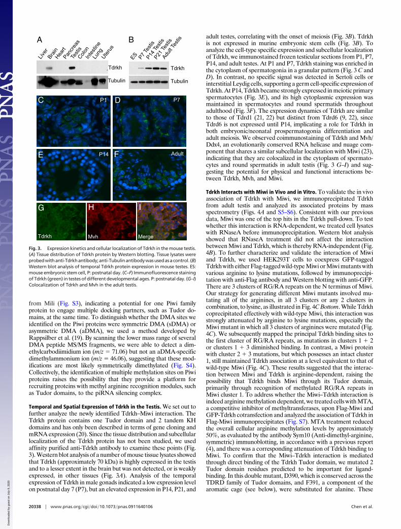
Temporal and Spatial Expression of Tdrkh in the Testis. We set out tofurther analyze the newly identified Tdrkh–Miwi interaction. TheTdrkh protein contains one Tudor domain and 2 tandem KHdomains and has only been described in terms of gene cloning andmRNA expression (20). Since the tissue distribution and subcellularlocalization of the Tdrkh protein has not been studied, we usedaffinity purified anti-Tdrkh antibody to examine these points (Fig.3). Western blot analysis of a number of mouse tissue lysates showedthat Tdrkh (approximately 70 kDa) is highly expressed in the testisand to a lesser extent in the brain but was not detected, or is weaklyexpressed, in other tissues (Fig. 3A). Analysis of the temporalexpression of Tdrkh in male gonads indicated a low expression levelon postnatal day 7 (P7), but an elevated expression in P14, P21, and
adult testes, correlating with the onset of meiosis (Fig. 3B). Tdrkhis not expressed in murine embryonic stem cells (Fig. 3B). Toanalyze the cell-type specific expression and subcellular localizationof Tdrkh, we immunostained frozen testicular sections from P1, P7,P14, and adult testes. At P1 and P7, Tdrkh staining was enriched inthe cytoplasm of spermatogonia in a granular pattern (Fig. 3 C andD). In contrast, no specific signal was detected in Sertoli cells orinterstitial Leydig cells, supporting a germ cell-specific expression ofTdrkh. At P14, Tdrkh became strongly expressed in meiotic primaryspermatocytes (Fig. 3E), and its high cytoplasmic expression wasmaintained in spermatocytes and round spermatids throughoutadulthood (Fig. 3F). The expression dynamics of Tdrkh are similarto those of Tdrd1 (21, 22) but distinct from Tdrd6 (9, 22), sinceTdrd6 is not expressed until P14, implicating a role for Tdrkh inboth embryonic/neonatal prospermatogonia differentiation andadult meiosis. We observed coimmunostaining of Tdrkh and Mvh/Ddx4, an evolutionarily conserved RNA helicase and nuage com-ponent that shares a similar subcellular localization with Miwi (23),indicating that they are colocalized in the cytoplasm of spermato-cytes and round spermatids in adult testis (Fig. 3 G–I) and sug-gesting the potential for physical and functional interactions be-tween Tdrkh, Mvh, and Miwi.
Tdrkh Interacts with Miwi in Vivo and in Vitro. To validate the in vivoassociation of Tdrkh with Miwi, we immunoprecipitated Tdrkhfrom adult testis and analyzed its associated proteins by massspectrometry (Figs. 4A and S5–S6). Consistent with our previousdata, Miwi was one of the top hits in the Tdrkh pull-down. To testwhether this interaction is RNA-dependent, we treated cell lysateswith RNaseA before immunoprecipitation. Western blot analysisshowed that RNaseA treatment did not affect the interactionbetween Miwi and Tdrkh, which is thereby RNA-independent (Fig.4B). To further characterize and validate the interaction of Miwiand Tdrkh, we used HEK293T cells to coexpress GFP-taggedTdrkh with either Flag-tagged wild-type Miwi or Miwi mutants withvarious arginine to lysine mutations, followed by immunoprecipi-tation with anti-Flag antibody and Western blotting with anti-GFP.There are 3 clusters of RG/RA repeats on the N terminus of Miwi.Our strategy for generating different Miwi mutants involved mu-tating all of the arginines, in all 3 clusters or any 2 clusters incombination, to lysine, as illustrated in Fig. 4C Bottom. While Tdrkhcoprecipitated effectively with wild-type Miwi, this interaction wasstrongly attenuated by arginine to lysine mutations, especially theMiwi mutant in which all 3 clusters of arginines were mutated (Fig.4C). We subsequently mapped the principal Tdrkh binding sites tothe first cluster of RG/RA repeats, as mutations in clusters 1 � 2or clusters 1 � 3 diminished binding. In contrast, a Miwi proteinwith cluster 2 � 3 mutations, but which possesses an intact cluster1, still maintained Tdrkh association at a level equivalent to that ofwild-type Miwi (Fig. 4C). These results suggested that the interac-tion between Miwi and Tdrkh is arginine-dependent, raising thepossibility that Tdrkh binds Miwi through its Tudor domain,primarily through recognition of methylated RG/RA repeats inMiwi cluster 1. To address whether the Miwi–Tdrkh interaction isindeed arginine methylation dependent, we treated cells with MTA,a competitive inhibitor of methyltransferases, upon Flag-Miwi andGFP-Tdrkh cotransfection and analyzed the association of Tdrkh inFlag-Miwi immunoprecipitates (Fig. S7). MTA treatment reducedthe overall cellular arginine methylation levels by approximately50%, as evaluated by the antibody Sym10 (Anti-dimethyl-arginine,symmetric) immunoblotting, in accordance with a previous report(4), and there was a corresponding attenuation of Tdrkh binding toMiwi. To confirm that the Miwi–Tdrkh interaction is mediatedthrough direct binding of the Tdrkh Tudor domain, we mutated 2Tudor domain residues predicted to be important for ligand-binding. In this double mutant, D390, which is conserved across theTDRD family of Tudor domains, and F391, a component of thearomatic cage (see below), were substituted for alanine. These
P1 bC D
E F
P7
P14 Adult
Live
rBr
ain
Hea
rtPa
ncre
asTe
stis
Col
onIn
test
ine
Lung
Ute
rus
Tdrkh
Tubulin
ES P7 T
estis
P14
Test
isP2
1 Te
stis
Adul
t Tes
tis
Tdrkh
Tubulin
A B
G H I
Tdrkh Mvh Merge
Fig. 3. Expression kinetics and cellular localization of Tdrkh in the mouse testis.(A) Tissue distribution of Tdrkh protein by Western blotting. Tissue lysates wereprobedwithanti-Tdrkhantibody;anti-Tubulinantibodywasusedasacontrol. (B)Western blot analysis of temporal Tdrkh protein expression in mouse testes. ES:mouse embryonic stem cell, P: postnatal day. (C–F) Immunofluorescence stainingof Tdrkh (green) in testes of different developmental ages. P: postnatal day. (G–I)Colocalization of Tdrkh and Mvh in the adult testis.
20338 � www.pnas.org�cgi�doi�10.1073�pnas.0911640106 Chen et al.
Dow
nloa
ded
by g
uest
on
July
9, 2
020

mutations of the Tudor domain completely abolished the bindingof Tdrkh to Miwi, consistent with the model that the Tudor domainis the protein-binding module involved in recognition of argininemethylated Miwi (Fig. 4C). Together, the endogenous coimmuno-precipitation results and in vitro cotransfection and immunopre-cipitation data clearly indicate that a single Tudor domain can directthe binding of Tdrkh to Miwi and therefore may influence andregulate Miwi and piRNA function.
Crystal Structure of the Tudor Domain of Human Tdrkh. To pursue thestructural basis for Tudor domain binding, the recombinant Tudordomain of Tdrkh was produced in Escherichia coli and its structuresolved by x-ray crystallography (Table S1). Sparse matrix screening
yielded crystals that diffracted to 1.75 Å and contained a singlemolecule in the asymmetric unit corresponding to residues 331 to418 of the full-length human Tdrkh protein (PDB 3fdr). Thecrystallized protein consists of a single Tudor barrel flanked by�-helices at both termini. Overall, the Tdrkh Tudor domain exhibitsexcellent structural similarity to the Tudor domain of Snd1 (PDB2hqx), with a root mean standard deviation of only 1.6 Å for themain-chain atoms despite the 30% sequence identity between thealigned regions (Fig. 5 A and B).
As with other Tudor domains, Tdrkh also appears to possess anintact aromatic cage that may be used for ligand recognition. Thecage comprises residues L364, T366, N367, Y371, F388, F391, andD393 (Fig. 5A). In contrast, the aromatic cage of the Snd1 Tudordomain comprises residues F715, V716, D717, Y721, Y738, Y741,and N743 (Fig. 5B). Calculation of the vacuum electrostatic po-tential of the protein surface surrounding the putative ligand-binding cavity also suggests that the nature of the protein targets forthese Tudor domains differs substantially. The Snd1 pocket israther hydrophobic and is surrounded by regions of both positiveand negative charge (Fig. 5C). That of Tdrkh, however, carriessignificant negative charges both within the pocket and over theentire surface of corresponding protein interface (Fig. 5D). This isin agreement with the possible interaction of the Tdrkh Tudordomain with the highly positively charged Miwi and Mili termini. As
Miwi MiwiR-K1,2,3
MiwiR-K 1,2
MiwiR-K 1,3
Flag-Miwis:
GFP-Tdrkh: + + + + +
GFP-Tdrkh
Flag-Miwi
-
IP F
lag
WC
L
GFP-Tdrkh
Tdr
kh
IgG
Miw
i
Tdr
kh
IgG
Miw
i
-RNaseA +RNaseA
IP:
Tdrkh
IgG
WB: Tdrkh
B
C MiwiR-K 2,3
Miwi-
+
GFP-Tdrkh mutant: (D390A, F391A)
- -- - - -
- -
+ +
MTGRARARARGRARGQE....VGRGRQRGV....AERGGRRRDF....4 6 8 10 12 14 49 51 53 80 83 84 85
RG Cluster1 RG Cluster2 RG Cluster3
Miwi N-terminus:
Miwi Mutants:
Miwi R-K 1,2,3
Miwi R-K 1,2
Miwi R-K 1,3
Miwi R-K 2,3
4 6 8 10 12 14 49 51 53 80 83 84 85
4 6 8 10 12 14 49 51 53 80 83 84 85
4 6 8 10 12 14 49 51 53 80 83 84 85
4 6 8 10 12 14 49 51 53 80 83 84 85
A Tdrkh IP #1 Tdrkh IP #2
Tdrkh 71 58% 76 64%Pcca 48 45% 47 38%Pccb 45 48% 40 53%Miwi 16 19% 23 23%4932413014Rik 13 13% 22 19%Mia3 9 5.70% 17 7.60%
Protein ID Total Peptide # Percent Coverage Total Peptide # Percent Coverage
Fig. 4. Tdrkh directly interacts with Miwi in vivo and in vitro through its Tudordomain. (A) Miwi is among the top specific interaction partners complexed withTdrkh. Immunoprecipitation of Tdrkh from adult testis lysate and gel-free massspectrometry were performed. Specific binding proteins are ranked based on thetotal peptide number identified. The top 5 Tdrkh interacting proteins with totalpeptide numbers and percentage of sequence coverage are shown for 2 inde-pendent immunoprecipitation experiments. (B) The interaction between Tdrkhand Miwi is RNA independent. Endogenous Tdrkh and Miwi were immunopre-cipitated from adult testis lysates treated with or without RNaseA using anti-Tdrkh and anti-Miwi antibodies, respectively and immunoblotted with anti-Tdrkh antibody. (C) Tdrkh binds to the first cluster of RG/RA repeats on Miwi viaits Tudor domain. HEK293T cells were cotransfected with Flag-Miwi or Flag-Miwi(R-K) mutants and GFP-Tdrkh or GFP-Tdrkh Tudor domain mutant (D390A,F391A). Flag-tagged protein complexes immunoprecipitated from cell extractsand whole cell lysates (WCL) were probed with anti-GFP and anti-Flag antibodies.The scheme of Miwi arginine mutations is shown in the bottom panel, with a redcross indicating an R-K mutant.
A B
C D
E
Tdrkh Snd1
Tdrkh Snd1
Tdrkh
Fig. 5. Crystal structure of the Tudor domain of Tdrkh. (A) Ribbon repre-sentation of the Tdrkh Tudor domain crystal structure. The residues compris-ing the aromatic binding pocket are shown in yellow. (B) Ribbon representa-tion of the Snd1 Tudor domain crystal structure. (C) Surface representation ofthe Tdrkh Tudor domain crystal structure. (D) Surface representation of theSnd1 Tudor domain crystal structure. (E) Molecular docking of a GRG peptidewith sDMA into the aromatic cage of the Tdrkh Tudor domain.
Chen et al. PNAS � December 1, 2009 � vol. 106 � no. 48 � 20339
CELL
BIO
LOG
Y
Dow
nloa
ded
by g
uest
on
July
9, 2
020

the Snd1 Tudor domain may target different cellular substratesfrom Tdrkh, such as snRNP-U in the RNA splicing pathway (24),variations in the chemical nature of their protein-binding surfacesare anticipated to confer distinct ligand specificity between theTudor domains of Tdrkh and Snd1.
Complementary to the biochemical and cell biology studies forthe Tdrkh protein, molecular docking simulations also suggest thatthe aromatic cage of the Tudor is capable of accepting peptidescarrying methylarginine modifications, although optimal dockingrequires simulation of flexibility of the residues lining this pocket.Docking simulations with a Gly-Arg-Gly (GRG) peptide carryingan sDMA suggest that the gamma carbons of L364, N367, Y371,and F391 would be displaced between 1.5–2.8 Å relative to thecrystal structure conformation (Fig. 5E). Similar experiments car-ried out with MMA or aDMA variants of the GRG peptide resultedin less preferred energies, as compared to the sDMA peptide, withthe lowest energy conformations excluding the arginine side chainfrom the binding pocket. This is consistent with the identified sitesof sDMA on the N-termini of Miwi and Mili.
DiscussionA principal function of posttranslational modifications (PTM) is tocreate binding sites for specific protein interaction domains andthus to regulate the dynamic assembly of multiprotein complexes.Although originally established in the context of phosphorylation-dependent protein–protein interactions, it is apparent that a rangeof PTMs, including acetylation, methylation, hydroxylation, andubiquitination, amongst others, can serve a similar purpose (25).Here, we have investigated the selective interactions of germlineTudor domain proteins with arginine methylated Piwi family pro-teins in the mouse testis. These interactions appear relativelyspecialized for germline Piwi proteins, as the related Argonauteproteins lack N-terminal RG/RA repeats, and are therefore un-likely to be regulated by Tudor domain interactions.
By undertaking a comprehensive proteomic survey of Miwi andMili complexes in adult germ cells, we identified a group ofgermline Tudor proteins among the major physiological bindingpartners of the mouse Piwi proteins. We also found multiple in vivosites of arginine methylation on the N-termini of Miwi and Mili,particularly symmetrically dimethylated arginines. These couldtherefore provide multiple modified motifs for binding the Tudordomains of the various proteins associated with the murine Piwifamily. Consistent with this view, mutation of the aromatic cageobserved in the structure of Tdrkh Tudor domain, which likelyserves as the binding site for methylated arginines in Miwi, abol-ished the Tdrkh-Miwi interaction. In this regard, gel-free massspectrometry of immunoprecipitated germline proteins has provento be an efficient tool for analyzing the in vivo protein interactionnetwork assembled around Piwi and Tudor domain proteins, whichcontains multiple components that are not naturally expressed intypical cultured cells.
The RG/RA clusters at the N-termini of Miwi and Mili arepotentially methylated by specific protein arginine methyltrans-ferases (PRMTs), especially PRMT5, which has a Drosophilacounterpart (dPRMT5) with crucial roles in germ cell specificationand maintenance, and gives a similar mutant phenotype to Dro-sophila tudor (26, 27). Kirino et al. (28) have demonstrated thatdPRMT5 is required for arginine methylation of Drosophila Piwiproteins and their stability, reinforcing the functional significance ofarginine methylation in germ cell biology. Our finding that multiplearginine methylation sites can be found on a single Mili peptidesuggests that individual Mili and Miwi protein chains containmultiple methylation sites, reminiscent of multisite phosphoryla-tion. Indeed, Miwi has 11 N-terminal RG/RA repeats, suggestingthat it could accumulate a high density of methylated arginine sites,and undergo different modes of interaction with Tudor domainproteins. In one scenario, proteins with multiple Tudor domainscould undergo multivalent interactions with methylated Piwi pro-
teins. For example, a single multiTudor protein, such as Tdrd1 orTdrd6, could simultaneously engage multiple sites on the same Piwiprotein through its individual Tudor domains, or could cross-linkdistinct Piwi proteins; conversely, each Piwi protein with multiplemethylated arginines could recruit Tudor domains from differentTudor proteins. Proteins with tandem Tudor domains may there-fore serve as scaffolds that form multipoint contacts with Piwiproteins, and thereby coordinate the formation and operation ofthe nuage/chromatoid body important for germ cell differentiationand development (7, 9, 29).
Although multiTudor proteins potentially have scaffolding func-tion in mammalian germinal granule formation, as discussed above,the means by which single Tudor domain proteins participate ingerm cell development is still unexplored. In this study, we haveidentified and characterized a single Tudor domain containingprotein, Tdrkh, and have shown that it interacts with Miwi in vivoand in vitro. Mutagenesis data indicate that this interaction involvesthe Tdrkh Tudor domain and the first cluster of Miwi N-terminalRG/RA repeats. Although our mass spectrometry analysis failed todetect arginine methylation sites on this first cluster of RG/RArepeats due to poor sequence coverage of the very N-terminalregion of Miwi, the last residue of the first cluster (R14) isreportedly methylated (30), consistent with the possibility that theTdrkh Tudor domain directly interacts with methylated Miwithrough R14 or other unidentified arginine methylation sites. Inaddition, we have obtained a crystal structure of the Tdrkh Tudordomain, which provides the first insight into the structural basis forligand recognition by the germline Tudor domain family. Given thatthe single Tdrkh Tudor domain interacts with a region of Miwicontaining numerous RG/RA motifs, representing actual or po-tential sites for methylation, it is possible that the Tdrkh domainbinds in a dynamic equilibrium to multiple methylated sites in Miwi,as we have previously shown for the Cdc4 F-box protein binding tomultiply phosphorylated motifs in the Sic1 protein during the yeastcell cycle (31, 32). It will be of considerable interest to investigatewhether this dynamic binding of a polyvalent ligand to a single sitereceptor applies to Tdrkh-Miwi interactions.
The observation of a direct association between Tdrkh and Miwicomplexes could add another layer of regulation to Piwi/piRNAcomplex formation and assembly by the recruitment of associatedfunctional protein domains. For example, Tdrkh has 2 KH domains,which are frequently associated with RNA recognition (33). Al-though it is not known whether the Tdrkh KH domains bind RNA,it is possible that there is an interplay between KH-mediated RNAinteractions and Piwi protein-associated piRNAs, which thereforecould influence piRNA biogenesis and functionality. It will beinteresting to examine whether the loss of Tdrkh impacts thepiRNA pathway through genetic and functional analysis.
Very recently, several groups have characterized the associationof specific germline multiTudor proteins with the Piwi familyproteins and have confirmed Mili–Tdrd1 and Miwi–Tdrd6 inter-actions, which coincides with our proteomic data (9, 17, 18). Duringthe preparation of our manuscript, an elegant study has shown acomprehensive analysis of mouse Piwi complexes and demon-strated that germline Tudor proteins direct critical protein–proteininteractions with Piwi proteins that are important for small RNAproduction and proper operation of the piRNA pathway (30). Theprofiles of Tudor domain proteins that associate with individualmouse Piwi proteins in this study are largely in accordance with ourresults, illustrating the biochemical and functional link between thePiwi family and the germline Tudor proteins. Here, we have focusedon the single Tudor domain protein Tdrkh as a member of therepertoire of Piwi-binding proteins and have defined the potentialbiochemical and structural basis for this interaction as a prototypefor germline Tudor-Piwi complexes.
In summary, our findings that various germline Tudor proteinsare in complex with Piwi proteins, and our detection of in vivo Piwimethylation sites and biochemical and structural analysis of Tdrkh
20340 � www.pnas.org�cgi�doi�10.1073�pnas.0911640106 Chen et al.
Dow
nloa
ded
by g
uest
on
July
9, 2
020

Tudor domain binding indicates that arginine methylation underliesa critical protein interaction network in germ cell development.Future studies on germline Tudor domain binding properties andspecificity, as well as the interaction dynamics of Tudor protein–Piwi complexes, will provide new insight into the construction andorganization of mammalian piRNA silencing pathways.
Materials and MethodsFull details of material and methods are discussed in the SI Text.
Antibodies. Miwi (Abcam), Mili/Piwil2 (MBL), Tdrkh (Protein Tech), Ddx4 (Ab-cam), �Tubulin (clone DM 1A, Sigma), Flag M2 agarose (Sigma), GFP (A290,Abcam), Sym10 (Millipore).
Tissue Immunoprecipitation and Western Blot Analysis. Adult testes were ho-mogenized in 1% Triton lysis buffer (10 mM Tris-HCl pH7.5, 150 mM NaCl, 1%Triton X-100, 1 mM PMSF, 10 �g/ml Leupeptin, 10 �g/ml Aprotinin, 10 �g/mlPepstatin) using Dounce homogenizer. After centrifugation, the supernatantwas filtered through 0.45 �m filter, precleared with Protein A Sepharose for 2 h,and incubatedwith3 �gofantibodiesovernightat4 °C.The immunoprecipitateswere recovered by incubation with 80 �l 10% Protein A slurry for 3 h. Afterextensive washing with the lysis buffer, the samples were eluted using 50 mMH3PO4 for mass spectrometry sample preparation or eluted using 1% SDS samplebuffer for Western blotting (34). For tissue extract used for Western blotting,mouse tissues and testes from different aged animals were lysed in RIPA bufferusing Polytron tissue homogenizer.
Immunofluorescence. Testes from different aged animals were fixed in naturalbuffered 10% formalin for 24 h and embedded in CYRO-OCT compound (SakuraFinetek). Frozen sections (6 �m) were cut, air dried, fixed, rehydrated with PBS,and blocked with 5% goat serum in PBS for 1 h. Primary antibody incubation(anti-Tdrkh, 1:200) was performed at room temperature for 2 h followed by 3 �10 min wash with PBS and 1 h secondary antibody incubation. After extensivewashing with PBS, sections were stained with DAPI and mounted. For doublestaining of Tdrkh and Ddx4, Zenon Rabbit IgG Labeling Kit (Invitrogen) was usedaccording to manufacturer’s instruction. Images were acquired with Leica DMIRE2 Microscope and processed with OpenLab software.
Plasmid Constructs. Miwi cDNA (BC129857) was cloned into pcDNA3 vector inframewithaN-terminalFlag-tag.MouseTdrkhcDNA(BC049363)wascloned intoAscI and PacI sites of a modified pDNR-Dual donor vector and shuttled to anacceptor vector with N-terminal GFP-tag. Tudor domain mutation (D390A,F391A) was generated by site directed mutagenesis. For Miwi R-K mutagenesis,various N-terminal Miwi mutant cDNA fragments encoding the first 101 aa ofMiwi were generated by DNA synthesis (Genscript) and cloned into XhoI and Blp1sites of parental pcDNA3-Flag-Miwi plasmid.
Cell Transfection and Immunoprecipitation. HEK293T cells were cultured inDMEM supplemented with 10% FBS and transfected with Flag-Miwi, Flag-Miwimutants, and GFP-Tdrkh or GFP-Tdrkh mutant using polyethyleneimine; 24–36 hafter transfection, cells were harvested and lysed using 1% Triton buffer asmentioned above. Flag-Miwi complex was immunoprecipitated by incubatingwith anti-Flag M2 agarose (Sigma) for 4 h at 4 °C and extensive washing. Proteinseluted by SDS/PAGE sample buffer were subject to Western blotting. For meth-ylation inhibition experiment, cells cotransfected with Flag-Miwi and GFP-Tdrkhwere treated with 5�-deoxy-5�-(methyl-thio) adenosine (MTA) (Sigma) at finalconcentrations of 250 �M, 750 �M, and 1 mM for 24 h before lysis.
Mass Spectrometry. Immunoprecipitated endogenous Miwi and Mili complexeswere analyzed by gel-free LC-MS/MS. All experiments were performed on aQSTAR Elite QqTOF mass spectrometer equipped with a nanospray III ion source(AppliedBiosystems/MDSSciex).Thedetailsonsamplepreparation,experimentalsetup and data analysis are described in the SI Text.
Crystallization and Structure Determination. Amino acids 327 to 420 of humanTdrkh was expressed and purified for crystallization. For details on proteinpurification, crystallization, structure determination and docking simulation, seeSI Text.
ACKNOWLEDGMENTS. We thank S. Zhang, V. Nguyen, and C. Virag, fortechnical assistance; M. Amaya and Y. Li for help with crystallography; and K.Colwill for reagents. We are grateful to L. Taylor and B. Larson for advice onmass spectrometry and T. Nott and X. Cheng for comments on the manuscript.C.C., J.J. and J.G.P. are recipients of Canadian Institutes of Health ResearchFellowships. This work is supported by grants from the Canadian Institutes ofHealth Research (Grant MOP-6849), the Canadian Cancer Society, GenomeCanada, the Premier’s Summit Award, and the Ontario Research Fund.
1. Maurer-Stroh S, et al. (2003) The Tudor domain ‘‘royal family’’: Tudor, plant agenet,chromo, PWWP and MBT domains. Trends Biochem Sci 28:69–74.
2. Kim J, et al. (2006) Tudor, MBT and chromo domains gauge the degree of lysinemethylation. EMBO Rep 7:397–403.
3. Adams-Cioaba MA, Min J (2009) Structure and function of histone methylation bindingproteins. Biochem Cell Biol 87:93–105.
4. Cote J, Richard S (2005) Tudor domains bind symmetrical dimethylated arginines. J BiolChem 280:28476–28483.
5. Huang Y, Fang J, Bedford MT, Zhang Y, Xu RM (2006) Recognition of histone H3 lysine-4methylation by the double tudor domain of JMJD2A. Science 312:748–751.
6. Boswell RE, Mahowald AP (1985) Tudor, a gene required for assembly of the germplasm in Drosophila melanogaster. Cell 43:97–104.
7. Chuma S, et al. (2006) Tdrd1/Mtr-1, a tudor-related gene, is essential for male germ-celldifferentiation and nuage/germinal granule formation in mice. Proc Natl Acad Sci USA103:15894–15899.
8. Pan J, et al. (2005) RNF17, a component of the mammalian germ cell nuage, is essentialfor spermiogenesis. Development (Cambridge, UK) 132:4029–4039.
9. Vasileva A, Tiedau D, Firooznia A, Muller-Reichert T, Jessberger R (2009) Tdrd6 isrequired for spermiogenesis, chromatoid body architecture, and regulation of miRNAexpression. Curr Biol 19:630–639.
10. Hutvagner G, Simard MJ (2008) Argonaute proteins: Key players in RNA silencing. NatRev Mol Cell Biol 9:22–32.
11. Deng W, Lin H (2002) Miwi, a murine homolog of piwi, encodes a cytoplasmic proteinessential for spermatogenesis. Dev Cell 2:819–830.
12. Kuramochi-Miyagawa S, et al. (2004) Mili, a mammalian member of piwi family gene,is essential for spermatogenesis. Development (Cambridge, UK) 131:839–849.
13. Carmell MA, et al. (2007) MIWI2 is essential for spermatogenesis and repression oftransposons in the mouse male germline. Dev Cell 12:503–514.
14. Kotaja N, Lin H, Parvinen M, Sassone-Corsi P (2006) Interplay of PIWI/Argonaute protein MIWIand kinesin KIF17b in chromatoid bodies of male germ cells. J Cell Sci 119(Pt 13):2819–2825.
15. Caudy AA, et al. (2003) A micrococcal nuclease homologue in RNAi effector complexes.Nature 425:411–414.
16. Vagin VV, et al. (2006) A distinct small RNA pathway silences selfish genetic elementsin the germline. Science 313:320–324.
17. Wang J, Saxe JP, Tanaka T, Chuma S, Lin H (2009) Mili interacts with tudor domain-containing protein 1 in regulating spermatogenesis. Curr Biol 19:640–644.
18. Reuter M, et al. (2009) Loss of the Mili-interacting Tudor domain-containing protein-1activates transposons and alters the Mili-associated small RNA profile. Nat Struct MolBiol 16:639–646.
19. Rappsilber J, Friesen WJ, Paushkin S, Dreyfuss G, Mann M (2003) Detection of argininedimethylated peptides by parallel precursor ion scanning mass spectrometry in positiveion mode. Anal Chem 75:3107–3114.
20. Lamb FS, et al. (2000) Complex RNA processing of TDRKH, a novel gene encoding theputative RNA-binding tudor and KH domains. Gene 246:209–218.
21. Chuma S, et al. (2003) Mouse Tudor Repeat-1 (MTR-1) is a novel component ofchromatoid bodies/nuages in male germ cells and forms a complex with snRNPs. MechDev 120:979–990.
22. Hosokawa M, et al. (2007) Tudor-related proteins TDRD1/MTR-1, TDRD6 and TDRD7/TRAP: Domain composition, intracellular localization, and function in male germ cellsin mice. Dev Biol 301:38–52.
23. Toyooka Y, et al. (2000) Expression and intracellular localization of mouse Vasa-homologue protein during germ cell development. Mech Dev 93:139–149.
24. Shaw N, et al. (2007) The multifunctional human p100 protein ‘‘hooks’’ methylatedligands. Nat Struct Mol Biol 14:779–784.
25. Seet BT, Dikic I, Zhou MM, Pawson T (2006) Reading protein modifications withinteraction domains. Nat Rev Mol Cell Biol 7:473–483.
26. Gonsalvez GB, Rajendra TK, Tian L, Matera AG (2006) The Sm-protein methyltransferase,dart5, is essential for germ-cell specification and maintenance. Curr Biol 16:1077–1089.
27. Anne J, Ollo R, Ephrussi A, Mechler BM (2007) Arginine methyltransferase Capsuleenis essential for methylation of spliceosomal Sm proteins and germ cell formation inDrosophila. Development 134:137–146.
28. Kirino Y, et al. (2009) Arginine methylation of Piwi proteins catalysed by dPRMT5 isrequired for Ago3 and Aub stability. Nat Cell Biol 11:652–658.
29. Arkov AL, Wang JY, Ramos A, Lehmann R (2006) The role of Tudor domains in germlinedevelopment and polar granule architecture. Development 133:4053–4062.
30. Vagin VV, et al. (2009) Proteomic analysis of murine Piwi proteins reveals a role for argininemethylation in specifying interaction with Tudor family members. Genes Dev 23:1749–1762.
31. Nash P, et al. (2001) Multisite phosphorylation of a CDK inhibitor sets a threshold forthe onset of DNA replication. Nature 414:514–521.
32. Mittag T, et al. (2008) Dynamic equilibrium engagement of a polyvalent ligand with asingle-site receptor. Proc Natl Acad Sci USA 105:17772–17777.
33. Valverde R, Edwards L, Regan L (2008) Structure and function of KH domains. Febs J275:2712–2726.
34. Smith MJ, Hardy WR, Murphy JM, Jones N, Pawson T (2006) Screening for PTB domainbinding partners and ligand specificity using proteome-derived NPXY peptide arrays.Mol Cell Biol 26:8461–8474.
Chen et al. PNAS � December 1, 2009 � vol. 106 � no. 48 � 20341
CELL
BIO
LOG
Y
Dow
nloa
ded
by g
uest
on
July
9, 2
020