Embed Size (px)
Citation preview

Mouse CCL2/JE/MCP-1 Immunoassay
Quantikine® ELISA
This package insert must be read in its entirety before using this product. For research use only. Not for use in diagnostic procedures.
Catalog Number MJE00B Catalog Number SMJE00B Catalog Number PMJE00B
For the quantitative determination of mouse Monocyte Chemotactic Protein 1 (MCP-1) concentrations in cell culture supernates, tissue culture supernates, tissue lysates, serum, and plasma.

MANUFACTURED AND DISTRIBUTED BY:
USA & Canada | R&D Systems, Inc. 614 McKinley Place NE, Minneapolis, MN 55413, USATEL: (800) 343-7475 (612) 379-2956 FAX: (612) 656-4400E-MAIL: [email protected]
DISTRIBUTED BY:
UK & Europe | R&D Systems Europe, Ltd.19 Barton Lane, Abingdon Science Park, Abingdon OX14 3NB, UKTEL: +44 (0)1235 529449 FAX: +44 (0)1235 533420E-MAIL: [email protected]
China | R&D Systems China Co., Ltd.24A1 Hua Min Empire Plaza, 726 West Yan An Road, Shanghai PRC 200050TEL: +86 (21) 52380373 FAX: +86 (21) 52371001E-MAIL: [email protected]
TABLE OF CONTENTS
SECTION PAGE
INTRODUCTION .....................................................................................................................................................................1
PRINCIPLE OF THE ASSAY ...................................................................................................................................................2
LIMITATIONS OF THE PROCEDURE .................................................................................................................................2
TECHNICAL HINTS .................................................................................................................................................................2
MATERIALS PROVIDED & STORAGE CONDITIONS ...................................................................................................3
OTHER SUPPLIES REQUIRED .............................................................................................................................................4
SUPPLIES REQUIRED FOR TISSUE LYSATE SAMPLES ...............................................................................................4
PRECAUTIONS .........................................................................................................................................................................4
SAMPLE COLLECTION & STORAGE .................................................................................................................................4
REAGENT PREPARATION .....................................................................................................................................................5
ASSAY PROCEDURE .............................................................................................................................................................6
CALCULATION OF RESULTS ...............................................................................................................................................7
TYPICAL DATA .........................................................................................................................................................................7
PRECISION ................................................................................................................................................................................8
RECOVERY.................................................................................................................................................................................8
LINEARITY .................................................................................................................................................................................8
SENSITIVITY .............................................................................................................................................................................9
CALIBRATION ..........................................................................................................................................................................9
SAMPLE VALUES .....................................................................................................................................................................9
SPECIFICITY ........................................................................................................................................................................... 10
REFERENCES ......................................................................................................................................................................... 11
PLATE LAYOUT ..................................................................................................................................................................... 12

www.RnDSystems.com 1
INTRODUCTIONThe mouse JE gene was originally described as a platelet-derived growth factor-inducible gene in mouse fibroblasts (1). The protein encoded by mouse JE was found to belong to the large CC chemokine family of inflammatory and immunoregulatory cytokines. Among CC chemokine family members, JE is functionally and structurally most closely related to the MCP/eotaxin subfamily of proteins. Within this MCP/eotaxin subfamily, five human (MCP-1, 2, 3, 4 and eotaxin) and four mouse (JE/MCP-1, MARC/MCP-3, MCP-5, and eotaxin) proteins have been identified (1-3). At the amino acid (aa) sequence level, mature human MCP-1 shows 55%, 59%, and 66% identity with the analogous regions of mouse JE, MARC, and MCP-5, respectively (1-3). Although JE has been presumed to be the mouse homolog of human MCP-1 (3-6), the more recently isolated mouse MCP-5 is actually more homologous and may be considered to be a second human MCP-1 homolog (7).
Mouse MCP-1 cDNA encodes a 148 aa residue precursor protein with a predicted 23 aa residue signal peptide that is cleaved to generate a putative mature protein of 125 aa residues (1-3). Compared to mature human MCP-1, mouse MCP-1 has a 49 aa residue carboxy-terminal extension that is rich in serine and threonine residues. Recombinant MCP-1 expressed in CHO cells (2) as well as natural MCP-1 purified from mouse astrocytes (4, 10) and a mouse thymic epithelial cell line (8), were shown to be approximately 30 kDa glycoproteins with multiple O-linked oligosaccharide chains added to the 49 aa residue C-terminal domain. Nonetheless, the natural form of MCP-1 produced by virus-stimulated mouse L929 fibroblasts occurs as a non-glycosylated 7-8 kDa protein that lacks the C-terminal domain (12). The carboxy-terminal domain has been found not to be required for mouse MCP-1 activity. Besides fibroblasts, astrocytes and epithelial cells, mouse MCP-1 has been found to be expressed in macrophages (4, 9), mast cells (7), endothelial cells (7), osteoblasts and ameloblasts (11). The expression of mouse MCP-1 is induced after stimulation with inflammatory stimuli including viruses, LPS, and cytokines such as TNF-α, IL-1, IFN-γ, and PDGF (1, 3, 4, 12, 13).
Mouse MCP-1 is a potent chemoattractant for monocytes/macrophages and lymphocytes (3, 4, 13, 17). It has also been shown to be involved in the regulation of Th1/Th2 lymphocyte differentiation, enhancing Th2 development by increasing IL-4 production and inhibiting IL-12 production (18-21). The activities of mouse MCP-1 have been shown to be mediated by the mouse CC chemokine receptor CCR2, a G protein-coupled, seven transmembrane domain receptor (6, 14, 15). Mouse CCR2 cDNA encodes a 373 aa residue protein that shows the highest (80%) overall identity at the aa sequence level with the human MCP-1 receptor, CCR2B (14-16). The gene for mouse CCR2 has been mapped to mouse chromosome 9, in close proximity with mouse CCR1 and CCR3. High levels of mouse CCR2 expression have been detected in monocytes/macrophages.
The Quantikine® Mouse CCL2/JE/MCP-1 Immunoassay is a 4.5 hour solid-phase ELISA designed to measure mouse MCP-1 in cell culture supernates, tissue culture supernates, tissue lysates, serum, and plasma. It contains E. coli-expressed recombinant mouse MCP-1 and antibodies raised against the recombinant factor. This immunoassay has been shown to accurately quantitate the recombinant mouse MCP-1. Results obtained using natural mouse MCP-1 showed dose response curves that were parallel to the standard curves obtained using the Quantikine® kit standards. These results indicate that this kit can be used to determine relative mass values for natural mouse MCP-1.
For research use only. Not for use in diagnostic procedures.2
PRINCIPLE OF THE ASSAYThis assay employs the quantitative sandwich enzyme immunoassay technique. A monoclonal antibody specific for mouse MCP-1 has been pre-coated onto a microplate. Standards, control, and samples are pipetted into the wells and any MCP-1 present is bound by the immobilized antibody. After washing away any unbound substances, an enzyme-linked polyclonal antibody specific for mouse MCP-1 is added to the wells. Following a wash to remove any unbound antibody-enzyme reagent, a substrate solution is added to the wells. The enzyme reaction yields a blue product that turns yellow when the Stop Solution is added. The intensity of the color measured is in proportion to the amount of MCP-1 bound in the initial step. The sample values are then read off the standard curve.
LIMITATIONS OF THE PROCEDURE• FOR RESEARCH USE ONLY. NOT FOR USE IN DIAGNOSTIC PROCEDURES.
• The kit should not be used beyond the expiration date on the kit label.
• Do not mix or substitute reagents with those from other lots or sources.
• If samples generate values higher than the highest standard, further dilute the samples with calibrator diluent and repeat the assay.
• Any variation in diluent, operator, pipetting technique, washing technique, incubation time or temperature, and kit age can cause variation in binding.
• Variations in sample collection, processing, and storage may cause sample value differences.
• This assay is designed to eliminate interference by other factors present in biological samples. Until all factors have been tested in the Quantikine® Immunoassay, the possibility of interference cannot be excluded.
TECHNICAL HINTS• When mixing or reconstituting protein solutions, always avoid foaming.
• To avoid cross-contamination, change pipette tips between additions of each standard level, between sample additions, and between reagent additions. Also, use separate reservoirs for each reagent.
• To ensure accurate results, proper adhesion of plate sealers during incubation steps is necessary.
• Substrate Solution should remain colorless until added to the plate. Keep Substrate Solution protected from light. Substrate Solution should change from colorless to gradations of blue.
• Stop Solution should be added to the plate in the same order as the Substrate Solution. The color developed in the wells will turn from blue to yellow upon addition of the Stop Solution.

www.RnDSystems.com 3
MATERIALS PROVIDED & STORAGE CONDITIONSStore the unopened kit at 2-8 °C. Do not use past kit expiration date.
PART PART #CATALOG # MJE00B
CATALOG # SMJE00B DESCRIPTION
STORAGE OF OPENED/ RECONSTITUTED MATERIAL
Mouse MCP-1 Microplate
898936 1 plate 6 plates 96 well polystyrene microplate (12 strips of 8 wells) coated with a monoclonal antibody specific for mouse MCP-1.
Return unused wells to the foil pouch containing the desiccant pack. Reseal along entire edge of zip-seal. May be stored for up to 1 month at 2-8 °C.*
Mouse MCP-1 Standard
898938 2 vials 12 vials Recombinant mouse MCP-1 in a buffered protein base with preservatives; lyophilized. Refer to the vial label for reconstitution volume. Use a fresh standard and
control for each assay. Discard after use.
Mouse MCP-1 Control
898939 2 vials 12 vials Recombinant mouse MCP-1 in a buffered protein base with preservatives; lyophilized. The assay value of the control should be within the range specified on the label.
Mouse MCP-1 Conjugate
898937 1 vial 6 vials 12 mL/vial of a polyclonal antibody specific for mouse MCP-1 conjugated to horseradish peroxidase with preservatives.
May be stored for up to 1 month at 2-8 °C.*
Assay Diluent RD1W
895117 1 vial 6 vials 11 mL/vial of a buffered protein base with preservatives.
Calibrator Diluent RD5-3
895436 1 vial 6 vials 21 mL/vial of a buffered protein base with preservatives.
Wash Buffer Concentrate
895003 1 vial 6 vials 21 mL/vial of a 25-fold concentrated solution of buffered surfactant with preservative. May turn yellow over time.
Color Reagent A 895000 1 vial 6 vials 12 mL/vial of stabilized hydrogen peroxide.
Color Reagent B 895001 1 vial 6 vials 12 mL/vial of stabilized chromogen (tetramethylbenzidine).
Stop Solution 895174 1 vial 6 vials 23 mL/vial of diluted hydrochloric acid.
Plate Sealers N/A 4 strips 24 strips Adhesive strips.
* Provided this is within the expiration date of the kit.
MJE00B contains sufficient materials to run ELISAs on one 96 well plate. SMJE00B (SixPak) contains sufficient materials to run ELISAs on six 96 well plates.
This kit is also available in a PharmPak (R&D Systems®, Catalog # PMJE00B). PharmPaks contain sufficient materials to run ELISAs on 50 microplates. Specific vial counts of each component may vary. Refer to the literature accompanying your order for specific vial counts.

For research use only. Not for use in diagnostic procedures.4
OTHER SUPPLIES REQUIRED• Microplate reader capable of measuring absorbance at 450 nm, with the correction
wavelength set at 540 nm or 570 nm.• Pipettes and pipette tips.• Deionized or distilled water.• Squirt bottle, manifold dispenser, or automated microplate washer.• 500 mL graduated cylinder.• Test tubes for dilution of standards and samples.
SUPPLIES REQUIRED FOR TISSUE LYSATE SAMPLES• Cell Lysis Buffer 2 (R&D Systems®, Catalog # 895347), Cell Lysis Buffer 3 (R&D Systems®,
Catalog # 895366), Lysis Buffer 16 (R&D Systems®, Catalog # 895935), or Lysis Buffer 17 (R&D Systems®, Catalog # 895943).
• PBS
PRECAUTIONSThe Stop Solution provided with this kit is an acid solution.
Some components in this kit contain a preservative which may cause an allergic skin reaction. Avoid breathing mist.
Color Reagent B may cause skin, eye, and respiratory irritation. Avoid breathing fumes.
Wear protective gloves, clothing, eye, and face protection. Wash hands thoroughly after handling. Refer to the SDS on our website prior to use.
SAMPLE COLLECTION & STORAGEThe sample collection and storage conditions listed below are intended as general guidelines. Sample stability has not been evaluated.
Cell Culture Supernates - Remove particulates by centrifugation and assay immediately or aliquot and store samples at ≤ -20 °C. Avoid repeated freeze-thaw cycles.
Tissue Culture Supernates - Tissues must be prepared prior to assay as described in the Sample Values section.
Tissue Lysates - Lysates were prepared prior to assay as described in the Sample Values section.
Serum - Allow blood samples to clot for 2 hours at room temperature before centrifuging for 20 minutes at 2000 x g. Remove serum and assay immediately or aliquot and store samples at ≤ -20 °C. Avoid repeated freeze-thaw cycles.
Plasma - Collect plasma using EDTA or heparin as an anticoagulant. Centrifuge for 20 minutes at 2000 x g within 30 minutes of collection. Assay immediately or aliquot and store samples at ≤ -20 °C. Avoid repeated freeze-thaw cycles.
Note: Citrate plasma has not been validated for use in this assay.
www.RnDSystems.com 5
REAGENT PREPARATIONBring all reagents to room temperature before use.
Mouse MCP-1 Control - Reconstitute the control with 1.0 mL deionized or distilled water. Assay the control undiluted. Mix thoroughly.
Wash Buff er - If crystals have formed in the concentrate, warm to room temperature and mix gently until the crystals have completely dissolved. Add 20 mL of Wash Buff er Concentrate to 480 mL of deionized or distilled water to prepare 500 mL of Wash Buff er.
Substrate Solution - Color Reagents A and B should be mixed together in equal volumes within 15 minutes of use. Protect from light. 100 μL of the resultant mixture is required per well.
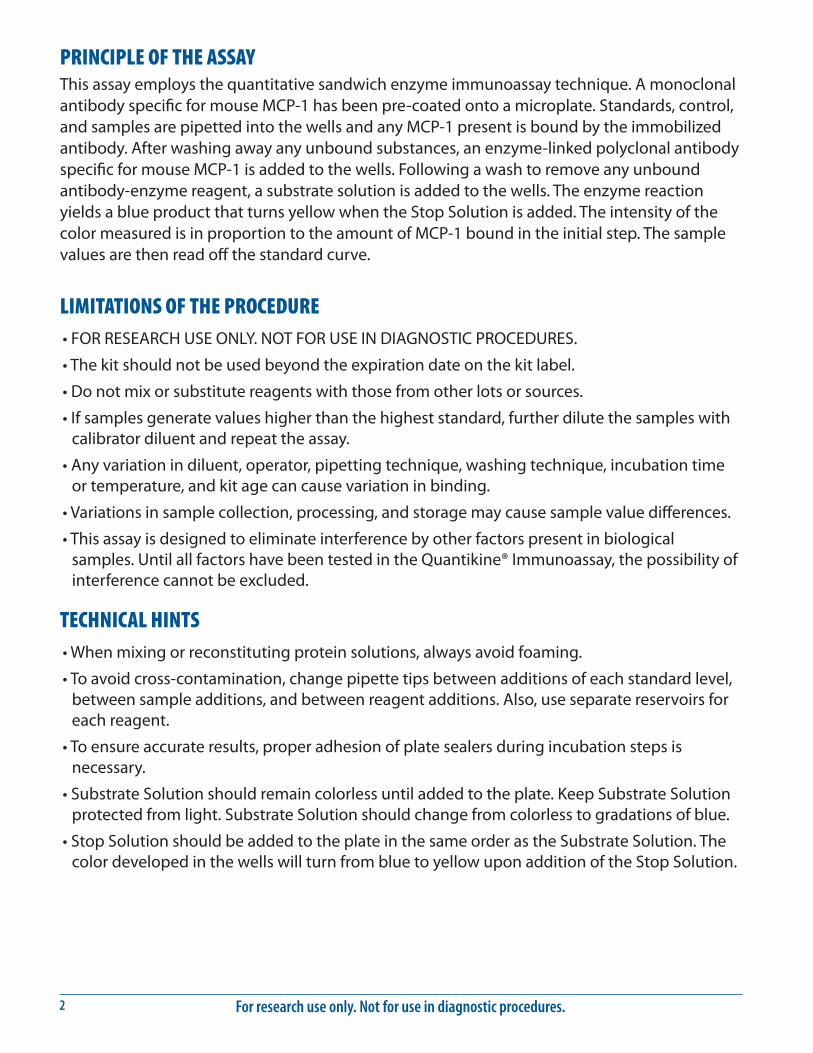
Mouse MCP-1 Standard - Refer to the vial label for reconstitution volume. Reconstitute the Mouse MCP-1 Standard with deionized or distilled water. Do not substitute other diluents. This reconstitution produces a stock solution of 5000 pg/mL. Allow the standard to sit for a minimum of 5 minutes with gentle mixing prior to making dilutions.
Pipette 450 μL of Calibrator Diluent RD5-3 into the 500 pg/mL tube. Pipette 200 μL into the remaining tubes. Use the stock solution to produce a dilution series (below). Mix each tube thoroughly before the next transfer. The 500 pg/mL standard serves as the high standard. Calibrator Diluent RD5-3 serves as the zero standard (0 pg/mL).
50 µL Std.
5000 pg/mL 500 pg/mL 250 pg/mL 125 pg/mL 62.5 pg/mL 31.3 pg/mL 15.6 pg/mL 7.81 pg/mL
200 µL 200 µL 200 µL 200 µL 200 µL 200 µL

For research use only. Not for use in diagnostic procedures.6
ASSAY PROCEDURE Bring all reagents and samples to room temperature before use. It is recommended that all standards, controls, and samples be assayed in duplicate.
1. Prepare all reagents, working standards, and samples as directed in the previous sections.
2. Remove excess microplate strips from the plate frame, return them to the foil pouch containing the desiccant pack, and reseal.
3. Add 50 μL of Assay Diluent RD1W to each well.
4. Add 50 μL of standard, control, or sample per well. Cover with the adhesive strip provided. Incubate for 2 hours at room temperature. A plate layout is provided to record standards and samples assayed.
5. Aspirate each well and wash, repeating the process three times for a total of four washes. Wash by filling each well with Wash Buffer (400 μL) using a squirt bottle, manifold dispenser, or autowasher. Complete removal of liquid at each step is essential to good performance. After the last wash, remove any remaining Wash Buffer by aspirating or decanting. Invert the plate and blot it against clean paper towels.
6. Add 100 μL of Mouse MCP-1 Conjugate to each well. Cover with a new adhesive strip. Incubate for 2 hours at room temperature.
7. Repeat the aspiration/wash as in step 5.
8. Add 100 μL of Substrate Solution to each well. Incubate for 30 minutes at room temperature. Protect from light.
9. Add 100 μL of Stop Solution to each well. Gently tap the plate to ensure thorough mixing.
10. Determine the optical density of each well within 30 minutes, using a microplate reader set to 450 nm. If wavelength correction is available, set to 540 nm or 570 nm. If wavelength correction is not available, subtract readings at 540 nm or 570 nm from the readings at 450 nm. This subtraction will correct for optical imperfections in the plate. Readings made directly at 450 nm without correction may be higher and less accurate.

www.RnDSystems.com 7
CALCULATION OF RESULTSAverage the duplicate readings for each standard, control, and sample and subtract the average zero standard optical density (O.D.).
Create a standard curve by reducing the data using computer software capable of generating a four parameter logistic (4-PL) curve-fit. As an alternative, construct a standard curve by plotting the mean absorbance for each standard on the y-axis against the concentration on the x-axis and draw a best fit curve through the points on the graph. The data may be linearized by plotting the log of the mouse MCP-1 concentrations versus the log of the O.D. and the best fit line can be determined by regression analysis. This procedure will produce an adequate but less precise fit of the data.
If samples have been diluted, the concentration read from the standard curve must be multiplied by the dilution factor.
TYPICAL DATAThis standard curve is provided for demonstration only. A standard curve should be generated for each set of samples assayed.
(pg/mL) O.D. Average Corrected0 0.007 0.007 —
0.0077.81 0.056 0.057 0.050
0.05715.6 0.100 0.101 0.094
0.10131.3 0.196 0.199 0.192
0.20262.5 0.365 0.372 0.365
0.379125 0.702 0.709 0.702
0.715250 1.320 1.326 1.319
1.332500 2.274 2.296 2.289
2.317

For research use only. Not for use in diagnostic procedures.8
PRECISIONIntra-assay Precision (Precision within an assay) Three samples of known concentration were tested twenty times on one plate to assess intra-assay precision.
Inter-assay Precision (Precision between assays) Three samples of known concentration were tested in twenty separate assays to assess inter-assay precision. Assays were performed by at least three technicians.
Intra-Assay Precision Inter-Assay Precision
Sample 1 2 3 1 2 3
n 20 20 20 20 20 20
Mean (pg/mL) 36.7 51.8 149 37.8 60.0 155
Standard deviation 1.45 1.40 3.66 2.85 4.37 7.86
CV (%) 4.0 2.7 2.5 7.5 7.3 5.1
RECOVERYThe recovery of mouse MCP-1 spiked to three levels throughout the range of the assay in various matrices was evaluated.
Sample Type Average % Recovery Range
Cell culture media (n=4) 105 95-112%
Lysis buffer (n=4) 88 79-101%
Serum (n=4) 91 83-100%
EDTA plasma (n=4) 84 75-93%
Heparin plasma (n=4) 93 80-101%
LINEARITYTo assess the linearity of the assay, samples containing and/or spiked with high concentrations of mouse MCP-1 were serially diluted with calibrator diluent to produce samples with values within the dynamic range of the assay.
Cell culture supernates* (n=4)
Lysis buffer (n=4)
Serum (n=4)
EDTA plasma (n=4)
Heparin plasma (n=4)
1:2Average % of Expected 105 99 96 96 107
Range (%) 102-107 95-108 94-99 93-104 104-109
1:4Average % of Expected 110 102 101 102 111
Range (%) 106-116 94-107 97-109 101-105 104-116
1:8Average % of Expected 109 109 105 107 104
Range (%) 105-113 103-116 100-116 100-110 98-110
1:16Average % of Expected 115 119 106 107 108
Range (%) 107-125 113-124 101-116 104-112 102-119
*Samples were diluted prior to assay.
www.RnDSystems.com 9
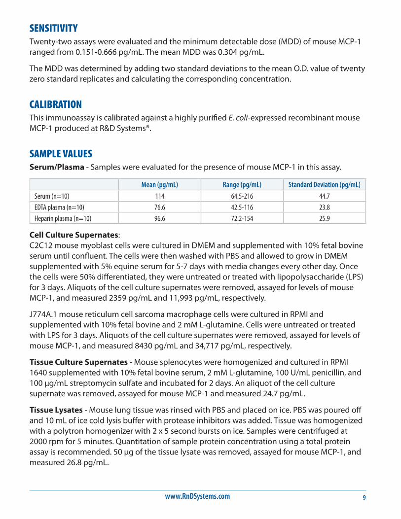
SENSITIVITYTwenty-two assays were evaluated and the minimum detectable dose (MDD) of mouse MCP-1 ranged from 0.151-0.666 pg/mL. The mean MDD was 0.304 pg/mL.
The MDD was determined by adding two standard deviations to the mean O.D. value of twenty zero standard replicates and calculating the corresponding concentration.
CALIBRATIONThis immunoassay is calibrated against a highly purified E. coli-expressed recombinant mouse MCP-1 produced at R&D Systems®.
SAMPLE VALUESSerum/Plasma - Samples were evaluated for the presence of mouse MCP-1 in this assay.
Mean (pg/mL) Range (pg/mL) Standard Deviation (pg/mL)
Serum (n=10) 114 64.5-216 44.7
EDTA plasma (n=10) 76.6 42.5-116 23.8
Heparin plasma (n=10) 96.6 72.2-154 25.9
Cell Culture Supernates: C2C12 mouse myoblast cells were cultured in DMEM and supplemented with 10% fetal bovine serum until confluent. The cells were then washed with PBS and allowed to grow in DMEM supplemented with 5% equine serum for 5-7 days with media changes every other day. Once the cells were 50% differentiated, they were untreated or treated with lipopolysaccharide (LPS) for 3 days. Aliquots of the cell culture supernates were removed, assayed for levels of mouse MCP-1, and measured 2359 pg/mL and 11,993 pg/mL, respectively.
J774A.1 mouse reticulum cell sarcoma macrophage cells were cultured in RPMI and supplemented with 10% fetal bovine and 2 mM L-glutamine. Cells were untreated or treated with LPS for 3 days. Aliquots of the cell culture supernates were removed, assayed for levels of mouse MCP-1, and measured 8430 pg/mL and 34,717 pg/mL, respectively.
Tissue Culture Supernates - Mouse splenocytes were homogenized and cultured in RPMI 1640 supplemented with 10% fetal bovine serum, 2 mM L-glutamine, 100 U/mL penicillin, and 100 μg/mL streptomycin sulfate and incubated for 2 days. An aliquot of the cell culture supernate was removed, assayed for mouse MCP-1 and measured 24.7 pg/mL.
Tissue Lysates - Mouse lung tissue was rinsed with PBS and placed on ice. PBS was poured off and 10 mL of ice cold lysis buffer with protease inhibitors was added. Tissue was homogenized with a polytron homogenizer with 2 x 5 second bursts on ice. Samples were centrifuged at 2000 rpm for 5 minutes. Quantitation of sample protein concentration using a total protein assay is recommended. 50 μg of the tissue lysate was removed, assayed for mouse MCP-1, and measured 26.8 pg/mL.

For research use only. Not for use in diagnostic procedures.10
SPECIFICITYThis assay recognizes natural and recombinant mouse MCP-1.
The factors listed below were prepared at 50 ng/mL in calibrator diluent and assayed for cross-reactivity. Preparations of the following factors prepared at 50 ng/mL in a mid-range mouse MCP-1 control were assayed for interference. No signifi cant cross-reactivity or interference was observed.
Recombinant mouse:EotaxinMARCMCP-2MCP-5MIP-1αMIP-1βMIP-1γMIP-3α
Other recombinants:human MCP-1rat MCP-1
IgG Heavy Chain
MCP-1
J774
A.1
J774
A.1
+ LP
S
C2C1
2
C2C1
2 +
LPS
250150100
75
50
37
2520
15
10
kDa
IP: MAB479
0
5000
10000
15000
20000
25000
30000
35000
40000
J774A.1 J774A.1 + LPS C2C12 C2C12 + LPS
pg/m
L
Conditioned media from J774A.1 and C2C12 cells left untreated or treated with LPS for 72 hours were analyzed by immunoprecipitation Western Blot and Quantikine® ELISA. Conditioned media was immunoprecipitated using Rat anti-Mouse CCL2/JE/MCP-1 (R&D Systems®, Catalog # MAB479). For Western Blot, samples were resolved under reducing SDS-PAGE conditions, transferred to a PVDF membrane, and immunoblotted with Goat anti-Mouse CCL2/JE/MCP-1 (R&D Systems®, Catalog # AF-479-NA). The immunoprecipitation Western Blot shows a direct correlation with ELISA value for these samples.

www.RnDSystems.com 11
REFERENCES1. Rollins, B.J. et al. (1988) Proc. Natl. Acad. Sci. USA 85:3738.
2. Ernst, C.A. et al. (1994) J. Immunol. 152:3541.
3. Luster, A.D. and M.E. Rothenberg (1997) J. Leukoc. Biol. 62:620.
4. Luo, Y. et al. (1994) J. Immunol. 153:3708.
5. Rollins, B.J. et al. (1989) Mol. Cell. Biol. 9:4687.
6. Boring, L. et al. (1996) J. Biol. Chem. 271:7551.
7. Sarafi, M.N. et al. (1997) J. Exp. Med. 185:99.
8. Liu, Z-G. et al. (1996) Eur. Cytokine Netw. 7:381.
9. Frazier-Jessen, M.R. and E.J. Kovacs (1995) J. Immunol. 154:1838.
10. Glabinski, A.R. et al. (1996) J. Immunol. 156:4363.
11. Volejnikova, S. et al. (1997) Am. J. Pathol. 150:1711.
12. Van Damme, J. et al. (1991) Eur. J. Biochem. 199:223.
13. Gu, L. et al. (1997) J. Leukoc. Biol. 62:577.
14. Gao, J-L. et al. (1995) J. Biol. Chem. 270:17494.
15. Kurihara, T. et al. (1996) J. Biol. Chem. 271:11603.
16. Charo, I.F. et al. (1994) Proc. Natl. Acad. Sci. USA 91:2752.
17. Bottazzi, B. et al. (1992) J. Immunol. 148:1280.
18. Chensue, S.W. et al. (1996) J. Immunol. 157:4602.
19. Karpus, W.J. et al. (1997) J. Immunol. 158:4129.
20. Lukacs, N.W. et al. (1997) Am. J. Pathol. 150:1861.
21. Karpus, W.J. and K.J. Kennedy (1997) J. Leukoc. Biol. 62:681.

For research use only. Not for use in diagnostic procedures.12
PLATE LAYOUTUse this plate layout to record standards and samples assayed.

www.RnDSystems.com 13
NOTES

For research use only. Not for use in diagnostic procedures.14
NOTES
03.18 753297.0 3/18
©2018 R&D Systems®, Inc.
All trademarks and registered trademarks are the property of their respective owners.


![[inserm-00630697, v1] The chemokine CCL2 protects against ... · The chemokine CCL2 protects against methylmercury neurotoxicity. David Godefroy, Romain-Daniel Gosselin, Akira Yasutake,](https://img.dokumen.tips/doc/110x75/5f071b327e708231d41b5617/inserm-00630697-v1-the-chemokine-ccl2-protects-against-the-chemokine-ccl2.jpg)