Embed Size (px)
Citation preview
J. Exp. Biol. (1973), 58. 599-625•H'ifA 1 plate and 13 text-figuresWrinted in Great Britain
MOTOR CONTROL OF TAIL SPINE ROTATION OF THEHORSESHOE CRAB, LIMULUS POLYPHEMUS
BY GERALD E. SILVEY*
Laboratory for Quantitative Biology, Department of Biology,University of Miami, Coral Gables, FL 33124 U.S.A.
(Received 11 October 1972)
INTRODUCTION
Rotation is a striking movement of the tail spine of the horseshoe crab, Limuluspolyphemus. Overturned animals use this movement to right their large bodies.Richter (1964, 1966) studied the righting behaviour, which consists of the followingactions. Upon coming to rest on the dorsum after being upset, the animal deflects itstail spine ventrally. For some time it holds the tail spine over the venter and flexesthe abdomen upon the prosoma. The animal next extends the abdomen relative to theprosoma and deflects the tail spine dorsally, slowly at first and then with increasingspeed, through the vertical plane. At the end of this movement the body is archedbetween an edge of the prosoma and tip of the tail spine. The animal then rotates thebody about the tail spine to one side or the other. During rotation of the body the tipof the tail spine sometimes loses contact with the substrate. When this occurs thearch of the body collapses and the animal rotates the tail spine opposite to the direc-tion in which the body was moving until the tip touches once again. It repeats thearching and rotating actions until, with contributions from grasping and pushing legsand beating gills, it pivots over upon a side or upon the anterior edge of the prosoma,thereby turning itself right side up.
Rotation of the tail spine is of neurophysiological interest because of its bearing onthe question of how motor control systems promote behaviour. This question requiresconsideration of the motor pattern and the muscle response, because behaviour isexpressed through integration of the two (Horridge, 1968). The importance of muscleresponse to motor control has been brought out most clearly by Harold Atwood. Hisstudies on the functional and structural properties of crustacean muscles has indicatedthe extent to which integration can occur peripherally. He has shown in crustaceanneuromuscular systems that a great variety of behavioural movements is possiblethrough activation at different frequencies and of different combinations of fast, slowand inhibitory axons to fast, intermediate and slow muscle fibres (Atwood, 1967).
The importance of motor patterns to motor control in determination of particularclasses of movement has been shown by studies of rhythmic and non-rhythmicsystems, for example, insect flight (Wilson, 1961) and the crab claw opener (Wilson& Davis, 1965) respectively. Nerve recordings from some of these systems, namely thecrayfish abdomen flexor (Gillary & Kennedy, 1969 a, b), the cockroach coxal levator
• Present address: Department of Neurobiology, Research School of Biological Sciences, TheAustralian National University, Canberra City, A.C.T., Australia.
38-2
600 G. E. SILVEY
and depressor (Pearson & lies, 1970) and the dragonfly larva expiratory systen1
(Mill, 1970), show distinct motoneurones firing at different times in the firing patterns.The consequent movement of these systems is an effect of the sequence in which eachmotoneurone fired in the pattern. Analysis of motoneurone sequence during naturalmovements together with the knowledge of the neuromuscular effects of the moto-neurones should provide a more full understanding of the significance of the firingpattern.
In at least one behavioural system both issues of motor control have been examined,and effective relationships have been shown among motor pattern, muscle propertiesand behaviour. Gillary & Kennedy (1969 a, b) demonstrated that the burst-firingpattern of the largest motoneurone innervating the superficial or tonic flexors of thecrayfish abdomen facilitated the response of these slow muscles (Kennedy & Takeda,1965^). Facilitation did not occur when the motoneurone was driven in a non-burstmode. Behaviourally, the muscle response to the burst firing pattern gave rise to rapidflexion or to strong flexion against a barrier (Larimer & Eggleston, 1971).
In approaching the question of motor control of the Limulus tail spine in this work,the integrative aspects of motor pattern and muscle response were of concern. Muchattention was directed to the motor pattern, and events in the firing pattern werecorrelated with movement of the tail spine, muscle tension, and activity of themuscles in order to show how the output pattern generated rotation. Attention tomuscle response was given through an investigation of neuromuscular and histologicalproperties in order to determine the mechanisms and structures through which theoutput was expressed. Movement and use of the tail spine during behaviour and thearrangement and action of the tail spine muscles were also examined, because theseproperties specified the behavioural and functional limits of the system. The result ofthese considerations provides an explanation of a motor control system which usesmany neurones to recruit functionally and structurally similar muscle fibres and excitesthem efficiently and in inverse sequences to rotate the tail spine appendage in twodirections, clockwise and counterclockwise.
MATERIALS AND METHODS
Limulus polyphemus (L.) was collected locally throughout the year on nights of thefull moon at the spring tide, when animals ascended beaches to mate as described byTeale (1957). Selected animals were fed on fish and squid and kept in running seawater until shortly before use when they were taken to a filtered and aerated sea-waterpond in the laboratory. Average-sized males - that is, 15-19 cm across the carapace -were used in experiments, because females were too large and powerful.
Results are given of some single experiments, which were conducted slightly dif-ferently from others in the same group of experiments. For example, in a set of experi-ments in which firing and tension were examined rotation was evoked in one animaland recorded, and this result is presented (Text-fig. 11). Furthermore, the analysis ofmotoneurones that is presented in Text-figs. 7-9 and in Text-fig. 12 are from singleexperiments, because it was impossible to identify the same motoneurones in differentanimals. Nevertheless, analysis of motoneurones in separate experiments did not differqualitatively.

Control of Limulus tail spine rotation 601
Electrical recording. The experiments required extracellular recording of muscleand nerve potentials and in some cases intracellular recording from muscle fibres.Extracellular muscle potentials were recorded by means of paired copper-wire elec-trodes approximately 95 /an in diameter. These were inserted through holes drilledin the carapace above the desired muscle. The wires were insulated to within 1 mmof the tip. These recordings were taken while animals moved without restraint in asea-water aquarium. In some experiments involving restrained animals musclepotentials were recorded by means of suction electrodes attached to the sides ofmuscle fibres.
Nerve potentials were recorded by means of long flexible polyethylene suctionelectrodes. About 5 in. of P.E. 60 tubing, into which a silver wire was inserted, wassufficiently flexible to stay on the nerve during the vigorous movements of the con-tracting muscles. Electrode tips were placed on branches of nerves which ran justbeneath the surface of muscles. Muscles were exposed by cutting windows in thedorsal or ventral wall of the carapace. In order to reduce bleeding and clotting aboutexposed tissue blood was withdrawn from the heart or pericardium by hypodermicsyringe until no more came with ease. The volume withdrawn was ordinarily 50 ml.This withdrawal reduced both the blood pressure and the amount of blood circulatingthrough the muscles but did not impair the behaviour of the animals during the courseof experiments lasting 2-6 h. In order to eliminate prosoma-opisthosoma flexionmovement, which would have dislodged electrodes and moved the tail spine in andout of the plane of view of the motion picture camera, a plate was bolted across thejoint of these two sections of the body. Animals were clamped rigidly in a sea-waterbath which was sufficiently deep to allow the tail spine to rotate without hindrance.
In both the muscle and nerve recording experiments movements of the tail spinewere filmed with a Beaulieu 16 mm cine camera at 25 frames/sec. A Texas Instru-ments LS 400 phototransistor placed in the viewfinder produced pulses on theoscilloscope between frames when light was deflected away from the shutter andthrough the viewfinder. Films were taken from the rear of the animal and examinedfrom this direction so that descriptions of rotations, namely clockwise and counter-clockwise, refer to a viewing position behind the animal.
Intracellular potentials were recorded by means of 3 M-KCl-filled glass capillariesof 4-14 Mii impedance. Only about 1 cm of a capillary was used and this was sus-pended from a flexible silver wire as a' floating' microelectrode. Signals were amplifiedby means of a Bioelectric NF 1 neutralized input-capacity amplifier. Most recordingswere made in tail-spine muscles of abdomens which had been separated from theprosoma and therefore deprived of circulation. Potentials were evoked in musclefibres of these preparations by tactile stimulation, which triggered output to the dorsalmuscles, or by directly stimulating nerves by means of suction electrodes. Theremainder of the recordings were made in muscles of whole animals.
Experiments on motor pattern and on neuromuscular properties were performed oneach of the muscles. The results suggested that motor pattern and neuromuscularproperties were the same in each of the muscles. Most of the experiments, however,were conducted on the dorsal lateral muscles because these were the most approach-able, provided at least one long nerve branch for recording and permitted work withthe animal in a position from which tail spine rotation was readily elicited.
38-3

602 G. E. SILVEY
Mechanical recording. Tension was recorded in some experiments. Recordings wer1
made with a Grass Ft. 03 force transducer tied with a nylon monofilament to a muscletendon, which had been severed at its insertion. This method was applied to musclesin isolated abdomens and in intact animals. In the latter the tail spine was cut veryclose to the base so that when it rotated it would not interfere with the thread connect-ing the tendon to the transducer, and the tendon was not severed at its insertion. Inorder to record force developed by the tail spine during rotation the end of the tailspine was tied to the tip of a rod which was fixed to the transducer so that the tip wassuspended only a few millimeters above the substrate. When the animal was turnedover the tail spine pushed down upon the rod as if it were the substrate and the forcemeasured was assumed to be the same as the force generated against a natural sub-strate. The animals were sometimes prevented from righting and the additional forcegenerated was accepted as the reserve force it could generate.
Histology. Nerve and muscle tissue from the dorsal lateral muscles was prepared forlight microscopy after the method of Fourtner & Sherman (1972). Muscles were firstrelaxed in a 5-5 mM-MgSO4 and 445 mM-NaCl solution. With the tail spine in itsresting position tissue was then pre-fixed in glutaraldehyde, which was made 4 % ino-i M sodium cacodylate buffer adjusted to pH 7-3 and osmotically adjusted with 2%NaCl and 0-2 % CaCl2. Tiny pieces of tissue were dissected out, and after washing inbuffer post-fixed in 1 % osmium tetroxide were washed, pre-stained in uranylacetate, dehydrated in ethanol and transferred to propylene oxide before embedding inAraldite (6005). Thick sections were cut at 0-250 /an, stained with toluidene blue andexamined with bright-field optics.
RESULTS
Functional morphology
Eight muscles move the tail spine of Limulus. These are housed in a chamber in theposterior of the abdomen and are symmetrically and adjacently arranged about the tailspine (Text-fig. 1 and inset diagram in Text-fig. 7). Patten & Redenbaugh (1899), whodescribed the anatomy of Limulus in some detail, recognized only six muscles. Con-nective tissue, however, does separate the muscle mass into eight discrete muscles orbundles of muscle fibres. Occasionally, however, a few muscle fibres which originatewith one muscle fuse or insert with an adjacent muscle. Patten and Redenbaughdesignated the muscles as extensors and flexors but these terms do not accuratelydescribe the action of the muscles. None of the muscles extends or straightens the tailspine relative to the longitudinal axis of the body yet all of them flex or deflect the tailspine relative to this axis. The action of each deflects the tail spine through some angleuntil the tail spine is perpendicular or nearly so to the normal resting position. Theresting position is characterized by the tail spine pointing posteriorly and slightlyventrally and in a plane parallel to the vertical plane of the body. The angle throughwhich any single muscle deflects the tail spine is given in Text-fig. 2.
The names dorsal medial, dorsal lateral, lateral and ventral have been given to thepairs of muscles relative to their position about the horizontal and vertical planes. Thedorsal medial muscle, which corresponds to the telson extensor muscle a of Patten &Redenbaugh (1899), originates on the medial side of the last three abdominal epicon-dyles and on the underside of the dorsal wall of the muscle chamber. The dorsal

Mot
or
ral c
ord-
gang
lion
14
c 14
Ven
tral
cord
-gan
glio
n 14
Ner
ve I
4 N
erve
15
Lute
ral
spin
e M
olor
bra
nch
of n
erve
14
Ven
tral
mus
cle-
dors
al h
ead
Ana
stom
osis
of
nerv
es 1
4 an
d 15
V
entra
l m
uscl
e-ve
ntra
l h
a
Sens
ory
bran
ch o
f ne
we
15
Sens
ory
bran
ch o
f ne
rve
14
Mot
or b
rnnc
h of
ner
ve I
S
Sens
ory
bran
ch o
f ne
rve
15
to d
orsi
l nn
d la
tera
l m
uscl
es
Mot
or b
ranc
h of
ner
ve 1
5 to
ven
tral
bran
ch o
f ne
rve
15 to
ven
tral
mus
cle
Mot
or b
mnc
h of
ner
ve 1
5 to
dor
sal
and
Iate
ral .
mus
cle9
M
otor
bra
nch
of n
erve
16
Dor
sal
med
ial
mus
cle
Ana
stom
osis
of
nerv
es 1
5 an
d 16
D
orsa
l Ia
tem
l m
uscl
e Pr
opri
ocep
tor
nerv
e L
iter
al m
uscl
e O
cclu
dor
ani m
. L
evnt
or a
ni m
.
Tai
l spi
ne n
erve
\D
orsu
l pr
ojec
tion
of th
e ta
il sp
ine
Tai
l sp
ine
Con
dyle
Ven
tral
proj
ectio
n of
the
tail
spin
e
Ner
ve 1
5
Lute
ral
spin
e M
olor
bra
nch
of n
erve
14
Ana
stom
osis
of
nerv
es 1
4 an
d 15
Sens
ory
bran
ch o
f ne
we
15
Sens
ory
bran
ch o
f ne
rve
14
Mot
or b
rnnc
h of
ner
ve I
S
Sens
ory
bran
ch o
f ne
rve
15
to d
orsi
l nn
d la
tera
l m
uscl
es
Mot
or b
ranc
h of
ner
ve 1
5 to
ven
tral
bran
ch o
f ne
rve
15 to
ven
tral
mus
cle
Mot
or b
mnc
h of
ner
ve 1
5 to
dor
sal
and
Iate
ral .
mus
cle9
D
orsa
l m
edia
l m
uscl
e D
orsa
l Ia
tem
l m
uscl
e
Lit
eral
mus
cle
Lev
ntor
ani
m.
Dor
sul
proj
ectio
n of
the
tail
spin
e
mus
cle
Tex
t-fi
g. I
. S
ketc
h of
the
ner
ves
and
mus
cles
of
the
tail
-spi
ne a
ppar
atus
fro
m a
dor
sal
aspe
ct.
Th
e ri
ght
side
sho
ws
dors
al a
nd la
tera
l m
uscl
es b
enea
th t
he w
all o
f th
e ca
rapa
ce. T
he
left
sid
e, a
dee
per
view
wit
h do
rsal
and
lat
eral
mus
cles
re
mov
ed a
nd d
orsa
l pro
ject
ion
of t
he t
ail
spin
e cu
t aw
ay, s
how
s a
vent
ral
mus
cle.
Sol
id l
ines
rep
rese
nt n
erve
s ru
nnin
g ab
ove
or
betw
een
mus
cles
in
the
plan
e of
vie
w.
Das
hed
line
s re
pres
ent
nerv
es r
unni
ng b
enea
th o
r w
ithi
n m
uscl
es.
Th
e ei
ght t
ail-
spin
e m
uscl
es a
re a
rran
ged
sym
met
rica
lly
and
cont
iguo
usly
(cf
. upp
er in
set i
n T
ext-
fig.
7).
Th
e do
rsal
and
la
tera
l m
uscl
es i
nser
t on
the
dor
sal
proj
ecti
ons
of t
he t
ail
spin
e, t
he v
entr
al m
uscl
es o
n t
he v
entr
al p
roje
ctio
ns.
Th
e co
-ope
rati
ve a
ctio
n of
the
se m
uscl
es m
oves
the
tail
spi
ne a
bout
the
cond
yles
.

604 G. E. SlLVEY
Muscles - left sideDM - 80°
DL-65"L-50°A
Muscles - right side100°-DM
I # »110°-DL* 130°-L
DM
220°-V
I
Text-fig. 2. Diagrams showing the angles to which individually contracted muscles deflect thetail spine and the arcs through which the major burst is active in each muscle during clockwiseand counterclockwise rotation of the tail spine. The diagrams are drawn as if one were lookingat the animal from behind. The isolated action of each muscle was elicited either by stimulationof the largest nerve innervating the muscle or by direct stimulation of the muscle. Rotationswere produced centrally in whole animals in response to sensory input. The period of majorburst activity in a given muscle was determined from several recordings of nerves innervatingthe muscle or of muscle potentials in the case of the left ventral muscle and was correlated withthe arc through which the tail spine moved during this period. The most striking feature isthat for each muscle the end-point of firing after rotation in either direction coincided closelywith the angle to which the individual contraction of the same muscle deflected the tail spine.This point is indicated by the short, heavy bars at the end of arcs. During rotation there wasno period of time when fewer than two muscles were active. DM, Dorsal medial; DL, dorsallateral; L, lateral; and V, ventral muscles.
lateral and lateral muscles, which correspond to the telson extensor muscle b ofPatten & Redenbaugh, originate on the underside of the dorsal wall of the musclechamber and on its anterior wall. These three muscles insert on the dorsal projectionof the tail spine (cf. right side of Text-fig, i). The ventral muscle, which correspondsto the telson flexor muscle of Patten & Redenbaugh, originates by small heads on thelateral side of the last three abdominal epicondyles and on the lower portion of theanterior wall of the muscle chamber and inserts on the ventral projection of the tailspine (cf. left side of Text-fig, i). Although the ventral muscle originates by dorsalheads, that is, by those on the epiconcyles (which are apodemes that project from thedorsal wall downward into the abdominal body cavity) as well as by a large ventralhead, all of its force is directed upon the ventral projection.
Each of the eight muscles inserts on a tail-spine projection by more than one tendon.The dorsal lateral muscles, for example, insert by five or six tendons. Functionally, thisarrangement allows the tail spine to be deflected to different angles by contraction ofdifferent portions of the muscle. Pulling on one and then the other of the most widelyseparated tendons of a dorsal lateral muscle deflects the tail spine to points separated by5° of arc.
The tail spine articulates upon two condyles, which arise from the sides of theabdominal wall that forms the tail spine and anal foramen. The condyles project intospacious sockets, which exist on both sides of the head of the tail spine between dorsal

Control of Limulus tail spine rotation 605
'and ventral projections. Thin cuticle unites the condyles to the tail spine. The tail spinecan swivel freely through 3600 at its articulation. The tail spine can be maximallydeflected to almost 90° in the vertical plane. However, as the tail spine is rotatedtoward the horizontal plane the angle of deflection is reduced to abouf6o° because thepointed, wing-like ends of the abdomen restrict lateral movement. This arrangementprevents the arc developed by the tip of a rotating, fully deflected tail spine fromdescribing a perfect circle.
Three pairs of nerves innervate the tail-spine muscles. Nerve pairs arise from eachof the last three ganglia of the ventral cord (Text-fig. 1). The nerves correspond tohaemal nerves 14, 15 and 16 of Patten & Redenbaugh (1899) but are here callednerves 14, 15 and 16, because these are the only nerves from the last three gangliainvolved with the tail-spine musculature. Nerve 14 innervates the dorsal medialmuscle by a motor branch and the wall of the abdomen in the region of the last lateralspine by a sensory branch. Nerve 15 innervates all of the muscles by motor branchesand also the wall of the abdomen posterior to the last lateral spine by a sensory branch.Nerve 16 innervates the medial portion of the ventral muscle by a motor branch.Nerve 16 also innervates a proprioceptor (Eagles, 1972, and personal observation) inthe ventral muscles, muscles of the gut and anus and the tail spine. Anastomoses occurbetween motor branches of nerves 14 and 15 and nerves 15 and 16.
Rotation consists of a clockwise or counterclockwise turning of the tail spine aboutits point of articulation. Rotation starts with a deflexion of the tail spine through anyplane perpendicular to the longitudinal axis at the point of articulation. The deflexionmovement is usually turned into a rotational movement before the tail spine reachesthe full extent of its deflexion. In nature, rotation occurs when the animal is turnedover and starts the struggle which results in righting. Rotation also occurs in naturewhen the animal is tipped. In such cases the animal deflects its tail spine and swings itdownward through the side of the body that is lower in order to bring this side up bythe force of the moving tail spine. In the laboratory full rotations could be elicitedwhen the animal was rigidly fixed, lifted above the substrate in order that the tailspine could revolve freely and stroked on the sides of the abdomen. Stroking on theright side evoked counterclockwise rotation and on the left side clockwise rotation.Persistent rotation required an intact central nervous system. However, followingseverance of the ventral cord as far posterior as ganglion 13, rotation could still beelicited by tactile stimulation of the abdomen but for only a brief period of time.
Righting requires the development of sufficient force to lift up the body and swivelit upon the tail spine. In some animals the force developed by the rotating tail spineapproximated to the body weight in air, which was about 500 g for an average-sizedmale. The weight in water, however, as determined from weighings of several animals,was only 6-5 % of that in air. The average force developed by the rotating tail spine offour animals in the course of righting was 65 g, but more than twice this was generatedwhen the tail spine was prevented from rotating. Thus, the animal is capable ofdeveloping sufficient force to lift itself upon its tail spine and to provide some addi-tional force if it encounters resistance.

606 G. E. SILVEY
RightDorsal muscleLateral muscleVentral- muscle
LeftDorsal muscleLateral muscle -Ventral muscle-
Right
Text-fig. 3. Extracellular recording of muscle potentials from six muscles during rightingof an unrestrained animal. The sketches below the records show movements of the animal asviewed from the rear and directions of movement are described from this viewpoint. Therecordings are from two separated righting sequences but each consisted of approximatelythe same movements and took about the same time. The records have been placed together inorder to show the sequential activation of each muscle during the production of rotation. Themuscles fired in clockwise sequence, beginning with the left ventral muscle, and the tail spinewas moved clockwise until it touched the substrate. At this point the body was moved counter-clockwise until it left the substrate. From the point of leaving the substrate the tail spine wasbrought further around clockwise. The dorsal muscle is the dorsal medial muscle. The fourthand eighth traces monitored the frames filmed. Time: 200 msec.
The motor pattern
Rotation of the tail spine is produced when muscles contract sequentially. Therecords in Text-fig. 3 of potentials from three contralateral pairs of muscles during arighting manoeuvre indicate that this is the case. Just prior to the records shown theanimal was turned ventral side up. At the start of the recording the animal rotated itstail spine clockwise until the spine touched and anchored. From that point on thebody moved counterclockwise until the body left the substrate. The animal then settledand brought the tail spine further around clockwise. During this action muscles wererecruited in a clockwise sequence. First the left ventral muscle fired, next the leftlateral, left dorsal and then muscles on the right, dorsal, lateral, ventral and onaround. Thus, sequential activation of the muscles in one direction leads to a rotationof the tail spine in the same direction or, when the tail spine becomes anchored, torotation of the body in the opposite direction.
Sequential firing in the nerves produces the muscle activity, which is responsiblefor rotation. This is shown by recordings in Text-fig. 4 from a pair of contralateralnerves, one of which innervated the right dorsal lateral muscle, the other the leftdorsal lateral muscle. In the upper record firing in the nerve to the left muscle pre-ceded that in the nerve to the right muscle and the tail spine moved clockwise asindicated by the figures below the frame-counting trace. In the lower record firing tothe right muscle preceded that to the left muscle and the tail spine was moved counter-clockwise.
Activity also occurs in sequence within single muscles. Fibres in a muscle becameactive at different times during rotations as shown in Text-fig. 5. The order of firing todifferent fibres corresponded to the direction of rotation of the tail spine. The examplein Text-fig. 5 is from the left dorsal lateral muscle and shows that lateral musclefibres fired before medial fibres during clockwise rotation and vice versa during coun-terclockwise rotation. The changes in time between the beginning and the end offiring of one portion of the muscle relative to the beginning and end of firing of anotherportion of the same muscle during opposite rotations differed from 100 to 500 msec in

Tex
t-fi
g. 4
. R
ecor
ding
s fr
om n
erve
s to
con
tral
ater
al m
uscl
es d
urin
g op
posi
te r
otat
ions
. T
he
figu
res
belo
w t
he fr
ame-
coun
ting
tra
ce i
ndic
ate
the
posi
tion
%
in
rot
atio
n an
d re
lati
ve d
egre
e of
def
lexi
on o
f th
e ta
il s
pine
at zoo
mse
c in
terv
als.
Fir
ing
to t
he l
eft
mus
cle
prec
eded
tha
t to
the
rig
ht m
uscl
e fo
r th
e ge
nera
tion
of
cloc
kwis
e ro
tati
on;
firi
ng t
o th
e ri
ght
prec
eded
tha
t to
the
lef
t fo
r co
unte
rclo
ckw
ise
rota
tion
. T
his
ind
icat
es t
hat n
ervo
us a
ctiv
ity
occu
rs in
gs
op
posi
te s
eque
nces
to
prod
uce
oppo
site
rot
atio
ns.
Not
e th
at th
e an
gle
of t
he t
ail
spin
e at
the
end
of
!iri
ng to
eac
h m
uscl
e w
as a
ppro
xim
atel
y th
at to
whi
ch
3 th
e ac
tion
of
each
mus
cle
alon
e dr
ew t
he t
ail
spin
e as
des
crib
ed i
n T
ext-
fig.
z.
Th
is p
oint
at t
he e
nd o
f fi
ring
of
a ne
rve
to a
mus
cle
also
cor
resp
onde
d to
3
the
peak
of
tens
ion
deve
lope
d by
a m
uscl
e (T
ext-
figs
. I
I, 12). N
ote
also
that
the
freq
uenc
y an
d du
rati
on o
f fi
ring
wer
e gr
eate
r in
a n
erve
whe
n th
e m
uscl
e it
inn
erva
ted
brou
ght
the
tail
spi
ne u
pwar
d o
n th
e si
de o
f th
e m
uscl
e th
an w
hen
the
mus
cle
brou
ght
the
tail
spin
e do
wnw
ard.
Fo
r ex
ampl
e, t
he le
ft d
orsa
l 8
late
ral m
uscl
e li
fted
the
tai
l sp
ine
duri
ng c
lock
wis
e ro
tati
on b
ut c
arri
ed i
t dow
nwar
d du
ring
cou
nter
cloc
kwis
e rot
atio
n. T
he
freq
uenc
y an
d du
rati
on o
f !i
ring
w
ere
grea
ter
to t
his
mus
cle
duri
ng t
he f
orm
er r
otat
ion
than
dur
ing
the
latt
er.
Th
is p
heno
men
on s
ugge
sts
that
the
tail
-spi
ne m
echa
nism
is c
ompe
nsat
ing
for
a lo
ad,
and
evid
ence
for
wm
pens
atio
n is
pre
sent
ed i
n g
reat
er d
etai
l in
Tab
le 2 fo
r al
l ro
tati
ons
of t
he s
erie
s fr
om w
hich
the
se r
ecor
ds w
ere
take
n.
Tim
e: zoo
mse
c. C
W, c
lock
wis
e; C
CW
, cou
nter
cloc
kwis
e; D
LM
, do
rsal
lat
eral
mus
cle.

608 G. E. SILVEY
LeftDLMCW
LM
ccwL
M
Text-fig. 5. Muscle potentials recorded with a suction electrode from laterally and mediallysituated fibres in the same muscle, the left dorsal lateral muscle (DLM), during three clockwiseand three counterclockwise rotations. During clockwise (CW) rotations the lateral fibres (L)fired slightly before the medial (M) but both stopped at about the same time. During counter-clockwise (CCW) rotation the medial fibres started and stopped firing before the lateralfibres. Thus, fibres of a muscle are excited sequentially and effect rotation in the same directionas that in which they are excited. Time: 200 msec.
lead and lag. Since each muscle inserts by several tendons, sequential activation offibres in a muscle means that the wave of contraction passing across a muscle pulls onthe tendons consecutively. Such action would move the tail spine through the arcthat can be described between the angles of deflexion created by pulling on the mostdistantly separated tendons of the muscle.
The motor pattern during rotation consists of a major burst of variously sized nervespikes followed by a minor burst of small spikes. Each burst ranged from 800 to 1800msec in duration. These features were seen in nerves to all of the muscles. A recordfrom a nerve during a single rotation is shown in Text-fig. 6. The record demonstratesa major burst with three distinct large spikes, a, b and c, and some smaller onesfollowed by a minor burst of small spikes. In most nerve recordings several unitsappeared in the major burst. Nevertheless, one or more of the largest units in such aburst could usually be distinguished and followed throughout the record. In theminor burst only a few units fired, and presumably these were small neurones, becausethe recorded amplitude of the units in minor bursts was consistently small. Theseunits did not fire during the major burst but began as the major burst ended. Theunits of the minor burst fired fastest at their onset and declined to a very low rate offiring or ceased altogether just before the next major burst began.
In major bursts motoneurones begin and finish firing consecutively and thesequence in which the units are recruited and drop out changes as the direction ofrotation changes. Relationships among the three largest units in the major burst ofText-fig. 6 and accompanying bursts from the same nerve during five clockwise andfive counterclockwise rotations are presented graphically in Text-fig. 7. Units a, b andc innervated some of the fibres of the left dorsal medial muscle and were distinguishableby their heights and waveforms (cf. lower inset in Text-fig. 7). Spikes of each unitwere counted during 200 msec intervals and plotted against time. The most interestingfeature shown by the graph is the relationship of spike b to spike c (Text-fig. 7;Table 1). During clockwise rotation b typically began to fire slightly before c andstopped sooner than c stopped. During counterclockwise rotation b did not beginfiring until a few hundred milliseconds after c began and stopped slightly after cstopped. Thus, firing among motoneurones that innervate the same muscle occurs insequences, which change during opposite rotations.
In major bursts units fire in clusters - that is, in groups of spikes. Each group usuallyincludes one large spike and several smaller ones. Clusters are separated by silent

Tex
t-fi
g. 6
. R
ecor
d sh
owin
g de
tail
s of
fir
ing
in a
ner
ve w
hich
inn
erva
ted
the
left
dor
sal m
edia
l m
uscl
e du
ring
one
com
plet
e ro
tati
on, t
he s
econ
d in
s
a se
ries
of
five
uni
nter
rupt
ed c
lock
wis
e ro
tati
ons.
Fir
ing
cons
iste
d of
a m
ajor
bur
st w
ith
larg
e, i
nter
med
iate
and
sm
all s
pike
s an
d a
min
or b
urst
k
of s
mal
l spi
kes.
Th
e m
inor
bur
st d
ecli
ned
in f
requ
ency
or c
ease
d al
toge
ther
bef
ore
onse
t of
the
next
maj
or b
urst
. In
the
maj
or b
urst
spi
kes
fire
d in
cl
uste
rs; t
hat i
s, in
gro
ups
cons
isti
ng o
f a
few
spi
kes
each
. Th
e cl
uste
rs a
re re
cogn
izab
le a
bout
th
e la
rges
t spi
ke, s
pike
a, w
hich
may
ser
ve a
s a
unit
of
t.3
refe
renc
e. T
he
firi
ng c
hara
cter
isti
cs a
nd r
elat
ions
hips
of
neur
ones
in
this
rec
ord
and
othe
rs i
n th
e sa
me
expe
rim
ent
are
desc
ribe
d in
the
next
thr
ee
figu
res.
Tim
e : 2
00 m
sec.
ii.

6 i o G. E. SlLVEY
; in
terv
3CO
200
CW
12
9
6
3
0
-
-
-
•
•
\l\l\i\l\.
12
Time (sec)
Text-fig. 7. Graph of the firing frequency of the three largest neurons shown in the recordof Text-fig. 6 during an unbroken series of five clockwise and five counterclockwise rotations.The neurones innervated the left dorsal medial muscle as indicated by the blackened muscle inthe upper inset which represents a posterior view into a cross-section of the abdomen andindicates the symmetrical and continuous arrangement of the muscles. The neurones could bedistinguished by their height and waveforms as indicated by the lower inset, which is an en-largement from Text-fig. 6. Spikes of the three neurones were counted during successive200 msec intervals and plotted against time. The figures above the graphs indicate the rota-tional position and relative deflexion of the tail spine at the end of each interval. The mostinteresting feature is the timing relation of spike 6 to spike e. b preceded c during clockwise rota-tion but followed c during counterclockwise rotation. Duration of lead and lag are presentedin Table 1. This relationship means that firing sequences of motoneurons which innervatefibres in the same muscle change during opposite rotations and presumably in such a way as tocause sequential firing in a muscle (Text-fig. 5). Note also the higher firing frequency of thethree units during clockwise rotation when the left muscles lifted the tail spine upward throughthe left side.
Table 1. Time in milliseconds at the start and finish of firing of unit b relative to unit c ofText-figs. 6 and 7
b
Mean
Start
before c b
—
2-5157-522-550-043'5
Clockwise
after c b
1 5 0————
bursts
Finish
before c b after c
177-5 —— I3S-0
142-5 —160-0 —197-5 —1085
Counterclockwise bursts
Start
b before c b after c
— 6oo-o— 2225— 285-0— 1800— 300-0
317-5
Finish
b before c b after
150 —
— 7-5— 50— 327-5— 72-5
79-5

Control of Limulus tail spine rotation
Clockwise
611
15 -1
10-
S 0
- i u - r
— grPl-A I I I I I I A " A I I I 1 I I • A
l - i o o io I l - i o o io T< —15 > 15 < —15
msec msecCross-correlation
Counterclockwise15n
A r r r r n I A " A ml-io o 10 I l-io
msecCross-correlation
msec
Text-fig. 8. Histograms of phase relationships and cross-correlations between spikes a and band a and c from the experiment described in Text-figs. 6 and 7. a was chosen as the referenceunit because it was the largest spike and fired most often. For cross-correlations spikes b and cwere counted in 2-5 msec intervals preceding (negative values) and following (positivevalues) spike a. Spikes falling near the middle of an interval was assigned to the closer a spike.Phase histograms show at best a weak association between units. Cross-correlation histo-grams, however, indicate that more than two-thirds of the firings occurred within ±10 msec ofthe reference unit and bear out the observation that motoneurones fire in clusters.
periods of several milliseconds, as can be seen in the record in Text-fig. 6 (see alsoText-figs. 4 and 11). Analysis of the experiment described in Text-figs. 6 and 7revealed the following relationships. The units were not tightly coupled as indicated inText-fig. 8 by the absence of definite phase relationships among them. However, cross-correlation histograms (Text-fig. 8), which show the time of firing of one unit relativeto another, indicated that more than two-thirds of the firings occurred within + iomsecof spike a, the reference unit and the largest spike in the record (cf. Text-figs. 6, 7).The average interspike interval of spike a was 32 + 15-6 (s.D., N = 151) msec forclockwise rotation and 40+ 14-4 (s.D., N = 154) msec for counterclockwise rotation.
6l2 G. E. SlLVEY
0-5Time (sec)
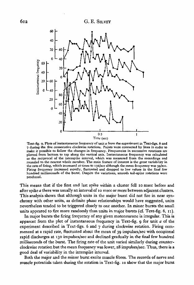
Text-fig. 9. Plots of instantaneous frequency of unit a from the experiment in Text-figs. 6 and7 during the five consecutive clockwise rotations. Points were connected by lines in order tomake it possible to follow the changes in frequency. Frequencies in successive rotations areplotted from bottom to top along the vertical axis. Instantaneous frequency was calculatedas the reciprocal of the interspike interval, which was measured from the recordings androunded to the nearest whole number. The main feature of interest is the great variability inthe rate of firing, which increased at times to 130/sec although the mean frequency was 30,/sec.Firing frequency increased rapidly, fluctuated and dropped to low values in the final fewhundred milliseconds of the burst. Despite the variations, smooth tail-spine rotations wereproduced.
This means that if the first and last spike within a cluster fell 10 msec before andafter spike a there was usually an interval of 10 msec or more between adjacent clusters.This analysis shows that although units in the major burst did not fire in near syn-chrony with other units, as definite phase relationships would have suggested, unitsnevertheless tended to be triggered closely to one another. In minor bursts the smallunits appeared to fire more randomly than units in major bursts (cf. Text-fig. 6, 11).
In major bursts the firing frequency of any given motoneurone is irregular. This isapparent from the plot of instantaneous frequency in Text-fig. 9 for unit a of theexperiment described in Text-figs. 6 and 7 during clockwise rotation. Firing com-menced at a rapid rate, fluctuated about the mean of 39 impulses/sec with occasionalrapid discharges at 130 impulses/sec and declined gradually in the final few hundredmilliseconds of the burst. The firing rate of the unit varied similarly during counter-clockwise rotation but the mean frequency was lower, 28 impulses/sec. Thus, there is agood deal of variability in the interspike interval.
Both the major and the minor burst excite muscle fibres. The records of nerve andmuscle potentials taken during the rotation in Text-fig. 10 show that the major burst

Tex
t-fi
g. 1
0. C
orre
lati
on o
f th
e fi
ring
of
neur
ones
in
the
maj
or a
nd m
inor
bur
sts
wit
h m
uscl
e po
tent
ials
dur
ing
thre
e cl
ockw
ise
rota
tion
s of
the
tail
spi
ne.
3
Th
e ex
trac
ellu
lar
reco
rdin
gs w
ere
mad
e w
ith
suct
ion
elec
trod
es, o
ne
atta
ched
to
a ne
rve
bran
ch (
top
trac
e) a
nd t
he o
ther
to
one
or
mor
e m
uscl
e fi
bres
2
(bot
tom
tra
ce)
near
the
ins
erti
on o
f th
e ri
ght
dors
al l
ater
al m
uscl
e. D
urin
g th
e m
ajor
bur
st la
rge
mus
cle
pote
ntia
ls o
ccur
red;
dur
ing
the
min
or b
urst
sm
all
mus
cle
pote
ntia
ls o
ccur
red.
Th
ese
pote
ntia
ls i
ndic
ate
the
exci
tato
ry n
atur
e of
th
e ne
uron
es i
n b
oth
burs
ts.
Th
e la
rge
mus
cle
pote
ntia
ls o
bscu
re th
e ac
tivi
ty
t-3
of t
he
maj
or b
urst
in
the
nerv
e tr
ace.
Tim
e: 2
00
mse
c.
8. Y
0,
Ei

Rig
ht D
LM
CW
5
--
--
.- -
-- --
--
-
.. ..
Y
I
F
Tex
t-fi
g. I
I.
Fir
ing
in t
he
sam
e ne
rve
du
rin
g o
ppos
ite
rota
tion
s w
hile
tens
ion
was
rec
orde
d fr
om t
he
rig
ht
dors
al l
ater
al m
uscl
e o
n t
he
seco
nd
F tr
ace.
Th
e p
eak
of t
ensi
on o
ccur
red
at t
he e
nd o
f th
e m
ajor
bu
rst
as in
dica
ted
by
the
ces
sati
on i
n fi
ring
of
the
larg
est
spik
e. T
ensi
on p
ersi
sted
$
4
for
a ti
me
as t
he
min
or b
urs
t be
gan.
Ten
sion
the
n de
clin
ed a
long
wit
h th
e m
inor
bur
st.
Th
e r
ate
of f
all
in t
ensi
on w
as a
ppro
xim
atel
y th
e sa
me
us t
he r
ate
of r
ise.
Not
e al
so th
e gr
eate
r du
rati
on o
f fi
ring
du
rin
g c
ount
ercl
ockw
ise
rota
tion
whe
n th
e ri
ght
dors
al l
ater
al m
uscl
e li
fted
th
e ta
il
spin
e up
war
d o
n t
he
righ
t si
de.
Not
e al
so t
he
chan
ge i
n or
der
of a
ppea
ranc
e of
lar
ge-s
ized
and
int
erm
edia
te-s
ized
spi
kes
at t
he
begi
nnin
g an
d
end
of
the
oppo
site
rot
atio
ns.
Th
is i
s an
othe
r ex
ampl
e of
cha
nged
fir
ing
sequ
ence
s am
ong
mot
oneu
rone
s to
mus
cle
fibr
es w
ithi
n a
mus
cle
as
seen
in T
ext-
fig.
7 a
nd
Tab
le I.
Th
ird
trac
e is
a b
ase
line
. Tim
e: zoo m
sec.
Ten
sion
: IOO g
.

Control of Limulus tail spine rotation 618
Table 2. Burst duration and frequency of the largest spikes in each of the nerves innervatingthe contralateral muscles of the animal of Text-fig. 4 during three clockwise and threecounterclockwise rotations
Clockwise Counterclockwise
Right dorsal lateralmuscle
Totals
Left dorsal lateralmuscle
Totals
Duration(msec)
560640880
2080
720880
11202720
No. offirings
141714
45
243236
92
Meanfrequency
———
22/sec———
34/sec
Duration(msec)
760640760
2160
840640840
2320
No. offirings
23253°78
24
2424
72
Meanfrequency
———
36/sec
———
31/sec
produced large and numerous muscle potentials and that the minor burst producedsmall and fewer muscle potentials.
The major burst occurs during the development of tension and the minor burstduring the decline of tension. This relationship of bursts to tension is shown by therecords in Text-fig. 11 and the graph in Text-fig. 12. In the experiment nervous out-put to the right dorsal lateral muscle and tension from the muscle were recordedtogether while the animal rotated its tail spine. The major burst excited the muscle torotate the tail spine to the same angle as that to which contraction of only that musclewould have brought the tail spine (Text-fig. 2). At the point of maximum tension themajor burst ceased and the minor burst began. Tension persisted for about 200 msecafter cessation of the major burst then declined as firing of the smaller units built up,peaked and then also declined. Tension decreased to zero and the minor burst ceased.This occurred at a point about 1800 of tail-spine rotation from the point wheremaximum tension occurred and the minor burst began. The next major burst beganat the end of the minor burst and tension rose once again. Thus, the major burst leadsto development of tension and to rotation of the tail spine through a segment of thepath of rotation. This burst, regardless of the direction of rotation, shuts off at thepoint to which the muscle it excites would draw the tail spine if the muscle contractedby itself (compare action of right DLM in Text-fig. 2 to (that in) Text-fig. 12). Theminor burst acts during extension of the muscle and presumably impedes lengthening,because of the excitatory nature of the small units.
The nervous output to a given muscle varies in duration and frequency dependingon the side of the body through which the tail spine moves. In general the duration(Text-figs. 11, 12), frequency (Text-fig. 7) or both (Text-fig. 4, Table 2) were greaterwhen the muscle lifted the tail spine through its own side rather than when the musclecarried the tail spine down through the same side as the muscle. For example, Text-fig. 4 and Table 2 show that the output to the right dorsal lateral muscle was greaterboth in duration frequency during counterclockwise rotations when the tail spine wasbrought up on the right side of the body than during clockwise rotation when the tailspine was brought up on the left side and across the body. The duration and fre-quency of output were greater to the left dorsal lateral muscle during clockwiserotation than during counterclockwise rotation. In water the weight of the tail spine

6i6 G. E. SlLVEY
CCW i CW
30
20
.S 10
o
8
3c SI
O
c
0200150100
500
6 9 12Time (sec)
15 18
Text-fig. 12. Graph of three clockwise and three counterclockwise rotations from the experi-ment in Text-fig. 11. The largest spike in the major burst and all spikes in the minor burst werecounted in 200 msec intervals. Tension was measured at the end of each interval. Both countsof spikes and measurements of tension were plotted against time. The main feature of interestis the correspondence of the minor burst with the period of relaxation when the muscleextended. This indicates that extension is actively resisted.
would appear to be negligible in itself because of the buoyancy of the body of Limulus.Yet when friction of the water is considered movement of the tail spine may developa sufficient load to evoke the observed compensation. The reciprocal results betweencontralateral muscles (Text-fig. 4; Table 2), which were treated similarly, indicatethat this phenomenon was not due to operative, recording and mechanical factors.The increased duration and frequency of firing to the muscle pulling the tail spineupward suggests that the tail-spine control system was compensating for load.
Neuromuscular properties
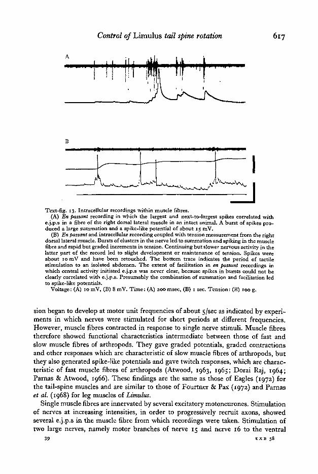
Responses of muscle fibres and patterns of innervation were studied by means ofintracellular recordings in order to determine the effect of motor output upon themuscles. Membrane potentials averaged 50+12 (S.D., N = 233) mV. Motoneuroneactivity, evoked centrally by tactile stimulation of the surface and spines of theabdomen, produced excitatory junctional potentials (e.j.p.s) no greater than 5 mV(Text-fig. 13). Junctional potentials summed and facilitated. During bursts of nervousactivity, when the units innervating a muscle fibre fired together rapidly, spike-likepotentials of 10—20 mV arose. Presumably these spikes initiated stronger contractionsthan junctional potentials because bursts in the nerve accompanied by spikes in themuscle fibre preceded large increments in tension (Text-fig. 13b). Tension increasedin a graded manner in response to centrally initiated firing (Text-figs. 11, 136). Ten-
Control of Limulus tail spine rotation 617
Text-fig. 13. Intracellular recordings within muscle fibres.(A) En passant recording in which the largest and next-to-largest spikes correlated with
e.j.p.s in a fibre of the right dorsal lateral muscle in an intact animal. A burst of spikes pro-duced a large summation and a spike-like potential of about 15 mV.
(B) En passant and intracellular recording coupled with tension measurement from the rightdorsal lateral muscle. Bursts of clusters in the nerve led to summation and spiking in the musclefibre and rapid but graded increments in tension. Continuing but slower nervous activity in thelatter part of the record led to slight development or maintenance of tension. Spikes wereabout 10 mV and have been retouched. The bottom trace indicates the period of tactilestimulation to an isolated abdomen. The extent of facilitation in en passant recordings inwhich central activity initiated e.j.p.s was never clear, because spikes in bursts could not beclearly correlated with e.j.p.s. Presumably the combination of summation and facilitation ledto spike-like potentials.
Voltage: (A) 10 mV, (B) 8 mV. Time: (A) 200 msec, (B) 1 sec. Tension: (B) 100 g.
sion began to develop at motor unit frequencies of about 5/sec as indicated by experi-ments in which nerves were stimulated for short periods at different frequencies.However, muscle fibres contracted in response to single nerve stimuli. Muscle fibrestherefore showed functional characteristics intermediate between those of fast andslow muscle fibres of arthropods. They gave graded potentials, graded contractionsand other responses which are characteristic of slow muscle fibres of arthropods, butthey also generated spike-like potentials and gave twitch responses, which are charac-teristic of fast muscle fibres of arthropods (Atwood, 1963, 1965; Dorai Raj, 1964;Parnas & Atwood, 1966). These findings are the same as those of Eagles (1972) forthe tail-spine muscles and are similar to those of Fourtner & Pax (1972) and Parnaset al. (1968) for leg muscles of Limulus.
Single muscle fibres are innervated by several excitatory motoneurones. Stimulationof nerves at increasing intensities, in order to progressively recruit axons, showedseveral e.j.p.s in the muscle fibre from which recordings were taken. Stimulation oftwo large nerves, namely motor branches of nerve 15 and nerve 16 to the ventral
39 EXB 58
618 G. E. SILVEY
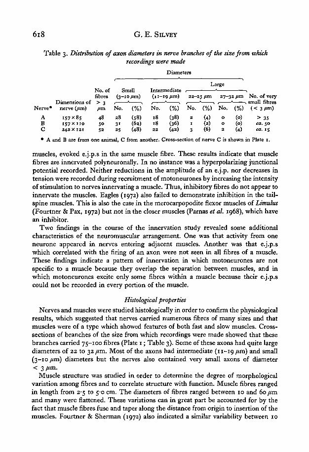
Table 3. Distribution of axon diameters in nerve branches of the size from whichrecordings were made
Diameters
LargeNo. of Small Intermediatefibres (3-10 /an) (11-19/ tm) 2 2 - 2 5 / a n 2 7 - 3 2 / o n N o . of very
Dimensions of > 3 , * , 1 * > < * > 1 * > small fibresNerve* nerve Qim) /tm N o . (%) N o . (%) No . (%) N o . (%) ( < 3 / « n )
A 157x85 48 28 (58) 18 (38) 2 (4) o (o) > 35B 157x110 50 31 (62) 18 (36) 1 (2) o (o) ea. 50C 242x121 52 25 (48) 22 (42) 3 (6) 2 (4) ca. 15
• A and B are from one animal, C from another. Cross-section of nerve C is shown in Plate 1.
muscles, evoked e.j.p.s in the same muscle fibre. These results indicate that musclefibres are innervated polyneuronally. In no instance was a hyperpolarizing junctionalpotential recorded. Neither reductions in the amplitude of an e.j.p. nor decreases intension were recorded during recruitment of motoneurones by increasing the intensityof stimulation to nerves innervating a muscle. Thus, inhibitory fibres do not appear toinnervate the muscles. Eagles (1972) also failed to demonstrate inhibition in the tail-spine muscles. This is also the case in the merocarpopodite flexor muscles of Limulus(Fourtner & Pax, 1972) but not in the closer muscles (Parnas et al. 1968), which havean inhibitor.
Two findings in the course of the innervation study revealed some additionalcharacteristics of the neuromuscular arrangement. One was that activity from oneneurone appeared in nerves entering adjacent muscles. Another was that e.j.p.swhich correlated with the firing of an axon were not seen in all fibres of a muscle.These findings indicate a pattern of innervation in which motoneurones are notspecific to a muscle because they overlap the separation between muscles, and inwhich motoneurones excite only some fibres within a muscle because their e.j.p.scould not be recorded in every portion of the muscle.
Histological properties
Nerves and muscles were studied histologically in order to confirm the physiologicalresults, which suggested that nerves carried numerous fibres of many sizes and thatmuscles were of a type which showed features of both fast and slow muscles. Cross-sections of branches of the size from which recordings were made showed that thesebranches carried 75-100 fibres (Plate 1; Table 3). Some of these axons had quite largediameters of 22 to 32 /an. Most of the axons had intermediate (11-19 jum) and small(3-10 /im) diameters but the nerves also contained very small axons of diameter< 3 /im.
Muscle structure was studied in order to determine the degree of morphologicalvariation among fibres and to correlate structure with function. Muscle fibres rangedin length from 2-5 to 5-0 cm. The diameters of fibres ranged between 10 and 60 /anand many were flattened. These variations can in great part be accounted for by thefact that muscle fibres fuse and taper along the distance from origin to insertion of themuscles. Fourtner & Sherman (1972) also indicated a similar variability between 10

Control of Limulus tail spine rotation 619
ind 60 fim in the range of diameters of leg-muscle fibres. The average sarcomerelength was 6-5 ± o-8 (S.D., N = 63) fim with a range in lengths between 4-9 and 8-4 /an.This indicates that the fibres are very similar. Similar uniform sarcomere lengthshave been found in other Limulus muscles (Eastwood, 1971; Fourtner & Sherman,1972). A mean sarcomere length of 6-5 fim indicates that Limulus muscle fibres are ofan intermediate type, because arthropod fast muscles typically have short sarcomerelengths of < 6 fim with a range between 2 and 6 fim; arthropod slow muscles havesarcomere lengths of > 6/im (Atwood, 1963, 1965; Doraj Raj, 1964; Kennedy &Takeda, 1965 a; Parnas & Atwood, 1966; Gilai & Parnas, 1970; Jahromi & Atwood,1969, 1971). In general, the histological properties of the tail-spine muscles allowthem to be described as an intermediate type and provide a structural correlation tothe observed intermediate neuromuscular properties.
DISCUSSION
The sequential firing pattern
Motor control of the tail spine in Limulus polyphemus produces rotation in oppositedirections by exciting muscle fibres in opposite sequences. Muscle fibres or groups offibres are activated one after the other in a clockwise or a counterclockwise sequence(Fig. 5), either of which can be driven continuously to generate several uninterruptedrotations. Presumably the changes observed in the order of recruitment of moto-neurones in a nerve (Fig. 7; Table 1) are responsible for the changed firing order infibres of a muscle. The firing sequences of the motoneurones during opposite rotationsare not mirror images, because the time of firing of a motoneurone changes dis-proportionately relative to the start and end of firing of other motoneurones (Table 1).
Functionally, the sequential firing observed in the time of activation of musclefibres within the same muscle is quite significant, because it means that fibres orgroups of fibres within a muscle contract separately rather than the whole muscle atonce. This further means that sequentially activated muscle fibres within a discretemuscle bundle sequentially exert tension through the several separate tendons bywhich the bundle inserts. This action would seem to contribute to a smoother rota-tion than if all the fibres in a muscle contracted at once.
Conceptually, the findings on sequential firing indicate that the functional con-tractile entity is not an entire muscle. Sequential firing shows that nerve and musclefibres function independently of muscle boundaries. The findings that single moto-neurones innervate portions of a muscle and not the whole muscle, that some nervefibres innervate muscle fibres in adjacent muscles and that some muscle fibres origi-nate with one muscle but insert with another show that the nerve and muscle fibresare arranged independently of muscle boundaries. Thus populations of fibres within amuscle appear to be the functional contractile entities. This finding extends theconcept that a muscle - that is, a discrete bundle of muscle fibres - is not necessarilythe functional entity. Davis (1968) pointed this out for the lobster swimmeret muscles,and Burrows' work (1967) on the eyecup muscles of the crab implies this but for adifferent reason than in the Limulus tail-spine system. In their systems two or moremuscles have the same innervation and these muscles act as the functional contractileentity. In Limulus, the functional muscle appears to be different sections of the same
39-2

620 G. E. SILVEY
muscle or of adjacent muscle bundles. A functional muscle unit in Limulus may1
possibly be the fibres which insert by a single tendon. The tail spine in this sense ismoved by a cone of muscle which differentiates movement through the separate con-traction of its muscle units. Rotation is effected by muscle units pulling consecutivelyupon the more than a few dozen tendons.
These findings have certain comparative and functional implications. Other motorsystems of arthropods show sequential firing patterns. Sequential firing occurs inantagonistic muscles in systems such as flight in insects (Wilson, 1961; Kammer,1968), righting in lobsters (Davis, 1968), swimming in crayfish (Schrameck, 1970),ventilation in dragonfly larvae (Mill, 1970), gill-plate beating in Limulus (Fourtner,Drews & Pax, 1971) and walking in the cockroach (Pearson & lies, 1970; Pearson,1972). In these systems one muscle or one group of muscles fires alternately withanother. In some of these systems a delay occurs between alternate bursts so thatcontractions in the sequence do not overlap. This delay is seen between the cockroachcoxal depressor and elevator muscles during walking (Pearson & lies, 1970). Non-overlapping of contraction is enhanced by peripheral inhibitors, which increase therate of muscle relaxation (lies & Pearson, 1971). Furthermore, in these systems move-ment typically does not occur in two directions as in the tail-spine system, althoughreverse movement occasionally takes place in walking systems (observations on crayfishand crabs in this laboratory; Davis & Ayers, 1972). Sequential firing is seen among theeleven muscles which sweep and pivot the scaphognathite or gill bailer in the branchialchamber of the crayfish in order to bring in water, move it across the gill and force itback out (Pasztor, 1968). The muscles fire one after the other to achieve movement,but this movement occurs in one direction and it is not continuous, because there is ashort period of inactivity between beats of the bailer. Thus, there is a single motorprogram that generates sequential but not uninterrupted firing to the muscles of thebailer. Sequential firing occurs among muscles in the crab eyecup motor system, whichpromotes optokinetic nystagmus in two directions (Burrows & Horridge, 1968 a). Notall the muscles, however, are active during any given fast or slow phase of a nystagmus,and opposite nystagmus movements require different muscles. Thus, only somemuscles in a given nystagmus fire one after the other. This means that there are twocentral programs which drive different muscles in two quite dissimilar sequences.Sequential firing among several muscles and requiring two motor programs probablyoccurs in the crayfish uropod motor system, because the uropods move in oppositedirections (Larimer & Kennedy, 1969 a, b) and presumably do so through the co-operation of their several muscles. Thus, among the arthropods rhythmic movementis produced by sequential activation of two or more muscles or groups of muscles.Tail-spine rotation in Limulus is similarly produced by sequential firing but differsin the number of functional contractile units activated. Many functional contractileentities firing consecutively produce rotations in such a way that the periods ofactivity in adjacent entities overlap.
Function of the minor burst and the significance of extension
The small excitatory units of the minor burst appear to contribute to rotationalmovement of the tail spine by maintaining a degree of tension in muscles as they arepassively extended. Activity in these units may affect both deflexion and rotation of

Control of Limulus tail spine rotation 621
the tail spine. The effect of deflexion would be to keep the tail spine at a desiredangle. Without some degree of tension in muscles other than those that are contractingthe tail spine might not be rotated steadily nor be held firmly at angles between restingand maximum deflexion, because support is needed in all directions about the base ofthe tail spine. Tension maintained in muscles being extended would provide thisstabilization, in much the same way as low-level firing in some antagonistic muscles ofthe leg of salamanders provides stabilization during walking (Szekely, Czeh & Voros,1969). Tension maintained to provide stability is also seen in some crab eyecupmuscles, which undergo no detectable change in impulse frequency and presumablycontribute to eyecup movement by resisting lengthening while other muscles changethe position of the eyecup (Burrows & Horridge, 1968 b). This function of maintainingtension in lengthening muscles seems to be necessary in other systems. In walkingcockroaches the coxal depressor muscle is inhibited during the contraction of itsantagonist, the coxal elevator (Pearson & lies, 1970; lies & Pearson, 1971).
The effect of the small excitatory units on rotation is based on the following rela-tionships. Tension reaches a maximum at the end of the major burst (Text-figs. 11,12).At this point, firing of the small units begins and is maximal. Activity in the smallunits and tension decline simultaneously until the tail spine is rotated about 1800 fromthe point of maximum tension (see sketches of the tail spine position and plots oftension and firing frequency in Text-fig. 12). Thus, during rotation a major burstcauses a muscle to draw the tail spine toward the position attained during activecontraction of the muscle and away from the position to which the previously con-tracting muscle brought it. The minor burst tends to keep the tail spine at the positionattained during active contraction of a muscle. Without the minor burst and the ten-sion it presumably maintains in extending muscles the tail spine might jerk as itmoves in rotation and fail to move in its normal smooth pattern. Moreover, it mighttend to fall toward the resting position rather than continue around the outer circum-ference of the circle which the tip of the rotating spine describes. The small units,therefore, possibly provide tension to extending muscles in order to maintain outwarddeflection and smooth rotation of the tail spine.
A similar role of excitatory activity during extension of muscles in another rotatingsystem is seen in the vertebrate eye. In the vertebrate eye continuous but decliningactivity is seen in extending eye muscles. For example, slow nerve fibres to the lateralrectus muscle fire at decreasing rates but never cease completely during extension ofthe lateral rectus in the slow lateral-to-medial tracking phase of nystagmus (Yamanka& Bach-y-Rita, 1968). During naso-temporal movements of the eye the intensity offiring in the lateral and medial recti muscles fluctuates reciprocally. Firing increasesin one muscle concomitantly with decreasing, but not terminating, firing in theantagonist (Bjork & Kugelberg, 1963). Boeder (1962) has pointed out that activemodulation of muscle extension is just as important from a mechanical point of viewas contraction. Without the resistance contributed by active muscles during extensionsome component of abduction or adduction, elevation or depression of the eye wouldbe lacking and the eye would deviate from its normal path of movement.

622 G. E. SlLVEY
Expression of the firing pattern
The firing pattern causes the greatest effect in this motor system which lacks greatneuromuscular and structural diversity by exciting small to large axons in clusters.Clusters evoke the strongest contractions in the muscles. Most spikes in the clusterfire within ± 10 msec of a reference unit and produce summed and facilitated e.j.p.sand frequently spike-like potentials in the muscle fibre (Text-fig. 13). A similarphenomenon of closely spaced firing affecting muscles more strongly than random orunspaced firing is seen in the motor patterns to some crustacean muscles. For example,the largest axon innervating the superficial flexors of the crayfish abdomen (Gillary& Kennedy, 1969a, b) fires in a bursting pattern. This neurone evokes small e.j.p.sthat undergo facilitation when the neurone fires in its normal bursting mode of two tofour occurrences per burst. Stronger contractions result from firing in bursts thanwhen the axon is driven at a constant rate for a number of firings equal to the total inthe bursts. Since this axon innervates more than 60% of the superficial flexors(Kennedy & Takeda, 19656), strong contractions of these muscles are produced by theactivity of this axon (Larimer & Eggleston, 1971). The effectiveness of closely spacedfiring is also seen in the paired firing patterns of motoneurones that innervate thecrayfish carpopodite extensor (Atwood & Wiersma, 1967), crayfish opener (Wilson &Davis, 1965) and some oculomotor muscles of the crab (Burrows & Horridge, 19686).The interspike interval between the pairs ranges between 5 and 13 msec and thiscorresponds to the most effective interval for increasing tension in muscle (Ripley &Wiersma, 1953). This interval is of the same order of magnitude as the intervalbetween some of the spikes in a cluster of the Limulus tail-spine system. Since some ofthe units in a cluster innervate the same muscle fibre (Text-fig. 13 a), the effect couldbe similar to the paired or burst firing in crustacean axons. That is, closely spaced firingamong the clustered units develops tension more effectively than if the units firedwholly at random.
In Limulus changes in the firing frequency of clusters and the duration of the firingperiod lead to variations in the rate and strength of movement of the tail spine. Suchvariations in frequency and duration of firing occurred (Text-figs. 2, 4, 5, 7, 11, 12;Table 2) when a muscle, in particular the dorsal lateral muscle, lifted the tail spineupward on the same side of the body rather than when the muscle brought the tailspine downward on the same side of the body. Changes in the firing frequency andduration of clusters suggest that these variations in the firing pattern are a motormechanism that compensates for load.
The firing pattern appears to express itself without peripheral inhibition. Thisabsence seems understandable if the excitatory activity during extension (Text-fig. 10)indicates that the main function of the system is to maintain tension in order toachieve desired tail-spine deflexions and to effect smoothness. Inhibition wouldrelease tension, which would interfere with stable movement of the tail spine andcause instability. Two other systems, which employ a sequential activation of muscles,namely those of the gill bailer (Pasztor, 1968) and the eye of the crab (Burrows &Horridge, 1968 a) also lack peripheral inhibition. These systems require a high degreeof co-ordination among muscles, which in the scaphognathite system fire in sequenceduring rhythmic beating. Inhibition appears in systems which tend to function with

Control of Limulus tail spine rotation 623
Igreater reciprocity between muscles than do these systems, for example, in lobsterswimmeret beating (Davis, 1968), cockroach walking (lies & Pearson, 1971) andcrayfish tail positioning (Page & Sokolove, 1972). However, this does not seem to be anabsolute generalization, because tonic muscles in the crayfish uropods, which wouldappear to require cooperative interaction of muscles in order to perform the variety ofmovements of which they are capable, have peripheral inhibitors (Larimer & Kennedy,1969 a, b).
Central organization of the motoneurones
The results of the experiments on motor output give some insight into the centralorganization of the motoneurones. The findings that motoneurones tend to fire inclusters suggests a common input which triggers several neurones at about the sametime. The rinding that individual motoneurones fire in different sequences in oppositerotations suggests that each motoneurone also receives a specific input. Thus, eachmotoneurone may be driven by both a common excitor and a specific excitor. Theinteraction of the two leads to a suitable duration, sequence and frequency of units inthe firing pattern for effecting rotations of the tail spine. Furthermore, since themotoneurones fire in inverse (although not mirror-image) patterns during oppositerotations, interactions appear to take place in the terminal fused ganglia 14—16 of theventral cord, because the nerves that innervate the muscles arise from these gangliaand because rotation continues to occur, at least for a short time, following severanceof the cord as far posterior as the 13th ganglion. Exploration of the centralmechanisms within these ganglia in quest of understanding the organization of themotoneurones, the origin of the motor pattern, sequencing and the motor programswill be interesting.
SUMMARY
1. Limulus polyphemns (L.), the horseshoe crab, rotates its tail spine in order to righttself and to keep itself balanced.
2. Eight muscles, discrete bundles of muscle fibres, move the tail spine. Fibres ofthe muscles contract in sequence and thereby pull consecutively on the severaltendons of each muscle in order to rotate the tail spine in either a clockwise or counter-clockwise direction.
3. Motoneurones in nerves to different muscles, fibres within a muscle and units ina nerve to a single muscle fire in different sequences during clockwise and counter-clockwise rotation of the tail spine.
4. The firing pattern consists of a major burst of small to large neurons whichfire in clusters, and a minor burst of small neurones which appear to fire randomly.Motoneurones in both bursts are excitatory. The major burst develops tension, theminor burst acts during extension of the muscle and presumably impedes relaxationin order to produce stable deflexion and smooth rotation of the tail spine.
5. Muscle fibres respond to motor output with small excitatory junctional potentialsof < 5 mV. E.j.p.s sum and show facilitation, and in some cases develop spike-likepotentials of 10-20 mV. Both spiking and the greatest increase in tension occur duringthe clustered firings in major bursts.
6. Muscle fibres have sarcomere lengths of 6-5 ± o-8 /tm and diameters of 10-60 ftm.

624 G. E. SILVEY
Nerve fibres range from less than 3 to 32 /tm in diameter in large nerve brancheswhich contain between 50 and 100 fibres.
7. These findings indicate that two different motor programs evoke contraction ofmuscle fibres in opposite sequences. Sequential contraction of fibres within a musclemeans that muscle fibres which are activated together, rather than whole muscles, arethe functional contractile entities.
I am grateful to Dr William H. Evoy under whose direction and in whose laboratorythis study was made. I also acknowledge help in a variety of forms from the studentsand staff of LQB. The study is the substance of a dissertation submitted for thedoctorate of philosophy at the University of Miami. Financial support was provided byNIH training grant HD00187 and facilities in part by NSF grant GB8447 to WilliamH. Evoy.
REFERENCES
ATWOOD, H. L. (1963). Differences in muscle fibre properties as a factor in ' fast' and' slow' contractionsin Carcinus. Comp. Biochem. Physiol. to, 17—32.
ATWOOD, H. L. (1965). Excitation and inhibition in crab muscle fibers. Comp. Biochem. Physiol. 16,409-26.
ATWOOD, H. L. (1967). Crustacean neuromuscular mechanisms. Am. Zool. 7, 527-52.ATWOOD, H. L. & WIERSMA, C. A. G. (1967). Command interneurons in the crayfish central nervous
system. J. exp. Biol. 46, 249-61.BJORK, A. & KUGELBERG, E. (1963). The electrical activity of the muscles of the eye and eyelids in
various positions and during movement. Electroenceph. Clin. Neurophysiol. 5, 595-602.BOEDER, P. (1962). Cooperative action of extraocular muscles. Br. J. Ophthal. 46, 397-403.BURROWS, M. (1967). Reflex withdrawal of the eyecup in the crab Carcinus. Nature, Lond. 215, 56-7.BURROWS, M. & HORRIDGE, G. A. (1968 a). The action of the eyecup muscles of the crab, Carcinus,
during optokinetic movements. J. exp. Biol. 49, 223—50.BURROWS, M. & HORRIDGE, G. A. (19686). Motoneurone discharges to the eyecup muscles of the crab
Carcinus. J. exp. Biol. 49, 251-67.DAVIS, W. J. (1968). The neuromuscular basis of lobster swimmeret beating. J. exp. Zool. 168, 363-78.DAVIS, W. J. & AYERS, J. L., JR. (1972). Locomotion: control by positive-feedback optokinetic responses.
Science, N.Y. 177, 183-5.DORAI RAJ, B. S. (1964). Diversity of crab muscle fibers innervated by a single motor axon. J. Cell.
Comp. Physiol. 64, 41-54.EAGLES, D. (1972). Sensory and motor aspects of the control of tail spine movement in Limulus poly-
phemus. Ph.D. Thesis, University of Massachusetts.EASTWOOD, A. B. (1971). The comparative physiology of muscles in the walking legs of Limulus poly-
phemus. Ph.D. Thesis, Lehigh University.FOURTNER, C. R., DREWES, C. D. & PAX, R. A. (1971). Rhythmic motor outputs co-ordinating the
respiratory movement of the gill plates of Limulus polyphemus. Comp. Biochem. Physiol. 38 A, 751-62.FOURTNER, C. R. & PAX, R. A. (1972). Chelicerate neuromuscular systems: the distal flexor of the mero-
carpopodite of Limulus polyphemus (L.). Comp. Biochem. Physiol. 41 A, 617—27.FOURTNER, C. R. & SHERMAN, R. G. (1972). Alight and electron microscopic examination of muscles in
the walking legs of the horseshoe crab, Limulus polyphemus (L.). Can. J. Zool. (in the Press).GILAI, A. & PARNAS, I. (1970). Neuromuscular physiology of the closer muscles in the pedipalp of the
scorpion Leiurus quinquestriatus. J. exp. Biol. 52, 325—44.GILLARY, H. L. & KENNEDY, D. (1969a). Pattern generation in a crustacean motoneuron. J. Neuro-
physiol. 32, 595-606.GILLARY, H. L. & KENNEDY, D. (19696). Neuromuscular effects of impulse pattern in a crustacean
motoneuron. J. Neurophysiol. 32, 607-12.HORRIDGE, G. A. (1968). Interneurons, p. 397. San Francisco: W. H. Freeman.ILES, J. F. & PEARSON, K. G. (1971). Coxal depressor muscles of the cockroach and the role of peri-
pheral inhibition. ,7. exp. Biol. 55, 151-64.JAHROMI, S. S. & ATWOOD, H. L. (1969). Correlation of structure, speed of contraction, and total
tension in fast and slow abdominal muscle fibers of the lobster (Homarus americanus). J. exp. Zool.171. 25-38.
JAHROMI, S. S. & ATWOOD, H. L. (1971). Structural and contractile properties of lobster leg-musclefibers. J. exp. Zool. 176, 475-86.


Journal of Experimental Biology, Vol. 58, No. 3 Plate 1
GERALD E. SILVEY (Facing p. 625)

Control of Limulus tail spine rotation 625KAMMER, A. E. (1968). Motor pattern during flight and warm-up in lepidoptera. J. exp. Biol. 48, 89-110.KENNEDY, D. & TAKEDA, K. (1965a). Reflex control of abdominal flexor muscles in the crayfish.
I. The twitch system, j . exp. Biol. 43, 211-27.KENNEDY, D. & TAKEDA, K. (19656). Reflex control of abdominal flexor muscles in the crayfish.
II. The tonic system. J. exp. Biol. 43, 229-46.LARIMER, J. L. & EGGLESTON, A. C. (1971). Motor programs for abdominal positioning in crayfish
Zeit. vergl. Physiol. 74, 388-402.LARIMER, J. L. & KENNEDY, D. (1969a). Innervation patterns of fast and slow muscle in the uropods of
crayfish. J. exp. Biol. 51, 119-33.LARIMER, J. L. & KENNEDY, D. (19696). The central nervous control of complex movements in the
uropods of crayfish. J. exp. Biol. 41, 135-50.MILL, P. J. (1970). Neural patterns associated with ventilatory movements in dragonfly larvae. J. exp.
Biol. 52, 167-75.PAGE, C. J. & SOKOLOVE, P. G. (1972). Crayfish muscle receptor organ: role in regulation of postural
flexion. Science, N. Y. 175, 647-50.PARNAS, I. & ATWOOD, H. L. (1966). Phasic and tonicneuromuscular systems in the abdominal extensor
muscles of the crayfish and rock lobster. Contp. Biochem. Physiol. 18, 701—23.PARNAS, I., ABBOTT, B. C , SHAPIRO, B. & LANG, F. (1968). Neuromuscular system of Limulus leg
closer muscle. Comp. Biochem. Physiol. 26, 467-78.PASZTOR, V. M. (1968). The neurophysiology of respiration in decapod Crustacea. I. The motor system.
Can. J. Zool. 46, 585-96.PATTEN, W. & REDENBAUGH, W. A. (1899). Studies of Limulus polyphemus. II. The nervous system of
Limulus polyphemus, with observations upon the general anatomy. J. Morph. 16, 91—200.PEARSON, K. G. (1972). Central programming and reflex control of walking in the cockroach. J. exp.
Biol. 56, 173-93.PEARSON, K. G. & ILES, J. F. (1970). Discharge patterns of coxal elevator and depressor motoneurones
of the cockroach, Periplaneta americana. J. exp. Biol. 52, 139—65.RICHTER, I.-E. (1964). Bewegungsstudien an Limulus. Verh. dt. zool. Ges. in Miinchen 1963. Zool.
Anz. 27, Suppl., 491-7.RICHTER, I.-E. (1966). Limulus polyphemus (Xiphosura) Undrehen und Eingraben. Film E 1033 der
Enc. Cin., Gottingen, pp. 249-54.RIPLEY, S. H. & WIERSMA, C. A. G. (1953). The effect of spaced stimulation of excitatory and inhibitory
axons of the crayfish. Physiol. Comp. Oecol. 3, 1—17.SCHRAMECK, J. E. (1970). Crayfish swimming: alternating motor output and giant fiber activity.
Science, N. Y. 169, 698-70.SZEKELY, G., CZEH, G. & VSRSS, G. (1969). The activity pattern of limb muscles in freely moving
normal and deafferented newts. Exptl. Brain Res. 9, 53-62.TEALE, E. W. (1957). The oldest migration. Nat. Hist. 66, 364-9.WILSON, D. M. (1961). The central nervous control of flight in a locust. J. exp. Biol. 38, 471-90.WILSON, D. M. & DAVIS, W. J. (1965). Nerve impulse patterns and reflex control in the crayfish claw
motor system. J. exp. Biol. 43, 193-210.YAMANKA, Y. & BACH-Y-RITA, P. (1968). Conduction velocities in the abducens nerve correlated with
vestibular nystagmus in cats. Expl. Neurol. 20, 143-155.
EXPLANATION OF PLATE
Light micrograph of a cross-section of a nerve branch in the dorsal lateral muscle stained withtoluidine blue. The section is from a branch of the size and in the position of a nerve commonly foundand recorded from. Fibres with a variety of diameters are seen. Size distributions of the fibres are givenin Table 3 under nerve C. Scale: 10/an.