Embed Size (px)
Citation preview
Motility and Signaling Systems in Uropathogenic Escherichia coli: the Role of
Chemotaxis and c-di-GMP Production in Colonization of the Urinary Tract
By
Erica L. Raterman
A dissertation submitted in partial fulfillment of
the requirements for the degree of
Doctor of Philosophy
(Microbiology)
at the
UNIVERSITY OF WISCONSIN-MADISON
2012
Date of final oral examination: 06/28/12 The dissertation is approved by the following members of the Final Oral Committee:
Rodney A. Welch, Professor and Chair, Medical Microbiology and Immunology Caitilyn Allen, Professor and Chair, Plant Pathology
Christina M. Hull, Associate Professor, Medical Microbiology and Immunology/Biomolecular Chemistry
Jorge C. Escalante-Semerena, Professor, Bacteriology Edward G. Ruby, Professor, Medical Microbiology and Immunolgy

ii
Motility and Signaling Systems in Uropathogenic Escherichia coli: the Role of
Chemotaxis and c-di-GMP Production in Colonization of the Urinary Tract
Erica Lynn Raterman
Under the supervision of Professor Rodney A. Welch
At the University of Wisconsin-Madison
Urinary tract infections are among the most common infections that occur in young,
healthy adults. The majority of urinary tract infections are caused by uropathogenic
Escherichia coli and likely progress by an ascending manner from the urethra, to the
bladder, and finally to the kidneys. Motility is a known fitness factor that aids in bacterial
survival in the urinary tract, and chemotaxis toward urine may help to efficiently guide
the bacteria up the urinary tract before expulsion by micturition. Chemotaxis capillary
assays using components of urine as ligands showed that a subset of amino acids acts
as the main chemoattractants for uropathogenic E. coli strain CFT073. Loss of the
chemoreceptors did not result in a loss of fitness in the mouse model of urinary tract
infection. However, a true ascending model of urinary tract infection is likely necessary
to conclusively determine whether chemotaxis improves the chance of successful
colonization of the urinary tract. Increased production of the second messenger c-di-
GMP also has an effect on motility by promoting a transition from planktonic, motile cells
to sessile biofilm populations. Deletion of yfiR, a gene that encodes a periplasmic
inhibitor of activity of the diguanylate cyclase YfiN, resulted in attenuation in vivo.

iii
Increased production of curli fimbriae and cellulose and decreased motility as a result
of increased c-di-GMP levels in the yfiR deletion mutant caused attenuation in this
strain. Likely the increased metabolic burden contributed to this reduction in fitness.
Since a plethora of diguanylate cyclases and phosphodiesterases are encoded in the
CFT073 genome, research into other aspects of c-di-GMP-mediated processes might
reveal new fitness and colonization factors that were not previously considered in the
pathogenesis of uropathogenic E. coli.
_______________________
Rodney A. Welch, Ph.D.

iv
Acknowledgements
Many people have supported and encouraged me in the pursuit of my graduate
degree. First and foremost, I would like to thank my thesis advisor, Rod Welch. You’ve
hung in there with me for seven long years and have displayed incredible patience while
I’ve tried to find my way past various setbacks in my thesis project. I thank for your
support during my time in graduate school and during the job search process. I regard
myself as lucky to have joined the Welch lab.
I would like to also thank my thesis committee members: Christina Hull, Ned
Ruby, Jorge Escalante, and Caitilyn Allen. Everyone has shown unwavering support
for me as a young researcher and your insights into and suggestions for the
improvement of my research have proved to be invaluable. I appreciate the interest you
all have shown in the progress of my projects even outside of committee meetings and
your continued support in the pursuit of my future career goals. Thank you all. My
thanks also extend to Dr. Ken Bayles, my academic advisor during my undergraduate
studies at the University of Idaho. He gave me my first chance to work in a research
lab, an experience without which I may not have decided to go (or been accepted to)
graduate school.
Thanks to the Welch lab and its members, both past and present, who have
made life in the lab enjoyable. Coming into work each day was made easier by this
great group of people, especially during the stretches when my research was not going
well. Special thanks go to Shai Pellett, our former lab manager, who made sure that the
lab ran smoothly and who maintained a sincere interest in the progression of my project
even after she had retired. If only Rod had convinced you to stay a little longer. Thank

v
you as well to the three undergraduates who worked on my various projects- Jeff
Gelhausen, Dan Shapiro, and Dan Stevens. I greatly appreciate all of your hard work.
My friends and family have also supported me throughout my time in graduate
school. Thanks are due mostly to my parents, who have provided me with
encouragement throughout my life that was “disproportionate to my looks and abilities”,
to quote Tina Fey. I think that they’re more excited about my graduation than I am. If
not for my Dad suggesting that I should try to get an undergraduate research job in a
lab, I might not have made it all the way to graduate school. Good job parents, and,
despite the good times I’ve had in graduate school, I echo their sentiments- thank god
it’s finally over.

vi
Table of Contents
Abstract of Dissertation……………………………………………………...….……...ii-iii
Acknowledgements ………………………………………………………...…....…..…iv-v
Table of Contents……....…………………………………………………...…….….…vi-vii
Index of Figures………………………………………………………………….….......viii-xi
Index of Tables........................................................................................................xii-xiii
Abbreviations...........................................................................................................xiv
Preface....................................................................................................................xv
Chapter 1.................................................................................................................1
Introduction
Chapter 2.................................................................................................................36
Chemoreceptors of Escherichia coli CFT073 play redundant roles in chemotaxis
toward urine and during urinary tract infection
Submitted to PLoS One
Chapter 3...................................................................................................................67
The yfiLRNB locus encodes a diguanylate cyclase and controls curli fimbriae and
cellulose production in Escherichia coli CFT073
In preparation for publication

vii
Chapter 4..................................................................................................................124
Discussion and Future Directions
Appendix A................................................................................................................144
Escherichia coli strain CFT073 displays chemotaxis toward physiological
concentrations of D-serine present in urine
Appendix B.................................................................................................................178
Reflux in the mouse model of urinary tract infection
In preparation for publication
Appendix C.................................................................................................................186
Motility and in vivo phenotypes of Escherichia coli CFT073 gene deletions first
identified in Escherichia coli K-12 motility screens
Appendix D.................................................................................................................199
Atypical Shigella boydii 13 encodes virulence factors seen in attaching and
effacing Escherichia coli
FEMS Microbiology Letters (2012) 328:20-25.

viii
Index of Figures
Chapter 1
Figure 1.1 Schematic of the chemotaxis machinery in Escherichia coli 8
Figure 1.2 Schematic of the downstream pathways affected by c-di-GMP 19
production
Chapter 2
Figure 2.1 Chemotaxis toward human urine 65
Figure 2.2 Phenotypes of chemoreceptor deletion mutants in the mouse 66
model of urinary tract infection
Chapter 3
Figure 3.1 Diagram of the yfiLRNB locus 104
Figure 3.2 Localization of YfiR 105
Figure 3.3 Phenotypes of yfiLRNB gene deletions in the mouse model 106
of urinary tract infection
Figure 3.4 Phenotypes of yfiL and and yfiB gene deletions and yfiR 107
mutant single infection in the mouse model of urinary tract
infection
Figure 3.5 In vitro growth phenotype of the yfiR deletion mutant 108
Figure 3.6 Expression of the yfiLRNB locus 109
Figure 3.7 Motility phenotypes of gene deletions in the yfiLRNB locus 110
Figure 3.8 Overexpression and production of curli fimbriae and cellulose 111

ix
in response to deletion of yfiR
Figure 3.9 Increased pellicle formation in the yfiR deletion mutant 113
Figure 3.10 Phenotypic complementation of the yfiR deletion mutant by 114
expression of yhjH
Figure 3.11 Colony morphologies of yfiR complement and yedQ and 116
adrA deletion mutants and expression of adrA
Figure 3.12 Expression of bcsA and bcsE in the yfiR deletion mutant 117
Figure 3.13 Sensitivity of the yfiR deletion mutant to hydrogen peroxide 118
Figure 3.14 Sensitivity of the yfiR deletion mutant to iron limitation 119
Figure 3.15 Motility phenotypes of ycgR, bcsA, and csgD deletions in the 120
yfiR deltion background
Figure 3.16 Phenotypes of ycgR, bcsA, and csgD deletions in the yfiR 121
deletion background in the mouse model of urinary tract
infection
Figure 3.17 Phenotypes of ycgR, bcsA, and csgD single deletion mutants 122
in the mouse model of urinary tract infection
Figure 3.18 Decreased expression of curli fimbriae and cellulose in the 123
presence of human urine
Appendix A
Figure A.1 Chemotaxis of E. coli CFT073 and MG1655 toward D-serine 169
and L-serine
Figure A.2 Effects of reciprocal cross of tsr in E. coli CFT073 and MG1655 170

x
tsr deletion backgrounds on chemotaxis toward D-serine and
L-serine
Figure A.3 Effect of deletion of dscO on chemotaxis toward D-serine 171
Figure A.4 Purification of DscO 173
Figure A.5 Southern blot analysis of the presence of dscO in the genomes 174
of E. coli isolates
Figure A.6 Expression of dscO 175
Figure A.7 Phenotype of the dscO deltion mutant in the mouse model 177
of urinary tract infection
Appendix B
Figure B.1 Bladder and kidney bacterial counts in different mouse strains 184
immediately following inoculation of live mice
Figure B.2 Bladder and kidneys bacterial counts immediately following 185
inoculation of post-mortem mice or live mice with a surgically
ligated ureter
Appendix C
Figure C.1 Motility phenotypes of mutant strains 196
Figure C.2 Phenotypes of motility mutants in the mouse model of urinary 198
tract infection

xi
Appendix D
Figure D.1 StcE activity of atypical Shigella B13 strains 217
Figure D.2 Invasion and pedestal formation of atypical Shigella B13 218
strains

xii
Index of Tables
Chapter 2
Table 2.1 List of primers 61
Table 2.2 Chemotaxis toward L-amino acids 62
Table 2.3 Chemotaxis toward D-amino acids, caffeine, and glucose 63
Table 2.4 Chemotaxis of single chemoreceptor mutants toward 64
identified attractants
Chapter 3
Table 3.1 List of primers 101
Appendix A
Table A.1 List of primers 166
Table A.2 Prevalence of dscO in E. coli isolates 168
Appendix C
Table C.1 List of primers 193
Table C.2 Swarming and swimming motility scores 195
Appendix D
Table D.1 List of primers 214

xiii
Table D.2 Prevalence of Escherichia coli- and Shigella-specific genes 216
detected by PCR and secreted StcE antigen and C1 cleavage
activity detected by immunoblot

xiv
Abbreviations
c-di-GMP- bis-(3’-5’)-cylcic dimeric guanosine monophosphate
CFU- colony forming unit
DGC- diguanylate cyclase
ExPEC- extraintestinal Escherichia coli
MCP- methyl-accepting chemotaxis protein
OD- optical density
PCR- polymerase chain reaction
PDE- phosphodiesterase
pGpG- 5’-phosphoguanylyl-(3’-5’)-guanosine
RDAR- red, dry and rough
UPEC- uropathogenic Escherichia coli
UTI- urinary tract infection

xv
Preface
This dissertation is organized into four chapters and four appendices. Chapter 1
provides background information that is necessary to understand the following research
chapters. Chapter 2 presents experiments that determined the components of human
urine that can act as chemoattractants for uropathogenic Escherichia coli. Chapter 3
describes analyses undertaken to determine the role of the yfiLRNB locus in c-di-GMP
production and persistence in vivo. Chapter 4 discusses the results contained in this
dissertation and recommends future projects that could extend the current research.
Appendix A specifically analyzes a possible mechanism for how E. coli CFT073 senses
and displays chemotaxis toward D-serine. Appendix B notes problems with reflux in the
current mouse model of urinary tract infection. Appendix C describes the motility and in
vivo phenotypes of a set of mutants that I did not pursued further. Appendix D is a
manuscript that contains some of my early work with atypical Shigella boydii strains and
that was published in FEMS Microbiology Letters (2012) 328:20-25.

1
Chapter 1
Introduction

2
The sections below provide background information necessary to evaluate the
following research chapters in this thesis. An overview of uropathogenic Escherichia
coli and urinary tract infections is given as is information about the current mouse model
of urinary tract infection and the virulence and colonization factors of uropathogenic E.
coli. These sections are followed by a review of chemotaxis in E. coli and of the second
messenger cyclic di-GMP. Finally, the research chapters on chemotaxis and c-di-GMP
signaling are summarized.
Pathogenic Escherichia coli
Escherichia coli is a well-studied gram-negative bacterium and is commonly
found in humans as a normal, nonpathogenic member of the intestinal microbiota.
However, some E.coli strains have acquired virulence factors that shift the lifestyle from
commensal/beneficial to pathogenic. These pathogenic strains of E. coli are divided
into two separate groups that distinguish between strains that cause disease within the
digestive tract (diarrheagenic E. coli) and those that cause disease outside of the
digestive tract (extra-intestinal pathogenic E. coli (ExPEC)). The diarrheagenic E. coli
strains are further divided into five pathotypes- enterohaemorrhagic E. coli (EHEC),
enteropathogenic E. coli (EPEC), enterotoxigenic E. coli (ETEC), enteroinvasisve E. coli
(EIEC), and enteroaggregative E. coli (EAEC)- that are distinguished by both the clinical
symptoms of the disease that they each cause and by the specialized set of virulence
and colonization factors used to infect the host (41). Included in the ExPEC group are
the uropathogenic E. coli (UPEC) strains that cause urinary tract infections. UPEC

3
strain CFT073, isolated from a patient with pyelonephritis (infection of the kidneys), is
one of the most characterized strains in this group.
Urinary tract infections
Urinary tract infections (UTIs) are among the most common infection of humans
and cost more than 1 billion dollars annually for diagnosis and treatment in the United
States alone (47, 66). Although men and children are able to contract UTIs, women
comprise the majority of UTI patients with about 40-50% of women experiencing a UTI
at least once in their lifetimes (24, 25, 47). About a third to half of women will have
recurrent UTIs throughout their lifetimes (21, 35). UTIs are believed to be ascending
infections in the vast majority of cases that start when the urethra is contaminated with
bacteria most likely from a fecal origin. After colonization of the urethra, the bacteria
then ascend to the bladder. For the most part, urinary tract infections are
uncomplicated cases of cystitis and remain localized in the bladder for a few days
before clearance from the urinary tract with the help of antibiotic treatment (7, 64).
Occasionally, these bladder infections can spread to the kidneys causing pyelonephritis
that can result in more serious complications such as renal scarring and kidney failure.
From the kidneys the bacteria can then spread to the bloodstream where they can
cause the patient to go into septic shock, potentially ending in death. Uropathogenic E.
coli account for the majority of community-acquired and hospital-acquired UTIs (7).
The bladder is normally considered a sterile environment, although a recent
study suggests that viable but non-culturable organisms may reside continually and

4
asymptomatically in this location (99). Whether they are commensal or pathogenic,
bacteria living in the urinary tract must overcome several hurdles to achieve a
measurable degree of colonization. Ascent of the urinary tract for access to the bladder
and kidneys is hampered by the turbulent and expulsive flow of urine. Once in the
urinary tract, the bacteria face growth inhibition due to limited amounts of free iron.
Attachment to the bladder lumen may also be prevented by sloughing of the
uroepithelial cells along with any bound bacteria. Those bacteria that stimulate an
immune response through signaling by pattern recognition receptors are also
susceptible to killing by antimicrobial peptides and invading neutrophils (17, 42, 62, 97).
Virulence and fitness factors of uropathogenic Escherichia coli
Several factors involved in the adherence of. E. coli to the bladder epithelium and
its persistence in the urinary tract are known. Type I fimbriae and Pap fimbriae,
although not the only fimbriae present in most UPEC genomes, are the two major
fimbriae thought to be responsible for the tight adherence to the umbrella cells of the
bladder and the renal cells of the kidneys, respectively (8). Additionally, iron acquisition
systems such as the yersiniabactin and heme uptake systems are factors necessary for
full fitness in the urinary tract (23). Toxins such as hemolysin and the cytopathic
secreted autotransporter toxin (Sat) are involved in UPEC infection process (26, 62, 83).
Flagella are also apparently needed for maintenance of infection as nonmotile strains
are attenuated within the urinary tract (50, 71, 80, 101). For the most part, the
virulence and fitness factors mentioned above contribute to persistence in the urinary

5
tract but not necessarily to initial colonization. For reasons detailed in the next section,
the current mouse model of UTI is unsuitable for analysis of factors needed for ascent
of the urinary tract and initial colonization.
Mouse model of urinary tract infection and its limitations
In the early 1980s, a murine model of urinary tract infection was developed by
Hagberg et al. that, with some modifications, is still used today to study UTIs. Although
human urinary pathogens are not natural pathogens in mice, this model was originally
chosen because mice express the same glycolipids on the bladder epithelium that are
needed for bacterial attachment in human bladders. Mice are also relatively
inexpensive compared to other lab animals. The model involves transurethral
inoculation of the bladder with a 50 µl inoculum delivered via a small catheter. Due to
the non-native nature of UPEC strains with regards to a mouse as a host, inocula
typically must contain upwards of 107 CFU of bacteria to attain measurable infection
levels and consistent results (27, 33, 34, 37). Notably, some mouse strains such as the
C3H/HeJ line are more susceptible to urinary tract infection than others for reasons that
still remain unclear (32, 87).
While this model has allowed researchers to gain valuable information regarding
the virulence factors necessary for attachment to the urinary tract epithelium as well as
other factors necessary for full fitness within the urinary tract, the model is insufficient to
assess which factors are necessary for initial ascent of the urethra. The inoculum size
of 50 µl is delivered directly to the bladder, bypassing the early colonization events that

6
would take place in the urethra during a natural infection entirely. The lowest
inoculum volume reported in the literature is 10 µl delivered to the urethra (33), although
given the short distance of the female mouse urethra (~1cm), even this small volume
undoubtedly reaches the bladder. Furthermore, as previous reports and Appendix B of
this thesis note, the 50 µl inoculum is sufficiently large enough to cause immediate
reflux into the kidneys upon infection, thus bypassing the steps in the ascent of the
ureters to the kidneys as well. Some labs argue that use of a Harvard pump that
delivers the 50 µl inoculum over 30 seconds eliminates the reflux (50). This assertion is
doubtful since conflicting results about reflux using this apparatus occur in the literature
(37). Despite this disagreement, it remains clear that a true model of ascending urinary
tract infection, from colonization of the urethra to ascent up to the kidneys, would
provide an improved model for analysis of colonization factors.
Chemotaxis in Escherichia coli
Chemotaxis in bacteria is simply defined as the net movement toward an
attractant or away from a repellent. First described by Englemann and Pfeffer in the
late 19th century and later elaborated on by Julius Adler in the 1960s (1, 2), the
chemotaxis field has seen many advances over the past 50 years in our understanding
of how E. coli senses attractants and repellants and how those signals are transmitted
to the flagella to produce movement in the appropriate direction. The core of the
chemotaxis machinery is comprised of integral membrane receptors, termed methyl-
accepting chemotaxis proteins (MCPs) or chemoreceptors, which usually possess a

7
periplasmic ligand binding domain and a cytoplasmic signal transduction domain. E.
coli K-12 has five known chemoreceptors: Tsr, Tar, Trg, Tap, and Aer. Tsr senses
serine and pH, Tar senses aspartate and maltose, Trg senses galactose and ribose,
Tap senses dipeptides, and Aer senses the oxidative state of the cell (4, 58, 59, 95). Of
these five chemoreceptors, E. coli CFT073 only encodes tsr, tar, and aer (51). Upon
binding of a ligand to its concomitant receptor, a conformational change occurs in the
chemoreceptor that inhibits the activity of CheA, a histidine kinase that is docked to the
cytoplasmic portion of the chemoreceptor by CheW. CheA phosphorylates the
cytoplasmic signal transducer CheY which can then bind to the flagellar motor and
reverse the rotation of the flagellar filament, causing the cell to tumble (Fig. 1). When
CheY is unphosphorylated, which can be accomplished through the activity of the CheZ
phosphatase, rotation of the flagella continues in a counter-clockwise fashion and the
cell swims smoothly. In the absence of any attractants or repellants, the cell undergoes
varying periods of smooth swimming and tumbling, depending on the ratio of
phophorylated and unphosphorylated CheY present. When an attractant is present the
number of phosphorylated CheY proteins is decreased, thereby increasing the periods
of smooth swimming and eventually biasing the net movement of the bacterium towards
the attractant. Additional control of the activity of CheA is mediated by CheB, a
methylesterase, and CheR, a methyltransferase, which remove and add methyl groups,
respectively, from glutamate residues in the cytoplasmic portion of the chemoreceptors.
Methylation of the chemoreceptors increases activity of CheA. CheB, like CheY, is also
activated by phosphorylation by active CheA (55, 95).

8
Figure 1: Schematic of the chemotaxis machinery. The membrane spanning
chemoreceptors are shown coupled with the core chemotaxis signaling pathway
components in the presence and absence of attractant (diamond). Each Che protein is
identified by its gene letter and phosphorylated proteins are denoted with attached P-
labeled circles. Demethylated and methylated glutamate residues are shown as open
9
and filled bubbles, respectively. Ultimate results for flagella filament rotation direction
and swimming behavior are indicated. IM= inner membrane
The chemoreceptors are associated in large clusters found at the cell poles and
smaller clusters arranged along the length of the cell. Within these clusters,
chemoreceptor homodimers are arranged in groups of three. Each trimer of
chemoreceptor dimers associates with CheW and CheA to form the signaling complex.
Organization into these complexes allows the cell increased sensitivity to altered
concentrations of ligands since the signal produced by one receptor bound to its
chemoeffector is amplified through allosteric interactions between the other
chemoreceptor dimers. Adaptation, or a return to pre-stimulus CheA activity levels, is
achieved in two ways. CheR methylates the exposed glutamate residues of the
chemoreceptors and increases the activity of CheA. Methylation also alters the
conformation of the chemoreceptor dimer such that ligand affinity for the receptor is
reduced. These changes lead to increased levels of phosphorylated CheY and a return
to the state before attractant binding. Active CheA also activates CheB and results in
removal of the methyl groups from the chemoreceptor, reducing CheA activity and
allowing adaptation to repellants (3, 18, 28, 43, 49, 65, 94).
Chemotaxis in colonization of the host by pathogenic organisms
Various studies show that chemotaxis is used to find preferred niches by a
pathogen in the host organism. In Vibrio cholerae, the chemotaxis system is used to

10
navigate to the crypts of the small intestine where the bacteria increase expression of
its virulence factors (14, 15, 22). Similarly, chemotaxis is needed for Campylobacter
jejuni to colonize the subcellular space of the gastric epithelium before initiation of
invasion of the host cell can occur (53). Aerotaxis is also used by Ralstonium
solaracerum, a tomato plant pathogen, for navigation to the plant roots in soil (103,
104). A cheW mutant in UPEC strain CFT073 was attenuated in the mouse model of
UTI, suggesting that chemotaxis may also be important for fitness in the urinary tract
(52). These examples provide evidence that chemotaxis and motility are widely applied
by pathogens to gain access to sites within the host where infection can be maintained.
Comparison of the regulation of flagellar genes in Escherichia coli strains CFT073
and K-12
Because chemotaxis and flagellar-mediated motility are important for bacterial
fitness in the host in general but can also stimulate the immune response (93), UPEC
have mechanisms in place to carefully control the expression of these systems. In
general, motility in E. coli CFT073 is more tightly controlled in comparison to the lab
strain of E. coli K-12. The master regulator of the flagellar regulon, flhDC, in E. coli K-
12 is expressed at constitutive levels due to an insertion sequence that interrupts the
regulator-binding regions (9). The flhDC promoter in CFT073 does not contain this
insertion sequence and is thus subject to the more stringent control of the ancestral
regulatory region. The flagellar regulon consists of three classes of flagellar genes.
The genes encoding flhDC comprise the class I flagellar genes and their expression is

11
necessary for transcription of class II genes which include fliA. FliA is a sigma factor
that is necessary for transcription of class III flagellar genes, which encode the proteins
for the chemotaxis system and the flagellin subunit FliC. The flhDC promoter responds
to multiple regulatory factors and its transcription can be altered in response to various
environmental stimuli including temperature, pH, and butyrate concentration. The
regulatory factors that act on the flhDC promoter include the nucleoid protein H-NS, the
RscBA response regulator system, the QseBC quorum-sensing system, and the
catabolite repressor protein, CRP (5, 20, 45, 85).
E. coli CFT073 also possesses type I fimbriae whose expression is inversely
regulated with flagella expression. The type I fimbriae operon is controlled by the fimS
switch, an invertible element that is positioned in either the ON or OFF position by
specific DNA recombinases. In the ON position, transcription is initiated off the fim
promoter and procedes through the type I fimbriae genes. While type I fimbriae are
being expressed, expression of the flagellar genes is down-regulated. In the OFF
position, the fim promoter is oriented away from the fim operon and expression of
flagellar genes is activated (12, 44). One other study reported the repression of the
flhDC operon by MatA, an activator of the mat fimbriae operon that is involved in biofilm
formation (54).
As a result of the additional regulation that is absent in many E. coli K-12 strains
due to the aforementioned insertion sequence in the flhDC operon, E. coli CFT073
motility is often repressed, resulting in only a small proportion of the population
expressing flagellar filaments in broth culture. In contrast, most of the population of an

12
E. coli K-12 strain containing the insertion element are flagellated in broth culture
because flhDC is nearly constitutively expressed. Growing the cells in 0.3% swim
plates before transferring them to growth in broth culture can enhance the number of
flagellated cells in the E. coli CFT073 population yet, even with the added manipulation,
elevated flagellar expression can remain difficult to obtain. This problem has
implications for capillary chemotaxis assays that monitor the chemotactic ability of the
bacteria. Because a lower number of bacteria remain flagellated in CFT073 compared
to a K-12 strain, a greater number of bacteria are needed for the assay to work. This
raises the background values in the assay, and standard-error rates are increased
compared to assays performed with K-12 strains. However, reliable, statistically
significant results can still be obtained under these conditions. Additional flagellar
regulation mechanisms may be present that can cause the bacteria to display different
forms of flagellar-based motility, namely swimming and swarming. Different in vitro
conditions stimulate each motility type, and evaluation of the contribution of each
motility phenotype to the infective process in UPEC may provide additional insight into
the ways motility contributes to fitness in vivo.
Swarming and swimming motility
Swarming motility is defined as the coordinated, multicellular movement of a
group of cells across a semisolid surface. Conversely, swimming is performed by
independent planktonic cells and is not dependent on association with other bacteria.
Swarming motility is observed in a variety of bacterial species including E. coli and

13
Salmonella species and swarmer cells are usually differentiated from independent
swimmer cells by an increase in cell length and hyperflagellaton (29, 30). However, E.
coli does not display the elongated cell morphotype that is seen in other bacterial
species. The swarming phenotype is assessed on 0.5% agar plates, although the
percentage of agar used can vary between different bacterial species. Swimming
motility is assessed in 0.3% agar tryptone plates. Non-swarmer cells form colonies that
can spread out by growth of the colony, but swarmer cells display enhanced coverage
of the agar surface and tendrils of swarming bacteria can shoot off from the main colony
on the plate surface. Under the microscope, rafts of coupled bacteria in the outer edge
of the swarming colony move in a swirling pattern that pushes the colony edge forward.
Recent studies show that individual bacteria in the swarming colony edge are
associated by interactions between the flagella of individual bacteria (13, 91).
Swarming motility could be important for colonization of the urinary tract because
groups of cells encased in the surfactants that are excreted by swarming cells could be
more difficult to expel from the urinary tract by the force of flowing urine than would
individual swimmer cells. A recent study in Pseudomonas aeruginosa showed that the
presence of small amounts of mucin, a glycoprotein often found in the mucus overlaying
epithelial surfaces, promoted swarming in in vitro plate assays (105). This finding
strengthens the possibility that swarming may take place in vivo across epithelial cell
layers.

14
Effect of the binding of second messenger c-di-GMP on motility
In addition to the regulatory controls mentioned above, cytoplasmic accumulation
of bis-(33’-5’)-cyclic dimeric guanosine monophosphate (c-di-GMP) can also inhibit both
swimming and swarming motility. Inhibition of motility is accomplished by the interaction
of c-di-GMP with YcgR, a cytoplasmic protein. When c-di-GMP is bound to the PilZ
domain of YcgR, YcgR reverses the rotation of the flagellar motor from clockwise to
counterclockwise by binding to components of the motor-stator complex. Reports
concluded that YcgR interacts directly with FliG and FliM, components of the flagellar
stator, to bias the motor to counterclockwise rotation, which causes smooth swimming
(19, 67). Without the ability to tumble and reverse the direction of swimming, the
bacteria may become stuck in an unfavorable environment.
Second messenger c-di-GMP metabolism and signaling
c-di-GMP is a small, diffusible molecule that is now recognized as a global
second messenger in the same category as cAMP; it primarily controls the transition
between motility and sessility. First identified as an activator of cellulose production in
Gluconacetobacter xylinus (76, 82), studies have since shown that c-di-GMP signaling
is common in diverse bacterial species, many of them pathogens, but is absent in
eukaryotes and archaea (31, 36, 76, 78). C-di-GMP synthesis and metabolism are
primarily regulated by two classes of proteins: diguanylate cyclases (DGCs) that
possess the conserved GGDEF (Gly-Gly-Asp-Glu-Phe) domain responsible for cyclizing
two molecules of GTP into c-di-GMP and phophosdiesterases (PDEs) that posses the

15
EAL (Glu-Ala-Leu) domain responsible for linearizing c-di-GMP to 5′-
phosphoguanylyl-(3'-5')-guanosine (pGpG). GGDEF and EAL domains are often found
in the same protein but can also occur separately (16, 68, 78, 79, 89). Since their
discovery GGDEF and EAL domains have been identified in many species of bacteria,
although their numbers in each individual species can vary widely. For example, E. coli
K-12 apparently encodes 19 GGDEF proteins and 16 EAL proteins and Vibrio cholerae
apparently encodes 31 GGDEF proteins, 22 EAL proteins and 10 GGDEF-EAL proteins;
in contrast, Bacillus subtilis apparently encodes only 4 GGDEF proteins and 3 EAL
proteins (36, 76).
Proteins with either of these domains respond to a diverse set of extra- and
intracellular stimuli including the redox state of the cell, starvation consitions, and the
levels of oxygen, light, antibiotics, and quorum sensing molecules; thus, they often
include domains involved in the sensing of these stimuli (i.e. PAS, FAD-associated
BLUF, and GAF domains, to name a few)(60). Production of c-di-GMP results in
modulation of motility, virulence gene expression, heavy-metal and reactive-oxygen
species resistance, fimbrial synthesis, exopolysaccharide production, and biofilm
formation. c-di-GMP can modulate the activity of various proteins by binding to
allosteric sites, such as PilZ domains, that alter the activity of those proteins (6, 39, 72,
76, 88, 100, 102). c-di-GMP can also feedback to control its own production by binding
to I sites on the DGCs (36, 102). In addition,some reports indicate that c-di-GMP may
be able to bind directly to riboswitches in the 5’ untranslated region of bacterial mRNA,
preventing translation of those messengers (81, 86, 102).

16
DGCs themselves are regulated at multiple levels. As mentioned above, c-di-
GMP can regulate its own production through feedback mechanisms. Additionally,
expression of DGCs is regulated at a transcriptional level; for instance, σS controls the
expression of many DGCs in E. coli (84, 96).
Increased c-di-GMP production increases curli fimbriae and cellulose synthesis
Curli fimbriae are found in E. coli and other enterobacteria, and are important
adhesins involved in biofilm formation, functioning both in initial adhesion and cell-cell
interaction. The genes that encode the structural and biosynthetic components of curli
fimbriae are found in two divergently transcribed operons. One operon encodes csgD,
a transcriptional regulator needed for expression of csgBA, the genes encoding the
structural subunits of the curli fibril, which are encoded on the opposite operon. The
operon that encodes csgD also encodes the genes csgEFG, which are needed for the
export and stabilization of the curli fibril. CsgD can also activate transcription of its own
operon. The activity of the csg promoter is also positively regulated by OmpR, MlrA,
and IHF, and repressed by H-NS and CpxR. Expression of the curli operon is
temperature sensitive with an optimum at 30°C (10, 11, 38, 84).
Multiple studies indicate that c-di-GMP accumulation is responsible for an
increase in curli production resulting from an increase in csgD expression (70, 73, 96).
Although it has never been experimentally shown, a possible mechanism for this
increase in transcription involves the binding of c-di-GMP to CsgD to increase its
activity. Such an increased expression and activity also has implications for the
17
expression of other genes. Microarray and other transcriptional analyses of csgD
over-expression identified both known and novel genes whose expression is increased
or decreased. The genes cspA, cspB and cspG, involved in the cold shock response,
and the flagellar gene fliE are all repressed, while the transcription of adrA and yedQ,
encoding DGCs, and yoaD encoding a putative PDE, are all increased, to name a few
(10, 11). AdrA and YedQ dependent production of c-di-GMP have been linked to an
increase in the production of cellulose, although these genes and their links to cellulose
synthesis are not present in every strain of E. coli (39, 61, 74).
Cellulose is also an important player in biofilm formation as it comprises much of
the extracellular matrix. The cellulose biosynthetic genes are arranged in two
divergently transcribed operons as well (bcsEFG and yhjRQbcsABZC), although many
of the encoded genes have unknown functions. Of these genes, BcsA was identified as
the catalytic subunit for cellulose biosynthesis and BcsB was implicated in the regulation
of BcsA activity (74, 75, 106). Most likely c-di-GMP binds to the PilZ domain of BcsA to
increase its activity (31, 77). A schematic of the downstream effects of c-di-GMP
production is provided in Fig. 2.
A simple plate assay can determine whether curli, cellulose, or both structures
are being produced by the bacteria. Congo-red dye can bind to both curli fimbriae and
cellulose. Bacteria that express both curli and cellulose produce red, dry and rough
(RDAR) colonies on Luria-Bertani (LB) agar plates containing the dye but no salt.
Expression of curli only produces a deep red colony that is rough but lacks the rugose
colony characteristics of a cellulose producing strain. Expression of cellulose only

18
produces a colony that displays the rugose quality but appears pink due to the
reduced binding of the dye. Lack of expression of both curli and cellulose produces a
white, smooth colony. Cellulose will also bind calcofluor, which then causes the
bacterial colonies to glow under UV light (74). Because c-di-GMP production is closely
tied to the expression of the curli and cellulose operons, the development of this method
has increased the ease with which potential DGCs and PDEs can be identified.
Effect of c-di-GMP signaling on virulence factor expression
Signalling by c-di-GMP can have diverse effects on the factors needed by bacterial
pathogens to mount a successful infection. As previously mentioned, c-di-GMP
production decreases the motility of the bacteria (19, 36, 70, 82, 100). Motility is
important for many bacterial pathogens to colonize their hosts both by providing
navigation to the pathogens preferred niche and by acting as an adhesin. Loss of
motility and flagellar genes has resulted in attenuation of pathogens in numerous
reports (14, 15, 22, 98, 101, 103). Curli fimbriae are immunogenic and trigger activation
of the host coagulation cascade on the bacterial surface (40, 56, 63, 69, 92). Activation
of this pathway results in infiltration of immune cells to the infection site with subsequent
inflammation.
One of the best-studied examples of c-di-GMP control of virulence factor
expression is found in V. cholerae. VieA is a response regulator with PDE activity that
is necessary for expression of toxT, which activates transcription of toxin-coregulated
pili and cholera toxin genes. Expression of both of these virulence factors was

19
Figure 2: Model of c-di-GMP effects on flagellar function and curli fimbriae and
cellulose production. Generic DGCs and PDEs are shown, although YedQ and AdrA
are specifically identified because of their known roles in cellulose production. CsgD
activation of yedQ and adrA expression is not present in all E. coli strains, and CsgD-
independent and YedQ/AdrA-independent pathways for cellulose production do exist.
For simplicity, additional regulatory factors involved in control of curli fimbriae and
cellulose gene expression are not shown. IM= inner membrane

20
repressed when the PDE activity of VieA was lost and c-di-GMP accumulated in the
cell (88-90). Mutations that cause alterations in c-di-GMP production in Pseudomonas
aeruginosa also result in attenuation of the organism in animal colonization models,
although loss of both DGC and PDE encoding genes can result in this phenotype.
Therefore, links between the regulation of c-di-GMP levels and its effect on the
virulence of the pathogen may not always be easily deduced (46).
Global analysis of the differential expression of genes in response to high levels
of c-di-GMP in E. coli K-12 has also been investigated. Genes involved in iron
acquisition were down-regulated creating a deficiency that could impair the growth of
the bacteria in vivo. This increase in c-di-GMP also led to defects in cell wall formation
and division (57). Another report showed that loss of a PDE, with the putative
concomittant increase in c-di-GMP levels, results in increased sensitivity of E. coli K-12
to hydrogen peroxide, t-butyl hydroperoxide, and cumene hydroperoxide (48). This
could mean that the bacteria may be more susceptible to killing by macrophages and
neutrophils in vivo. These studies highlight the possible importance of the regulation of
c-di-GMP levels during the infectious process.
Research Summary
The primary goal of the research in this thesis was to determine the motility-
related factors in uropathogenic E. coli strain CFT073 that are important for colonization
and persistence in the urinary tract. Chapter 2 describes the components of urine that
can act as attractants for E. coli CFT073 in the urinary tract, and also details which
chemoreceptors are needed for response to each of those attractants. The effect of the

21
loss of the chemoreceptors on the ability of E. coli CFT073 to mount a successful
infection is also described. Chapter 3 details the effect that accumulation of c-di-GMP
as a result of the loss of the DGC activity inhibitor YfiR has on motility, curli and
cellulose production, as well as virulence in vivo. Appendix A gives further detail on
chemotaxis of E. coli CFT073 toward D-serine. Appendix B details the attempts to
obtain a true ascending mouse model of urinary tract infection, and the impediment that
reflux into the kidneys during infection places on this goal. Appendix C assesses the
competitive fitness of various motility mutants. Finally, Appendix D relates the similarity
between atypical Shigella boydii strains and diarrheagenic strains of E. coli.

22
References
1. Adler, J. 1969. Chemoreceptors in bacteria. Science 166:1588-1597.
2. Adler, J. 1966. Chemotaxis in bacteria. Science 153:708-716.
3. Adler, J. 1966. Effect of amino acids and oxygen on chemotaxis in Escherichia
coli. J. Bacteriol. 92:121-129.
4. Adler, J., G. L. Hazelbauer, and M. M. Dahl. 1973. Chemotaxis toward sugars
in Escherichia coli. J. Bacteriol. 115:824-847.
5. Aldridge, P., and K. T. Hughes. 2002. Regulation of flagellar assembly. Curr.
Opin. Microbiol. 5:160-165.
6. Amikam, D., and M. Y. Galperin. 2006. PilZ domain is part of the bacterial c-di-
GMP binding protein. Bioinformatics 22:3-6.
7. Bacheller, C. D., and J. M. Bernstein. 1997. Urinary tract infections. Med. Clin.
N. Am. 81:719-730.
8. Bahrani-Mougeot, F. K., E. L. Buckles, C. V. Lockatell, J. R. Hebel, D. E.
Johnson, C. M. Tang, and M. S. Donnenberg. 2002. Type 1 fimbriae and
extracellular polysaccharides are preeminent uropathogenic Escherichia coli
virulence determinants in the murine urinary tract. Mol. Microbiol. 45:1079-1093.
9. Barker, C. S., B. M. Pruss, and P. Matsumura. 2004. Increased motility of
Escherichia coli by insertion sequence element integration into the regulatory
region of the flhD operon. J. Bacteriol. 186:7529-7537.
10. Brombacher, E., A. Baratto, C. Dorel, and P. Landini. 2006. Gene expression
regulation by the Curli activator CsgD protein: modulation of cellulose

23
biosynthesis and control of negative determinants for microbial adhesion. J.
Bacteriol. 188:2027-2037.
11. Brombacher, E., C. Dorel, A. J. Zehnder, and P. Landini. 2003. The curli
biosynthesis regulator CsgD co-ordinates the expression of both positive and
negative determinants for biofilm formation in Escherichia coli. Microbiology
149:2847-2857.
12. Bryan, A., P. Roesch, L. Davis, R. Moritz, S. Pellett, and R. A. Welch. 2006.
Regulation of type 1 fimbriae by unlinked FimB- and FimE-like recombinases in
uropathogenic Escherichia coli strain CFT073. Infect. Immun. 74:1072-1083.
13. Burkart, M., A. Toguchi, and R. M. Harshey. 1998. The chemotaxis system, but
not chemotaxis, is essential for swarming motility in Escherichia coli. Proc. Natl.
Acad. Sci. U. S. A. 95:2568-2573.
14. Butler, S. M., and A. Camilli. 2004. Both chemotaxis and net motility greatly
influence the infectivity of Vibrio cholerae. Proc. Natl. Acad. Sci. U. S. A.
101:5018-5023.
15. Butler, S. M., and A. Camilli. 2005. Going against the grain: chemotaxis and
infection in Vibrio cholerae. Nat. Rev. Microbiol. 3:611-620.
16. D'Argenio, D. A., and S. I. Miller. 2004. Cyclic di-GMP as a bacterial second
messenger. Microbiology 150:2497-2502.
17. Donnenberg, M. S., and R. A. Welch. 1996. Virulence determinants of
uropathogenic Escherichia coli, p. 135-174. In H. L. T. Mobley and J. W. Warren

24
(ed.), Urinary Tract Infections: Molecular Pathogenesis and Clinical
Management. ASM Press, Washington, D. C.
18. Endres, R. G., and N. S. Wingreen. 2006. Precise adaptation in bacterial
chemotaxis through "assistance neighborhoods". Proc. Natl. Acad. Sci. U. S. A.
103:13040-13044.
19. Fang, X., and M. Gomelsky. 2010. A post-translational, c-di-GMP-dependent
mechanism regulating flagellar motility. Mol. Microbiol. 76:1295-1305.
20. Ferris, H. U., and T. Minamino. 2006. Flipping the switch: bringing order to
flagellar assembly. Trends Microbiol. 14:519-526.
21. Foxman, B. 1990. Recurring urinary tract infection: incidence and risk factors.
Am. J. Public Health 80:331-333.
22. Freter, R., P. C. O'Brien, and M. S. Macsai. 1979. Effect of chemotaxis on the
interaction of cholera vibrios with intestinal mucosa. Am. J. Clin. Nutr. 32:128-
132.
23. Garcia, E. C., A. R. Brumbaugh, and H. L. Mobley. 2011. Redundancy and
specificity of Escherichia coli iron acquisition systems during urinary tract
infection. Infect. Immun. 79:1225-1235.
24. Griebling, T. L. 2007. Urinary tract infection in men. Urologic Diseases in
America:621-646.
25. Griebling, T. L. 2007. Urinary tract infection in women. Urologic Diseases in
America:587-620.

25
26. Guyer, D. M., s. Radulovic, F. E. Jones, and H. L. Mobley. 2002. Sat, the
secreted autotransporter toxin of uropathogenic Escherichia coli, is a vacuolating
cytotoxin for bladder and kidney epithelial cells. Infect. Immun. 70:4539-4546.
27. Hagberg, L., I. Engberg, R. Freter, J. Lam, S. Olling, and C. Svanborg Eden.
1983. Ascending, unobstructed urinary tract infection in mice caused by
pyelonephritogenic Escherichia coli of human origin. Infect. Immun. 40:273-283.
28. Hansen, C. H., V. Sourjik, and N. S. Wingreen. 2010. A dynamic-signaling-
team model for chemotaxis receptors in Escherichia coli. Proc. Natl. Acad. Sci.
U. S. A. 107:17170-17175.
29. Harshey, R. M. 1994. Bees aren't the only ones: swarming in gram-negative
bacteria. Mol. Microbiol. 13:389-394.
30. Harshey, R. M., and T. Matsuyama. 1994. Dimorphic transition in Escherichia
coli and Salmonella typhimurium: surface-induced differentiation into
hyperflagellate swarmer cells. Proc. Natl. Acad. Sci. U. S. A. 91:8631-8635.
31. Hengge, R. 2009. Principles of c-di-GMP signalling in bacteria. Nat. Rev.
Microbiol. 7:263-273.
32. Hopkins, W. J., A. Gendron-Fitzpatrick, E. Balish, and D. T. Uehling. 1998.
Time course and host responses to Escherichia coli urinary tract infection in
genetically distinct mouse strains. Infect. Immun. 66:2798-2802.
33. Hopkins, W. J., J. A. Hall, B. P. Conway, and D. T. Uehling. 1995. Induction of
urinary tract infection by intraurethral inoculation with Escherichia coli: refining
the murine model. J. Infect. Dis. 171:462-465.

26
34. Hung, C. S., K. W. Dodson, and S. J. Hultgren. 2009. A murine model of
urinary tract infection. Nat. Protoc. 4:1230-1243.
35. Ikaheimo, R., A. Siitonen, T. Heiskanen, U. KArkkainen, P. Kuosmanen, and
P. Lipponen. 1996. Recurrence of urinary tract infection in a primary care
setting: analysis of a 1-year follow-up of 179 women. Clin. Infect. Dis. 22:1051-
1056.
36. Jenal, U., and J. Malone. 2006. Mechanisms of cyclic-di-GMP signaling in
bacteria. Annu. Rev. Genet. 40:385-407.
37. Johnson, J. R., and J. J. Brown. 1996. Defining inoculation conditions for the
mouse model of ascending urinary tract infection that avoid immediate
vesicoureteral reflux yet produce renal and bladder infection. J. Infect. Dis.
173:746-749.
38. Jubelin, G., A. Vianney, C. Beloin, J. M. Ghigo, J. C. Lazzaroni, P. Lejeune,
and C. Dorel. 2005. CpxR/OmpR interplay regulates curli gene expression in
response to osmolarity in Escherichia coli. J. Bacteriol. 187:2038-2049.
39. Kader, A., R. Simm, U. Gerstel, M. Morr, and U. Romling. 2006. Hierarchical
involvement of various GGDEF domain proteins in rdar morphotype development
of Salmonella enterica serovar Typhimurium. Mol. Microbiol. 60:602-616.
40. Kai-Larsen, Y., P. Luthje, M. Chromek, V. Peters, X. Wang, A. Holm, L.
Kadas, K. O. Hedlund, J. Johansson, M. R. Chapman, S. H. Jacobson, U.
Romling, B. Agerberth, and A. Brauner. 2010. Uropathogenic Escherichia coli

27
modulates immune responses and its curli fimbriae interact with the
antimicrobial peptide LL-37. PLoS Pathog. 6:e1001010.
41. Kaper, J. B., J. P. Nataro, and H. L. T. Mobley. 2004. Pathogenic Escherichia
coli. Nat. Rev. Microbiol. 2:123-140.
42. Kau, A. L., D. A. Hunstad, and S. J. Hultgren. 2005. Interaction of
uropathogenic Escherichia coli with host uroepithelium. Curr. Opin. Microbiol.
8:54-59.
43. Khursigara, C. M., G. Lan, S. Neumann, X. Wu, S. Ravindran, M. J. Borgnia,
V. Sourjik, J. Milne, Y. Tu, and S. Subramaniam. 2011. Lateral density of
receptor arrays in the membrane plane influences sensitivity of the E. coli
chemotaxis response. EMBO J. 30:1719-1729.
44. Klemm, P. 1986. Two regulatory fim genes, fimB and fimE, control the phase
variation of type 1 fimbriae in Escherichia coli. EMBO J. 5:1389-1393.
45. Ko, M., and C. Park. 2000. Two novel flagellar components and H-NS are
involved in the motor function of Escherichia coli. J. Mol. Biol. 303:371-382.
46. Kulasakara, H., V. Lee, A. Brencic, N. Liberati, J. Urbach, S. Miyata, D. G.
Lee, A. N. Neely, M. Hyodo, Y. Hayakawa, F. M. Ausubel, and S. Lory. 2006.
Analysis of Pseudomonas aeruginosa diguanylate cyclases and
phosphodiesterases reveals a role for bis-(3'-5')-cyclic-GMP in virulence. Proc.
Natl. Acad. Sci. U. S. A. 103:2839-2844.
47. Kunin, C. M. 1994. Urinary tract infections in females. Clin. Infect. Dis. 18:1-10.

28
48. Lacey, M. M., J. D. Partridge, and J. Green. 2010. Escherichia coli K-12 YfgF
is an anaerobic cyclic di-GMP phosphodiesterase with roles in cell surface
remodelling and the oxidative stress response. Microbiology 156:2873-2886.
49. Lan, G., S. Schulmeister, V. Sourjik, and Y. Tu. 2011. Adapt locally and act
globally: strategy to maintain high chemoreceptor sensitivity in complex
environments. Mol. Syst. Biol. 7:475.
50. Lane, M. C., C. J. Alteri, S. N. Smith, and H. L. Mobley. 2007. Expression of
flagella is coincident with uropathogenic Escherichia coli ascension to the upper
urinary tract. Proc Natl Acad Sci U S A 104:16669-16674.
51. Lane, M. C., A. L. Lloyd, T. A. Markyvech, E. C. Hagan, and H. L. Mobley.
2006. Uropathogenic Escherichia coli strains generally lack functional Trg and
Tap chemoreceptors found in the majority of E. coli strains strictly residing in the
gut. J. Bacteriol. 188:5618-5625.
52. Lane, M. C., V. Lockatell, G. Monterosso, D. Lamphier, J. Weinert, J. R.
Hebel, D. E. Johnson, and H. L. T. Mobley. 2005. Role of motility in the
colonization of uropathogenic Escherichia coli in the urinary tract. Infect. Immun.
73:7644-7656.
53. Lee, A., J. L. O'Rourke, P. J. Barrington, and T. J. Trust. 1986. Mucus
colonization as a determinant of pathogenicity in intestinal infection by
Campylobacter jejuni: a mouse cecal model. Infect. Immun. 51:536-546.

29
54. Lehti, T. A., P. Bauchart, U. Dobrindt, T. K. Korhonen, and B. Westerlund-
Wikstrom. 2012. The fimbriae activator MatA switches off motility in Escherichia
coli by repression of the flagellar master operon flhDC. Microbiology.
55. Li, M., and G. L. Hazelbauer. 2004. Cellular stoichiometry of the components of
the chemotaxis signaling complex. J. Bacteriol. 186:3687-3694.
56. Lloyd, S. J., J. M. Ritchie, M. Rojas-Lopez, C. A. Blumentritt, V. L. Popov, J.
L. Greenwich, M. K. Waldor, and A. G. Torres. 2012. A double, long polar
fimbria mutant of Escherichia coli O157:H7 expresses curli and exhibits reduced
in vivo colonization. Infect. Immun. 80:914-920.
57. Mendez-Ortiz, M. M., M. Hyodo, Y. Hayakawa, and J. Membrillo-Hernandez.
2006. Genome-wide transcriptional profile of Escherichia coli in response to high
levels of the second messenger 3',5'-cyclic diguanylic acid. J. Biol. Chem.
281:8090-8099.
58. Mesibov, R., and J. Adler. 1972. Chemotaxis toward amino acids in Escherichia
coli. J. Bacteriol. 112:315-326.
59. Mesibov, R., G. W. Ordal, and J. Adler. 1973. The range of attractant
concentrations for bacterial chemotaxis and the threshold and size of response
over this range. Weber law and related phenomena. J. Gen. Physiol. 62:203-223.
60. Mills, E., I. S. Pultz, H. D. Kulasekara, and S. I. Miller. 2011. The bacterial
second messenger c-di-GMP: mechanisms of signalling. Cell. Microbiol.
13:1122-1129.

30
61. Monteiro, C., I. Saxena, X. Wang, A. Kader, W. Bokranz, R. Simm, D.
Nobles, M. Chromek, A. Brauner, R. M. Brown, Jr., and U. Romling. 2009.
Characterization of cellulose production in Escherichia coli Nissle 1917 and its
biological consequences. Environ. Microbiol. 11:1105-1116.
62. Nielubowicz, G. R., and H. L. Mobley. 2010. Host-pathogen interactions in
urinary tract infection. Nat. Rev. Urol. 7:430-441.
63. Olsen, A., A. Jonsson, and S. Normark. 1989. Fibronectin binding mediated by
a novel class of surface organelles on Escherichia coli. Nature 338:652-655.
64. Orenstein, R., and E. S. Wong. 1999. Urinary tract infections in adults. Am.
Fam. Physician 59:1225-1234.
65. Parkinson, J. S., P. Ames, and C. A. Studdert. 2005. Collaborative signaling by
bacterial chemoreceptors. Curr. Opin. Microbiol. 8:116-121.
66. Patton, J. P., D. B. Nash, and E. Abrutyn. 1991. Urinary tract infection:
economic considerations. Med. Clin. N. Am. 75:495-513.
67. Paul, K., V. Nieto, W. C. Carlquist, D. F. Blair, and R. M. Harshey. 2010. The
c-di-GMP binding protein YcgR controls flagellar motor direction and speed to
affect chemotaxis by a "backstop brake" mechanism. Mol. Cell. 38:128-139.
68. Paul, R., S. Weiser, N. C. Amiot, C. Chan, T. Schirmer, B. Giese, and U.
Jenal. 2004. Cell cycle-dependent dynamic localization of a bacterial response
regulator with a novel di-guanylate cyclase output domain. Genes Dev. 18:715-
727.

31
69. Persson, K., W. Russell, M. Morgelin, and H. Herwald. 2003. The
conversion of fibrinogen to fibrin at the surface of curliated Escherichia coli
bacteria leads to the generation of proinflammatory fibrinopeptides. J. Biol.
Chem. 278:31884-31890.
70. Pesavento, C., G. Becker, N. Sommerfeldt, A. Possling, N. Tschowri, A.
Mehlis, and R. Hengge. 2008. Inverse regulatory coordination of motility and
curli-mediated adhesion in Escherichia coli. Genes Dev. 22:2434-2446.
71. Pichon, C., C. Hechard, L. du Merle, C. Chaudray, I. Bonne, S. Guadagnini,
A. Vandewalle, and C. Le Bouguenec. 2009. Uropathogenic Escherichia coli
AL511 requires flagellum to enter renal collecting duct cells. Cell. Microbiol.
11:616-628.
72. Povolotsky, T. L., and R. Hengge. 2011. 'Life-style' control networks in
Escherichia coli: Signaling by the second messenger c-di-GMP. J. Biotechnol.
73. Prigent-Combaret, C., E. Brombacher, O. Vidal, A. Ambert, P. Lejeune, P.
Landini, and C. Dorel. 2001. Complex regulatory network controls initial
adhesion and biofilm formation in Escherichia coli via regulation of the csgD
gene. J. Bacteriol. 183:7213-7223.
74. Romling, U. 2005. Characterization of the rdar morphotype, a multicellular
behaviour in Enterobacteriaceae. Cell Mol. Life Sci. 62:1234-1246.
75. Romling, U. 2002. Molecular biology of cellulose production in bacteria. Res.
Microbiol. 153:205-212.

32
76. Romling, U., M. Gomelsky, and M. Y. Galperin. 2005. C-di-GMP: the
dawning of a novel bacterial signalling system. Mol. Microbiol. 57:629-639.
77. Ryjenkov, D. A., R. Simm, U. Romling, and M. Gomelsky. 2006. The PilZ
domain is a receptor for the second messenger c-di-GMP. The PilZ domain
protein controls motility in enterobacteria. J. Biol. Chem. 281:30310-30314.
78. Ryjenkov, D. A., M. Tarutina, O. V. Moskvin, and M. Gomelsky. 2005. Cyclic
diguanylate is a ubiquitous signaling molecule in bacteria: insights into
biochemistry of the GGDEF protein domain. J. Bacteriol. 187:1792-1798.
79. Schmidt, A. J., D. A. Ryjenkov, and M. Gomelsky. 2005. The ubiquitous
protein domain EAL is a cyclic diguanylate-specific phosphodiesterase:
enzymatically active and inactive EAL domains. J. Bacteriol. 187:4774-4781.
80. Schwan, W. R. 2007. Flagella allow uropathogenic Escherichia coli ascension
into murine kidneys. Int. J. Med. Microbiol. 298:441-447.
81. Shanahan, C. A., B. L. Gaffney, R. A. Jones, and S. A. Strobel. 2011.
Differential analogue binding by two classes of c-di-GMP riboswitches. J. Am.
Chem. Soc. 133:15578-15592.
82. Simm, R., M. Morr, A. Kader, M. Nimtz, and U. Romling. 2004. GGDEF and
EAL domains inversely regulate cyclic di-GMP levels and transition from sessility
to motility. Mol. Microbiol. 53:1123-1134.
83. Smith, Y. C., S. B. Rasmussen, K. K. Grande, R. M. Conran, and A. D.
O'Brien. 2008. Hemolysin of uropathogenic Escherichia coli evokes extensive

33
shedding of the uroepithelium and hemorrhage in bladder tissue within the first
24 hours after intraurethral inoculation of mice. Infect. Immun. 76:2978-2990.
84. Sommerfeldt, N., A. Possling, G. Becker, C. Pesavento, N. Tschowri, and R.
Hengge. 2009. Gene expression patterns and differential input into curli fimbriae
regulation of all GGDEF/EAL domain proteins in Escherichia coli. Microbiology
155:1318-1331.
85. Soutourina, O. A., and P. N. Bertin. 2003. Regulation cascade of flagellar
expression in gram-negative bacteria. FEMS Microbiol. Rev. 27:505-523.
86. Sudarsan, N., E. R. Lee, Z. Weinberg, R. H. Moy, J. N. Kim, K. H. Link, and R.
R. Breaker. 2008. Riboswitches in eubacteria sense the second messenger
cyclic di-GMP. Science 321:411-413.
87. Suhs, K. A., B. R. Marthaler, R. A. Welch, and W. J. Hopkins. 2011. Lack of
association between the Tlr4 (Lpsd/Lpsd) genotype and increased susceptibility
to Escherichia coli bladder infections in female C3H/HeJ mice. MBio. 2:e00094-
00011.
88. Tamayo, R., J. T. Pratt, and A. Camilli. 2007. Roles of cyclic diguanylate in the
regulation of bacterial pathogenesis. Annu. Rev. Microbiol. 61:131-148.
89. Tamayo, R., A. D. Tischler, and A. Camilli. 2005. The EAL domain protein VieA
is a cyclic diguanylate phosphodiesterase. J. Biol. Chem. 280:33324-33330.
90. Tischler, A. D., and A. Camilli. 2005. Cyclic diguanylate regulates Vibrio
cholerae virulence gene expression. Infect. Immun. 73:5873-5882.

34
91. Turner, L., R. Zhang, N. C. Darnton, and H. C. Berg. 2010. Visualization of
Flagella during bacterial swarming. J. Bacteriol. 192:3259-3267.
92. Uhlich, G. A., N. W. t. Gunther, D. O. Bayles, and D. A. Mosier. 2009. The
CsgA and Lpp proteins of an Escherichia coli O157:H7 strain affect HEp-2 cell
invasion, motility, and biofilm formation. Infect. Immun. 77:1543-1552.
93. Vijay-Kumar, M., and A. T. Gewirtz. 2009. Flagellin: key target of mucosal
innate immunity. Mucosal Immunol. 2:197-205.
94. Vladimirov, N., and V. Sourjik. 2009. Chemotaxis: how bacteria use memory.
Biol. Chem. 390:1097-1104.
95. Wadhams, G. H., and J. P. Armitage. 2004. Making sense of it all: bacterial
chemotaxis. Nat. Rev. Mol. Cell. Biol. 5:1024-1037.
96. Weber, H., C. Pesavento, A. Possling, G. Tischendorf, and R. Hengge. 2006.
Cyclic-di-GMP-mediated signalling within the sigma network of Escherichia coli.
Mol. Microbiol. 62:1014-1034.
97. Wiles, T. J., R. R. Kulesus, and M. A. Mulvey. 2008. Origins and virulence
mechanisms of uropathogenic Escherichia coli. Exp. Mol. Pathol. 85:11-19.
98. Williams, S. M., C. Y., T. Andermann, J. E. Carter, D. J. McGee, and K. M.
Ottemann. 2007. Helicobacter pylori chemotaxis modulates inflammation and
gastric-epithelium interactions in infected mice. Infect. Immun. 75:3747-3757.
99. Wolfe, A. J., E. Toh, N. Shibata, R. Rong, K. Kenton, M. Fitzgerald, E. R.
Mueller, P. Schreckenberger, Q. Dong, D. E. Nelson, and L. Brubaker. 2012.

35
Evidence of uncultivated bacteria in the adult female bladder. J. Clin. Microbiol.
50:1376-1383.
100. Wolfe, A. J., and K. L. Visick. 2008. Get the message out: cyclic-Di-GMP
regulates multiple levels of flagellum-based motility. J. Bacteriol. 190:463-475.
101. Wright, K. J., P. C. Seed, and S. J. Hultgren. 2005. Uropathogenic Escherichia
coli flagella aid in efficient urinary tract colonization. Infect. Immun. 73:7657-
7668.
102. Yan, H., and W. Chen. 2010. 3',5'-Cyclic diguanylic acid: a small nucleotide that
makes big impacts. Chem. Soc. Rev. 39:2914-2924.
103. Yao, J., and C. Allen. 2006. Chemotaxis is required for virulence and
competitive fitness of the bacterial wilt pathogen Ralstonia solanacearum. J.
Bacteriol. 188:3697-3608.
104. Yao, J., and C. Allen. 2007. The plant pathogen Ralstonia solanacearum needs
aerotaxis for normal biofilm formation and interactions with its tomato host. J.
Bacteriol. 189:6415-6424.
105. Yeung, A. T., A. Parayno, and R. E. Hancock. 2012. Mucin promotes rapid
surface motility in Pseudomonas aeruginosa. MBio. 3.
106. Zogaj, X., M. Nimtz, M. Rohde, W. Bokranz, and U. Romling. 2001. The
multicellular morphotypes of Salmonella typhimurium and Escherichia coli
produce cellulose as the second component of the extracellular matrix. Mol.
Microbiol. 39:1452-1463.

36
Chapter 2
Chemoreceptors of Escherichia coli CFT073 Play Redundant Roles in Chemotaxis
toward Urine and during Urinary Tract Infection
Submitted to PLoS ONE
Authors and contributions:
Erica L. Raterman: Wrote manuscript and designed and performed all experiments.
Rodney A. Welch: Supervised all work and contributed to the writing of the
manuscript.

37
Abstract
Community-acquired urinary tract infections (UTIs) are commonly caused by
uropathogenic Escherichia coli (UPEC). We hypothesize that chemotaxis toward ligands
present in urine could direct UPEC into and up the urinary tract. Wild-type E. coli
CFT073 and chemoreceptor mutants with tsr, tar, or aer deletions were tested for
chemotaxis toward human urine in the capillary tube assay. Wild-type CFT073 was
attracted toward urine, and Tsr and Tar were the chemoreceptors mainly responsible for
mediating this response. The individual components of urine, including L-amino acids,
D-amino acids and various organic compounds, were also tested in the capillary assay
with wild-type CFT073. Our results indicate that CFT073 is attracted toward glucose
and some L- and D-amino acids, but not toward other common compounds found in
urine such as urea, creatinine and glucuronic acid. In the murine model of UTI, the
presence of any single chemoreceptor is adequate to achieve wild-type colonization
levels. Our data suggest that the presence of any strong attractant and its cognizant
chemoreceptor is sufficient for colonization of the urinary tract, and that amino acids are
the main chemoattractants for E. coli strain CFT073 in this niche.

38
Introduction
Urinary tract infections (UTIs) are some of the most commonly diagnosed and
treated infections in the United States, with 40-60% of women experiencing at least one
UTI in their lifetime (25, 38). Most community-acquired UTIs are caused by
uropathogenic Escherichia coli (UPEC)(23). UPEC likely gain access to the urinary tract
when the urethra is contaminated with intestinal microorganisms. After colonization of
the urethra, the bacteria ascend the urethra to the bladder and, in some cases, continue
up the ureters to the kidneys (6). Known UPEC virulence and fitness factors include
type I fimbriae, Pap pili, flagella, hemolysin, iron acquisition systems and toxins (16, 22,
23, 47), though factors important for early ascension of the urethra and the ureters and
subsequent colonization of the bladder and kidneys remain largely unidentified.
Previous reports indicate that motility is important for ascension of the ureters as non-
flagellated bacteria were unable to reach the kidneys in the murine model of UTI (26,
28, 43). Because motility is important for a successful infection, chemotaxis may also
play a part in the efficient and rapid colonization of the urinary tract by directing the
bacteria up the urethra and ureters.
Chemotaxis is defined as the movement toward an attractant or away from a
repellant, and the mechanism behind this behavior is well characterized in non-
pathogenic E. coli strain K-12. The chemotaxis system consists of chemoreceptors, or
methyl-accepting chemotaxis proteins, that possess a variable periplasmic ligand
binding domain and a conserved cytoplasmic signaling domain. When the periplasmic
binding site of the chemoreceptor is occupied, a conformational change occurs that

39
inhibits the activity of CheA, a histidine kinase that is docked to the chemoreceptor
through CheW. CheY is the cytoplasmic protein that is phosphorylated by CheA and
shuttles between the chemoreceptor complexes and the flagellar motor to complete
transduction of the signal from the chemoreceptor. When it is phosphorylated, CheY
binds to a component of the flagellar motor and changes the direction of flagellar
rotation to produce a tumble (17, 45). To further increase the magnitude of the
response to a ligand, dimers of individual chemoreceptors form teams of mixed trimers
that in turn form large signaling clusters capable of amplifying the signal from a few
chemoreceptors. Signaling through the chemoreceptors is therefore a cooperative
process (39).
E. coli strain K-12 possesses five known chemoreceptors including Tsr (L-
serine), Tar (aspartate/maltose), Trg (galactose/ribose), Tap (dipeptides) and Aer
(oxygen/redox state) (33, 45). The prototypical UPEC strain CFT073 encodes only
three of the five chemoreceptors: tsr, tar and aer. A previous study looking at the
prevalence of chemoreceptors across E. coli strain groups found that the loss of the Trg
and Tap receptors is common among UPEC strains (27), suggesting that the remaining
three chemoreceptors are useful for survival in the urinary tract. Loss of cheW was also
found to attenuate UPEC in the murine model of UTI, indicating that chemotaxis is
important for a successful infection (28). However, it is unknown which components of
urine act as chemoattractants for E. coli, and which chemoreceptors are responsible for
detecting those attractants.

40
In this study, we show that the L-forms of a subset of amino acids act as strong
attractants, while other components of urine, including some D-amino acids, caffeine
and glucose act as weak to moderate attractants. We also demonstrate that only one
chemoreceptor is needed for chemotaxis toward urine as well as for successful infection
of the urinary tract in the murine model. Taken together, this evidence indicates that the
chemoreceptors of E. coli strain CFT073 perform redundant functions in the urinary
tract.
Materials and Methods
Bacterial strains and growth conditions
E. coli strain CFT073 was isolated from the blood and urine of a woman
admitted to the University of Maryland Medical System with pyelonephritis (34). E. coli
strain RP437 was a gift from Alan Wolfe. All strains were grown in either Luria Broth
(LB) or filter sterilized human female urine from a single volunteer. When growing
RP437 in urine, 0.25 mg/mL of L-histidine, L-leucine and L-methionine had to be added
to allow for growth. All strains were grown at 37˚ C with shaking at 250 rpm. Urine
swim plates were made by mixing 3 parts sterile urine to 1 part sterile agar solution to a
final concentration of 0.3% agar. All swim plates were incubated at 37˚ C.
Construction of strains and complements
All gene deletions were performed using the λ-Red recombination system
developed by Datsenko and Wanner (15). Plasmids pKD3 and pKD4 were used to

41
generate the specific λ-Red PCR products for transformation. The antibiotic
resistance cassette used to replace the target gene was removed using a Flp
recombinase encoded on pCP20, leaving a small, nonpolar scar sequence in place of
the deleted gene (15). All gene deletions were verified by PCR and loss of antibiotic
resistance on LB agar containing the appropriate antibiotic. Primers used for PCR
generation of the λ-Red fragments are listed in Table 1
Allelic repair of the tsr, tar and aer deletion mutants were constructed by
transducing each chemoreceptor gene linked to a kanamycin marker outside of the
targeted operon into its corresponding deletion mutant using the E. coli CFT073
transducing phage EB49 (8). The marker was removed via electroporation of the
tranductants with pCP20. Insertion of each gene was verified by PCR and sequencing
of the chemoreceptor gene. Primers used for insertion of antibiotic cassettes via λ-Red
are listed in Table 1.
Capillary chemotaxis assay
Capillary assays were performed according to Adler’s original method (1) with a
few modifications. Briefly, strains were grown overnight in urine swim plates (0.3%
agar). The following morning, the outer ring of the swim colony was aseptically
collected and incubated for a further 3-4 hours in 8 mL urine at 37˚ C with shaking.
Bacteria were then harvested and washed 1X in chemotaxis buffer (10-2 M phosphate
buffer, 10-4 M EDTA in ddH20). Bacteria were resuspended to OD600~0.387 in
chemotaxis buffer. Capillary tubes (1mm) were sealed on one end and filled with 0.5 µl

42
of the attractant solution at the indicated concentrations. Buffer alone was used as a
control for random motility and undiluted urine or 500 µg/mL L-aspartate were used as
positive controls for all assays. Capillary tubes were then incubated with 100 µl of the
bacterial suspension at 30˚ C for 45 minutes. Capillary tubes were washed and the
contents were dilution plated. Reported values for each strain and attractant represent
combined data from at least three independent assays. Response levels above the
buffer control were graphed and analyzed using a two-way ANOVA and Bonferroni post
tests with Prism (GraphPad).
Murine model of UTI
Colonization of the urinary tract was determined using the competitive murine
model of urinary tract infection as described previously (44). CFT073∆lacZYA was used
as the wild-type strain and the chemotaxis mutants had an intact lacZYA locus. To
select for piliated bacteria, all bacterial strains were grown statically in 3 mL LB at 37˚ C
for 2 days. The pellicle formed on the rim of the test tube was then passaged to fresh
LB, incubated for 2 more days, and finally passaged again to 40 mL LB for a final 2-day
incubation. The broths were adjusted to OD600~0.4 with 1XPBS and the wild-type strain
and the mutant strain were mixed equally. The mixed broth was then centrifuged and
the cell pellet washed 1X in 1XPBS and resuspended in 500µl 1XPBS. Isofluorane-
anesthetized 6-7 week-old female Swiss Webster mice (Harlan, USA) were inoculated
via urethral catheterization with 50 µl (108 CFU) of the mixed bacterial suspension.
Mice were euthanized via CO2 asphyxiation and the bladder and kidneys were

43
aseptically harvested and placed in 1XPBS. The organs were homogenized, serially
diluted in 1XPBS and plated on MacConkey agar medium (Fisher, USA). Surviving
strain ratios were determined by counting white (wild-type) and red (mutant) colonies.
Colonization levels were graphed and analyzed using a paired Wilcoxon signed ranked
test and Prism (GraphPad). All animal experiments were approved by the UW-Madison
Animal Care and Use Committee.
Results
Chemotaxis toward human urine is mainly mediated by Tsr and Tar in E. coli
CFT073.
While it is known that E. coli CFT073 displays chemotaxis toward urine (27), it
was unclear if all three chemoreceptors were involved in mediating this attraction. To
determine whether one or all chemoreceptors are involved in chemotaxis toward urine,
a series of chemoreceptor mutants with all combinations of the genes encoding Tsr, Tar
and Aer were deleted from the CFT073 genome via the λ-Red system (15). Each
mutant strain was then tested in the capillary assay for chemotaxis toward undiluted
human urine, and compared to the wild-type CFT073 response. Because CFT073,
unlike K-12, does not flagellate well when grown in broth culture, the bacteria were first
grown in swim plates and then transferred to broth culture for all capillary assays. The
results showed that Tsr and Tar, even when present alone, are capable of mediating
>90% of wild-type chemotaxis toward urine. When Aer is the only chemoreceptor
present, however, chemotaxis toward urine is almost completely abolished (<1%) (Fig.

44
1). Therefore, Tsr and Tar, but not Aer, appear to mediate most of the observed
attraction toward urine.
Physiological levels of a subset of L-amino acids are chemoattractants for E. coli
CFT073.
As was first reported by J. Adler, E. coli K-12 is strongly attracted to a variety of
L-amino acids (33). All 22 L-amino acids are excreted in human urine, although some
are found in higher concentrations than others (9, 20, 44, 50). To determine which of
these amino acids could act as attractants for CFT073 during infection, each individual
amino acid at its upper physiological concentration found in human urine was used as
the attractant for wild-type CFT073 in the capillary assay. As was previously reported
by Lane et al. (27), aspartate is an attractant for CFT073. We also identified seven
other amino acids that act as strong attractants for CFT073 including alanine,
asparagine, cysteine, glutamate, glycine, methionine and serine. Histidine and
threonine elicited weak responses that may not be biologically significant, while all other
L-amino acids tested elicited no chemotactic response above the buffer control (Table
1). These results concur with the earlier research by J. Adler done in E. coli K-12 (33)
except for the positive response of CFT073 toward histidine (Table 1). Because the
responses at the high concentrations found in urine were low, serine, aspartate and
methionine were tested at alternate concentrations that are comparable to the peak
concentrations used in Adler’s studies (1 mg/mL, 500 µg/mL and 15 mg/mL,
respectively) to verify that these amino acids act as attractants at lower concentrations

45
as well (Table 1). Capillary assay results for K-12 strain RP437 cells prepared in the
same way as CFT073 are shown in parentheses in Table 1 to illustrate the similarity of
responses to attractants between the two strains.
The chemoreceptor(s) responsible for sensing each individual L-amino acid were
investigated using the chemoreceptor knock-out mutants in the capillary assay. These
experimental results agree with previously published reports with E. coli K-12 with
regards to amino acids sensed by Tsr and Tar (33). The tar deletion mutant showed
decreased chemotaxis toward asparagine, aspartate and glutamate, and loss of either
tsr or tar resulted in a decreased response toward alanine, cysteine, glycine and serine.
Additionally, although the results are not statistically significant, the tsr mutant did not
respond to threonine. Our results indicate that loss of aer also lowers the chemotactic
response toward alanine and cysteine (Table 3). Allelic repair with the appropriate
chemoreceptor gene for each mutant strain restored the altered responses of all of the
L-amino acids to wild-type levels (data not shown).
D-amino acids elicit a positive chemotactic response from E. coli CFT073.
In addition to L-amino acids, some D-amino acids (D-alanine, D-asparagine, D-
phenylalanine, D-proline, D-serine, D-threonine, D-tryptophan, D-tyrosine, and D-valine)
are also found in urine, with D-serine and D-alanine present in the highest
concentrations (5, 11, 21, 24). These nine D-amino acids were tested at their
physiological concentrations with wild-type CFT073 in the capillary assay to determine
whether any were attractants. D-asparagine, D-tryptophan and D-valine were the only

46
D-amino acids tested that did not show any response above the buffer control.
Physiological concentrations of D-alanine, D-phenylalanine, D-proline, D-threonine, and
D-tyrosine all produced a weak and possibly non-biologically significant response while
D-serine produced a moderate response (Table 2). Adler also noted a positive
response toward D-serine by K-12 in his report (33).
Because D-serine is present in much higher concentrations in urine than any of
the other D-amino acids, the greater effect observed may due to the higher
concentration tested and not to a specific preference for D-serine by CFT073.
Therefore, all D-amino acids were tested at 300-500 µg/mL to determine whether a
concentration increase alone could amplify the chemotactic response toward each D-
amino acid. When tested at the higher concentration, the chemotactic response to D-
alanine and D-asparagine increased relative to that at their physiological concentration
(Table 2). D-phenylalanine, D-proline, D-threonine, D-tryptophan, D-tyrosine, and D-
valine did not produce a response above buffer at the higher concentration (data not
shown). The results obtained for K-12 strain RP437 toward the D-amino acids again
displays the similarity of response to attractants between the two strains (shown in
parenthesis in Table 2). These data indicates that D-amino acids, in addition to L-amino
acids, are sensed by the chemosensory machinery of CFT073 and may influence the
chemotactic behavior of E. coli in vivo.
The chemoreceptor(s) mediating the response to the D-amino acids were
determined using the chemoreceptor mutants with the amino-acid concentration that
elicited the greatest response. Loss of tsr diminished the response to D-alanine, D-

47
phenylalanine, D-proline, D-serine, D-threonine, and D-tyrosine. Loss of tar lessened
the response toward D-alanine, D-asparagine, D-phenylalanine, D-proline, D-serine, D-
threonine, and D-tyrosine, while the loss of aer partially diminished chemotaxis toward
D-alanine, D-asparagine, D-phenylalanine, D-proline and D-tyrosine (Table 3). The
complemented deletion strains were tested in the capillary assay for a return to the wild-
type phenotype. Unlike the results for the L-amino acids, the tsr complement showed a
return to wild-type response levels for only D-alanine and D-serine, and the tar
complement responded like the wild-type strain to just D-alanine, D-asparagine and D-
serine. The aer complement did not return the response of any of the D-amino acids
tested to wild-type levels (data not shown).
Most non-amino acid components of urine do not act as chemoattractants for E.
coli CFT073.
A substantial portion of the solutes in human urine consists of organic
compounds in addition to amino acids (14, 37, 41, 46). We tested a large subset of the
compounds found in the highest abundance in human urine as there were no reports of
their study as chemoattractants. Urea, creatine, creatinine, glucuronic acid, uric acid,
imidazole, glucose, inositol, urobilin, allantoin, 2'-deoxyadenosine 5'-triphosphate, 2'-
deoxyguanosine 5'-triphosphate and caffeine were tested in the capillary assay, using
values at the higher range of their physiological concentrations. As reported previously
in K-12 (3), glucose elicited a response from wild-type CFT073, but this response was
much weaker than observed for K-12 at the tested concentration. Caffeine also

48
produced a very weak response from wild-type CFT073 that may not be biologically
significant (Table 2). No other component tested produced a response above the buffer
baseline (data not shown). When caffeine was tested as an attractant with the
chemoreceptor knock out mutants, only the tsr mutant displayed a decrease in its
response (Table 3). However, complementation of the tsr mutant did not return the
response to caffeine to wild-type levels (data not shown). Because it has been
determined that glucose is detected through the interaction of unphosphorylated
Enzyme I of the phosphoenolpyruvate-dependent carbohydrate phosphotransferase
system with the chemotaxis machinery (2, 3, 9, 32), the chemoreceptor mutants were
not tested with glucose as an attractant.
A variety of hormones are also secreted in urine in the ng/mL range, including
dopamine, norepinephrine, epinephrine, estrogens and androsterone (35, 36, 40, 51).
Another lab has shown that enterohemorrhagic E. coli is attracted to norepinephrine and
epinephrine (7). To determine whether CFT073 is also attracted to hormones,
dopamine, norepinephrine, epinephrine, estradiol, estrone, estriol and androsterone
were tested as potential attractants for CFT073 in the capillary assay. Unlike
enterohemorrhagic E. coli, CFT073 did not respond to epinephrine, norepinephrine or
any of the other hormones at their physiological concentrations (data not shown).
Therefore, hormones are unlikely to act as attractants for uropathogenic E. coli in the
urinary tract.

49
Presence of any single chemoreceptor imparts a fitness advantage in vivo.
Studies with Vibrio cholerae, Helicobacter pylori and Campylobacter jejuni
reported the necessity for chemotaxis in the colonization of each pathogens’ infectious
niche (4, 12, 13, 29, 48). To determine whether loss of any chemoreceptor results in
attenuation in the urinary tract, female Swiss-Webster mice were co-infected with an
equal mix of wild-type CFT073 and a CFT073 chemoreceptor mutant via urethral
catheterization. All animals were sacrificed at 48 hours post-infection. The ratios of the
wild-type strain to the mutant strain recovered from the bladder and the kidneys
revealed that loss of any two chemoreceptors did not affect the ability of the mutant
strain to achieve wild-type colonization levels in the urinary tract of mice (Fig. 2 A-C).
Discussion
Initial events in the colonization of the urinary tract by UPEC remain to be
characterized in detail. We hypothesize that navigation to and in the urinary tract via
chemotaxis are among the first steps to aid UPEC colonization of the urethra and
bladder. As previously shown, uropathogenic E. coli CFT073 is attracted to undiluted
human urine, demonstrating that the attraction of CFT073 to urine may aid the bacteria
in gaining access to the urinary tract (27). In support of this hypothesis, other bacterial
species use chemotaxis to direct colonization of specific regions of the digestive tract
(i.e. V. cholerae to the deep crypts of the small intestine and C. jejuni to the mucus of
the gastric epithelium) (4, 12, 13). Additionally, the plant pathogen Ralstonia

50
solanacearum uses aerotaxis to quickly find the roots of its host plant to initiate
colonization (48, 49). Multiple species of bacteria use chemotaxis to direct colonization
to their preferred niches, highlighting chemotaxis as an important colonization factor
during pathogenesis.
We sought to determine which chemoreceptor(s) is responsible for the attraction
of CFT073 to urine. CFT073 deletion mutants missing one of the three chemoreceptors
(∆tsr, ∆tar, or ∆aer) were subsequently tested for chemotaxis toward human urine.
Loss of any one chemoreceptor did not have a significant effect on the attraction to
urine, indicating that there were redundant ligands shared between the chemoreceptors
and/or that there were multiple strong attractants in urine that are sensed by different
chemoreceptors. When both Tsr and Tar were absent, the positive response to urine
was almost abolished, indicating that these two chemoreceptors are mainly responsible
for chemotaxis toward urine. The loss of Aer did not change the response to urine.
One explanation for the loss of a response toward urine when only aer is present may
be that the small number of Aer receptors, which is markedly lower than Tsr and Tar
levels (30), is simply too small to direct an efficient chemotactic response without the aid
of Tsr and Tar to form the receptor clusters (19). Therefore, urine could still stimulate a
response through Aer, but the resulting signal would be too small to produce a
chemotactic response that could be detected by the capillary assay. Aer could also be
important for sensing oxygen within the urinary tract, but aerotaxis was not tested in this
study.

51
The results obtained in this study with E. coli CFT073 mostly concur with those
obtained previously with E. coli K-12. Just as with K-12, CFT073 is attracted to a
variety of L-amino acids including alanine, asparagine, aspartate, cysteine, glutamate,
glycine, methionine, serine and threonine. Unlike K-12, CFT073 did positively respond
to histidine. The responses obtained in this report were also slightly lower than in
Adler’s original report, a difference that might be accounted for by growth in urine
instead of MOPS glycerol, as well as some slight differences in the purity of the
attractants used. However, the experiments using chemoreceptor mutant to determine
whether Tsr or Tar mediates the response to each L-amino acid did agree with previous
reports (33). Additionally, this study identified Tar as the chemoreceptor most likely to
mediate chemotaxis toward L-histidine, and Aer as a partial mediator of chemotaxis
toward L-alanine and L-cysteine in E. coli CFT073.
In addition to the response to L-amino acids, this study also identified several D-
amino acids and two non-amino acid components of urine that produced chemotactic
responses above buffer for E. coli CFT073 within their physiological ranges. These
components include D-alanine, D-phenylalanine, D-proline, D-serine, D-threonine, D-
tyrosine, glucose and caffeine with D-serine exhibiting the strongest response. The
other D-amino acids on this list, as well as glucose and caffeine, weakly responded
even at increased concentrations, although D-asparagine generated a response that
surpassed that of D-serine when tested at a concentration that far exceeded its reported
concentration range in urine. Because the urine concentrations of each amino acid can
vary widely from person to person, the reported range of D-asparagine in human urine

52
is by no means absolute. Therefore, D-asparagine may act as an attractant in urine
for UPEC when its concentration is sufficiently high. Capillary assays using the
complemented receptor strains showed that only the responses toward D-alanine, D-
asparagine and D-serine could be returned to wild-type levels. This suggests that, from
the above group, only these three D-amino acids elicit a true response through the
chemoreceptors.
Our results also indicate that the presence of any one chemoreceptor is sufficient
for successful colonization of the urinary tract in the mouse model. These results
indicate that a certain amount of flexibility is inherent in the use of chemotaxis by the
bacteria to colonize the urinary tract, and suggest that any strong attractant present in
urine would be sufficient to enhance the efficiency of colonization. However, the direct
inoculation of the bladder and potential reflux of bacteria into the kidneys of the mice
immediately after inoculation prevents any conclusions that can be made about the
necessity of chemotaxis for ascension of the urethra and ureters, and could mask any
potential colonization defects. A better model for ascension of the urinary tract is
needed to address this issue. Additionally, the chemotaxis system could be important
not only for ascension of the urinary tract but also for penetration of the mucus layer to
achieve close contact with the bladder epithelium (18, 31). Without the ability to switch
rotation of the flagella and the direction of net movement, the bacteria could become
stuck delayed in the mucus layer leading to eventual expulsion of the bacteria from the
urinary tract by micturition. One final curious observation is that, while the
CFT073∆tsr∆tar mutant was attenuated for chemotaxis toward urine in vitro, this strain

53
performed like wild-type CFT073 in the mouse model. Although Aer protein levels
appear insufficient to produce a detectable response in the capillary assay, they may be
sufficient to produce a chemotactic response in vivo that keeps the bacteria competitive
during infection.
This study has shown that amino acids, both D and L forms, act as attractants for
E. coli CFT073 within their physiological concentrations in urine, and that Tsr and Tar
are primarily responsible for sensing these attractants. Loss of any one chemoreceptor
did not result in a fitness defect in vivo, although the current mouse model may be a
poor tool to evaluate the contribution of each chemoreceptor in the infectious process.
The ability to navigate the urinary tract may result in more efficient colonization,
lessening the possibility that the bacteria would be dispelled by urine flow before being
able to colonize the bladder. An improved method for monitoring early events in the
colonization of the urinary tract is needed to determine whether chemotaxis plays a part
in the ascension of the urethra and ureters, resulting in a colonization of the urinary
tract.
Acknowledgements.
We thank Dr. Alan Wolfe for kindly providing E. coli strain RP437 for use in this study.
This research was funded by the National Institutes of Health Grant R01 DK063250-06
and by the National Institutes of Health, National Research Service Award AI55397.

54
References
1. Adler, J. 1973. A method for measuring chemotaxis and use of the method to
determine optimum conditions for chemotaxis by Escherichia coli. J. Gen.
Microbiol. 74:77-91.
2. Adler, J., and W. Epstein. 1974. Phosphotransferase-system enzymes as
chemoreceptors for certain sugars in Escherichia coli chemotaxis. Proc. Natl.
Acad. Sci. U. S. A. 71:2895-2899.
3. Adler, J., G. L. Hazelbauer, and M. M. Dahl. 1973. Chemotaxis toward sugars
in Escherichia coli. J. Bacteriol. 115:824-847.
4. Alphen, L. B. v., N. M. C. Bleumink-Pluym, K. D. Rochat, B. W. M. v. Balkom,
M. M. S. M. Wösten, and J. P. M. V. Putten. 2008. Active migration into the
subcellular space precedes Campylobacter jejuni invasion of epithelial cells. Cell.
Microbiol. 10:53-66.
5. Armstrong, D. W., J. D. Duncan, and S. H. Lee. 1991. Evaluation of D-amino
acid levels in human urine and in commercial L-amino acid samples. Amino
Acids 1:97-106.
6. Bacheller, C. D., and J. M. Bernstein. 1997. Urinary tract infections. Med. Clin.
N. Am. 81:719-730.
7. Bansal, T., D. Englert, J. Lee, M. Hegde, T. K. Wood, and A. Jayaraman.
2007. Differential effects of epinephrine, norepinephrine, and indole on
Escherichia coli O157:H7 chemotaxis, colonization, and gene expression. Infect.
Immun. 75:4597-4607.

55
8. Battaglioli, E.J., G.A. Baisa, A.E. Weeks, R.A. Schroll, A.J. Hryckowian
and R.A. Welch. Isolation of generalized transducing bacteriophages for
uropathogenic strains of Escherichia coli. Appl. Environ. Microbiol. 77:6630-5.
9. Black, R. A., A. C. Hobson, and J. Adler. 1983. Adenylate cyclase is required
for chemotaxis to phosphotransferase system sugars by Escherichia coli. J.
Bacteriol. 153:1187-1195.
10. Braun, P., S. Kisfaludy, and M. Dubsky. 1955. Quantitative analysis of free
amino acids in normal and pathological human serum and urine. Acta. Med.
Acad. Sci. Hung. 7:147-159.
11. Brückner, H., S. Haasmann, and A. Friedrich. 1994. Quantification of D-amino
acids in human urine using GC-MS and HPLC. Amino Acids 6:205-211.
12. Butler, S. M., and A. Camilli. 2004. Both chemotaxis and net motility greatly
influence the infectivity of Vibrio cholerae. Proc. Natl. Acad. Sci. U. S. A.
101:5018-5023.
13. Butler, S. M., and A. Camilli. 2005. Going against the grain: chemotaxis and
infection in Vibrio cholerae. Nat. Rev. Microbiol. 3:611-620.
14. Chambaz, A., I. Meirim, and J. Décombaz. 2001. Urinary caffeine after coffee
consumption and heat dehydration. Int. J. Sports Med. 22:366-372.
15. Datsenko, K. A., and B. L. Wanner. 2000. One-step inactivation of
chromosomal genes in Escherichia coli K-12 using PCR products. Proc. Natl.
Acad. Sci. U. S. A. 97:6640-6645.

56
16. Donnenberg, M. S., and R. A. Welch. 1996. Virulence determinants of
uropathogenic Escherichia coli, p. 135-174. In H. L. T. Mobley and J. W. Warren
(ed.), Urinary Tract Infections: Molecular Pathogenesis and Clinical
Management. ASM Press, Washington, D. C.
17. Eisenbach, M. 2007. A hitchhiker's guide through advances and conceptual
changes in chemotaxis. J. Cell. Physiol. 213:574-580.
18. Freter, R. 1981. Mechanisms of association of bacteria with mucosal surfaces.
Ciba. Found. Symp. 80:36-55.
19. Gosink, K. K., M. C. Burón-Barral, and J. S. Parkinson. 2006. Signaling
interactions between the aerotaxis transducer Aer and heterologous
chemoreceptors in Escherichia coli. J. Bacteriol. 188:3487-3493.
20. Greco, F. A. June 2009, posting date. Aminoaciduria. MedlinePlus, NIH.
http://www.nlm.nih.gov/medlineplus/ency/article/003366.htm.
21. Huang, Y., T. Nishikawa, K. Satoh, T. Iwata, T. Fukushima, T. Santa, H.
Homma, and K. Imai. 1998. Urinary excretion of D-serine in human: comparison
of different ages and species. Biol. Pharm. Bull. 21:156-162.
22. Johnson, J. R. 1991. Virulence factors in Escherichia coli urinary tract infection.
Clin. Microbiol. Rev. 4:80-128.
23. Kaper, J. B., J. P. Nataro, and H. L. T. Mobley. 2004. Pathogenic Escherichia
coli. Nat. Rev. Microbiol. 2:123-140.
24. Kato, S., Y. Kito, H. Hemmi, and T. Yoshimura. 2010. Simultaneous
determination of D-amino acids by the coupling method of D-amino acid oxidase

57
with high-performance liquid chromatography. J. Chromatogr. B Analyt.
Technol. Biomed. Life Sci., in press.
25. Kunin, C. M. 1994. Urinary tract infections in females. Clin. Infect. Dis. 18:1-10.
26. Lane, M. C., J. C. Alteri, S. N. Smith, and H. L. T. Mobley. 2007. Expression of
flagella is coincident with uropathogenic Escherichia coli ascension to the upper
urinary tract. Proc. Natl. Acad. Sci. U. S. A. 104:16669-16674.
27. Lane, M. C., A. L. Lloyd, T. A. Markyvech, E. C. Hagan, and H. L. Mobley.
2006. Uropathogenic Escherichia coli strains generally lack functional Trg and
Tap chemoreceptors found in the majority of E. coli strains strictly residing in the
gut. J. Bacteriol. 188:5618-5625.
28. Lane, M. C., V. Lockatell, G. Monterosso, D. Lamphier, J. Weinert, J. R.
Hebel, D. E. Johnson, and H. L. T. Mobley. 2005. Role of motility in the
colonization of uropathogenic Escherichia coli in the urinary tract. Infect. Immun.
73:7644-7656.
29. Lee, A., J. L. O'Rourke, P. J. Barrington, and T. J. Trust. 1986. Mucus
colonization as a determinant of pathogenicity in intestinal infection by
Campylobacter jejuni: a mouse cecal model. Infect. Immun. 51:536-546.
30. Li, M., and G. L. Hazelbauer. 2004. Cellular stoichiometry of the components of
the chemotaxis signaling complex. J. Bacteriol. 186:3687-3694.
31. Linden, S. K., P. Sutton, N. G. Karlsson, V. Korolik, and M. A. McGuckin.
2008. Mucins in the mucosal barrier to infection. Mucosal Immunol. 1:183-197.

58
32. Lux, R., V. R. N. Munasinghe, F. Castellano, J. W. Lengeler, J. E. T. Corrie,
and S. Khan. 1999. Elucidation of a PTS-carbohydrate chemotactic signal
pathway in Escherichia coli using a time-resolved behavioral assay. Mol. Biol.
Cell 10:1133-1146.
33. Mesibov, R., and J. Adler. 1972. Chemotaxis toward amino acids in Escherichia
coli. J. Bacteriol. 112:315-326.
34. Mobley, H. L., D. M. Green, A. L. Trifillis, D. E. Johnson, G. R. Cippendale, C.
V. Lockatell, B. D. Jones, and J. W. Warren. 1990. Pyelonephritogenic
Escherichia coli and killing of cultered human renal proximal tubular epithelial
cells: role of hemolysin in some strains. Infect. Immun. 58:1281-1289.
35. Moran, V. H., H. L. Leathard, and J. Coley. 2001. Urinary hormone levels
during natural menstrual cycle: the effect of age. J. Endocrinol. 170:157-164.
36. Munion, G. L., J. F. Seaton, and T. S. Harrison. 1983. HPLC for urinary
catecholamines and metanephrines with alph-methyldopa. J. Surg. Res. 35:507-
514.
37. Murad, F., and C. Y. C. Pak. 1972. Urinary excretion of adenosine 3',5'-
monophosphate and guanosine 3',5'-monophosphate. N. Engl. J. Med.
286:1382-1387.
38. Orenstein, R., and E. S. Wong. 1999. Urinary tract infections in adults. Am.
Fam. Physician 59:1225-1234.
39. Parkinson, J. S., P. Ames, and C. A. Studdert. 2005. Collaborative signaling by
bacterial chemoreceptors. Curr. Opin. Microbiol. 8:116-121.

59
40. PeaceHealth. July 2010, posting date. Catecholamines in urine. http://
www.peacehealth.org/xhtml/content/medicaltest/hw6078.html#hw6130.
41. Putnam, D. F. 1971. Composition and concentrative properties of human urine.
McDonnell Douglas Astronautics Company, Huntington Beach, CA.
42. Redford, P., and R. A. Welch. 2006. Role of sigma E-regulated genes in
Escherichia coli uropathogenesis. Infect. Immun. 74:4030-4038.
43. Schwan, W. R. 2007. Flagella allow uropathogenic Escherichia coli ascension
into murine kidneys. Int. J. Med. Microbiol. 298:441-447.
44. Slocum, R. H., and J. G. Cummings. 1991. Amino Acid Analysis of
Physiological Samples, p. 87-126. In F. A. Hommes (ed.), Techniques in
Diagnostic Human Biochemical Genetics: A Laboratory Manual. Wiley-Liss, Inc.,
New York, NY.
45. Wadhams, G. H., and J. P. Armitage. 2004. Making sense of it all: bacterial
chemotaxis. Nat. Rev. Mol. Cell. Biol. 5:1024-1037.
46. Weimann, A., M. Sabroe, and H. E. Poulsen. 2005. Measurement of caffeine
and five of the major metabolites in urine by high-performance liquid
chromatography/tandem mass spectrometry. J. Mass Spectrom. 40:307-316.
47. Yamamoto, S. 2007. Molecular epidemiology of uropathogenic Escherichia coli.
J. Infect. Chemother. 13:68-73.
48. Yao, J., and C. Allen. 2006. Chemotaxis is required for virulence and
competitive fitness of the bacterial wilt pathogen Ralstonia solanacearum. J.
Bacteriol. 188:3697-3608.

60
49. Yao, J., and C. Allen. 2007. The plant pathogen Ralstonia solanacearum
needs aerotaxis for normal biofilm formation and interactions with its tomato host.
J. Bacteriol. 189:6415-6424.
50. Young, S. E., A. C. Griffin, A. N. Milner, and J. John S. Stehlin. 1967. Free
amino acids and related compounds in the blood and urine of patients with
malignant melanoma. Cancer Res. 27:15-17.
51. Zhang, H., and J. Henion. 1999. Quantitative and qualitative determination of
estrogen sulfates in human urine by liquid chromatography/tandem mass
spectrometry using 96-well technology. Anal. Chem. 71:3955-3964.

61
Table 1: Primers used in this study
Gene/Application Sequence
λλλλ-Red deletions tsr deletion
tsr deletion ck
tar deletion
tar deletion ck
aer deletion
aer deletion ck
φφφφ restorations tsr (tsr-c5431)
tar (argS-flhE)
aer (aer-ygjL)
5’- GGCCGAAAATCTTGCATCGGTCCACAGGAAAGAGAAA CCTGTGTAGGCTGGAGCTGCTTC-3’ 5’-AAAACGCCGGATGAAATACTCATCCGGCATCATTACGC ATATGAATATCCTCCTTAG-3’ 5’-CGAGAACCGGACCCACCAG-3’ 5’-CCTGGCCTGCGCTGTTCC-3’ 5’- CCCATCAGGCGGCAATGACCGCTTTAGTAAATACTCGT GTGTAGGCTGGAGCTGCTTCG-3’ 5’- AATAAAGTTTCCCCCCTCCTTGCCGATAACGAGATCAA CCATATGAATATCCTCCTTAG-3’ 5’-CCAATGTATCAGCCAGCGTC-3’ 5’-CTACGCCTTGCTGGTGGATC-3’ 5’-GGCATTGTGCTCCAACCGCTGGATCCGGCATACCGAT GTGTAGGCTGGAGCTGCTTCG-3’ 5’- GAAGTTAACAACCATATAACCTGCACAGGACGCGAACC ATATGAATATCCTCCTTAG-3’ 5’-CGATGACAGCCACGGTTACG-3’ 5’-GCTTGCCACTCTACGGCTC-3’ 5’-ACGTAATGATGCCGGATGAGTATTTCATCCGGCGTTTT TGTGTAGGCTGGAGCTGCTTCG-3’ 5’-ATATCCCGGCGATTTCACTGTTCCTGGTTTAATAATAAA CCATATGAATATCCTCCTTAG-3’ 5’-AGCGTATGTAATCGCGTTATACGGCAACAGCCGATGTT TGTGTAGGCTGGAGCTGCTTCG-3’ 5’-AATTATCGTTAATGATAAAAAAGCCGATGTCTGCAATAA CCATATGAATATCCTCCTTAG-3’ 5’-ATATTACGCAACTGGATTAATCGCCGCATCCGCCAGTG TGTGTAGGCTGGAGCTGCTTCG-3’ 5’-GGCGCGTCAAGCGTCGCATCCGGCAATTGCACCGCGC AACCATATGAATATCCTCCTTAG-3’

62
Table 2. Chemotaxis by E. coli CFT073 toward L-amino acids
Componentd
Reported High Value
in Urine (mg/mL)
Concentration
Tested (mg/mL)
CFT073 CFU
Accumulated (x103)a,b,c
CFT073 Fold
Change to Buffera
RP437 CFU
Accumulated (x103)a,b,c,e
L-alanine
L-asparagine
L-aspartate
L-cysteine
L-glutamate
L-glycine
L-histidine
L-methionine
L-serine
L-threonine
6.2
2.9
1.2
4.0
1.7
18.0
23.3
2.3
7.3
3.8
5
3
1
0.5
1
1.5
10
20
2
15
5
1
0.12
284±150c
211±130c
62±51
209±68c
320±154c
157±108c
161±87c
10±2
5±14
58±29
15±10
249±42c
8±3
10.7±4.8
9.8±5.1
2.7±1.2
8.1±1.4
15.1±8.6
6.3±2.4
6.8±2.5
1.4±0.2
1.1±0.4
2.9±0.8
1.6±0.4
9.0±1.3
1.4±0.2
256±87c
104±27c
86±19c
192±24c
312±87c
130±17c
167±43c
BB
12±5
60±9
135±61c
222±35c
6±1 a Values are means ± standard deviations (n=3-6). b Normalized to buffer control. cStatistical significance (p<0.05) from buffer control was calculated using a 2-way ANOVA analysis with Bonferroni post-tests on CFU/capillary data. dResponses toward arginine, cystine, glutamine, isoleucine, leucine, lysine, phenylalanine, proline, taurine, tryptophan, tyrosine, and valine were all below the buffer control. eBB= Below buffer

63
Table 3: Chemotaxis by E. coli CFT073 toward D-amino acids, caffeine and glucose
Component
Reported Range in
Urine
(µg/mL)
Concentration
Tested
(µg/mL)
CFT073 CFU
Accumulated (x103)a,b,c
CFT073 Fold
Change to Buffera
RP437 CFU
Accumulated (x103)a,b,c
D-alanine
D-asparagine
D-phenylalanine
D-proline
D-serine
D-threonine
D-tyrosine
Caffeine
D-glucose
0.68-4.79
0-0.1189
0.02-2.04
0.03-1.34
3.16-114
0.03-0.69
0.008-
2.51
1.33-
10.26
30-200
500
10
5
300
0.3
0.15
4
2
2
1
200
100
1.4
0.7
5
2.5
20
5
250
125
60±71
2±3
3±6
92±68
BB
BB
4±3
4±2
5±3
3±4
96±68
17±8
3±1
4±5
BB
3±4
2±3
4±2
3±9
0.3±9
2.8±1.8
1.0±0.6
1.0±0.6
3.9±2.1
1.4±0.3
1.4±0.4
1.5±0.6
1.2±0.4
5.8±4.6
2.8±1.2
1.4±0.3
1.3±0.5
1.3±0.4
1.1±0.2
1.2±0.1
1.4±0.6
1.3±0.7
23±8
178±43d
BB
BB
10±2
11±14
BB
BB
a Values are means ± standard deviations (n=3-6). b Normalized to buffer control. cBB= Below buffer dStatistical significance (p<0.05) from buffer control was calculated using a 2-way ANOVA analysis with Bonferroni post-tests on CFU/capillary data.

64
Table 4: Chemotaxis by E. coli CFT073 receptor mutants toward identified attractants
Fold Change to Buffera Component Wild-type ∆tsr ∆tar ∆aer
L-alanine
L-asparagine
L-aspartate
L-cysteine
L-glutamate
L-glycine
L-histidine
L-methionine
L-serine
L-threonine
D-alanine
D-asparagine
D-phenylalanine
D-proline
D-serine
D-threonine
D-tyrosine
Caffeine
10.7±4.8
9.8±5.1
8.1±1.4
15.1±8.6
6.3±2.4
6.8±2.5
1.4±0.2
2.9±0.8
9.0±1.3
1.4±0.2
2.8±1.8
3.9±2.1
1.4±0.3
1.5±0.6
5.8±4.6
1.4±0.3
1.3±0.3
1.1±0.2
1.2±0.2b
5.9±4.2
6.7±1.1
2.8±1.8b
9.7±6.9
1.0±0.1b
2.8±2.5
3.3±1.4
1.0±0.01b
0.6±0.1
0.7±0.1
8.3±3.7
1.0±0.3
0.9±0.2
0.9±0.1
0.9±0.2
0.8±0.3
.96±0.2
1.7±1.0b
2.5±2.1b
0.5±0.2b
2.2±1.5b
0.4±0.2b
1.5±0.9
0.7±0.5
0.8±0.2
2.7±1.3
1.8±0.7
1.5±0.7
1.2±0.7
0.7±0.3
0.6±0.3
1.7±0.9
0.6±0.2
0.6±0.2
1.1±0.1
4.3±4.4b
7.4±5.1
5.4±1.1
6.4±4.7b
4.4±4.7
5.0±2.7
2.1±0.8
1.6±0.4
6.7±2.2
1.9±0.4
1.6±0.3
2.0±0.3
1.8±0.2
0.9±0.5
3.1±1.3
1.0±0.6
1.1±0.6
1.1±0.1 a Values are means ± standard deviations (n=3-6). b Statistical significance (p<0.05) from wild-type CFT073 was calculated using a 2-way ANOVA analysis with Bonferroni post-tests using CFU/capillary above buffer data.

65
Figure 1: Chemotaxis of wild-type CFT073 and CFT073 chemoreceptor mutants
toward undiluted human urine. The colony forming units (CFUs) normalized to the
buffer control (A) and the fold increase relative to the buffer control (B) are shown for
each strain. Each strain was tested in triplicate, and a nonparametric T-test used to
calculate statistical significance between the wild-type and mutant strains.

66
Figure 2: Chemoreceptor mutants compete like the wild-type strain in vivo. The
results for competitive co-infection with wild-type CFT073 and CFT073∆tsr∆tar (n=9)
(A), CFT073∆tsr∆aer (n=8) (B), CFT073∆tar∆aer (n=8) (C) in the mouse model of UTI
are shown. The competitive indices were calculated by the following equation: (mutant
CFU recovered/wild-type CFU recovered)/(mutant inoculum CFU/wild-type inoculum
CFU). The line indicates the median value.

67
Chapter 3
YfiR-dependent Regulation of YfiN Diguanylate Cyclase Activity Mediates Control
of Processes Important for Perisitence in the Urinary Tract by Escherichia coli
CFT073
In preparation for publication
Authors and contributions:
Erica L. Raterman: Wrote manuscript and performed all experiments.
Rodney A. Welch: Supervised all work and contributed to the writing of the
manuscript.

68
Abstract
During urinary tract infections (UTIs) uropathogenic Escherichia coli must maintain a
delicate balance between sessility and motility to achieve successful infection of both
the bladder and kidneys. Previous studies showed that c-di-GMP levels aid in the
control of the transition between motile and non-motile states in Escherichia coli. A
survey of genes purported to be involved in motility identified the yfiRNB locus in E. coli
CFT073, which encodes genes for YfiN, a diguanylate cyclase, and its activity
regulators, YfiR and YfiB. Deletion of yfiR yielded a mutant that was attenuated in both
the bladder and the kidneys when tested in competition with the wild-type strain in the
murine model of UTI. A double yfiRN mutant was not attenuated in the mouse model,
suggesting that YfiR is a repressor of YfiN activity, and unregulated YfiN activity causes
a survival defect in vivo. Curli fimbriae and cellulose production were increased in the
yfiR mutant. Additional deletion of csgD and bcsA, genes necessary for curli fimbriae
and cellulose production, respectively, returned colonization levels of the yfiR deletion
mutant to those of wild type in the mouse model. Peroxide sensitivity assays and iron
acquisition assays displayed no significant differences between the yfiR mutant and the
wild-type strain. These findings indicate that disregulation of c-di-GMP production
results in pleiotropic effects that disable E. coli in the urinary tract ,and implicate the c-
di-GMP regulatory system as an important factor in the persistence of uropathogenic E.
coli in vivo.

69
Introduction
Uropathogenic Escherichia coli (UPEC) are the most prevalent isolates of
patients with urinary tract infections (UTIs). Unlike many other bacterial infections, UTIs
are common in young healthy women and are among the most frequently diagnosed
infections in the United States (2, 12, 17, 22, 28). The contribution of colonization and
virulence factors such as type I fimbriae, iron acquisition systems, and hemolysin to
successful infection of the urinary tract by UPEC are well documented (3, 11, 13, 21,
26, 41, 42, 44, 45). However, the contribution of second messengers such as bis-(3'-
5')-cyclic dimeric guanosine monophosphate (c-di-GMP) to virulence have thus far not
been documented in UPEC. Given the recent advances in the understanding of the
downstream effectors of c-di-GMP, interest in the role of c-di-GMP signaling in the
virulence of bacterial pathogens has increased.
c-di-GMP is created by diguanylate cyclases (DGCs) from two GTP molecules,
and is broken down to 5’-phosphoguanylyl-(3’-5’)-guanosine (pGpG) by specific
phosphodiesterases (PDEs). Other enzymes are responsible for breaking down pGpG
to two GMP molecules. All DGCs possess the GGDEF (Gly-Gly-Asp-Glu-Phe) active
site domain, and PDEs are associated with the EAL (Glu-Ala-Leu) domain (40). Various
external or internal stimuli are sensed by the DGCs and translated to the concentration
of c-di-GMP effector molecules to alter the behavior of the bacteria. Because c-di-GMP
effectors are present at all levels of regulation, including transcriptional, translational
and post-translational functions, c-di-GMP signaling can produce rapid responses to
quickly changing environmental conditions. The most common alterations made by c-

70
di-GMP signaling in E. coli are changes in motility and production of adhesins and
exopolysaccharides. In general, increased levels of c-di-GMP decrease motility and
increase multicellular community behaviors, while the reverse is true when c-di-GMP
levels are low (1, 9, 16, 18, 19, 24, 31, 36, 38, 39, 43, 46).
Groups working with nonpathogenic K-12 strains of E. coli have established a
link between curli fimbriae production and c-di-GMP levels (7, 19, 33). Curli fimbriae
are coiled surface structures that promote both adhesion to surfaces and cell-cell
interactions. The curli operon is organized in two divergent operons (csgDEFG and
csgBA), and the transcriptional activator CsgD stimulates expression of csgBA as well
as its own operon. CsgA is the major subunit of the curli fiber and CsgB is needed for
the nucleation of the CsgA oligomer. Increased levels of c-di-GMP lead to increased
activity of CsgD (8, 15, 19, 33, 35). Various studies have demonstrated the ability of
curli fimbriae to induce an immune response through generation of proinflammatory
fibrinopeptides and by activating inducible nitric oxide synthase, a defense mechanism
that produces large amounts of nitric oxide (27, 30). Increased bacterial resistance to
the antimicrobial peptide LL-37 is also a characteristic of curli fimbriated E. coli (20).
Increased activity of CsgD also indirectly affects the production of cellulose.
Expression of adrA and yedQ, two genes that encode DGCs, is activated by CsgD. The
subsequent production of c-di-GMP by either or both AdrA and YedQ, depending on the
strain of E. coli, stimulates increased activity of BcsA, the catalytic subunit of the
cellulose biosynthetic machinery. Transcription levels of the cellulose operons (bcsEFG
and yhjRQbcsABZC) are not affected by c-di-GMP levels (1, 8, 19, 34, 37, 47).

71
Production of cellulose in concert with curli fimbriae dampens the immune response
produced by the curli fibers (20). The co-production of these factors also produces a
distinctive in vitro plate phenotype termed the RDAR morphotype for the red, dry and
rough colonies observed on agar plates that contain congo red dye (33). The RDAR
phenotype is synonymous with the small colony variant (SCV) phentotype seen in
Pseudomonas aeruginosa isolates.
This study investigates the links between the DGC YfiN and its activity regulator
YfiR that were first identified in a screen for loss of motility in E. coli K-12 (14) and the
changes observed in curli fimbriae production, cellulose production and motility. The
implications of the expression and activity of these systems for the virulence of UPEC
strain CFT073 in the urinary tract is also assessed using the mouse model of UTI.
Deletion of yfiR and the subsequent increased activity of YfiN, lead to increased
production of curli fimbriae and cellulose, and a near total loss of motility. We further
show that these phenotypes lead to attenuation in the competitive mouse model of UTI.
This study is the first report to demonstrate that proper control of c-di-GMP production is
essential for UPEC to mount successful infections in the urinary tract.
Materials and Methods
Strains and Media
E. coli strain CFT073 was isolated from a patient with pyelonephritis at the
University of Maryland Medical System. Strains were grown in Luria-Bertani broth (LB),
MOPS glycerol broth, tryptone broth (2 g tryptone, 1 g NaCl in 200 mL ddH2O), or filter

72
sterilized human urine collected from a female volunteer. Curli fimbriae and cellulose
expression were determined by growth on modified LB agar plates (10 g tryptone, 5 g
yeast extract, 15 g agar per 1 L ddH2O with or without 40 µg/mL congo red and 20
µg/mL coomassie blue added after autoclaving) or urine agar plates (750 mL sterile
human urine mixed with autoclaved 15 g agar in 250 mL ddH2O with or without 40
µg/mL congo red and 20 µg/mL coomassie blue). Swimming motility was assessed in
0.3% tryptone agar plates (1 g tryptone, 0.5 g NaCl, 0.3 g agar per 100 mL ddH2O), and
swarming motility was assessed on 0.45% swarm agar (0.3 g meat extract, 1 g bacto-
peptone, 0.45 g agar per 100 mL ddH2O with 0.5 g glucose added after autoclaving).
Antibiotics were added in the following concentrations as appropriate: 50 µg/mL
kanamycin, 20 µg/mL chloramphenicol, 200 µg/mL carbenicillin. 10 mM arabinose was
added to the media to induce expression from the pBAD promoter when necessary.
Construction of mutant strains,allelic repair and complementation systems
All primers used for the generation of deletion mutants and cloning are listed in
Table 2. All nonpolar gene deletions were performed using the λ-Red recombination
system developed by Datsenko and Wanner (10). The PCR products used for
transformations in CFT073 were generated off of pKD3 or pKD4 templates using
primers specific for the targeted gene. The kanamycin or chloramphenicol-marked
gene deletions were then transduced into a fresh CFT073 background using the EB49
transducing phage (4). The antibiotic resistance cassette used to replace the target
gene was removed using a Flp recombinase encoded on pCP20 (10). All gene

73
deletions were verified by PCR and loss of antibiotic resistance on LB agar containing
the appropriate antibiotic. The yfiR locus was repaired in the yfiR deletion mutant by
insertion of the kanamycin cassette via λ-Red near, but outside of, the yfiLRNB locus,
being careful not to disrupt any other loci. The EB49 transducing phage was used to
transduce the wild-type yfiLRNB locus into the yfiR deletion mutant background.
Restoration of the locus was confirmed by PCR and sequencing.
Complementation of the yfiR deletion strain was obtained by cloning PCR-
amplified yfiR into the NcoI-PstI restriction sites of pBAD24d. Sequencing of the
resulting plasmid confirmed the insertion of yfiR. pBAD30-yhjH and pBAD30-yhjHE136A
were obtained from Ute Römling (40). The lacZ transcriptional fusion strains used for
the β-galactosidase assays were obtained using the pFUSE system (5). Briefly,
approximated 500 bp of the 5’ end of the targeted gene was cloned into the XbaI/SmaI
sites of pFUSE to link the gene to the plasmid-encoded lacZYA locus. The resulting
plasmids were used in a tetraparental mating with two other strains carrying the
transposase on a suicide plasmid for homologous recombination into a CFT073
∆lacZ::Kan background strain. This method produces a transcriptional fusion of the
lacZYA operon with the targeted gene in the CFT073 genome. Kanamycin and
chloramphenicol were used to select positive strains and inserts were verfied by PCR
and sequencing.

74
Murine model of UTI
Colonization of the urinary tract was determined using the competitive murine
model of urinary tract infection as described previously (32). CFT073∆lacZYA was used
as the wild-type strain and the chemotaxis mutants had an intact lacZYA locus. To
select for piliated bacteria, all bacterial strains were grown statically in 3 mL LB at 37˚ C
for 2 days. The pellicle formed on the rim of the test tube was then transferred to fresh
LB, incubated for 2 more days, and finally passaged again to 40 mL LB for a final 2-day
incubation. The broths were adjusted to OD600 ~0.4 with 1XPBS and the wild-type strain
and the mutant strain were mixed equally. The mixed broth was then pelleted and
washed 1X in 1XPBS and resuspended in 500 µl 1XPBS. Isofluorane-anesthetized 6-7
week-old female Swiss Webster mice (Harlan, USA) were inoculated via urethral
catheterization with 50 µl (~108 CFU) of the mixed bacterial suspension. Mice were
euthanized via CO2 asphyxiation and the bladder and kidneys were aseptically
harvested in 1XPBS. The organs were homogenized, serially diluted in 1XPBS, and
plated on MacConkey agar medium (Fisher, USA). Surviving strain ratios were
determined by counting white (wild-type) and red (mutant) colonies. Colonization levels
were graphed and analyzed using a paired Wilcoxon signed ranked test and Prism
(GraphPad). Competitive indices were calculated by dividing mutant CFUs by wild-type
CFUs from the mice and then dividing this ratio by the ratio of mutant to wild-type in the
original inoculum. All animal experiments were approved by the UW-Madison Animal
Care and Use Committee.

75
Preparation and western blotting of cell fractions
Cellular fractionation experiments were performed as previously described. Briefly, 200
mL of LB broth with 50 µg/mL kanamycin, 10 µM IPTG, and 0.2% maltose were
inoculated with 2 mL of an overnight broth grown in the same medium at 37°C. The
broths were incubated with shaking at 37°C to ~OD600 0.8. Cells were then pelleted at
8,000 rpm and resuspended in 5 mL spheroplasting buffer (0.2 M Tris-HCl, pH 8.0, 1 M
sucrose, 1 mM EDTA, 1 mg/mL lysozyme). The samples were incubated at room
temperature with shaking for 5 minutes followed by addition of 40 mL of ddH2O and
another 5 minute incubation on ice. Cells were centrifuged at 200,000 xg for 45 minutes
at 4°C to separate the periplasmic fraction from the spheroplasts. The spheroplasts
were resuspended in 7.5 mL ice cold cell fraction buffer (10 mM Tris-HCl, pH 8.0, 5 mM
EDTA, 0.2 mM DTT) with 50 µl 1 mg/mL DNase. All fractions were stored at -20°C.
Protein concentration of each sample was determined using Bradford assays (Biorad,
USA)
Before loading on SDS-PAGE gels, each sample was mixed 1:1 with 2X CRACK
buffer and boiled for 20 minutes. Samples were run on 15% SDS-PAGE gels at 125 V
for ~3 hours. Gels were either coomassie stained or transferred to nitrocellulose
membranes at 40 V for 1 hour. Membranes were blocked in 5% milk in 1X TBST for
one hour at room temperature before addition of 1:4000 anti-FLAG antibody.
Membranes were then incubated overnight at 4°C. Membranes were washed in 1X
TBST and probed with 1:20,000 anti-rabbit HRP for 30 minutes at room temperature
before being washed again and exposed to ECL reagent (GE Healthcare, USA).

76
Membranes were exposed to film and processed. Bound antibodies were removed
from the membranes by suspension in 100 mL stripping solution (100 mM 2-
mercaptoethanol, 2% (w/v) SDS, 62.5 mM Tris-HCl, pH 6.7) and incubation at 50°C for
1 hour with agitation. The probing and development process was repeated with 1:5,000
anti-maltose binding protein and 1:10,000 anti-rabbit HRP or 1:1,000 anti-RpoS and
1:10,000 anti-mouse HRP to ensure accurate fractionation of periplasmic- and
cytoplasmic-associated proteins, respectively.
ββββ-galactosidase assay
All β-galactosidase assays were performed as previously described (2). Briefly,
bacterial strains were grown on LB agar plates without salt containing 200 µg/mL
carbenicillin and 10 mM arabinose if necessary. Plates were incubated 16-20 hours at
30° C. The bacteria were then collected with cotton swabs and suspended in 3 mL
tryptone broth without salt to an OD600 between 0.4 and 0.8. 500µl of each broth was
mixed with 500 µl Z buffer (60 mM Na2HPO4, 60 mM NaH2PO4, 10 mM KCL, 1 mM
MgSO4, β-mercaptoethanol, pH 7.0), 20 µl chloroform, and 10 µl 0.1% SDS and
vortexed for 10 seconds. Samples were then incubated at 28° C for 5 minutes, and 200
µl of 4 mg/mL ONPG were then added. The reaction was stopped with 500 µl Na2CO3.
The OD420 and OD550 were then recorded and the Miller units were calculated according
to the manual.

77
Congo red binding assay
Congo red binding assays were performed as previously described. Briefly,
strains were swabbed on LB agar plates without salt containing 200 µg/mL carbenicillin
and 10 mM arabinose if necessary. Plates were incubated 16-20 hours at 30˚ C. The
bacteria were collected with cotton swabs and suspended in 5 mL tryptone broth with 40
µg/mL congo red and without NaCl. Samples were taken for dilution plating on LB
plates, and broths were separated into two tubes and incubated with shaking at 37° C
for 2 hours. The bacteria were pelleted at 8,000 rpm and the OD490 was determined for
the supernatants. The amount of congo red dye left in the supernatant was determined
using a standard curve of known concentrations of the dye.
Congo red plates were used to determine the RDAR morphotype of each strain
as previously described. Strains were grown statically in 3 mL LB at 37˚ C for 2 days.
The pellicle formed on the rim of the test tube was then transferred to fresh LB,
incubated for 2 more days, and finally passaged again to 3 mL LB for a final 2 day
incubation. Broths were dilution plated onto the indicated congo red plates and
incubated for 5-7 days at 30° C. Colony morphology was then recorded.
Pellicle Assay
Strains were grown for 16 hours at 37° C at 250 rpm in 3 mL of MOPS glycerol broths in
glass test tubes. Expression from plasmids was induced with 10 mM arabinose. After
growth, broths were removed from the test tubes and the pellicles were stained for 15
minutes with 0.1% crystal violet. Tubes were then washed 3X with 1X PBS. Bound

78
crystal violet was released by addition of 5 mL of ethanol-acetone solution (80:20, v/v)
and the OD 590nm was measured.
Western blot analysis of CsgA expression
Strains were grown overnight at 30°C on LB plates without salt with carbenicillin
and arabinose for induction of pBAD-yfiR strains. Bacteria were collected using cotton
swabs and resuspended in 1 mL 1XPBS. A sample of each broth was taken for plate
counts and the remaining sample was prepared according to recommendations from the
lab of Dr. Matt Chapman at the University of Michigan, Ann Arbor (personal
communication). The bacteria were pelleted at 8,000 rpm, resuspended in 100 µl 99%
formic acid, and incubated for 10 minutes on ice. Samples were then evaporated to
dryness using a Speedvac. Pellets were resuspended in 200 µl 2X CRACK buffer. The
dual precision plus protein standard (Biorad, USA) and the samples were then loaded
on 15% SDS-PAGE gels and run at 250 V for 3 hours. Amounts loaded were
normalized using the CFUs obtained from the plate counts. Gels were either coomassie
stained or transferred to nitrocellulose at 40 V for 1 hour. Nitrocellulose membranes
were then blocked with 5% milk in 1X TBST for 1 hour at room temperature. Anti-CsgA
antibody (1:7000) (obtained courtesy of the Chapman lab) was added and blots were
incubated overnight at 4° C. Blots were then washed 3 times with 1X TBST and probed
with anti-rabbit HRP (1:20,000) in 5% milk in 1X TBST for 30 minutes at room
temperature. Blots were washed with 1X TBST 3 more times and developed using an
ECL kit (GE Healthcare, USA).

79
Motility assays
Swimming motility was assessed in 0.3% agar tryptone plates. Strains were
grown overnight at 37°C in tryptone broth. Broths were adjusted to OD600 ~0.1 with
fresh tryptone broth before 2 µl was injected in the center of the agar swim plates. Ten
microliters of the same broths were placed on the surface of swarm agar plates to
assess swarming motility. Swim plates were incubated 12 hours at 30° C, and swarm
plates were incubated at 30° C for 2 days. Assays were independently repeated at
least three times.
Hydrogen peroxide sensitivity assay
Overnight cultures grown in 3 mL LB were diluted 1:1000 in 1XPBS. Samples
were taken for dilution plating to establish CFUs present before addition of H2O2. About
0.51 µl of 30% H2O2 was added to diluted broths. Samples were incubated statically at
37° C and aliquots were taken every half hour for dilution plating. Diluted broths without
H2O2 added were used as controls. Two independent assays were performed for each
strain.
Iron limitation assay
Growth in iron-limited environments was assessed using LB and MOPS glycerol
broths containing either 0, 200, or 400 µM 2,2-dipyridyl, an iron chelator. Strains were
grown overnight in LB broths at 37° C with shaking. Broths were diluted with fresh LB to

80
OD600 ~0.1, and 10 µl of each broth was added to 200 µl of the indicated media with
the various amounts of 2,2-dipyridyl in a 96 well flat-bottom plate (Corning, USA).
Plates were incubated at 37° C with shaking, and the OD600 of each well was read and
recorded each hour using the Synergy HT multi-mode microplate reader (BioTek, USA).
Two independent assays were performed for each strain.
Results
Predicted functions of the YfiLRNB system of E. coli CFT073 are similar to the
YfiRNB system of P. aeruginosa.
A recent report in P. aeruginosa identified the YfiRNB system as a regulatory
complex that plays a role in the development of the SCV phenotype important for
persistence in a subcutaneous catheter model. YfiN was identified as an active
membrane-associated DGC and YfiR was identified as a periplasmic repressor of YfiN
activity. The authors proposed that YfiB promotes dimerization of YfiR, which in turn
relieves repression of YfiN activity. In the analogous operon, E. coli encodes an
additional gene, yfiL,that has no predicted function (Fig. 1A). Like their P.aeruginosa
counterparts, E. coli YfiN has a predicted active site domain with the conserved GGDEF
sequence, while YfiR has a predicted signal sequence with a predicted cleavage site
between amino acids 22 and 23; this processing prepares the protein for export to the
periplasm (Fig. 1B). Export of YfiR to the periplasm in CFT073 was confirmed by cell
fractionation of FLAG-tagged YfiR (Fig. 2). YfiN also has a predicted allosteric inhibition

81
site for feedback inhibition by c-di-GMP (Fig. 1B). However, the sequence identity at
both the nucleotide and protein levels between the P. aeruginosa and E. coli proteins is
low, with only 30% and 39% amino acid identity between the two predicted YfiR proteins
and YfiN proteins, respectively (Fig. 1B).
A yfiR mutant is attenuated in vivo.
A yfiR mutant in P. aeruginosa was attenuated in the acute phase of a skin
infection model. To test this phenotype in E. coli CFT073, a nonpolar yfiR deletion
mutant was constructed a tested in competition with the wild-type strain in the murine
model of urinary tract infection. As shown in figure 2, the yfiR mutant was attenuated in
both the bladder (p=0.0008) and kidneys (p=0.004) by an average of 7.4-fold and 24.8-
fold, respectively, at 48 hours post-infection (Fig. 3A). Single deletions of yfiN (Fig. 3B),
yfiL (Fig. 4A), and yfiB (Fig. 4B) were not attenuated in vivo. Similarly, deletion of yfiN
from the yfiR mutant reversed the competitive defect of the yfiR single mutant (Fig. 3C),
indicating that unrestricted activity of YfiN c-di-GMP production leads to attenuation in
the urinary tract. Allelic repair of the yfiR mutation to its wild-type form by transduction
also resulted in loss of attenuation in vivo (Fig. 3D). Single infections with the yfiR
mutant and wild-type CFT073 also showed that the yfiR mutant is carried at lower loads
in the bladder and kidneys than the wild-type strain at 48 hour post-infection (Fig. 4C).
Growth curves performed in LB, minimal media, and urine showed no difference in the
growth rates between wild-type CFT073 and the yfiR mutant (Fig. 5A). The yfiR mutant
was slightly out-competed by the wild-type strain in competitive LB broth cultures but
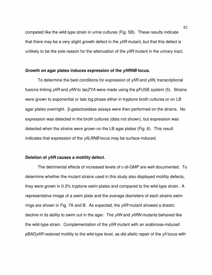
82
competed like the wild-type strain in urine cultures (Fig. 5B). These results indicate
that there may be a very slight growth defect in the yfiR mutant, but that this defect is
unlikely to be the sole reason for the attenuation of the yfiR mutant in the urinary tract.
Growth on agar plates induces expression of the yfiRNB locus.
To determine the best conditions for expression of yfiR and yfiN, transcriptional
fusions linking yfiR and yfiN to lacZYA were made using the pFUSE system (5). Strains
were grown to exponential or late log phase either in tryptone broth cultures or on LB
agar plates overnight. β-galactosidase assays were then performed on the strains. No
expression was detected in the broth cultures (data not shown), but expression was
detected when the strains were grown on the LB agar plates (Fig. 6). This result
indicates that expression of the yfiLRNB locus may be surface-induced.
Deletion of yfiR causes a motility defect.
The detrimental effects of increased levels of c-di-GMP are well documented. To
determine whether the mutant strains used in this study also displayed motility defects,
they were grown in 0.3% tryptone swim plates and compared to the wild-type strain. A
representative image of a swim plate and the average diameters of each strains swim
rings are shown in Fig. 7A and B. As expected, the yfiR mutant showed a drastic
decline in its ability to swim out in the agar. The yfiN and yfiRN mutants behaved like
the wild-type strain. Complementation of the yfiR mutant with an arabinose-induced
pBADyfiR restored motility to the wild-type level, as did allelic repair of the yfi locus with

83
a CFT073 transducing phage. The swimming defect observed with the yfiR deletion
strain does not seem to be due to an inability to assembly the flagella because intact
flagellar filaments can be observed in electron micrographs of the cells, and motility is
seen in live bacteria observed under a light microscope (Fig. 7C).
Deletion of yfiR causes increased production of cellulose and curli fimbriae.
In other strains of E. coli as well as other species of bacteria an increase in c-di-
GMP levels usually results in increase in fimbriation and exopolysaccharide production.
E. coli strains specifically up-regulate production of curli fimbriae and cellulose. An easy
assay to test whether curli fimbriae and cellulose are being produced involves plating
the bacteria on LB plates that contain congo red dye, but lack NaCl. After 5-8 days of
growth, colonies that express both products will have a deep red, rugose phentotype
that is referred to as the RDAR morphotype. Colonies that produce only curli fimbriae
will lack the rugose characteristic, but will still be deep red in color and slightly rough.
Expression of cellulose only results in a rugose colony that is pink instead of deep red,
and loss of expression of both products results in a colony that is white and smooth. All
colonies plated for the yfiR mutant displayed the RDAR morphotype while the wild-type,
yfiN and yfiRN mutants produced a mixture of both RDAR and smooth white colonies.
Restoration of yfiR via transduction returned the phenotype of the yfiR mutant to a
mixture of both RDAR and smooth white colonies (Fig. 8A and B). Over-expression yfiR
encoded on pBAD in the yfiR mutant produced all smooth colonies, although most were
slightly rough in the center presumably from loss of arabinose induction as a result of

84
metabolism (Fig. 11A). Congo red binding assays in liquid culture were also
performed as described in the materials and methods section. As expected, the yfiR
mutant bound more congo red dye per CFU than the other strains, and this increase in
bound congo red was mitigated by either transduction of the yfi locus to wild type or by
plasmid-expressed yfiR (Fig. 8C).
To determine whether increased c-di-GMP levels increased csgD expression as
previously described, the lacZYA locus was inserted directly downstream of csgD to
create a transcriptional fusion using the pFUSE system. Expression levels of csgD
could then be monitored by β-galactosidase assays. Deletion of yfiR resulted in a
drastic increase in the Miller units expressed from the csgD operon (Fig. 8D), indicating
that elevated c-di-GMP production does increase csgD expression levels in E. coli
CFT073. Deletion of both yfiR and yfiN in the csgD::lacZYA strain returned csgD
expression to wild-type levels as did complementation with pBADyfiR in the yfiR
deletion strain (Fig. 8D). Increased production of the curli fibrils was verified by western
blotting using an antibody specific for CsgA, the major curlin subunit (Fig. 8E). Deletion
of adrA, but not yedQ, reduced cellulose production by congo red plating (Fig. 11A).
The link between increased csgD expression and increased adrA expression was also
confirmed using an adrA::lacZYA fusion in the β-galactosidase assay (Fig. 11B).
Expression off of the bcs operon was also monitored using the same system to place
lacZYA downstream of bcsA. However, bcsA and bcsE expression levels in the mutant
strains remained unchanged from the wild-type strain (Fig. 12AB). This result is not
surprising given that c-di-GMP is thought to bind directly to BcsA, increasing its activity

85
levels and, thus, elevating cellulose production (24, 33). Because cellulose and curli
fimbriae are also components of biofilms, pellicle formation on glass test tubes was
analyzed via a crystal violet-staining assay. Deletion of yfiR caused increased pellicle
formation (Fig. 9AB), indicating that this strain has increased biofilm formation.
Expression of a phosphodiesterase reverses the phenotypes of CFT073∆yfiR to
wild type.
The increase in curli fimbriae and cellulose production and pellicle formation in
the yfiR deletion strain is likely due to increased cytoplasmic levels of c-di-GMP. To test
if this is the case, wild-type yhjH, which encodes a proven phosphodiesterase (40), was
expressed from a plasmid in CFT073∆yfiR in the congo red binding assay, pellicle
formation assay, and swimming motility assay. As predicted, expression and,
presumably, phosphodiesterase activity of YhjH reduced the amount of congo red dye
bound, reduced pellicle formation, and increased swimming motility in comparison to the
yfiR deletion strain (Fig. 10A-C). Expression of yhjHE136A, an active site mutant that
cannot degrade c-di-GMP, did not have an effect on any of the noted phenotypes when
expressed in the yfiR deletion strain, indicating that the phosphodiesterase activity and
the resulting reduction in c-di-GMP levels is indeed required for the reversal of the
observed phenotypes (Fig. 10 A-C).

86
Deletion of yfiR does not increase sensitivity to hydrogen peroxide or iron
limitation.
Past reports have indicated that increased levels of c-di-GMP lead to an increase
in sensitivity to peroxide stress and a decrease in the expression of iron acquisition
genes. To determine whether the yfiR mutant was more sensitive to peroxides than
wild-type CFT073, hydrogen peroxide sensitivity assays were performed in both liquid
culture and disc-diffusion agar. No differences between the wild-type strain and the yfiR
mutant were observed in either assay (Fig. 13AB). To determine whether the yfiR
mutant would have trouble growing in an iron-limited environment like the host, both the
wild-type and mutant strains were grown in LB broth with 2,2-dipyridyl, an iron chelator.
Again, no differences in growth were observed between the wild-type and yfiR mutant
strains (Fig. 14).
Deletion of ycgR in the yfiR mutant partially restores colonization levels in the
mouse model.
Motility is a known colonization factor of uropathogenic E. coli strains in the
urinary tract, and is needed for ascension of the ureters to the kidneys. Multiple studies
have recently showed that YcgR can bind c-di-GMP and then interact with the flagellar
motor (6, 29). This interaction acts either to inhibit rotation or to keep the flagella
turning counterclockwise, causing smooth swimming cells that may become stuck in the
agar matrix. To determine whether the swimming defect seen in the yfiR mutant causes
the attenuated phenotype in vivo, ycgR was deleted from the yfiR mutant and tested in

87
the competitive mouse model of UTI. Swim plates of this mutant confirmed that
motility was partially restored (Fig. 15). As shown in Figure 16A, this partial restoration
of motility also resulted in the partial recovery of the yfiR mutant to wild-type
colonization levels in vivo. By itself, a single ycgR deletion mutant was not attenuated
in the competitive mouse model against wild-type CFT073 (Fig. 17A). Given that the
colonization levels were only partially restored, full motility may be needed for full
virulence, or another factor may also be partially responsible for the observed yfiR
mutant attenuation.
Deletion of both bcsA and csgD in the yfiR mutant returns colonization to wild-
type levels.
Previous studies have shown that curli fibers produce an immune response by
activating the host coagulation system and, thus, promoting an influx of white blood
cells (27, 30). However, curli fimbriae can also promote resistance to the host
antimicrobial peptide LL-37 (20). Additional mutations in the yfiR deletion strain were
made to determine whether the overexpression of curli fimbriae, or possibly even the
increase in cellulose production, could be responsible for the attenuation of the yfiR
deletion strain in vivo. Deletion of csgD or bcsA from the yfiR mutant does not return
colonization to wild-type levels in the competitive UTI model, although perhaps a slight
improvement in the mutant’s fitness is seen in the kidneys for both double deletion
strains (Fig. 16B and C). Single-deletion strains of csgD or bcsA compete well with the
wild-type strain (Fig. 17B and C). Additional deletion of csgD from the ∆yfiR∆ycgR

88
double deletion strain did not relieve the attenuated phenotype in the mouse model
(Fig. 17D). However, when both csgD and bcsA are deleted from the yfiR mutant,
colonization is restored to wild-type levels (Fig. 16D). The motility of these double and
triple mutants was determined using tryptone swim plates, on which they remained
poorly motile like the yfiR single mutant (Fig. 15).
Urine inhibits the RDAR morphotype and csgD expression.
In addition to growing the mutant strains on LB congo red plates, we inoculated
them on urine agar congo red plates to see whether exposure to urine caused a change
in the RDAR phenotype. All of the wild-type CFT073 colonies grown on the urine congo
red plates displayed a smooth, white phenotype, indicating that urine represses the
expression or activity of the proteins needed for production of cellulose and curli
fimbriae (Fig. 18A). Similar results were obtained when the wild-type strain was grown
in liquid urine, and then plated on LB congo red plates (Fig. 18B), suggesting that the
effect urine has on the regulatory controls for curli fimbriae and cellulose is long-lasting.
The change was not permanent, however, because a return of the RDAR phenotype
was observed after growth in LB liquid culture followed by growth on LB congo red
plates (data not shown). Congo red plates that combined urine and LB were also tested
with the wild-type strain as a control to ensure that the loss of the RDAR phenotype was
not due to growth inhibition on the urine only plates (Fig. 18A). The presence of urine
also affected the phenotype of the yfiR mutant on the urine congo red plates, but all
colonies were still deep red and somewhat rugose (Fig. 18A), which indicated that curli

89
fimbriae and cellulose were both still being produced. Growth in liquid urine followed
by growth on LB congo red plates produced no differences in the RDAR phenotype for
the yfiR mutant (Fig. 18B). Because salt is a known inhibitor of the RDAR phenotype,
we determined which salts found in urine might be responsible for the all smooth and
white phenotype seen in wild-type CFT073. Individual salt components of urine (NaCl,
Ca3(PO4)2, K2SO4, KCl, KHCO3, MgCO3, and MgSO4) at their physiological
concentrations were added to LB congo red plates for testing with the wild-type strain.
Of the salts tested, only NaCl produced a phenotype similar to that found on the urine
congo red plates (Fig. 18A).
To determine whether urine and NaCl affected the expression of csgD, the wild-
type and yfiR mutant csgD::lacZYA strains were employed. β-galactosidase assays
performed on cells grown on urine plates showed a decrease in expression from the csg
promoter in the presence of urine compared to LB. However, expression levels in the
yfiR mutant remained elevated compared to the wild-type strain under all conditions
tested (Fig. 18C). Growth on LB-NaCl plates caused no change in expression when
compared to growth on LB only plates (Fig. 18C).
Discussion
This study describes the effects that elevated levels of c-di-GMP brought on by
loss of inhibition of YfiN by YfiR have on the ability of E. coli CFT073 to mount a
successful infection in the urinary tract. Results show that loss of YfiR likely relieves
inhibition of the activity of the membrane bound DGC YfiN, leading to an increase in

90
cellular c-di-GMP levels. This increase leads to an elevation in production of both
curli fimbriae and cellulose, a reduction in the motility, and ultimately attenuation in the
mouse model of UTI. Single deletions of yfiL, yfiB, or yfiN did not attenuate E. coli
CFT073 in vivo. Additional deletion of yfiN from the yfiR deletion strain, or restoration to
the wild-type operon of the yfiR deletion strain, brought colonization back to wild-type
levels. This indicates that YfiR exerts an inhibitory effect on the activity of YfiN that is
important for maintaining control of c-di-GMP levels in vivo. Without this control, the
bacteria become impaired in the urinary tract, presumably due to disregulation of
downstream genes and functions that are sensitive to c-di-GMP signaling.
A study of the homologous P. aeruginosa system postulated that YfiR prevents
the dimerization of YfiN, a state that is necessary for activity of most DGCs. This same
report also indicated that YfiB promotes the dimerization of YfiR, which would
presumably inhibit its ability to disrupt YfiN dimerization (23). Overall, though, the
function of YfiB and the mechanism by which the YfiRNB system operates is still largely
unknown. Further studies would be necessary to determine the hierarchy of the YfiRNB
system and the set of external stimuli that may activate or repress YfiN activity. Our
work indicates that transcription of the yfi locus may be surface induced. If attachment
to the host epithelium also induces expression of this locus, the regulation of both
yfiLRNB transcription and YfiN activity could give insights into the series of events
necessary for successful colonization of and persistence in the urinary tract.
The downstream targets of increased c-di-GMP levels in E. coli CFT073 are
similar to those that have been previously described in other E. coli strains (31). The

91
loss of yfiR in CFT073 resulted in decreased motility, increased expression of the
csgD promoter, and increased production of both curli fimbriae and cellulose.
Expression of yhjH, which encodes a phosphodiestrase, in the CFT073∆yfiR
background returned these phenotypes to the wild-type forms. The phosphodiesterase
activity of YhjH was required for this reversal, indicating that increased c-di-GMP levels
is indeed the factor driving the over-expression of curli fimbriae and cellulose and the
observed decrease in motility in the yfiR deletion strain. The motility defect and in vivo
attenuation of the yfiR deletion strain was also partially rescued by additional deletion of
ycgR. YcgR is capable of interacting with the flagellar motor to either promote smooth
swimming or act as a braking mechanism when it is bound to c-di-GMP. Deletion of
ycgR therefore prevented alteration of flagellar rotation by this mechanism. This
observation indicates that motility and the effect of increased c-di-GMP levels on
flagellar rotation do impair the ability of CFT073 to successfully infect the urinary tract.
The overproduction of curli fimbriae and cellulose also appears to have a detrimental
effect on CFT073 in vivo. The link between the expression of csgD and the activity of
the cellulose biosynthetic machinery was established previously in E. coli K-12 strains.
CsgD activates not only expression of the curli operon, but also expression of adrA and
yedQ, genes that encodes DGCs. The c-di-GMP produced by either AdrA or YedQ
would then stimulate increased activity of the cellulose synthetic machinery by binding
to components of its core catalytic module (25, 33, 47). Deletion of adrA, but not yedQ,
resulted in a loss of cellulose production on LB congo red plates. Therefore, we

92
postulate that AdrA expression stimulated by CsgD activity is the likely link involved in
CsgD-dependent increases of cellulose production in E. coli CFT073.
Curli fibers are known activators of the host coagulase system that lead to
increased influx of immune cells and inflammation (30). Another report also implicated
the expression of curli fimbriae in the absence of cellulose production as producing an
immune stimulant that resulted in increased clearance of uropathogenic E. coli strain
UTI89 from the urinary tract (20). In contrast to these reports, a ∆yfiR∆csgD mutant still
displayed attenuation in the competitive mouse model of UTI, even though it was
confirmed that curli fimbriae were no longer being produced. Only additional deletion of
bcsA from the ∆yfiR∆csgD mutant rescued the attenuated phenotype. Although the
yfiR single-deletion mutant did not display any growth defects in various media when
grown singly or in competition with the wild-type strain in vitro, it is possible that the
increased stress of the urinary tract environment attenuated the metabolically over-
burdened mutant. The significant amount of cellular resources being consumed for the
production of large and unnecessary amounts of curli fimbriae and cellulose may have
therefore diverted resources away from the production of other virulence or colonization
factors vital for prolonged infection of the urinary tract. Further studies into alterations in
the global production of bacterial proteins in the yfiR deletion mutant could give insight
into whether or not the expression of any other virulence factors were altered.
Given that single deletions of csgD or bcsA did not attenuate CFT073 in vivo,
biofilm formation associated with curli or cellulose is likely not a persistence factor
involved in colonization in the urinary tract. Additionally, growth in urine decreases

93
expression of csgD and inhibits the RDAR morphology. The salt content of urine
appears to be the main suppressor leading to this outcome. Growth at 37° C also
inhibits the production of these biofilm components. Taken together, these data indicate
that biofilms are not formed by E. coli CFT073 in vivo and may, instead, be necessary
for adaptation and persistence in environments outside of the urinary tract.
This study not only emphasizes the importance of regulation of curli fimbriae,
cellulose production, and motility in the colonization of UPEC in the urinary tract but also
highlights c-di-GMP production as a key regulator of bacterial processes during
infection. The abundance of DGCs (~19) and PDEs (~17) in E. coli alone suggests that
control of c-di-GMP production and its downstream effects are likely important at a
number of times in different environments. Further studies into the distinct or perhaps
redundant roles played by the various DGCs and PDEs would illuminate the steps in the
fine-tuned control that the bacteria have over their expression of both colonization and
virulence factors to mount successful infections in the host.
Acknowledgements
This research was funded by the National Institutes of Health Grant R01 DK063250-06
and by the National Institutes of Health, National Research Service Award AI55397.

94
References
1. Amikam, D., and M. Y. Galperin. 2006. PilZ domain is part of the bacterial c-di-
GMP binding protein. Bioinformatics 22:3-6.
2. Bacheller, C. D., and J. M. Bernstein. 1997. Urinary tract infections. Med. Clin.
N. Am. 81:719-730.
3. Bahrani-Mougeot, F. K., E. L. Buckles, C. V. Lockatell, J. R. Hebel, D. E.
Johnson, C. M. Tang, and M. S. Donnenberg. 2002. Type 1 fimbriae and
extracellular polysaccharides are preeminent uropathogenic Escherichia coli
virulence determinants in the murine urinary tract. Mol Microbiol 45:1079-1093.
4. Battaglioli, E. J., G. Baisa, A. E. Weeks, R. Schroll, A. J. Hryckowian, and R.
A. Welch. 2011. Isolation of generalized transducing bacteriophages for
uropathogenic strains of Escherichia coli. Appl Environ Microbiol.
5. Baumler, A. J., R. M. Tsolis, A. W. M. van der Velden, I. Stojiljkovic, S. Anic,
and F. Heffron. 1996. Identification of a new iron regulated locus of Salmonella
typhi. Gene 183:207-213.
6. Boehm, A., M. Kaiser, H. Li, C. Spangler, C. A. Kasper, M. Ackermann, V.
Kaever, V. Sourjik, V. Roth, and U. Jenal. 2010. Second messenger-mediated
adjustment of bacterial swimming velocity. Cell 141:107-116.
7. Bokranz, W., X. Wang, H. Tschape, and U. Romling. 2005. Expression of
cellulose and curli fimbriae by Escherichia coli isolated from the gastrointestinal
tract. J Med Microbiol 54:1171-1182.

95
8. Brombacher, E., A. Baratto, C. Dorel, and P. Landini. 2006. Gene
expression regulation by the curli activator CsgD protein: modulation of cellulose
biosynthesis and control of negative determinants for microbial adhesion. J
Bacteriol 188:2027-2037.
9. D'Argenio, D. A., and S. I. Miller. 2004. Cyclic di-GMP as a bacterial second
messenger. Microbiology 150:2497-2502.
10. Datsenko, K. A., and B. L. Wanner. 2000. One-step inactivation of
chromosomal genes in Escherichia coli K-12 using PCR products. Proc. Natl.
Acad. Sci. U. S. A. 97:6640-6645.
11. Donnenberg, M. S., and R. A. Welch. 1996. Virulence determinants of
uropathogenic Escherichia coli, p. 135-174. In H. L. T. Mobley and J. W. Warren
(ed.), Urinary Tract Infections: Molecular Pathogenesis and Clinical
Management. ASM Press, Washington, D. C.
12. Faro, S., and D. E. Fenner. 1998. Urinary tract infections. Clinical Obstetrics and
Gynecology 41:744-754.
13. Garcia, E. C., A. R. Brumbaugh, and H. L. Mobley. 2011. Redundancy and
specificity of Escherichia coli iron acquisition systems during urinary tract
infection. Infect Immun 79:1225-1235.
14. Girgis, H. S., Y. Liu, W. S. Ryu, and S. Tavazoie. 2007. A comprehensive
genetic characterization of bacterial motility. PLoS Genet 3:1644-1660.
15. Hammar, M., A. Arnqvist, Z. Bian, A. Olsen, and S. Normark. 1995.
Expression of two csg operons is required for production of fibronectin- and

96
congo red-binding curli polymers in Escherichia coli K-12. Mol Microbiol
18:661-670.
16. Hengge, R. 2009. Principles of c-di-GMP signalling in bacteria. Nat Rev
Microbiol 7:263-273.
17. Hooton, T. M. 2000. Pathogenesis of urinary tract infections: an update. J
Antimicrob Chemother 46 Suppl 1:1-7; discussion 63-65.
18. Jenal, U., and J. Malone. 2006. Mechanisms of cyclic-di-GMP signaling in
bacteria. Annu Rev Genet 40:385-407.
19. Kader, A., R. Simm, U. Gerstel, M. Morr, and U. Romling. 2006. Hierarchical
involvement of various GGDEF domain proteins in rdar morphotype development
of Salmonella enterica serovar Typhimurium. Mol Microbiol 60:602-616.
20. Kai-Larsen, Y., P. Luthje, M. Chromek, V. Peters, X. Wang, A. Holm, L.
Kadas, K. O. Hedlund, J. Johansson, M. R. Chapman, S. H. Jacobson, U.
Romling, B. Agerberth, and A. Brauner. 2010. Uropathogenic Escherichia coli
modulates immune responses and its curli fimbriae interact with the antimicrobial
peptide LL-37. PLoS Pathog 6:e1001010.
21. Kau, A. L., D. A. Hunstad, and S. J. Hultgren. 2005. Interaction of
uropathogenic Escherichia coli with host uroepithelium. Curr Opin Microbiol 8:54-
59.
22. Kunin, C. M. 1994. Urinary tract infections in females. Clin. Infect. Dis. 18:1-10.
23. Malone, J. G., T. Jaeger, C. Spangler, D. Ritz, A. Spang, C. Arrieumerlou, V.
Kaever, R. Landmann, and U. Jenal. 2010. YfiBNR mediates cyclic di-GMP

97
dependent small colony variant formation and persistence in Pseudomonas
aeruginosa. PLoS Pathog 6:e1000804.
24. Mills, E., I. S. Pultz, H. D. Kulasekara, and S. I. Miller. 2011. The bacterial
second messenger c-di-GMP: mechanisms of signalling. Cell Microbiol 13:1122-
1129.
25. Monteiro, C., I. Saxena, X. Wang, A. Kader, W. Bokranz, R. Simm, D. Nobles,
M. Chromek, A. Brauner, R. M. Brown, Jr., and U. Romling. 2009.
Characterization of cellulose production in Escherichia coli Nissle 1917 and its
biological consequences. Environ Microbiol 11:1105-1116.
26. Nielubowicz, G. R., and H. L. Mobley. 2010. Host-pathogen interactions in
urinary tract infection. Nat Rev Urol 7:430-441.
27. Olsen, A., A. Jonsson, and S. Normark. 1989. Fibronectin binding mediated by
a novel class of surface organelles on Escherichia coli. Nature 338:652-655.
28. Orenstein, R., and E. S. Wong. 1999. Urinary tract infections in adults. Am.
Fam. Physician 59:1225-1234.
29. Paul, K., V. Nieto, W. C. Carlquist, D. F. Blair, and R. M. Harshey. 2010. The
c-di-GMP binding protein YcgR controls flagellar motor direction and speed to
affect chemotaxis by a "backstop brake" mechanism. Mol Cell 38:128-139.
30. Persson, K., W. Russell, M. Morgelin, and H. Herwald. 2003. The conversion
of fibrinogen to fibrin at the surface of curliated Escherichia coli bacteria leads to
the generation of proinflammatory fibrinopeptides. J Biol Chem 278:31884-
31890.

98
31. Povolotsky, T. L., and R. Hengge. 2011. 'Life-style' control networks in
Escherichia coli: Signaling by the second messenger c-di-GMP. J Biotechnol.
32. Redford, P., and R. A. Welch. 2006. Role of sigma E-regulated genes in
Escherichia coli uropathogenesis. Infect. Immun. 74:4030-4038.
33. Romling, U. 2005. Characterization of the rdar morphotype, a multicellular
behaviour in Enterobacteriaceae. Cell Mol Life Sci 62:1234-1246.
34. Romling, U. 2002. Molecular biology of cellulose production in bacteria. Res
Microbiol 153:205-212.
35. Romling, U., Z. Bian, M. Hammar, W. D. Sierralta, and S. Normark. 1998.
Curli fibers are highly conserved between Salmonella typhimurium and
Escherichia coli with respect to operon structure and regulation. J Bacteriol
180:722-731.
36. Romling, U., M. Gomelsky, and M. Y. Galperin. 2005. c-di-GMP: the dawning
of a novel bacterial signalling system. Mol Microbiol 57:629-639.
37. Ross, P., R. Mayer, and M. Benziman. 1991. Cellulose biosynthesis and
function in bacteria. Microbiol Rev 55:35-58.
38. Ryjenkov, D. A., R. Simm, U. Romling, and M. Gomelsky. 2006. The PilZ
domain is a receptor for the second messenger c-di-GMP. The PilZ domain
protein controls motility in enterobacteria. J Biol Chem 281:30310-30314.
39. Ryjenkov, D. A., M. Tarutina, O. V. Moskvin, and M. Gomelsky. 2005. Cyclic
diguanylate is a ubiquitous signaling molecule in bacteria: insights into
biochemistry of the GGDEF protein domain. J Bacteriol 187:1792-1798.

99
40. Simm, R., M. Morr, A. Kader, M. Nimtz, and U. Romling. 2004. GGDEF and
EAL domains inversely regulate cyclic di-GMP levels and transition from sessility
to motility. Mol Microbiol 53:1123-1134.
41. Smith, Y. C., S. B. Rasmussen, K. K. Grande, R. M. Conran, and A. D.
O'Brien. 2008. Hemolysin of uropathogenic Escherichia coli evokes extensive
shedding of the uroepithelium and hemorrhage in bladder tissue within the first
24 hours after intraurethral inoculation of mice. Infect Immun 76:2978-2990.
42. Wiles, T. J., R. R. Kulesus, and M. A. Mulvey. 2008. Origins and virulence
mechanisms of uropathogenic Escherichia coli. Exp Mol Pathol 85:11-19.
43. Wolfe, A. J., and K. L. Visick. 2008. Get the message out: cyclic-Di-GMP
regulates multiple levels of flagellum-based motility. J Bacteriol 190:463-475.
44. Wright, K. J., P. C. Seed, and S. J. Hultgren. 2005. Uropathogenic Escherichia
coli flagella aid in efficient urinary tract colonization. Infect Immun 73:7657-7668.
45. Yamamoto, S., K. Nakata, K. Yuri, H. Katae, A. Terai, H. Kurazono, Y.
Takeda, and O. Yoshida. 1996. Assessment of the significance of virulence
factors of uropathogenic Escherichia coli in experimental urinary tract infection in
mice. Microbiol Immunol 40:607-610.
46. Yan, H., and W. Chen. 2010. 3',5'-Cyclic diguanylic acid: a small nucleotide that
makes big impacts. Chem Soc Rev 39:2914-2924.
47. Zogaj, X., M. Nimtz, M. Rohde, W. Bokranz, and U. Romling. 2001. The
multicellular morphotypes of Salmonella typhimurium and Escherichia coli

100
produce cellulose as the second component of the extracellular matrix. Mol
Microbiol 39:1452-1463.

101
Table 1: Primers used in this study Gene/Application Sequence
λλλλ-Red Deletions yfiR deletion
yfiR deletion ck
yfiN deletion
yfiB deletion
yfiL deletion
yfi locus deletion ck
csgD deletion
csgD deletion ck
csgA deletion
csgA deletion ck
bcsA deletion
bcsA deletion ck
yedQ deletion
5’-CGATGCATTTCGAGCGAAGATGGTGAGGATCCCTGA ATGTGTGTAGGCTGGAGCTGCTTC-3’ 5’-ATTAAGAGAATTATCGTTATCCATCATTTTTCTTCCGT GCCATATCAATATCCTCCTTAG-3’ 5’-GCCAACTGCATGAAGTATGGC-3’ 5’-CTGCAGTTGAAAATTGGCCTTGC-3’ 5’-CAACCCGGATGTCTTAATGCTCGCACGGAAGAAAAAT GTGTGTAGGCTGGAGCTGCTTC-3’ 5’-GTGCTAGCAGGTGCTTTATCATATCGATATATCCTTGT TACATATGAATATCCTCCTTAG-3’ 5’-GCCGAAAAGCTGGTGAGATAACAAGGATATATCGATA TGTGTGTAGGCTGG AGCTGCTTC-3’ 5’-AATTGGCTGGCCCTTTTTTTGTCTGTTTGCTCGTGTTT TACATATGAATATCCTCCTTAG-3’ 5’-AAATTTACACCAGCTTTAAAAACAATCCGCTATGCTTT GTGTGTAGGCTGGAGCTGCTTC-3’ 5’-TGAGTAAAATCTCATGCTTAATTGATTTGGGGGGTAAT TACATATGAATATCCTCCTTAG-3’ 5’-CTCAGCGAGGATGACGTAACG-3’ 5’-CGTGAGCGTACTGGTAAGGC-3’ 5’-TCAGGTGTGCGATCAATAAAAAAAGCGGGGTTTCATC ATGGTGTAGGCTGGAGCTGCTTC-3’ 5’-TGCCGCCACAATCCAGCGTAAATAACGTTTCATGGCT TTACATATGAATATCCTCCTTAG-3’ 5’-CACACAGCAGTGCAACATCTGTCA-3’ 5’-GCCGCCACAATCCAGCGTAAATAA-3’ 5’-CCATTCGACTTTTAAATCAATCCGATGGGGGTTTTAC ATGGTGTAGGCTGGAGCTGCTTC-3’ 5’-AGGGCTTGCGCCCTGTTTCTTTCATACTGATGATGTA TTACATATGAATATCCTCCTTAG-3’ 5’-GCAAATGGCTATTCGCGTGACACA-3’ 5’-GGCTTGCGCCCTGTTTCTTTCATA-3’ 5’-CGGGCTGAAAACGCCAGTCGGGAGTGCATCATGAGT ATCTGTGTAGGCTGGAGCTGCTTC-3’ 5’-GTTTCGCTCCGCTCCGGGCGGCGCGGAATAAACGAT ACAACATATGAATAT CCTCCTTAG-3’ 5’-GCTGAGTTTGTGCGATCACTCG-3’ 5’-CTGGCTCAGCATTGATCAGTGG-3’ 5’-AAACTCGCACGCCGCCTGGGGCCTGGTCATATCGTT AATTGT GTAGGCTGGAGCTGCTTC-3’ 5’-GCTGGCGTGAAGGGCTGGACCATTTTTTCTCCGCCC

102
yedQ deletion ck
adrA deletion
adrA deletion ck
ycgR deletion
ycgR deletion ck
fliC deletion
fliC deletion ck
lacZ deletion
lacZ deletion ck
yfiR φ restoration (pheA-tyrA)
pBAD24d Cloning
yfiR-NcoI yfiR-PstI
Insertion site ck
pFUSE Cloning csgD
bcsA
GTTACATATGAATATCCTCCTTAG -3’ 5’-GGATCACATCCGGCCTGG-3’ 5’-CCCAACTGGATTATCGCGACC-3’ 5’-AGCCCCGTCTCTATAATTTGGGAAAATTGTTTCTGAA TGTGTGTAGGCTGGAGCTGCTTC-3’ 5’-GAAAAACTCAGTAAATCCTGATGACTTTTGCCGGACG TCACATATGAATATCCTCCTTAG-3’ 5’-GCAACAACAGCGTATGACCTCC-3’ 5’-CGGTGTGAGCGGTTCTTCG-3’ 5’-AACCAAAGACAGTTTGTCAGTCAGGAGTTTTTCCGCG TGTGTGTAGGCTGGAGCTGCTTC-3’ 5’-ATCTACAAACTTGAGCAGGCACTGGACGCGATGTAAA TCACATATGAATATCCTCCTTAG-3’ 5’-CCTCGTTTCGATGAATCGGACG-3’ 5’-CTGGTGGTGTCATACGCTAACG-3’ 5’-GGCGTTGCCGTCAGTCTCAGTTAATCAGGTTACGGC GATGTGTAGGCTGGAGCTGCTTC-3’ 5’-AAAACGTAATCAACGACTTGCAATATAGGATAACGAA TCCATATGAATATCCTCCTTAG-3’ 5’-CGCAATGGGTCTGGCTGTG-3’ 5’-GAGTGATGGTCAGCGTCTGGTTGC-3’ 5’-GTTGGTCTGGTGTCAAAAATAATAATAACCTGCGGCC GCATCTAG-3’ 5’-ACGCGAAATACGGGCAGACATAGCCTGCCCATTCCG GGGATCCGT-3’ 5’-GCAGACCAAACAGCGGTTGG-3’ 5’-TTGAATTATGGCCCACACCAGTG-3’ 5’-GAATGACAATCGCCAGTAATAATCCAGTGCCGGATGA TTTGTGTAGGCTGGAGCTGCTTC-3’ 5’-CTGTTGATCCAACCTGATGAAAAAGTGCCGGATGATG TGACATATGAATATCCTCCTTAG-3’ 5’-GTGAGGCCATGGGAATGCGTTTTTCTCACCGACTG-3’ 5’-AGAGAACTGCAGTTATCCATCATTTTTCTTCCGTGCGA G-3’ 5’-GGACCAAAGCCATGACAAAAACG-3’ 5’-CTACTCAGGAGAGCGTTCACC-3' 5’-AACGTTCCCGGGCTTTATCGCCTGAGGTTATCGTTTGC-3’ 5’-TTCTTTTCTAGAGACAGCTCTCTTGCAGCACC-3’ 5’-GAAAGTCCCGGGCCACATATCATTGCCGTCCTGC-3’

103
bcsE
adrA
yfiR
yfiN
yfiB
5’-TGAAACTCTAGAGTGGATTGTGCTGGTGCTCG-3’ 5’-GATATCCCCGGGGGTCATCATGATGAGCGCTCC-3’ 5’-AAGATATCTAGACCTTGCTGTCAATGACCCAGC-3’ 5’-AAATCCCCCGGGCTTTTGCCGGACGTCAGG-3’ 5’-ATCGTCTCTAGACCTCTGCTGTTTGGCTGG-3’ 5’-TAAGAGCCCGGGCGTTATCCATCATTTTTCTTCCGTGC-3’ 5’-GATGGTTCTAGACCCTGAATGCGTTTTTCTCACC-3’ 5’-CATATCCCCGGGTCCTTGTTATCTCACCAGCTTTTCG-3’ 5’-CGTTTCTCTAGAGCATCAACACGTTGATGAACAATTCC-3’ 5’-CTGTTTCCCGGGGTTTTAAGGGGTTGTAATCACCACTG C-3’ 5’-GATAAATCTAGAGCTAGCACCCCTGATTTTCACC-3’

104
Figure 1: Organization of the yfiLRNB locus and predicted protein functions. (A)
The yfiLRNB locus consists of four genes that are expressed off a single predicted
promoter. (B) YfiR is a predicted periplasmic protein of unknown function with a signal
sequence (S.S.), and YfiN is a predicted membrane-bound diguanylate cyclase with an
intact GGDEF active site and an allosteric inhibition site (I site). YfiL and YfiB are
predicted lioproteins of unknown function.

105
Figure 2: YfiR localizes to the periplasm. Wild-type CFT073 producing YfiR with an
C-terminus FLAG tag via IPTG-mediated induction of expression of yfiR-FLAG off of the
pJMP69 plasmid was spheroplasted and separated into their inner
membrane/cytoplasmic and periplasmic fractions by centrifugation. Western blots of the
SDS-PAGE gel-separated fractions were probed with antibodies against either the
FLAG tag, periplasmic maltose-binding protein (MBP), or cytoplasmic RpoS.

106
Figure 3: Deletion of yfiR causes attenuation in the mouse model of competitive
UTI. Results for co-infection of female Swiss Webster mice with CFT073∆yfiR (n=21)
(A), CFT073∆yfiN (n=26) (B), CFT073∆yfiRN (n=16) (C), or the CFT073∆yfiR phage
restoration mutant (n=18) (D) and CFT073∆lacZYA using the mouse model of UTI. The
competitive indices were calculated by the following equation: (yfi mutant CFU
recovered/wild-type CFU recovered)/(mutant inoculum CFU/wild-type inoculum CFU).
The line indicates the median value.

107
Figure 4: Deletion of yfiL or yfiB produces no in vivo defect and CFT073∆∆∆∆yfiR is
carried at lower loads than wild-type CFT073 in single infections. Results for co-
infection of female Swiss Webster mice with CFT073∆yfiL (n=8) (A) and CFT073∆yfiB
(n=7) (B) and CFT073∆lacZYA using the mouse model of UTI. The competitive indices
were calculated by the following equation: (yfi mutant CFU recovered/wild-type CFU
recovered)/(mutant inoculum CFU/wild-type inoculum CFU). (C) Single infections of

108
wild-type CFT073 (n=8) and CFT073∆yfiR (n=9) in female Swiss Webster mice. The
line indicates the median value.
Figure 5: Deletion of yfiR does not cause a significant in vitro growth defect. (A)
CFT073∆yfiR grew much like wild-type CFT073 in both LB and urine in single-strain
broths. (B) Competitve growth broths with wild-type CFT073 and CFT073∆yfiR showed
no differences in nutrient acquisition by either strain when grown in LB and urine. The
line indicates the median.

109
Figure 6: The yfi locus is expressed on agar plates. β-galactosidase assays using
the CFT073 yfiR::lacZ, CFT073 yfiB::lacZ, and yfiN::lacZ transcriptional fusions
displayed locus expression when grown on LB agar plates without salt at 30° C.
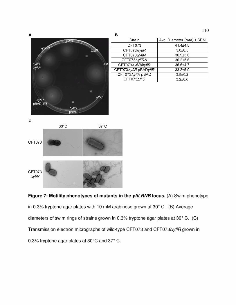
110
Figure 7: Motility phenotypes of mutants in the yfiLRNB locus. (A) Swim phenotype
in 0.3% tryptone agar plates with 10 mM arabinose grown at 30° C. (B) Average
diameters of swim rings of strains grown in 0.3% tryptone agar plates at 30° C. (C)
Transmission electron micrographs of wild-type CFT073 and CFT073∆yfiR grown in
0.3% tryptone agar plates at 30°C and 37° C.
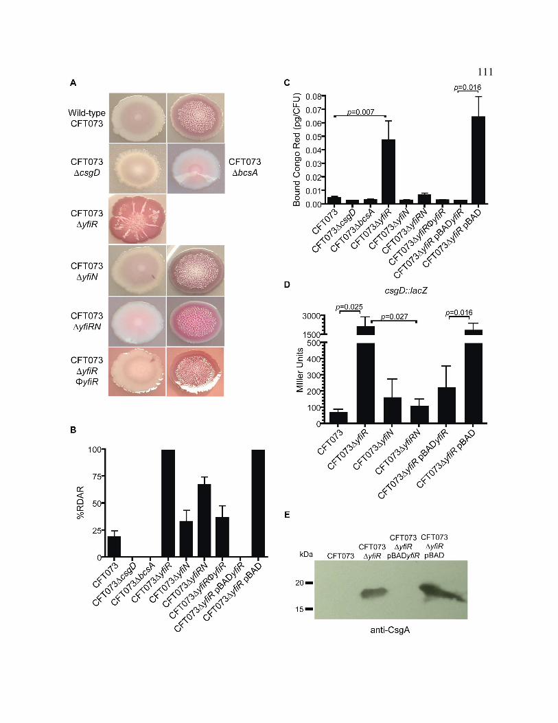
111

112
Figure 8: Curli fimbriae and cellulose production are increased in
CFT073∆∆∆∆yfiR. (A) Phenotypes of yfiLRNB locus mutants grown on LB congo red plates
at 30°C. All colony phenotypes that were observed are pictured. (B) Percentage of
mutant colonies from the LB congo red plates displaying the RDAR phenotype. (C)
Congo red binding assay in liquid broth. The amount of congo red dye bound is
presented as pg of bound dye per CFU. P values were calculated using the student’s T-
test. (D) β-galactosidase assays performed on wild-type CFT073 and CFT073∆yfiR
strains with the csgD::lacZYA transcriptional fusion. P values were calculated using the
student’s T-test. (E) Western blot of acid-treated, whole-cell preparations grown on LB
no-salt plates at 30°C. A rabbit anti-CsgA antibody was used to detect CsgA
monomers.

113
Figure 9: Pellicle formation is increased in CFT073∆∆∆∆yfiR. (A) Optical density of
crystal violet stain bound and released from pellicles formed by each strain after growth
in MOPS glycerol broths for 16 hours at 37° C. Expression of yfiR from pBAD plasmids
was induced with 10 mM arabinose. P values were calculated using the student’s T-
test. (B) Observed pellicles of each strain dyed with crystal violet.
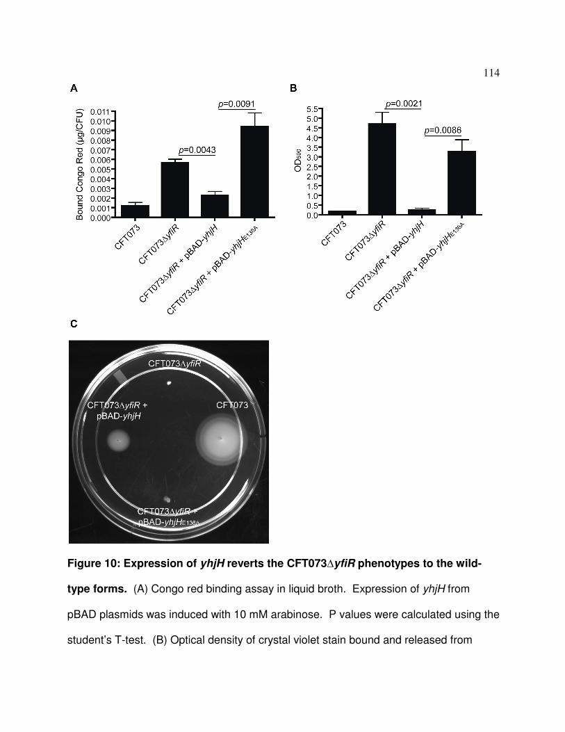
114
Figure 10: Expression of yhjH reverts the CFT073∆yfiR phenotypes to the wild-
type forms. (A) Congo red binding assay in liquid broth. Expression of yhjH from
pBAD plasmids was induced with 10 mM arabinose. P values were calculated using the
student’s T-test. (B) Optical density of crystal violet stain bound and released from

115
pellicles formed by each strain after growth in MOPS glycerol broths for 16 hours at
37° C. Expression of yhjH from pBAD plasmids was induced with 10 mM arabinose. P
values were calculated using the student’s T-test. (C) Swim phenotype in 0.3% tryptone
agar plates with 10 mM arabinose grown at 30° C.

116
Figure 11: Loss of adrA reduces cellulose production. (A) Strains grown on LB
congo red plates at 30° C. All colony phenotypes observed for each strain are pictured.
(B) β-galactosidase assays with adrA::lacZYA fusion strains. The p value was
calculated using a nonparametric student’s T test.

117
Figure 12: Loss of yfiR has no effect on bcsA or bcsE expression. β-galactosidase
assays performed on wild-type CFT073 and CFT073∆yfiR strains with the bcsA::lacZYA
(A) or bcsE::lacZYA (B) transcriptional fusion.

118
Figure 13: Deletion of yfiR does not increase sensitivity to hydrogen peroxide.
(A) Disc diffusion assays with wild-type CFT073 and CFT073∆yfiR grown aerobically at
37° C. 1X PBS was spotted on the left-side disc and 300 mM H2O2 was spotted on the
right-side disc. The average diameters of the zones of inhibition are given below the
plate pictures. (B) Liquid broths assays performed with wild-type CFT073 and
CFT073∆yfiR. Diluted cultures were exposed to either PBS or 300 mM H2O2. Percent
survival was calculated by dividing the CFUs at each time point by the CFUs obtained
from the pre-exposure diluted broths.

119
Figure 14: Deletion of yfiR does not cause a defect in growth in an iron-limited
environment. Wild-type CFT073 and CFT073∆yfiR were grown in LB at 37°C with two
different concentrations of the iron chelator 2,2-dipyridyl added to the growth medium.

120
Figure 15: Deletion of ycgR from the yfiR single-deletion mutant partially restores
motility. Strains were grown in 0.3% agar tryptone swim plates at 30° C.

121
Figure 16: Deletion of both bcsA and csgD from the yfiR deletion mutant restores
fitness to wild-type CFT073 levels in the mouse model of competitive UTI. Results
for co-infection of female Swiss Webster mice with CFT073∆yfiR∆ycgR (n=14) (A),
CFT073∆yfiR∆csgD (n=11) (B), CFT073∆yfiR∆bcsA (n=7) (C), or
CFT073∆yfiR∆csgD∆bcsA (n=24) (D) and CFT073∆lacZYA using the mouse model of
UTI. The insets show the phenotype of each mutant on congo red plates grown at 30°
C. The competitive indices were calculated by the following equation: (yfi mutant CFU
recovered/wild-type CFU recovered)/(mutant inoculum CFU/wild-type inoculum CFU).
The line indicates the median value.

122
Figure 17: Deletion of bcsA, csgD, or ycgR from wild-type CFT073 does not affect
fitness in the mouse model of competitive UTI. Results for co-infection of female
Swiss Webster mice with CFT073∆ycgR (n=8) (A), CFT073∆csgD (n=11) (B),
CFT073∆bcsA (n=18) (C), or CFT073∆yfiR∆csgD∆ycgR (n=8) (D) and CFT073∆lacZYA
using the mouse model of UTI. The insets show the phenotype of each mutant on
congo red plates grown at 30° C. The competitive indices were calculated by the
following equation: (yfi mutant CFU recovered/wild-type CFU recovered)/(mutant
inoculum CFU/wild-type inoculum CFU). The line indicates the median value.

123
Figure 18: Urine inhibits the RDAR phenotype in CFT073. (A) Phenotypes of wild-
type CFT073 or CFT073∆yfiR grown in LB broth before plating on urine, LB + urine, or 8
mg/mL NaCl congo red plates. (B) Phenotypes of wild-type CFT073 or CFT073∆yfiR
grown in urine broths before plating on LB congo red plates. All colony phenotypes are
pictured for each strain grown under each condition. (C) β-galactosidase assays
performed on wild-type CFT073 and CFT073∆yfiR strains with the csgD::lacZYA
transcriptional fusion. P values were calculated using the student’s T-test.

124
Chapter 4
Discussion and Future Directions

125
The preceding chapters in this thesis discussed the importance of motility and
signaling systems on the persistence of uropathogenic E. coli (UPEC) strain CFT073 in
the urinary tract during infection. Chapter 2 identified the most likely candidates to act
as attractants for CFT073 in the urinary tract, and provided evidence for the functional
redundancy of the chemoreceptors in the chemotactic response toward urine. Chapter
3 detailed the downstream effects of the over-production of c-di-GMP on motility, curli
fimbriae and cellulose production, and the consequences of this disregulation for the
persistence of the bacteria in vivo. This chapter will further discuss the implications of
chemotaxis, c-di-GMP signaling, and motility in general on the ability of uropathogenic
E. coli to colonize and persist in the host. Complications with the use of the mouse
model to detect colonization factors will also be discussed, and suggestions for further
improvement of the model will be made. Suggestions for future investigation of the role
of c-di-GMP production in the pathogenic process will also be considered.
Signaling systems are important for adaptation to the host environment
A single bacterium may encounter many different environments throughout its life
cycle and must be able to adapt to changing conditions to improve its chances of
survival. UPEC, specifically, must be able to survive the transition from the relatively
nutritionally rich environment of the host intestine to the nutritionally poor but nitrogen
rich environment of the urinary tract (2). Besides the metabolic concerns, life in the
urinary tract is also more challenging due to the near constant expulsive force of
discharged urine, and an influx of antimicrobial peptides and neutrophils in response to

126
the presence of the pathogenic bacteria. While uropathogens reside in the intestinal
tract they are not faced with these defenses (26). To deal with this change in
environmental conditions, the bacteria often employ a variety of two-component
systems and other sensory systems that can trigger the expression of the set of genes
and processes most able to deal with the added stresses encountered in the urinary
tract. I discuss below the possible roles that the chemotaxis sensory system and the
regulation of c-di-GMP production by a periplasmic repressor of diguanylate cyclase
activity may play in the virulence of UPEC strain CFT073 in the urinary tract.
Amino acids are the main attractants found in urine for E. coli CFT073
Results in Chapter 2 of this thesis demonstrated that amino acids act as the main
attractants for E. coli CFT073 in human urine. This finding is perhaps not surprising
given that amino acids and small peptides are also the major nutrients used by UPEC in
the urinary tract. In general, E. coli tend to display chemotaxis towards compounds that
they can also metabolize, mainly amino acids, peptides, and sugars (1, 25). Because
sugars are largely absent within the urinary tract, amino acids are left as the main easily
metabolized substances in urine. The absence of sugars in urine may also explain the
common loss of trg, the gene encoding the chemoreceptor responsible for the
chemotactic response to ribose, in UPEC strains, including CFT073 (21). The absence
of Trg may also indicate that chemotaxis is more important for the colonization of the
urinary tract then for the time spent in the intestinal tract in UPEC strains, perhaps
because sugars are the main compounds that are metabolized by E. coli in the digestive

127
tract (2). The gene encoding the Tap chemoreceptor, which is responsible for
sensing dipeptides, is also absent from many/most UPEC strains (21), although an
explanation for its absence is less clear. Small peptides are present in the urine, and
UPEC strains should be capable of transporting and metabolizing them (2). However,
the smaller amino acids may take less energy to transport and metabolize. This
perhaps lessens the incentive for UPEC to maintain a chemoreceptor that is not highly
expressed even when present in the genome and whose ligand is most likely not the
best energy source for the bacteria.
Hormones present in the urine were also potential candidates as
chemoattractants because epinephrine and norepinephrine are both chemoattractants
for enterohemorrhagic E. coli strain O157:H7 (4). However, neither epinephrine nor
norepinephrine elicited a chemotactic response from CFT073 in the analysis included in
this thesis. Other hormones in urine such as estrogen derivatives, androsterone, and
dopamine also did not act as attractants at their physiological concentrations.
Therefore, hormones would likely not be stimulants involved in the ascension of the
urinary tract by UPEC. One more avenue in the analysis of hormones as
chemoattractants may be worth further study, though: while pregnant women
experience simple cases of cystitis at about the same rate as the rest of the female
population, they are at increased risk for development of pyelonephritis (32, 38).
Progesterone, a hormone produced during pregnancy, was not included in the analyses
of chemoattractants in this thesis but might act as an attractant when present in high
concentrations in urine. However, progesterone also induces the relaxation of smooth

128
muscle, including the ureters. As a result, peristalsis of the ureters decreases,
which, along with the increased dilation in the renal pelvis and ureters, may allow
increased reflux of bacteria from the bladder into the kidneys (8, 15). Perhaps a
combination of increased attraction and chemotaxis on the part of the bacteria, and
decreased physical barriers to ascension of the urinary tract, play roles in the increased
number of cases of pyleonephritis in pregnant women. The direct effects of
progesterone on other bacterial processes and gene expression, if there are indeed any
effects, would also be interesting to investigate. In addition, glycosuria and
aminoaciduria are also increased in pregnant women (15), raising the possibility that
concentrations of established growth substrates and chemoattractants are increased in
pregnant women and enhance ascension of the ureters to the kidneys via increased
chemotactic behavior of the bacteria.
As shown in Chapter 2 of this thesis, E. coli CFT073 chemoreceptor mutants did
not show a decrease in competitive fitness compared to the wild-type strain. However,
these results may not indicate that the chemotaxis system is not needed to establish a
successful infection. The current mouse model of urinary tract infection can inform us
which bacterial factors may be needed for fitness and persistence in the urinary tract,
but the model is inadequate to assess factors that are needed for ascension of the
urinary tract. Under the typical inoculation protocol, bacteria can be found in the kidneys
of the infected mice as a result of reflux of the bacterial inoculum up the ureters
immediately upon infection. Any steps, be they from the urethra to the bladder or the
bladder to the kidneys, needed for colonization are bypassed in this model and, thus, it

129
is difficult to draw any conclusions about the contribution of any chemoreceptor (or
chemotaxis in general) in the ascension of the urinary tract. The following section
discusses this topic more thoroughly and introduces steps that could be taken to
improve the model.
Limitations of the mouse model of UTI and suggestions for improvement
The current murine model of urinary tract infection is a good tool for identifying
bacterial factors needed for persistence in the urinary tract; however, it is lacking in its
usefulness for discerning which factors are needed for initial colonization. Early studies
in the development of the mouse model of UTI noted that reflux of bacteria into the
kidneys occurred during inoculation with as little as 10 µl of the bacterial inoculum (10,
12, 16, 17). Experiments performed in Appendix B of this thesis showed reflux into the
kidneys with as little as 2 µl of inoculum in certain mouse strains. This reflux bypasses
the need for active ascension of the ureters to gain access to the kidneys, making
determination of bacterial ascension factors difficult.
Appendix B of this thesis details the attempts made to obtain a reliable mouse
model of ascending urinary tract infection that would eliminate the possibility of reflux
into the kidneys. Ideally, the inoculum would be swabbed on the entrance of the
urethra, avoiding penetration of the bladder altogether, and eliminating the chance for
reflux. Unfortunately, mice exposed to the bacteria in this manner never developed a
detectable urinary tract infection. Even placement of a 2 µl inoculum in the urethra
distal to the sphincter, and repeated inoculation of the same mouse, failed to produce

130
reliable infection of the urinary tract. The 2 µl inoculum had to be placed proximal to
the sphincter and directly into bladder to achieve consistent infections; however, this
inoculation method also resulted in reflux into the kidneys in C3H/HeJ mice. The same
infection method was tried with Swiss Webster mice that are less likely to experience
reflux during the inoculation procedure, but all of these mice quickly cleared the
infection. Some of these obstacles may be overcome by alteration of the inoculation
process and of the bacterial strain used for the infections. Although repeated
inoculation distal to the urethral sphincter did not provide improved results, the
inoculation step was only repeated over the course of a few hours. Expansion of the
time frame to a few days or weeks of repeated inoculation using this procedure may
result in an eventual increase in the number of bacteria that stably reside in the urethra.
Once they have reached a sufficient colonization level in the urethra, the bacteria may
eventually progress to colonize the bladder and kidneys. This method would be time-
consuming and would also make the analysis of the ability of mutants to ascend the
urinary tract more difficult. Careful determination of how many inoculations over a given
time period result in successful infections in the majority of mice with the wild-type strain
would have to be undertaken. Cutoffs and statistical analyses of the number of mice
with ascending UTIs using mutant bacterial strains could then be employed to assess
defects in ascension. This method also has the disadvantage of most likely requiring
more mice per experiment to make accurate conclusions. Some of the negatives of this
proposed model could be mitigated with a UPEC strain enhanced for colonization of the
urinary tract. A recent report found that UPEC exposed to acidified sodium nitrite or

131
cadaverine before inoculation displayed increased colonization in the mouse model
(5). Isolation of a UPEC strain from a naturally infected mouse or conditioning of the
human pathogenic strain to the mouse urinary tract by repeatedly infecting new mice
with the expelled bacteria from another infected mouse may also result in a strain of
UPEC that is better suited for study in the mouse model of UTI. Any of these methods
could produce a strain that could be capable of establishing an ascending infection with
fewer or even one inoculation event. Development of a true ascending mouse model of
UTI would mark a major advancement in the study of UPEC colonization factors in vivo.
c-di-GMP production has an effect on the persistence of UPEC in the urinary tract
Although the mouse model of UTI is not a true ascending model of infection,
motility has still arisen as an important fitness factor during infection by UPEC (20, 22,
40). An initial project aimed at deciphering the roles that swimming motility (single
planktonic cells moving through liquid) and swarming motility (population movement
across a surface) using motility mutants originally identified in two motility screens (9,
13) eventually led to the study of the yfiLRNB locus, the subject of Chapter 3 of this
thesis. As is presented in Appendix C, the other mutants identified by the motility
screens either did not display the expected motility phenotypes in CFT073 or were not
attenuated in the mouse model of UTI and were therefore not pursued. The lack of the
ability to separate the two forms of motility in the mouse model prohibited any
meaningful conclusions from being made about the contributions of each motility type to
fitness in the urinary tract. Perhaps further insight into either the differences in gene

132
expression or the identification of a reliable genetic or phenotypic marker for
swarming motility will allow future studies to differentiate between swimming motility and
swarming motility in vivo.
As mentioned above, preliminary experiments on a mutant in the yfiLRNB locus
looked promising enough that further evaluation of this locus was undertaken. Deletion
of yfiR resulted in attenuated swimming and swarming motility, and also caused
attenuation in the mouse model of UTI when competed against the wild-type strain. The
locus is comprised of genes encoding a diguanylate cyclase, YfiN, and a predicted
periplasmic protein, YfiR. YfiL and YfiB are both predicted to be outer membrane
proteins with unknown functions. A similar system in Pseudomonas aeruginosa with the
same gene names verifies YfiN as an active diguanylate cyclase and YfiR as the
periplasmic inhibitor of YfiN activity in that organism (23). Although the sequence
identities between the E. coli CFT073 genes and the P. aeruginosa genes are low
(~30% identity), the same relative phenotypes are obtained when yfiR is deleted from
the genome of each organism, suggesting that despite the sequence dissimilarity the
proteins appear to perform the same function in each organism. As was shown in the
P. aeruginosa mutant, removal of YfiR inhibition likely results in both increased activity
of YfiN and cellular increases in levels of c-di-GMP, a bacterial second messenger, in E.
coli CFT073. Ultimately, production of two biofilm components, curli fimbriae and
cellulose, were increased via the curli operon regulator CsgD and the diguanylate
cyclase AdrA-dependent pathways that were previously discovered (18, 29, 41).

133
In vivo studies showed that further deletion of csgD and bcsA, encoding the
catalytic unit of the cellulose biosynthetic machinery, from the yfiR deletion mutant
diminished production of curli fimbriae and cellulose and restored competition to wild-
type levels in the mouse model of UTI. Because the yfiR deletion mutant did not display
increased sensitivity to either hydrogen peroxide stress or iron limitation when
compared to the wild-type strain, the attenuation displayed in vivo is likely due to the
inability of a metabolically over-burdened cell to successfully compete for nutrients with
the wild-type strain and not due to increased susceptibility to these host defenses. The
added stress of producing an excess of curli fimbriae and cellulose in a nutrient-limited
environment while simultaneously trying to defend against the host immune response
likely contributed to a decreased in vivo growth rate and eventual loss of competitive
parity with the wild-type strain. However, the increase in surface-exposed curli fibers on
the yfiR mutant surface may facilitate binding of host anti-microbial peptides to the
mutant bacteria. This effect would inhibit attachment of the mutant bacteria to the host
epithelium and lead to eventual expulsion by the flow of urine from the urinary tract.
The P. aeruginosa yfiR mutant was also out-competed by the wild-type strain in an
acute subcutaneous infection model but surprisingly persisted longer than the wild-type
strain in a chronic model of subcutaneous infection. Perhaps a chronic model of UTI
would reveal increased persistence of the yfiR mutant over the wild-type strain in vivo,
as well.
Overall, the study of the yfiLRNB operon illustrated the tight control that UPEC
likely have over the production of c-di-GMP as well as the downstream processes

134
affected by intracellular levels of this ubiquitous second messenger. Loss of motility
as a result of high c-di-GMP levels may decrease fitness of the bacteria in vivo.
Production of curli fimbriae and cellulose also likely needs to be tightly controlled so as
to conserve resources. Curli fimbriae can also be immunogenic in the right context (19,
27), giving another reason to regulate their expression in vivo. Curiously, curli fimbriae
and cellulose are not well expressed in vitro until temperatures are dropped to 30° C or
lower, and their expression is inhibited at 37° C (29, 33). These data suggest that curli
fimbriae and cellulose production in vivo would likely be expressed at low levels, if at all.
Deletion of the genes necessary for the synthesis of these extracellular factors did not
attenuate CFT073 in vivo in the two-day infection model, which also suggests that the
role of biofilm formation during urinary tract infections is minimal. Vibrio cholerae also
decrease the expression of genes needed for biofilm formation in vivo while increasing
the expression of genes that promote a motile lifestyle or enhance virulence. Biofilm
formation is instead presumably important for persistence in the estuary environment (6,
7, 34-37). Like V. cholerae, expression of biofilm genes may be more important for
persistence in the environment after the UPEC have been expelled from the urinary
tract.
Future directions for the study of c-di-GMP signaling networks in E. coli CFT073
Many interesting avenues for the further study of the regulation of c-di-GMP
production and its effects on downstream processes exist. Of the 19 diguanylate
cyclases (DGCs) and 16 phosphodiesterases (PDEs) predicted to be encoded by E. coli

135
K-12, E. coli CFT073 shares 17 and 14 of these enzymes, respectively. One of the
most perplexing aspects of c-di-GMP signaling is why the cell encodes so many of the
same type of proteins. Some researchers postulate that certain DGCs and PDEs are
expressed only under certain conditions, giving the bacteria a finer degree of control of
the downstream processes in a greater number of possible environments. Others
theorize that c-di-GMP is locally produced and protein bound, limiting the range of their
diffusion, and thus infuence, in the cell. Therefore, different DGCS and PDEs and the c-
di-GMP levels that they control have effects only on a subset of possible c-di-GMP
effectors. This specificity would allow the cell to more precisely control the intended
target, whether that involves modulation of flagellar rotation and motility, biofilm
formation, or expression of virulence factors. In line with this theory, not all DGCs and
PDEs exhibit the same effects on the bacteria that express them (3, 11, 14, 28, 30, 33,
35, 39).
So far little is known either about the stimuli that DGCs and PDEs respond to or,
outside of the curli fimbriae and cellulose, about the factors that are affected by c-di-
GMP levels. Analysis of possible stimulants and inhibitors in both the expression and
activity of each DGC and PDE could clarify whether a specific set of DGCs or PDEs is
needed for survival in or outside of the host. Alternative pathways affected by c-di-GMP
levels could also give insight into the factors needed for adaptation to various
environments and stresses. In addition to the effects on the bacteria themselves, c-di-
GMP can also stimulate an inflammatory response through the host innate immune
system (24, 31). Determination of the levels of c-di-GMP being produced by UPEC

136
during infection in the mouse model might therefore not only give new information
about the lifestyle maintained by the bacteria in vivo but may also indicate whether part
of the inflammatory response to infections in the urinary tract might be mediated by
sensing of bacterial c-di-GMP.
A systematic evaluation of the contribution of each DGC and PDE to the
regulation of the known c-di-GMP-controlled pathways and functions, as well as to the
fitness and infectivity of UPEC in vivo, might highlight the factors that are important for
colonization, fitness, and virulence in the urinary tract. Microarray and proteomic
analysis as well as phenotypic arrays of the DGC and PDE mutants and other proteins
that may be encoded in the same operon may highlight novel regulatory networks in
UPEC that could inform both how the bacteria sense and respond to the urinary tract
environment.
Conclusion
The studies contained in this thesis originally sought to determine the role of
chemotaxis and swimming and swarming motility in the colonization of the urinary tract
by E. coli strain CFT073. Although lack of a true ascending mouse model of UTI
precluded any conclusions that could be made concerning the initial colonization steps
in the urinary tract, chemotaxis studies showed that amino acids are likely the main
chemoattractants for CFT073 in human urine. Examination of various swimming and
swarming mutants also expanded the scope of these projects to include signaling by c-
di-GMP. Experiments performed with mutants in the yfiLRNB locus, which encodes a

137
diguanylate cyclase involved in the synthesis of c-di-GMP, highlighted the
importance of controling of the production of this second messenger in order to achieve
optimal infection levels in the urinary tract. This research into c-di-GMP signaling has
opened new lines of investigation into global signaling networks that could potentially
inform new lines of study into how UPEC coordinate expression and activity of various
factors needed for adaptation to both in vivo and ex vivo environments.

138
References
1. Adler, J., G. L. Hazelbauer, and M. M. Dahl. 1973. Chemotaxis toward sugars
in Escherichia coli. J. Bacteriol. 115:824-847.
2. Alteri, C. J., and H. L. Mobley. 2012. Escherichia coli physiology and
metabolism dictates adaptation to diverse host microenvironments. Curr. Opin.
Microbiol. 15:3-9.
3. Amikam, D., O. Steinberger, T. Shkolnik, and Z. Ben-Ishai. 1995. The novel
cyclic dinucleotide 3'-5' cyclic diguanylic acid binds to p21ras and enhances DNA
synthesis but not cell replication in the Molt 4 cell line. Biochem. J. 311 (Pt
3):921-927.
4. Bansal, T., D. Englert, J. Lee, M. Hegde, T. K. Wood, and A. Jayaraman.
2007. Differential effects of epinephrine, norepinephrine, and indole on
Escherichia coli O157:H7 chemotaxis, colonization, and gene expression. Infect.
Immun. 75:4597-4607.
5. Bower, J. M., H. B. Gordon-Raagas, and M. A. Mulvey. 2009. Conditioning of
uropathogenic Escherichia coli for enhanced colonization of host. Infect. Immun.
77:2104-2112.
6. Butler, S. M., and A. Camilli. 2004. Both chemotaxis and net motility greatly
influence the infectivity of Vibrio cholerae. Proc. Natl. Acad. Sci. U. S. A.
101:5018-5023.
7. Butler, S. M., and A. Camilli. 2005. Going against the grain: chemotaxis and
infection in Vibrio cholerae. Nat. Rev. Microbiol. 3:611-620.

139
8. Dafnis, E., and S. Sabatini. 1992. The effect of pregnancy on renal function:
physiology and pathophysiology. Am. J. Med. Sci. 303:184-205.
9. Girgis, H. S., Y. Liu, W. S. Ryu, and S. Tavazoie. 2007. A comprehensive
genetic characterization of bacterial motility. PLoS Genet. 3:1644-1660.
10. Hagberg, L., I. Engberg, R. Freter, J. Lam, S. Olling, and C. Svanborg Eden.
1983. Ascending, unobstructed urinary tract infection in mice caused by
pyelonephritogenic Escherichia coli of human origin. Infect. Immun. 40:273-283.
11. Hengge, R. 2009. Principles of c-di-GMP signalling in bacteria. Nat. Rev.
Microbiol. 7:263-273.
12. Hopkins, W. J., J. A. Hall, B. P. Conway, and D. T. Uehling. 1995. Induction of
urinary tract infection by intraurethral inoculation with Escherichia coli: refining
the murine model. J. Infect. Dis. 171:462-465.
13. Inoue, T., R. Shingaki, S. Hirose, K. Waki, H. Mori, and K. Fukui. 2007.
Genome-wide screening of genes required for swarming motility in Escherichia
coli K-12. J. Bacteriol. 189:950-957.
14. Jenal, U., and J. Malone. 2006. Mechanisms of cyclic-di-GMP signaling in
bacteria. Annu. Rev. Genet. 40:385-407.
15. Jeyabalan, A., and K. Y. Lain. 2007. Anatomic and functional changes of the
upper urinary tract during pregnancy. Urol. Clin. N. Am. 34:1-6.
16. Johnson, J. R. 1998. Reflux in the mouse model of urinary tract infection. Infect.
Immun. 66:6063-6064.

140
17. Johnson, J. R., and J. J. Brown. 1996. Defining inoculation conditions for
the mouse model of ascending urinary tract infection that avoid immediate
vesicoureteral reflux yet produce renal and bladder infection. J. Infect. Dis.
173:746-749.
18. Kader, A., R. Simm, U. Gerstel, M. Morr, and U. Romling. 2006. Hierarchical
involvement of various GGDEF domain proteins in rdar morphotype development
of Salmonella enterica serovar Typhimurium. Mol. Microbiol. 60:602-616.
19. Kai-Larsen, Y., P. Luthje, M. Chromek, V. Peters, X. Wang, A. Holm, L.
Kadas, K. O. Hedlund, J. Johansson, M. R. Chapman, S. H. Jacobson, U.
Romling, B. Agerberth, and A. Brauner. 2010. Uropathogenic Escherichia coli
modulates immune responses and its curli fimbriae interact with the antimicrobial
peptide LL-37. PLoS Pathog. 6:e1001010.
20. Lane, M. C., C. J. Alteri, S. N. Smith, and H. L. Mobley. 2007. Expression of
flagella is coincident with uropathogenic Escherichia coli ascension to the upper
urinary tract. Proc. Natl. Acad. Sci. U. S. A. 104:16669-16674.
21. Lane, M. C., A. L. Lloyd, T. A. Markyvech, E. C. Hagan, and H. L. Mobley.
2006. Uropathogenic Escherichia coli strains generally lack functional Trg and
Tap chemoreceptors found in the majority of E. coli strains strictly residing in the
gut. J. Bacteriol. 188:5618-5625.
22. Lane, M. C., V. Lockatell, G. Monterosso, D. Lamphier, J. Weinert, J. R.
Hebel, D. E. Johnson, and H. L. T. Mobley. 2005. Role of motility in the

141
colonization of uropathogenic Escherichia coli in the urinary tract. Infect.
Immun. 73:7644-7656.
23. Malone, J. G., T. Jaeger, C. Spangler, D. Ritz, A. Spang, C. Arrieumerlou, V.
Kaever, R. Landmann, and U. Jenal. 2010. YfiBNR mediates cyclic di-GMP
dependent small colony variant formation and persistence in Pseudomonas
aeruginosa. PLoS Pathog. 6:e1000804.
24. McWhirter, S. M., R. Barbalat, K. M. Monroe, M. F. Fontana, M. Hyodo, N. T.
Joncker, K. J. Ishii, S. Akira, M. Colonna, Z. J. Chen, K. A. Fitzgerald, Y.
Hayakawa, and R. E. Vance. 2009. A host type I interferon response is induced
by cytosolic sensing of the bacterial second messenger cyclic-di-GMP. J. Exp.
Med. 206:1899-1911.
25. Mesibov, R., and J. Adler. 1972. Chemotaxis toward amino acids in Escherichia
coli. J. Bacteriol. 112:315-326.
26. Nielubowicz, G. R., and H. L. Mobley. 2010. Host-pathogen interactions in
urinary tract infection. Nat. Rev. Urol. 7:430-441.
27. Persson, K., W. Russell, M. Morgelin, and H. Herwald. 2003. The conversion
of fibrinogen to fibrin at the surface of curliated Escherichia coli bacteria leads to
the generation of proinflammatory fibrinopeptides. J. Biol. Chem. 278:31884-
31890.
28. Povolotsky, T. L., and R. Hengge. 2011. 'Life-style' control networks in
Escherichia coli: Signaling by the second messenger c-di-GMP. J. Biotechnol.

142
29. Romling, U. 2005. Characterization of the rdar morphotype, a multicellular
behaviour in Enterobacteriaceae. Cell. Mol. Life Sci. 62:1234-1246.
30. Romling, U., M. Gomelsky, and M. Y. Galperin. 2005. C-di-GMP: the dawning
of a novel bacterial signalling system. Mol. Microbiol. 57:629-639.
31. Sauer, J. D., K. Sotelo-Troha, J. von Moltke, K. M. Monroe, C. S. Rae, S. W.
Brubaker, M. Hyodo, Y. Hayakawa, J. J. Woodward, D. A. Portnoy, and R. E.
Vance. 2011. The N-ethyl-N-nitrosourea-induced Goldenticket mouse mutant
reveals an essential function of Sting in the in vivo interferon response to Listeria
monocytogenes and cyclic dinucleotides. Infect. Immun. 79:688-694.
32. Schnarr, J., and F. Smaill. 2008. Asymptomatic bacteruria and symptomatic
urinary tract infections in pregnancy. Eur. J. Clin. Invest. 38:50-57.
33. Sommerfeldt, N., A. Possling, G. Becker, C. Pesavento, N. Tschowri, and R.
Hengge. 2009. Gene expression patterns and differential input into curli fimbriae
regulation of all GGDEF/EAL domain proteins in Escherichia coli. Microbiology
155:1318-1331.
34. Srivastava, D., R. C. Harris, and C. M. Waters. 2011. Integration of cyclic di-
GMP and quorum sensing in the control of vpsT and aphA in Vibrio cholerae. J.
Bacteriol. 193:6331-6341.
35. Tamayo, R., J. T. Pratt, and A. Camilli. 2007. Roles of cyclic diguanylate in the
regulation of bacterial pathogenesis. Annu. Rev. Microbiol. 61:131-148.
36. Tischler, A. D., and A. Camilli. 2004. Cyclic diguanylate (c-di-GMP) regulates
Vibrio cholerae biofilm formation. Mol. Microbiol. 53:857-869.

143
37. Tischler, A. D., and A. Camilli. 2005. Cyclic diguanylate regulates Vibrio
cholerae virulence gene expression. Infect. Immun. 73:5873-5882.
38. Whalley, P. 1967. Bacteruria of pregnancy. Am. J. Obstet. Gynecol. 97:723-738.
39. Wolfe, A. J., and K. L. Visick. 2008. Get the message out: cyclic-di-GMP
regulates multiple levels of flagellum-based motility. J. Bacteriol. 190:463-475.
40. Wright, K. J., P. C. Seed, and S. J. Hultgren. 2005. Uropathogenic Escherichia
coli flagella aid in efficient urinary tract colonization. Infect. Immun. 73:7657-
7668.
41. Zogaj, X., M. Nimtz, M. Rohde, W. Bokranz, and U. Romling. 2001. The
multicellular morphotypes of Salmonella typhimurium and Escherichia coli
produce cellulose as the second component of the extracellular matrix. Mol.
Microbiol. 39:1452-1463.

144
Appendix A
Escherichia coli strain CFT073 displays chemotaxis toward physiological
concentrations of D-serine present in urine
Authors and contributions:
Erica L. Raterman: Wrote manuscript and designed and performed all experiments not
noted below.
Jeff Gelhausen: Performed chemotaxis experiments for CFT073 and MG1655
chemoreceptor reciprocol crosses.
Shahaireen Pellett: Helped develop the chemotaxis assay.
Daniel Shapiro: Performed PCR screen and southerns for dscO in clinical isolates.
Rodney A. Welch: Supervised all work and contributed to the writing of the
manuscript.

145
Abstract
Community-acquired urinary tract infections (UTIs) are commonly caused by
uropathogenic Escherichia coli (UPEC). We hypothesize that chemotaxis toward a
ligand present in urine could direct UPEC into and up the urinary tract. Compared to
other amino acids, D-serine is found in uncommonly high concentrations in the urine of
mammals and is a candidate chemotactic attractant for UPEC. We found that UPEC
strain CFT073 chemotaxed toward D-serine but not L-serine at concentrations typically
found in human urine. In contrast, E. coli strain MG1655 chemotaxed toward L-serine
but not D-serine. Deletion of tsr, the known MG1655 L-serine chemoreceptor,
eliminated the positive CFT073 chemotactic response to D-serine. Exchanges of tsr
alleles revealed that expression of tsrCFT073 in the MG1655∆tsr background did not result
in chemotaxis toward D-serine. Further mutational analyses revealed that dscO, a gene
linked to tsr in UPEC genomes but missing in MG1655, encodes a putative periplasmic
binding protein that is needed for CFT073 chemotaxis toward D-serine when grown in
minimal medium. However, deletion of dscO did not inhibit chemotaxis of CFT073
toward D-serine when grown in rich medium nor could expression of dscO in MG1655
produce chemotaxis toward D-serine. Attempts at purification of DscO were
unsuccessful and it remains to be shown through biochemical methods if DscO can bind
D-serine and interact with Tsr.

146
Introduction
Urinary tract infections (UTIs) are some of the most commonly diagnosed and
treated infections in the United States with 40-60% of women will experience at least
one UTI in their lifetime (23, 30). Approximately 80-90% of community-acquired UTIs
are caused by uropathogenic Escherichia coli (UPEC)(11, 12). UPEC likely gain access
to the urinary tract when the urethra is contaminated with intestinal microorganisms.
After colonization of the urethra, the bacteria ascend the urethra to the bladder and, in
some cases, continue up the ureters to the kidneys (2). Known UPEC virulence factors
include type I fimbriae, Pap pili, flagella, hemolysin, iron acquisition systems, and toxins
(8, 20, 21, 35), though factors important for early ascension of the urethra and the
ureters and subsequent colonization of the bladder and kidneys remain largely
unidentified. Previous reports indicate that motility is important for ascension of the
ureters as non-flagellated bacteria were unable to reach the kidneys in the murine
model of UTI (24, 26, 32). Since motility is important for a successful infection,
chemotaxis may also play a part in the efficient and rapid colonization of the urinary
tract by directing the bacteria up the urethra and ureters.
Chemotaxis is defined as the movement toward an attractant or away from a
repellant. The chemotaxis machinery has been well characterized in non-pathogenic E.
coli strain K-12. The chemotaxis system consists of chemoreceptors, or methyl-
accepting chemotaxis proteins, that possess a variable periplasmic ligand binding
domain and a conserved cytoplasmic signaling domain. Cytoplasmic components of the
chemotaxis system sense the occupation state of the chemoreceptor and, based on

147
their phosphorylation state, change the direction of flagellar rotation to produce
tumbling or smooth swimming (9, 34).
E. coli strain K-12 possesses five known chemoreceptors including Tsr (L-
serine), Tar (aspartate/maltose), Trg (glactose/ribose), Tap (dipeptides), and Aer
(oxygen/redox state). Each chemoreceptor is specific for the indicated set of ligands
(28, 34). The prototypical UPEC strain CFT073 encodes only three of the five
chemoreceptors: tsr, tar, and aer. A previous study looking at the prevalence of
chemoreceptors across E. coli strain groups found that the loss of the Trg and Tap
receptors is common among UPEC strains (25), suggesting that only these three
chemoreceptors are useful for survival in the urinary tract. Loss of cheW was also
found to attenuate UPEC in the murine model of UTI, indicating that chemotaxis is
important for a successful infection (26).
Our previous work demonstrated that D-serine serves as a signal for up-
expression of virulence and colonization genes of UPEC strain CFT073 (14, 33). This
study examines if D-serine is a possible attractant for UPEC strain CFT073. We show
that CFT073 chemotaxes toward D-serine in vitro and that a putative periplasmic
binding protein, DscO (D-serine chemotaxis), is necessary to produce this response.
Furthermore, we show that deletion of all three chemoreceptors causes a fitness defect
in the murine model of UTI. This work highlights the fitness advantage gained by the
presence of the chemotaxis system as well as the possible gain in colonization
efficiency imparted by expedited ascension of the urinary tract.

148
Materials and Methods
Bacterial strains and growth conditions
E. coli strain CFT073 was isolated from the blood and urine of a woman admitted
to the University of Maryland Medical System with pyelonephritis (29). E. coli strain
MG1655 was obtained from Ian Blomfield. Clinical isolates tested in the PCR screen
were obtained from Ann Stapleton at the University of Washington. All strains were
grown in either Luria Broth (LB) or 1XMOPS glycerol minimal medium with 50µg/mL
kanamycin, 20µg/mL chloramphenicl, or 200µg/mL carbenicillin as appropriate. All
strains were grown at 37˚C and 250rpm.
Construction of strains
All gene deletions were performed using the λ-Red recombination system
developed by Datsenko and Wanner (7). The primers used for each deletion are listed
in Table 1. The antibiotic resistance cassette used to replace the target gene was
removed using a Flp recombinase encoded on pCP20 (7). All gene deletions were
verified by PCR and loss of antibiotic resistance on LB agar containing the appropriate
antibiotic.
Complementation of tsr in E. coli strains CFT073 and MG1655 were performed
by inserting each strains tsr allele under the control of the native promoter into the PstI-
HincII site of pACYC177 (New England Biolabs, USA). Complementation of the dscO
deletion strain was performed by inserting the promoterless dscO gene into the NcoI-
HindIII restriction sites of pBAD24d (Invitrogen, USA). The vector for expression and

149
purification of DscO was constructed by cloning promoterless dscO into the BamHI-
HindIII restriction sites of the pMAL-c2x vector (New England Biolabs, USA). The
sequence for a C-terminal histidine tag was included in the reverse primer. All
constructs were verified by sequencing.
The dscO::lacZYA strain used for the ß-galactosidase experiments was
constructed by inserting the pFUSE lacZYA vector downstream of c5433 in the genome
of a CFT073 lacZ deletion strain to create a transcriptional fusion, as previously
described (3). The insertion event was verified by PCR.
The MG1655∆tsr + Tn7::tsrCFT073 strain was constructed by mating the CFT073
tsr region carried on a Tn7 plasmid into the MG1655∆tsr background. The same
plasmid used to complement the dscO deletion strain was electroporated into the
MG1655∆tsr + Tn7::tsrCFT073 strain to produce a MG1655 background strain expressing
dscO.
Capillary chemotaxis assay
Capillary assays were performed according to Adler’s original method (1) with a
few modifications. Briefly, strains were grown aerobically overnight in 1xMOPS glycerol
with the appropriate antibiotic at 37˚C. Bacteria were harvested at OD600~0.4 (approx.
107 CFUs) and washed 1X in chemotaxis buffer (10-2 phosphate buffer, 10-4 EDTA).
Bacteria were resuspended to OD600~0.387 in chemotaxis buffer. Capillary tubes
(1mm) were sealed on one end and filled with 0.5ul attractant solution (L-serine or D-
serine, 31-500µg/mL). Capillary tubes were then incubated with 100µl of the bacterial

150
suspension at 30˚C for 30 minutes. Capillary tubes were washed and the contents
were dilution plated. Graphs for each strain represent combined data from at least
three independent assays, unless otherwise noted. Response levels above the buffer
control were graphed and analyzed using a two-way ANOVA and Bonferroni post tests
with Prism (GraphPad).
PCR analysis
PCR analysis of the region downstream of tsr was performed using the primers
listed in SI Table 1. Reactions were run using GoTaq Master Mix (Promega, USA) and
the appropriate primers with cells taken either from an LB plate for the UTI and fecal
isolates or a freezer stock for the DEC isolates.
Purification of DscO
Purification was performed according to the NEB pMAL expression manual (New
England Biosciences, USA). Briefly, a DH5α cloning strain carrying the pMAL-c4--dscO
plasmid with C-terimus histidine tag was grown in 6 L of LB broth with 200 µg/mL
carbinecillin and 1% glucose at 15°C. At OD600~0.4, the dscO expression was induced
by the addition of 0.3 mM IPTG and growth was continued for a further 12 hours. Cells
were then pelleted at 4,000 x g and resuspended in 300 mL column buffer (20 mM Tris-
HCl, 200 mM NaCl, 1mM EDTA). Cell suspensions were stored at -20°C overnight and
then lysed by sonication. Cell debris was pelleted by centrifugation at 9,000 x g and the
supernatant was diluted 1:5 in column buffer. The diluted crude cell extract was run

151
over an amylose column (New England Biolabs, USA) and the bound MBP-DscO
fusion protein was eluted with 10 mM maltose. The eluted protein was further purified
by binding the fusion protein on a nickel column (Fisher Scientific, USA) to remove any
MBP degradation fragments. Protein concentration and purity were determined by
Bradford assay (Biorad, USA) and separation on a 10% SDS-PAGE gel followed by
coomassie staining. Protein was also transferred to nitrocellulose and probed with
1:5000 α-penta His-tag (Qiagen, USA) and 1:5000 α-MBP (New England Biolabs, USA)
antibodies to confirm the presence of the fusion protein.
Southern analysis
Southerns were performed by digesting genomic DNA isolated using a genomic
DNA preparation kit (Promega, USA) with PstI (New England Biolabs, USA). Reactions
were run on a 0.8% gel and transferred to a Hybond-N+ nylon membrane (Amersham
Biosciences, USA). After UV-crosslinking, the blot was probed overnight at 60°C using
a PCR product spanning the entire putative transport locus. The probe was labeled with
∝-32P using the Prime-It random primer labeling kit (Stratagene). Blots were visualized
using the Typhoon Imager 8600 phosphorimager.
β-galactosidase assays
All β-galactosidase assays were performed as previously described (2). Briefly,
bacterial strains were grown in 1XMOPS glycerol with the appropriate antibiotic and
were grown either aerobically or anaerobically at 37˚C. Anaerobic media was prepared

152
by pumping nitrogen gas over media aliquoted in test tubes. The test tubes were
then sealed and autoclaved. Broths were inoculated with bacteria, antibiotics, and IPTG
by syringe after the tubes had cooled. Broths were placed on ice for 20 minutes after
the OD600 reached 0.3-0.7. 500µl of each broth was mixed with 500µl Z buffer and 20µl
chloroform and 10µl 0.1% SDS was added. Samples were then incubated at 28˚C for 5
minutes and then 200µl 4mg/mL ONPG was added. The reaction was stopped with
500µl Na2CO3. The OD420 and OD550 were then recorded and the Miller units were
calculated according to the manual.
Murine model of UTI
Colonization of the urinary tract was determined using the competitive murine
model of urinary tract infection as described previously (31). CFT073∆lacZYA was used
as the wild-type strain and CFT073∆tsr∆tar∆aer was used as the mutant strain.
Bacterial strains were grown statically in 3mL LB at 37˚C. Strains were passaged to
fresh LB every two days for 6 days with the final passage into 40mL LB. After 2 days of
growth, the broths were adjusted to OD600~0.4 and the wild-type strain and the mutant
strain were mixed equally. The mixed broth was then washed 1X in 1XPBS and
resuspended in 500µl 1XPBS. Isofluorane-anesthetized 6-7 week-old female Swiss
Webster mice (Harlan, USA) were inoculated via urethral catheterization with 50µl (108
CFU) of the mixed bacterial suspension. Mice were euthanized via CO2 asphyxiation
and the bladder and kidneys were aseptically harvested and placed in 1XPBS. The
organs were homogenized, serially diluted in 1XPBS, and plated on MacConkey agar

153
medium (Fisher, USA). Surviving strain ratios were determined by counting white
(wild-type) and red (mutant) colonies. Colonization levels were graphed an analyzed
using a paired Wilcoxon signed ranked test and Prism (GraphPad). All animal
experiments were approved by the UW-Madison Animal Care and Use Committee.
Results
Tsr mediates E. coli strain CFT073 chemotaxis toward D-serine.
Human urine consists of amino acids, peptides, urea, and salts (5). In particular,
D-serine is found in high concentrations in the urine (4-115 µg/mL)(S. Pellett and R. A.
Welch, unpublished data) and brain tissue of mammals (6, 13). Though a variety of D-
amino acids are found in urine, D-serine is unusual in that its D- to L-serine molar ratio
is much higher than the D- to L-enantiomer ratios of other amino acids. D-serine even
accounts for more than half of the total serine in the urine of some individuals (6, 18).
We hypthesize that D-serine is used as a specific signal for UPEC to ascend the urinary
tract. To test if uropathogenic E. coli strain CFT073 and K-12 substrain MG1655 could
chemotax toward L-serine or D-serine, the capillary chemotaxis assay described by
Julius Adler was performed using various concentrations of L-serine and D-serine (31-
500 ug/mL) with buffer alone serving as a baseline control for random motility. Within
the concentration range tested, MG1655 chemotaxed toward L-serine but did not
chemotax toward D-serine (Fig. 1B), while CFT073 chemotaxed toward D-serine but not
L-serine (Fig. 1A). E. coli chemotaxis toward L-serine is well documented in E. coli K-
12 substrain RP437 and is mediated by the Tsr chemoreceptor (16, 17, 28). A

154
CFT073∆tsr deletion strain did not chemotax toward D-serine or L-serine, confirming
that Tsr mediates the chemotactic response toward both forms of serine (Fig. 1D).
Chemotaxis toward D-serine was reported in the early literature on chemotaxis in E. coli
strain RP437 (28). However, in our lab, RP437 did not chemotax toward D-serine, but
did display strong chemotaxis toward L-serine (Fig. 1C). The discrepancy could be
attributed to the purity of D-serine used in the previous study because contamination
with L-serine could have produced the apparent response to D-serine.
Single amino acid changes in the periplasmic binding domain of TsrK-12 lessen or
even abolish chemotaxis toward L-serine (17, 27). Therefore, a single or a few amino
acid substitutions in TsrCFT073 could account for the difference seen between CFT073
and MG1655 chemotaxis toward D and L forms of serine. When the Tsr amino acid
sequences were aligned, three amino acid differences between the two strains were
found in the periplasmic binding domain (Fig. 2A). To determine if these differences
could account for the difference in attraction to D-serine and L-serine, a MG1655∆tsr
strain expressing tsrCFT073 and a CFT073∆tsr strain expressing tsrMG1655 were tested in
the capillary chemotaxis assay. CFT073 complemented with tsrMG1655 responded
positively to L-serine, but not to D-serine (Fig. 2B). However, MG1655 complemented
with tsrCFT073 did not respond D-serine but still chemotaxed toward L-serine, albeit to a
lesser degree than the wild-type MG1655 strain (Fig. 2C). Each tsr deletion mutant
behaved like its wild-type parent when complemented with its own allele of tsr (Fig.
2D,E). These results suggest that an additional component present in CFT073 but not
in MG1655 is required for chemotaxis toward D-serine.

155
A putative periplasmic binding protein, DscO, may be necessary for chemotaxis
toward D-serine.
Occasionally a periplasmic binding protein is necessary to mediate ligand binding
to chemoreceptors. For example, MalE, a maltose periplasmic binding protein, works
together with the Tar chemoreceptor to produce a chemotactic response to maltose.
MalE is a component of the maltose transport system that binds maltose to form a
closed complex. In the closed complex, MalE can either dock at the maltose transport
machinery or it can bind with Tar to produce a chemotactic signal (4, 15, 22).
Examination of the MG1655 and CFT073 genomes revealed a potential transport
system containing dscO, a putative periplasmic binding protein (locus tag c5433 and
c5434), immediately downstream of tsr in the CFT073 genome but not in the MG1655
genome. The MG1655 genome instead has yjjL, a predicted transporter of the Major
Facilitator Superfamily, in the place of the putative transport locus (Fig. 3A). To
determine if the loss of dscO resulted in altered chemotaxis to D-serine, the gene was
deleted from the CFT073 genome via the λ-Red system (7). Absence of dscO resulted
in the loss of chemotaxis toward D-serine in the capillary chemotaxis assay (Fig. 3B).
Complementation of the deletion mutant with dscO under the control of an arabinose-
inducible promoter resulted in the return of chemotaxis toward D-serine and a loss of
chemotaxis toward L-serine (Fig. 3C). However, sequencing of the complementing
plasmid exposed a frameshift mutation that would result in an additional 15 amino acids
being added to the C-terminus of DscO. Despite this alteration, the protein seems to be

156
a functional mutant that can result in a successful complementation. If TsrCFT073 and
DscO are the only components needed to produce a chemotactic response to D-serine,
the expression of these two genes in the MG1655∆tsr background should impart the
ability to chemotax toward D-serine on this strain. However, an attempt to complement
strain MG1655∆tsr with tsrCFT073 inserted at the Tn7::att site and plasmid-encoded dscO
did not result in chemotaxis toward D-serine or L-serine (Fig. 3D). MG1655∆tsr
complemented with only tsrCFT073 at the Tn7::att site regained chemotaxis toward L-
serine (Fig. 3E), suggesting that recombinant dscO in multiple copies had a deleterious
effect on the ability of the strain to be attracted toward serine in general. Additionally,
although CFT073 shows a DscO-dependent response toward D-serine when the strain
is grown in minimal medium, deletion of dscO does not seem to have an effect on
chemotaxis toward D-serine by CFT073 when grown in rich medium (Fig. 3F). To
determine if DscO does bind D-serine, an attempt to purify DscO using the pMAL
expression system was made. Unfortunately, the fusion MBP-DscO protein could only
be obtained in small quantities insufficient to undertake isothermal calorimetry
experiments to determine binding affinity of DscO for D-serine (Fig. 4).
A protein BLAST search for dscO across all sequenced and annotated strains of
E. coli revealed that dscO is most often present in extraintestinal pathogenic E. coli
(ExPEC) strains over the nonpathogenic or diarrheagenic strains of E. coli (p=0.0073
and p=0.00003, respectively, for ExPEC comparison to nonpathogenic and
diarrheagenic strains, Fisher’s exact test). Additionally, dscO occurs in the same
genomic context (i.e. downstream of tsr) in every strain observed (Table 2). To further
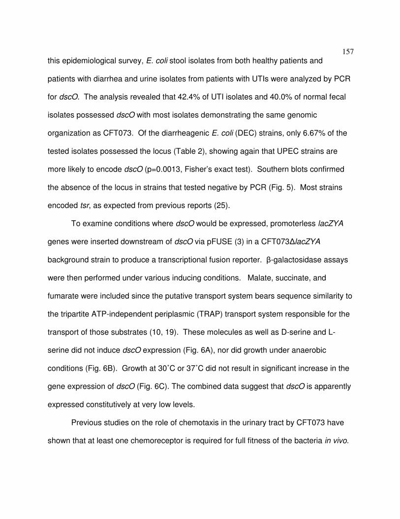
157
this epidemiological survey, E. coli stool isolates from both healthy patients and
patients with diarrhea and urine isolates from patients with UTIs were analyzed by PCR
for dscO. The analysis revealed that 42.4% of UTI isolates and 40.0% of normal fecal
isolates possessed dscO with most isolates demonstrating the same genomic
organization as CFT073. Of the diarrheagenic E. coli (DEC) strains, only 6.67% of the
tested isolates possessed the locus (Table 2), showing again that UPEC strains are
more likely to encode dscO (p=0.0013, Fisher’s exact test). Southern blots confirmed
the absence of the locus in strains that tested negative by PCR (Fig. 5). Most strains
encoded tsr, as expected from previous reports (25).
To examine conditions where dscO would be expressed, promoterless lacZYA
genes were inserted downstream of dscO via pFUSE (3) in a CFT073∆lacZYA
background strain to produce a transcriptional fusion reporter. β-galactosidase assays
were then performed under various inducing conditions. Malate, succinate, and
fumarate were included since the putative transport system bears sequence similarity to
the tripartite ATP-independent periplasmic (TRAP) transport system responsible for the
transport of those substrates (10, 19). These molecules as well as D-serine and L-
serine did not induce dscO expression (Fig. 6A), nor did growth under anaerobic
conditions (Fig. 6B). Growth at 30˚C or 37˚C did not result in significant increase in the
gene expression of dscO (Fig. 6C). The combined data suggest that dscO is apparently
expressed constitutively at very low levels.
Previous studies on the role of chemotaxis in the urinary tract by CFT073 have
shown that at least one chemoreceptor is required for full fitness of the bacteria in vivo.

158
Similarly, the CFT073 dscO deletion mutant was co-infected with wild-type CFT073
in the competitive mouse model of UTI to see if loss of this putative periplasmic binding
protein would result in attenuation of the mutant strain. Although the results of three
combined experiments show that there may be a slight trend toward attenuation (Fig. 7),
the results are not statistically significant (bladder p=0.09, kidney p=0.30). This result
suggests that DscO is not critical for successful infection of the urinary tract.
Discussion
Initial events in the colonization of the urinary tract by UPEC remain relatively
undetermined. Navigation up the urinary tract via chemotaxis could be the first step to
aid UPEC entry into the urethra and bladder. A UPEC-specific ligand present in urine
but found minimally in the digestive tract could provide the needed stimulus for UPEC
fecal contaminants occupying the urethra to initiate movement up the urinary tract. The
ability to chemotax toward a urine-specific attractant could also give UPEC an
advantage over other fecal organisms with a limited or different chemotactic repertoire.
D-serine is found in high concentrations in the urine but, by comparison, is present in
the digestive tract in trace amounts (13). Although various UPEC strains, including
CFT073, chemotax towards the same attractants that K-12 strains chemotax towards,
no UPEC-specific attractants capable of acting as a ligand for UPEC when tested within
their physiological concentrations were previously identified. Therefore, D-serine, while
not the sole possible attractant for UPEC, is a comparatively unique signal that could

159
stimulate ascension of the urinary tract by UPEC when present in its physiological
concentration.
In this study, we reported that chemotaxis of uropathogenic E.coli strain CFT073
toward D-serine is mediated by the Tsr chemoreceptor and a putative periplasmic
binding protein, DscO, when grown in minimal medium but not rich medium. The range
in which CFT073 responds to D-serine is within the physiological range found in urine
(4-114µg/mL). Chemotaxis toward D-serine could not be established in MG1655 in the
presence of tsrCFT073 and dscO. However, this inability could be attributed to copy
number effects that could disrupt the Tsr-DscO interaction. Attempts to purify DscO for
further biochemical analysis were unsuccessful, leaving the true function of DscO
uncertain.
Uropathogenic and nonpathogenic fecal isolates were more likely to encode
dscO than DEC isolates. Although the fecal isolates were isolated from healthy
patients, the ability of these isolates to cause disease in the urinary tract remains
undetermined. Urinary tract infections are hypothesized to originate from fecal
contamination of the urethra, leaving open the possibility that the fecal strains
possessing dscO could be capable of causing UTIs. However, as has been shown in
this study, the presence of dscO is not necessary for successful infection of the urinary
tract.
This study provides D-serine as one possible attractant in urine that may
stimulate ascension of the urinary tract. The ability to navigate the urinary tract may
result in more efficient colonization, lessening the possibility that the bacteria would be

160
dispelled by urine before being able to gain a foothold in the bladder. Further
biochemical studies are needed to determine if dscO is capable of binding D-serine.
Without this data, the actual function of DscO, especially when grown in rich medium,
remains in doubt.

161
Reference
1. Adler, J. 1973. A method for measuring chemotaxis and use of the method to
determine optimum conditions for chemotaxis by Escherichia coli. J. Gen.
Microbiol. 74:77-91.
2. Bacheller, C. D., and J. M. Bernstein. 1997. Urinary tract infections. Med. Clin.
N. Am. 81:719-730.
3. Baumler, A. J., R. M. Tsolis, A. W. M. van der Velden, I. Stojiljkovic, S. Anic,
and F. Heffron. 1996. Identification of a new iron regulated locus of Salmonella
typhi. Gene 183:207-213.
4. Boos, W., and H. Shuman. 1998. Maltose/maltodextrin system of Escherichia
coli: transport, metabolsim, and regulation. Micro. Mol. Biol. Rev. 62:204-229.
5. Brooks, T., and C. W. Keevil. 1997. A simple artificial urine for the growth of
urinary pathogens. Let. Appl. Microbiol. 24:203-206.
6. Brückner, H., S. Haasmann, and A. Friedrich. 1994. Quantification of D-amino
acids in human urine using GC-MS and HPLC. Amino Acids 6:205-211.
7. Datsenko, K. A., and B. L. Wanner. 2000. One-step inactivation of
chromosomal genes in Escherichia coli K-12 using PCR products. Proc. Natl.
Acad. Sci. U. S. A. 97:6640-6645.
8. Donnenberg, M. S., and R. A. Welch. 1996. Virulence determinants of
uropathogenic Escherichia coli, p. 135-174. In H. L. T. Mobley and J. W. Warren
(ed.), Urinary Tract Infections: Molecular Pathogenesis and Clinical
Management. ASM Press, Washington, D. C.

162
9. Eisenbach, M. 2007. A hitchhiker's guide through advances and conceptual
changes in chemotaxis. J. Cell. Physiol. 213:574-580.
10. Forward, J. A., M. C. Behrendt, N. R. Wyborn, R. Cross, and D. J. Kelly.
1997. TRAP transporters: a new family of periplasmic solute transport systems
encoded by the dctPQM genes of Rhodobacter capsulatus and by homologs in
diverse gram-negative bacteria. J. Bacteriol. 179:5482-5493.
11. Griebling, T. L. 2007. Urinary tract infection in men. Urologic Diseases in
America:621-646.
12. Griebling, T. L. 2007. Urinary tract infection in women. Urologic Diseases in
America:587-620.
13. Hashimoto, A., and T. Oka. 1997. Free D-aspartate and D-serine in the
mammalian brain and periphery. Prog. Neurobiol. 52:325-353.
14. Haugen, B. J., S. Pellett, P. Redford, H. L. Hamilton, P. L. Roesch, and R. A.
Welch. 2007. In vivo expression analysis identifies genes required for enhanced
colonization of the mouse urinary tract by uropathogenic Escherichia coli strain
CFT073 dsdA. Infect. Immun. 75:278-289.
15. Hazelbauer, G. L. 1975. Maltose chemoreceptor of Escherichia coli. J. Bact.
122:206-214.
16. Hazelbauer, G. L., R. E. Mesibov, and J. Adler. 1969. Escherichia coli mutants
defective in chemotaxis toward specific chemicals. Proc. Natl. Acad. Sci. U. S. A.
64:1300-1307.

163
17. Hedblom, M. L., and J. Adler. 1980. Genetic and biochemical properties of
Escherichia coli mutants with defects in serine chemotaxis. J. Bacteriol.
144:1048-1060.
18. Huang, Y., T. Nishikawa, K. Satoh, T. Iwata, T. Fukushima, T. Santa, H.
Homma, and K. Imai. 1998. Urinary excretion of D-serine in human: comparison
of different ages and species. Biol. Pharm. Bull. 21:156-162.
19. Janausch, I. G., E. Zientz, Q. H. Tran, A. Kroger, and G. Unden. 2002. C4-
dicarboxylate carriers and sensors in bacteria. Biochimica et Biophysica Acta
1553:39-56.
20. Johnson, J. R. 1991. Virulence factors in Escherichia coli urinary tract infection.
Clin. Microbiol. Rev. 4:80-128.
21. Kaper, J. B., J. P. Nataro, and H. L. T. Mobley. 2004. Pathogenic Escherichia
coli. Nat. Rev. Microbiol. 2:123-140.
22. Kossmann, M., C. Wolff, and M. D. Manson. 1988. Maltose chemoreceptor of
Escherichia coli: interaction of maltose-binding protein and the tar signal
transducer. J. Bacteriol. 170:4516-4521.
23. Kunin, C. M. 1994. Urinary tract infections in females. Clin. Infect. Dis. 18:1-10.
24. Lane, M. C., J. C. Alteri, S. N. Smith, and H. L. T. Mobley. 2007. Expression of
flagella is coincident with uropathogenic Escherichia coli ascension to the upper
urinary tract. Proc. Natl. Acad. Sci. U. S. A. 104:16669-16674.
25. Lane, M. C., A. L. Lloyd, T. A. Markyvech, E. C. Hagan, and H. L. Mobley.
2006. Uropathogenic Escherichia coli strains generally lack functional Trg and

164
Tap chemoreceptors found in the majority of E. coli strains strictly residing in
the gut. J. Bacteriol. 188:5618-5625.
26. Lane, M. C., V. Lockatell, G. Monterosso, D. Lamphier, J. Weinert, J. R.
Hebel, D. E. Johnson, and H. L. T. Mobley. 2005. Role of motility in the
colonization of uropathogenic Escherichia coli in the urinary tract. Infect. Immun.
73:7644-7656.
27. Lee, L., T. Mizuno, and Y. Imae. 1988. Thermosensing properties of Escherichia
coli tsr mutants defective in serine chemoreception. J. Bacteriol. 170:4769-4774.
28. Mesibov, R., and J. Adler. 1972. Chemotaxis toward amino acids in Escherichia
coli. J. Bacteriol. 112:315-326.
29. Mobley, H. L., D. M. Green, A. L. Trifillis, D. E. Johnson, G. R. Cippendale, C.
V. Lockatell, B. D. Jones, and J. W. Warren. 1990. Pyelonephritogenic
Escherichia coli and killing of cultered human renal proximal tubular epithelial
cells: role of hemolysin in some strains. Infect. Immun. 58:1281-1289.
30. Orenstein, R., and E. S. Wong. 1999. Urinary tract infections in adults. Am.
Fam. Physician 59:1225-1234.
31. Redford, P., and R. A. Welch. 2006. Role of sigma E-regulated genes in
Escherichia coli uropathogenesis. Infect. Immun. 74:4030-4038.
32. Schwan, W. R. 2007. Flagella allow uropathogenic Escherichia coli ascension
into murine kidneys. Int. J. Med. Microbiol. 298:441-447.
33. Snyder, J. A., B. J. Haugen, E. L. Buckles, C. V. Lockatell, D. E. Johnson, M.
S. Donnenberg, R. A. Welch, and H. L. Mobley. 2004. Transcriptome of

165
uropathogenic Escherichia coli during urinary tract infection. Infect. Immun.
72:6373-6381.
34. Wadhams, G. H., and J. P. Armitage. 2004. Making sense of it all: bacterial
chemotaxis. Nat. Rev. Mol. Cell. Biol. 5:1024-1037.
35. Yamamoto, S. 2007. Molecular epidemiology of uropathogenic Escherichia coli.
J. Infect. Chemother. 13:68-73.
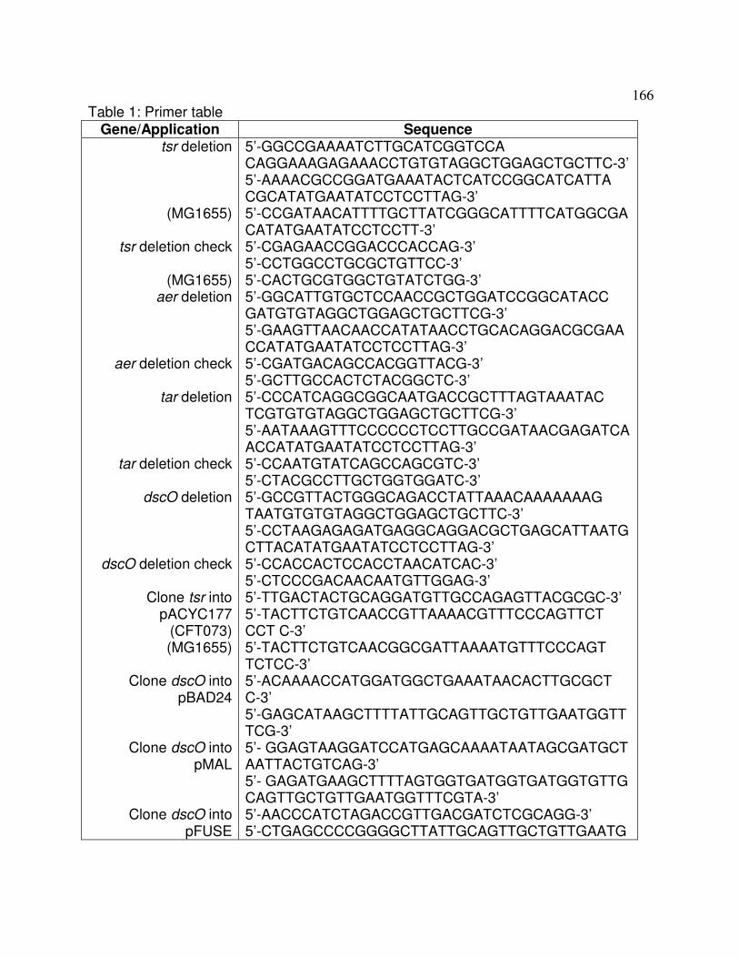
166
Table 1: Primer table Gene/Application Sequence
tsr deletion
(MG1655)
tsr deletion check
(MG1655) aer deletion
aer deletion check
tar deletion
tar deletion check
dscO deletion
dscO deletion check
Clone tsr into pACYC177
(CFT073) (MG1655)
Clone dscO into
pBAD24
Clone dscO into pMAL
Clone dscO into pFUSE
5’-GGCCGAAAATCTTGCATCGGTCCA CAGGAAAGAGAAACCTGTGTAGGCTGGAGCTGCTTC-3’ 5’-AAAACGCCGGATGAAATACTCATCCGGCATCATTA CGCATATGAATATCCTCCTTAG-3’ 5’-CCGATAACATTTTGCTTATCGGGCATTTTCATGGCGA CATATGAATATCCTCCTT-3’ 5’-CGAGAACCGGACCCACCAG-3’ 5’-CCTGGCCTGCGCTGTTCC-3’ 5’-CACTGCGTGGCTGTATCTGG-3’ 5’-GGCATTGTGCTCCAACCGCTGGATCCGGCATACC GATGTGTAGGCTGGAGCTGCTTCG-3’ 5’-GAAGTTAACAACCATATAACCTGCACAGGACGCGAA CCATATGAATATCCTCCTTAG-3’ 5’-CGATGACAGCCACGGTTACG-3’ 5’-GCTTGCCACTCTACGGCTC-3’ 5’-CCCATCAGGCGGCAATGACCGCTTTAGTAAATAC TCGTGTGTAGGCTGGAGCTGCTTCG-3’ 5’-AATAAAGTTTCCCCCCTCCTTGCCGATAACGAGATCA ACCATATGAATATCCTCCTTAG-3’ 5’-CCAATGTATCAGCCAGCGTC-3’ 5’-CTACGCCTTGCTGGTGGATC-3’ 5’-GCCGTTACTGGGCAGACCTATTAAACAAAAAAAG TAATGTGTGTAGGCTGGAGCTGCTTC-3’ 5’-CCTAAGAGAGATGAGGCAGGACGCTGAGCATTAATG CTTACATATGAATATCCTCCTTAG-3’ 5’-CCACCACTCCACCTAACATCAC-3’ 5’-CTCCCGACAACAATGTTGGAG-3’ 5’-TTGACTACTGCAGGATGTTGCCAGAGTTACGCGC-3’ 5’-TACTTCTGTCAACCGTTAAAACGTTTCCCAGTTCT CCT C-3’ 5’-TACTTCTGTCAACGGCGATTAAAATGTTTCCCAGT TCTCC-3’ 5’-ACAAAACCATGGATGGCTGAAATAACACTTGCGCT C-3’ 5’-GAGCATAAGCTTTTATTGCAGTTGCTGTTGAATGGTT TCG-3’ 5’- GGAGTAAGGATCCATGAGCAAAATAATAGCGATGCT AATTACTGTCAG-3’ 5’- GAGATGAAGCTTTTAGTGGTGATGGTGATGGTGTTG CAGTTGCTGTTGAATGGTTTCGTA-3’ 5’-AACCCATCTAGACCGTTGACGATCTCGCAGG-3’ 5’-CTGAGCCCCGGGGCTTATTGCAGTTGCTGTTGAATG

167
dscO PCR screen
tsr PCR screen
DEC strain tsr region
PCR
G-3’ 5’-TTATTGCAGTTGCTGTTGAATGGTTTCG-3’ 5’-ATGAGCAAAATAATAGCGATGCTAATTACTGTC-3’ 5’-GGCCGAAAATCTTGCATCGG-3’ 5’-CGTTAAAACGTTTCCCAGTTCTCCTC-3’ 5’-CCTTGATGCCACCCTTGC-3’ 5’-CACCGATGATTCCGAGAATCACC-3’

168
Table 2: Prevalence of dscO in E. coli
tsr dscO
CFT073 context
Screened
UTI (n=33)
Fecal
(n=15)
DEC (n=30)
87.9%
93.3%
90.0%
42.4%*
40.0%*
6.7%
42.4%
40.0%
6.7%
Sequenced
ExPEC (n=9)
Non-Path
(n=13)
DEC (n=28)
100%
92.3%
100%
77.8%†‡
15.4%†
3.6%‡
77.8%
15.4%
3.6%
Fisher’s exact test: *p=0.0013, †p=0.0073, ‡p=0.00003

169
Figure 1: Chemotaxis of CFT073 and MG1655 toward D-serine and L-serine.
Capillary chemotaxis assay results for (A) CFT073, (B) MG1655, (C) RP437 and (D)
CFT073∆tsr. *, p<0.05; ***, p<0.001. Bars indicate standard error (s.e.m.).

170
Figure 2: Chemotaxis of MG1655∆∆∆∆tsr+ptsrCFT073, CFT073∆∆∆∆tsr+ptsrMG1655,
MG1655∆tsr+ ptsrMG1655 and CFT073∆tsr+ptsrCFT073 toward D-serine and L-serine. (A)
Amino acid sequence differences between MG1655 Tsr (left) and CFT073 Tsr (right).
Capillary chemotaxis assay results for (B) CFT073∆tsr+ptsrMG1655, (C)
MG1655∆tsr+ptsrCFT073, (D) MG1655∆tsr+ptsrMG1655 and (E) CFT073∆tsr+ptsrCFT073.
The tsr alleles were cloned into pACYC177 under the control of their native promoters.

171
The result shown for MG1655∆tsr+Tn7::tsrCFT073 represents a single experiment. *,
p<0.05; **, p<0.01; ***, p<0.001. Bars indicate standard error (s.e.m.).
Figure 3: Effect of dscO on chemotaxis toward D-serine and L-serine. (A)
Genomic comparison of the region downstream of tsr in MG1655 and CFT073. Arrows

172
above genes show the placement of primers for the PCR screen reported in Table 1.
Capillary chemotaxis assay results for (B) CFT073∆dscO, (C)
CFT073∆dscO+pBAD24dscO, (D) MG1655∆tsr att::Tn7-tsrCFT073+pBAD24dscO (E)
MG1655∆tsr att::Tn7-tsrCFT073 and (F) CFT073∆dscO grown in rich medium.
Expression of dscO for the complementing strain was induced with 10µM arabinose and
all strains were grown in MOPS minimal media unless otherwise noted. *, p<0.05; **,
p<0.01. Bars indicate standard error (s.e.m.).

173
Figure 4: Attempted purification of MBP-DscO. (A) Coomassie-stained SDS-PAGE
gel of purified protein from amylose and nickel columns. (B) Immunoblot of purified
protein (~1mg total purified) from amylose and nickel columns using α-penta His tag
antibodies.

174
Figure 5: Southern blot analysis of strains tested in the dscO PCR screen. Blots
were probed with the dscO region of the CFT073 genome, including the entire putative
transport system. Each lane contains the digested genomic DNA of an individual E. coli
isolate. Results of the PCR screen for dscO and the strain origin are indicated above
each lane. PCR positive strains were included for comparison. A representative set of
strains was used for the DEC isolates. P=probe, D=DEC strain, F=fecal strain, U=UTI
strain, M=MG1655, and C=CFT073.

175

176
Figure 6: Expression of dscO under varying conditions. ß-galactosidase assay
results for dscO expression under (A) aerobic (n=3), (B) anaerobic (n=3), (C) and
different temperatures (n=1). Bars indicate standard error (s.e.m.).
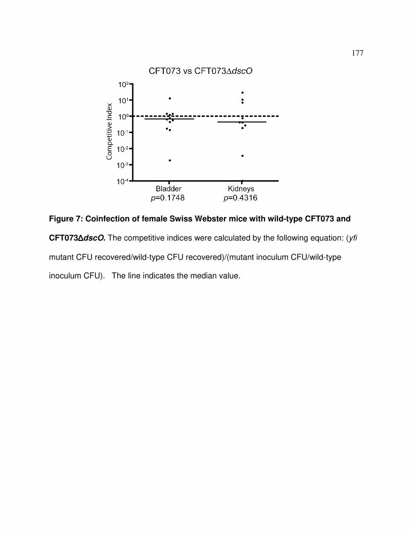
177
Figure 7: Coinfection of female Swiss Webster mice with wild-type CFT073 and
CFT073∆∆∆∆dscO. The competitive indices were calculated by the following equation: (yfi
mutant CFU recovered/wild-type CFU recovered)/(mutant inoculum CFU/wild-type
inoculum CFU). The line indicates the median value.

178
Appendix B
Reflux in the Mouse Model of Urinary Tract Infection
In preparation for publication
Authors and contributions:
Erica L. Raterman: Wrote manuscript and performed all experiments.
Rodney A. Welch: Supervised all work and helped in writing of the manuscript

179
Abstract
The mouse model of urinary tract infection has endured controversy over whether it is
an accurate model for ascending urinary tract infections. This report provides additional
evidence that the kidneys are infected during inoculation via reflux, making this model
suitable only for reliable prediction of persistence but not ascension factors.

180
The mouse model of urinary tract infection was first developed in 1983 by
Hagberg et al. (1). Over the following two decades, several reports emerged showing
the immediate infection of the kidneys, presumably due to reflux up the ureters as a
result of the pressure administered during inoculation. Although the model was still
deemed suitable for the determination of virulence factors such as fimbriae for
attachment to epithelial cells, the model did not constitute a true model of ascending
urinary tract infection due to this immediate infection of the entire urinary tract.
Subsequently, several reports gave different inoculation volumes (10 µl, 25 µl, and 50
µl) that purportedly would cause limited to no immediate reflux into the kidneys, thus
allowing assessment of factors needed for ascension from the bladder to the kidneys (2-
5). However, these reports disagreed on both the volume of inoculation that would
produce reflux and the method of inoculation. One report alleged that administration of
a 50 µl inoculum by a Harvard pump to control flow rate would limit reflux (5, 6), yet a
second report found that, even when using the Harvard pump, the inoculum volume had
to be reduced to 25 µl to avoid reflux (3). A third report found that the inoculum had to
be reduced to 10 µl to avoid reflux when administering the inoculum by hand, and had
to be placed in the urethra, not the bladder (2). Other factors such as mouse strain,
mouse weight, and euthanasia method also were also noted as variables that could
affect reflux during and subsequent to inoculation (3).
With no clear consensus on which method would reliably prevent reflux, we
sought to determine whether a small volume of only 2 µl (~2 x 10⁷ CFU) administered

181
into the urethra instead of the bladder of 6-7 week old female mice could be used to
simulate an ascending urinary tract infection. Attempts were made to place the
inoculum in the urethra distal to the urethral sphincter; however, the infection did not
progress to the bladder and kidneys even after two weeks of monitoring. Therefore, the
2 µl inoculum was instead placed proximal to the sphincter, resulting in direct
inoculation of the bladder. Immediately after inoculation, mice were euthanized by CO2
asphyxiation and the spleen, kidneys, and bladder were aspectically harvested in that
order. Organs were homogenized and dilution plated on MacConkey’s agar for
incubation overnight at 37ºC. This procedure was used with three different mouse
strains (C3H/HeJ (Jackson Labs), Swiss Webster (Harlan), and CBA/J (Harlan)), and
results showed that bacteria could be detected in the bladder and kidneys of all three
strains, although fewer bacteria were present in mice inoculated with 50 µl (~5x10⁷
CFU) of inoculum (Fig. 1 A-C). All mice with bacteria present in the spleen were
removed from the analysis and the peritonea of all mice were sterilized before extraction
of the organs to ensure that no contaminating bacteria were present. Because bacteria
in the bloodstream may not always be detected in the spleen, two other experiments
were performed to rule out the possibility that bacteria had gained access to the
bloodstream during the trauma of inoculation, and thereby reached the kidneys via a
venous route instead of by reflux up the ureters. Firstly, the mice were asphyxiated
prior to inoculation to remove blood flow as a possible carrier of the bacteria to the
kidneys. One ureter was then ligated in two places by tying a knot with silk sutures to
differentiate between bacteria in the kidney acquired through reflux or through some

182
other means. As shown in Figure 2A, inoculation post-mortem did eliminate bacteria
in the kidneys with the 2 µl inoculum, but not with the 50 µl inoculum in the non-ligated
kidney. Finally, live mice with one ureter surgically ligated in two places were
purchased from Jackson Labs (USA) and inoculated with either 2 µl or 50 µl of
inoculum. Bacteria were found only in the non-ligated kidney (Fig. B) of the surgically
altered mice. Together these results indicate that reflux via the ureters is indeed the
route that the bacteria gain access to the kidneys at the time of inoculation.
Our data indicate that as little as 2 µl of a bacteria inoculum is sufficient to reach
the kidneys during inoculation. We also, for the first time, have shown that the bacteria
reach the kidneys via reflux up the ureters, as was previously assumed. Although
bacteria are not found in the kidneys in all animals tested, enough of the mice had
bacteria in the kidneys to make this method an unreliable model for assessing bacterial
factors needed for ascension of the urinary tract during infection.
Acknowledgements
This research was funded by the National Institutes of Health Grant R01 DK063250-06
and by the National Institutes of Health, National Research Service Award AI55397.

183
References
1. Hagberg, L., I. Engberg, R. Freter, J. Lam, S. Olling, and C. Svanborg Eden.
1983. Ascending, unobstructed urinary tract infection in mice caused by
pyelonephritogenic Escherichia coli of human origin. Infect. Immun. 40:273-283.
2. Hopkins, W. J., J. A. Hall, B. P. Conway, and D. T. Uehling. 1995. Induction of
urinary tract infection by intraurethral inoculation with Escherichia coli: refining
the murine model. J. Infect. Dis. 171:462-465.
3. Johnson, J. R., and J. J. Brown. 1996. Defining inoculation conditions for the
mouse model of ascending urinary tract infection that avoid immediate
vesicoureteral reflux yet produce renal and bladder infection. J. Infect. Dis.
173:746-749.
4. Johnson, J. R., and J. C. Manivel. 1991. Vesicoureteral reflux induces renal
trauma in a mouse model of ascending, unobstructed pyelonephritis. J. Urol.
145:1306-1311.
5. Lane, M. C., C. J. Alteri, S. N. Smith, and H. L. Mobley. 2007. Expression of
flagella is coincident with uropathogenic Escherichia coli ascension to the upper
urinary tract. Proc. Natl. Acad. Sci. U. S. A. 104:16669-16674.
6. Mobley, H. L., K. G. Jarvis, J. P. Elwood, D. I. Whittle, C. V. Lockatell, R. G.
Russell, D. E. Johnson, M. S. Donnenberg, and J. W. Warren. 1993. Isogenic
P-fimbrial deletion mutants of pyelonephritogenic Escherichia coli: the role of

184
alpha Gal(1-4) beta Gal binding in virulence of a wild-type strain. Mol.
Microbiol. 10:143-155.
Figure 1: Colony counts in the bladder and kidneys 5 minutes after inoculation.
Results for the CFUs found in each organ in C3H/HeJ mice (n=10, n=9) (A), Swiss
Webster mice (n=3, n=9) (B), and CBA/J mice (n=5, n=4) (C) for both 2 µl and 50 µl
inoculation volumes are shown. The line indicates the median value.

185
Figure 2: Surgical ligation of the left ureter blocks reflux into the kidneys. Bladder
and kidney CFUs recovered 5 minutes after inoculation for mice infected post-mortem
(A) and live (B) are shown. The line indicates the median value.

186
Appendix C
In vitro and in vivo phenotypes for swim and swarm mutants not used in
manuscripts

187
Appendix C summarizes data obtained for other identified swimming or swarming
mutants that will not be published.
One of the original projects of this thesis sought to determine whether swimming
motility and/or swarming motility was important for colonization and persistence in the
urinary tract. To differentiate between the two motility types in vivo, a series of mutants
that could either swim but not swarm, or could swarm but not swim, were sought. Two
screens for motility performed in E. coli K-12 identified a number of mutants that were
defective for either swimming motility or swarming motility (3, 5). Other papers studying
Salmonella motility also identified promising motility mutants (1, 7). A subset of these
mutants was selected for study in CFT073 motility based on their predicted phenotypes
of swim+/swarm- or swim-/swarm+. Care was also taken to select mutants that would
not interfere with core metabolic processes. The final set of genes selected to be
deleted from the CFT073 genome included yhjH (predicted phosphodiesterase), yehP
(unknown function), fliR (flagellar biosynthesis protein), fliL(flagellar basal body-
associated protein), flhE (flagellar protein), and yojN (previously called rcsD in E. coli K-
12) (predicted sensor-like histidine kinase) (primers for deletion of each gene via λ-Red
are listed in Table 1). The expected motility phenotypes from the previous reports and
the experimentally derived motility phenotypes in CFT073 are summarized in Table 2.
While mutants that were defective for swarming motility but not swimming motility were

188
found, no mutants resulted in a non-swimmer that could still swarm (Fig. 1).
Swarming motility phenotype was confirmed by microscopic examination of the swarm
front movement. As a result, the project overall was not pursued. However, a few
mutants were selected for testing in the competitive mouse model of urinary tract
infection. The in vivo results for the yojN, yhjH, and flhE deletion mutants in competition
with the wild-type CFT073 strain are shown in Figure 2. The yhjH (Fig. 2C) and flhE
(Fig. 2B) mutants both colonized as well as the wild-type strain, although the yhjH did
yield a slightly attenuated phenotype in the bladder that was statistically significant. The
yojN mutant was also attenuated in the bladder and kidneys (Fig. 2A). However, this
phenotype is likely due to more than just a defect in motility as YojN is part of the Rcs
signaling system whose regulon has multiple effects on bacterial virulence factor
expression (2, 4, 6). Despite the complexity involved, further study with this mutant
could yield potential insights into how E. coli CFT073 regulates expression of its
virulence and colonization factors in response to the urinary tract environment.
Materials and Methods
Construction of mutant strains
All primers used for the generation of deletion mutants and cloning are listed in
Table 2. All nonpolar gene deletions were performed using the λ-Red recombination
system developed by Datsenko and Wanner . The PCR products used for
transformations in CFT073 were generated off of pKD3 or pKD4 templates using
primers specific for the targeted gene. The kanamycin or chloramphenicol-marked

189
gene deletions were then transduced into a fresh CFT073 background using the
EB49 transducing phage. The antibiotic resistance cassette used to replace the target
gene was removed using a Flp recombinase encoded on pCP20. All gene deletions
were verified by PCR and loss of antibiotic resistance on LB agar containing the
appropriate antibiotic.
Murine model of UTI
Colonization of the urinary tract was determined using the competitive murine
model of urinary tract infection as described previously. CFT073∆lacZYA was used as
the wild-type strain and the chemotaxis mutants had an intact lacZYA locus. To select
for piliated bacteria, all bacterial strains were grown statically in 3 mL LB at 37˚C for 2
days. The pellicle formed on the rim of the test tube was then transferred to fresh LB,
incubated for 2 more days, and finally passaged again to 40 mL LB for a final 2 day
incubation. The broths were adjusted to OD600~0.4 with 1XPBS and the wild-type strain
and the mutant strain were mixed equally. The mixed broth was then pelleted and
washed 1X in 1XPBS and resuspended in 500 µl 1XPBS. Isofluorane-anesthetized 6-7
week-old female Swiss Webster mice (Harlan, USA) were inoculated via urethral
catheterization with 50 µl (~108 CFU) of the mixed bacterial suspension. Mice were
euthanized via CO2 asphyxiation and the bladder and kidneys were aseptically
harvested in 1XPBS. The organs were homogenized, serially diluted in 1XPBS, and
plated on MacConkey agar medium (Fisher, USA). Surviving strain ratios were
determined by counting white (wild-type) and red (mutant) colonies. Colonization levels

190
were graphed and analyzed using a paired Wilcoxon signed ranked test and Prism
(GraphPad). Competitive indices were calculated by dividing mutant CFUs by wild-type
CFUs from the mice and then dividing this ratio by the ratio of mutant to wild-type in the
original inoculum. All animal experiments were approved by the UW-Madison Animal
Care and Use Committee.
Motility Assays
Swimming motility was assessed in 0.3% agar tryptone plates. Strains were
grown overnight at 37°C in tryptone broth. Broths were adjusted to OD600~0.1 with
fresh tryptone broth before 2 µl was injected in the center of the agar swim plates. 10 µl
of the same broths were placed on the surface of swarm agar plates to assess
swarming motility. Swim plates were incubated 12 hours at 30°C, and swarm plates
were incubated at 30°C for 2 days. Assays were independently repeated at least three
times.

191
References
1. Attmannspacher, U., B. E. Scharf, and R. M. Harshey. 2008. FliL is essential
for swarming: motor rotation in absence of FliL fractures the flagellar rod in
swarmer cells of Salmonella enterica. Mol. Microbiol. 68:328-341.
2. Dominguez-Bernal, G., M. G. Pucciarelli, F. Ramos-Morales, M. Garcia-
Quintanilla, D. A. Cano, J. Casadesus, and F. Garcia-del Portillo. 2004.
Repression of the RcsC-YojN-RcsB phosphorelay by the IgaA protein is a
requisite for Salmonella virulence. Mol. Microbiol. 53:1437-1449.
3. Girgis, H. S., Y. Liu, W. S. Ryu, and S. Tavazoie. 2007. A comprehensive
genetic characterization of bacterial motility. PLoS Genet. 3:1644-1660.
4. Hagiwara, D., M. Sugiura, T. Oshima, H. Mori, H. Aiba, T. Yamashino, and T.
Mizuno. 2003. Genome-wide analyses revealing a signaling network of the
RcsC-YojN-RcsB phosphorelay system in Eschrichia coli. J. Bacteriol. 185:5735-
5746.
5. Inoue, T., R. Shingaki, S. Hirose, K. Waki, H. Mori, and K. Fukui. 2007.
Genome-wide screening of genes required for swarming motility in Escherichia
coli K-12. J. Bacteriol. 189:950-957.
6. Mouslim, C., M. Delgado, and E. A. Groisman. 2004. Activation of the
RcsC/YojN/RcsB phosphorelay system attenuates Salmonella virulence. Mol.
Microbiol. 54:386-395.

192
7. Stafford, G. P., and C. Hughes. 2007. Salmonella typhimurium flhE, a
conserved flagellar regulon gene required for swarming. Microbiology 153:541-
547.

193
Table 1: Primers used in this study
Gene/Application Sequence
flhE deletion
flhE deletion ck
yojN deletion
yojN deletion ck
yehP deletion
yehP deletion ck
yhjH deletion
yhjH deletion ck
fliR deletion
fliR deletion ck
fliL deletion
fliL deletion ck
fliC deletion
5’-CATATCCGTATGACGGCGACGATTGGCGGCAAATAATG TGTGTAGGCTGGAGCTGCTTC-3’ 5’-CCGATGTTATTGCAGACATCGGCTTTTTTATCATTACAT ATGAATATCCTCCTTAG-3’ 5’-CACGCCGCATGTGATGTG-3’ 5’-CGATACACCGCTGGAACG-3’ 5’-ATCACTTCGCGAAGTTTTAACAGGTCATAAACACGAAT GTGTGTAGGCTGGAGCTGCTTC-3’ 5’-GCTACAGCAAGCTCTTGACATAACTGTCAATGTCGCTG ATCATATGAATATCCTCCTTAG-3’ 5’-CTGCTACCAGCAGAGCTGG-3’ 5’-GAGAGCCTTCGGCAAATCGG-3’ 5’-CATCGCTGCAACACTGGGGAGTTTTCCATGTCTGAACT GTGTGTAGGCTGGAGCTGCTTC-3’ 5’-TTCCGGACGTAGTGAATTCATGACTGAAGATTCTCCGC AACATATGAATATCCTCCTTAG-3’ 5’-CCATCAACTGGCCTGTCAGC-3’ 5’-GCAGGTGCACTGAGCCTC-3’ 5’-TACACCTGGCAGCCGATCTATCAAACATGCGGGCGGT TATGTGTAGGCTGGAGCTGCTTC-3’ 5’-CCTAAAGATAGTCCAGCCAGGCGGAAAATGAGGCAGC TTACATATGAATATCCTCCTTAG-3’ 5’-GCGAACGGACGATTCAACTCC-3’ 5’-GAAACCGCGCTTAACGTCC-3’ 5’-CACTAACCTGCCGTATATCATCGGGTAGCCCGTACAAT GTGTGTAGGCTGGAGCTGCTTC-3’ 5’-GGATAATCCTTAGGGTAGCATGATAAACGTTACGGAAT TACATATGAATATCCTCCTTAG-3’ 5’-GATGAAAGTCGCGCTGGC-3’ 5’-CACCTAATGCGCCACTCTCG-3’ 5’-CGATAAGCAGTAGCGACACAGGAAGACCGCAACACAT GTGTGTAGGCTGGAGCTGCTTCG-3’ 5’-TTCAGCTTGAGAAAGAATACTATCGCCCATGTCGTTAA TGAATATCCTCCTTAG-3’ 5’-GCGGCGTTGATATTTTCGCC-3’ 5’-CAGCGCCTGCAAACGTTC-3’ 5’-GGCGTTGCCGTCAGTCTCAGTTAATCAGGTTACGGCGA TGTGTAGGCTGGAGCTGCTTC-3’ 5’-AAAACGTAATCAACGACTTGCAATATAGGATAACGAATC CATATGAATATCCTCCTTAG-3’

194
fliC deletion ck
5’-CGCAATGGGTCTGGCTGTG-3’ 5’-GAGTGATGGTCAGCGTCTGGTTGC-3’
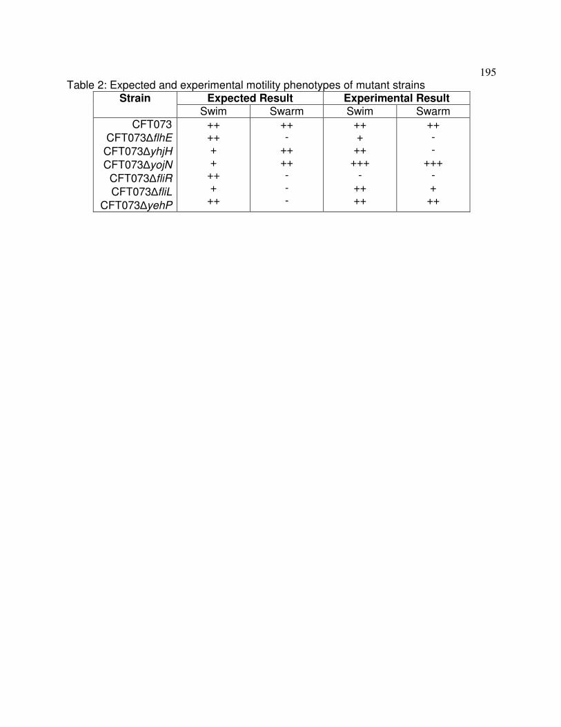
195
Table 2: Expected and experimental motility phenotypes of mutant strains Expected Result Experimental Result Strain
Swim Swarm Swim Swarm CFT073
CFT073∆flhE
CFT073∆yhjH
CFT073∆yojN
CFT073∆fliR
CFT073∆fliL
CFT073∆yehP
++ ++ + +
++ +
++
++ -
++ ++ - - -
++ +
++ +++
- ++ ++
++ - -
+++ - +
++

196

197
Figure 1: Swim and swarm phenotypes of predicted motility mutants in E. coli
CFT073. Strains were inoculated either in 0.3% tryptone agar swim plates (A) or on
0.45% agar swarm plates (B) and incubated at 30° C for either 12 hours or 2 days,
respectively. The wild-type CFT073 strain is shown on the left and the motility mutant is
shown on the right for the swarm plates.

198
Figure 2: Coinfection of female Swiss Webster mice with wild-type CFT073 and
the motility mutants. Results for the CFT073∆yojN (n=12) (A), CFT073∆yhjH (n=21)
(B), and CFT073∆flhE (n=13) (C) are shown. The competitive indices were calculated
by the following equation: (yfi mutant CFU recovered/wild-type CFU recovered)/(mutant
inoculum CFU/wild-type inoculum CFU). The line indicates the median value.

199
Appendix D
Atypical Shigella boydii 13 encodes virulence factors seen in attaching and
effacing Escherichia coli
Published in FEMS Microbiology Letters (2012) 328: 20-5.
Authors and contributions:
Laura L. Walters: Wrote manuscript and performed all experiments except as noted
below.
Erica L. Raterman: Revised manuscript and performed some of the PCR and southern
screens for stcE.
Thomas E. Grys: Initially developed project.
Rodney A. Welch: Supervised all work and contributed to the writing of the manuscript.

200
Abstract
Enterohemorrhagic E. coli (EHEC) is a foodborne pathogen that causes watery
diarrhea and hemorrhagic colitis. In this study, we identified StcE, a secreted zinc
metalloprotease that contributes to intimate adherence of EHEC to host cells, in culture
supernatants of atypical Shigella boydii 13 (Shigella B13) strains. Further examination
of the Shigella B13 strains revealed that this cluster of pathogens does not invade but
forms pedestals on HEp-2 cells similar to EHEC and enteropathogenic E. coli. This
study also demonstrates that atypical Shigella B13 strains are more closely related to
attaching and effacing E. coli and that their evolution recapitulates the progression from
ancestral E. coli to EHEC.

201
Introduction
Enterohemorrhagic Escherichia coli (EHEC) cause diarrheal disease that ranges
from watery diarrhea to hemorrhagic colitis. Virulence factors of EHEC include the
chromosomally-encoded Shiga toxin and the locus of enterocyte effacement (LEE). LEE
is a 35 kb pathogenicity island that confers the attaching and effacing phenotype to both
EHEC and enteropathogenic E. coli (EPEC), wherein intimate adherence of the bacteria
to host cells induces formation of actin-rich pedestals beneath the bacteria. The majority
of the clinical EHEC disease in United States is caused by serotype O157:H7 (Manning
et al., 2007), which carries a 92 kb virulence plasmid, pO157, that encodes many
potential virulence factors, including stcE (Burland et al., 1998).
The stcE gene is encoded on the large virulence plasmids of E. coli O157:H7,
O157:H-, ON:H7, and O55:H7 (Lathem et al., 2003). In all cases, stcE is found linked to
etpD, which encodes the subunit of the type II secretion apparatus responsible for the
secretion of StcE protein (Lathem et al., 2002). StcE is a 96 kDa zinc metalloprotease
that cleaves specific O-linked glycoproteins and contributes to the intimate adherence of
E. coli O157:H7 to HEp-2 cell surfaces (Grys et al., 2005). Evidence supports a role for
StcE in EHEC disease in clearing the mucus that forms a protective barrier over the
colonic epithelium. Following colonization, intimate adherence, and pedestal formation
by EHEC, the clinical syndrome progresses from watery diarrhea to hemorrhagic colitis.
At this stage, StcE plays an anti-inflammatory role by localizing the human complement
regulator, C1 esterase inhibitor (C1-INH), to cell surfaces, decreasing the complement-
mediated lysis of both bacteria and host cells (Lathem et al., 2004; Grys et al., 2006).

202
Shigella, another enteropathogen, is indistinguishable from E. coli by DNA-
DNA hybridization techniques, with the exception of Shigella boydii 13 (Shigella B13)
(Pupo et al., 2000). Shigella B13 is more closely related to Escherichia albertii than the
E. coli-Shigella group, and lacks the large virulence plasmid, (pINV), that confers the
invasion phenotype in all other Shigella. Hyma et al. demonstrated that Shigella B13
and E. albertii strains carry eae, a marker for LEE (Hyma et al., 2005). A small subset of
analyzed Shigella B13 strains encoding eae were more related to the E. coli-Shigella
group and labeled atypical Shigella B13. Many of these strains also carried markers for
the pO157 plasmid, such as ehxA and toxB, suggesting that atypical Shigella B13 may
be similar to EHEC and, thus, may encode stcE. This study describes the identification
of stcE in atypical Shigella B13 strains and the genetic and phenotypic profile of this
unique cluster of Shigella.
Materials and Methods
Bacterial culture, DNA extraction and PCR amplification
The S. boydii 7 and 13 and E. albertii strains used in this study are listed in table
2 and were provided by Thomas Whittam. E. coli O157:H7 EDL933 and E. coli O127:H6
E2348/69 were provided by Alison O’Brien. E. coli K12 MG1655 and S. flexneri 5a
M90T were provided from Fred Blattner. Internal fragments of Shigella (Venkatesan et
al., 2001) and E. coli (Burland et al., 1998) genes were amplified using the primers
shown in table 1. Strains stored at −80°C in Luria-Bertani (LB) medium with 50%
glycerol were directly inoculated into PCR reactions with GoTaq polymerase (Promega).

203
The stcE gene was sequenced from PCR products amplified with primers IR ApaI 5’
1 and etpD 3’ 1803 (table 1) and TripleMaster polymerase (Eppendorf) from plasmid
DNA extracted from the atypical Shigella B13 strains using a Maxi Prep Kit (Qiagen).
The nucleotide sequence for the stcE gene from the atypical Shigella B13 strains 3556-
77, 3557-77, 3052-94, and 3053-94 have been submitted to GenBank under accession
numbers EU159265, EU159266, EU159267, EU159268, respectively. For Southern blot
analysis, plasmid DNA isolated from the atypical Shigella B13 strains was
electrophoresed on a 0.6% agarose gel. Gel and stcE probe preparation and
hybridization were performed as previously described (Lathem et al., 2003).
StcE activity assay
To examine secretion of StcE, strains were grown in 25 ml Lennox L broth
overnight at 37°C with aeration and cells removed by centrifugation. Three mls of
culture supernatant were precipitated with 10% trichloroacetic acid on ice for ≥1 hour.
To measure StcE activity, 12 ml of culture supernatant were incubated with 0.5 µg C1-
INH protein (CompTech) overnight at room temperature prior to TCA precipitation.
Precipitated protein was separated by SDS-PAGE, transferred to nitrocellulose, and
immunoblotted with polyclonal anti-rStcE’ antisera (Grys et al., 2005) or anti-C1-INH IgG
(Cedarlane Laboratories).

204
Invasion assay
The gentamicin protection assay was used to determine the invasion phenotypes
of the atypical Shigella B13 strains (Elsinghorst et al., 1994). A colony of each strain
grown overnight on LB agar was inoculated into 2 ml of LB broth and incubated
statically overnight at 37oC. Overnight culture (40 µl) was diluted into a total volume of 1
ml of HEp-2 media (EMEM, 1 mM sodium pyruvate, 10% FBS) prior to the addition to a
monolayer of HEp-2 cells in a 24-well tissue culture plate (MOI of 14-95) and incubated
at 37°C in 5% CO2 for 2 hours. Monolayers were washed with Dulbecco’s PBS (D-PBS)
and fresh media containing 100 µg/ml gentamicin added for an additional 2 hours. The
monolayers were washed with D-PBS and lysed with 1 ml 0.1% Triton X-100 per well.
Suspensions were serially diluted and plated onto LB agar. Results are presented as
the average percent of inoculum recovered after gentamicin treatment and are
representative of duplicate samples in three independent experiments. Statistical
analysis was preformed using a one-way ANOVA with a Tukey’s post hoc test.
Pedestal formation assay
To determine the ability of atypical Shigella B13 strains to form pedestals, HEp-2
cells were seeded onto 8-well microscope slides (Nalge Nunc International) 48 hours
prior to infection so that cells would reach 50-80% confluency. Overnight bacterial
cultures (10 µl of 2.5x10⁸ to 9.0x10⁸ CFUs/ml) grown as for the invasion assay were
diluted into a total volume of 250 µl with HEp-2 media and added to each well of
washed HEp-2 cells. The mixtures were incubated at 37°C in 5% CO2 for a total of 6-7

205
hours with a media exchange after 3 hours. Wells were washed with D-PBS and the
cells fixed with 3% paraformaldehyde and permeabilized with 0.1% Triton X-100.
Bacterial cells were stained with 1:200 goat anti-lipid A (Abcam), followed by 1:200 anti-
goat-Alexa 488 and HEp-2 cells stained with 1:100 phalloidin-Alexa 594 (Invitrogen).
Preparations were mounted with Prolong Gold (Invitrogen) and analyzed by
epifluorescence microscopy (Carl Zeiss MicroImaging Inc.).
Results
PCR screen for presence of stcE in atypical S. boydii and E. albertii strains.
We set out to identify stcE in other bacterial species recently found to carry eae,
the gene that encodes the bacterial adhesin (intimin) required for pedestal formation. A
PCR screen of numerous S. boydii and E. albertii strains showed that an internal
fragment of stcE can be PCR amplified from only a subset of the S. boydii strains known
as atypical S. boydii 13 (table 2). Atypical Shigella B13 strains 3557-77, 3556-77, 3052-
94, and 3053-94, which form a distinct phylogenetic cluster, were all positive for the
stcE gene. Atypical Shigella B13 strain 5216-70, which is phylogenetically clustered with
enteroinvasive E. coli strains, was negative for the stcE gene. The presence or absence
of the stcE gene in all strains was confirmed by Southern blot (data not shown).
Analysis of isolated plasmid DNA by Southern blot demonstrated that stcE was encoded
on the large plasmid of the four atypical Shigella B13 strains (data not shown).
Sequence analysis of the 2.7 kb stcE gene showed only three synonymous
substitutions shared among the atypical Shigella B13 strains and a Q727L substitution

206
in strain 3556-77 compared to the EHEC EDL933 allele (data not shown). Six
substitutions within 220 nucleotides of the intergenic region upstream of the predicted
stcE promoter are present in the plasmids of all four atypical Shigella B13 strains
compared to pO157.
Activity of StcE in atypical S. boydii B13 strains.
To determine if the StcE protein was expressed and secreted by the atypical
Shigella B13 strains, TCA-precipitated supernatants of overnight cultures were analyzed
by immunoblot. StcE protein was identified in supernatants from strains 3556-77, 3052-
94, and 3053-94, but not from 3557-77 or 5216-70 (table 2). StcE activity in culture
supernatants was assayed for C1-INH proteolysis by immunoblots, and detected with all
atypical Shigella B13 strains except 3557-77 and 5216-70 (Fig. 1, table 2).
Atypical Shigella B13 strains encode other EHEC virulence factors.
To determine if the atypical Shigella B13 plasmid encoding stcE is similar to the
large invasion plasmid of Shigella (pINV), several pINV-encoded virulence factors were
sought by PCR amplification (table 2). None of the pINV-encoded virulence factors
could be amplified from the atypical Shigella B13 strains. PCR analysis using primers
specific for pO157-encoded genes resulted in amplification of etpD, but not katP. The
gene, traC, which is an F plasmid gene that is also encoded on the large virulence
plasmid of E. coli O157:H-, pSFO157, did not PCR amplify from any of the atypical
Shigella B13 strains tested.

207
The presence of additional E. coli-specific chromosomally-encoded genes
was determined by colony PCR (table 2). The LEE-encoded regulator (Ler) is a global
virulence regulator that has been shown to positively regulate the expression of LEE
(Mellies et al., 1999), stcE, and the etp operon in E. coli O157:H7 (Lathem et al., 2002).
PCR analysis of the atypical Shigella B13 strains identified the ler gene in the four
atypical Shigella B13 strains encoding eae and stcE. An additional LEE-encoded gene,
espA, encodes a subunit of the type III secretion system unique to EPEC and EHEC
and is encoded by the atypical Shigella B13 strains encoding eae and stcE. PCR
analysis of cadA, which encodes lysine decarboxylase and is universally absent in
Shigella but present in most E. coli strains (Day et al., 2001), revealed that none of the
atypical Shigella B13 strains encoded cadA.
Some atypical Shigella B13 strains invade and form pedestals on HEp-2 cells like
E. coli.
The abilities of the atypical Shigella B13 strains to invade HEp-2 cells were
determined. Strains 3556-77 and 3557-77 showed invasion levels below the level of
detection, whereas strains 3052-94 and 3053-94 showed relative levels of invasion
more similar to E. coli than S. flexneri strains (Fig. 2A). The presence of the LEE operon
and stcE suggested that the atypical Shigella B13 strains might form pedestals on host
cells. We tested this hypothesis by infecting HEp-2 cells and observing for co-
localization of bacteria with actin bundles on the surface of cells. Pedestal formation on

208
HEp-2 cells could be detected for atypical Shigella B13 strains 3556-77, 3052-94,
and 3053-94, but not 3557-77 (Fig. 2B).
Discussion
In this study, we discovered the stcE gene in the atypical Shigella B13 cluster.
The relatively low incidence of three nucleotide substitutions within the 2.7 kb stcE gene
compared to the six nucleotide substitutions within 220 nucleotides of the upstream
intergenic region suggests selection for the preservation of StcE function. The
acquisition of the large plasmid carrying stcE and the etp operon, in combination with
the LEE element encoded on the chromosome, may provide a selective advantage by
increasing the level of intimate adherence to host cells. A role of StcE in intimate
adherence is further supported by the observation that a lack of extracellular StcE
coincides with absence of pedestal formation by strain 3557-77.
The current model of Shigella evolution proposes that multiple ancestral E. coli
clones acquired the pINV Shigella invasion plasmid, leading to selection for the loss of
traits such as motility and lysine decarboxylation (Pupo et al., 2000). In contrast, the
atypical Shigella B13 strains show loss of E. coli traits in the apparent absence of pINV
selective forces. Furthermore, strains 3556-77 and 3557-77 display metabolic
phenotypes intermediate between Shigella and E. coli, and atypical Shigella B13 DNA is
more similar to E.coli than other Shigella B13 strains based on DNA-DNA hybridization
assays (Brenner et al., 1982). These atypical Shigella B13 strains also form a distinct
phylogenetic cluster and possess intermediate chromosomal genotypes between E. coli

209
and Shigella groups (Hyma et al., 2005). As was previously suggested by Hyma et
al., these data indicate that the atypical Shigella B13 strains were misclassified as
Shigella and that they actually represent a lineage that evolved from ancestral forms of
Shigella and attaching and effacing E. coli. The data presented here strengthen this
argument by showing the acquisition of LEE and a pO157-like plasmid encoding stcE
which we suggest recapitulates the model of EHEC evolution, described as the step-
wise acquisition of the LEE element, followed by pO157 and then the Shiga toxin phage
(Reid et al., 2000). We therefore propose to reclassify the atypical Shigella B13 strains
as an E. coli group that, through convergent evolution or horizontal transfer of virulence
genes on an ancestral background that shared both E. coli and Shigella characteristics,
has evolved to closely resemble pathotypes of E. coli that form attaching and effacing
lesions.
Sequelogs of stcE, historically named tagA, have been functionally characterized
in other pathogens, including Vibrio cholerae and Aeromonas hydrophila (Szabady et
al., 2010 and Pillai et al., 2006). Like StcE, V. cholerae TagA is a secreted mucinase
and contributes to colonization of the intestinal epithelium (Szabady et al., 2010). The A.
hydrophilia TagA exhibits an additional StcE function by cleaving and localizing C1-INH
the surface of bacterium, increasing the serum resistance of the bacterium in vitro. An
isogenic deletion mutant of tagA decreased the mortality of mice compared to wild-type
A. hydrophila in a mouse model of peritonitis (Pillai et al., 2006). Thus, StcE-like
metalloproteases play a role in the virulence phenotypes of A. hydrophila, V. cholerae
and E. coli O157:H7. In this study, we identified stcE as a possible virulence factor in

210
atypical Shigella B13 strains, and further characterized this unique cluster of
attaching and effacing pathogens.
Acknowledgements
We would like to thank Thomas Whittam, Alison O’Brien, and Fred Blattner for
bacterial strains, Nancy Strockbine for information regarding the atypical Shigella B13
strains, Jay Bangs for use of his epifluorescence microscope, and Rose Szabady and
Becca Moritz for insightful discussions regarding the project and critical reading of the
manuscript. This work was supported by NIH grant RO1 AI051735.

211
References
Brenner DJ, Steigerwalt AG, Wathen HG, Gross RJ & Rowe B (1982) Confirmation of
aerogenic strains of Shigella boydii 13 and further study of Shigella serotypes by
DNA relatedness. J Clin Microbiol 16: 432-36.
Burland V, Shao Y, Perna NT, Plunkett G, Sofia HJ & Blattner FR (1998) The complete
DNA sequence and analysis of the large virulence plasmid of Escherichia coli
O157:H7. Nucleic Acids Res 26: 4196-204.
Day WA, Fernández RE & Maurelli AT (2001) Pathoadaptive mutations that enhance
virulence: genetic organization of the cadA regions of Shigella spp. Infect Immun 69:
7471-80.
Elsinghorst EA (1994) Measurement of invasion by gentamicin resistance. Methods
Enzymol 236: 405-20.
Grys TE, Walters LL & Welch RA (2006) Characterization of the StcE protease activity
of Escherichia coli O157:H7. J Bacteriol 188: 4646-53.
Grys TE, Siegel MB, Lathem WW & Welch RA (2005) The StcE protease contributes to
intimate adherence of enterohemorrhagic Escherichia coli O157:H7 to host cells.
Infect Immun 73: 1295-303.
Hyma KE, Lacher DW, Nelson AM, et al. (2005) Evolutionary genetics of a new
pathogenic Escherichia species: Escherichia albertii and related Shigella boydii
strains. J Bacteriol 187: 619-28.

212
Lathem WW, Bergsbaken T & Welch RA (2004) Potentiation of C1 esterase inhibitor
by StcE, a metalloprotease secreted by Escherichia coli O157:H7. J Exp Med 199:
1077-87.
Lathem WW, Bergsbaken T, Witowski SE, Perna NT & Welch RA (2003) Acquisition of
stcE, a C1 esterase inhibitor-specific metalloprotease, during the evolution of
Escherichia coli O157:H7. J Infect Dis 187: 1907-14.
Lathem WW, Grys TE, Witowski SE, et al. (2002) StcE, a metalloprotease secreted by
Escherichia coli O157:H7, specifically cleaves C1 esterase inhibitor. Mol Microbiol
45: 277-88.
Manning D, Madera T, Schneider, et al. (2007) Surveillance for Shiga Toxin-producing
Escherichia coli, Michigan, 2001-2005. Emerg Infect Dis 13: 318-321.
Mellies JL, Elliott SJ, Sperandio V, Donnenberg MS & Kaper JB (1999) The Per regulon
of enteropathogenic Escherichia coli : identification of a regulatory cascade and a
novel transcriptional activator, the locus of enterocyte effacement (LEE)-encoded
regulator (Ler). Mol Microbiol 33: 296-306.
Pillai L, Sha J, Erova TE, Fadl AA, Khajanchi BK & Chopra AK (2006) Molecular and
functional characterization of a ToxR-regulated lipoprotein from a clinical isolate of
Aeromonas hydrophila. Infect Immun 74: 3742-55.
Pupo GM, Lan R & Reeves PR (2000) Multiple independent origins of Shigella clones of
Escherichia coli and convergent evolution of many of their characteristics. Proc Natl
Acad Sci U S A 97: 10567-72.
213
Reid SD, Herbelin CJ, Bumbaugh AC, Selander RK & Whittam TS (2000) Parallel
evolution of virulence in pathogenic Escherichia coli. Nature 406: 64-7.
Venkatesan MM, Goldberg MB, Rose DJ, Grotbeck EJ, Burland V & Blattner FR (2001)
Complete DNA sequence and analysis of the large virulence plasmid of Shigella
flexneri. Infect Immun 69: 3271-85.

214
Table 1. Primers used in this study.
Primer Name Sequence
stcE 5’ 693 5’-CCGCTCCGGTGAACTGGAGAATA-3’
stcE 3’ 1841 5’-CCTTATCTGCGGAGGCTGTAGGG-3’
etpD SacI 5’ 1 5’-CCGAGCTCCGTGTTCACTACAGTAATTTTG-3’
etpD XbaI 3’
1929
5’-CCTCTAGATTACATCTCCTGCGCATAAA-3’
katP 5’ 16 5’-CTTCCTGTTCTGATTCTTCTGG-3’
katP 3’ 2141 5’-AACTTATTTCTCGCATCATCC-3’
ler 5’ 38 5’-CACATACAACAAGTCCATACATTCAGC-3’
ler 3’ 378 5’-CAGCGGTATTATTTCTTCTTCAGTGTCC-3’
espA 5’ 81 5’-GTCGAAGGATGAGGTGGTTAAGCTA-3’
espA 3’ 535 5’-ATTGCACATCAGAACGTGCACTCG-3’
cadA 5’ 455 5’-ACATGGGTGGTACTGCATTCCAGA-3’
cadA 3’ 1603 5’-ACAGCAGGTTATACGGACCGGTTT-3’
ipaB 5’ 1 5’-ATGCATAATGTAAGCACCACAACC-3’
ipaB 3’ 1743 5’-TCAAGCAGTAGTTTGTTGCAAAAT-3’
ipaD 5’ 1 5’-ATGAATATAACAACTCTGACTAAT-3’
ipaD 3’ 999 5’-TCAGAAATGGAGAAAAAGTTTATC-3’
virK 5’ 188 5’-TTCTGGCAATACAACCCACGTTGC-3’
virK 3’ 915 5’-TGCATCCAAAGAGCGGATAGCAGT-3’
icsA 5’ 348 5’-AGGTCATGGTGGTGCTGGTGATAA-3’

215
icsA 3’ 2002 5’-CTGCAATTTCCAGCCGGTCAGTTT-3’
ipaH7.8 5’ 500 5’-ACAGGCTGACAACATTACCCGACT-3’
ipaH7.8 3’ 1624 5’-TCTGCTGTTCAGTCTCACGCATCA-3’
mxiM 5’ 27 5’-TGCTCTGCAGCAAAGATTAAATAGTGAAGA-3’
mxiM 3’ 407 5’-TACCATGTCGAATCATCTGCCTCTCTC-3’
traC 5’ 318 5’-TGGTGACAGGATTGAATACGGGCT-3’
traC 3’ 1732 5’-GCAACAGCAGACCTTCATGCACTT-3’
IR ApaI 5’ 1 5’-AAGGGCCCCTCTGAGGTGTCTGTTAAACCCGTGG-
3’
etpD 3’ 1803 5’-CGACTGCACCTGTTCCTGATTA-3’

216
Table 2. Prevalence of E. coli and Shigella specific genes detected by PCR and secreted StcE antigen and C1 cleavage activity detected by immunoblot.
E. coli O157:H7 S. flexneri 5a plasmid chromosome plasmid
Straina Species stcE/ etpD
katP ler/ espA
cadA ipaBD/ icsA/mxi
M
virK/ ipaH7.8
StcE Sec.
StcE Act.
EDL 933
E. coli O157:H7
+ + + +
- - + +
M90T S. flexneri
5a
- - - - + + - -
5216-70
Atypical S. boydii
13
- - - - - + - -
3556-77
Atypical S. boydii
13
+ - + - - - + +
3557-77
Atypical S. boydii
13
+ - + - - - - -
3052-94
Atypical S. boydii
13
+ - + -
- - + +
3053-94
Atypical S. boydii
13
+ - + - - - + +
astcE was not detected in the following additional strains tested: S. boydii 13 3103-99, S. boydii 13 ATCC 12032, S. boydii 13 2045-54, S. boydii 13 K-694, S. boydii 7 K-1, E. albertii 9194, E. albertii 10790, E. albertii 10457, E. albertii 12502, E. albertii 19982.

217
Figure 1: StcE activity of atypical Shigella B13 strains. Immunoblot of overnight
culture supernatants of atypical Shigella B13 strains 5216-70, 3556-77, 3557-77, 3052-
94, and 3053-94 incubated overnight at room temperature with purified C1-INH and
probed with anti-C1-INH IgG. E. coli O157:H7 EDL933 (EHEC) and S. flexneri 5a M90T
were used as positive and negative controls for C1-INH cleavage, respectively.

218

219
Figure 2: Invasion and pedestal formation of atypical Shigella B13 strains. A,
Percent HEp-2 invasion by E. coli O157:H7 EDL933 (EHEC), E. coli O127:H6 E2348/69
(EPEC), E. coli K12 MG1655, S. flexneri 5a M90T, and the atypical Shigella B13 strains
3556-77, 3557-77, 3052-94, and 3053-94 as determined by the gentamicin protection
assay with duplicate samples in three independent experiments. Statistical analysis was
preformed using a one-way ANOVA with a Tukey’s post hoc test. S. flexneri invasion is
significantly different from all other strains (P<0.001). Invasion was not significantly
different among all other strains (P>0.05). B, Pedestal formation of the atypical Shigella
B13 strains and E. coli O157:H7 EDL933. Infected HEp-2 cells were fixed and
permeabilized and were stained with anti-lipid A antibody, followed by anti-goat-Alexa
488 and phalloidin-Alexa 594 antibodies. The scale bar located in the lower right corner
of the EDL933 actin field represents 5 µm and is applicable to all micrographs.