Embed Size (px)
Citation preview
J Appl Oral Sci. 540
ABSTRACT
www.scielo.br/jaos������������ �������������������������
Morphology and morphometry of the human �������������� ��� ���� ������������of edentulous patients
���������������� !������"#1, Elen de Souza TOLENTINO2, Luciana Reis AZEVEDO-ALANIS3$�%� ��&'()&�*&%+62, Vanessa Soares LARA4, José Humberto DAMANTE4
1- Aeronautical Central Hospital. Rio de Janeiro, RJ, Brazil.2- Department of Dentistry, Maringá State University, Maringá, PR, Brazil.����������� ����������� ���������� ������������� �����������������������!��"4- Department of Stomatology, Bauru School of Dentistry, University of São Paulo, Bauru, SP, Brazil.
���������� ��������� Elen de Souza Tolentino - Faculdade de Odontologia de Bauru - Universidade de São Paulo - Al. Dr. Otávio Pinheiro Brisolla 9-75 ��#$"%#&�'%#����������(������!������� ��)��*+�/00�3#68��&�6�:&0#���������+�����; ����� <� ����"� �
"!78�������9�:���$��������9��;<�������!=:���$��������(<<�������(! !�����$�����
A����� ����� ���� ���������������� ���������������� ���������������������������� ������� ����� ���� ����� ���������������� ���������
and require surgery. Whether this “entity” may be considered an anatomical variation ����� ���� � ����������������������� ����������������������� ��� ������������!����� � ����� �� �� � ���������� ���� ��� � ������� ��� ������������ ������������������ �������� �������������� ���� ��������������� ������������ ���������������������������������� ������������"�������"��� ��!"��� �� ��������� ������� ���� ��������������������� �������� �������������� ���� ������������#�$%&'��������� ��������#�$%&'����������������������� ��������������������� ����������Mann-Whitney U, Fisher’s exact and Student’s t tests (p<0.05). Results: Acinar atrophy, ����*���� ������������ � ������ ���������� ���������� ���������������� ��+��� ������������ ���/������ ��� �� ��� ������������������������� ���(p>0.05). Only the variables “autolysis” and “congested blood vessels” presented statistical ������������������� ���#�$&�&123�$&�&24'����� ��� ������������������������� ������������� ��������������� ������ ����������������������������groups (p>0.05). Conclusion: The microscopic characteristics of the sublingual glands in � ���� ��������������������� ���������� ����� ��� ��������������� ������������� ��������� ���������������� ���� � ������������������*������������ ���� ��������� ����� ������������� ����� ���� �������������
Key words: Salivary glands. Sublingual gland. Morphology. Anatomy.
INTRODUCTION
6����� ����� ���� ������������������to the sublingual glands (MFERSG) are observed in inferior edentulous and partially edentulous patients2,6������������������� ������� ����� ���� ������ �������� ���*���������� ����� ���������������� �����������require pre-prosthetic surgery6,8 (Figure 1).
������ � �� �������������������� ��� ���and the terminology is often unsuitable. Some of
the names used to describe these enlargements include adenomatoid serous hyperplasia15���������or hypertrophy of the sublingual gland3, mouth � � enlargement related to sublingual glands in edentulous and partially edentulous patients5 and idiopathic hyperplasia of the sublingual gland in edentulous or partially edentulous patients6. Campos2#1??@'�������������������������������as pathological entities and denoted them as “hyperplasia of the sublingual glands” related to the absence of posterior teeth. The aging process may also contribute to the etiopathogenesis of MFERSG6.
2013;21(6):540-546

J Appl Oral Sci. 541
B��� D��� � �� ��6 (2006) microscopically ���������������������������������� ����� � ��� ������� � ��� � �� � 1? ��������These enlargements exhibited acinar atrophy,
an increase in the number of duct-like structures and substitution of the glandular parenchyma ���� �� �� � �������� ������� 6H���� � �� ��1 (2005) and Moreira, et al.9#%&&@'���������������characteristics as a result of the physiological aging �� ���� �� ���� ��������� ����� ��� ����obtained from necropsies. Moreira, et al.9 (2006) ��������� ������� �� ��� � �� � ���� � ���������������������������������� �������������������� ����������� ��������������
"��� �� ���� ����� ����������������� �MFERSG patients and those of inferior edentulous �������������� �������������������/����the aetiology of these enlargements. Whether this “entity” may be considered an anatomical variation � ��� � ��� � � ���� ����� � �� � ���������������� ��� �� ��� �� ���� �� ��� ������ ���� ��� ��������� ���� �� � � �� ��� ������ ��this study examined sublingual glands in MFERSG ������� �� � ����� ���� ���� ����� �� �������� �������� ������������ ���� �present the enlargements.*� !��� ��� = ��� > � ����?����� ��� �� @&����� �G�
woman
*� !����� Sublingual glands of cadavers and MFERSG patients (hematoxylin & eosin staining). a: Normal lobule (*) and atrophic acini (arrows). b: True duct (excretory interlobular). c: Intense acinar atrophy, presence of duct-like structures and �������������� �� �������������������� ��������"�G+������������������Y����� �����G��G�� ���������� ������?��� ��Y� ����� ������������3Z8"��+����G����� � ����� � ������� ������� 3[=8"� �+���������� � ������� ������� 3Z8���G�congested blood vessels. g: Acinar autolysis (*). h: Oncocytosis (*) in ductal cells. i: Mucous extravasation (*)=\]�(^_� ���> �����?����������G� ����������?����?���G�
9���= :�����8���8���:�F������!8����!7=�� !�=� =��������8!���G����=�� �8�����F������!=!����������
2013;21(6):540-546
J Appl Oral Sci. 542
MATERIAL AND METHODS
The Human Research Ethics Committee of the Bauru School of Dentistry - University of São Paulo (process no 010/2006) approved this study, �� ��� ���� ������� � ����� �� ���� �� ��������� ������ ���������� �� ������ �� � ���patient. ���QV X�� Y���Z������� �Q������* [��������� � QV X�� � ����� ��� ����� � ������ ���� ������� �� ��� ��� ���research. All experiments � �� ����������������of the Helsinki Declaration.
The sample set included 20 human sublingual ������������ �������� �����������������MFERSG patients6 (MFERSG group). These patients ����������� �� ��������������� ��������referred by the prosthodontist because they presented difficulties in fitting the mandibular dentures. Analysis of the their medical history revealed only systemic diseases as anemia and arterial hypertension. All patients had clinically � ����� ������������������������������� ��������� � �/����� �� ����� � ��milking. Physical aspects of the saliva such as � ������ � ���������������� ������� ���6. The "D\]Q^������������������������/���� �under local anaesthesia during pre-prosthetic procedures. The specimens containing mucosa of ���� ���� ���� ����� � ��� ������������������������������������ �6.
������ ���� ���� ��������� ����� ����obtained from edentulous cadavers during autopsies (Control group). B��������� ���� ��� � �� ��������������� ����� ����������������/������from the study, using the methods and the exclusion criteria of Azevedo, et al.1 (2005): lymphoma, leukemia, mucoviscidosis, rheumatic diseases, Sjögren’s syndrome, obesity, cachexia, diabetes mellitus, alcoholic cirrhosis, collagen diseases, history of head or neck cancer-related surgery, and history of cytotoxic drug administration or previous radiotherapy of the head or neck during the last 3 months and macroscopic autolysis. Data regarding �������������� ������������� ���������obtained from the familiar and autopsy reports. The main causes of death included pulmonary edema, bronchopneumonia, acute myocardial infarction, cerebral vascular accident, ischemic heart disease, cerebral edema and congestive heart failure. The ������� ������� ��� ���� � ���� �� �� ���������� �@!&_� ?%!__��������� �1@!2@h. The cadave����������� ����������� �������� ��� � ���� ����������� � ����� ���� ��
Q�������� �� � � �� �� ��� ���� ��* ��gender-���������� �������� ��� �� � � ��groups ranged from 40 to 79 years (±59.5 years). Only inferior*� ����� �������� ������������
included because the enlargements are limited to edentulous individuals2,6�{ ����������������� ��������� ���������������������� ����� �detected. �����������������/����1&|� �����and processed using routine procedures. The slides ���� �������������� /����*� ��� #{�\�'� ��a single pathology expert of the Bauru School of Dentistry performed the microscopic examinations.
Morphological study����������� ����� ��� � ��������������
established in a previous study1 and the same pathologist performed all examinations. Only �����������#����� ����������� ����� �'����processed for histology regardless of the gland ��H�� ��� ������ ���� �� ��� ����� ��� � �� ����criteria: 1- acinar atrophy; 2- duct-like structures; 4* � � ������ �������� �3 2* ���������� �������������� ��������3_*���������� �parenchyma by adipose tissue; 6- oncocytosis; 7- congested blood vessels; 8- acinar autolysis; and 9- mucous extravasation (Figure 2).
In this study, ������� �������������decrease in the size of the acinar cells and/or the number of acini (Figure 2a). Many authors1,2,5,9,13,16,17
prefer to denote doubtful structures as “duct-like structures” because there is no guarantee that some of these structures are atrophic acini or ducts. We � ������������*���������������� ����������������� ��� ��������������������� ����������lumens that are adjacent to ductal epithelial cells and resemble intralobular non-striated ducts (Figure %�'��������� �����������������������H�� �used in our previous study1. We excluded true ����������������������� ���H�������������normal parenchyma from the variable “duct-like structures” because true ducts exhibited a dilated ����� ����� �� ��� ��� ��������� ������ ������and an eosinophilic cytoplasm (Figure 2e). The ��������� ������������������*������������������ ������� �������������������������� ������������������/������ ������������������� ��� ����� ������ ��� ��� ����� ������������������������������ ��� � ����������
A degree of severity of the alterations in ��� ����� �� ��������� � ��� �������� ������� �� ��� ������� ���� �������� � ������������ ���� ���� ������� �� �� � 1+4 � �������� ����������������� ���������1+4�%+4 ��������� ������ ������������������ �����%+4 ��������� ������������������������������������������ ��������������� ����gland to describe the microscopic variables that �� ���� ����������� ��� ����� Q� ��� ��������the microscopic characteristics and ranged from 0 to 3: 0- absent; 1- discrete; 2- moderate; and 3- intense1.
The scores of the three slices of each gland
"#����$�K6%PQK&Q6�P"$�(UPWPX6�(%(Q&"�%�$�&'()&�*&%+6�%$�%(�(�W"$�X(9(QKP��+
2013;21(6):540-546
J Appl Oral Sci. 543
�������������� � ������ ������ �� ����one representative score for each variable in each gland. The scores of the three slices from each ������������� �� ���� ���������������� �&#��������������� �������&*�����'� ?#���� ����4�������� �������4*�������'����������� ��������� � ��� � �� ���� � ������ �����!�������� ����&����������&31����������1�43%����������2�@3��4������������?�
Morphometrical study6���� �� ������2&/ ������������/
Zeiss Kpl eyepiece containing a Zeiss II integration ��������1&�������������1&&� �����������symmetrically distributed over a quadrangular ��������� ������������ �������������51 histological fields per gland by systematic randomization19, and the points (Pi) that coincided ���� ��� ����� � ��� � �� ���� � �� �����������������!����#��� ������ �� ���/��'�ducts and duct-like structures, stroma (connective tissue, blood vessels, septa, cells, nerves and ������ �����������'���� ��������#��������separately because of the frequent substitution of the parenchyma) and others (artefacts, points in the void and tissues not related to the gland, e.g. � ��� � � ����������'1,9. The total number of � ����#X�'���� �������Z �����������#ZZ�'����������������������� �ZZ�$X�+X����expressed as a percentage or a fraction of one9.
Statistical analysis��� ������� ���� ������ ����� ��� Q���
Stat JadelTM Q��������� ������ ����� ��#����Corporation, Chicago, IL, USA). The variables ������ ����������&��4������ ��� � ����
����� ���� ������ ����� ��� "��*������� [�����X������������������� ������������values for both groups in the morphometric study ���������������Q���������*������������� �����������������_|� ���������
RESULTS
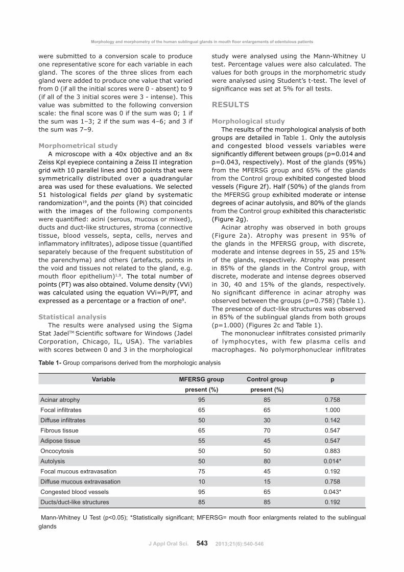
Morphological studyThe results of the morphological analysis of both
groups are detailed in Table 1. Only the autolysis �� � ������� �� � ������� ������� ��������������������������������� ���#�$&�&12��p=0.043, respectively). Most of the glands #?_|'�� � ���"D\]Q^�� ����@_| � ��������from the Control group exhibited congested blood �������#D�����%�'�{��#_&|' ����glands from the MFERSG group exhibited moderate or intense ������� ������� ���������&| ����glands from the Control group exhibited this characteristic (Figure 2g).
6���� �� ��� �� ������� �� � �� �� ���#D����� %'� 6�� ��� �� ������� �� ?_| ���� ����� �� ��� "D\]Q^ �� ��� ���� ���������� �����������������������__�%_��1_| � ��������� �������������6�� ���������������_| � �������� �� ���� ��� � �� �������discrete, moderate and intense degrees observed �� 4&� 2& �� 1_| � ��� ������ �������������� ��������� ���������� �� ���� �� ��� �� ������������������� ���#�$&��_�'#����1'������������ �����*���������������� ����������_| �������������������� �� ���� ���(p=1.000) (Figures 2c and Table 1).
���� � ���������������� ��������������� � ����� ������ ���� ��� ���� ����� ����� ������ � � ��� ��� ������ ���������
Variable 9*P�"Y� �!� ����=� �!� ppresent (%) present (%)
Acinar atrophy 95 85 0.758
\ ����������� 65 65 1.000
��������������� 50 30 0.142
Fibrous tissue 65 70 0.547
Adipose tissue 55 45 0.547
Oncocytosis 50 50 0.883
Autolysis 50 80 0.014*
Focal mucous extravasation 75 45 0.192
Diffuse mucous extravasation 10 15 0.758
Congested blood vessels 95 65 0.043*
Ducts/duct-like structures 85 85 0.192
Table 1- Group comparisons derived from the morphologic analysis
�=����}��������~��� 3��%"%08�� Z(������������?��������=\]�(^_�� ���> �����?����� ����G� � ���������?����glands
9���= :�����8���8���:�F������!8����!7=�� !�=� =��������8!���G����=�� �8�����F������!=!����������
2013;21(6):540-546
J Appl Oral Sci. 544
���� ����������� ��������������������Y������� �� ������ ��������������� ��������� @_| � ��� ����� �� � �� �� ��� #�$1�&&&'#D�����%�'�Y������������������� ���������_&��4&| ��������������"D\]Q^��� ��� �groups, respectively (p=0.142) (Figure 2f and Table 1).
Moderate or intense replacement of the glandular ��������������� ���������� ���������@_���&| �����������������������"D\]Q^��Control groups, respectively (p=0.547) (Figure 2c, 2d and Table 1). The replacement of the glandular �������� ���� ��� �� ������ �� ���������__��2_| ��������������"D\]Q^��Control groups, respectively (p=0.547) (Figure 2d and Table 1).
No gland exhibited a severe degree of oncocytosis, but discrete oncocytosis (Figure 2h) �� ������������ /������_&| ���������of both groups (p=0.883). Both groups primarily exhibited focal mucous extravasation (Figure 2i), ��� ���������������������������� �����observed (p=0.192) (Table 1).
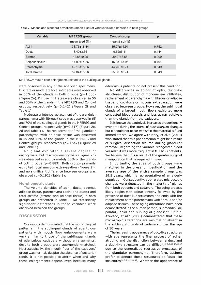
Morphometric studyThe volume densities of acini, ducts, stroma,
adipose tissue, parenchyma (acini and ducts) and total stroma (stroma and adipose tissue) in both groups are presented in Table 2. No statistically ���������� ����������� �� ����� ������� ���� ������������������� ����
DISCUSSION
Our results demonstrated that the morphological patterns in the sublingual glands of edentulous ������� ���� � ��� �� � ����������� ����very similar to those of the sublingual glands � ������� �� ������ ���� �� ������������������� � �� �� ��� ���� ��+������*�������"�� �� ����������� ���� � ������������� ����� �������������������� �� ����� ������� B� ��� �� ������ � �������������these enlargements appear, even because many
edentulous patients do not present this condition.No differences in acinar atrophy, duct-like
��������������������� � �� � �������������������������� ���������������� �� ���� ��������� �� ��� ��� ���� ���/������ ����� ���������������� ����{ ����������������������� � ������� � ��� � �� �/������� � ��congested blood vessels and less acinar autolysis than the glands from the cadavers.
B����� ������� ��������������� � ��� ����over time during the course of post-mortem changes but it should not occur ex vivo��������������/��immediately10��������������������10 (2010) �� ���������������� ��� ��������������of surgical dissection trauma during glandular removal. Regarding the variable “congested blood �������������� ������������"D\]Q^��������We believe that it is a result of the greater surgical manipulation that is required in vivo.
B�� ������� ��� ��� � � �� �� ��� ����matched in the present investigation and the ����� �� � ��� ������ ����� �� �� ��_?�_���������� ��������������� ���������population. Consequently, age-related microscopic ������������������ ������� ���� ������from both patients and cadavers. The aging process ���������������������� ���� �� ���������������� �����*������������������������������������� ������������������� ����+ �adipose tissue1. These aging alterations have been demonstrated in the human parotid, submandibular, palatal, labial and sublingual glands3,5,9,12-14,16-18. Azevedo, et al.1 (2005) demonstrated that these microscopic alterations are minimal or absent in the sublingual glands of cadavers under the age of 30 years.
The increasing appearance of duct-like structures ���� �� ���������� ��� ��� �� ���� � ������ ���� �� ������������ ������������ �� ����*���� ��������� �� �� ��������1,3,5,9,12-14,16,17
due to the generalized regressive processes of the glandular parenchyma. Therefore, authors prefer to denote these structures as “duct-like structures”1,3,5,9,13,16,17. Whether the appearance of
Variable 9*P�"Y� �!� ����=� �!� pmean ± sd (%) mean ± sd (%)
Acini 33.76±18.84 35.07±14.91 0.752
Ducts 8.40±3.36 9.62±5.11 0.444
Stroma 42.85±9.25 39.27±8.58 0.209
Adipose tissue 14.99±14.86 16.03±13.96 0.794
Parenchyma 42.16±18.26 44.70±16.74 0.649
Total stroma 57.84±18.26 55.30±16.74 0.649
Table 2- Means and standard deviations (mean ± sd) of various volume densities in both groups
"#����$�K6%PQK&Q6�P"$�(UPWPX6�(%(Q&"�%�$�&'()&�*&%+6�%$�%(�(�W"$�X(9(QKP��+
=\]�(^_�� ���> �����?���������G� ����������?����?���G�
2013;21(6):540-546

J Appl Oral Sci. 545
these structures and the increase in their number ���� �� �� ��� ������ � ���������� �� ����������������������� ��� �������� ��������������������� ������� ���������� ��� ���6����� ���������� �����������������is required.
� �������������������������� ������������� � ����� ������ ��� � �� ��� �� ������ ���� ������� ������� ��� �� ��� �� �� ������ ���sublingual glands exhibited a minor quantity of parenchyma in relation to the total stroma volume (stroma and adipose tissue) in both groups. This ������� ���� ��������������� �" ���������9 (2006) �� ��� �������������� ���� ����������������������������� ���� �ducts, stroma and adipose tissue increases. These authors also found that the glandular volume decreases, � ������ 44���| ������ ��� ���� �� ����������������������������������������� ����� ������� ����� ���� �������������
Our data demonstrated that the sublingual glands ��� ���� ��������������������� ������������� not morphologically and morphometrically different from the glands of edentulous cadavers ���� �� ����������� �� � �� ���� � �����exhibited age-related changes. Therefore, the aging process cannot explain the etiopathogenesis � ��� � ��� � � ����������� �� ������� ��individuals. Moreira, et al.9 #%&&@' �� ��� ������������������ � ������������H�������despite the enlargements. Conversely, the total volume of the glands decreases9. We agree that the intrinsic biologic characteristics of the sublingual glands are not responsible for the enlargements although these glands occupied almost the entire �������� ��� �� ��������� � � ���� �� ���examination. In both groups, a portion of the � ���� ���� ����/����������������������� ������������� �� �����������������������normal. We do not believe that the surrounding tissues are responsible for the enlargements. The enlargements may arise from local external factors ��� �� ������� ��� ����� ��� ������ �posterior-inferior teeth and the degree of alveolar ridge resorption2,4,6�{ �������������� ����� ����������� �� ���� ������� ��� ������������������������������� ������������� ��resorption in the alveolar margin do not exhibit "D\]Q^�6�� ������������� ����������� �� ��������������� ������� ��������������������clinically. We speculated that the accommodation of � ������������������� ��������������� ������ ��������������������� ������������� ����movements, might also contribute to the MFERSG. Therefore, the enlargements may be a clinical manifestation of an adaptation to the current anatomical situation since the placement of a dental �� ������������� ���� ����� ����� ���������
��������������� ���������������� � ��� � � ����������� �� ������� ��
patients are not described in academic books, but surgical interventions are frequent. Commonly, surgeons treat and describe these enlargements ���������� �������� ����{ ������ ���pathologists often describe the microscopic images as a ��� �������� ����� �� � ����� ������� ��the salivary glands during the aging process1,9. The terms “hyperplasia” and “hypertrophy” are �� ��� ����� ���� ���������� ������������� �abnormal increase in the number of normal cells in normal arrangement in an organ or tissue and an enlargement of an organ or part resulting from an increase in the size of the cells. The sialodenitis ������������������ �� ������� ����� ���� �������������������������� ������������������������������������������������glands biopsies11�6��������������������������������� � �� ������� ���� "D\]Q^6 �� ���that all these terms are incorrect. The sublingual ����� �� ��� ����������� � � � �� � ���������� ������� ������� ����� �� ���������� �������������������� ����1,9. We suggest that pathologists provide a descriptive microscopic report that emphasizes the possibility of age-related changes in the sublingual gland in MFERSG patients. ���������� ������������� ����������������and microscopic information.
The microscopic characteristics of these glands rule out the diagnosis of Sjögren Syndrome. First of ������������������������������� ���� �������� ��������� ������������� ��������������������������/�������� �����������B��������������� ������ � ���������������������� ������� ������ ����������������� ����� ��plasma cells and macrophages. The presence of a ���������� �������������� � �������������� probably corresponds to IgA-secreting plasma cells and duct-associated lymphoid tissue1. Ductal obstruction in adult life might explain the increase of ���� � ���������������������6H���� �����1 (2005) found that the presence of a mononuclear ������������ �������������� ���� ������� ��������������������������������� ����of parenchymal replacement progressed. When ���������� ��������������� ������������������������������������ ��� ������Vered, et al.17 (2001).
The nomenclature of these volume alterations in ���� ���� ����������� �������������������� ������������B����������������������������� � ��� ������B���D��� �����6 (2006). ������������������� �� �������������� �� ��� ������������������������������ ���������� ���� ����� �������������������������gland. This term does not refer to a disturbance �������� ��� ���H�������� ���������������
9���= :�����8���8���:�F������!8����!7=�� !�=� =��������8!���G����=�� �8�����F������!=!����������
2013;21(6):540-546

J Appl Oral Sci. 546
the best nomenclature for these enlargements is �� �*��� � ��������������� ����� ���� �in edentulous individuals”. Academic books should present these enlargements as normal variations of ����� �� ����� ���� ��Q���������������� ��� �� �������� ��� ���� ��� ������������������������������
CONCLUSION
The present study demonstrated that the microscopic characteristics of the sublingual glands ��� ���� ��������������������� ���������� ����� ��� ��������������� ��������� ���normal aging process. The sublingual glands do not represent pathological changes; they represent an ��*������������ ���� ��������� ����� ������������� ����� ���� �������������
ACKNOWLEDGEMENTS
������ ��� �������� ����D6X\QX#����&_+@&221*2'� ����������� ������ ����� like to thank the professors and employees of the Department of Biological Sciences of the Bauru School of Dentistry and Dr. José Roberto Lauris, Dr. Tânia Mary Cestari and Dr. Carla Ruffeil Moreira for their contributions to this study.
CONFLICT OF INTEREST STATEMENT
��� ��� �� ��� � � ������ � �������� � report.
REFERENCES
1- Azevedo LR, Damante JH, Lara VS, Lauris JRP. Age-related changes in human sublingual glands: a post mortem study. Arch Oral Biol. 2005;50(6):565-74.2- Campos LA. Hyperplasia of the sublingual glands in adult patients. Oral Surg Oral Med Oral Pathol Oral Radiol Endod. 1996;81(5):584-5.3- Dayan D, Vered M, Paz T, Buchner A. Aging of human palatal salivary glands: a histomorphometric study. Exp Gerontol. 2000;35(1):85-93.
4- Domaneschi C, Maurício AR, Modolo F, Migliari AD. Idiopathic hyperplasia of the sublingual glands in totally or partially edentulous individuals. Oral Surg Oral Med Oral Pathol Oral Radiol Oral Endod. 2007;103(3):374-7.5- Drummond JR, Chisholm DM. A qualitative and quantitative study of the ageing human labial salivary glands. Arch Oral Biol. 1984;29(2):151-5.@* B��� D��� �� Y���� �{� � �� �� 6� � ����� ���Y�����6�" ���� ������������������� ������������glands in edentulous or partially edentulous patients: a microscopic study. J Appl Oral Sci. 2006;14(4):264-9.7- Mandel L, Romao M. Sublingual salivary gland enlargement. NY State Dent J. 2004;70(7):24-7.�* "�� �� �D� ^��� 66� B��������� � � � ������ �������problems: a summary. Br Dent J. 2000;189:128-34.9- Moreira CR, Azevedo LR, Lauris JRP, Taga R, Damante JH. Quantitative age-related differences in human sublingual gland. Arch Oral Biol. 2006;51(11):960-6.10- Nery LR, Moreira CR, Cestari TM, Taga R, Damante JH. Post-mortem acinar autolysis in rat sublingual gland: a morphometric study. J Appl Oral Sci. 2010;18(5):509-14.11- Radfar L, Kleiner DE, Fox PC, Pillemer SR. Prevalence and ����������������� ������ ������ ������� ������������ �healthy volunteers. Arthritis Rheum. 2002;47(5):520-4.12- Scott J. A morphometric study of age changes in the histology of the ducts of human submandibular salivary glands. Arch Oral Biol. 1977;22(4):243-9.13- Scott J. Qualitative and quantitative observations on the histology of human labial salivary glands obtained post mortem. J Biol Buccale. 1980;8(3):187-200.12*Q� ����D� ���\6��������6��������������� ����� � ��������������������� ������� ��������������������Oral Pathol Med. 1987;16(10):505-10.1_* ��� Q� B��� "� " �� 6� Q��� �� "��� �� ��� ��Adenomatoid serous hyperplasia of sublingual gland: a case report. Oral Surg Oral Med Oral Pathol Oral Radiol Endod. 1996;82(4):437-40.16- Takahashi S, Shinzato K, Nakamura S, Domon T, Yamamoto T, Wakita M. The roles of apoptosis and mitosis in atrophy of the rat sublingual gland. Tissue Cell. 2002;34(5);297-304.17- Vered M, Buchner A, Haimovici E, Hiss Y, Dayan D. Focal ����� ������������� ��������������������������!� ������������������������������������X�� �"���2001;30(1):7-11.18- Waterhouse JP, Chisholm DM, Winter RB, Patel M, Yale RS. Replacement of functional parenchymal cells by fat and connective tissue in human submandibular salivary glands: an age-related change. J Oral Pathol Med. 1973;2(1):16-27.19- Weibel ER. Stereological principles for morphometry in electron microscopic cytology. Int Rev Cytol. 1969;26:235-302.
"#����$�K6%PQK&Q6�P"$�(UPWPX6�(%(Q&"�%�$�&'()&�*&%+6�%$�%(�(�W"$�X(9(QKP��+
2013;21(6):540-546