Embed Size (px)
Citation preview
Original Paper Veterinarni Medicina, 58, 2013 (1): 16–24
16
Morphological pattern of the livers of diff erent lines
of broiler chickens during rearing
M. Gesek1, J. Szarek1, I. Otrocka-Domagala1, I. Babinska1, K. Pazdzior1,
M. Szweda1, A. Andrzejewska2, B. Szynaka2
1Faculty of Veterinary Medicine, University of Warmia and Mazury, Olsztyn, Poland2Medical University, Bialystok, Poland
ABSTRACT: The aim of this study was to analyse the microscopic and ultrastructural lesions of the livers of
broiler chickens during fattening. Three genetic lines of broiler chickens (Ross 308, Cobb 500, Hubbard F15) were
investigated. The liver samples were taken on the 3rd, 10th, 17th, 24th, 31st, and 38th day of life from six healthy broiler
chickens from each commercial broiler flock. The dominant microscopic lesions were associated with prolonged
hypoxia and bile ductules, including: fatty, vacuolar and parenchymatous degeneration, necrosis of epithelial cells
of bile ductules, necrosis of hepatocytes around the proliferating bile ductules, lymphoid cell infiltration around
the bile ductules and blood vessels, proliferation of the bile ductules, proliferation of the connective tissue around
bile ductules and stimulation of the lymph nodules. Ultrastructural evaluation revealed abnormalities involving
mitochondria and rough endoplasmic reticulum. The mitochondria underwent swelling, polymorphism, prolifera-
tion and damage. The rough endoplasmic reticulum underwent defragmentation and acinar transformation. The
cytoplasm of most hepatocytes showed vacuoles of varying size or lipid droplets and the presence of cytoplasmic
myelin-like structures. This study shows that the livers of broiler chickens are the most predisposed to the occur-
rence of lesions on the 17th, 31st and 38th days of life.
Keywords: broiler chicken; Cobb; Ross; Hubbard; liver; pathomorphology
Supported by the Ministry of Science and Higher Education of Poland in 2009–2010 (Project No. N308 230236) and
by the EU within the European Social Foundation.
The growing demand for poultry meat has
prompted producers of breeding lines of broiler
chickens to seek to optimise breeding through
genetic means. Modern broiler lines are expected
to grow fast (shorter breeding period), be healthy
(characterised by a good metabolism and a high
degree of utilisation of nutrients), have a strong
skeleton, high survival rate, be resistant to dis-
ease, have high adaptive capacity and show steady
growth. Currently, broiler chickens reach an av-
erage body mass of between 2.2–2.5 kg within
42 days of rearing and consume 1.60–1.80 kg feed
per kg of growth, depending on the breeding line.
Clear genetic progress can be seen when compar-
ing this data with the results from 10 years ago. At
that time, the final body mass of broiler chickens
reached 1.95–2.15 kg, the average fattening period
was 43–45 days and the average feed consumption
per kg of body mass ranged from 2.00–2.10 kg. The
current production results are even more impres-
sive when viewed in the long term (Havenstein et al.
2003a,b). In 1957, the average body mass of broilers
after 42 days of rearing was 539 g and the average
feed consumption per kg of body mass was 2.34 kg.
These data shows how much progress has been
made in the field of breeding and genetic selection.
Unfortunately, genetic progress has also led to
several negative effects. Thus, changes are now
found in broiler chickens which not occurred be-
fore or were rather uncommon. These include:
sudden death syndrome, pulmonary hypertension
syndrome, abnormal bone growth, limb disease,
the occurrence of diseases in subclinical form
(Clostridium perfringens, Escherichia coli), immu-
nosuppression and susceptibility of birds to my-
cotoxin intoxication. Subclinical infections are a

Veterinarni Medicina, 58, 2013 (1): 16–24 Original Paper
17
major problem in broiler fattening (Onderka et al.
1990; Lovland and Kaldhusdal 1999) and also re-
sult in liver damage (Hutchison and Riddell 1990;
Onderka et al. 1990; Sasaki et al. 2003).
Th e aim of this study was to analyse the morpho-
logical pattern during fattening of the livers of broiler
chickens which did not show clinical signs of disease.
Furthermore, the aim was to determine the most sus-
ceptible period for the occurrence of morphological
lesions in the liver during fattening and determine
whether these changes may accompany disorders oc-
curring in these birds in a subclinical form.
MATERIAL AND METHODS
Broiler chickens of three genetic lines (Cobb
500, Hubbard F15, and Ross 308) were reared in
three commercial broiler flocks – each line in a
separate house. Rearing parameters were regulated
in accordance with the recommendations of the
producers of breeding lines. These factors did not
differ significantly from each other. The quantita-
tive composition of the feed was slightly modified
depending on the farm with reagard to the percent-
age of total protein in the feed and the amount of
metabolisable energy (ME).
All three broiler flocks had the same vaccination
program: on the 1st day of life – against Marek’s dis-
ease, Infectious Bursal Disease (IBD) and Infectious
Bronchitis (IB, H-120 strain), on the 16th day –
against IB 4/91 strain and the IBD vaccination was
repeated on the 18th day of life.
From each genetic line, six broiler chickens were
taken randomly for morphological examination
on the 3rd, 10th, 17th, 24th, 31st and 38th days of life
(n = 36). Th e birds were weighed and slaughtered fol-
lowing the protocols established by the Local Ethics
Committee in Olsztyn, No. 3/N dated 22.01.2008).
Samples of liver for microscopic evaluation were
fixed in 10% neutralised formalin and embedded in
paraffin blocks. The paraffin sections (5 μm) were
stained with haematoxylin, eosin (HE) and frozen
sections of livers on the 3rd day of life were also
stained with Oil Red O to detect lipids (Bancroft
and Gamble 2008). Histologically, changes were
graded subjectively as focal, multifocal and diffuse,
and the intensity of these lesions was classified as
low (mild), medium (moderate) and high (severe).
Liver sections were also fixed for ultrastructural
examination for two hours in 2.5% paraformalde-
hyde and 2% glutaraldehyde in a pH 7.4 phosphate
buffer. Samples were post-fixed in 2% osmium
tetraoxide in a pH 7.4 phosphate buffer and the
material was embedded in Epon 812. Semi-thin
sections were obtained from the blocks and were
subsequently stained according to the method de-
scribed by Lewis and Knight (1977) and viewed
under an optic microscope in order to establish the
appropriate place for preparing ultrathin sections.
The ultrastructural analysis was carried out with
an Opton 900 PC TEM (Germany).
Due to the relatively small number of groups and
deviations from normal distributions of variables,
a Kruskal-Wallis non-parametric ANOVA test was
performed.
Figure 1. 3rd day (Ross 308): hepatic lipidosis, fatty
degeneration (arrows); HE. 1a (box): 3rd day (Cobb 500):
lipid vacuoles; Oil Red O
Figure 2. 17th day (Hubbard F15): cholangiohepatitis
chronica – infi ltration of lymphoid cells (asterisks), pro-
liferation of the connective tissue (long arrows), hyper-
trophy of the endothelial cells in artery (short arrows),
hypertrophy of smooth muscle in artery (arrowheads); HE

Original Paper Veterinarni Medicina, 58, 2013 (1): 16–24
18
RESULTS
Clinical examination showed that broiler chick-
ens on the 3rd, 10th, 17th, 24th, 31st and 38th days of
life were active, had an appropriate appetite and
did not show clinical signs of disease. Macroscopic
examination showed normal morphology of tissues
and organs in most birds. By the third day, all livers
were light-brown with a loamy/brittle consistency
and the cross-sectional lobular pattern had disap-
peared.
The results of microscopic evaluation
During rearing, the livers of the examined broiler
chickens showed several microscopic lesions (pre-
sented in Tables 1–3 and Figures 1–4). Th e livers of
the examined birds mainly showed parenchymatous,
vacuolar (Figure 4) and fatty degeneration (Figure 1).
Th ese were observed with high intensity and most
often vacuolar degeneration throughout the breeding
(92 birds) was described along with parenchymatous
degeneration from the 10th day (88 chickens). In addi-
Table 1. Morphological lesions in the livers of the broiler chicken Cobb 500 genetic line
Type of morphological lesions
Number of lesions in Cobb 500 line broiler
chicken livers
day of life
3 10 17 24 31 38
A – parenchymatous degeneration 0/6 5/6 6/6 6/6 6/6 3/6
B – vacuolar degeneration 6/6 6/6 5/6 6/6 0/6 4/6
C – fatty degeneration 6/6 4/6 3/6 0/6 0/6 4/6
D – necrosis of the epithelium cells of the bile ductules 0/6 0/6 0/6 2/6 0/6 0/6
E – congestion 0/6 5/6 5/6 5/6 4/6 6/6
F – infi ltration of lymphoid cell around bile ductules and blood vessels 0/6 3/6 4/6 3/6 3/6 3/6
G – stimulation of the lymph nodules 0/6 0/6 6/6 2/6 3/6 0/6
H – proliferation of the bile ductules 0/6 4/6 6/6 4/6 5/6 6/6
I – proliferation of the connective tissue around the bile ductules 0/6 0/6 2/6 0/6 0/6 2/6
J – hypertrophy of the endothelium cells in arteries 0/6 4/6 6/6 4/6 5/6 2/6
K – hypertrophy of the smooth muscle in arteries 0/6 4/6 6/6 3/6 0/6 3/6
Figure 3. 24th day (Cobb 500): necrosis of epithelium
cells in the bile ductules (short arrows), hypertrophy
of smooth muscle in arteries (long arrows), coagulative
necrosis of hepatocytes around the bile ductules (N),
dilatation of the bile ducts (asterisk); HE
Figure 4. 38th day (Ross 308): proliferation of the bile
ductules (short arrows), infi ltration of lymphoid cells
(arrowheads) and myeloid cells (long arrows) around the
portal area, vacuolar degeneration; HE

Veterinarni Medicina, 58, 2013 (1): 16–24 Original Paper
19
tion, fatty degeneration was diffuse on the 3rd day,
while on the 10th day it was multifocal near blood
vessels and the terminal portal venules and focal
near the blood vessels and terminal portal venules
on the 17th day. Fatty degeneration was multifocal
on the 31st and 38th days in 14 chickens. Changes
were particularly intense from the 10th day and
included bile ductule proliferation (Figure 4), hy-
pertrophy of endothelium cells in arteries and hy-
pertrophy of the smooth muscle in arteries (Figure
2 and 3). Lesions in the bile ductules were recorded
together with the proliferation of the connective
tissue around bile ductules (lower intensity after
the 10th day) (Figure 2), lymphoid cell infiltration
around bile ductules and blood vessels (medium
intensity after the 10th day) (Figure 2 and 4). These
were associated with infiltration of myeloid cells
around the proliferating bile ductules (observed
focally after the 10th day), with interstitial lym-
phoid hepatitis diagnosed in seven chickens and
outbreaks of coagulative necrosis of hepatocytes
around the proliferating bile ductules (occurring
from the 10th day). It is noteworthy that multifocal
necrosis of epithelial cells in bile ductules after the
17th day (Figure 3) and multifocal stimulation of the
lymph nodules (10th day) occurred in 38 chickens.
Table 2. Morphological lesions in the livers of the broiler chicken Hubbard F15 genetic line
Type of morphological lesions
Number of lesions in Hubbard F15 line broiler
chicken livers
day of life
3 10 17 24 31 38
A – parenchymatous degeneration 0/6 6/6 6/6 6/6 6/6 6/6
B – vacuolar degeneration 6/6 6/6 6/6 6/6 3/6 6/6
C – fatty degeneration 6/6 4/6 0/6 2/6 0/6 5/6
D – necrosis of epithelium cells of the bile ductules 0/6 0/6 3/6 0/6 5/6 4/6
E – congestion 0/6 4/6 6/6 6/6 6/6 4/6
F – infi ltration of lymphoid cell around bile ductules and blood vessels 0/6 0/6 3/6 5/6 5/6 6/6
G – stimulation of the lymph nodules 0/6 2/6 5/6 0/6 6/6 0/6
H – proliferation of the bile ductules 0/6 6/6 6/6 4/6 6/6 6/6
I – proliferation of the connective tissue around the bile ductules 0/6 0/6 2/6 0/6 4/6 4/6
J – hypertrophy of the endothelium cells in arteries 0/6 4/6 6/6 0/6 5/6 5/6
K – hypertrophy of the smooth muscle in arteries 0/6 0/6 3/6 0/6 3/6 2/6
Figure 5. 3rd day (Ross 308): rarefi cation of cytoplasm,
myelin-like structures (long arrows), mitochondrial
polymorphism with swollen and damaged mitochondria
(M), acinar transformation of the RER (arrowheads) and
lipid vacuoles (asterisk); 7500×
Figure 6. 10th day (Hubbard F15): acinar transformation
of RER (arrowheads), lipid vacuoles (long arrows) and
rarefi cation of cytoplasm; 7500×

Original Paper Veterinarni Medicina, 58, 2013 (1): 16–24
20
A Kruskal-Wallis nonparametric ANOVA test
(Table 4; Figure 11) showed that the number of
lesions in the form of parenchymatous and vacu-
olar degeneration was statistically significantly
higher than the necrosis of the bile ductule epi-
thelium, hypertrophy of the smooth muscle in
arteries as well as the stimulation of the lymph
nodules.
Furthermore, the number of animals with necrosis
of the bile ductule epithelium cells was statistically
significantly lower than those with congestion and
proliferation of the bile ductules. In addition, the
number of birds with proliferation of the connec-
tive tissue around the bile ductules was statistically
significantly lower than those with parenchymatous
and vacuolar degeneration, congestion and prolif-
eration of the bile ductules. A statistical analysis of
the results, using the day of the fattening as a vari-
able (Table 5; Figure 12) showed that the number
of broilers with changes on day 3 was statistically
significantly lower than the number of chickens on
the 17th, 31st and 38th days.
There were no significant statistical differences
between the lines, although the largest number of
lesions in the liver was found in the Ross 308 line,
followed by Hubbard F15 and Cobb 500. Important
statistical differences may also emerge with further
tests on a larger number of flocks.
Table 3. Morphological lesions in the livers of the broiler chicken Ross 308 genetic line
Type of morphological lesions
Number of lesions in Ross 308 line broiler
chicken livers
day of life
3 10 17 24 31 38
A – parenchymatous degeneration 0/6 6/6 6/6 6/6 6/6 5/6
B – vacuolar degeneration 6/6 6/6 4/6 4/6 6/6 6/6
C – fatty degeneration 6/6 4/6 2/6 0/6 6/6 0/6
D – necrosis of epithelium cells of the bile ductules 0/6 0/6 2/6 2/6 4/6 4/6
E – congestion 4/6 3/6 6/6 6/6 3/6 6/6
F – infi ltration of lymphoid cell around bile ductules and blood vessels 0/6 0/6 5/6 5/6 6/6 4/6
G – stimulation of the lymph nodules 0/6 6/6 0/6 2/6 0/6 3/6
H – proliferation of the bile ductules 0/6 2/6 6/6 6/6 6/6 6/6
I – proliferation of the connective tissue around the bile ductules 0/6 2/6 0/6 2/6 2/6 6/6
J – hypertrophy of the endothelium cells in arteries 0/6 2/6 6/6 2/6 0/6 2/6
K – hypertrophy of the smooth muscle in arteries 0/6 4/6 2/6 4/6 2/6 3/6
Figure 7. 17th day (Ross 308): rarefi cation of the cyto-
plasm, myelin-like structures (long arrows), polymor-
phic and swollen mitochondria and damage to some of
the mitochondria (short arrows) and the defragmenta-
tion of the RER (arrowheads); 7500×
Figure 8. 31st day (Cobb 500): acinar transformation of
RER (long arrows), myelin-like structures (short arrows)
and rarefi cation of the cytoplasm; 7500×
Veterinarni Medicina, 58, 2013 (1): 16–24 Original Paper
21
The results of ultrastructural evaluation
Ultrastructural lesions in hepatocytes are shown
in Figures 5–10. The relatively most frequent ul-
trastructural abnormalities concerned mitochon-
dria and rough endoplasmic reticulum (RER).
Mitochondria underwent swelling (Figure 5),
polymorphism (Figures 5, 7 and 9), proliferation
(Figures 9) and damage (Figure 7). Almost all mi-
tochondria in the examined hepatocytes were ob-
served as dense bodies. Broilers from the Hubbard
F15 genetic line showed the greatest intensity of
these lesions (Figure 9). Mitochondria changed
most frequently in chickens on the 10th, 17th, 24th,
31st and 38th day of life. RER often underwent de-
fragmentation (Figure 7) or acinar transformation
(Figures 5, 6 and 9). These lesions were particularly
intensified on the 17th and 31st day, especially in
chickens from the Ross 308 line. Occasionally, RER
was devoid of ribosomes or its channels under-
went widening. The cytoplasm of most hepatocytes
showed vacuoles of varying size (Figure 9) or lipid
droplets (Figures 5, 6 and 10) and the presence of
cytoplasmic myelin-like structures (Figures 5, 7, 8
and 10). Necrosis of hepatocytes, as well as necro-
sis of parts of the hepatocytes were visible (often
in chickens of the Ross 308 line), especially in the
second half of the breeding period, and was visible
Table 4. P-value for multiple comparisons (bilateral) between the morphological lesions. Kruskal-Wallis test; H (10,
n = 198) = 58.10, P = 0.00; abbreviations discribed in Tables 1 to 3
A B C D E F G H I J K
A 1.00 0.94 0.00 1.00 1.00 0.03 1.00 0.00 0.99 0.05
B 1.00 0.28 0.00 1.00 0.36 0.01 1.00 0.00 0.29 0.01
C 0.94 0.28 1.00 1.00 1.00 1.00 1.00 1.00 1.00 1.00
D 0.00 0.00 1.00 0.02 1.00 1.00 0.01 1.00 1.00 1.00
E 1.00 1.00 1.00 0.02 1.00 0.23 1.00 0.03 1.00 0.34
F 1.00 0.36 1.00 1.00 1.00 1.00 1.00 1.00 1.00 1.00
G 0.03 0.01 1.00 1.00 0.23 1.00 0.14 1.00 1.00 1.00
H 1.00 1.00 1.00 0.01 1.00 1.00 0.14 0.01 1.00 0.22
I 0.00 0.00 1.00 1.00 0.03 1.00 1.00 0.01 1.00 1.00
J 0.99 0.29 1.00 1.00 1.00 1.00 1.00 1.00 1.00 1.00
K 0.05 0.01 1.00 1.00 0.34 1.00 1.00 0.22 1.00 1.00
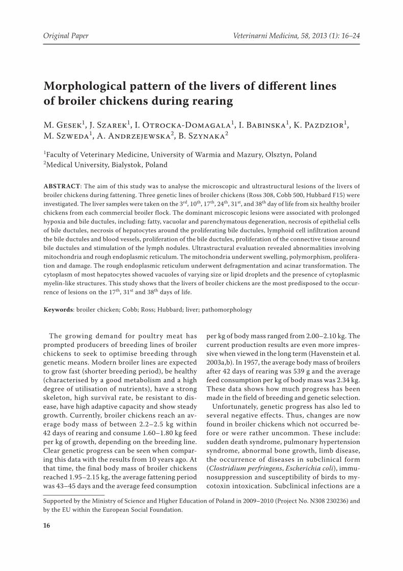
Figure 9. 31st day (Hubbard F15): proliferation and poly-
morphism of the mitochondria, with numerous dense
bodies in the mitochondria, acinar transformation of the
RER (long arrows), myelin-like structure (arrowhead)
and vacuoles (short arrows); 15 400×
Figure 10. 38th day (Hubbard F15): lipid droplets (short
arrows), myelin-like structures (long arrows) and rarefi -
cation of the cytoplasm; 3200×

Original Paper Veterinarni Medicina, 58, 2013 (1): 16–24
22
Figure 11. Kruskal-Wallis test. Grouping variable: type of
lesion. Abbreviations discribed in Tables 1 to 3
Figure 12. Kruskal-Wallis test. Grouping variable: day of
fattening
Nu
mb
er o
f b
ird
s
A B C D E F G H I J K
Type of lesion
Mediana25–75%Min–Max
7
6
5
4
3
2
1
0
–1
3 10 17 24 31 38
Day of fattening
Nu
mb
er o
f b
ird
s
Mediana25–75%Min–Max
7
6
5
4
3
2
1
0
–1
Table 5. P-values for multiple comparisons (bilateral) between days. Kruskal-Wallis test; H (5, n = 198) = 26.08, P = 0.00
3 10 17 24 31 38
3 0.08 0.00 0.06 0.01 0.00
10 0.08 1.00 1.00 1.00 1.00
17 0.00 1.00 1.00 1.00 1.00
24 0.06 1.00 1.00 1.00 1.00
31 0.01 1.00 1.00 1.00 1.00
38 0.00 1.00 1.00 1.00 1.00
as damage to mitochondria and defragmentation of
the RER with the occurence of myelin-like struc-
tures. The presence of macrophages was quite often
noted close to necrotic hepatocytes and lysosomes
were usually present in the hepatocytes which were
partially damaged.
DISCUSSION
All the lesions described in this report were simi-
lar to those reported in subclinical Clostridium
perfringens infection in broiler chickens. In birds
with microbiologically confi rmed C. perfringens in-
fection, Hutchison and Ridell (1990) found massive
proliferation of bile ductules, proliferation of con-
nective tissue around ductules, hepatocyte necrosis
and massive lymphocyte and heterophil infi ltration.
In an experiment on direct C. perfringens infec-
tion of the bile duct, Onderka et al. (1990) report-
ed lesions similar to Hutchison and Ridell (1990).
Sasaki et al. (2003) microbiologically confirmed C.
perfringens infections in the newly hatched chicks
and described the necrosis of hepatocytes (from fo-
cal to diffuse), proliferation of the connective tissue
and infiltration of heterophils and macrophages.
They also noted that necrotic enteritis induced by
C. perfringens was accompanied by lesions in the
liver, which are a major problem in broilers at the
age of 2–5 weeks.
Changes such as infiltration of lymphoid and
myeloid cells, proliferation of bile ductules and
connective tissue around ductules, necrosis of
epithelial cell in bile ductules and hepatocytes
around ductules are described as cholangiohepa-
titis (Sasaki et al. 2000; Abdul-Aziz et al. 2008).
Particular changes can occur with varying intensity
and the cause is usually C. perfringens infection.
Thus, the changes observed in our study should
also be classified as a cholangiohepatitis chronica.
The examined livers also demonstrated vascular
remodelling, with hypertrophy and hyperplasia of
endothelial cells in arteries (especially from the
10th day of life) and hypertrophy and hyperplasia
of the smooth muscle in arteries (intensively after
the 10th day). In addition, hypertrophy and hyper-
plasia of the smooth muscle were observed in the
wall of terminal portal venules in the liver, but with
lower intensity. All these processes should be in-
terpreted as a vascular remodelling. Confirmation
Veterinarni Medicina, 58, 2013 (1): 16–24 Original Paper
23
of this hypothesis can be found in the reports of
Julian (2007), which describe pulmonary vascular
remodelling in pulmonary hypertension syndrome
and showed that blood vessels in other organs may
also undergo these changes. It was also stated in
this report that the reason for these phenomena is
the reaction of the endothelium and smooth muscle
to changing blood pressure and blood flow.
A significant group of lesions found in the liver
were regressive changes, not associated with sub-
clinical forms of C. perfringens infection. Steatosis
degenerativa on day 3 accompanied steatosis sim-
plex of the liver, which is a physiological phenom-
enon (Cullen 2007). The cause of degenerative
changes in fast-growing broiler chickens is a pro-
longed state of hypoxaemia leading to hypoxia
(Olkowski et al. 2005). Under conditions of con-
tinuous and high demand for oxygen and nutrients,
the liver tissue may respond with regressive lesions
(parenchymatous, vacuolar and fatty degeneration,
and necrosis of hepatocytes) (Madej et al. 2007).
Prolonged hypoxia causes swelling of the mito-
chondria, Golgi apparatus and endoplasmic reticu-
lum canals. Water penetrates these structures and
they become larger and acinar (parenchymatous
and vacuolar degeneration). Under conditions of
hypoxia, fatty hepatocytes develop rapidly and the
process is described as infiltratio adiposa degen-
erativa hypoxaemica (Madej et al. 2007). On the
3rd day, hepatocyte damage due to lipid accumula-
tion in 13 chicks was also observed and the dam-
age was sometimes accompanied by infiltration of
lymphoid cells. Madej et al. (2007) reported that
lipids could be used in the combustion process, if
the damaging agent has been removed. The cur-
rent results confirmed this view. In the first days
of rearing fatty degeneration occured, which was
accompanied by steatosis simplex. In the following
days, fatty degeneration had a less extensive char-
acter and occurred multifocally near blood vessels
and terminal portal venules. The presence of fatty
degeneration in older birds on the 31st and 38th day
of life was also noteworthy. It is suspected that the
reason could be poorly balanced feed. An inappro-
priate ratio of metabolic energy and protein, as well
as an excessive concentration of energy in the feed
may result in the accumulation of lipid vacuoles in
hepatocytes and ultrastructural damage (Cullen
2007; Madej et al. 2007).
In analysing the liver lesions reported in the pre-
sent study, mycotoxin contamination (aflatoxin B1,
fumonisin) should also be taken into considera-
tion. Mollenhauer et al. (1989) described the ex-
perimental intoxication of broilers aflatoxin and
found fatty degeneration, enlarged bile ductules,
lymphoid infiltration and necrosis of hepatocytes.
In the course of aflatoxin intoxication observed
in the liver, Ortatatli et al. (2005) observed focal
necrosis of hepatocytes, inflammatory cell infiltra-
tion, vacuolar and fatty degeneration, proliferation
of bile ductules and proliferation of the connective
tissue around blood vessels. The authors concluded
that no hepatocyte necrosis was observed around
proliferating bile ductules or ductules becoming
necrotic (as in the current study); therefore, the
presence of mycotoxins in the feed seems to be
doubtful.
In summary, the greatest threat to health in the
livers of the examined chickens involved changes
usually associated with subclinical C. perfringens
infection. Lack of microbiological examination of
the examined livers does not allow us to be ab-
solutely certain regarding the occurrence of this
infection. However, our data are supported by other
reports in which microbiological studies were per-
formed where similar results were obtained. In ad-
dition, prolonged hypoxia of hepatocytes resulted
in regressive lesions of liver tissue throughout the
fattening period. The statistical analysis revealed
that the livers of broiler chickens are the most
predisposed to the occurrence of morphological
lesions on the 17th, 31st and 38th days.
Acknowledgement
The authors are indebted to Ms Krystyna Dublan,
MSc and Mr A. Penkowski, MSc (University of
Warmia and Mazury, Olsztyn, Poland) for their
excellent technical assistance.
REFERENCES
Abdul-Aziz T, Fletcher O, Barnes HJ (2008): Hepatobil-
iary system. In: Fletcher OJ (eds.): Avian Histopathol-
ogy. 3rd ed. AAAP, Jacksonville, FL. 203–275.
Bancroft JD, Gamble M (2008): Theory and Practice of
Histological Techniques. 6th ed. Churchill Livingstone
Elsevier, Philadelphia, PA.
Cullen JM (2007): Liver, biliary system, and exocrine
pancreas. In: McGavin MD, Zachary JF (eds.): Patho-
logic Basis of Veterinary Diseases. 4th ed. Mosby El-
sevier, St. Louis, Missouri. 393–462.

Original Paper Veterinarni Medicina, 58, 2013 (1): 16–24
24
Havenstein GB, Ferket PR, Qureshi MA (2003a): Carcass
composition and yield of 2001 vs 1957 broilers when
fed representative 1957 and 2001 broiler diets. Poultry
Science 82, 1509–1518.
Havenstein GB, Ferket PR, Qureshi MA (2003b): Growth,
livability and feed conversion of 1957 versus 2001
broilers when fed representative 1957 and 2001 broiler
diets. Poultry Science 82, 1500–1508.
Hutchison TWS, Riddell C (1990): A study of hepatic
lesions in broiler chickens at processing plants in Sas-
katchewan. Canadian Veterinary Journal 31, 20–25.
Julian RJ (2007): The response of heart and pulmonary
arteries to hypoxia, pressure and volume. A short re-
view. Poultry Science 86, 1006–1011.
Lewis PR, Knight DP (1977): Staining Methods for Sec-
tioned Material. North Holland Publishing Company,
134–186.
Lovland A, Kaldhusdal M (1999): Liver lesions seen at
slaughter as an indicator of necrotic enteritis in broiler
folks. Immunology and Medical Microbiology 24,
345–351.
Madej JA, Rotkiewicz T, Nozdryn-Plotnicki Z (2007):
Systemic Pathology of Animals (in Polish). Publisher of
University of Warmia and Mazury, Olsztyn. 359–391.
Mollenhauer HH, Corrier DE, Huff WE, Kubena LF, Har-
vey RB, Droleskey RE (1989): Ultrastructure of hepatic
and renal lesions in chickens fed afl atoxin. American
Journal of Veterinary Research 50, 771–777.
Olkowski AA, Duke T, Wojnarowicz C (2005): The ae-
tiology of hypoxaemia in chickens selected for rapid
growth. Comperative Biochemistry and Physiology
Part A 141, 122–131.
Onderka DK, Carmen CL, Hanson JA (1990): Fibrosing
cholehepatitis in broiler chickens induced by bile duct
ligations or inoculation of Clostridium perfringens.
Canadian Journal of Veterinary Research 54, 285–290.
Ortatatli M, Oguz H, Hatipoglu F, Karaman M (2005):
Evaluation of pathological changes in broilers during
chronic aflotoxin (50 and 100 ppb) and clinoptilolite
exposure. Research in Veterinary Science 78, 61–68.
Sasaki J, Goryo M, Honda J, Okoshi N, Okada K, Furu-
kawa H (2000): Cholangiohepatitis in broiler chickens
in Japan: histopathological, immunohistochemical and
microbiological studies of spontaneous disease. Acta
Veterinaria Hungarica 48, 59–67.
Sasaki J, Goryo M, Makara M, Nakamura K, Okada K
(2003): Necrotic hepatitis due to Clostridium perfrin-
gens infection in new hatched broiler chick. Journal
of Veterinary Medical Science 65, 1249–1251.
Received: 2012–04–27
Accepted after corrections: 2012–12–08
Corresponding Author:
Michal Gesek, University of Warmia and Mazury in Olsztyn, Faculty of Veterinary Medicine,
Department of Pathological Anatomy, Oczapowskiego St. 13, 10-719 Olsztyn, Poland
Tel: +48 895 246 141, E-mail: [email protected]