Embed Size (px)
Citation preview

The Egyptian Journal of Hospital Medicine (2007) Vol., 29: 255 – 536
Morphological, Biochemical and Ultrastructural Changes in the Pregnant
Rat Placenta and the Liver of their Fetuses Treated with Folic
Acid and / or Gamma Radiation
Fatma, L. Ramadan and Seham M. Abu Nour National Center for Radiation Research and Technology (NCRRT), Atomic Energy Authority
(AEA) Cairo, Egypt.
Abstract
Backgrounds: The efficacy of antioxidant supplementation and oxidative stress of
gamma irradiation for and during pregnancy is poorly established. The present study aimed to
detect the toxic effects of high dose of folic acid and / or gamma radiation on the placenta of
pregnant rat and the liver of their fetuses. Material and Methods: Pregnant albino rats were divided into four groups. The first
group served as a control, the second group received oral intake of folic acid (5 mg/kg) from the
5th to 20
th day of gestation, the third group was irradiated with gamma radiation (3Gy, as
fractionated doses (1Gy/ 3 times) on each 5th, 10
th and 15
th days of gestation, the fourth group
was imanaged with combined treatment.
The pregnant rats were sacrified after 20 days of pregnancy and samples were taken from
the blood, placenta and the fetal liver for the morphological, biochemical and electron microscopic studies.
Results: The present results showed a significant elevation in serum gamma
glutamyltransferase (GT), lactate dehydrogenase (LDH) in placental tissue of pregnant rats associated with an increase of phosphorus content in liver of fetuses. Fetal malformations
including: protrusion, anotia, short neck dactylomegaly, subcutaneous haemorrhage, paralysis in the fore limbs and congested blood vessels.
The ultrastructural changes revealed sever damage in the placenta following folic acid
administration and / or exposure to whole body gamma radiation. Also the fetal liver showed an appearent signs of damage under the combined treatment.
The obtained changes were represented by: dilatation of the blood sinsoids, swollen
mitochondria, fragmented rough endoplasmic reticulum and necrosis.
Conclusion: It could be concluded that administration of folic acid and/or exposure to gamma radiation during pregnancy induced morphological, biochemical and ultrastructural
changes in both placenta of the pregnant rats and liver of their fetuses.
Key words: Folic acid, radiation, Pregnancy, placenta, liver, fetus, -GT, LDH, phosphorus, Teratology, electron microscop.
Introduction
Folic acid (B vitamin) needed for cell replication and growth. Folic acid helps
from building blocks DNA baby's genetic
information and building blocks of RNA
and needed for protein synthesis in all cells. The growing tissues such as those of
the fetus and rapidly regenerating cells like
red blood cells have a high need for folic acid (Health notes, 2005). The requirement
for folic acid increases considerably during
pregnancy (Truswell, 1985). Vitamin B also
helps the body to convert carbohydrates
into glucose which is burned to produce energy.
B complex vitamins are essential in
the breakdown of fats and proteins.
B complex vitamins also play an important role in maintaining muscle tone along the
lining of the digestive tract and promoting
the health of the nervous system, skin, hair, eyes, mouth and liver (Living Naturally,
2006). Hathcock (1997) stated that
supplemental folic acid should not exceed
1.000 mcg for adult men and women and a
522

Morphological, Biochemical and Ultrastructural………
523
800 mcg for pregnant and lactating women
less than 18 years of age to prevent folic acid from masking symptoms of vitamin
B12 deficiency.
In association with a continual
increase in the environmental radiation load, mainly as a consequence of long-
lasting radioactive fall out coming from the
testing of nuclear weapons and accidents of nuclear fittings in different parts of the
world, the question of the transmission of
radiation-induced genetic damage to the next generation becomes very real.
Different types of radiation cause damage
to different organs and production of
genetic changes which affecting future generation (Omran and Abu-Zied 2006). In
addition, Rezk and Ibrahim (2006)
observed that folic acid at a dose 4mg/kg b.wt was found to offer protection during
pregnancy and suppressing the embryonic
mortality rates and serious fetal malformations when pregnant rat were
exposed to gamma rays at dose 3 Gy on day
10 of gestation.
Aldeen and Konermann (1978), studied the effect of acute X-irradiation
during pre and post-implantation stage on
mouse embryos by exposing pregnant mice to a single dose of 3 Gy. They found that
direct effect of irradiation on mother must
lead to embryonic death. Moreover, Walash
et al. (1989) stated that whole body -irradiation of pregnant rats at dose of 2 Gy in organogenesis stage caused malfor-
mations in the skull, anophthalmia and
defects in the central nervous system. Moreover, Gaber (1990) found that exposed
pregnant rats to gamma irradiation showed
an increase in the incidence of intrauterine
foetal death, as well as induced uterine growth retardation. Placenta is a complex
mammalian tissue that performs many
metabolic function as it is the major transporting system of nutrients necessary
for foetal growth (Dorothy, 1983). Abu
Gabal et al. (1994) noted severe
degeneration in maternal and foetal rat placenta and marked loss of DNA in
different layers. Ashry (1997) stated that
gamma irradiation with 1 Gy induced degenerative signs in placenta represented
by pyknotic nuclei, fibrosis and vacuolation
of the cytoplasm. While fractionated dose of 2Gy showed progressive degenerative
features including: haemorrhage and
congestion of blood vessels. Walash et al. (1988) and Abu Gabal et al. (1995) studied
the effect of gamma irradiation on liver of
pregnant rats during organogenesis period.
Their results showed degenerative and necrotic lesions in hepatic lobules. In
addition Katarina et al. (2002) reported that
irradiation with dose of 3 Gy -irradiation caused latent cytogentic damage to the liver.
The present study was performed to
identify the toxic effects of folic acid
(5mg/kg b.w) and / or gamma irradiation at dose of 3 Gy on the placenta of pregnant
rats and the liver of their fetuses.
Material and Methods
Experimental animals
A total number of 40 adult pregnant albino rats weighing (150-170g) were used.
They were housed at room temperature and
allowed food and water.
Experiment was performed according to the international guidelines of animal
handling and care.
Irradiation processing:
Whole body gamma irradiation at a
dose level of 3 Gy was performed using an indoor shielded Cs-137 irradiator (Gamma
cell-40 installed in the National Center for
Radiation Research and Technology
(NCRRT) Atomic Energy Authority, Cairo, Egypt, which emitting a dose of gamma
radiation at the rate of 1.26 Gy/min
Folic Acid:
Folic acid was purchased from the
Nile Company Pharmaceutical and Chemical Industries. It was dissolved in
distilled water and administered to pregnant
rats at a dose level of 5 mg/kg b.wt using an
oral stomach tube.
Experimental design: Pregnant rats were classified into 4 groups
each of five animals:
Group 1 (Control): Pregnant rats served as a control untreated group.
Group 2 (treated): Pregnant rats treated
orally with folic acid at a dose of 5 mg/kg
b.wt /day from 5th to 20
th day of gestation.
Group 3 (irradiated): Pregnant rats

Fatma, L. Ramadan & Seham M. Abu Nour
524
exposed to whole body gamma rays
delivered as 1 Gy increament on 5th, 10
th
and 15th days of gestation up to total
cumulative dose of 3 Gy.
Group 4 (Irradiated and treated): Pregnant rats treated orally with folic acid at dose 5mg/kg b.wt from 5
th to 20
th
gestational day and irradiated on the 5th,
10th and 15
th days of pregnancy.
Experimental parameters: Five rats of each group were scarified
at the end of day 20 of gestation and blood
samples were collected in heparnized tube by heart puncture. Placenta and liver were
dissected out, and homogenized in 10%
sucrose buffer 0.25µ. The activity of
gamma glatamyel transferase in serum was measured calorimetrically (Tietz, 1986),
whereas phosphorus is measured using
commercial kits (Henry, 1974), and kits were used for colorimetric determination of
lactate dehydrogenase activity (Diamond-
Diagnostic, Egypt).
Morphological studies:
Pregnant rats of each experimental group, were dissected then the embryos
were observed externally and the morpho-
logically prominent abnormalities or deformities were photographed for detailed
evaluation.
For electron microscopy, the specimens from liver fetal and placenta of
adult rats were washed with cacodylate
buffer and then immersed for 2h in 4%
glutraldhyde fixative (pH 7.2) which, contained 4% glutrldehyde. In a 0.2 M
caccodylate buffer. They were washed 3
times (5 min each time) and post-fixation was done in 2% osmium tetroxide with
0.3M caccodylate buffer for 2h. After
dehydration in ascending grades of ethyl
alcohol, the specimens were embedded in Epon 812. Semi-thin sections were
performed for purpose of orientation. Ultra-
thin sections were examined under the (Transmission JE M100 CX) electron
microscope at the National Center for
Radiation Research and Technology (NCRRT) after staining with uranyl acetate
and lead citrate (Hayut, 1986).
Statistical analysis: Student t-test was applied for the
statistical analysis of the results obtained
according to Snedecor and Cochron (1978).
Results
Biochemical observations:
The results of the present
investigations showed that folic acid administration at dose 5 mg/kg b.wt./day on
gestational days 5th to 20
th and / or -
irradiation at a fractionated dose rate 1 Gy
up to 3 on each days 5th, 10
th and 15
th of
gestation affected the metabolic activates in
the placenta of pregnant rats and this in turn
affected the metabolism of liver embryo.
These metabolic disorders were manifested by elevation of the level gamma glutamyl
transferase in serum and lactate dehydro-
genase in placenta tissues of pregnant rats (P < 0.01) when compared to control
groups. The same results were obtained due
to elevated level of the folic acid adminis-
tration either alone or after irradiation (P < 0.05, P < 0.001) respectively (table 1).
Fetuses exposed to gamma irradiation
on gestational days 5th, 10
th and 15
th showed
elevation in phosphorus content in liver
tissue (P < 0.01).
Moreover, treatments with folic acid discerned a significant change (P < 0.05) as
compared with the control groups (table 1).
Morphological observations: The mortality rate in the fetuses in
the rats treated with folic acid (5mg/kg
b.wt.) and irradiated accumulative dose 3 Gy reached zero percentage. There was no
data reported in this group.
Morphological observations of the uterus of the control pregnant rats revealed
healthy bright the appearance and normal
distribution of the implanted fetuses
between the two horns and normal development of fetuses (Figs. 1 and 2).
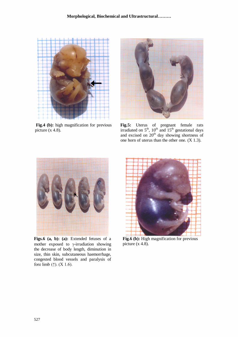
In figures (3) and (4a, b) the uterus of
group (2) showed frequent implantation sites. The fetuses of the same group showed
subcutaneous haemorrhage, abnormal
bending of body (protrusion), anotia, short
nect and dactylomegaly. The uterus of the group (3) displayed
shorting of one horn of uterus than the other
one Fig (5) with decreased fetal number. However the number of surviving
Morphological, Biochemical and Ultrastructural………
525
fetuses of irradiated mothers showed a
number of malformations including: conge-sted blood vessels, paralysis of fore limb,
thin skin and decreased body length (Fig.
6a, b).
On the other hand, gamma irradiation on days 5
th, 10
th and 15
th of gestation during
folic acid treatment showed high incidence
of prenatal mortality which appeared as residual bodies Fig (7).
Furthermore, the fetuses of this group
showed pre-natal death and embryos replaced by residual bodies Fig (8).
Ultrastructural studies
1- Control group: Electron microscopic examination of
the liver of control rat foetuses showed the
normal ultrastructure. The cytoplasm of the hepatocytes contains mitochondria, rough
endoplasmic reticulum and small amount of
glycogen. Also, the nuclei are rounded and central in position and have a coarse pattern
of heterochromatin (Figs. 9, 10).
2- Folic acid -treated group: A remarkable alteration could be observed on examined sections of treated
group with folic acid at a dose of 5mg/kg
b.wt/day revealing loss of normal archite-cture with marked degeneration of
hepatocytes. (Figs. 11, 12).
3- Irradiated group: At the ultrasturtural level the irradiated animals an accumulative dose 1
Gy on 5th, 10th and 15th days up to 3 Gy
during pregnancy showed severe damage in the fetal hepatocytes with hydropic dege-
neration (Fig. 13). In such cells, the
cytoplasmic matrix became lytic and the cytoplasmic organelles are highly degene-
rated. The mitochondria were swollen with
ruptured outer membranes and cristae. The
rough endoplasmic reticulum was broken into fragments. The nuclei contained
translucent fragmented and marginal
chromatin and the nucleolus was prominent at the periphery. The blood sinusoids were
damaged and contained phagocytic kupffer
cells, and the nuclei of the endothelial cells were mostly pyknotic with disturbed
erythrocytes (Fig. 14).
Placenta 1- Control group:
The placenta is a temporary organ,
consisting of a maternal portion and a fetal
portion. (Fig. 15) showed the general ultrastructural of the trophobalast
comprising, cytotrophoblastic cell and
syncytotrophoblastic cell.
2- Folic acid-treated group: Administration of folic acid at a dose
of 5mg/kg b.wt from days 5th to 20th
induced marked ultrastructural abnorm-
alities in the placental cells. They appeared
with degenerated cytoplasmic matrix.
Whereas many lysosomal bodies were represented (Fig. 16). Also, the degenerated
cytoplasm organoids were occupied by
numerous heterogenous vesicles and vacuoles with variable sizes (Fig. 17).
3- Irradiated group: The electron microscopic examination of the placental sections of the irradiated
animals at accumulative dose 1 Gy on 5th,
10th and 15th days up to 3 Gy showed the
placental cells which were greatly injuried, the nuclei contained fragmented chromatin
with corrugated nuclear membrane .The
cytoplasm was disorganized with increased amount and different sizes and shapes of
lysosomal bodies (Fig. 18). In some of the
placental cells the cytoplasm became lytic
and the cytoplasmic organelles are highly degenerated with lots of lysosomes. The
mitochondria were small and electron
dense. The endoplasmic reticulum was broken into fragments. Cell nuclei showed
chromatin disintegration (Fig. 19).
4-Folic acid - irradiated group: This group was given folic acid at a
dose of 5mg/kg and then irradiated at
accumulative dose 3 Gy revealed severely
effected placental cells. As represented in figs. (20 & 21) degenerated cyncytiotr-
ophoblast and highly atrophied and
karyolytic nuclei with corrugated outer and inner membranes were well marked.
Furthermore, fibroid areas and increased
collagen fibers were observed.

Fatma, L. Ramadan & Seham M. Abu Nour
526
Table (1): effect of folic acid and/or gamma irradiation exposure on serum level of gamma
glutamyl transferase; placenta tissues content of lactate dehydrogenase of
pregnant rats and phosphorus content in the liver tissue of feuses.
parameters Goup 1
(control)
Group 2
(folic treated)
Group 3
(Irradiated)
Group 4
(irradiated and folic
treated)
Gamma glutamyl transferase
(U/l) 4.3240.769 6.8580.206a 8.5400.447c 11.0260.425c
Lactate dehydrogenase
(U/dI) 0.1380.006 0.1440.018c 0.3020.005c 0.3800.015c
Phosphorus content (mg/dI) 47.771.312 51.4820.746a 55.5062.110b
Each value represents the mean of five rats S.E a-Significant changes from control at P < 0.05
b-Significant changes from control at P < 0.01 c-Significant change from control at P < 0.001
Fig.1: Uterus of a control pregnant rat
excised on the 20th gestational day showing
normal distribution of 9 implanted embry-os distributed between the 2 hours (X 0.7).
Fig.2: Embryos of a control mother rat
illustrating normal development
(X 0.7).
Fig.3: The uterus of animal received folic
acid during gestation period showing frequent implantation sites (X 1).
Figs.4 (a, b): (a): Fetuses of rat treated with
folic acid note: Subcutaneous haemorrhage abnormal bending of the body (protrusion),
absence of ear (anotia), short neck and abnor-
mally large size of finger (dactylomegaly) (↑). (X 2.2).
Morphological, Biochemical and Ultrastructural………
527
Fig.4 (b): high magnification for previous
picture (x 4.8).
Fig.5: Uterus of pregnant female rats
irradiated on 5th, 10
th and 15
th gestational days
and excised on 20th day showing shortness of
one horn of uterus than the other one. (X 1.3).
Figs.6 (a, b): (a): Extended fetuses of a
mother exposed to -irradiation showing the decrease of body length, diminution in
size, thin skin, subcutaneous haemorrhage, congested blood vessels and paralysis of
fore limb (↑). (X 1.6).
Fig.6 (b): High magnification for previous
picture (x 4.8).

Fatma, L. Ramadan & Seham M. Abu Nour
528
Fig.7: Uterus of pregnant rats exposed to -
irradiation on days 5th, 10
th and 15
th of
gestation during folic acid treatment
exhibiting high incidence of prenatal
mortality which appeared as residual bodies.
(X 1.6).
Fig.8: Extended rat fetuses of mother exposed
to γ-irradiation during folic acid treatment
showing pre-natal death and embryos
replaced by residual bodies. (X. 2).
Fig.9: Electron micrograph of the liver of a fetus showing a normal architecture of
hepatocytes. Notice large nuclei (N) with
dense matrix. (X-8000).
Fig.10: Electron micrograph of the liver of a fetus showing mitotic figure. (X -6000).

Morphological, Biochemical and Ultrastructural………
529
Fig.11: Electron micrograph of the liver of
a fetus whose mother which received folic
acid during pregnancy showing the cytoplasm is severely degenerated . Note
pyknotic (p) and karyolytic (k)
nuclei,degenerated cristae of mitochondria (M) and dilated cisternae of rough
endoplasmic retculium. (X-2800)
Fig.12: Electron micrograph of the liver of
fetus whose mother received folic acid during
pregnancy showing irregular architechteur of hepatocytes. Note swollen mitochondria (M)
with degenerated cristae pyknotic (p) and
karyolytic nuclei (k) (X-2800).
Fig.13: Electron micrograph of the liver
of fetus whose mother exposed to γ-
radiation during pregnancy showing hydropic degeneration of the cytoplasm.
Swollen and degenerated mitochondria
(M) and fragmented rough endoplasmic
retieulum (RER). Note lysosomes (ly) (X-6000).
Fig.14: Electron micrograph of the liver of
fetus whose mother exposed to γ-radiation
during pregnancy showing blood sinusoid (BS) is dilated and contains kupffer cell (K)
and inflammatory cells (IC) (X-4600).

Fatma, L. Ramadan & Seham M. Abu Nour
530
Fig.15: Electron micrograph of control rat
Placenta showing the ultrastructure of the
tropoblastic components. The
syncytiotrophoblast (S) has irregular microvilli (v) and the cytotrophoblast (C).
blood vessel (V) which contain
erythrocytes (E). (X- 2800).
Fig.16: Electron micrograph of the placenta
of an adult rat treated with folic acid
showing disorganization of the cytoplasmic
structure and it contained the debris of degenerated organoids and lots of
lysosomes (y) . The nucleus (N) contained
fragmented chromatin. (X 2800).
Fig.17: Electron micrograph of the
placenta of an adult rat treated with folic acid showing the cytoplasm which
contained lots of irregular vacuoles
indicating degenerated organoids . The
nucleus (N) had fragment chromatin. (X 3600).
Fig.18: Electron micrograph of the placenta
of an adult rat exposed to γ-radiation showing highly degenerated
cytoplasmic organelles. Note increased
amount of lysosomal bodies (Ly).
Karyolytic nucleus (N) had undulating membranes. (X 3600).

Morphological, Biochemical and Ultrastructural………
531
Fig.19: Electron micrograph of the
placenta of an adult rat exposed to
γ-radiation showing fibroid areas and
collagen fiber (F), increased of lysosomal bodies (Ly) and atrophied and karyolytic
nuclei (N) with irregular nuclear
membrane. (X 4600).
Fig. 20: Electron micrograph of the
placenta of an adult rat treated with folic
acid perior to irradiation showing
degenerated syncytiotrophoblast with highly atrophied and karyolytic nuclei (N)
and increased collagen fibers (F). Note the
maternal blood spaces (bs) which contain deformed RBC (X 2800).
Discussion
The results of the current study
revealed that whole body gamma irradiation of pregnant rats at an accumulative dose 1
Gy/3 times on both 5th, 10
th and 15
th days of
gestation resulted in a significant
biochemical disturbance with considerable ultrastructural damage in the placenta of the
pregnant rats and liver of their fetuses
associated with embryonic disorders. Data obtained from the present study
revealed a significant elevation in maternal
serum of -GT and LDH in the placental tissue together with an elevation in
phosphorus content in the liver of fetuses as compared to those of control. This was a
correlation to Omran and Abu-zied (2006)
who stated that the increase in -GT has been considered as an indicator for tissue
injury. The synthesis of -GT is regulated by presence of reactive oxygen species since the enzyme is essential for the
metabolism of antioxidant glutathione.
It was clamid that glutathione
depletion led to increased -GT level which indicated hepatocellular necrosis (Whilfield, 2000).
On the other hand, Said and Hanafy
(2006), demonstrated decreased cellular
glutathione altering protein thiols which
disordering the transport and storage of
Ca+2
in mitochondria, endoplasmic reticu-lum and cell membrane of liver so the
content of Ca+2
increasing and ultimately
causing cell death and -GT was released into the blood.
Radiation exposure induced marked elevation in LDH in placental tissue of
pregnant female rats. This is usually used as
marker to placental dysfunction and also
cytotoxic effect of exposure to irradiation (Gharib, 2007).
In this respect, Yoshid et al. (1995)
attributed the disorders in the placenta circulation to involvement of endocrine
mechanisms and decreased blood follow in
the placenta which affect the pituitary function. In addition to the possible
pathological changes which can observed in
the structure and function of the epithelial
cells of renal tubules with subsequent alterations in the active transport of calcium
and phosphorus through these organs.
These findings one supported by Abdel Gawad et al. (2005), who recorded that
under effect of irradiation the maternal
calcium and phosphorus content were not sufficient to satisfy the fetuses with its
requirement for normal growth. The various

Fatma, L. Ramadan & Seham M. Abu Nour
532
degrees of damage observed in the liver of
rat embryos in the present study are most probably due to the direct effect of radiation
on the mitochondria and endoplasmic
reticulum which leading to the release of
enzymes from cellular organelles like lysosomes, microsomes and others.
It is possible to assume that severe
malformations observed in the fetuses were due to vascular disturbance, amino acid
abnormalities and hormone inbalance
(Ramadan and Rezk, 2006) Otherwise , the excessive production of oxygen free
radicals in fetuses of irradiated mother
might increase the risk of fetal
abnormalities (Rezk and Ibrahim, 2006). Diamant (1981) noticed decreased
total protein in the placenta post-exposure
to -rays and this may be due to the destructive effect of these rays. The
significant decrease in fetal size related to radiation which induced depression in the
cellular differentiation process taking place
in the embryonic cells were noticed by Rezk et al. (2005).
These observation were similar to
those described by Amvrsene et al. (1994) who reported that total body irradiation of
rat embryos at the pre-implantation and
organogenesis stages produced increased
incidence of malform- ations due to irradia-tion which caused death to the zygote while
during organogenesis the cell death may be
due to disruption in the morphog-enetic movements of differentiation and caused
pre and neonatal mortality. Furthermore,
Suitor and Bailey (2000) found that the vertebra of vertebral column did not close
properly after fertilization and exposure to
gamma irradiation so the spinal fluid bulge
and cause paralysis this observation are correlated to our present results.
Results of the present study suggest
that susceptibility of tissue and embryonic development have been influenced by folic
acid administration. The current study
despicted that folic acid administration at
dose 5 mg/kg b. wt / day revealed a
significant elevation in serum of -GT level, LDH in the placental tissues of
pregnant female rats and also recorded an
elevation in phosphours content in the liver tissue of fetuses.
Abdel Gawad et al. (2005) reported
that degenerated cells of placenta taken
from exposed pregnant rats may be
responsible for reduced size of fetuses and may be due to the disturbance in transport
of nutrients to the fetuses and resulted in
fetal disorders.
Accordingly, Rezk and Ibrahim (2006) reported that after absorption of the
drug from the gut caused teratogensis
which associated with reproductive and developmental toxicity as a result of
exposure liver to damage
The present results which indicated the dystrophic changes in placenta and fetal
liver exposed to -rays or treated with folic acid are in agreement with many authors.
Rowell and Clark (1982), Khera, (1991)
and Ubbink (1995) stated that irradiation caused fetal growth and embryo lethality
due to inhibition of protein synthesis or
placental dysfunction On the other hand,
Hartridge, et al.(1999) noticed that administration of folic acid at dose
10mg/100g b.wt can precipitated in the
nervous system and caused degeneration of the spinal cord which linked to the
increased risk of neural tube defect because
folic acid at this dose caused blocked permeability which did not allow easier
transfer of mineral and protein from blood
to the bone and cells. This view is
supported by the work of Baggot et al. (1992) who reported that high blood supply
levels were associated with spontaneous
abortion and developmental abnormalities. Whether administration of folic acid and
exposure to -irradiation inhibit the synthesis of nucleic acids and cell can not
complete its mitosis and also radiation
inhibit the conversion of folic acid to folinic acid (Kamen, 1997). In this respect increase
of malformation following folic acid
administration and radiation exposure as result of decreased hepatic metabolism
(Chevias et al. 1987). The severity of these
changes might be attributed to synergistic
effect of folic acid and gamma rays exposure.
At the ultrastructural level, folic acid
caused variable degrees of damage in the fetuses liver. The commen lesions which
were observed are: hydropic degeneration,
deformation of the cytoplasmic organelles and nuclear damage. The cytoplasmic
organelles which were markedly affected
by folic acid treatment in the fetal liver and

Morphological, Biochemical and Ultrastructural………
533
placenta cells are the mitochondria and the
endoplasmic reticulum. The mitochondria are the most sensitive structures to any
affecting chemical factor. Stevens and lowe
(1995) considered the mitochondrial
damage as an early event for the cell injury. Also mitochondria are the main sites of the
energy production in the cells, their damage
may results in lowed energy output. This in turn may be a factor in inducing other
changes observed in the cells.
Moreover, mitochondria are known to contain fatty acid oxidases, these enzymes
are necessary for the metabolism of
triglycerides. This leads to another
suggestion that the mitochondrial damage observed in the present study may be
involved in the lipid changes.
The observed alterations of the rough endoplasmic reticulum constitute the main
adverse effect of -ray and folic acid on the fetal liver cells and placental cells due to its
important role in protein synthesis. In
addition smooth endoplasmic reticulum represents the main site of metabolism of
various drugs (Davis, 1984), so it was
markedly affected following folic acid administration. Rubin (2000), attributed the
damage of cell membranes and organelles
following drug treatment to the O2 metabo-
lites which react with the unsaturated fatty acids in phospholipids (lipid peroxidation).
He also attributed it to the deficiency of the
protective enzymes, glutathione peroxidase and superoxide dismutase, from the liver
following drug treatment.
The present study indicates that folic acid has an dystrophic effect on the nuclei
of hepatocytes and placental cells, which
showed variable changes in their amount of
chromatin and others showed signs of shrinkage pyknosis and karyolysis. These
changes may be due to the interference of
folic acid with nuclear DNA in liver cells and placenta cells. On the contrary Kamen
(1997), reported that folic acid is necessary
for the production and maintenance of new
cells. This is especially important during periods of rapid cell divion and growth such
as infancy and pregnancy.
Also, Frenech et al. (1998) recorded that folate is needed to make DNA and
RNA, the building blocks of cells. It also
helps prevent changes to DNA that may lead to cancer. Several authors suggested
that a deficiency in folate might predispose
people to develop cancer of the cervix, colon, lung; breast and mouth (Butterworth
et al., 1992; Heimburger et al., 1992; Kim
and Mason 1995 and Zhang et al., 1999).
Whereas, some observations suggested that folate supplements may prevent colon
cancer, especially when taken for many
years (Ginovannucci et al., 1998). Moreover, high-dose folate (10 mg/daily)
might be helpful for normalizing
abnormalities in the appearance of the cervix in women taking oral contraceptive,
but it dose not appear to reverse actual
cervical dysplasia (Burrerworth, et al.,
1982, Burrerworth 1992 and Butterworth et al., 1992). Very high dosages of folate may
be helpful for gout (Oster, 1973), although
some authorities suggested that these was actually a contaminant of folate that caused
the benefit seen in some studies (Boss et
al., 1980). Furthermore, other studies have found no benefit at all (Boss et al., 1980).
The present findings revealed that
when pregnant rats were exposed to
gamma-radiation, the fetal liver and plac-enta cells induced ultrastructural changes in
the hepatocytes and placenta cells.
The results obtained in the present study pointed to prominent cellular changes
which were observed in the distortion of the
nuclei, swollen mitochondria, increased lys-
osomes and fragmentation of endoplasmic reticulum.
Exposure to ionizing radiation is
characterized by excessive production of ROS associated with an increase in the
process of lipid peroxidation (Saada et al.,
2001 & 2003). ROS such as hydroxyl radicals (OH), superoxide anion (O2) and
hydrogen peroxide (H2O2) produced in the
living cell during normal metabolic
functions or as a consequence of response to abnormal stress symbolized a great threat
to biomembranes and interact with vital
molecule causing their alteration and destruction (Benderitter et al., 1995). The
polyunsaturated fatty acids of cell memb-
rane phospholipids are major targets of the highly reactive (OH) attack (Haliwell and
Gutteridge, 1989). This is associated with a
decrease in the activity of antioxidant enzy-
mes of the body with consequent damage of cellular biomemb-ranes (El Habit et al.,
2000 and Saade and Azab, 2001). Radiation

Fatma, L. Ramadan & Seham M. Abu Nour
534
induced damage to membrane of the
subcellular organelles which may be attributed to peroxidation of membrane
lipid protion monitored by the increase in
the thiobarbituric acid reactive substances
(TBARS) concentration (Azab, 2007).
References
1. Abdel-Gawad I T, Badr H M. and
Shabon M H. (2005): Physiological effect
of natural thumic acid during pregnancy on
fetuses and maternal by irradiation in rats.
Isotope & Rad. Res., 37 (3): 722-749.
2. Abu-Gabal H A, Eid F A, Gaber S H A.
and Mahmoud N H. (1994): Effect of
polytrin and -rays on nucleic acid in
placenta of albino rats. Egypt. J. Rad. Applic., (7) 1: 69-73.
3. Abu-Gabal H A, Zaki M M, Gaber S H,
Roushdy, H M. and Ramadan F L.
(1995): Changes induced in total proteins
and liver tissues of pregnant rats following
radiation and caffeine treatment. Al-Azhar
Bull. Sci., 6: 901-917.
4. Aldeen K A M. and Konermann G.
(1978): Effect of acute X-irradiation on
pre-implantation embryos and on the impl-
antation reaction on the mouse Radiation Environment. Biophys., 15 (1): 47-56.
5. Amvrosene A P, Rogov IU I, Dorokhina
R I. and Pavlenko V S. (1994): Effect of
single-dose external gamma-irradiation of
0.5 Gy on the development of rat, embryos.
Rad. Biol. Radio. Col., 33: 623-628.
6. Ashry O M. (1997): Control of organ
functional changes in radiation exposed
pregnant mice Ph.D. Thesis Faculty of
Science Ain Shams University.
7. Azab K S. (2007): Modulation of radiation
injuries in rats receiving multiple doses of aloe vera. Egypt. J. Rad. Sci. Applic., 20
(1): 17-28.
8. Baggott J E, Morgan S L, Ha T. (1992):
Inhibition on of folate dependent enzymes
by non-steroidal anti inflammatory drugs.
Biochem., 282, (11): 147-202.
9. Benderitter M, Assem M. and Maupoil
V. (1995): Effect of in vivo heart irradi-
ation on the development of antioxidant
defenses and cardiac functions in the rat.
Rad. Res., 144, 64.
10. Boss G R, Ragsdale R A. and Zettner A.
(1980): Failure of folic acid (pteroylg-
lutamic acid) to affect hyperuricemia. J.
Lab. Clin. Med., 96: 783-789.
11. Butterworth C E, Hatch K D. and Gore
H. (1982): Improvement in cervical
dynsplasia associated with folic acid
therapy in users of oral contraceptive. Am.
J. Clin. Nutr., 35: 73-82.
12. Butterworth C E. (1992): Effect of folate
on cervical cancer. Synergism among risk
factors. Ann. Y. Acad. Sci., 669: 293-299.
13. Butterworth C E, Hatch K D. and Soong S J. (1992): Oral folic acid supplem-
entation for cervical dysplasia. A clinical
intervention trial. Am. J. Obstet. Gynecol.,
166: 803-809.
14. Chevais M, Reinert P. and Randeau M
C. (1987): Cirtical risk / benefit analysis of
pefloxacin use in children under 15 years.
The problem of arthralgias. Int. J. Clin.
Pharmacol. Toxicol., 25: 306-609.
15. Davis M. (1984): Drug and hepatotoxicity.
In Maddrey W. C. ED: liver, London, Buta.
Worth, pp: 133-164. 16. Diamant Y Z. (1981): The placenta in
intrauterine fetal deprivation, Acta. Obst.
Gynecol. Scand., 60: 141-150.
17. Dorothy R H. (1983): Alterations of
maternal metabolism in normal and diabetic
pregnancies. Am. J. Obst. Gynecol., 146:
417-421.
18. El-Habit O H, Saada H N, Azab Kh Sh,
Abdel-Rahman M. and El-Malah D F.
(2000): The modifying effect of B-carotene
on gamma radiation-induced elevation reactions and genotoxicity in male rat. Mut.
Res., 466: 179.
19. Frenech M, Aitken C. and Rinald J.
(1998): Folate, vitamin B12, homocysteine
status and DNA damage in young Austr-
alian adults. Carcinogenesis., 19: 1163-71.
20. Gaber S H. (1990): Synergistic effect of a
chemical pollutaint substance and whole
body irradiation of mother on the placenta
and development of albino rat embryo.
Ph.D thesis, Faculty of Science. Al-Azhar
Univ. Girl's. Branch. 21. Gharib O A. (2007): Does kombucha Tea
reduce the damage induced by radiation
exposure?. Egypt. J. Rad. Sci. Applic., 20
(1): 141-157.
22. Ginovannucci E, Stampfer M J. and
Colditz G A. (1998): Multivitamin use,
folate, and colon cancer in women in the
Nurses, Health Sludy. Ann. Intern. Med.,
129: 517-524.
23. Haliwell B. and Gutteridge J M. (1989): Free radicals in biology and medicine 2nd ed., Oxford, U. K. Clarendon Press.
24. Hartridge T, Rling M.Sc, ORth F D.
(1999): The role of folic acid in oral
clefling. British Journal of orthodontics,
vol.26 No. 2, 115-120.
25. Hathcock J N. (1997): Vitamins and
minerals: Efficacy and safety. Am. J. Clin.
Nutr; 66: 427-464.
26. Hayut M A. (1986): Basic techniques for

Morphological, Biochemical and Ultrastructural………
535
transmission electron microscopy. New
York. Harcourt Brace Jovanovich.
27. Health notes (2005): Folic acid. W. W. W.
Healthnotes. Com.
28. Heimburger D C. (1992): Localized
deficiencies of folic acid in aerodingestive tissues. Ann. N. Y. Acad. Sci., 669: 87-96.
29. Henry J B. (1974): Clinical diagnosis and
measurement by laboratory methods. Todd.
Sanford. Davidsohn. 16th ed. W. B.
Saunders. Philadephia. Pp. 260.
30. Kamen B. (1997): Folate and anlifolate
pharmacology. Semin. Oncol., 24: 30-39.
31. Katarina K, Lucia S. and Eva M. (2002): Cytogenetic changes in the liver of progeny
of irradiated male rats. J. Radiat. Res., 43:
125-133.
32. Khera K. S. (1991): Chemically induced alteration in maternal homeostasis and
histology of conceptus. Their etiologic
significance in rat fetal anomalies. Teratol.,
44 (3): 259-297.
33. Kim Y I. and Mason J B. (1995): Folate
epithelial dysplasia and colon cancer. Proc.
Assoc. Am. Physicians. 107: 218-227.
34. Living Naturally (2006): Living Naturally
and Nature's Garden Natural Foods.
35. Omran M F. and Abu-Zied N M. (2006):
Modulation of the antioxidant system efficacy in irradiated rats supplemented
with vitamin B12 cobalamin and folic acid.
Egypt. J. Rad. Sci. Applic., 19 (1): 61-71.
36. Oster K A. (1973): Evaluation of serum
cholesterol reduction and xanthine oxidase
inhibition in the treatment of atheros-
clerosis. Recent. Adv. Stud. Cardiac Struct.
Melab., 3: 73-80.
37. Ramadan F L. and Rezk R G. (2006): Ameliorating role of chromium ingestion
on biochemical, histological and terato-
genic disorders induced by diabetes and / or gamma irradiation in pregnant albino rats
and their fetuses. Isotope & Rad. Res., 38,
1, 43-69.
38. Rezk R G. and Ibrahim M F. (2006): Potency of maternal folic acid suppleme-
ntation on gamma irradiation induced
histological and emleryological anomalies
in albino rats. Egypt. J. Rad. Sci. Applic. 19
(2): 38-413
39. Rezk R G, Ibrahim M F. and Darwish M
M. (2005): Efficacy of ginger in alleviating the severe radiation-induced biochemical,
Histological and embryological Impacts in
pregnant female albino rats. Isotope of Rad.
Res., 37 (3): 625-645.
40. Rowell P P, clark M. (1982): The effect of
chronic oral nicotine administration on fetal
weight and placental amino acid accumu-
lation in mice. Toxicol and Appl.
Pharmacol., 66: 30-38.
41. Rubin E. (2000): Essential pathology. 3rd
ed., lippincott Williams and Wilkins,
Philadelphia. pp: 24-25.
42. Saada H N. and Azab Kh Sh. (2001): Role of lycopene in recovery of radiation
induced injury of mammalin cellular organelles. Pharmazie., 56: 239.
43. Saada H N, Azab Kh Sh. And Zahran A
M.(2003):Post-irradiation effect of Bronco-
Vaxom, OM-85 and its relationship to anti-
oxidant activities. Pharmazie.56(8): 654.
44. Saada H N, Ussama Z S. and Mahdy A
M. (2003): Effectiveness of Aloe vera on
the anlioxidant status of different tissues in
irradiation rats. Pharmazie., 58 (12): 929.
45. Said V Z. and Hanafy N. (2006): Effect of
grape seed extract on hepatic function and
antioxidant status of mouse bearing ehrlich ascites carcinoma and exposed to gamma
radiation. Isotope &Rad. Res.,38 (1):225-
240.
46. Snedecor G W. and Cochron W G.
(1978): "Statistical Methods" 8th ed. Louis
State Univ., Press, Ames, IAWO, USA.
47. Stevens A A. and Lowe J. (1995): Pathology. Alan Steven/ James Lowe.
Mosbe, St. Louis. Baltimore, Philadelphia,
Toronto: 23-33.
48. Suitor C W. and Bailey L B. (2000): Dietary folate equivalents interpretation and
application. J. Am. Diet. Assoc. 100: 88-49.
49. Tietz N W. (1986): "Textbook Clinical
chemistry" Saunders Comp. Philadelphia.
50. Truswell A S. (1985): ABC of nutrition.
Nutrition for pregnancy. Br. Med. J., 291:
263-269.
51. Ubbink J B. (1995): Elevated circulating
maternal homocystene concentration a risk
factor for neural tube defects. Nut. Rec., 53,
173-175.
52. Walash M N, Abu-Gabal H S. and Moustafa U A.(1989): Synergistic effect of
nicotine and radiation exposure on a mam-
malian embryo. Proc. Zool. Soc. Egypt., 16:
337-340.
53. Walash M N, Eid F A, Abu Gabal H N.
and Moustafa N A. (1988): Teratogenic
effect of nicotin and gamma rays on the
liver of albino rat embryo. Proc. Zool. Soc.
A. R. Egypt, 16: 337-346.
54. Whilfield J B.(2000): Gamma glutamyl
transferase. Grit. Rev. Clin. Sci.,38(4): 263.
55. Yoshida Y, Seto T, Ohsu W, Hayashi S.
and Nakamura H. (1995): Endocrine mec-
hanism of placental or circulatory disturb-
ances induced by microwave in pregnant
rats. Nippon-Sanka-Zasshi, 47 (2):101-108.
56. Zhang S, Hunter D J. and Hankinson S
E. (1999): A prospective study of folate
intake and the risk of breast cancer. JAMA.,
281: 1632-1637.

Fatma, L. Ramadan & Seham M. Abu Nour
536
مشيمة الجرذان فى التغيرات المىرفىلىجية والبيىكيميائية والنسيجية الدقيقة
أو آشعة جاما/ الفىليك و ا المعاملة بحمضالحىامل وكبد أجنته
فاطمة لطفى رمضان ـ سهام محمد أبى نىر
المشمض القم لجخس رنىلجيب اإلشؼبع ـ يئخ الطبقخ الزسيخ
لؼتء الزأمسذ آلشؼخ جبمب مب صالذ ف حبجخ إل المضيدذ مده فبػليخ معبداد األمسذح ا
أ / يذف زا الجخش إل رقيدي الزدأصيشاد العدبسح للةشػدخ النجيدشح مده حمدط ال ليدل . األثخبس
. آشؼخ جبمب ػل مشيمخ الةشران الخامل مجذ أجىز
د المةمػدخ المةمػدخ األلد. ر رقسي إوبس الةشران الخامل إل أسثؼخ مةمػدبد
مةد مده صن الةسد مده اليد /مةد 2العبثطخ أػطيذ المةمػخ الضبويخ حمط ال ليل ثةشػدخ
جدشا 3المةمػخ الضبلضخ رؼشظذ للزشؼيغ الةبم ثةشػدخ . الخبمس إل الي الؼششيه مه الخمل
. مدلفد اليد الخدبمس الؼبشدش الخدبمس ػشدش مده الخ( صدثس مدشاد/ جدشا 1)مةشػخ مةضئخ
المةمػخ الشاثؼخ ػملذ ثخمط ال ليل رؼشظذ آلشؼخ جبمب مؼب ثى س الةشػبد الز ػملدذ
.ثب المةمػزيه الضبويخ الضبلضخ
ردد رثددج الةددشران فدد اليدد الؼشددشيه مدده الخمددل م أتددزد ػيىددبد الددذ لزؼيدديه الز يددشاد
زلل مجدذ األجىدخ رد رةيضدب الجيميميبئيخ فيد م أتدزد أيعدب ػيىدبد مده مشديمخ اإلودبس الجبل دخ مد
.للذساسخ ثبلمينشسنة اإللنزشو
أظخذ الىزدبئ أن األمدبد المؼبملدخ ثخمدط ال ليدل أ المؼشظدخ لاشدؼبع الةدبم قدذ
ظشد ثب صيدبدح فد وشدبغ إودضي الةبمدب جلربميدل رشاوسد يشيض فد السديش مدغ إسر دبع ملخدظ فد
يمخ لأل الخبمل مصخثب ثإسر بع فد مخزد ال سد س فد الثمزيذ دييذسجيىيض ف أوسةخ المش
جذ أن المؼبملخ ثخمط ال ليل أ الزؼشض لاشؼبع قذ أدد إلد الؼذيدذ مده . أوسةخ النجذ للةىيه
الزشبد مىب إوخىبء الظش إتز بء األرن قصش الشقجخ م وضيف رخدذ الةلدذ شدلل فد األغدشاف
.ػيخ الذميخاألمبميخ مزلل إحزقبن ف األ
ممدب أظدخذ الىزدبئ أظدشاسا سدزلجيخ ثبل دخ فد وسدي مشديمخ إودبس الةدشران مدزلل
ثيىمدب رسدجت . وسي النجذ لألجىخ وزيةخ رؼبغ حمدط ال ليدل أ رؼدشيط أجسدبمب للزشدؼيغ الةدبم
الزؼشض المشزشك ف المضيذ مه األظشاس الزشبد شملذ إرسبع الةية الذميخ جدد تثيدب
الزبثيددخ رخلدددل سددديزثثص النضيددش مددده الخثيدددب حددذس ظدددشس لندددل مدده الميزمودددذسيب الشدددجنخ
.اإلوذثثصميخ ممب شمل حذس ر يشاد ف ثؼط األويخ ف وسي المشيميخ
مةدد مدده /مةدد 2وسددزىز مدده ددزي الذساسددخ أن إػطددبء ال ئددشان الخامددل جشػددخ مقددذاسب
اإلصىدديه مؼددب قدذ أد إلدد ظددس ر يددشاد ظددبسح سدداء فدد حمدط ال ليددل أ الزؼددشض لاشددؼبع أ
.مسفلجيخ األجىخ أ الز يشاد الزشميجيخ مزلل القيبسبد الجيميميبئيخ الز ر قيبسب