Embed Size (px)
Citation preview
International Journal of Fisheries and Aquaculture Sciences. ISSN 2248-9975 Volume 3, Number 1 (2013), pp. 45-62 © International Research Publication House http://www.irphouse.com
Morphological and Skeletal Deformities in Labeo Rohita (Ham. Buch.) from Fresh Water Fish
Ponds of Punjab
Prof. S.P.S. Dutta Department of Environmental Sciences, University of Jammu
Dr. Deepika Slathia Department of Environmental Sciences, University of Jammu
Vaseem Katoch Department of Environmental Sciences, University of Jammu
ABSTRACT
Six aberrant adult specimens of Labeo rohita, a commercially important major Indian carp cultured in Punjab ponds were seen at Gumat fish market, Jammu and have been described. These morphological and vertebral deformities are most probably induced by water degradation caused by excessive use of pesticides in leading grain producing state of the country and predatory effects in one of the specimen.
INTRODUCTION Fish anomalies are of serious concern in aquaculture, as deformed animals are not preferred by consumers, have low market value and are ultimately a loss to the farmer. Study of such aberrant animals and exploring this problem is important fish biological aspect as accumulation of various pollutants through biomagnifications, is not only harmful to the fish, but can have ill effects on its consumers like birds and ultimately man. Although there is no documented report on health problems to man caused by consumption of such deformed fishes, but we should not forget the Minamata bay episode. Early records of aberrations in fresh water carps from India are those of Hora (1942) and Law (1944), who described hump backed Catla. Kapoor and Sarkar (1955) were the first to describe morphological and vertebral anomalies

46 Prof. S.P.S. Dutta et al
in four specimens of Labeo rohita from river Yamuna and its neighbouring ponds, within Delhi. Pillai and Thampy(1990) noticed deformities in the vertebral column at the tail region of Labeo rohita in the fish culture systems of the College of fisheries, Panangad, Cochin. Due to degradation of water quality, there is steep rise in fish deformities in various fish species in Punjab and have been documented by Tandon and Sharma (1971) and Dutta et al. (2011). During the survey of deformed fishes, brought from Punjab, at Gumat fish market, Jammu, six adult deformed specimens of Labeo rohita were seen along with normal fish and have been described to aware the scientists and society about degradation of environment and its impact on fish. MATERIALS AND METHODS Six adult deformed specimens of Labeo rohita were purchased from fish market at Gumat, Jammu, and studied for morphological characteristics and photographed. X-ray of these specimens was taken using Fuji Green Base Film with SOFTEX CMB-2 at 10 milliamps and 50kv. for 0.25 sec. Specimens were then fixed in 10% neutral buffered formalin. RESULTS Morphological characteristics of normal and abnormal specimens of Labeo rohita (Ham Buch.) are tabulated in Table 1. Other features like length, weight, number of vertebrae in vertebral column, region of skeletal deformity and deformity type in aberrant specimen of Labeo rohita (Ham Buch.) have been depicted in Table 2.
Table1. Comparison of morphological features of normal and abnormal specimens of Labeo rohita (Ham. Buch.)
Ratios Normal
fish Specimen
1 Specimen
2 Specimen
3 Specimen
4 Specimen
5 Specimen
6 Head length in total body length Body height in total body length Pre-dorsal length in total body length Post-dorsal length in total body length Pre-anal length in total body length Post-anal length in total body length Length of dorsal fin in total body length Length of pectoral fin in total body length Length of pelvic fin in total body length Length of anal fin in total body length Length of caudal fin in total body length Pectoral-pelvic origin distance in total body length Pelvic-anal origin distance in total body length Anal-caudal origin distance in total body length
5.27 4.21 2.68 1.59 1.68 2.46 5.9 6.70 7.19 6.56 4.04 5.27 4.54 6.55
3.97 3.26 2.17 1.86 1.56 2.77 4.43 4.30 5.34 5.64 4.43 5.0 5.96 7.38
4.57 3.19 2.46 1.68 1.67 2.5 4.92 5.51 6.15 5.33 4.58 4.85 5.0 8.0
3.64 2.78 1.78 2.27 1.18 6.67 3.64 3.92 5.0 4.76
- 3.33 2.94
-
4.30 2.90 2.25 1.80 1.41 3.44 4.77 5.64 5.53 5.17 4.77 3.73 4.56 8.86
4.85 4.0 2.60 1.62 1.68 2.46 5.52 6.15 6.67 5.92 3.90 5.33 4.44 7.11
4.0 2.96 2.42 1.86 1.6 3.08 4.88 4.82 5.55 4.76 3.88 4.44 5.48 10.81

Morphological and Skeletal Deformities in Labeo Rohita (Ham. Buch.) 47
Table 2. Length, weight, number of vertebrae in vertebral column, region of skeletal deformity and deformity type in Labeo rohita(Ham. Buch.).
Specimen Length
(cm) Weight
(g) Number of
Fin rays Number of vertebrae
in Vertebral Column
Normal Deformed
Region of Skeletal Deformation
Percentage of Deformity
Deformity Type Spinal Caudal Fin
Jaw Rib
Specimen 1 31 480 Deviation 15 17 Post Dorsal region 0.5 Y - Y - Y Specimen 2 32 380 Deviation 11 21 Post Dorsal region 0.6 Y - Y - - Specimen 3 20 240 Deviation 19 10 Caudal Peduncle 0.3 - Y Y - - Specimen 4 31 520 Deviation 16 17 Post Dorsal region 0.5 Y - Y - - Specimen 5 32 410 Normal 17 15 Post Dorsal region 0.4 Y - Y - - Specimen 6 40 770 Normal 4 26 Post Dorsal region Y - Y - -
In a normal streamlined Labeo rohita, dorsal fin origin is more towards the snout than the caudal fin base, the longest pectoral fin ray falls short of pelvic fin origin, longest pelvic fin ray falls short of anal fin base and longest anal fin ray falls short of caudal fin base (Fig. 1A). The numbers of rays in dorsal, pectoral, pelvic, anal and caudal fin are 15, 15, 9, 8 and 19, respectively. Lateral line, with 42 scales, extends from anterior to the posterior end of the body. Body scales are equal in size. Stream lined vertebral column, having 35 vertebrae, follows the pattern of body shape and runs from anterior to the posterior end of the body. Air bladder is bilobed. Anterior lobe is large and posterior is tubular and elongated (Fig. 1B). Various deformed fishes collected during the present study are described as below:

48 Prof. S.P.S. Dutta et al
Fig. 1. Labeo rohita (Ham. Buch.) A: Normal specimen of Labeo rohita (Ham. Buch.) B: X-ray photograph of normal specimen of Labeo rohita (Ham. Buch.)

Morphological and Skeletal Deformities in Labeo Rohita (Ham. Buch.) 49
Fig.2 Specimen 1(Aberrant Labeo rohita) A: Truncated post-dorsal region, a post-dorsal depression, a lateral bulge and disposition of dorsal, pectoral, pelvic and anal fins. B: Showing truncated post-dorsal region and post-dorsal depression. C: X-ray photograph showing lordosis, scoliosis and kyphosis.
This deformed specimen of Labeo rohita was recognized by highly truncated caudal peduncle, a post-dorsal depression, a lateral bulge and disposition of dorsal, pectoral, pelvic and anal fin. There is no deviation in the number of rays in paired and unpaired fins from a normal fish. Vertebral column in the normal and abnormal fish has 35 and 33 vertebrae, respectively. X-ray study shows various aberrations in vertebral column (Fig. 2C) and are given below:
1. First 12 vertebrae, after complex vertebrae, are normal. 2. Between 13th -26th vertebrae, vertebral column forms a table (kyphosis)
with a smooth top. a. Vertebral column between 13th – 18th vertebrae is coiled and
forms anterior deformed side of the table, 13th and 14th vertebra is well demarcated and vertebral column forms inward curvature (lordosis). 15th to 18th vertebrae coiled and form lateral curvature (scoliosis) and are undifferentiated to form fused mass (ankylosis). Spines are degenerated.
b. Vertebral column between 19th to 23rd vertebrae forms top of the table. Vertebrae are short.
c. Vertebral column between 23rd to 26th vertebrae forms posterior side of the table.
d. Vertebrae have reduced inter-vertebral spaces. At the base vertebral column shows inward curvature (lordosis)
3. Posteriorly, vertebral column between 27th to 32nd vertebrae is normal.
Vertebrae, however, have reduced intervertebral spaces and vertebral thickness.

50 Prof. S.P.S. Dutta et al
4. Urostyle is normal. X- ray analysis of the fish has revealed bi-lobed air bladder with normal anterior lobe and degenerated posterior lobe divided into two smaller lobes. The study indicates that truncated post-dorsal depression and bulge and disposition of fins are caused by table like formation in the vertebral column between 13th -26th vertebrae.

Morphological and Skeletal Deformities in Labeo Rohita (Ham. Buch.) 51
Fig.3 Specimen 2(Aberrant Labeo rohita). A,B: Truncated post dorsal body and disposition of dorsal and anal fins. C: X-ray photograph showing kyphosis, lordosis and scoliosis .
This aberrant specimen of Labeo rohita was characterized by truncated post-dorsal body and disposition of dorsal and anal fin. Pectoral fin extends almost upto pelvic fin origin, pelvic extends beyond anal origin and the latter extends caudal fin base. Radiological examination (Fig.3C) of the abnormal fish has shown the presence 35 vertebrae with following aberrations:
1. First nine vertebrae, after the complex one, are normal. 2. Vertebral column between 10th to 14th vertebrae wedge shaped
(kyphosis) and forms an undifferentiated mass. 3. Between 15th to 31th vertebrae, vertebral column irregular and forms a
semicircular trough(lordosis), a dome(kyphosis) and a trough(lordosis) in which:
a. Vertebral column between 15th to 23rd vertebrae forms a semicircular trough
i. 15th to 17th vertebrae represent anterior side of the trough. ii. 18th to 20th vertebrae form bottom
iii. 21st to 23rd vertebrae represent posterior side of trough. The vertebrae have reduced inter-vertebral spaces and vertebral thickness.
b. Ascending limb of the trough forms a dome upto 26th vertebra. i. 23rd and 24th vertebrae form top of dome with reduced
intervertebral spaces and vertebral thickness. Spines are degenerated.
ii. 25th and 26th vertebrae, represent posterior side of the dome.
c. Vertebral column between 27th to 31st vertebrae, along with 25th

52 Prof. S.P.S. Dutta et al
to 26th vertebrae of posterior side of dome, forms trough with smooth bottom. 28th to 31st vertebrae form bottom. At the angle of the bottom, vertebral column is overlapping. 32nd and 35th vertebrae form posterior side of the trough. Vertebrae have reduced intervertebral spaces and vertebral thickness.
4. 34th and 35th vertebrae are normal. 5. Urostyle normal.
In this fish, anterior lobe of air bladder is normal and posterior is degenerated. From the above discussion, it is revealed that morphological aberrations are caused by formation of two troughs and a dome in vertebral column between 15th to 35th vertebrae.

Morphological and Skeletal Deformities in Labeo Rohita (Ham. Buch.) 53
Fig. 4 Specimen 3(Aberrant Labeo rohita) A: Showing split pectoral fin and absence of caudal peduncle and caudal fin. B: Split left pectoral fin. C: X-ray photograph showing lordosis and caudal fin absence This deformed specimen of Labeo rohita was recognized by the absence of caudal peduncle and caudal fin and split in left pectoral fin. Outer lobe of pectoral fin has 6 rays and inner lobe 8 rays. Placement and number of fins in paired and unpaired fins are like a normal fish, except left pectoral fin. Radiographical study of the normal and abnormal fish has revealed the presence of 35 and 29 vertebrae, respectively (Fig.1B, 4C). Abnormal fish shows various deformities in the vertebral column as given below.
1. After anterior complex vertebrae, vertebral column placement between 1st to 19th vertebrea is like a normal fish.
2. Posteriorly, vertebral column between 20th to 29th vertebrae is directed upward giving a V-shaped structure (lordosis).
3. Urostyle absent. Anterior lobe of air bladder in this fish is normal and posterior absent. As revealed by X-ray study, the absence of caudal peduncle and caudal fin is due to the loss of vertebral column in the post-dorsal region.
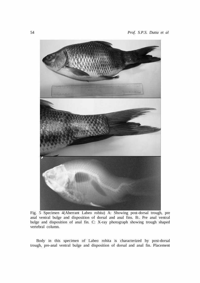
54 Prof. S.P.S. Dutta et al
Fig. 5 Specimen 4(Aberrant Labeo rohita) A: Showing post-dorsal trough, pre anal ventral bulge and disposition of dorsal and anal fins. B:. Pre anal ventral bulge and disposition of anal fin. C: X-ray photograph showing trough shaped vertebral column. Body in this specimen of Labeo rohita is characterized by post-dorsal trough, pre-anal ventral bulge and disposition of dorsal and anal fin. Placement

Morphological and Skeletal Deformities in Labeo Rohita (Ham. Buch.) 55
of other paired and unpaired fins is like a normal fish. X-ray analysis has revealed the presence of 35 and 33 vertebrae, respectively, in normal and abnormal fish (Fig. 1B, 5C). In abnormal fish, vertebral column shows various aberrations as given below:
1. First twelve vertebrae, after complex one, are normal. 2. Vertebral column between 13th to 17th vertebrae truncated and vertebrae
show reduced inter-vertebral space and vertebral thickness. 3. Between 13th to 30th vertebrae, vertebral column forms a trough
(lordosis) with smooth bottom. a. Vertebral column between 13th to 16th vertebrae forms anterior
side of the trough. Vertebrae have reduced inter-vertebral spaces. b. 17th to 25th normal vertebrae form smooth bottom of the trough. c. Posterior side of the trough is formed by 26th to 30th normal
vertebrae.
4. Posteriorly, vertebrae are normal. 5. Urostyle normal.
In this fish, anterior lobe of air bladder is normal and posterior normal lobe is directed downwards due to trough shaped vertebral column. From the radiological study, it is clear that the morphological aberrations are caused by trough like formation in vertebral column between 13th to 30th vertebrae.

56 Prof. S.P.S. Dutta et al
Fig. 6 Specimen 5(Aberrant Labeo rohita) A,B: Showing truncated post dorsal trough and disposition of dorsal and anal fins. C: X-ray photograph showing trough shaped(lordosis) vertebral column.
This deformed specimen of Labeo rohita, was recognized by irregular body with post-dorsal trough and ventral bulge between pelvic and anal fin. X-ray examination of the normal fish reveals a streamlined vertebral column with 35 amphiceolus vertebrae. In the abnormal fish, vertebral column has 32 vertebrae (Fig. 6C) and shows various anomalies:
1. First thirteen vertebrae, after the complex vertebrae, are normal. 2. Vertebral column between 14th to 28th vertebrae form a semi-circular
trough(lordosis) in which: . a. Vertebral column between 14th to 19th vertebrae forms anterior
side of the trough. Intervertebral spaces between 13th to 15th vertebrae are slightly reduced.
b. 20th to 25th normal vertebrae forms bottom of the trough. c. 26th to 28th normal vertebrae form posterior side of the trough.

Morphological and Skeletal Deformities in Labeo Rohita (Ham. Buch.) 57
3. Posteriorly, vertebral column between 29th to 32nd vertebrae is normal. 4. Urostyle is well developed.
Anterior and posterior lobe of air bladder of this fish is normal. However, posterior lobe is directed downwards due to trough shaped vertebral column. X-ray analysis has revealed that truncated body and disposition of fins is caused by vertebral trough between 14th to 28th vertebrae.
Fig.7 Specimen 6(Aberrant Labeo rohita) A, B: Showing truncated post-dorsal

58 Prof. S.P.S. Dutta et al
body with lateral bulge and disposition of dorsal and anal fins. C: X-ray photograph showing kyphois lordosis and kylonosis. In this deformed specimen of Labeo rohita post-dorsal truncated body is characterized by a lateral bulge and disposition of dorsal and anal fin. X-ray analysis of normal and abnormal fish reveals the presence of 35 and 30 vertebrae, respectively (Fig.1B, 7C). In the aberrant fish, vertebral column shows various abnormalities and is described below:
1. After complex vertebrae, first seven vertebrae have reduced inter-vertebral spaces and vertebral thickness.
2. 8th to 10th vertebrae clustered (ankylosis). 3. Vertebral column between 11th to 14th vertebrae forms a dome
(kyphosis). Vertebrae have reduced inter-vertebral spaces and vertebral thickness.
a. Vertebral column between 15th to 23rd vertebrae forms a trough in which:
i. 15th to 17th vertebrae form descending limb of the trough(lordosis). Vertebrae have reduced inter-vertebral spaces.
ii. 18th and 19th normal vertebrae form bottom of the trough. iii. 20th to 25th normal vertebrae form ascending limb of the
trough.
4. Posteriorly, between 24th to 30th vertebrae, vertebral column forms a depression(lordosis).
5. Urostyle is well developed. Air bladder in this fish is bi-lobed. Anterior lobe is degenerated and elongated with wide space between the two lobes as compared to normal fish. X-ray study has revealed that morphological aberrations like truncated body with lateral bulge and disposition of fins are caused by formation of dome a trough and a depression in vertebral column between 11th to 27th vertebrae. In all the deformed fishes, there are deviations in various body ratios (Table 2) DISCUSSION All the fish specimens studied during the present investigation are adults which indicate that these deformities are not fatal. Survival of these deformed fishes to the adult stage may also be due to absence of predatory fishes in these fish ponds. Multiple factors such as currents (Hilger, 1992; Divanach 1997 and Valverde et al,. 2005), temperature variations(Gluth and Hanke, 1983; Hore and Ahmad, 2010), salinity fluctuation(Silverstone and Hammell, 2002), low
Morphological and Skeletal Deformities in Labeo Rohita (Ham. Buch.) 59
dissolved oxygen (Turner and Farley, 1971; Alderdice et al. 2011), high CO2 concentration in water(Martens et al., 2006), environmental imbalances or toxicant (Sarkar and Konar, 1993; Kessabi, 2009, Sun et al. 2009), exposure to ultraviolet radiations (Torres et al., 2012), nutritional deficiency(Azzaydi et al. 1999; Lall and Lewis – McCrea, 2007 ), parasitic infestations(Yokoyama et al., 2005; Cunningham et al., 2005), culturing techniques (Pearson and Hopley, 1999), hereditary factors(Schultz, 1963, Daisai et al., 1995), developmental error(Dutta and Kumar, 1991; Shekhar and Dutta 1993 and Gupta et al. 2002) and injury (Dutta et al. 2011) have been considered to induce anomalies in fishes. Absence of caudle peduncle and caudle fin, as noticed in a solitary specimen of Labeo rohita, appears to be caused by predation. Fish aberrations due to predatory effects have also been described by Lemly 1993, Sulaiman and Weizhong 2010 and. Silverstone and Hammell, 2002. Most of the anomalies viz. truncated post-dorsal region, a post-dorsal depression, a lateral bulge and disposition of dorsal, pectoral, pelvic and anal fins; truncated post-dorsal body and disposition of dorsal and anal fins; absence of caudal peduncle and caudal fin and left split pectoral fin; post-dorsal trough, pre-anal ventral bulge and disposition of dorsal and anal fins; truncated post-dorsal trough and disposition of dorsal and anal fins and recognized by truncated post-dorsal body, lateral bulge anal disposition of dorsal and anal fins as seen in various specimen of Labeo rohita, under discussion, are most probably induced by water quality degradation caused by extensive use of pesticides in Punjab (Dutta and Gupta, 2010 and Dutta et al., 2011). From the above discussion, it is it is clear that the fish abnormalities are very complex and are caused by multiple factors. Taking into consideration the present study, it is suggested that the State Fishery Department should undertake an extensive survey of fish deformities in different water bodies of Punjab and determine the concentration of pesticides in these water bodies and fish bodies, respectively. ACKNOWLEDGMENT Sincere thanks are due to Head, Department of Environmental Sciences, for laboratory and other facilities. Financial assistance provided by Jammu University is greatly acknowledged. REFERENCES
[1] Alderdice, D.F., Wickett, W.P., Brett, J.R., 2011, “Some effects of temporary exposure to low dissolved oxygen levels on Pacific Salmon eggs”. Journal of the Fisheries Research Board of Canada, 15(2), pp.229-250.
[2] Azzaydi, M,, Martínez, F., Zamora S., Sánchez-Vázquez, F.J. and Madrid, J.A., 1999, ”Effect of meal size modulation on growth

60 Prof. S.P.S. Dutta et al
performance and feeding rhythms in European sea bass (Dicentrarchus labrax, L.)”. Aquacult., 170 (3-4), pp. 253-266.
[3] Cunningham, M.E., Markle, D.F., Watral, V.G., Kent, M.L. and Curtis, L.R., 2005, Patterns of fish deformities and their associates with trematode cysts in The Williamette River, Oregon. Env. Biol. Fishes, 73, pp. 9-19.
[4] Divanach, P,, Papandroulakis, N. , Anastasiadis, P., Koumoundouros, G. and Kentouri, M., 1997, “Effect of water currents on the development of skeletal deformities in sea bass (Dicentrarchus labrax L.) with functional swimbladder during postlarval and nursery phase”. Aquacult.156(1&2), pp.145-155.
[5] Dutta, S.P.S., Gupta, N., 2010, “Morphological and skeletal deformities in some specimens of Mrigal, Cirrhinus mrigala (Ham.Buch.) inhabiting fish ponds of Gurdaspur district, Punjab”. J. Aqua. Biol., 25(2), pp. 163-172.
[6] Dutta, S.P.S., Kumar, S. 1991, “Deformity in dorsal fin in Puntius conchonius (Ham.) from Jammu”. Geobios New Reports, 5, pp. 173-174.
[7] Dutta, S.P.S., Slathia, D., Chander, G. and Kumar, H., 2011, “Anomilies in Cirrhinus mrigala(Ham.Buch.), a commercially important fresh water food fish, from Gurdaspur District, Punjab”. The Bioscan, 6(3), pp.405-411.
[8] Gluth, G. and Hanke, W. 1983, “The effect of temperature on physiological changes in carp, Cyprinus carpio L.,induced by phenol”. Ecotoxicol and Environ Saf, 7(4), pp. 373-389.
[9] Gupta, S.C., Dutta, S.P.S. and Sharma, N. 2000, “ A report on some morphological deformities in silver carp Hypophthalmicthyes molitric (Vallenciennes) inhabiting aquatic environment of Jammu (J&K)”. Himalayan J.Env. and Zool., 14, pp. 25-30.
[10] Gupta, S.C., Dutta, S.P.S., Sharma, N. and Bala, N., 2002, Morphological deformities in Cirrhinus mrigala (Ham.Buch.) inhabiting lentic environments of Jammu, Aquacult. 3(2), pp.149-154.
[11] Hilger, I. 1992, “ Spinal compression of Atlantic cod, Gadus morhua from the German Wadden Sea”. Diseases of Aquatic Org., 13 (2), pp. 83-88.
[12] Hora, S.L., 1944, A hump backed Catla. Year-book Roy. As. Soc. Bengal for 1941, 8, pp.176.
[13] Hore, A. , Ahmad, M.F., 2010, “A wild specimen of Indian Carp, Cirrhinus mrigala(Ham.) 1822 with multiple vertebral deformities”. World journal of Zoology, 5(3), pp.167-171.
[14] Kapoor, B.G. and Sarkar, H.L., 1955, “Notes on four deformed specimens of the Indian Carp, Labeo rohita (Hamilton)”. Proc. Nat. Inst. Sci. India 21B , (3), pp.129-136.
[15] Kessabi, K., Kerkani, A., Said, K.. and Messaoudi, I., 2009, “Involvement of cadmium bioaccumulation in spinal deformities
Morphological and Skeletal Deformities in Labeo Rohita (Ham. Buch.) 61
occurrence in natural population of mediteranean killifish”. Biol. Trace Elem. Res., 128, pp. 72-81.
[16] Lall, S.P. and Lewis – McCrea, L.M. 2007, “ Role of nutrients in skeletal metabolism pathology in fish”. An overview Aquaculture 267 (1 - 4), pp. 3 – 19.
[17] Law, N.C., 1944, “A hump backed carp, Catla catla”. Proc. Nat. Inst. Sci. India, 10(1), pp. 97-103.
[18] Lemly, A.D., 1993, “Teratogenic effects of selenium in natural populations of fresh water fish”. Ecotoxicol Environ Saf, 26(2), pp. 181-204.
[19] Martens, L.G., Witten, P.E., Fivelstad, S. , Huysseume, A., Savedreid, B., Vikessa, V. and Obach, A., 2006, “Impact of high carbon dioxide on Atlantic Salmaon smolts (Salmo salar L.): effect on fish performance, vertebral composition and structure”. Aquacult., 261(1), pp. 80-88.
[20] Pearsons, T.N., Hopley, C.W., 1999, “A Practical Approach for Assessing Ecological Risks Associated with Fish Stocking Programs”. Fisheries, 24, pp. 16-23
[21] Pillai, C.T. and Thampy, D. M., 1990, “Cases of deformities in some cultivable fishes”. Indian Journal of fisheries, 37(2), pp.171-173.
[22] Sarkar, U.K. and Konar, S.K., 1993, “Sub-lethal effects of pesticides, heavy metals, detergents, and petroleum products on three combinations of fishes”. Env. Ecol. 11(3), pp. 609-615.
[23] Schultz, R.J., 1963, “Stubby, a Hereditary vertebral deformity in the Viviparous Fish Poeciliopsis prolific”. R COPEIA, 1963( 2), pp. 325-330.
[24] Shekhar, C. and Dutta, S.P.S., 1993, “ An abnormal specimen of Schizothorax richardsonii (Gray and Hard) with vertebral deformity”. Himalayan J. Env. and Zool., 7, pp. 101-102.
[25] Silverstone, A.M., and Hammell, L., 2002, “Spinal deformities in farmed Atlantic Salmon”. Can. Vet J,. 43(10), pp. 782-784.
[26] Sulaiman, A. and Weizhong, C., 2010, “Deformities in silver pomfret Pampus argenteus caught from Kuwait waters”. Chinese J Oceanology and Limnology, 28(6), pp.1227-1229.
[27] Sun, P.L., Hawkins, W.E., Overstreet, R.M. and Brown, P.N.J., 2009, “Morphological deformities as biomarkers in fish from contaminated rivers in Taiwan”. Int. J. Environ. Res. Public Health, 6, pp. 2307-2331.
[28] Tandon , K.K. and Sharma, K. ,1971, “A specimen of Callichrous macrophthalamus (Blyth) with forked right barble”. Madras J. Fisheries, 40(16), pp.438.
[29] Torres, N.E., Sobrino, C., Neale, P.J., Ceinos, R.M., Du, S. and Rotlant, J., 2012, “Molecular response to ultraviolet radiation exposure in fish embryos: implications for survival and morphological development”. Photochem Photobiol 88(3), pp. 701-707.
[30] Turner, J.L. and Farley, T.C., 1971, “Effects of temperature, salinity and dissolved oxygen on the survival of striped bass eggs and larvae”. Califonia Fish and Game, 57, pp. 268-73.

62 Prof. S.P.S. Dutta et al
[31] Valverde, C.J., Mendiola, L.P. and Costa, R.J.D., 2005, “Effect of periodical water current on the phasing of demand feeding rhythms in sea bass (Dicentrarchus labrax L.)”.Physiology & Behavior, 85(4), pp.394-403.
[32] Yokoyama, H., Freeman, M.A., Itoh, N. and Fukuda, Y., 2005, “Spinal curvature of cultured Japanese mackerel, Scomber japanicus associated with a brain myxosporean, Myxobolus acenthogobii”. Dis. Aquat. Org., 66, pp. 1-7.