Embed Size (px)
Citation preview

Morphological and Ion-Transporting Plasticity of Branchial Mitochondrion-Rich Cells in the Euryhaline Spotted Green Pufferfish, Tetraodon nigroviridisCheng-Hao Tang1 and Tsung-Han Lee1,2,*1Department of Life Sciences, National Chung-Hsing University, Taichung 402, Taiwan2Department of Biological Science and Technology, China Medical University, Taichung 404, Taiwan
(Accepted September 1, 2010)
Cheng-Hao Tang and Tsung-Han Lee (2011) Morphological and ion-transporting plasticity of branchial mitochondrion-rich cells in the euryhaline spotted green pufferfish Tetraodon nigroviridis. Zoological Studies 50(1): 31-42. Morphological characteristics and chloride regulatory functions of gill mitochondrion-rich (MR) cells of the pufferfish (Tetraodon nigroviridis) were investigated in this study, because T. nigroviridis are widely used in studies of fish osmoregulation. In scanning electron micrographs, the apical membrane structures of MR cells could be distinguished into 3 phenotypes of flat, intermediate-indentation, and concave-hole. The apical surfaces of most MR cells were flat with microvilli in fresh water (FW)-acclimated pufferfish, whereas most apical surfaces of MR cells were concave to form holes in brackish water (BW) and seawater (SW)-acclimated pufferfish. Observed changes in the proportion of the intermediate indentation type suggested that when transferred from FW to BW or SW, pufferfish MR cells were transformed from the flat type into the concave-hole type via different stages of the intermediate indentation type. Meanwhile, no significant difference in cell size and density of gill MR cells were found in pufferfish acclimated to various salinities. To compare the ion-transporting functions of MR cells, both the chloride test and double immunofluorescent staining of Na+/K+-ATPase (NKA) and the chloride-secreting channel, cystic fibrosis transmembrane conductance regulator (CFTR), were examined. The CFTR was present in the apical membrane of NKA-immunoreactive cells in gills of SW-acclimated pufferfish, but completely disappeared in FW-acclimated fish. Moreover, the chloride test directly demonstrated the chloride secretion function of concave-hole MR cells. Taken together, our findings suggested that different types of MR cells in pufferfish gills expressed various ion-transport proteins to conduct distinct functions necessary for salinity acclimation of this euryhaline species. http://zoolstud.sinica.edu.tw/Journals/50.1/31.pdf
Key words: Spotted green pufferfish, Tetraodon nigroviridis, Mitochondrion-rich cells, Cystic fibrosis transmembrane conductance regulator, Na+/K+-ATPase.
*To whom correspondence and reprint requests should be addressed. E-mail:[email protected]
Teleosts have evolved to extend their habitats into various environments. Euryhaline teleosts maintain ionic concentrat ions and osmolality of body fluids at constant levels that differ from those of external environments (Kaneko et al. 2008). In response to changes in environmental conditions, the ion-transporting epithelia play a role of modulating ion fluxes. Although ionoregulation in euryhaline teleosts
is mediated by a group of organs including the intestines and kidneys, the gills are the major site for balancing ion movements between gains and losses (Evans et al. 2005). Because gill epithelial mitochondrion-rich (MR) cells are the main sites of ion absorption and secretion in freshwater (FW) and seawater (SW) fish, respectively, they are important in the adaptation of euryhaline teleosts to environments with various salinities. In SW
Zoological Studies 50(1): 31-42 (2011)
31
MR cells, the apical chloride channel, cystic fibrosis transmembrane conductance regulator (CFTR), plays a crucial role in carrying out chloride secretion (Hirose et al. 2003, Evans et al. 2005).
Some studies (Laurent and Perry 1990, Perry et al. 1992) reported that injection of exogenous cortisol or treatment using ion-deficient water (Avella et al. 1987, Perry and Laurent 1989, Greco et al. 1996, Tang et al. 2008) altered the morphology of the apical surfaces of branchial MR cells. Other studies demonstrated the correlations between MR cells and Cl− uptake in different species (Goss et al. 1992, Morgan et al. 1994, Morgan and Potts 1995, Chang et al. 2002 2003). Apical structures of MR cells are closely related to ion-transporting activities (Kaneko et al. 2008). Scanning electron microscopy (SEM) was used to observe the structures of apical membranes and identify functional MR cells including FW and SW types in many species (reviewed in Hwang and Lee 2007, Kaneko et al. 2008). On SEM observations, FW MR cells generally displayed flat or slightly invaginated surfaces like membrane patches with short cellular projections on them, while the SW type exhibited deeply invaginated surfaces with smaller orifices (Hwang and Lee 2007). In addition, Na+/K+-ATPase (NKA) is thought to be a marker of MR cells, because it is a primary active transport pump providing the major driving force for ion-transporting functions in branchial MR cells (McCormick 1995, Marshall 2002, Hirose et al. 2003). Many studied euryhaline species alter the cell size, density, or NKA responses of their MR cells suggesting adjustments of their ion-transporting functions when acclimated to environments with different salinities (Langdon and Thorpe 1985, Richman et al. 1987, McCormick 1995, Uchida et al. 1996 1997 2000, Heljden et al. 1997, Sasai et al. 1998, Katoh et al. 2001, Lee et al. 2003 2006, Kaneko et al. 2008).
The spotted green pufferfish (Tetraodon nigroviridis) is an advanced tetraodontid teleost whose native range covers the rivers and estuaries of Southeast Asia (Rainboth 1996). Our previous studies demonstrated that T. nigroviridis can survive when transferred from FW to SW and vice versa, indicating that this species is an efficient osmoregulator (Lin et al. 2004, Lin and Lee 2005, Tang and Lee 2007a b, Wang et al. 2008). The pufferfish was therefore used as a model organism to examine osmoregulatory mechanisms in several studies (Lin et al. 2004, Lin and Lee 2005, Tang and Lee 2007a b, Bagherie-Lachidan et al. 2008 2009, Wang et al. 2008). However, morphological
characterizations (i.e., apical membrane structure, cell size and density) of branchial MR cells of this euryhaline pufferfish are not well described. The present set of experiments was designed to clarify morphological features of MR cells and their implied functions in this euryhaline teleost.
MATERIALS AND METHODS
Experimental animals and environments
The spotted green pufferfish T. nigroviridis were obtained from a local aquarium with total length of 5.2 ± 0.6 cm. After being reared in brackish water (BW; 15‰) for 1 mo, the pufferfish were separated and reared in fresh water (FW), BW, or seawater (SW; 35‰) at 27 ± 1°C with a daily 12-h photoperiod for 2 wk before sampling. SW and BW used in this study were prepared from local tap water by adding proper amounts of synthetic sea salt (Instant Ocean, Aquarium Systems, Mentor, OH, USA). The water was continuously circulated through fabric-floss filters. The fish were fed with commercial dried shrimp daily.
Ultrastructure of apical membrane of mitochondrion-rich (MR) cells in gill filaments
After anesthetization with MS-222, fish were killed by spinal pithing, and their gills were excised. The 1st gill arch from each side was fixed at 4°C in the fixative consisting of 5% (w/v) glutaraldehyde and 4% (w/v) paraformaldehyde in 0.1 M phosphate buffer (PB, pH 7.3) for 12 h. After rinsing in 0.1 M PB, specimens were postfixed with 1% (v/v) osmium tetroxide in 0.1 M PB for 1 h. Subsequently, the specimens were serially dehydrated in ethanol, followed by critical-point drying using liquid CO2 in a critical-point dryer (Hitachi HCP-2, Tokyo, Japan). The gills were mounted on an aluminum specimen plate and coated with gold by ion sputter (JFC-1600, JEOL, Tokyo, Japan). Then the samples were examined by scanning electron microscope (SEM) (JSM-6700F, JEOL, Tokyo, Japan).
Antibodies
The following primary antibodies were used in this study. (1) Anti-Na+/K+-ATPase (NKA) antibody, α5, is a mouse monoclonal antibody (mAb) (Developmental Studies Hybridoma
Zoological Studies 50(1): 31-42 (2011)32

Bank, Iowa City, IA, USA) raised against the α-subunit of avian NKA. The dilution was 1:200 for immunofluorescent staining. (2) Anti-cystic fibrosis transmembrane conductance regulator (CFTR) is a mouse mAb to human CFTR (R&D Systems, Minneapolis MN, USA). This antibody was raised against a carboxy-terminal sequence of human CFTR and had been successfully applied to several fish species (Katoh and Kaneko 2003, McCormick et al. 2003, Hiroi et al. 2008). The dilution was 1:100 for immunofluorescent staining. (3) NKA #11 is a rabbit polyclonal antiserum kindly provided by Prof. P.P. Hwang (Institute of Cellular and Organismic Biology, Academia Sinica, Taipei, Taiwan). This antiserum was raised against 565 amino acids (aa) corresponding to the 392-939-aa sequence of the α-subunit of tilapia. This region shares high sequence identity with the NKA α-subunit of vertebrates (Hwang et al. 1998). The dilution was 1:100 for immunofluorescent staining. For immunofluorescent staining, the secondary antibodies were Alexa-Fluor 546-conjugated goat anti-mouse IgG or Alexa-Fluor 488-conjugated goat anti-rabbit IgG (Invitrogen, Carlsbad, CA, USA). The dilution of the 2 secondary antibodies was 1:200 for immunofluorescent staining.
Whole-mount double immunofluorescent staining
T h e p r o c e d u r e o f w h o l e - m o u n t immunofluorescent staining was carried out as described by Tang and Lee (2007b). The gill filaments were removed from gill samples and fixed in 4% paraformaldehyde in 0.1 M phosphate buffer (pH 7.4). After washing in phosphate-buffered saline (PBS), the gill filaments were postfixed and permeated with 70% ethanol for 10 min at -20°C. The gill filaments were rinsed with phosphate buffered saline (PBS) and then incubated in 5% bovine serum albumin (BSA; Sigma, St. Louis, MO, USA). To detect MR cells in whole-mount preparations, the gill filaments were incubated at room temperature for 2 h with the primary mAb of NKA (α5). Following incubation, the gill filaments were washed several times with PBS and then labeled with the Alexa Fluor 546-conjugated goat anti-mouse secondary antibody (Invitrogen) at room temperature for 2 h. To detect the chloride channel, CFTR, the gill filaments were first stained with the NKA#11 pAb and labeled with the Alexa Fluor 488-conjugated goat-anti-rabbit secondary antibody at room temperature for 2 h. After the 1st staining, the gill filaments were washed several
times with PBS before proceeding to the 2nd staining. The gill filaments were subsequently incubated with the anti-CFTR mAb overnight at 4°C followed by labeling with the Alexa Fluor 546-conjugated goat anti-mouse secondary antibody (Invitrogen) at room temperature for 2 h. The samples were then washed with PBS, mounted with a coverslip, and observed with a Leica TCS-NT confocal laser scanning microscope (Leica Lasertechnik, Heidelberg, Germany).
Quantitative analysis of MR cells
MR cells stained by whole-mount immuno-fluorescent staining were measured on stored images using Leica TCS NT software. The cell size was determined as the greatest l inear diameters of MR cells, and was obtained from 10 cells per individual (n = 5), which were randomly selected from gill filaments. To determine the density of MR cells, an area corresponding to 100 × 100 μm was randomly selected from the region of the afferent-vascular edge of gill filaments of each experimental fish (n = 5). MR cells in the selected areas were counted, and the number of cells per square millimeter was reported.
Chloride test
To determine the chloride-secreting sites in gill epithelium, SW pufferfish were subjected to the chloride test modified from Kaneko and Shiraishi (2001). The fish were first immersed in deionized water (DW) 3 times (for 1 min each) to remove Cl− on the gill surface, and immersed in 0.25% AgNO3 in DW for 1 min. Newly secreted Cl− reacted with Ag+ to form photosensitive AgCl during the period of incubation in AgNO3 solution. After a brief rinse with DW, the gills were removed and placed in a glass vial containing 4% paraformaldehyde in 0.1 M phosphate buffer (pH 7.4) and exposed to strong light for 30 min because light exposure causes the reduct ion o f AgCl to form Ag precipitates. To observe the details of chloride-secreting activity of MR cells in gills of SW pufferfish, samples were examined using X-ray microanalysis. The gills subjected to the chloride test were fixed as described above, dehydrated in ethanol, and dried using a Hitachi HCP-2 critical-point drier (Tokyo, Japan). The gills were mounted on an aluminum specimen stub, coated with gold by ion sputter (JFC-1600, JEOL, Tokyo, Japan), and examined with SEM (JSM-6700F, JEOL) equipped with an energy-dispersive X-ray
Tang and Lee – Gill MR Cells of an Euryhaline Pufferfish 33
microanalyzer and detector (Oxford Inca Energy 350, Oxford Instruments, Oxfordshire, UK). The elemental profile of Ag was examined by detecting X-ray characteristic of Ag at 2.644 keV (LI) and 2.984 keV (Lα1). For mapping the Ag profile, the X-ray signals were accumulated for 375 s.
Statistical analysis
Values were compared using a one-way analysis of variance (ANOVA), and post-hoc analyses were conducted using Tukey’s pairwise method. Values are expressed as the means ± SEM. The significant difference was set as p < 0.05.
RESULTS
The apical structure of gill mitochondrion-rich (MR) cells
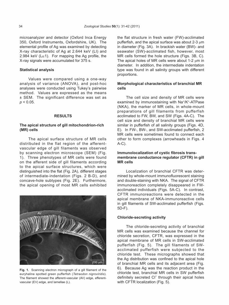
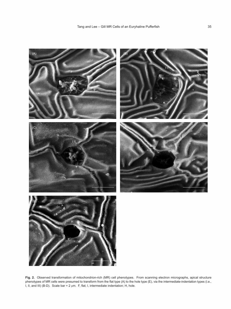
The apical surface structure of MR cells distributed in the flat region of the afferent-vascular edge of gill filaments was observed by scanning electron microscope (SEM) (Fig. 1). Three phenotypes of MR cells were found on the afferent side of gill filaments according to the apical surface structures, which were distinguished into the flat (Fig. 2A), different stages of intermediate-indentation (Figs. 2 B-D), and concave-hole subtypes (Fig. 2E). Furthermore, the apical opening of most MR cells exhibited
the flat structure in fresh water (FW)-acclimated pufferfish, and the apical surface was about 2-3 μm in diameter (Fig. 3A). In brackish water (BW)- and seawater (SW)-acclimated fish, however, most MR cells formed the hole structure (Figs. 3B, C). The apical holes of MR cells were about 1-2 μm in diameter. In addition, the intermediate indentation type was found in all salinity groups with different proportions.
Morphological characteristics of branchial MR cells
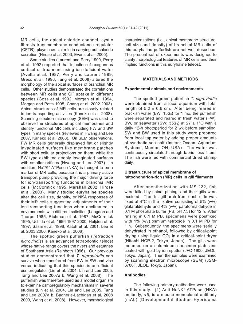
The cell size and density of MR cells were examined by immunostaining with Na+/K+-ATPase (NKA), the marker of MR cells, in whole-mount preparations of gill filaments from pufferfish acclimated to FW, BW, and SW (Figs. 4A-C). The cell size and density of branchial MR cells were similar in pufferfish of all salinity groups (Figs. 4D, E). In FW-, BW-, and SW-acclimated pufferfish, 2 MR cells were sometimes found to connect each other to form complexes (arrowheads in Figs. 4 A-C).
Immunolocalization of cystic fibrosis trans-membrane conductance regulator (CFTR) in gill MR cells
Localization of branchial CFTR was deter-mined by whole-mount immunofluorescent staining and double-staining with NKA. The signal of CFTR immunoreaction completely disappeared in FW-acclimated individuals (Figs. 5A-C). In contrast, CFTR immunoreactions were detected in the apical membrane of NKA-immunoreactive cells in gill filaments of SW-acclimated pufferfish (Figs. 5D-F).
Chloride-secreting activity
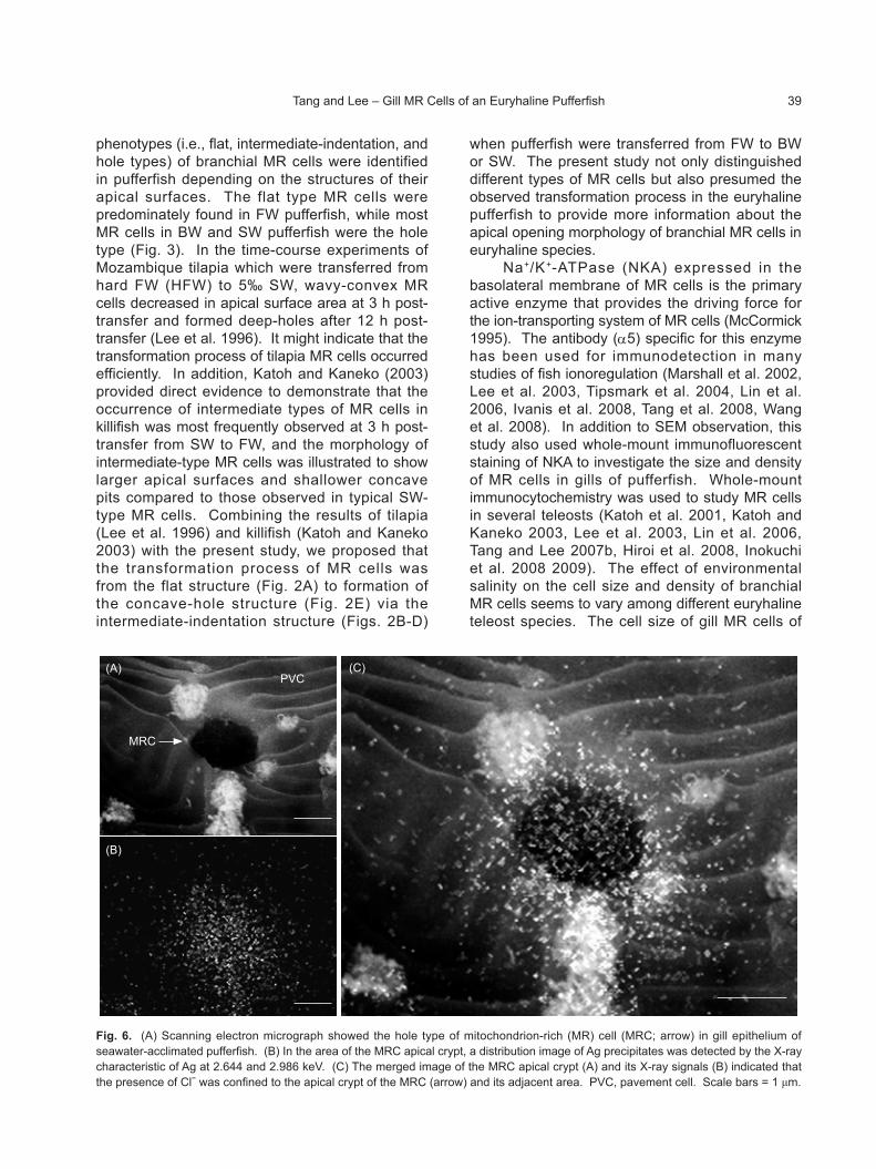
The chloride-secreting activity of branchial MR cells was examined because the channel for chloride secretion, CFTR, was expressed in the apical membrane of MR cells in SW-acclimated pufferfish (Fig. 5). The gill filaments of SW-acclimated pufferfish were subjected to the chloride test. These micrographs showed that the Ag distribution was confined to the apical hole of branchial MR cells and its adjacent area (Fig. 6). Because Ag was the reaction product in the chloride test, branchial MR cells in SW pufferfish definitely secreted Cl− through their apical holes with CFTR localization (Fig. 5).
Fig. 1. Scanning electron micrograph of a gill filament of the euryhaline spotted green pufferfish (Tetraodon nigroviridis). The filament showed the afferent-vascular (AV) edge, efferent-vascular (EV) edge, and lamellae (L).
AV
L
EV
200 μm
Zoological Studies 50(1): 31-42 (2011)34
(A)
FIⅠ
(B)
(C)
IⅡ
(D)
IⅢ
Fig. 2. Observed transformation of mitochondrion-rich (MR) cell phenotypes. From scanning electron micrographs, apical structure phenotypes of MR cells were presumed to transform from the flat type (A) to the hole type (E), via the intermediate-indentation types (i.e., I, II, and III) (B-D). Scale bar = 2 μm. F, flat; I, intermediate indentation; H, hole.
(E)
H
Tang and Lee – Gill MR Cells of an Euryhaline Pufferfish 35

DISCUSSION
The gill filaments and lamellae are covered by epithelium which not only provides a distinct boundary between the external environment and body fluids of fish but also plays a critical role in the physiological function of the fish gills (Evans et al. 2005). The mitochondrion-rich (MR) cell is one of the major cell types in gill epithelium and considered to be one of the predominant sites of active physiological mechanisms (Wilson and Laurent 2002, Hirose et al. 2003, Evans et al. 2005, Marshall and Grosell 2006). Scanning electron microscopy (SEM) was used in the present study to observe MR cells, which were mainly distributed in the afferent-vascular edge of gill filaments in the spotted green pufferfish (Tetraodon nigroviridis), similar to previous findings in many other teleostean species (Wilson and Laurent 2002, Evans et al. 2005, Marshall and Grosell 2006). In addition, branchial MR cells were generally not found in gill lamellae of T. nigroviridis acclimated to fresh water (FW), brackish water (BW; 15‰), and seawater (SW; 35‰) by either immunohistochemical (Lin et al. 2004) or SEM observations (data not shown). In other species, such as milkfish (Chanos chanos) (Lin et al. 2003) and tilapia (Oreochromis mossambicus) (Tang et al. 2008), MR cells were scarcely found on the epithelium of gill lamellae except under particular environmental conditions that are associated with the presence of lamellar MR cells (Evans et al. 2005, Hwang and Lee 2007). Thus, it will be intriguing to detect whether MR cells are present in gill lamellae when pufferfish are acclimated to hypersaline water (45‰) in future studies and to compare the results to the other species (i.e., Japanese eel, Anguilla japonica, Sasai et al. 1998; sea bass, Latealabrax japonicus, Hirai et al. 1999; Atlantic salmon, Salmo salar, Hiroi et al. 2007; and tilapia, O. mossambicus, Tang et al. 2008) to clarify the roles of MR cells in pufferfish gill lamellae.
MR cells located in gill epithelium play a role in ionoregulation because they are in contact with the external environment (via the apical membrane) and blood (via the basolateral membrane) (Evans et al. 2005, Kaneko et al. 2008). Therefore, the apical membrane structure of MR cells in many species varies greatly with diverse environmental salinities, generally with an apical concave crypt in SW and a convex surface equipped with numerous microvilli in FW (Marshall and Grosell 2006, Hwang and Lee 2007, Kaneko et al. 2008). From SEM micrographs, 3
(A)
FW
I I
F
F
FF
I
F
F
F
F F
F
F
F
I I
F
H
FF
BW
(B)
I H H I
H
H
H
H IH
I
I
II
HH
H
I
I
H
HH
H
HI
HH
SW
(C)
H
HH
HH
HHH
I
H
H
I
H
I
H
H
H
H
H
HH
I
Fig. 3. Scanning electron micrographs of the afferent-vascular area from gill filaments of pufferfish acclimated to fresh water (FW; A), brackish water (BW; B), and seawater (SW; C) for 2 wk. The letters in the micrographs indicate subtypes of mitochondrion-rich (MR) cells with different apical structures. Most MR cells of FW pufferfish were the flat type (F), almost all of the MR cells of SW fish were the hole type (H), and both the hole and intermediate-indentation types (I) were found in BW individuals. Scale bar = 10 μm.
Zoological Studies 50(1): 31-42 (2011)36

Fig. 4. Confocal laser scanning micrographs of whole-mount preparations of gill filaments in pufferfish acclimated to fresh water (FW; A), brackish water (BW; B), and seawater (SW; C). Gill filaments were stained with anti-Na+/K+-ATPase (NKA) antibody (α5). Arrows indicate the multicellular complex of mitochondrion-rich (MR) cells consisting of 2 adjacent cells. No significant differences (n = 5; mean ± SEM) were found in cell size (D) and or density (E) among all groups.
(B)
BW
(C)
SW 20 μm
(A)
FW
(D)
FW BW SW
Environments
Cel
l siz
e (μ
m)
12
8
4
0
(E)
FW BW SW
Environments
103 c
ells
/mm
2
8
6
4
2
0
Tang and Lee – Gill MR Cells of an Euryhaline Pufferfish 37

Fig. 5. Confocal laser scanning micrographs of whole-mount preparations of gill filaments in freshwater- (FW; A-C) and seawater (SW; D-F)-acclimated pufferfish. Gill filaments were double-stained with anti-Na+/K+-ATPase (NKA; green; A, D) (NKA #11) and anti-cystic fibrosis transmembrane conductance regulator (CFTR) (CFTR; red; B, E). The merged images (C, F) of double-stained gill filaments showed that the CFTR was localized in apical membrane of NKA immunoreactive cells in SW pufferfish (F), while the signal of CFTR expression completely disappeared in FW pufferfish (C).
FW
(A)
FW
(B)
FW
(C)
SW
(D)
SW
(E)
SW
(F)
20 μm
Zoological Studies 50(1): 31-42 (2011)38
phenotypes (i.e., flat, intermediate-indentation, and hole types) of branchial MR cells were identified in pufferfish depending on the structures of their apical surfaces. The flat type MR cells were predominately found in FW pufferfish, while most MR cells in BW and SW pufferfish were the hole type (Fig. 3). In the time-course experiments of Mozambique tilapia which were transferred from hard FW (HFW) to 5‰ SW, wavy-convex MR cells decreased in apical surface area at 3 h post-transfer and formed deep-holes after 12 h post-transfer (Lee et al. 1996). It might indicate that the transformation process of tilapia MR cells occurred efficiently. In addition, Katoh and Kaneko (2003) provided direct evidence to demonstrate that the occurrence of intermediate types of MR cells in killifish was most frequently observed at 3 h post-transfer from SW to FW, and the morphology of intermediate-type MR cells was illustrated to show larger apical surfaces and shallower concave pits compared to those observed in typical SW-type MR cells. Combining the results of tilapia (Lee et al. 1996) and killifish (Katoh and Kaneko 2003) with the present study, we proposed that the transformation process of MR cells was from the flat structure (Fig. 2A) to formation of the concave-hole structure (Fig. 2E) via the intermediate-indentation structure (Figs. 2B-D)
when pufferfish were transferred from FW to BW or SW. The present study not only distinguished different types of MR cells but also presumed the observed transformation process in the euryhaline pufferfish to provide more information about the apical opening morphology of branchial MR cells in euryhaline species.
Na+/K+-ATPase (NKA) expressed in the basolateral membrane of MR cells is the primary active enzyme that provides the driving force for the ion-transporting system of MR cells (McCormick 1995). The antibody (α5) specific for this enzyme has been used for immunodetection in many studies of fish ionoregulation (Marshall et al. 2002, Lee et al. 2003, Tipsmark et al. 2004, Lin et al. 2006, Ivanis et al. 2008, Tang et al. 2008, Wang et al. 2008). In addition to SEM observation, this study also used whole-mount immunofluorescent staining of NKA to investigate the size and density of MR cells in gills of pufferfish. Whole-mount immunocytochemistry was used to study MR cells in several teleosts (Katoh et al. 2001, Katoh and Kaneko 2003, Lee et al. 2003, Lin et al. 2006, Tang and Lee 2007b, Hiroi et al. 2008, Inokuchi et al. 2008 2009). The effect of environmental salinity on the cell size and density of branchial MR cells seems to vary among different euryhaline teleost species. The cell size of gill MR cells of
Fig. 6. (A) Scanning electron micrograph showed the hole type of mitochondrion-rich (MR) cell (MRC; arrow) in gill epithelium of seawater-acclimated pufferfish. (B) In the area of the MRC apical crypt, a distribution image of Ag precipitates was detected by the X-ray characteristic of Ag at 2.644 and 2.986 keV. (C) The merged image of the MRC apical crypt (A) and its X-ray signals (B) indicated that the presence of Cl− was confined to the apical crypt of the MRC (arrow) and its adjacent area. PVC, pavement cell. Scale bars = 1 μm.
(A)
MRC
PVC
(B)
(C)
Tang and Lee – Gill MR Cells of an Euryhaline Pufferfish 39

the euryhaline Mozambique tilapia increased but the density decreased with elevated environmental salinity (Heijden et al. 1997, Uchida et al. 2000). Moreover, the size of gill MR cells was larger in FW- than in SW-acclimated killifish, but the cell density was similar between FW- and SW-acclimated groups (Katoh et al. 2001). In Atlantic salmon, both the cell size and density of MR cells were found to increase after acclimation to SW or during smolting (Langdon and Thorpe 1985, Pelis et al. 2001). However, the cell size and number of MR cells on primary filament decreased in postsmolts (Pelis et al. 2001). Interestingly, this study found no significant difference in the cell size and density of branchial MR cells in pufferfish acclimated to different salinities (Fig. 4). Meanwhile, the abundance of branchial NKA α-subunit protein was the lowest in BW-acclimated pufferfish compared to FW- and SW-acclimated individuals (Lin et al. 2004). These findings suggested that modulating protein amounts of branchial NKA of the pufferfish acclimated to different environmental salinities occurred through modulating the protein amounts of NKA per cell rather than altering the size or density of MR cells, as reported in tilapia and killifish (Uchida et al. 2000, Katoh et al. 2001).
Cystic fibrosis transmembrane conductance regulator (CFTR) is a chloride channel expressed in apical membranes of MR cells and which is responsible for chloride secretion in teleosts acclimated to SW (reviewed in Hirose et al. 2003, Evans 2008, Kaneko et al. 2008). The localization and expression of ion transporters (i.e., NKA and CFTR) were used to classify the function of MR cells in euryhaline teleosts acclimated to different environmental salinities (Marshall et al. 2002, Katoh and Kaneko 2003, Hiroi et al. 2005a b, Wilson et al. 2007a). In the present study, localization of branchial CFTR was determined by counter-staining with NKA. In SW-acclimated pufferfish, CFTR was expressed in apical membranes of MR cells. In contrast, CFTR was undetectable in FW-acclimated pufferfish (Fig. 5). This finding is similar to previous studies of tilapia (Hiroi et al. 2005a b) and killifish (Katoh and Kaneko 2003). Wilson et al. (2007b) also reported that the immunoreaction of CFTR in apical membranes of branchial MR cells was positive in glass-eel, while it was negative in the elvers stage of the European eel (Anguilla anguilla). Furthermore, SW-acclimated pufferfish were also examined with the chloride test to demonstrate the chloride secretion of SW MR cells. The method
used in the chloride test was also used in other euryhaline teleosts to detect the site of chloride secretion (Wong and Chan 1999, Kaneko and Shiraishi 2001). In this study, the presence of large amounts of silver precipitates was confined to apical crypt of the hole-type MR cells of SW pufferfish (Fig. 6) with CFTR localized in apical membrane (Fig. 5). In FW pufferfish, however, a few silver precipitates were sprinkled on gill filaments rather than being specifically located on the apical surface (data not shown), and CFTR was immunonegative (Fig. 5). Taken together, our results provided direct evidence indicating different ion-transporting functions for SW- and FW-type MR cells in gills of the euryhaline pufferfish.
Tetraodon nigroviridis is a peripheral FW species often found in estuaries and FW rivers (Helfman et al. 1997). Thus, the pufferfish was demonstrated to be an efficient osmoregulator in experimental conditions, as it could tolerate direct transfer from FW to SW and vice versa (Lin et al. 2004, Lin and Lee 2005, Tang and Lee 2007a b, Wang et al. 2008). Although previous studies investigated the iono- and osmoregulatory mechanisms of gills of T. nigroviridis (Lin et al. 2004, Tang and Lee 2007a b, Wang et al. 2008, Bagherie-Lachidan et al. 2008 2009), the morphological features of gill MR cells were not addressed in this species. The present study investigated the types of MR cells and also proposed the transformation process and further discussed the cell size and number of branchial MR cells in pufferfish acclimated to environments with various salinities. Taken together, changes in gill MR cell phenotypes, with positive or negative expression of the apically located chloride secretion channel indicating altered functions of MR cells, are crucial for efficient responses to salinity challenge in pufferfish. We also concluded that expression of the key enzyme, NKA, and other ion-transport proteins, rather than modulation of the cell size and number of MR cells, were involved in regulating the ion-transporting capacity of branchial MR cells of pufferfish acclimated to different salinities.
Acknowledgments: The α5 monoclonal antibody was purchased from the Developmental Studies Hybridoma Bank (DSHB) maintained by the Department of Pharmacology and Molecular Sciences, John Hopkins University School of Medicine (Baltimore, MD, USA), and the Department of Biological Sciences, University of Iowa (Iowa City, IA, USA), under contract
Zoological Studies 50(1): 31-42 (2011)40

N01-HD-6-2915, NICHD, USA. This study was supported by a grant from the National Science Council of Taiwan to T.H.L. (NSC95-2313-B-005-040-MY3).
REFERENCES
Avella M, A Masoni, M Bornancin, N Mayer-Gostan. 1987. Gill morphology and sodium influx in the rainbow trout (Salmo gairdneri) acclimated to artificial freshwater environments. J. Exp. Zool. 241: 159-169.
Bagherie-Lachidan M, SI Wright, SP Kelly. 2008. Claudin-3 tight junction proteins in Tetraodon nigroviridis: cloning, tissue-specific expression, and a role in hydromineral balance. Am. J. Physiol. Regul. Integr. Comp. Physiol. 294: R1638-R1647.
Bagherie-Lachidan M, SI Wright, SP Kelly. 2009. Claudin-8 and -27 tight junction proteins in puffer fish Tetraodon nigroviridis acclimated to freshwater and seawater. J. Comp. Physiol. B 179: 419-431.
Chang IC, TH Lee, HC Wu, PP Hwang. 2002. Effects of environmental Cl− levels on Cl− uptake and mitochondria-rich cell morphology in gills of the stenohaline goldfish, Carassius auratus. Zool. Stud. 46: 236-243.
Chang IC, YY Wei, FI Chou, PP Hwang. 2003. Stimulation of Cl− uptake and morphological changes in gi l l mitochondria-rich cells in freshwater tilapia (Oreochromis mossambicus). Physiol. Biochem. Zool. 76: 544-552.
Evans DH. 2008. Teleost fish osmoregulation: What have we learned since August Krogh, Homer Smith, and Ancel Keys. Am. J. Physiol. Regul. Integr. Comp. Physiol. 295: R704-R713.
Evans DH, PM Piermarini, KP Choe. 2005. The multifunctional fish gill: dominant site of gas exchange, osmoregulation, acid-base regulation, and excretion of nitrogenous waste. Physiol. Rev. 85: 97-177.
Goss GG, SF Perry, CM Wood, P Laurent. 1992. Mechanisms of ion and acid-base regulation at the gills of freshwater fish. J. Exp. Zool. 263: 143-159.
Greco AM, JC Fenwick, SF Perry. 1996. The effects of soft-water acclimation on gill structure in the rainbow trout Oncorhynchus mykiss. Cell. Tissue. Res. 285: 75-82.
Heijden A, P Verbost, J Eygensteyn, J Li, S Bonga, G Flik. 1997. Mitochondria-rich cells in gills of tilapia (Oreochromis mossambicus) adapted to fresh water or sea water: quantification by confocal laser scanning microscopy. J. Exp. Biol. 200: 55-64.
Helfman GS, BB Collette, DE Facey. 1997. The diversity of fishes. Oxford, UK: Blackwell Science.
Hirai N, M Tagawa, T Kaneko, T Seikai, M Tanaka. 1999. Distributional changes in branchial chloride cells during freshwater adaptation in Japanese sea bass Lateolabrax japonicus. Zool. Sci. 16: 43-49.
Hiroi J, SD McCormick. 2007. Variation in salinity tolerance, gill Na+/K+-ATPase, Na+/K+/2Cl− cotransporter and mitochondria-rich cell distribution in three salmonids Salvelinus namaycush, Salvelinus fontinalis and Salmo salar. J. Exp. Biol. 210: 1015-1024.
Hiroi J, SD McCormick, R Ohtani-Kaneko, T Kaneko. 2005b. Functional classification of mitochondrion-rich cells in euryhaline Mozambique tilapia (Oreochromis mossambicus) embryos, by means of triple immuno-
fluorescence staining for Na+/K+-ATPase, Na+/K+/2Cl− cotransporter and CFTR anion channel. J. Exp. Biol. 208: 2023-2036.
Hiroi J, H Miyazaki, F Katoh, R Ohtani-Kaneko, T Kaneko. 2005a. Chloride turnover and ion-transporting activities of yolk-sac preparations (yolk balls) separated from Mozambique tilapia embryos and incubated in freshwater and seawater. J. Exp. Biol. 208: 3851-3858.
Hiroi J, S Yasumasu, SD McCormick, PP Hwang, T Kaneko. 2008. Evidence for an apical Na-Cl cotransporter involved in ion uptake in a teleost fish. J. Exp. Biol. 211: 2584-2599.
Hirose S, T Kaneko, N Naito, Y Takei. 2003. Molecular biology of major components of chloride cells. Comp. Biochem. Physiol. B Biochem. Mol. Biol. 136: 593-620.
Hwang PP, MJ Fang, JC Tsai, CJ Huang, ST Chen. 1998. Expression of mRNA and protein of Na+-K+-ATPase α subunit in gills of tilapia (Oreochromis mossambicus). Fish. Physiol. Biochem. 18: 363-373.
Hwang PP, TH Lee. 2007. New insights into fish ion regulation and mitochondrion-rich cells. Comp. Biochem. Physiol. A Mol. Integr. Physiol. 148: 479-497.
Inokuchi M, J Hiroi, S Watanabe, PP Hwang, T Kaneko. 2009. Morphological and functional classification of ion-absorbing mitochondria-rich cells in the gills of Mozambique tilapia. J. Exp. Biol. 212: 1003-1010.
Inokuchi M, J Hiroi, S Watanabe, KM Lee, T Kaneko. 2008. Gene expression and morphological localization of NHE3, NCC and NKCC1a in branchial mitochondria-rich cells of Mozambique tilapia (Oreochromis mossambicus) acclimated to a wide range of salinities. Comp. Biochem. Physiol. A Mol. Integr. Physiol. 151: 151-158.
Ivanis G, AJ Esbaugh, SF Perry. 2008. Branchial expression and localization of SLC9A2 and SLC9A3 sodium/hydrogen exchangers and their possible role in acid-base regulation in freshwater rainbow trout (Oncorhynchus mykiss). J. Exp. Biol. 211: 2467-2477.
Kaneko T, K Shiraishi. 2001. Evidence for chloride secretion from chloride cel ls in the yolk-sac membrane of Mozambique tilapia larvae adapted to seawater. Fish. Sci. 67: 541-543.
Kaneko T, S Watanabe, KM Lee. 2008. Functional morphology of mitochondrion-rich cells in euryhaline and stenohaline teleosts. Aqua-BioSci. Monogr. 1: 1-62.
Katoh F, S Hasegawa, J Kita, Y Takagi, T Kaneko. 2001. Distinct seawater and freshwater types of chloride cells in killifish, Fundulus heteroclitus. Can. J. Zool. 79: 822-829.
Katoh F, T Kaneko. 2003. Short-term transformation and long-term replacement of branchial chloride cells in killifish transferred from seawater to freshwater, revealed by morphofunctional observations and a newly established “time-differential double fluorescent staining” technique. J. Exp. Biol. 206: 4113-4123.
Langdon JS, JE Thorpe. 1985. The ontogeny of smoltification: developmental patterns of gill Na+, K+-ATPase, SDH and chloride cells in juvenile Atlantic salmon Salmo salar L. Aquaculture. 45: 83-95.
Laurent P, SF Perry. 1990. Effects of cortisol on gill chloride cell morphology and ionic uptake in the freshwater trout, Salmo gairdneri. Cell. Tissue. Res. 259: 429-442
Lee KM, T Kaneko, F Katoh, K Aida. 2006. Prolactin gene expression and gill chloride cell activity in fugu Takifugu rubripes exposed to a hypoosmotic environment. Gen. Comp. Endocrinol. 149: 285-293.
Tang and Lee – Gill MR Cells of an Euryhaline Pufferfish 41

Lee TH, SH Feng, CH Lin, YH Hwang, CL Huang, PP Hwang. 2003. Ambient salinity modulates the expression of sodium pumps in branchial mitochondria-rich cells of Mozambique tilapia, Oreochromis mossambicus. Zool. Sci. 20: 29-36.
Lee TH, PP Hwang, HC Lin, FL Huang. 1996. Mitochondria-rich cells in the branchial epithelium of the teleost, Oreochromis mossambicus, acclimated to various hypotonic environments. Fish. Physiol. Biochem. 15: 513-523.
Lin CH, TH Lee. 2005. Sodium or potassium ions activate different kinetics of gill Na, K-ATPase in three seawater- and freshwater-acclimated euryhaline teleosts. J. Exp. Zool. A Comp. Exp. Biol. 303: 57-65.
Lin CH, RS Tsai, TH Lee. 2004. Expression and distribution of Na, K-ATPase in gill and kidney of the spotted green pufferfish, Tetraodon nigroviridis, in response to salinity challenge. Comp. Biochem. Physiol. A Mol. Integr. Physiol. 138: 287-295.
Lin LY, JL Horng, JG Kunkel, PP Hwang. 2006. Proton pump-rich cell secretes acid in skin of zebrafish larvae. Am. J. Physiol. Cell. Physiol. 290: C371-C378.
Lin YM, CN Chen, TH Lee. 2003. The expression of gill Na, K-ATPase in milkfish, Chanos chanos, acclimatized to seawater, brackish water and fresh water. Comp. Biochem. Physiol. A 135: 489-497.
Marshall WS, EM Lynch, RR Cozzi. 2002. Redistribution of immunofluorescence of CFTR anion channel and NKCC cotransporter in chloride cells during adaptation of the killifish Fundulus heteroclitus to sea water. J. Exp. Biol. 205: 1265-1273.
Marshall WS. 2002. Na+, Cl−, Ca2+ and Zn2+ transport by fish gills: retrospective review and prospective synthesis. J. Exp. Zool. 293: 264-283.
Marshall WS, M Grosell. 2006. Ion transport, osmoregulation, and acid-base balance. In: DH Evans, JB Claiborne, eds. The physiology of fishes. Boca Raton, FL: CRC Press, pp. 179-214.
McCormick SD. 1995. Hormonal control of gill Na+, K+-ATPase and chloride cell function. In: CM Wood, TJ Shuttleworth, eds. Cellular and molecular approaches to fish ionic regulation. New York: Academic Press.
McCormick SD, K Sundell, BT Bjornsson, CL Brown, J Hiroi. 2003. Influence of salinity on the localization of Na+/K+-ATPase, Na+/K+/2Cl− cotransporter (NKCC) and CFTR anion channel in chloride cells of the Hawaiian goby (Stenogobius hawaiiensis). J. Exp. Biol. 206: 4575-4583.
Morgan II, W Potts. 1995. The effects of thiocyanate on the intracellular ion concentrations of branchial epithelial cells of brown trout. J. Exp. Biol. 198: 1229-1232.
Morgan II, W Potts, K Oates. 1994. Intracellular ion concentrations in branchial epithelial cells of brown trout (Salmo trutta L.) determined by x-ray microanalysis. J. Exp. Biol. 194: 139-151.
Pelis RM, J Zydlewski, SD McCormick. 2001. Gill Na+/K+/2Cl− cotransporter abundance and location in Atlantic salmon: effects of seawater and smolting. Am. J. Physiol. Regul. Integr. Comp. Physiol. 280: R1844-R1852.
Perry SF, GG Goss, P Laurent. 1992. The interrelationships between gill chloride cell morphology and ionic uptake in four freshwater teleosts. Can. J. Zool. 70: 1775-1786.
Perry SF, P Laurent. 1989. Adaptational responses of rainbow trout to lowered external NaCl concentration: contribution of the branchial chloride cell. J. Exp. Biol. 147: 147-168.
Rainboth WJ. 1996. Fishes of the Cambodian Mekong. FAO species identification field guide for fishery purposes. Rome: Food and Agricultural Organisation (FAO).
Richman NH, S Tai de Dias, RS Nishioka, P Prunet, HA Bern. 1987. Osmoregulatory and endocrine relationships with chloride cell morphology and density during smoltification in coho salmon (Oncorhynchus kisutch). Aquaculture. 60: 265-285.
Sasai S, T Kaneko, S Hasegawa, K Tsukamoto. 1998. Morphological alteration in two types of gill chloride cells in Japanese eels (Anguilla japonica) during catadromous migration. Can. J. Zool. 76: 1480-1487.
Tang CH, IC Chang, CH Chen, TH Lee, PP Hwang. 2008. Phenotypic changes in mitochondrion-rich cells and responses of Na+/K+-ATPase in gills of tilapia exposed to deionized water. Zool. Sci. 25: 205-211.
Tang CH, TH Lee. 2007a. The effect of environmental salinity on the protein expression of Na+/K+-ATPase, Na+/K+/2Cl− cotransporter, cystic fibrosis transmembrane conductance regulator, anion exchanger 1, and chloride channel 3 in gills of a euryhaline teleost, Tetraodon nigroviridis. Comp. Biochem. Physiol. A Mol. Integr. Physiol. 147: 521-528.
Tang CH, TH Lee. 2007b. The novel correlation of carbonic anhydrase II and anion exchanger 1 in gills of the spotted green pufferfish, Tetraodon nigrovirids. J. Exp. Zool. Part. A Ecol. Genet. Physiol. 307: 411-418.
Tipsmark CK, SS Madsen, RJ Borski. 2004. Effect of salinity on expression of branchial ion transporters in striped bass (Morone saxatilis). J. Exp. Zool. A Comp. Exp. Biol. 301: 979-991.
Uchida K, T Kaneko, H Miyazaki, S Hasegawa, T Hirano. 2000. Excellent salinity tolerance of Mozambique tilapia (Oreochromis mossambicus): elevated chloride cell activity in the branchial and opercular epithelia of the fish adapted to concentrated seawater. Zool. Sci. 17: 149-160.
Uchida K, T Kaneko, K Yamauchi, T Hirano. 1996. Morpho-metrical analysis of chloride cell activity in the gill filaments and lamellae and changes in Na+, K+-ATPase activity during seawater adaptation in chum salmon fry. J. Exp. Zool. 276: 193-200.
Uchida K, T Kaneko, A Yamaguchi, T Ogasawara, T Hirano. 1997. Reduced hypoosmoregulatory ability and alteration in gill chloride cell distribution in mature chum salmon (Oncorhynchus keta) migrating upstream for spawning. Mar. Biol. 129: 247-253.
Wang PJ, CH Lin, HH Hwang, TH Lee. 2008. Branchial FXYD protein expression in response to salinity change and its interaction with Na+/K+-ATPase of the euryhaline teleost Tetraodon nigroviridis. J. Exp. Biol. 211: 3750-3758.
Wilson JM, P Laurent. 2002. Fish gill morphology: inside out. J. Exp. Zool. 293: 192-213.
Wilson JM, A Leitão, AF Gonçalves, C Ferreira, P Reis-Santos, AV Fonseca et al. 2007a. Modulation of branchial ion transport protein expression by salinity in glass eels (Anguilla anguilla L.). Mar. Biol. 151: 1633-1645.
Wilson JM, P Reis-Santos, AV Fonseca, JC Antunes, PD Bouça, J Coimbra. 2007b. Seasonal changes in ionoregulatory variables of the glass eel Anguilla anguilla following estuarine entry: comparison with resident elvers. J. Fish. Biol. 70: 1239-1253.
Wong CK, DK Chan. 1999. Isolation of viable cell types from the gill epithelium of Japanese eel Anguilla japonica. Am. J. Physiol. 276: R363-R372.
Zoological Studies 50(1): 31-42 (2011)42