Embed Size (px)
Citation preview

8/12/2019 Monograph Series No. 1 - 1st Meeting of European Equine Gamete Group on Reproduction
http://slidepdf.com/reader/full/monograph-series-no-1-1st-meeting-of-european-equine-gamete-group-on-reproduction 1/85
8/12/2019 Monograph Series No. 1 - 1st Meeting of European Equine Gamete Group on Reproduction
http://slidepdf.com/reader/full/monograph-series-no-1-1st-meeting-of-european-equine-gamete-group-on-reproduction 2/85
Proceedings of the First Meeting of the
EUROPEAN EQUINE
GAMETE GROUP (EEGG) 5th - 8th September 1999
Lopuszna, Poland
Editors: W. R. Allen and J. F. Wade
Havemeyer Foundation
Monograph Series No. 1
H a v e m e y e r F o u nda t i o
n
8/12/2019 Monograph Series No. 1 - 1st Meeting of European Equine Gamete Group on Reproduction
http://slidepdf.com/reader/full/monograph-series-no-1-1st-meeting-of-european-equine-gamete-group-on-reproduction 3/85
© 2000 by R & W Publications (Newmarket) LimitedSuites 3 & 4, 8 Kings Court, Willie Snaith Road, Newmarket, Suffolk CB8 7SG, UK
No part of this publication may be reproduced, stored in a retrieval system, or transmitted, in any form or by any means,electronic, mechanical, photocopying, recording or otherwise, without the prior permission of the copyright owner.Authorisation to photocopy items for internal or personal use, or the internal or personal use of specific clients, isgranted by R & W Publications (Newmarket) Limited for libraries and other users registered with the CopyrightClearance Center (CCC) Transactional Reporting Service, provided that the base fee of £0.02 per copy (no additionalfee per page) is paid directly to CCC, 21 Congress Street, Salem, MA 01970. This consent does not extend to otherkinds of copying, such as copying for general distribution, for advertising or promotional purposes, for creating newcollective works, or for resale.
First published 2000
ISSN 1472-3158
Published by R & W Publications (Newmarket) Limited
Printed in Great Britain by Quality Print Services (Anglia) Limited
8/12/2019 Monograph Series No. 1 - 1st Meeting of European Equine Gamete Group on Reproduction
http://slidepdf.com/reader/full/monograph-series-no-1-1st-meeting-of-european-equine-gamete-group-on-reproduction 4/85
iii
Havemeyer Foundation Monograph Series No. 1
CONTENTS
PREFACE.............................................................................................................................................Page v
EDITORS’ FOREWORD .....................................................................................................................Page ix
SESSION I: STALLION SEMEN - RESEARCH ASPECTS
Induction of the acrosome reaction by progesterone binding to a non-genomic receptor exposedon the apical plasma membrane of capacitated sperm cells B. Colenbrander, R. Rathi, M. M. Bevers and B. M. Gadella ...........................................................Page 3
The effect of seminal plasma on the motility and cryopreservability of horse epididymal spermT. A. E. Stout, L. H. A. Morris, X. Li and W. R. Allen .......................................................................Page 5
Pre-ovulatory storage of stallion spermatozoa in the fallopian tube isthmus R. H. F Hunter ....................................................................................................................................Page 7
Successful low-dose insemination by hysteroscopy in the mare L. H. A. Morris, R. H. F. Hunter and W. R. Allen .............................................................................Page 9
SESSION II: STALLION SEMEN - PRESERVATION
Assessing the potential fertility of frozen stallion semen M. S. Boyle ........................................................................................................................................Page 13
Relationship between sperm chromatin structure, morphology and membrane quality in stallionspermatozoa before and after cryopreservation H. Torner, C. Warnke and S. Blottner ...............................................................................................Page 17
Evaluation of frozen stallion semenT. Katila, P. Kuisma and M. Andersson ............................................................................................Page 19
SESSION III: STALLION SEMEN - COMMERCIAL ASPECTS
Effect of glucocorticoid treatment on fresh and deep frozen semen and on endocrine
parameters in the stallion J. Juhasz, M. Vidament, P. Nagy, M. Kulcsar, D. Guillaume, M. Magistrini and
Gy. Huszenicza..................................................................................................................................Page 25
Fertility data in the horseK. Darenius .......................................................................................................................................Page 27
Pregnancy rates following insemination with fresh versus frozen semen Z. Müller and J. Müller.....................................................................................................................Page 29
Large scale commercial application of fresh, cooled and deep frozen stallion semen H. Sieme, E. Klug and E. Töpfer-Petersen .......................................................................................Page 32
8/12/2019 Monograph Series No. 1 - 1st Meeting of European Equine Gamete Group on Reproduction
http://slidepdf.com/reader/full/monograph-series-no-1-1st-meeting-of-european-equine-gamete-group-on-reproduction 5/85
iv
Equine European Gamete Group (EEG)
SESSION IV: OOCYTES - RECOVERY AND MATURATION
Assessment of equine follicles and oocytes collected at a slaughter house A. Okólski and W. Mlodawska ..........................................................................................................Page 35
Zone pellucida hardening and sperm binding assay for equine oocytes incubated in vitro
W. Mlodawska, J. Bezard, M. Walul and A. Okólski ........................................................................Page 37
In vivo oocyte aspiration from mares before and after treatment with crude equinegonadotrophins (CEG) I. Brück, J. Bézard, M. Baltsen, B. Synnestvedt, I. Couty, T. Greve, and G. Duchamp ...................Page 39
Comparison of different staining methods to evaluate horse oocytes H. Alm, H. Torner and W. Kanitz......................................................................................................Page 42
Dialysis for the study of follicular development in maresW. Kanitz, F. Becker, J. Kurth and F. Schneider ..............................................................................Page 44
SESSION V: OOCYTES - FERTILISATION IN VITRO
Equine embryo production using transfer of oocytes into a pre-ovulatory follicle:a retrospective study J. Bézard, G. Duchamp, I. Couty, N. Morais, J-M Yvon, P. Daels and E. Palmer...........................Page 49
Intracytoplasmic sperm injection (ICSI) of horse oocytes X. Li, L. H. A. Morris and W. R. Allen .............................................................................................Page 51
SESSION VI: EMBRYO PRESERVATION
Confocal microscopic analysis of microfilament damage induced by cryopreservation ofequine embryos M. Huhtinen and J. Paranko.............................................................................................................Page 55
SESSION VII: COMMERCIAL DEVELOPMENT IN EQUINE EMBRYO TRANSFER
Commercial development of an equine embryo transfer centre in SpainG. Formiguera...................................................................................................................................Page 59
Factors affecting embryo recovery, embryo development and pregnancy rate in acommercial embryo transfer programmeS. Meadows, H. Lisa and C. Welsh...................................................................................................Page 61
A failed attempt to collect, transport and transfer embryos from competing maresW. R. Allen and T. A. E. Stout ...........................................................................................................Page 63
WORKSHOP SUMMARY...................................................................................................................Page 67
AUTHOR INDEX ...............................................................................................................................Page 73

8/12/2019 Monograph Series No. 1 - 1st Meeting of European Equine Gamete Group on Reproduction
http://slidepdf.com/reader/full/monograph-series-no-1-1st-meeting-of-european-equine-gamete-group-on-reproduction 6/85
v
Havemeyer Foundation Monograph Series No. 1
Dorothy Russell Havemeyer, the founder of the Dorothy Russell HavemeyerFoundation, Inc. was born in Rumson,
New Jersey, on 24th July 1906. After graduatingfrom Miss Porter’s School, Farmington,Connecticut, she decided not to attend college. In1958 she married Edward McConville. Nochildren were born of the marriage. She continued
to use her maiden name as well as her marriedname after her marriage. She died on 1st August1985, survived by her husband.
For a number of years, Mrs McConville racedand bred horses in the States of New Jersey andFlorida. She owned a farm in Spring Lake, NewJersey, until 1972 when she decided to sell it.During that season, she sold her horses at auctionin Ocala, Florida. After those sales werecompleted, she never again owned horses andrarely attended the races.
In 1979 she founded The Dorothy RussellHavemeyer Foundation, Inc., an operatingfoundation. Its purpose is to conduct equineresearch and Mrs McConville was President untilher death. During her lifetime the Foundationestablished its reputation for conducting ‘cuttingedge’ scientific workshops. These were attendedby a small number of participants who shared theirknowledge, thoughts and ideas in an informalatmosphere. A list of the workshops conducted bythe Foundation over the years follows this preface.
A generous bequest under Mrs McConville’sWill has allowed the Foundation to expand itsactivities. It is at the forefront of the equinegenome project, having conducted severalinternational workshops on the subject. It alsoparticipates in the annual USDA InternationalPlant and Animal Genome Conference. Othercurrent projects include studies of equineneonatology, placentitis and behaviouralproblems. The results of its research programmehave been published in a wide variety of journals,
including Equine Veterinary Journal, AnimalGenetics, Veterinary Immunology andImmunopathology and Veterinary Microbiology.
Mrs McConville was particularly proud of thebarn dedicated to her at Cornell University, Ithaca,New York, known as The Dorothy HavemeyerMcConville Barn. The Equine Genetics Center issituated at this barn which is the focus for the
Foundation’s research activities at Cornell. She feltan affinity for the College of Veterinary Medicine,Cornell University, because of care given to one of her horses as a patient at the Large Animal Hospital.Doug Antczak, VMD, PhD, Director of the JamesA. Baker Institute for Animal Health at Cornell, wasthe first Principal Investigator retained by theFoundation in 1981. Since then, Sue McDonnell,PhD (University of Pennsylvania, School of Veterinary Medicine, New Bolton Center,Pennsylvania), Mary Rose Paradis, DVM, MS
(Tufts University, School of Veterinary Medicine,North Grafton, Massachusetts), W. R. (Twink)Allen (Thoroughbred Breeders’ Association EquineFertility Unit, Newmarket, England) and MichelleLeBlanc, DVM, PhD (University of Florida,VMTH Large Animal Clinic, Gainesville, Florida)have joined the roster of Principal Investigatorsunder retainer to the Foundation.
Mrs McConville also felt strong ties to theUniversity of Pennsylvania, School of VeterinaryMedicine, New Bolton Center, which had also
cared for one of her horses. In 1992 The DorothyRussell Havemeyer Barn at New Bolton Centerwas named in her honour. The behaviourlaboratory, where the Foundation’s study onstallion behaviour is conducted, is based in theHavemeyer Barn.
The Foundation is justifiably proud of itsresearch activities and its programme of workshops. This series of monographs has beenlaunched to ensure that the information gained isdisseminated as widely as possible.
PREFACE
Gene PranzoPresident, Dorothy Russell Havemeyer Foundation
8/12/2019 Monograph Series No. 1 - 1st Meeting of European Equine Gamete Group on Reproduction
http://slidepdf.com/reader/full/monograph-series-no-1-1st-meeting-of-european-equine-gamete-group-on-reproduction 7/85

8/12/2019 Monograph Series No. 1 - 1st Meeting of European Equine Gamete Group on Reproduction
http://slidepdf.com/reader/full/monograph-series-no-1-1st-meeting-of-european-equine-gamete-group-on-reproduction 8/85
vii
Havemeyer Foundation Monograph Series No. 1
1995 Equine Perinatology
July - Cambridge, EnglandOrganiser: Dr P. D. Rossdale
Second International Equine Leucocyte Antigen Workshop
July - Lake Tahoe, California, USA
Organisers : Drs D. F. Antczak, P. Lunn and M. Holmes
First International Workshop on Equine Gene Mapping
October - Lexington, Kentucky, USAOrganisers: Drs D. F. Antczak and E. Bailey
Erection and Ejaculation in the Human Male and Stallion: A Comparative
Study
October - Mount Joy, Pennsylvania, USAOrganiser: Dr S. M. McDonnell
Bone Remodelling WorkshopOctober - Corcord, Massachusetts, USAOrganiser: Dr H. Seeherman
1997 Second International Workshop on Equine Gene Mapping
October - San Diego, California, USAOrganisers: Drs D. F. Antczak and E. Bailey
Maternal Recognition of Pregnancy in the Mare
January - Dominican RepublicOrganisers: Drs W. R. Allen and T. A. E. Stout
Uterine Clearance
March - Gainesville, Florida, USAOrganiser: Dr M. M. LeBlanc
Trophoblast Differentiation
September - Edinburgh, ScotlandOrganisers: Drs D. F. Antczak and F. Stewart
1998 Third International Genome Workshop
January - San Diego, California, USA
Organisers: Drs D. F. Antczak and E. Bailey
Third International Workshop on Perinatology: Genesis and Post Natal
Consequences of Abnormal Intrauterine Developments: Comparative
Aspects
February - Sydney, AustraliaOrganiser: Dr P. D. Rossdale
Horse Genomics and the Genetic Factors Affecting Race Horse Performance
March - Banbury Center, Cold Spring Harbor, New York, USAOrganisers: Drs D. F. Antczak, E. Bailey and J. Witkowski
8/12/2019 Monograph Series No. 1 - 1st Meeting of European Equine Gamete Group on Reproduction
http://slidepdf.com/reader/full/monograph-series-no-1-1st-meeting-of-european-equine-gamete-group-on-reproduction 9/85
viii
Equine European Gamete Group (EEG)
Allergic Diseases of the Horse
April - Lipica, SloveniaOrganisers: Drs D. F. Antczak, S. Lazary and E. Marti
Equine Placentitis Workshop
October - Lexington, Kentucky, USA
Organisers: Drs D. F. Antczak, W. R. Allen and W. Zent
Septicemia II Workshop
November - Boston, Massachusetts, USAOrganiser: Dr M. R. Paradis
1999 Equine Genome Project
January - San Diego, California, USAOrganisers: Drs D. F. Antczak and E. Bailey
Third International Equine Genome Workshop
June - Uppsala, SwedenOrganisers: Drs D. F. Antczak, E. Bailey and K. Sandberg
Fourth International Meeting of OIE and WHO Experts on Control of
Equine Influenza
August - Miami, Florida, USAOrganiser: Dr. J. Mumford
European Equine Gamete Workshop
September - Lopuszna, PolandOrganisers: Drs W. R. Allen and M. Tischner
Fetomaternal Control of Pregnancy
November - Barbados, West IndiesOrganisers: Drs T. Stout and W. R. Allen
2000 Equine Genome Project
January - San Diego, California, USAOrganisers: Drs D. F. Antczak and E. Bailey
Uterine Infections in Mares and Women: A Comparative Study
March - Naples, Florida, USA
Organiser: Dr M. M. LeBlanc

8/12/2019 Monograph Series No. 1 - 1st Meeting of European Equine Gamete Group on Reproduction
http://slidepdf.com/reader/full/monograph-series-no-1-1st-meeting-of-european-equine-gamete-group-on-reproduction 10/85
ix
Havemeyer Foundation Monograph Series No. 1
M any significant advances in the field of equine gamete research have beenmade by individual scientists, or
groups of scientists, working within Europe.However, until now there has been no formalliaison between these individuals and groupswhich may have hampered progress and resultedin unnecessary duplication of effort.
This lack of communication was themotivation for forming the European EquineGamete Group (EEGG) and it is to be hoped that,by facilitating a means of regular dialogue andactive collaboration between all those working inEurope in this very specialised field, it will bepossible to enhance research efforts considerablyand expedite increases in understanding andknowledge of the subject.
It is particularly appropriate that the firstmeeting of EEGG has been held in the idyllic
village of Lopuszna, in southern Poland, theancestral home of Professor Marian Tischner. He,together with his old mentor the late WladislawBielanski, were pioneers in the field of equinegamete technology and their classical studies inthe 1960s and 1970s were a great source of encouragement for so many who are currentlyactively involved in the field.
The meeting was made possible by thegenerous financial support of the HavemeyerFoundation and sincere thanks are due to theFoundation Trustees, for their foresight inrecognising the importance of the topic to theinternational horsebreeding industry. TheFoundation has initiated a series of Monographsto publish information arising from itsWorkshop Meetings and we are very pleasedthat these proceedings represent the first in theseries.
EDITORS’ FOREWORD
W. R. Allen J. F. Wade

8/12/2019 Monograph Series No. 1 - 1st Meeting of European Equine Gamete Group on Reproduction
http://slidepdf.com/reader/full/monograph-series-no-1-1st-meeting-of-european-equine-gamete-group-on-reproduction 11/85
x
Equine European Gamete Group (EEG)
8/12/2019 Monograph Series No. 1 - 1st Meeting of European Equine Gamete Group on Reproduction
http://slidepdf.com/reader/full/monograph-series-no-1-1st-meeting-of-european-equine-gamete-group-on-reproduction 12/85
1
Havemeyer Foundation Monograph Series No. 1
Stallion semen- research aspects
Chairman: W. R. Allen
SESSION 1:

8/12/2019 Monograph Series No. 1 - 1st Meeting of European Equine Gamete Group on Reproduction
http://slidepdf.com/reader/full/monograph-series-no-1-1st-meeting-of-european-equine-gamete-group-on-reproduction 13/85
2
Equine European Gamete Group (EEG)
8/12/2019 Monograph Series No. 1 - 1st Meeting of European Equine Gamete Group on Reproduction
http://slidepdf.com/reader/full/monograph-series-no-1-1st-meeting-of-european-equine-gamete-group-on-reproduction 14/85
Havemeyer Foundation Monograph Series No. 1
INDUCTION OF THE ACROSOME REACTION BY PROGESTERONE BINDING TO A NON-GENOMICRECEPTOR EXPOSED ON THE APICAL PLASMA
MEMBRANE OF CAPACITATED SPERM CELLSB. Colenbrander, R. Rathi, M. M. Bevers and B. M. Gadella*†
Department of Equine Sciences, *Department of Farm Animal Health and † Department of
Biochemistry and Cell Biology, Graduate School of Animal Health, Faculty of Veterinary Medicine,
Utrecht University, Yalelaan 12, 3584 CL, Utrecht, The Netherlands
3
The sperm acrosome reaction (AR) is a calciumdependent, exocytotic event required forfertilisation. The AR facilitates the penetration of the zona pellucida by spermatozoa and subsequentfusion of the sperm plasma membrane with theoocyte's oolemma. Ejaculated spermatozoa requirea series of preparatory changes in order to undergothe AR. These physiological changes arecollectively termed ‘capacitation’. The capacitationprocess of stallion sperm involves complexchanges in the composition and orientation of molecules at the surface of the sperm cell. Thesemembrane changes relate to intrinsic membraneproperties such as fluidity and permeability and arerequired for the induction of sperm hypermotilityand increased affinity of sperm cells for the zonapellucida.
Capacitation can either occur in vivo duringthe passage of spermatozoa through the femalegenital tract, or in vitro, during incubation of washed spermatozoa under proper conditions. The
AR of bovine, and in vitro capacitating humansperm suspensions is induced by adding follicularfluid from mature Graafian follicles.
In stallion sperm we observed similar changesinduced by follicular fluid from mature follicles(fluid obtained by ovum pick up techniques).However, this potential was diminished when thefollicular fluid was treated with charcoal in order toextract lipids and steroids from the fluid. Thefollicular fluid regained its full potential by re-addition of physiological concentrations of
progesterone to the fluid. This fits well withpreviously published observations describing thataddition of progesterone to an in vitro capacitation
medium induced the AR in mouse and humansperm cells. Therefore, progesterone is thought toplay a part in the induction of AR after capacitationalthough its receptor has not been identified orlocalised on the stallion sperm cell. Furthermore,the mechanism by which progesterone can inducethe AR in sperm cells remains unknown. Normallysteroid receptors are found in the nucleus andcytosol of somatic cells and steroid receptorbinding will change gene expression patternsresulting in cellular responses. However, such amechanism cannot be proposed for sperm cellsbecause these cells lack an endoplasmic reticulumand ribosomes for protein synthesis. Also, theentire DNA in the nucleus is fully compactedto protamines so RNA synthesis and DNAduplification are completely blocked. In factmRNA is absent from the cytosol. Althoughprogesterone may diffuse through the plasmamembrane and might bind to a cytosolic receptorinvolved in the induction of certain genes in
somatic cells, it clearly will not have any effect onthe gene expression of the sperm cell. Therefore,the progesterone has to elicit its AR inducing effecton capacitated sperm cells by other intracellularsignalling pathways. Recently, non-genomicsteroid receptors (including the progesteronereceptor) have been characterised on the surface of diverse somatic cell types.
The localisation of the progesterone receptor,as well as the cellular responses to progesterone(progesterone induced AR in capacitated sperm
cells), was studied using stallion semen as amodel. Progesterone conjugated to a fluorescein-albumin complex (P-BSA-FITC) in combination

8/12/2019 Monograph Series No. 1 - 1st Meeting of European Equine Gamete Group on Reproduction
http://slidepdf.com/reader/full/monograph-series-no-1-1st-meeting-of-european-equine-gamete-group-on-reproduction 15/85
Equine European Gamete Group (EEG)
4
with a vital stain ethidium homodimer was appliedto visualise the presence of the progesteronereceptor on living spermatozoa.
Alternatively, indirect immunofluorescencelocalisation of a monoclonal antibody (C-262) forthe intracellular progesterone receptor was
studied. Immunogold labelling enabledultralocalisation of P-BSA-FITC or C-262 usingelectron microscopy. The dynamic changes inlabelling patterns on sperm cells were monitoredby fluorescence microscopy and flow cytometryduring a 5 h capacitation period. An increasingnumber of viable cells showed affinity for P-BSA-FITC at the acrosomal plasma membrane region of the sperm head, while a decreasing number of viable cells was not labelled. This was due to acapacitation dependent release of a post
epididymal coating factor of the progesteronereceptor. The P-BSA-FITC binding could beblocked by pre-incubation with unconjugatedprogesterone in a dose-dependent manner showingcompetition for the same binding site.
Furthermore, BSA-FITC (ie withoutconjugated progesterone) did not have any affinityfor stallion sperm cells. These observations madeus confident that P-BSA-FITC binds to aprogesterone receptor on the plasma membrane of sperm cells (the BSA-FITC moiety of P-BSA-
FITC is membrane insoluble) and only via theprogesterone moiety of the P-BSA-FITC reporterprobe. Incubation of capacitated sperm suspensionswith 3.2 µM progesterone did not induce spermhypermotility but resulted in increased affinity forthe zona pellucida and induced the AR (a similareffect was elicited by P-BSA-FITC). Therefore,coupling of progesterone to its receptor on thesperm plasma membrane seems to be an importantstep during sperm capacitation.
From studies on human and mouse sperm cells
it has been established that progesterone induces the
entry of calcium from the extracellular environmentinto the sperm cytosol by opening voltage dependentcalcium channels (probably the T-type). This influxof calcium is an absolute requisite for the initiationof membrane fusions involved in the AR. Mostlikely the T type voltage dependent calcium
channels are opened after a depolarisation of thesperm plasma membrane. Currently, it is believedthat progesterone after coupling to its receptorinduces this depolarisation because the receptorcommunicates or forms a complex with a GABAareceptor-like Cl- channel. It should be mentionedthat the sperm cell will physiologically encounter anenvironment with very high progesteroneconcentrations at the site of fertilisation.
During ovulation the oocyte is released intothe oviduct from a follicle filled with a fluid that
contains very high levels of progesterone. Thezona pellucida is, therefore, impregnated withprogesterone. Probably, progesterone has asynergistic effect on the AR induction by zonaproteins. In order to get more fundamental insightsinto how progesterone induces AR, we would liketo isolate and characterise the molecular structureof its receptor in the plasma membrane.
Conclusively, an increasing amount of stallionspermatozoa exposes a plasma membraneprogesterone receptor during in vitro capacitation
treatment. The coupling of progesterone to itsreceptor is an important step toward the ARinduction. The exposure of the progesteronereceptor and the subsequent coupling of progesterone are probably involved in spermbinding to the zona pellucida as well as zona-induced AR.
ACKNOWLEDGEMENTS
Dr B.M. Gadella is supported by the Royal Dutch
Academy of Sciences and Arts (KNAW).

8/12/2019 Monograph Series No. 1 - 1st Meeting of European Equine Gamete Group on Reproduction
http://slidepdf.com/reader/full/monograph-series-no-1-1st-meeting-of-european-equine-gamete-group-on-reproduction 16/85
Havemeyer Foundation Monograph Series No. 1
5
THE EFFECT OF SEMINAL PLASMA ON THEMOTILITY AND CRYOPRESERVABILITY OF HORSEEPIDIDYMAL SPERM
T. A. E. Stout*, L. H. A. Morris, X. Li and W. R. AllenUniversity of Cambridge Department of Clinical Veterinary Medicine Equine Fert ility Unit, Mertoun
Paddocks, Woodditton Road, Newmarket, Suffolk CB8 9BH, UK; *University of Utrecht Department
of Equine Sciences, Section Reproduction, Yalelaan 12, 3584 CM Utrecht, The Netherlands
INTRODUCTION
The epididymes of the stallion contain large
numbers of morphologically normal and viablespermatozoa and they therefore represent anavailable source of germ plasm in the event of deathor castration. Unfortunately, these sperm exhibitvery poor motility which makes them unsuitable forconventional intrauterine insemination. Thisprobably results either from their lack of exposureto specific ‘activating factors’ present in seminalplasma or a failure to dilute or remove ‘quiesence-inducing factors’ present in epididymal secretions.Not surprisingly, Braun et al. (1994) found that
exposure of stallion epididymal spermatozoa toseminal plasma markedly improved their motility.However, the effect of seminal plasma onthe cryopreservability of stallion epididymalspermatozoa has not been investigated and is muchmore difficult to predict. It is generally believedthat seminal plasma is detrimental to sperm survivalafter freezing and thawing and hence, prior toconventional cryopreservation of stallion semen, thebulk of the seminal plasma is removed bycentrifugation. However, Aurich et al. (1996) found
that treating the semen of ‘poor-freezing’ stallionswith the seminal plasma of ‘good-freezing’ stallionsmarkedly improved post thaw sperm motility. Theaim of the present experiment was to determine if seminal plasma recovered from stallions exhibitinggood sperm motility and post freeze fertility, wouldimprove the motility and freezeability of epididymal spermatozoa.
MATERIALS AND METHODS
Immediately after elective castration, the testes of 8 2-year-old, one 3-year-old and one 6-year-oldstallions were recovered and the cauda epididymes
dissected free. Spermatozoa were flushed fromeach cauda using 8 ml of either a skim-milkextender or a 1:1 mixture of extender and seminal
plasma that had been prepared by centrifuging thegel-free ejaculates of 3 fertile stallions at 4,000 g
for 15 min. The motility of both the rawepididymal spermatozoa and those flushed out of the epididymis with the extender was assessed andsmears were stained with nigrosin-eosin andpropidium iodide for the assessment of,respectively, morphology and viability. Theextended semen was centrifuged at 800 g for 10min and the sperm pellet resuspended in a diluentcontaining 20% v:v egg-yolk and 5% v:v glycerol.
Sperm motility was re-assessed before theextended semen was loaded into 0.5 ml strawswhich were frozen in liquid nitrogen vapour andthen plunged into liquid nitrogen. Subsequently,one straw from each treatment for each stallionwas thawed and the motility, morphology andviability of the sperm re-assessed.
RESULTS
Seminal plasma had a dramatic effect on sperm
motility and, overall, it increased the mean totalprogressive motility (TPM) estimate of epididymalspermatozoa from 12 to 48%. Although spermmotility parameters were only measuredsubjectively, the most obvious components of theimproved TPM were marked increases in both thevelocity and progessiveness of sperm movement.This enhancement of motility conferred byseminal plasma was still evident aftercentrifugation and freeze-thawing although theeffect was less pronounced (18% versus 10%). By
contrast, exposure to seminal plasma did not affecteither the morphology or the viability of epididymal spermatozoa, before or after freeze-

8/12/2019 Monograph Series No. 1 - 1st Meeting of European Equine Gamete Group on Reproduction
http://slidepdf.com/reader/full/monograph-series-no-1-1st-meeting-of-european-equine-gamete-group-on-reproduction 17/85
Equine European Gamete Group (EEG)
6
thawing. Marked differences between both pre-and post thaw motility of epididymal spermatozoawere evident between the stallions from which thetestes originated.
CONCLUSIONS
In summary, seminal plasma improved themotility of epididymal spermatozoa dramaticallyand this effect was maintained partially aftercryopreservation. Because sperm viability was notaltered, it is likely that seminal plasma inducedmotility in live, but quiescent, sperm. It may,therefore, be useful to pre-treat epididymal spermdestined for cryopreservation with seminalplasma although, as the motility enhancing effectsof human seminal plasma seem to be lost after
freezing and thawing (Check et al. 1991) it wouldbe interesting to examine the effects of treatingepididymal sperm with fresh seminal plasma
after, rather than before, freezing and thawing.The marked inter-stallion difference inepididymal sperm motility and freezeability wasinteresting and it suggests that some between-stallion differences in semen quality have theirorigin prior to ejaculation.
REFERENCES
Aurich, J.E., Kuhne, A., Hoppe, H. and Aurich, C.(1996) Seminal plasma affects membrane integrityand motility of equine spermatozoa aftercryopreservation. Theriogenol. 46, 791-797.
Braun, J., Torres-Boggino, F., Hochi, S. and Oguri, N.(1994) Effect of seminal plasma on motioncharacteristics of epididymal and ejaculated stallionspermatozoa during storage at 5°C. DTW Dtsch
Tierarztl Wochenschr. 101, 319-22.
Check, D.J., Check, J.H. and Bollendorf, A. (1991) Freshversus frozen seminal plasma for enhancing spermmotility in asthenozoospermic males. Arch Androl
26, 79-81.

8/12/2019 Monograph Series No. 1 - 1st Meeting of European Equine Gamete Group on Reproduction
http://slidepdf.com/reader/full/monograph-series-no-1-1st-meeting-of-european-equine-gamete-group-on-reproduction 18/85
Havemeyer Foundation Monograph Series No. 1
7
PRE-OVULATORY STORAGE OF STALLION
SPERMATOZOA IN THE FALLOPIAN TUBE ISTHMUS
R. H. F Hunter
Faculty of Veterinary Medicine, University of Cambridge, Madingley Road, Cambridge CB3 0ES, UK
The principal focus of this paper is the site of storage of a fertilising spermatozoon during theprotracted pre-ovulatory interval in the mare. In
particular, it questions the extent to which spermtransport strategies in equids parallel those inother mammalian species with intra-uterineaccumulation of the ejaculate during a prolongedperiod of coitus. The paper also considers whetherthe mare’s Fallopian tube acts rather precisely toregulate ad-ovarian sperm passage beforeovulation, as is now known to be the case inlaboratory rodents, ruminants such as sheep andcattle, and in pigs. A related aspect concernsovarian control mechanisms that influence peri-
ovulatory sperm activation and release fromstorage sites, and the question of whether thesemechanisms function predominantly in a unilateral(ipsilateral) manner. This is not to deny thatsystemic endocrine influences are also at play, butlocal vascular and lymphatic pathways would offera more sensitive and incisive means of controllingevents within the Fallopian tube; they would alsoavoid extensive complexing of follicular steroidhormones with binding proteins in the systemiccirculation.
Although the classical literature suggestedstorage of stallion spermatozoa in uterine glands,and more recent work has suggested a storage rolefor tissues of the utero-tubal junction, there arestrong arguments in favour of the caudal (distal)isthmus of the Fallopian tube serving as afunctional sperm reservoir during the pre-ovulatoryinterval. These include siting of the reservoir:
1) beyond gross influences of seminal plasma;
2) largely free of infiltrated polymorphonuclear
leucocytes;3) isolated from metabolic stimulation by uterine
and ampullary fluids;
4) in a viscous glycoprotein secretion withsuppressed motility; and
5) in a portion of the tract whose epitheliumavidly binds viable spermatozoa.
After a pre-ovulatory mating, the spermsuspension in seminal plasma and uterine fluidwould bathe the utero-tubal junction for asufficient period until spermatozoa are establishedin the Fallopian tube; thereafter the seminalcontents of the uterine lumen are expendable.Activation and release of a fertilisingspermatozoon arrested by binding in the caudalisthmus could be programmed locally by a
Graafian follicle on the verge of ovulation (andthus about to shed an oocyte): hormonalinformation would be transduced through themucosa of the Fallopian tube and could reachbound sperm cells via endosalpingeal microvilli.Such a system would permit close synchronisationin the final maturation and release of male andfemale gametes.
Completion of capacitation would be a logicalpart of this ovarian activation scenario. As acorollary, suppression of full capacitation would
be a significant requirement during the pre-ovulatory interval. Although ovarian endocrinecontrol of stallion sperm capacitation appears asthe most probable physiological means of coordinating this final phase of maturation, analternative model would involve sequential wavesof capacitation in sub-populations of spermatozoa,ie spontaneous but staggered waves of ripeningleading to optimum membrane status, followed bydecay and death of groups of spermatozoa in theupper reaches of the female tract. An essential pre-
requisite in this less-favoured of the 2 modelswould be the presence of a sufficientlyheterogeneous population of spermatozoa in the
8/12/2019 Monograph Series No. 1 - 1st Meeting of European Equine Gamete Group on Reproduction
http://slidepdf.com/reader/full/monograph-series-no-1-1st-meeting-of-european-equine-gamete-group-on-reproduction 19/85
Equine European Gamete Group (EEG)
8
Fallopian tubes to permit such a series of curves of asynchronous ripening and decay. A strongargument against this model is that progressionthrough the utero-tubal junction selects apopulation of isthmus spermatozoa far morehomogeneous than those in the uterus.
Apart from the conventional approaches of serial histology and scanning electron microscopyto establish details of an isthmus sperm reservoirin the mare, and its imposition of a quantitativecontrol of sperm transport to the site of fertilisation, there should be scope for the delicateand demanding surgical approaches already usedin sheep, cows and pigs. These involve:
1) transection of the isthmus progressively closerto the ampulla at progressively later post coital
intervals to establish the extent of pre-ovulatory progression of viable spermatozoa;this would be monitored by subsequentrecovery of the egg and examination forfertilisation and accessory sperm numbers;
2) overcoming the sperm regulatory functions of the isthmus by direct insemination of knownnumbers of spermatozoa into the ampulla tonote the incidence of polyspermic fertilisationand thereby to deduce the extent of the spermgradient along the Fallopian tube. The stability
of the zona block to polyspermy could also beassessed by this method.
The specific nature of sperm head binding to theisthmus epithelium is being examined by a groupat Cornell University, as is the nature of sperm
activation and release from the epithelium.However, there remains the important question of the passage and fate of follicular fluid released atovulation. Does it impact in whole or in part on thesperm reservoir and does it contribute in somedirect manner to activation of the fertilisingspermatozoon? Evidence from extensive studies insheep, cows, pigs and rabbits indicates that spermrelease in tightly controlled numbers from theisthmus reservoir commences shortly before
ovulation. Experimental introduction of
microdroplets of peri-ovulatory follicular fluidinto the isthmus lumen of mated animals causesa massive wave of sperm release, this inturn inducing the pathological condition of polyspermic fertilisation. In the physiologicalsituation, by contrast, large numbers of viablespermatozoa are released from the caudal isthmusafter fertilisation is completed and an irreversibleblock to polyspermy established. Suchspermatozoa do not compromise development of the zygote.
8/12/2019 Monograph Series No. 1 - 1st Meeting of European Equine Gamete Group on Reproduction
http://slidepdf.com/reader/full/monograph-series-no-1-1st-meeting-of-european-equine-gamete-group-on-reproduction 20/85
Havemeyer Foundation Monograph Series No. 1
9
SUCCESSFUL LOW-DOSE INSEMINATION BY
HYSTEROSCOPY IN THE MARE
L. H. A. Morris, R. H. F. Hunter* and W. R. AllenUniversity of Cambridge, Department of Clinical Veterinary Medicine Equine Fert ility Unit, Mertoun
Paddocks, Woodditton Road, Newmarket Suffolk CB8 9BH; *Faculty of Veterinary Medicine,
University of Cambridge, Madingley Road, Cambridge CB3 0ES, UK
INTRODUCTION
A normal stallion ejaculate contains 2–15 x 109
spermatozoa and is deposited directly into theuterus of a mare in oestrous during natural mating.Satisfactory conception rates can be achieved byinseminating mares with 200–500 x106 motilespermatozoa into the body of the uterus.Unfortunately, this requirement for relatively highnumbers of spermatozoa for conventionalinsemination limit the potential exploitation of newly available technologies such as thefluorescent activated cell sorting separation of
spermatozoa into their X- and Y- chromosomebearing populations, which can currently beundertaken at a rate of only 2–4 x 10⁶ cells/h. Thepresent study was undertaken to determine theminimum number of spermatozoa in anintrauterine inseminate needed to achieve highconception rates in mares.
MATERIALS AND METHODS
In 2 successive breeding seasons a hysteroscopic
insemination technique was used to deposit dosesas low as 10, 5, 1, 0.5, 0.1 or 0.001 x 10⁶ motilespermatozoa onto the utero-tubal papilla at the tipof the uterine horn ipsilateral to the ovarycontaining a dominant >35 mm pre-ovulatoryfollicle in oestrous mares. Semen was collectedfrom one of 2 identical twin stallions and extended1:1 with a skim-milk diluent containing glucoseand antibiotics. A 1.5 ml aliquot of the extendedsemen was centrifuged at 200 g for 5 min and thenat 800 g for 10 min through a discontinuous
90:45% Percoll density gradient to provide a veryconcentrated fraction of motile spermatozoa.These were resuspended in 30–150 µl Tyrode’s
medium supplemented with albumin, lactate andpyruvate (TALP), to give the pre-determined doseof spermatozoa. The aliquant was aspirated into an
equine gamete intrafallopian tube (GIFT) catheter.This was passed through the working channel of aPentax EPM 3000 videoendoscope which, in turn,was directed up the ipsilateral uterine horn todeposit the minimal volume of inseminate onto theuterine papilla of the utero-tubal junction. Thecombination of the TALP medium and the methodof its deposition resulted in the formation of a‘clutch’ of bubbles, which appeared to beimportant in retaining the inseminate on thesurface of the papilla and hence close to the site of
the potential sperm reservoir in the caudal isthmusof the oviduct. Coincidentally with this singleuterotubal insemination, each mare was given anovulation-inducing dose of either human chorionicgonadotrophin (hCG) or gonadotrophin-releasinghormone (GnRH). Daily blood samples formeasurement of serum progesteroneconcentrations and daily ultrasound scanning of the ovaries were carried out to determine the timeof ovulation.
10 5 1 0.5 0.1 0.001
Insemination dose (x106
)
1009080
7060
5040
3020
100
Fig 1: Conception rates from low-dose hysteroscopic
insemination.
8/12/2019 Monograph Series No. 1 - 1st Meeting of European Equine Gamete Group on Reproduction
http://slidepdf.com/reader/full/monograph-series-no-1-1st-meeting-of-european-equine-gamete-group-on-reproduction 21/85
10
RESULTS
In total, 79% of the 78 mares used in the study hadovulated within 48 h after the inseminationprocedure. The total mean progressive motility of the spermatozoa ultimately used for insemination
was increased by centrifugation through thePercoll gradient from 58% to 70%. As shown inthe graph, hysteroscopic insemination of oestrous
mares with 10, 5, 1, 0.5, 0.1 or 0.001 x10⁶ motilespermatozoa resulted in conception rates of,respectively, 60, 75, 64, 29, 22 and 10%. Therewere no differences in the conception rates in themares treated with hCG or GnRH, or in mares thatovulated within 24 or 48 h after administration of
these ovulation inducing agents. Indeed, one of 2mares that ovulated 4 days after the inseminationprocedure also became pregnant.
Equine European Gamete Group (EEG)
8/12/2019 Monograph Series No. 1 - 1st Meeting of European Equine Gamete Group on Reproduction
http://slidepdf.com/reader/full/monograph-series-no-1-1st-meeting-of-european-equine-gamete-group-on-reproduction 22/85
11
Havemeyer Foundation Monograph Series No. 1
Stallion semen- preservation
Chairman: M . Tischner
SESSION 1I:

8/12/2019 Monograph Series No. 1 - 1st Meeting of European Equine Gamete Group on Reproduction
http://slidepdf.com/reader/full/monograph-series-no-1-1st-meeting-of-european-equine-gamete-group-on-reproduction 23/85
12
Equine European Gamete Group (EEG)
8/12/2019 Monograph Series No. 1 - 1st Meeting of European Equine Gamete Group on Reproduction
http://slidepdf.com/reader/full/monograph-series-no-1-1st-meeting-of-european-equine-gamete-group-on-reproduction 24/85
13
Havemeyer Foundation Monograph Series No. 1
ASSESSING THE POTENTIAL FERTILITY OF FROZENSTALLION SEMEN
M. S. Boyle
Stallion Reproduction Services, 143 Huntingdon Road, Cambridge CB3 0DH, UK
INTRODUCTION
When semen is frozen for commercial use, there isa duty on the producer to supply a viable product.This can prove difficult as the only guarantee thata stallion's frozen semen has retained the ability toachieve conception is to use thawed samples for aseries of successful test inseminations before thesemen is offered for sale. In many commercialsituations, this will not be a practical option. Inaddition, because there is often low repeatabilitybetween ejaculates from individual stallions(Vidament et al. 1997), it may not be valid to make
general predictions about the fertility of frozensemen on the evidence of test inseminations usingdoses from a single ejaculate. Consequently, manyproducers of frozen semen rely on a post thawassessment of sperm motility in each ejaculate toindicate whether the sample is still likely to befully viable. Some authorities claim, however, thatpost thaw motility correlates poorly with fertility(Squires et al. 1987; Samper et al. 1991).
To overcome the perceived unreliability of visual motility assessments, various in vitro tests
have been used in an attempt to improve theaccuracy of post thaw semen evaluation. In manycases, there is little evidence to show that thesetests are more reliable than visual motilityassessments at predicting the fertility of stallionsemen. In addition, many are too complicated orexpensive to be of use in a busy commercialfreezing programme.
In order to re-assess whether post thaw spermmotility could be used to indicate the potentialfertility of frozen semen with sufficient
consistency to be of use commercially, the resultsof a large scale commercial semen freezingprogramme were reviewed.
MATERIALS AND METHODS
Over a 4 year period, semen was collected from227 stallions in a commercial freezing programme.The number of ejaculates frozen from individualstallions varied between one and 114. Overall,1,702 ejaculates were processed and frozen.
Evaluation of fresh semen
Within 3 min of collection, gel free volume andsperm concentration of each ejaculate weremeasured and the semen sample was diluted atleast 1:1 with either a skimmed milk/glucose
(Kenney et al. 1975) or an egg yolk extender (deVries 1987) in which penicillin and streptomycinwere substituted for the sulphanilamide. Thepercentage of progressively motile spermatozoawas determined by microscopic examination of astandard drop (0.12 µl) of the diluted semen placedunder a 22 x 22 mm cover slip on a heated stage.One experienced observer carried out all motilityassessments.
Semen freezing
After centrifugation and removal of thesupernatant, the sperm pellet was re-suspended inan egg yolk freezing medium (DV11) at aconcentration to give a nominal insemination doseof 3 ml. The extended semen was loaded into 0.5ml straws which were then cooled to -110°C in aprogrammable freezing machine before beingplunged in liquid nitrogen for storage.
Post thaw evaluation
After freezing, at least one straw from eachejaculate was thawed at 37°C for evaluation.
8/12/2019 Monograph Series No. 1 - 1st Meeting of European Equine Gamete Group on Reproduction
http://slidepdf.com/reader/full/monograph-series-no-1-1st-meeting-of-european-equine-gamete-group-on-reproduction 25/85
14
Equine European Gamete Group (EEG)
Sperm concentration was determined using ahaemocytometer. A 100 µl aliquot of the thawedsemen was mixed 1:4 with warmed skimmedmilk/glucose extender for microscopic evaluationof progressive sperm motility under similarconditions to those used for the fresh semen. The
thawed semen was stored at 5°C and motility wasre-assessed (at 37°C) at 24 and 48 h.
Selection of semen samples
Fresh semen: Following a visual assessment of sperm motility in the fresh semen, each ejaculatewas allotted to one of the following categoriesdepending on the percentage of progressively motilespermatozoa in the sample: good >65%; acceptable60%–65%; poor <60%. Ejaculates in which
progressive motility was <55% were discarded.
Frozen semen: Each batch of frozen semen wasallotted to one of the following categories based onvisual assessment of progressive sperm motility inthe thawed sample: good >35%; acceptable30%–35%; no commercial use <30%. A subjectiveassessment of sperm velocity based on a scale of 1(unacceptable) to 4 (excellent) was used whenallocating samples judged to be on the borderlinebetween categories.
Semen for insemination: After the post thawevaluation of each ejaculate, the actual number of 0.5 ml straws to use per dose was calculated toensure a minimum of 300 x 106 progressivelymotile spermatozoa per insemination. Batches inwhich progressive sperm motility was >30% withsatisfactory velocity were cleared for commercialuse. Semen in which progressive sperm motilitywas less than 25% or in which sperm velocity was
judged to be less than 2 were discarded. Borderlinesamples, which did not quite reach the standard forcommercial use, were kept for test matings at thediscretion of stallion owners.
Comparison of results between fresh and frozen
semen characteristics
When analysing the data for either fresh or frozensemen, the results were pooled for each stallion inorder to determine overall stallion potential rather
than to provide information on individualejaculates.
Fertility evaluation of frozen semen
To assess the fertility of the frozen semen, theconception rate per cycle achieved at 2 artificialinsemination stations over a 4 year period wasanalysed. In total, frozen semen from 35 stallionswas used to inseminate 137 mares in more than190 cycles. In the majority of cases, insemination
was carried out within 8 h following ovulation.
RESULTS
Fresh semen characteristics
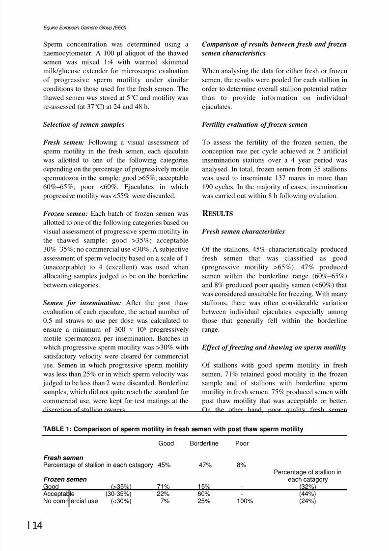
Of the stallions, 45% characteristically producedfresh semen that was classified as good(progressive motility >65%), 47% producedsemen within the borderline range (60%–65%)and 8% produced poor quality semen (<60%) thatwas considered unsuitable for freezing. With manystallions, there was often considerable variationbetween individual ejaculates especially amongthose that generally fell within the borderlinerange.
Effect of freezing and thawing on sperm motility
Of stallions with good sperm motility in freshsemen, 71% retained good motility in the frozen
sample and of stallions with borderline spermmotility in fresh semen, 75% produced semen withpost thaw motility that was acceptable or better.On the other hand, poor quality fresh semen
TABLE 1: Comparison of sperm motility in fresh semen with post thaw sperm motility
Good Borderline Poor
Fresh semen
Percentage of stallion in each catagory 45% 47% 8%Percentage of stallion in
Frozen semen each catagoryGood (>35%) 71% 15% - (32%)
Acceptable (30-35%) 22% 60% - (44%)No commercial use (<30%) 7% 25% 100% (24%)
8/12/2019 Monograph Series No. 1 - 1st Meeting of European Equine Gamete Group on Reproduction
http://slidepdf.com/reader/full/monograph-series-no-1-1st-meeting-of-european-equine-gamete-group-on-reproduction 26/85
8/12/2019 Monograph Series No. 1 - 1st Meeting of European Equine Gamete Group on Reproduction
http://slidepdf.com/reader/full/monograph-series-no-1-1st-meeting-of-european-equine-gamete-group-on-reproduction 27/85
16
Equine European Gamete Group (EEG)
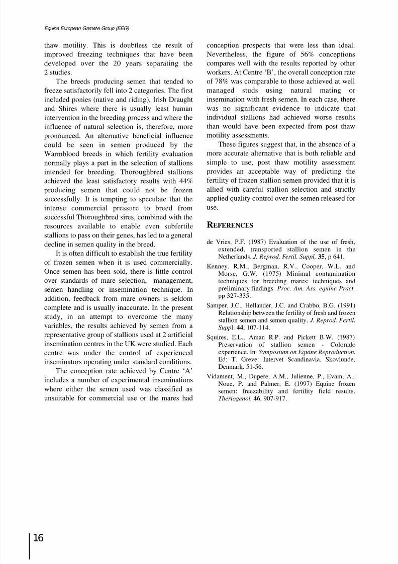
thaw motility. This is doubtless the result of improved freezing techniques that have beendeveloped over the 20 years separating the2 studies.
The breeds producing semen that tended tofreeze satisfactorily fell into 2 categories. The first
included ponies (native and riding), Irish Draughtand Shires where there is usually least humanintervention in the breeding process and where theinfluence of natural selection is, therefore, morepronounced. An alternative beneficial influencecould be seen in semen produced by theWarmblood breeds in which fertility evaluationnormally plays a part in the selection of stallionsintended for breeding. Thoroughbred stallionsachieved the least satisfactory results with 44%producing semen that could not be frozen
successfully. It is tempting to speculate that theintense commercial pressure to breed fromsuccessful Thoroughbred sires, combined with theresources available to enable even subfertilestallions to pass on their genes, has led to a generaldecline in semen quality in the breed.
It is often difficult to establish the true fertilityof frozen semen when it is used commercially.Once semen has been sold, there is little controlover standards of mare selection, management,semen handling or insemination technique. In
addition, feedback from mare owners is seldomcomplete and is usually inaccurate. In the presentstudy, in an attempt to overcome the manyvariables, the results achieved by semen from arepresentative group of stallions used at 2 artificialinsemination centres in the UK were studied. Eachcentre was under the control of experiencedinseminators operating under standard conditions.
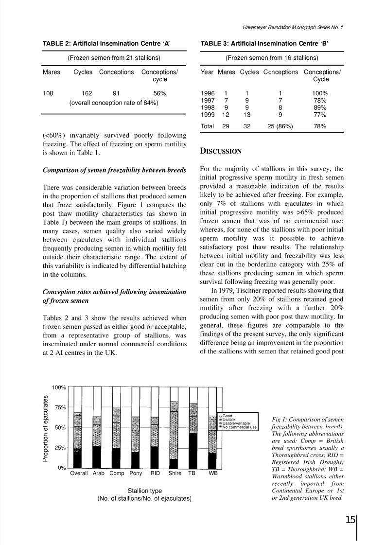
The conception rate achieved by Centre ‘A’includes a number of experimental inseminationswhere either the semen used was classified as
unsuitable for commercial use or the mares had
conception prospects that were less than ideal.Nevertheless, the figure of 56% conceptionscompares well with the results reported by otherworkers. At Centre ‘B’, the overall conception rateof 78% was comparable to those achieved at wellmanaged studs using natural mating or
insemination with fresh semen. In each case, therewas no significant evidence to indicate thatindividual stallions had achieved worse resultsthan would have been expected from post thawmotility assessments.
These figures suggest that, in the absence of amore accurate alternative that is both reliable andsimple to use, post thaw motility assessmentprovides an acceptable way of predicting thefertility of frozen stallion semen provided that it isallied with careful stallion selection and strictly
applied quality control over the semen released foruse.
REFERENCES
de Vries, P.F. (1987) Evaluation of the use of fresh,extended, transported stallion semen in theNetherlands. J. Reprod. Fertil. Suppl. 35, p 641.
Kenney, R.M., Bergman, R.V., Cooper, W.L. andMorse, G.W. (1975) Minimal contaminationtechniques for breeding mares: techniques andpreliminary findings. Proc. Am. Ass. equine Pract.
pp 327-335.Samper, J.C., Hellander, J.C. and Crabbo, B.G. (1991)
Relationship between the fertility of fresh and frozenstallion semen and semen quality. J. Reprod. Fertil.
Suppl. 44, 107-114.
Squires, E.L., Aman R.P. and Pickett B.W. (1987)Preservation of stallion semen - Coloradoexperience. In: Symposium on Equine Reproduction.
Ed: T. Greve: Intervet Scandinavia, Skovlunde,Denmark. 51-56.
Vidament, M., Dupere, A.M., Julienne, P., Evain, A.,Noue, P. and Palmer, E. (1997) Equine frozensemen: freezability and fertility field results.Theriogenol. 46, 907-917.
8/12/2019 Monograph Series No. 1 - 1st Meeting of European Equine Gamete Group on Reproduction
http://slidepdf.com/reader/full/monograph-series-no-1-1st-meeting-of-european-equine-gamete-group-on-reproduction 28/85
17
Havemeyer Foundation Monograph Series No. 1
RELATIONSHIP BETWEEN SPERM CHROMATINSTRUCTURE, MORPHOLOGY AND MEMBRANEQUALITY IN STALLION SPERMATOZOA BEFOREAND AFTER CRYOPRESERVATION
H. Torner, C. Warnke* and S. Blottner†
Research Institute for Biology of Farm Animals, Department of Reproductive Biology, 18196 Dummerstorf, Germany; *University of Rostock, 18069 Rostock, Germany ; † Institute for Zoo Biology and Wildlife Research, 10315 Berlin, Germany
INTRODUCTION
Current methods of evaluating stallion fertility are
based on morphological examination of spermatozoa and by calculating the number of normal, progressively motile sperm ejaculated.These methods provide basic information onlyabout sperm quality. Therefore it is very importantto develop more sensitive tests for characterisationof semen quality in stallions. The evaluation of cryopreserved semen is particularly complexbecause good post thaw motility does not alwayscorrelate with good fertility, indicating thatsubcellular damage can affect the fertility without
a concurrent impact on motility (Christensen et al.1995). A better understanding of both thephysiological and the morphological changes seenin spermatozoa would aid the development of improved techniques for the cryopreservation of stallion spermatozoa. The aim of the presentinvestigation therefore was to describe the changesin sperm chromatin structure, membrane quality,morphology and motility of stallion spermatozoaduring routine procedures for cryopreservation.Furthermore the possible correlation between
morphological and physiological parameters forstallion sperm evaluation was analysed.
MATERIALS AND METHODS
Semen samples were collected from 6 stallionsvia an artificial vagina. Three replications werecarried out. Each ejaculate was dilutedimmediately 1:1 in skim milk extender up to theadjustment of sperm concentration to 25 x 106 /ml.Extended semen were centrifuged at 900 g for 20
min at room temperature. After removing thesupernatant, one part was re-suspended in skimmilk extender for transport of chilled
spermatozoa, the other was re-suspended in eggyolk extender, packaged in 0.5 ml polyvinylchloride straws and frozen.
After transport of chilled spermatozoa orthawing of frozen spermatozoa from the sameejaculate the samples were diluted, centrifuged andwashed with TALP. Forward progression wasassessed microscopically in the dark field of a phasecontrast microscope. Head and tail abnormalitiesand condition of the acrosome of spermatozoa wereassessed from smears stained by saturated congo-red (2 min), tannin (3 min) and 3% aqueous brilliantcresyl-blue (30–45 s). Damaged plasma membrane(membrane integrity) was detected by fluorescence
staining in suspension with propidium iodide. Theintegrity and resistance of DNA (chromatinintegrity) was examined by the microscopicacridine orange test (Tejada et al. 1984). Toinvestigate chromatin stability, an artificial spermdecondensation test was used. Before the acridineorange test was carried out, the spermatozoa weretreated with 0.32% DTE/Papain (Hingst andBlottner 1993). Percentages were estimated from200 spermatozoa/sample. To investigate themembrane potential (+inside), the sperm cells were
stained with 2.5 µM DIBAC (Molecular Probes)and the part of fluorescence labelled cells werecounted by flow cytometry. The intracellular Ca2+
concentration was evaluated after pre-treatment of sperm cells with pluronic, followed by staining withFluo 3 AM and measurement of fluorescent spermcells by flow cytometry (10.000 counts/ measurement).
RESULTS
Stallion spermatozoa from different donors with thesame motility before freezing (about 60%) showeddifferent suitability for cryopreservation (forward
8/12/2019 Monograph Series No. 1 - 1st Meeting of European Equine Gamete Group on Reproduction
http://slidepdf.com/reader/full/monograph-series-no-1-1st-meeting-of-european-equine-gamete-group-on-reproduction 29/85
18
Equine European Gamete Group (EEG)
progression after thawing ranged from 9–27.5%).Cryopreservation led to a significant decrease inprogressive motility and morphologically normalspermatozoa (P<0.001). The main portion of abnormal spermatozoa after freezing was withoutacrosome (52–69%). Regardless of donor, the
freezing process led to a drastic increase of membrane defects (12.5% vs 67%, P<0.001). Only6.5–11.15% of spermatozoa showed chromatindefects after freezing.
After decondensation treatment with 0.32%DTE/Papain the portion of spermatozoa withchromatin defects increased (10.8–20.7%) andvaried between donors.
After freezing, the percentage of spermatozoawhich were DIBAC-labelled (membrane potential+ inside) increased (7.9% vs 26.4%; P<0.01). The
percentage of spermatozoa with a high level of intracellular Ca2+ (Fluo 3AM labelled) decreasedafter cryopreservation (78.1% vs 58%, P<0.01).
DISCUSSION
The data indicate that the freezing process led todrastic changes in physiological andmorphological parameters in stallionspermatozoa. The experiment has shown thatchromatin integrity and chromatin stability are
highly correlated in stallion spermatozoa (r=0.69;P<0.005). The results suggest that the motility of spermatozoa after cryopreservation depends on
the membrane quality and is negatively correlatedwith the percentage of spermatozoa withmembrane damage (r= -0.71; P<0.001). It seemsthat the increasing amount of spermatozoa withpositive membrane potential after thawing reflectsa higher passive diffusion potential in sperm
(Cooper et al. 1990). Additionally the Ca2+ levelin spermatozoa after freezing decreases, mainlycaused by the increased passive diffusionpotential of the spermatozoa membrane. Furtherinvestigations are focusing on the characterisationof functional and morphological changes installion spermatozoa, in relation to differentmethods of cryopreservation.
REFERENCES
Cooper, C.E., Bruce, D. and Nicholls, P. (1990) Use of Oxonal V as a probe of membrane potential inproteoliposomes containing cytochrome oxidase inthe submitochondrial orientation. Biochem. 29,3859-3865.
Christensen, P., Parlevliet, J., Van Butten, A. andHyttel, P. (1995) Ultrastructure of fresh and frozen-thawed stallion spermatozoa. Biol. Reprod.
Monograph Series 1, 769-777.
Hingst, O. and Blottner, S. (1993) Assessment of thechromatin structure of cat sperm before and aftercryopreservation. J. Reprod. Fert . 12, 59.
Tejada, R.I., Mitchel, J.C., Norman, A., Marik, J.J. andFriedman, S. (1984) A test for the practicalevaluation of male fertility by acridine orangefluorescence. Fertil. Steril. 42, 87-91.
8/12/2019 Monograph Series No. 1 - 1st Meeting of European Equine Gamete Group on Reproduction
http://slidepdf.com/reader/full/monograph-series-no-1-1st-meeting-of-european-equine-gamete-group-on-reproduction 30/85
19
Havemeyer Foundation Monograph Series No. 1
EVALUATION OF FROZEN STALLION SEMEN
T. Katila, P. Kuisma and M. Andersson
University of Helsinki, Department of Clinical Veterinary Sciences, Saari Unit, Pohjoinen Pikatie 800,
FIN-04920 Saarentaus, Finland
INTRODUCTION
Some semen evaluation methods work reasonably
well for the assessment of fresh equine semen, butnot of frozen semen. The best example is motilityevaluation. It is still the most commonly usedmethod for evaluation of frozen-thawed stallionsemen, both in laboratories and studfarms, becauseit is the only technique available in practiceconditions. There are very few data about thecorrelation between semen evaluation tests andpregnancy rates after insemination with frozensemen. Although good correlations have beenfound between different tests, it does not mean
necessarily that they would correlate with fertility.The purpose of this study was to find out
which tests used to evaluate stallion semen wouldcorrelate with foaling rates of mares inseminatedwith frozen semen.
MATERIALS AND METHODS
Frozen semen doses were available from 40commercially used stallions. The semen had beenfrozen between 1988 and 1997 in 5 different
countries: 29 in Sweden; 5 in Finland; 3 in Italy;one in Denmark; one in Germany; and one in theUSA. Semen from 2 stallions had been frozen in 5ml straws, 30 in 2.5 ml straws and 8 in 0.5 mlstraws. Twenty stallions were AmericanStandardbreds and the remaining 20 were differentkinds of riding horses. Sufficient foaling data – atleast 5 mares and 10 cycles – were available from31 stallions. Foaling data originated from Finlandand Sweden from the years 1989 to 1998. Theaverage number of mares/stallion was 33 (range
5–121). The average foaling rate was 58% (range0–83%). Twelve stallions had a foaling rate >50%and 19 had a foaling rate <50%.
Semen evaluation
Usually 3 straws were needed to perform all the
analyses. If only one or 2 straws were available,2 laboratory technicians worked in the laboratorysimultaneously to perform all the tests in a freshlythawed sample. From 14 stallions all tests wereconducted on the same batch. For the rest of thestallions, the batches had been frozen within thesame month.
The 0.5 ml straws were thawed at +37°C for30 s in the 2.5 and 5 ml straws at +50°C for 40 or45 s, respectively. An aliquot of 0.1 ml semen wascultured on a blood agar at +37°C for 48 h. Colony
forming units (CFU) were counted after anincubation of 24 and 48 h. Semen concentrationwas measured in a counting chamber and the totalnumber of spermatozoa/straw calculated. Aninsemination (AI) dose was one straw when using2.5 or 5 ml straws and 1–10 of the 0.5 ml straws.
Semen was extended with warm (+30°C) skimmilk extender (Kenney et al. 1975) to aconcentration of 20–30 x 106 spermatozoa /ml.Post thaw motility was evaluated subjectively witha light microscope: percentage of progressively
motile spermatozoa, percentage of total motilityand a velocity score from 1 to 3. Motility wasmeasured also with an automatic sperm analyser(Hamilton Thorn Motility Analyser, HTM-S,version 7.2). A 10 ml semen sample was placedonto a Makler chamber and 2 chambers wereprepared from the same sample. Sixfields/chamber were video taped for 15 s/field. Thevideo tapes were analysed for total motility,progressive motility and path velocity.
The sample for the longevity test was
prepared by placing 0.5 ml of extended semen intoa 3 ml vial enclosed with a cap. The samplewas kept in a water bath at +37°C for 4 h. The total

8/12/2019 Monograph Series No. 1 - 1st Meeting of European Equine Gamete Group on Reproduction
http://slidepdf.com/reader/full/monograph-series-no-1-1st-meeting-of-european-equine-gamete-group-on-reproduction 31/85
20
Equine European Gamete Group (EEG)
and progressive motility and velocity wereevaluated by light microscope every hour.
A second straw was thawed for the evaluationof plasma membrane integrity by CFDA/PIstaining (carboxyfluoresceindiacetate/propidiumiodide). Semen was extended with skim milk
extender to a concentration of 50 spermatozoa/ml,of which 950 µl were taken and mixed with 20 µlof formol citrate (1.7 mM). An aliquot of 20 µlCFDA stock solution consisting of 0.46 mg CFDAin 1 ml of DMSO (dimethylsulphoxide) and 10 µlof PI stock solution (0.5 mg PI in 1 ml of 0.9%NaCl-solution) were taken, mixed with semen-formol solution and incubated for 8 min at +30°C.A drop of 5 µl was placed on a slide and overlaidwith a cover slip (Harrison and Vickers 1990). Theproportion of fluorescent cells was counted in 200
cells in a fluorescence microscope (Olympus BH2with epifluorescence optics) using oil immersionand a fluorescein filter set.
A third straw was thawed for the plasmamembrane viability test by an automaticfluorometer (Fluoroscan Ascent), which reads a96 well microtitration tray and has an incubationcompartment. The interference filter at theexitation path and that of the emission filter had amaximum transmission at 544 nm and 590 nm,respectively. For the fluorometric assay, 20 mg of
PI was dissolved in 1:l of BTS (BeltsvilleThawing Solution) and dispensed in 3 mlaliquots. Equal aliquots (50 ml) of BTS extendedsemen sample (80 x 106 spermatozoa/ml) and PIsolution were dispensed into the well plate, andthe well was shaken gently for 2 min.Spermatozoa from the same samples were killedby unprotected rapid freezing-thawing to provideinternal control samples consisting of only non-viable cells (100% fluorescence). The controlsample was immersed into liquid nitrogen for1 min and thereafter it was allowed to stand atroom temperature for 30 s and then 3 min in awaterbath (37°C). Blanks containing 50 µl of diluted extender and 50 µl of PI were analysedseparately for every experiment in 4 replicates.The incubation time was 8 min. The percentageof fluorescence was calculated from the ratio of fluorescence intensities of the rapidly frozencontrol sample and the sample to be analysed,taking into account the blank values (Juonala et
al. 1999). For the resazurin reduction test, 400mg of resazurin were dissolved in 1:1 distilledwater. One part of this solution and 9 parts of
0.9% NaCl were mixed. An equal volume of thismixture and extended sperm were combined,shaken for 2 min, incubated for 30 min at 34°Cand measured with the fluorometer using thesame fluorometer settings as in the plasmamembrane viability test (Eriksson et al. 1998).
Data analysis
The results were analysed by Statgraphigs usingcorrelation matrix. The level of significance wasset at P<0.05. Only the results of the 31 stallionswith sufficient foaling data were included in thestatistical analysis.
RESULTS
Twenty straws showed no bacterial growth,9 yielded slight growth (<10 CFU), 5 mediumgrowth (10–100 CFU) and 6 straws yielded heavygrowth (>100 CFU). There was a wide range of bacteria in the positive samples. The mostcommon organisms were Enterobacteria,
Corynebacteria and coliforms. The occurrence of bacteria had no correlation with foaling rates.
The total number of spermatozoa in an AI dosewas, on average, 655 x 106 (ranging from 202 to1,772 x 106). The number of progressively motile
sperm was, on average, 254 x 106 (ranging from 81to 612 x 106). The dose had no correlation with thefoaling rate.
The only parameter that correlatedsignificantly with foaling rate was motility duringstorage at 37°C, both at 2 h (P=0.0092) and at 4 h(P=0.0286).
Some of the measured parameters had highcorrelations with each other. When all parameterswere analysed in the same semen batch (14stallions), very highly significant correlations(P<0.001) were detected between initial total andprogressive motility measured by HTM-S and bylight microscopy. Fluorometric viabilitymeasurement had a good correlation with 0 h lightmicroscope motility (P=0.0006), CFDA(P=0.0025), with HTM-S progressive (P=0.0165)and total motility (P=0.0238) and betweenresazurin reduction test (P=0.0170). CFDAcorrelated also with initial motility parameters(P<0.05). Progressive motility at 2 h correlatedsignificantly with initial total and progressivemotility and with 4 h motility, but motility at 4 hdid not correlate with any other parameters.
8/12/2019 Monograph Series No. 1 - 1st Meeting of European Equine Gamete Group on Reproduction
http://slidepdf.com/reader/full/monograph-series-no-1-1st-meeting-of-european-equine-gamete-group-on-reproduction 32/85

8/12/2019 Monograph Series No. 1 - 1st Meeting of European Equine Gamete Group on Reproduction
http://slidepdf.com/reader/full/monograph-series-no-1-1st-meeting-of-european-equine-gamete-group-on-reproduction 33/85
22
Equine European Gamete Group (EEG)

8/12/2019 Monograph Series No. 1 - 1st Meeting of European Equine Gamete Group on Reproduction
http://slidepdf.com/reader/full/monograph-series-no-1-1st-meeting-of-european-equine-gamete-group-on-reproduction 34/85
23
Havemeyer Foundation Monograph Series No. 1
Stallion semen- commercial aspects
Chairman: B. Colenbrander
SESSION III:

8/12/2019 Monograph Series No. 1 - 1st Meeting of European Equine Gamete Group on Reproduction
http://slidepdf.com/reader/full/monograph-series-no-1-1st-meeting-of-european-equine-gamete-group-on-reproduction 35/85
24
Equine European Gamete Group (EEG)
8/12/2019 Monograph Series No. 1 - 1st Meeting of European Equine Gamete Group on Reproduction
http://slidepdf.com/reader/full/monograph-series-no-1-1st-meeting-of-european-equine-gamete-group-on-reproduction 36/85
25
Havemeyer Foundation Monograph Series No. 1
EFFECT OF GLUCOCORTICOID TREATMENT ONFRESH AND DEEP FROZEN SEMEN AND ONENDOCRINE PARAMETERS IN THE STALLION
J. Juhasz, M. Vidament*, P. Nagy, M. Kulcsar, D. Guillaume*, M. Magistrini* andGy. Huszenicza
University of Veterinary Science, Budapest, Hungary; *INRA, Equine Reproduction, Nouzilly, France
The aim of the study was to determine the possibleeffects of a glucocorticoid treatment on thereproductive function of breeding stallions. Nine
stallions of various breeds, aged 3–17 years wereincluded. They were divided into 2 groupsaccording to their age and semen quality (5treated and 4 control stallions). Treated stallionswere given dexamethasone-sodium phosphate (30mg/stallion/day, Dexadreson, Intervet, TheNetherlands) for 7 days in mid-November. Semenwas collected and frozen twice a week from mid-September until the end of January. For freezing,lactose-EDTA-egg-yolk freezing extender wasused. Freezing was carried out in 0.5 ml straws in
200 x 106 spermatozoa/ml final concentrationabove N2 vapour.
The following parameters were evaluated fromfresh semen: total and gel-free volume, pH,progressive motility (under light microscope),concentration, total sperm number, morphology(fixed in formol solution, determined under phaseinterference microscope), acrosome integrity(stained with FITC-PSA). After thawing thefollowing parameters were evaluated: post thawmotility (under light microscope), membrane
functional integrity (HOS test), acrosome integrity(stained with FITC-PSA) and morphology (fixedin formol solution, determined under phaseinterference microscope).
GnRH (40 mg Receptal, Hoescht, Germany)and hCG (10,000 iu Chorulon, Intervet, TheNetherlands) challenge tests were carried outbefore, immediately after, one month and 2months after the end of the treatment. During thetreatment period, daily blood collections werecarried out. From the plasma samples LH, FSH
and testosterone were determined. TRH- (Sigma,USA) and ACTH-stimulations (60 mg Cortrosyn,Organon, The Netherlands) were carried out
before, one day and 16 days after treatment. Fromthe plasma samples T3, T4 and cortisol weredetermined.
In fresh semen, total sperm number perejaculate was significantly (P<0.001) higher in thetreated group on the sixth week after treatment.Midpiece abnormalities tended to be increased in 2treated stallions between Days 10 and 40,proximal protoplasmic droplet tended to be higherin one treated stallion between Days 14 and 55after treatment. No other parameters differedsignificantly or tended to be lower or higherbetween the pre-treatment and post treatmentperiods or between the treated and control groups.
Post thaw motility tended to be decreased in 2 of 4treated stallions beginning on Day 10 aftertreatment and returned to pre-treatment levelaround Day 60. Results of HOS tests showed asimilar pattern and tendency to post thaw motilityin all stallions. No effect of treatment could beobserved on morphology or acrosome integrity.
During treatment, plasma testosterone levelswere significantly lower and LH levelssignificantly higher in treated stallions (P<0.05).Plasma FSH concentration was not affected by the
treatment. LH response to GnRH-stimulation wassignificantly higher immediately after the end of the treatment in the treated group (P<0.05).Testosterone response to GnRH-stimulation wassignificantly higher in the treated group one monthafter the end of the treatment (P<0.05). FSHresponse to GnRH-stimulation was not altered bythe treatment. Testosterone response to hCG-stimulation was the same in the treated and controlgroups in all challenge periods. Basal cortisolconcentrations and maximal response to ACTH
were significantly decreased one day aftertreatment in treated stallions (P<0.001). Thisdifference disappeared by Day 16. Basal T4-levels
8/12/2019 Monograph Series No. 1 - 1st Meeting of European Equine Gamete Group on Reproduction
http://slidepdf.com/reader/full/monograph-series-no-1-1st-meeting-of-european-equine-gamete-group-on-reproduction 37/85
26
Equine European Gamete Group (EEG)
and maximal response to TRH were significantlylower (P<0.05) in treated stallions by Day 16. Asimilar but not significant tendency was alsoobserved for T3 in the same group.
It is well known and accepted thatcorticosteroids in general suppress the function of
the hypothalamus-pituitary-gonadal axis (Moberg1987). They can act at the level of hypothalamus,pituitary and the gonads altering their hormonesynthesis and/or release function. The site of theireffect depends on dose and length of exposure.Most studies have investigated the effect of stressor various adrenal diseases (Cushing’s syndrome)on reproductive function (McKenna et al. 1979).Our experiment did not aim to model the stress,but to investigate the side effect of dexamethasonetreatment on the reproductive function of
stallions.Dexamethasone treatment with the applied
dose primarily altered the testosterone productionof the testes in the short term. There areglucocorticoid binding receptors in the testeswhich can alter the testosterone producingfunction of Leydig-cells after binding has occurred(Welsh et al. 1979; Sapolsky 1985; Moberg 1987).This could be why the testosterone levels weresignificantly lower in the treated stallions duringthe treatment period. During treatment, LH levels
were significantly higher in the treated groupwhich could be caused by the decreasedtestosterone level or by the direct effect of dexamethasone on hypothalamus or pituitary.
This elevated LH level and storage could bethe reason for the significantly higher LH responseto GnRH-stimulation immediately after thetreatment. Unchanged testosterone response tohCG-stimulation after treatment indicates thatdexamethasone at the applied dose did not have a
negative influence on the testosterone producingcapacity of the testes. There were no significant ormarked alterations in semen parameters in thetreated stallions although, in some cases, slightdifferences were observed which could be causedby individual sensitivity to the drug. Decreasedtestosterone level during treatment is probablysufficient to maintain normal spermatogenesis.
In conclusion, dexamethasone applied at therecommended therapeutic dose does not causelong term alterations in the endocrine profiles or
significant changes in semen parameters inbreeding stallions.
REFERENCES
Moberg, G. P. (1987) Influence of the adrenal axis uponthe gonads. Oxford Rev. Reprod. Biol. 9, 456-496.
McKenna, T. J., Lorber, D., Lacroix, A. and Rabin, D.(1979) Testicular activity in Cushing’s disease. Acta
endocr. 91, 501-510.
Sapolsky, R. M. (1985) Stress-induced suppression of testicular function in the wild baboon: role of glucocorticoids. Endocrinology 116, 2273-2278.
Welsh, T. H., McGraw, R. L. and Johnson, B.H. (1979)Influence of corticosteroids on testosteroneproduction in the bull. Biol. Reprod. 21, 755-763.
8/12/2019 Monograph Series No. 1 - 1st Meeting of European Equine Gamete Group on Reproduction
http://slidepdf.com/reader/full/monograph-series-no-1-1st-meeting-of-european-equine-gamete-group-on-reproduction 38/85
27
Havemeyer Foundation Monograph Series No. 1
FERTILITY DATA IN THE HORSE
K. Darenius
Department of Obstetrics and Gynaecology, Swedish University of Agricultural Sciences, 8-750 07 Uppsala, Sweden
INTRODUCTION
The aim of the present work was to study the
occurrence of pregnancy and neonatal loss withregard to differences between stallions as well asbetween various categories of mares and breedingtechniques, using field data from one carefullycontrolled studfarm.
MATERIALS AND METHODS
Fertility data from 1,206 mares (Standardbredtrotters [SB], Swedish Warmbloods [SWB] andThoroughbreds [TB]) were collected during
7 breeding seasons. The mares were mated/ inseminated at one stud, under the samemanagement conditions. The stud is in centralSweden and most mares were mated during Mayto July. Different breeding techniques were used,natural breeding as well as artificial insemination(Al) with fresh, chilled or frozen semen.
During oestrus, gynaecological examinationswere generally performed on alternate days tomonitor follicular and endometrial activity, todetect any pathological conditions before and after
mating/insemination and to confirm ovulation. Inthe case of natural breeding and Al with fresh andchilled semen, matings/inseminations wereperformed every second day. Most of the mareswere inseminated only before ovulation.
For frozen semen, mares were examined daily.Close to ovulation they were examined 2–4 timesa day to determine the approximate time of ovulation. They were inseminated within 12 h pre-ovulation (39% of the mares), within 6–8 h postovulation (37%) or both pre- and post ovulation
(24%).Ultrasound scanning for pregnancy diagnosis
was performed around Day 14–17 and, in most of
the mares, also Day 40–45 post ovulation.Additional examinations were carried out in mareswith twins or where early fetal death was
suspected. The conception rates are based on theresults from examinations performed at the earliestconvenient opportunity.
The variations among stallions, breedingmethods and categories of mares, as regards thevarious fertility measures used, was analysedusing normal X2 tests in order to prove statisticallyany heterogeneity among classes. Levels of significance are given as follows: ns=notsignificant, *=P<0.05, **=P<0.01,***=P<0.001.
DEFINITIONS
The following terms are used in this paper:
Early fetal death (EFD): embryonic and fetal deathup to 150 days gestation, except visible abortions;
Abortion: visibly expelled fetus at any time of gestation;
Live foal: a foal that is alive 10 days post foaling;
Dead foal: a foal that is born dead or dies within
10 days post foaling;
Miscellanous loss: various reasons for pregnancyloss, such as induced abortion or death of mareduring pregnancy;
Total loss: EFD + abortions + dead foals.
RESULTS
Fertility data are expressed on a seasonal basis withregard to total conception and live foal rates. The
results are also expressed on a per cycle basis forconception, twinning, early fetal death, abortion,dead foal, miscellanous loss and live foal rates.

8/12/2019 Monograph Series No. 1 - 1st Meeting of European Equine Gamete Group on Reproduction
http://slidepdf.com/reader/full/monograph-series-no-1-1st-meeting-of-european-equine-gamete-group-on-reproduction 39/85
8/12/2019 Monograph Series No. 1 - 1st Meeting of European Equine Gamete Group on Reproduction
http://slidepdf.com/reader/full/monograph-series-no-1-1st-meeting-of-european-equine-gamete-group-on-reproduction 40/85
29
Havemeyer Foundation Monograph Series No. 1
PREGNANCY RATES FOLLOWING INSEMINATION
WITH FRESH VERSUS FROZEN SEMEN
Z. Müller and J. Müller
Equine Reproduction Centre, 530 02 Pardubice - Mnetice 126, Czech Republic
INTRODUCTION
Artificial insemination in horses is a well
established technique which is now considered tobe a routine breeding procedure. Notably theinfluence of insemination with frozen semen ongene migration is very apparent in the internationalbreeding of top sport horses.
Despite this, insemination methods using bothcooled and frozen semen are still being developed.There are several variations in methods of semencollection, freezing and the use of extenders. Todate, no single method has been shown to be themost advantageous. The aim of our work has been
to identify factors that influence the success orotherwise of specific strategies.
MATERIALS AND METHODS
Semen collection
For the purpose of shipping of cooled semen,different types of artificial vagina were used forcollection. Semen collection for freezing of insemination doses was carried out using the
Krakow model of an open artificial vagina, in thepresence of a mare. Semen was collected 4 daysper week for cooled semen or 3 days per week forfrozen semen.
Semen processing and shipping
Cooled semen: Kenney’s (instant non-fat dry milk2.4 g, glucose monohydrate 4.9 g, sodiumbicarbonate 7.5% sol 2.0 ml, gentamicin sulphate50 mg/ml 2.0 ml, distilled water 92 ml) or LC
(lactose 11 g, disodium EDTA 0.1 g, sodiumcitrate dihydrate 0.089 g, sodium bicarbonate0.008 g, deionised water 100 ml, egg yolk 1.6 g)
extenders were used for diluting raw semen. Aminimal dilution ratio of 1:2 (raw semen:extender)was used and one insemination dose contained at
least 200 million progressively motile sperms.Diluted insemination doses presenting 10–20 mlwere cooled to 4°C and shipped. On collectiondays, semen was sent by train around the CzechRepublic. Following telephone orders prior to 9.00am, the insemination doses were usually at theirdestination by 7.00 pm.
Frozen semen: LC extender containing 3.5%glycerol was used to dilute raw semen yielded byfractionated collection. Therefore the procedure
did not include centrifugation. The ratio of dilutioncorresponded to a minimum of 200 millionprogressively motile sperms per insemination doseafter thawing. Semen was frozen in 6 mlaluminium tubes.
Insemination: Insemination was carried out byveterinarians and insemination technicians. In1998 breeders could, following training at aworkshop, carry out insemination of their ownmares with fresh semen only. Insemination with
frozen semen was carried out by 21 workers andwith cooled semen by 48 workers.
Palpation and ultrasonography of the ovariesand uterus were used to determine the optimal timeof insemination (approaching ovulation). Insporadic cases when using cooled semen, theinsemination was timed on the basis of positiveteasing and viscosity of cervical gel. Inseminationwas always repeated after 48 h up to the detectedovulation or the end of heat.
Timing of insemination with frozen semen was
based on regular ultrasonographic examination of mares during oestrus. Examination frequencyvaried according to practical conditions from once
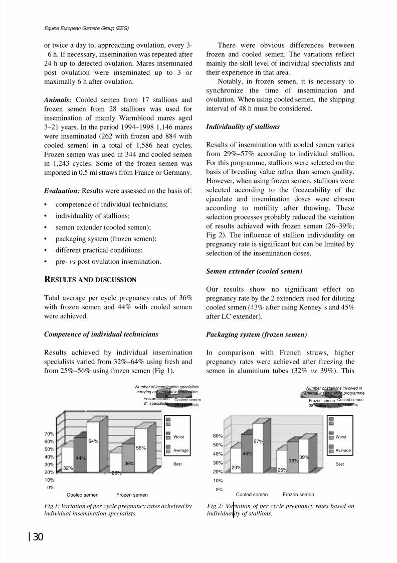
8/12/2019 Monograph Series No. 1 - 1st Meeting of European Equine Gamete Group on Reproduction
http://slidepdf.com/reader/full/monograph-series-no-1-1st-meeting-of-european-equine-gamete-group-on-reproduction 41/85
30
Equine European Gamete Group (EEG)
or twice a day to, approaching ovulation, every 3-–6 h. If necessary, insemination was repeated after24 h up to detected ovulation. Mares inseminatedpost ovulation were inseminated up to 3 ormaximally 6 h after ovulation.
Animals: Cooled semen from 17 stallions andfrozen semen from 28 stallions was used forinsemination of mainly Warmblood mares aged3–21 years. In the period 1994–1998 1,146 mareswere inseminated (262 with frozen and 884 withcooled semen) in a total of 1,586 heat cycles.Frozen semen was used in 344 and cooled semenin 1,243 cycles. Some of the frozen semen wasimported in 0.5 ml straws from France or Germany.
Evaluation: Results were assessed on the basis of:
• competence of individual technicians;
• individuality of stallions;
• semen extender (cooled semen);
• packaging system (frozen semen);
• different practical conditions;
• pre- vs post ovulation insemination.
RESULTS AND DISCUSSION
Total average per cycle pregnancy rates of 36%with frozen semen and 44% with cooled semenwere achieved.
Competence of individual technicians
Results achieved by individual inseminationspecialists varied from 32%–64% using fresh andfrom 25%–56% using frozen semen (Fig 1).
There were obvious differences betweenfrozen and cooled semen. The variations reflectmainly the skill level of individual specialists andtheir experience in that area.
Notably, in frozen semen, it is necessary tosynchronize the time of insemination and
ovulation. When using cooled semen, the shippinginterval of 48 h must be considered.
Individuality of stallions
Results of insemination with cooled semen variesfrom 29%–57% according to individual stallion.For this programme, stallions were selected on thebasis of breeding value rather than semen quality.However, when using frozen semen, stallions wereselected according to the freezeability of the
ejaculate and insemination doses were chosenaccording to motility after thawing. Theseselection processes probably reduced the variationof results achieved with frozen semen (26–39%;Fig 2). The influence of stallion individuality onpregnancy rate is significant but can be limited byselection of the insemination doses.
Semen extender (cooled semen)
Our results show no significant effect on
pregnancy rate by the 2 extenders used for dilutingcooled semen (43% after using Kenney’s and 45%after LC extender).
Packaging system (frozen semen)
In comparison with French straws, higherpregnancy rates were achieved after freezing thesemen in aluminium tubes (32% vs 39%). This
Number of insemination specialistscarrying out artificial insemination
Frozen semen21 specialists
Cooled semen48 specialists
Fig 1: Variation of per cycle pregnancy rates acheived by
individual insemination specialists.
Number of stallions involved in
artificial insemination programme Frozen semen28 stallions
Cooled semen17 stallions
Fig 2: Variation of per cycle pregnancy rates based on
individuality of stallions.
Cooled semen Frozen semen
70%
60%
50%
40%
30%
20%
10%
0%
32%
44%
64%
25%
36%
56%
Worst
Average
Best
Cooled semen Frozen semen
29%
44%
57%
26%
36%39%
Worst
Average
Best
60%
50%
40%
30%
20%
10%
0%

8/12/2019 Monograph Series No. 1 - 1st Meeting of European Equine Gamete Group on Reproduction
http://slidepdf.com/reader/full/monograph-series-no-1-1st-meeting-of-european-equine-gamete-group-on-reproduction 42/85
31
Havemeyer Foundation Monograph Series No. 1
difference may relate to the number of straws usedor damage arising from transport and storage,rather than to different fertilising capacities.
Different practical conditions
Significantly better results (54% for frozen and39% for cooled semen) were achieved by thespecialised clinic compared to inseminationscarried out at the breeders’ operations (42% and33% per cycle pregnancy rate).
This may be, in part, because of theopportunity to monitor oestrus and predictovulation. Also, the post insemination treatment of a mare can be an important factor, especially afterusing frozen semen.
Pre- and post ovulation insemination
Pregnancy rates of 37% and 33% were achievedbefore and after ovulation, respectively. Therefore,although the rate decreases, insemination carriedout up to 6 h after ovulation can still producesatisfactory results and has the advantage of reducing the number of insemination doses used.
COMPARISON OF FROZEN AND COOLED
SEMEN
Cooled versus frozen semen
• higher fertility rate;
• tolerance of a less precise inseminationprocedure - based on the presumption thatcooled semen maintains its fertilising capacityfor 48 h after insemination;
• permanent breeding occupation of the stallion -a stallion must be available for semencollection any time during the breeding season;
• problems relating to transport for long distances
• dependence of insemination on semen
distribution (mare’s insemination management)- partially it can be solved by daily semencollection.
Frozen versus cooled semen
• precise timing of insemination - frozen semencan be possible thawed any time to inseminatea mare just before or after ovulation;
• transport is simple and can be planned beforelong term semen storage;
• production is possible at any time during theyear - a stallion can be used while competing;
• lower fertility rate;• necessity of precise insemination technique;
• more expensive - semen processing andstorage.
CONCLUSIONS
Insemination with cooled semen is a method usedsuccessfully at present. It is suitable for a wide andquick extension of selected stallions.
Insemination with frozen semen is a methodwhich will become increasingly popular and may,as in cattle breeding, replace cooled semeneventually. At present it is used mainly forbreeding top sport horses.
Much work is still required in the field of optimising insemination technology.

8/12/2019 Monograph Series No. 1 - 1st Meeting of European Equine Gamete Group on Reproduction
http://slidepdf.com/reader/full/monograph-series-no-1-1st-meeting-of-european-equine-gamete-group-on-reproduction 43/85
32
Equine European Gamete Group (EEG)
LARGE SCALE COMMERCIAL APPLICATION OFFRESH, COOLED AND DEEP FROZEN STALLIONSEMEN
H. Sieme, E. Klug* and E. Töpfer-Petersen†
Celle National Stud; *Clinic for Horses, Veterinary School, Hannover; † Institute for Reproductive Medicine, Veterinary School, Hannover, Germany
In the last decade, equine artificial insemination(AI) has become the main zootechnique of equinebreeding practice in the Warmblood and
Standardbred breeding industry in Germany,France, The Netherlands, Belgium, Denmark andSweden.
In Germany in 1999, approximately 180 AIcentres were registered; 60 licensed by theEuropean Government (92/65EG modified by95/172EG).
Among riding horses in 1998, 48,395 matingswere registered (riding horse mare population in1997 was 83,349). A total of 3,460 registeredprivate stallions mated 28,193 mares (58.2%) and
623 state-owned stallions covered 20,202 mares(41.8%). AI was used for 64.6% of all coverings.In 1998, 30,552 live foals (61.6%) were registeredout of 49,494 matings in 1997.
During the pre-breeding season, pubertal and allother sires are subjected to a full physicalexamination, including monitoring of sexualbehaviour and bacteriological examination of thegenital tract. Spermiograms are conducted on allpubertal stallions and those which achieved belowaverage conception rates in the previous season. Inaddition, all stallions older than 18 years and allstallions used for AI are examined for semen quality.
Stallions are not allowed to servesimultaneously in artificial and natural breedingprogrammes. Semen collection is confined to one
daily collection per stallion to adjust daily spermoutput to a predictable level.
Factors influencing the success of AI in the
horse include stallion fertility and management,semen preservation techniques and management of insemination and mare. Regardless of technicalfactors like extender, dilution ratio, preparationtechnique (dilution, centrifugation, alternatives),cooling rate, storage conditions and inseminationvolume, it is obvious that sperm dose andfrequency of pre-ovulatory inseminations areimportant limiting factors.
Effect of time of insemination, relative toovulation, is under study. Published fertility data
from different countries are available but the basisof fertility calculation differs between countries(fertility per cycle, end of season results, foalingrate), which complicates a comparative estimate of system efficacy.
There is even variation in stud fee strategiesbetween countries. In the German riding horsepopulation, the stud fee is paid per mare andseason and prices differ between stallions. Barrenmares normally receive a discount in the followingseason. Price fixing for exported frozen semen isusually calculated per insemination dose. Anotherstrategy would be a stud fee split into: 1) an initialfe for AI/service per mare and season combinedwith 2) a secondary fee per pregnancy evaluatedby veterinary examination in the autumn.

8/12/2019 Monograph Series No. 1 - 1st Meeting of European Equine Gamete Group on Reproduction
http://slidepdf.com/reader/full/monograph-series-no-1-1st-meeting-of-european-equine-gamete-group-on-reproduction 44/85
33
Havemeyer Foundation Monograph Series No. 1
Oocytes - recoveryand maturation
Chairman: R. H. F. Hunt er
SESSION 1V:

8/12/2019 Monograph Series No. 1 - 1st Meeting of European Equine Gamete Group on Reproduction
http://slidepdf.com/reader/full/monograph-series-no-1-1st-meeting-of-european-equine-gamete-group-on-reproduction 45/85
34
Equine European Gamete Group (EEG)
8/12/2019 Monograph Series No. 1 - 1st Meeting of European Equine Gamete Group on Reproduction
http://slidepdf.com/reader/full/monograph-series-no-1-1st-meeting-of-european-equine-gamete-group-on-reproduction 46/85
35
Havemeyer Foundation Monograph Series No. 1
ASSESSMENT OF EQUINE FOLLICLES AND OOCYTESCOLLECTED AT A SLAUGHTER HOUSE
A. Okólski and W. Mlodawska
Department of Animal Reproduction, University of Agriculture, 30-059 Cracow, Al. Mickiewicza
24/28, Poland
INTRODUCTION
One of the cheapest and most simple ways tocollect horse oocytes is to obtain mare ovariesfrom a slaughter house. The aim of this study is topresent the results of our investigation on differentmethods of oocyte collection from ovarianfollicles after slaughter. Also, we determined thechronology and stage of maturity of equine oocytenuclei and the degree of atresia in the ovarianfollicles from which some of the oocytes werecollected.
MATERIALS AND METHODS
The ovaries of mares of unknown reproductivehistory were collected from a local slaughter houseonce a week for a period of 3 years. Immediatelyafter slaughter the ovaries were transported in athermos flask at room temperature to thelaboratory. Here, they were washed 3 times infresh Dulbecco’s phosphate buffered salinemedium supplemented with decreasing doses of penicillin and streptomycin
Two different methods of oocyte recoverywere used. In the first one the follicular fluidcontaining oocytes was aspirated with a syringefrom the ovaries of 900 mares. Then, under astereomicroscope, the oocytes were separatedfrom the follicular fluid.
In the second method, follicles were isolatedfrom the ovaries of 319 mares, placed separatelyon a Petri dish, measured and cut. The cumulusoocyte complex (COC) was located on the folliclewall or floating in the follicular fluid. The COCs
were scraped from the wall using a scalpel blade.The morphology of the COCs was assessed usingthe system of Leibfried and First (1979) and
Hinrichs et al. (1993), modified by our ownobservations. Two hundred and forty four oocyteswere cultured in vitro in TCM-199 medium, with
FSH, LH and E2 from 3–48 h to evaluate thechronology of mare oocyte maturation in vitro.
Data were analysed by chi-square test andP<0.05, was taken as the level of statisticalsignificance.
RESULTS AND DISCUSSION
As a result of aspiration of 7,798 follicles, a totalof 3,158 (40.5%) oocytes per mare were collected,of which only 1,125 (35.6%) qualified for culture.
By cutting 2,043 isolated follicles, a total of 1,872(91.6%) oocytes were obtained (5.9 per mare) of which 1,235 (66.0%) were accepted for culture.Thus, by cutting it was possible to obtain asignificantly higher percentage of oocytes suitablefor culture than by aspiration (P<0.01). Thepercentage of oocytes accepted for cultureincreased with increasing follicle diameter, being30%, 54%, 61% and 78% (P<0.01) for follicleswith diameters of 4–10, 11–15, 16–20 and 21-–35mm, respectively.
When 564 follicles and oocytes were evaluatedfor the macroscopic degree of atresia, thepercentage of oocytes with compact cumuliincreased significantly with increasing diameter of the follicles being 23.3% for the follicles <10 mmand 64.2% for the follicles > 21 mm (P<0.01). Thepercentage of oocytes floating in the follicularfluid decreased from 52.0% in the smallestfollicles to 9.9% in the largest ones. However,Hinrichs (1997) found that oocytes havingexpanded cumulus mature in vitro more quickly
and in higher proportion than do cumulus compactoocytes, and the period of protein synthesisnecessary for maturation is shorter for expanded
8/12/2019 Monograph Series No. 1 - 1st Meeting of European Equine Gamete Group on Reproduction
http://slidepdf.com/reader/full/monograph-series-no-1-1st-meeting-of-european-equine-gamete-group-on-reproduction 47/85
8/12/2019 Monograph Series No. 1 - 1st Meeting of European Equine Gamete Group on Reproduction
http://slidepdf.com/reader/full/monograph-series-no-1-1st-meeting-of-european-equine-gamete-group-on-reproduction 48/85
37
Havemeyer Foundation Monograph Series No. 1
ZONA PELLUCIDA HARDENING AND SPERMBINDING ASSAY FOR EQUINE OOCYTES INCUBATEDIN VITRO
W. Mlodawska, J. Bezard*, M. Walul and A. Okólski
Department of Animal Reproduction, Agriculture University, Al. Mickiewicza 24/28, 30-059 Kraków,
Poland, * INRA Reproductive Physiology and National Studs, 37380 Nouzilly, France
INTRODUCTION
Zona pellucida (ZP) hardening expressed as anincrease in its resistance to enzymatic digestionoccurs following fertilisation, parthenogeneticactivation and in vivo and in vitro ageing of the egg-cell. Spontaneous ZP hardening can also beinduced during in vitro oocyte incubation (DeFelici and Siracusa 1982). One consequence of ZPhardening is a considerable decrease of oocytecapacity for in vitro fertilisation and embryonicdevelopment (Gianfortoni and Gulyas 1985;Katska et al. 1989). It is supposed that spontaneous
ZP hardening of oocytes may be one reason for lowefficiency of in vitro fertilisation in horses. The aimof the study was to determine whether exposure of equine oocytes to culture medium supplementedwith various sera would affect ZP hardening and itscapacity to bind spermatozoa.
MATERIALS AND METHODS
The study was performed on oocytes collected byaspiration of individual follicles (≥ 5 mm) from the
ovaries of 28 mares, within 2–4 h after slaughter(Mlodawska et al. 1999). The age and reproductivehistory of mares were unknown.
Estimation of ZP hardening was performeddirectly after oocyte collection, following 45–48 hin vitro maturation (IVM) or after IVM +additional co-culture with stallion spermatozoa.Oocytes (3–5) were matured in 100 ml TCM 199with Earle’s salts, 2.2 g/ml NaHCO3 and L-glutamine (Gibco) supplemented with CEG (9.5µg/ml eFSH, 15 µg/ml eLH), oestradiol-17ß (1
µg/ml; Sigma), antibiotics (100 IU/ml penicillin,100 µg/ml streptomycin, 0.25 µg/ml fungizone;Gibco) and 20% of inactivated FCS (fetal calf
serum) or EMS (estrus mare serum), underparaffin oil at 38.5°C in 5 % CO2 in air.
For oocyte insemination frozen semen from the
same ejaculate of a fertile stallion was thawed,washed twice in HGLLBSA and then in HHBSA(Mlodawska et al. 1999). Before insemination eachoocyte was mechanically denuded of all cumulusand corona cells by aspiration through a fine borepipette and incubated individually with 2.5 x 10⁵
spermatozoa/ml in 100 µl HHBSA for 20 h. Afterco-culture each oocyte was washed in 9 sequentialdroplets of PBS to remove the loosely boundspermatozoa. The stage of oocyte maturity wasestimated and the number of spermatozoa binding
ZP counted under phase-contrast microscopy. Timenecessary for complete dissolution of ZP of eachoocyte was measured individually in 10 ml of Ringer-Krebs solution containing 1 mg/ml protease(Sigma).
Data were analysed by ANOVA using SAS.
RESULTS AND DISCUSSION
ZP hardening was assessed on a total of 122oocytes, 25 of which were estimated directly after
recovery from immature ovarian follicles. Themean (± se) digestion time of ZP of these oocyteswas 255 ± 14 s.
After 45–48 h of IVM in the presence of FCSor EMS, 62.5% (15/24) and 54% (13/24) oocytes,respectively, possessed polar body (PB) clearlyvisible under ZP, thus reaching the metaphase IIstage (Table 1).
The presence of various sera in culturemedium did not significantly affect oocytematuration to metaphase II stage. The average
digestion time of ZP of oocytes matured with FCSor EMS was 252±17 s and 239±13 s, respectively.The applied sera had no significant effect on ZP

8/12/2019 Monograph Series No. 1 - 1st Meeting of European Equine Gamete Group on Reproduction
http://slidepdf.com/reader/full/monograph-series-no-1-1st-meeting-of-european-equine-gamete-group-on-reproduction 49/85
38
Equine European Gamete Group (EEG)
TABLE 1: Equine ZP hardening and sperm binding after oocytes IVM with various sera
Maturation After IVM (45–48 h) After IVM + 20 h incubation with spermatazoa
medium Total without PB with PB Total without PB with PB
n t(s) n t(s) n t(s) n t(s) n t(s) spz/ooc n t(s) spz/ooc
TCM+FCS 24 252±17 9 291±36 15 228±13 23 273±16 8 316±41 9±1.8 15 250±9 10±1.5
TCM+EMS 24 239±13 11 245±25 13 234±14 26 302±17 11 300±25 10±2.4 15 303±25 15±2.3
n - number of oocytes; PB - polar body visible under ZP; t (s) - digestion time (mean ± se) of ZP; spz/ooc - number of
spermatazoa (mean ± se) bound with ZP of oocyte
This research is supported by KBN Grant No 5 P06D 019 11
solubility. On the contrary, Dell’Aquila et al.
(1998) observed a longer ZP digestion time for theoocytes matured with EMS or BSA, than withFCS, fetal equine serum or follicular fluid.
However, increased ZP resistance to enzymaticdigestion, connected with seasonal reproductivefunction and cycle stage of donor mares, wasobserved by Fusco et al. (1998) only for oocytesmaturing with BSA. Oestrus in mares lasts 6–7days, so anti-hardening properties of EMS maydepend both on blood sampling day and individualhormonal condition of donor mare.
Studies of human, monkey and mare oocyteshave shown that oocyte maturation stage mayaffect ZP capacity to bind spermatozoa (Oehninger
et al. 1991; Mlodawska et al. 1999). Therefore,assessment of ZP hardening should allow for stageof oocyte maturation.
After IVM, the average digestion time of ZPof oocytes with visible PB was 228±13 s and234±14 s for oocytes matured in the presence of FCS or EMS, respectively (Table 1). That timewas not significantly shorter than that for oocyteswithout visible PB, ie degenerated or immatureoocytes. In the mouse, ZP of immature oocytes indictyate stage is less soluble, than that of in vivo
matured oocytes tested directly after ovulation(De Felice and Siracusa 1982). In cattle, however,ZP of post ovulatory oocytes is more resistant toenzymatic digestion than ZP of immature pre-ovulatory ones (Katska et al. 1989).
After 20 h of additional co-culture of oocyteswith stallion spermatozoa a non-significantelongation of ZP dissolution time was observed.Mean number of spermatozoa bound with ZP of oocytes inseminated after IVM in the presence of FCS was 9±1.8 without visible PB and 10±1.5
with visible PB. This means from 10±2.4 to15±2.3 were found on ZP of oocytes inseminatedafter IVM in the presence of EMS (Table 1).
Oocytes with visible PB bound slightly morespermatozoa than oocytes without PB, but this wasnot statistically significant.
CONCLUSIONS
Our study suggested that in vitro culture of mares’oocytes in TCM supplemented with FCS orEMS did not significantly affect ZP digestion timeor capacity to bind spermatozoa.
REFERENCES
De Felici, M. and Siracusa, G. (1982) ‘Spontaneous’hardening of the zona pellucida of mouse oocytesduring in vitro culture. Gamete Research 6, 107-113.
Dell’Aquila, M.E., De Felici, M., Maritato, F.,Lacalandra, G. and Minoia, P. (1998) Effects of culture conditions on zona pellucida hardening of in
vitro matured equine oocytes Proc. 14th Symp.
A.E.T.E., Venice, 140.
Fusco, S., Dell’Aquilla, M.E., De Felici, M., Lacalandra,G.M. and Minoia, P. (1998) Zona pellucidahardening of in vitro matured equine oocytes asinfluenced by season and stage of estrous cycle.Proc. 14th Symposium A.E.T.E., Venice, 158.
Gianfortoni, J.G. and Gulyas, B.J. (1985) The effects of short-term incubation (ageing) of mouse oocytes on
in vitro fertilisation, zona solubility and embryonicdevelopment. Gamete Research 11, 59-68.
Katska, L., Kauffold, P., Smorag, Z., Duschinski, U.,Torner, H. and Kanitz, W. (1989) Influence of hardening of the zona pellucida on in vitro fertilisationof bovine oocytes. Theriogenol. 32, 767-777.
Mlodawska, W., Palmer, E., Duchamp, G., Okólski, A.and Bézard, J. (1999) Zona pellucida - spermbinding assay for equine oocytes, J. Reprod. Fert.
Suppl. (in press).
Oehninger, S., Veeck, L., Franken, D., Kruger, T.F.,Acosta, A.A. and Hodgen, G.D. (1991) Human
preovulatory oocytes have a higher sperm-bindingability than immature oocytes under hemizona assayconditions: evidence supporting the concept of ‘zonamaturation’. Fertil. Steril. 55, 1165-1170.
8/12/2019 Monograph Series No. 1 - 1st Meeting of European Equine Gamete Group on Reproduction
http://slidepdf.com/reader/full/monograph-series-no-1-1st-meeting-of-european-equine-gamete-group-on-reproduction 50/85
39
Havemeyer Foundation Monograph Series No. 1
IN VIVO OOCYTE ASPIRATION FROM MARESBEFORE AND AFTER TREATMENT WITH CRUDEEQUINE GONADOTROPHINS (CEG)
I. Brück, J. Bézard*, M. Baltsen†, B. Synnestvedt, I. Couty*, T. Greve, and G. Duchamp*Depar tment of Clinical Studies, Section for Reproduction, Royal Veterinary and Agricultural University, 1870
Frederiksberg C, Denmark; *INRA, PRMD 37380 Nouzilly, France; Laboratory of Reproductive Biology,
Center for Children; Women and Reproduction, Rigshospitalet, Blegdamsvej 9, 2100 Copenhagen, Denmark
INTRODUCTION
In the mare, the shortage of oocytes and thevariability in nuclear maturation at a certain stageof the oestrous cycle hinders the optimisation of methods for in vitro maturation and in vitro
fertilisation. Increasing the number of small tomedium sized follicles available for in vivo
aspiration could possibly increase the overalloocyte yield. Equine pituitary extract has a super-stimulatory effect on follicular growth whentreatment is commenced after temporarysuppression of follicular development and before
the selection of the pre-ovulatory follicle(Pierson and Ginther 1990; Dippert et al. 1992).Thus, administration of gonadotrophins duringearly dioestrus, in the absence of a dominantfollicle, might stimulate the growth of smallfollicles.
Two meiosis-activating sterols isolated frompre-ovulatory follicular fluid of women and mares(FF-MAS) and testes of bulls (T-MAS) are able toinduce resumption of meiosis in hypoxanthine-arrested mouse oocytes (Byskov et al. 1995;
Baltsen et al. 1997). Furthermore, FSH stimulatesthe production of FF-MAS in cultured murinecumulus cells (Byskov et al. 1997).Gonadotrophin administration might therefore bereflected in an increase in the follicularconcentration of FF-MAS.
The aims of the present study were toinvestigate whether initial follicular removalduring oestrus synchronises in vivo oocytematuration and whether administration of crudeequine gonadotrophins (CEG) affects follicular
development, oocyte recovery rate, in vivo oocytematuration and follicular concentrations of meiosis-activating sterols (MAS).
MATERIALS AND METHODS
At the beginning of September, the oestrous cyclesof 19 Welsh pony mares (3–16 years old) weresynchronised by the intra vaginal insertion of asponge containing 0.5 g altrenogest (Regumate,Roussel UCLAF, Romainville, France) and 50 mgoestradiol benzoate (ß-oestradiol 3-benzoate;Sigma, La Verpillère, France) for 7 days. On theday of sponge removal, the mares received 150 µgcloprostenol (Estrumate, Pitman-Moore, Meaux,France) im to induce luteolysis. During oestrusand when the largest follicle measured 33 mm in
diameter, each mare was treated once with 25 mgCEG in 5 ml physiological saline, iv (Duchamp et
al. 1987).Between 22 and 30 h after CEG administration,
all follicles measuring 4 mm or more in diameterwere evacuated by transvaginal ultrasound-guidedfollicular aspiration (first aspiration=A1) aspreviously described by Duchamp et al. (1995).The day of first aspiration (A1) was characterisedas Day 0. On Day 8 (early dioestrus), all follicles 4mm in diameter were aspirated again (second
aspiration=A2). Between Days 1 and 8, follicularnumber and diameter (5 mm) was monitored dailyin all mares by ultrasonography. For each day,follicles from both ovaries were pooled.
The ponies were separated randomly into 2groups: CEG group (n=10) and control group(n=9). From Day 0 to Day 7, mares in the CEGgroup received daily 25 mg CEG in 5 mlphysiological saline im; control mares wereinjected im with 5 ml physiological saline. Thebatch used for the present experiment contained
0.5 mg eFSH (2%) and 1.5 mg eLH (6%).All follicular fluid samples were examined
individually for the presence of COCs. Upon

8/12/2019 Monograph Series No. 1 - 1st Meeting of European Equine Gamete Group on Reproduction
http://slidepdf.com/reader/full/monograph-series-no-1-1st-meeting-of-european-equine-gamete-group-on-reproduction 51/85
40
Equine European Gamete Group (EEG)
recovery, the cumulus aspect of each COC wasclassified under light microscopy as follows: CC =the oocyte partly or entirely enclosed by severallayers of compact cumulus cells; CR = the oocyteenclosed by corona radiata cells only; EX = theoocyte entirely enclosed by an expanded cumulus
investment; DE = the oocyte denuded.Immediately after classification, the COCs wereplaced in 500 µl PBS supplemented with 525 iu/mlhyaluronidase (type III, 875 iu/mg; Sigma, LaVerpillère, France) at 37°C for 2 min. Thereafter,the COCs were completely denuded with smallglass pipettes. All oocytes were fixed in aceticacid:methanol (1:3) for approximately 2 days, andstained with 1% w/v orcein.
At A2, the follicular fluid samples fromfollicles >15 mm were centrifuged at 2000 g for 10
min to remove cell debris and the supernatantswere stored at - 20°C for subsequent analysis. FF-MAS and T-MAS in follicular fluid weredetermined quantitatively by HPLC and photodiode array detection as described by Baltsen andByskov (2000).
For statistical analysis of the folliculardevelopment, the follicles were grouped accordingto their diameter: 10 mm, 11–15 mm, 16–20 mm,21–25 mm and >25 mm. All results are presentedas mean ± se. Differences in number of follicles,
number of recovered oocytes, oocyte recoveryrate, cumulus expansion and nuclear maturationwere analysed using independence in 2-dimensional contingency tables applying a Yatescorrected chi-square model. Student-t -test wasused to compare mean number of follicles on acertain day after A1, follicular size, sum of follicular diameters on Day 8, follicular FF-MASand T-MAS concentrations. In all analyses theprobability P<0.05 was considered to expressstatistical significance.
RESULTS
There was no significant difference in the numberof follicles within the follicular size groups on anyof the 8 days between control and CEG treatedmares. On Day 8, the mean follicular size was10.2 ± 0.6 mm and 10.9 ± 0.9 mm for the controland the CEG group, respectively. The largestfollicle on Day 8 measured 18.3 ± 0.5 and 18.9 ±0.7 mm, respectively for the control and the CEG
group.The oocyte recovery rates at A1 (94/257) and
A2 (80/184) were similar. At A2, oocyte recovery
rate and in vivo oocyte maturation were notaffected by CEG treatment.
The number of expanded COCs recoveredfrom follicles 29 mm was significantly higher atA1 than at A2. The number of oocytes in germinalvesicle was significantly higher in A2 (42.5%)
than in A1 (17.8%) and the number of oocytes inmetaphase (I+II) was significantly higher in A1(27.4%) than in A2 (3.1%; P<0.005).
FF-MAS and T-MAS were identified infollicular samples of control and CEG treatedmares. CEG treatment did not affect follicular FF-MAS or T-MAS concentrations. Mean follicularFF-MAS concentrations were higher than mean T-MAS concentrations. The FF-MAS and T-MASconcentrations were similar in follicular fluidcontaining oocytes in germinal vesicle, germinal
vesicle breakdown and MII stage.
CONCLUSIONS
The present study demonstrates that follicularaspiration during oestrus allowed a new follicularpopulation to develop and resulted in a higherdegree of synchronisation of oocyte developmentin respect to cumulus expansion and nuclearmaturation. The availability of a morehomogeneous population of oocytes might
facilitate a better optimisation of in vitromaturation and in vitro fertilisation techniques inthe mare. Exogenous CEG administered duringearly dioestrus did not affect the growth of smallfollicles, the oocyte yield after in vivo aspiration orin vivo oocyte maturation.
ACKNOWLEDGEMENTS
We wish to thank Monique Ottogalli for thepreparation of the sponges and the pituitary
extract, Christine Briant for analysis of the CEGand the technical staff of the equine stables atINRA for assistance with the mares. This studywas supported by the Danish Agricultural andVeterinary Research Council, The DanishEnvironmental Research Program and The DanishResearch Councils.
REFERENCES
Baltsen, M., Brück Bøgh, I. and Byskov, A.G. (1997)
Assay for meiosis activating sterol (MAS) infollicular fluids: presence of MAS is correlated topreovulatory follicular maturation. Biol.Reprod. 56,Suppl 1, 95.
8/12/2019 Monograph Series No. 1 - 1st Meeting of European Equine Gamete Group on Reproduction
http://slidepdf.com/reader/full/monograph-series-no-1-1st-meeting-of-european-equine-gamete-group-on-reproduction 52/85
41
Havemeyer Foundation Monograph Series No. 1
Baltsen, M. and Byskov, A.G. (2000) Quantitation of meiosis activating sterols, MAS, in humanfollicular fluid using HPLC and photo diodearray detection. Biomedical Chromatography (in
press).
Byskov, A.G., Andersen, C.A., Hossaini, A. andGuoliang, X. (1997) Cumulus cells of oocyte-
cumulus complexes secrete a meiosis-activatingsubstance when stimulated with FSH. Molecular
Reprod. Develop. 46, 296-305.
Byskov, A.G., Andersen, C.Y., Nordholm, L., Thøgersen,H., Guoliang, X., Wassman, O., Andersen, J.V.,Guddal, E. and Roed, T. (1995) Chemical structureof sterols that activate oocyte meiosis. Nature 374,559-562.
Dippert, K.D., Hofferer, S., Palmer, E., Jasko, D.J. and
Squires, E.L. (1992) Initiation of superovulation inmares 5 or 12 days after ovulation using equinepituitary extract with or without GnRH analogue.Theriogenol. 38, 695-710.
Duchamp, G., Bézard, J. and Palmer, E. (1995) Oocyteyield and the consequences of puncture of allfollicles larger than 8 millimeters in mares. Biol.
Reprod. Monograph Series 1, 233-241.Duchamp, G., Bour, B., Combarnous, Y. and Palmer, E.
(1987) Alternative solutions to hCG induction of ovulation in the mare. J. Reprod. Fert. Suppl 35,221-228.
Pierson, R.A. and Ginther, O.J. (1990) Ovarian follicularresponse of mares to an equine pituitary extract aftersuppression of follicular development. Anim.
Reprod. Sci. 22, 131-144.
8/12/2019 Monograph Series No. 1 - 1st Meeting of European Equine Gamete Group on Reproduction
http://slidepdf.com/reader/full/monograph-series-no-1-1st-meeting-of-european-equine-gamete-group-on-reproduction 53/85
42
Equine European Gamete Group (EEG)
COMPARISON OF DIFFERENT STAINING METHODSTO EVALUATE HORSE OOCYTES
H. Alm, H. Torner and W. Kanitz
Research Institute for the Biology of Farm Animals, Dummerstorf-Rostock 18196, Germany
INTRODUCTION
After recovery of cumulus oocyte complexes, the
proportion of immature oocytes differs betweeninvestigators. For example, reports of germinalvesicle stages found at the time of recovery haveranged from 6% (King et al. 1990), 37% (Tornerand Alm 1995) and 28% (Zhang et al. 1989) to60% (Del Campo et al. 1992), 75% (Hinrichs et al.1993) and 80% (Willis et al. 1994). This widevariation could relate to different staining methodsand/or different interpretation of meioticconfiguration. Few descriptions of oocytemorphology and stages of meiotic configuration in
horse oocytes are available.The aim of this study was to compare different
staining methods and assess whether these affectchromatin configuration.
MATERIALS AND METHODS
Recovery of cumulus oocyte complexes
Oocytes were recovered either by ultrasoundguided follicle aspiration or by follicle scraping of
ovaries post mortem. After recovery, the oocyteswere classified according to cumulus morphology,and oocytes with compact and expanded cumuluswere used for investigations.
Staining procedure and evaluation
For chromatin evaluation, the cumulus cells wereremoved by pipetting in a solution of trypsin/EDTA. Oocytes were stained with Hoechst33258, orcein or lacmoid and evaluated with
fluorescence microscopy or phase contrastmicroscopy. In one group the oocytes werefluorescence labelled with Hoechst 33254, then
fixed and stained with orcein or lacmoid to assesswhether the meiotic configurations werecomparable between the 2 methods.
RESULTS
Fixation with acetic acid and staining with orceinor lacmoid identified clear meiotic configuration.The portion of immature oocytes with diplotenechromatin and germinal vesicle membrane was11.9%. The portion of oocytes with visible nuclearmembrane, but condensed chromatin was 43.1%.Using Hoechst 33254, the clear diplotenechromatin was visible in 6.9% of the oocytes.
Condensed chromatin in different configurationwas observed in 52.8% of the oocytes.
A direct comparison was made betweenHoechst 33254 and orcein on the same oocytes(n=35). Of these 35 oocytes, 7 showed diplotenechromatin both after Hoechst and orcein. In all7 oocytes, a good visible membrane was observed.Twenty six oocytes showed condensed chromatinafter Hoechst and, in 19 of these oocytes (73.1%),a membrane could be observed after orceinstaining only.
It is still unclear whether this condensedchromatin represents a healthy stage or signs of degeneration. Maybe the presence of themembrane can help to distinguish between intactand degenerated condensed chromatin.
CONCLUSION
As the DNA-specific fluorochrome Hoechst 33258is described to be non-toxic, and given thehypothesis that diffuse fluorescent nucleus is an
early stage of immature oocytes, and thecondensed chromatin is a progress in the meioticcompetence of immature oocytes (Hinrichs 1997),
8/12/2019 Monograph Series No. 1 - 1st Meeting of European Equine Gamete Group on Reproduction
http://slidepdf.com/reader/full/monograph-series-no-1-1st-meeting-of-european-equine-gamete-group-on-reproduction 54/85
43
Havemeyer Foundation Monograph Series No. 1
it would be good to find a culture system whichallows mature oocytes after staining to show thatthese oocytes are really meiotic competent.
In summary, the reason for the different resultsbetween studies may be the method of staining andinterpretation of condensed chromatin.
REFERENCES
Del Campo, M.R., Denose, M.X. and Parrish, J.J. (1992) In vitro maturation of equine oocytes. Theriogenol.
37, 200.
Hinrichs, K., Schmidt, A.L., Friedman, P.P., Selgrath, J.P.and Martin, M.G. (1993) In vitro maturation of horseoocytes: characterisation of chromatin configurationusing fluorescence microscopy. Biol. Reprod. 48,363-370.
Hinrichs, K. (1997) Cumulus expansion, chromatinconfiguration and meiotic competence in horseoocytes: A new hypothesis. Equine vet. J., Suppl. 25,43-46.
King, W.A., Desjardins, M., Xu, K.P. and Bousquet D.(1990) Chromosome analysis of horse oocytescultured in vitro. Genet. Sel. Evol. 22, 151-160.
Torner, H. and Alm, H. (1995) Meiotic configuration of horse oocytes in relation to the morphology of thecumulus-oocyte-complex. Biol. Reprod. Monograph
Series 1, 253-259.
Willis, P., Caudle, A.B. and Fayrer-Hosken, R.A. (1994)Fine structure of equine oocytes matured in vitro for15 hours. Mol. reprod. Dev. 37, 87-92.
Zhang, J.J., Boyle, M.S., Allen, W.R. and Galli, C.(1989) Recent studies on in vitro fertilisation of invitro matured horse oocytes. Equine vet. J. Suppl. 8,101-104.
8/12/2019 Monograph Series No. 1 - 1st Meeting of European Equine Gamete Group on Reproduction
http://slidepdf.com/reader/full/monograph-series-no-1-1st-meeting-of-european-equine-gamete-group-on-reproduction 55/85
44
Equine European Gamete Group (EEG)
DIALYSIS FOR THE STUDY OF FOLLICULAR
DEVELOPMENT IN MARES
W. Kanitz, F. Becker, J. Kurth and F. Schneider
Research Institute for Biology of Farm Animals, Unit of Reproductive Biology, 18196 Dummerstorf,
W-Stahl-Allee 2, Germany
INTRODUCTION
Knowledge about changes in local steroidconcentrations in follicular fluid and early corporalutea are important for the study of follicularmaturation and corpora lutea formation in vivo.The method of microdialysis offers the possibilityof recovering samples from the extracellular spaceor from body fluids. The method is based onprocesses of diffusion. In farm animals,microdialysis was originally used for the study of corpus luteum function in pigs (Jarry et al. 1990).Liebermann and Schams (1994) and Khandawood
et al. (1994) applied the method in vitro. Theauthors investigated the function of luteal tissuefrom the bovine/ovine species.
The aims of our study were to design a catheterfor microdialysis in horse follicles, to calibrate theprobe and to develop a technique for implantationof the catheter without interrupting maturation of the follicle.
MATERIALS AND METHODS
Construction and calibration of the probe
We used hollow fibres (Althin Medical Inc.,Philadelphia) for the construction of the probe.The diameter of the probe was 200 µm and thelength of the probe was 1 cm. The probe wasconnected via a catheter with an electrical pump,which ensures a volume of 1–2 µl NaCl per min.Because results of microdialysis depend on thearea of the probe, the environmental temperature,the volume per time and the gradient between the
concentrations of the substances in the probe andthe follicular fluid, a calibration of the system isnecessary before it can be used in vivo. Therefore
we used a standard solution with 1,000 pgoestradiol and 500 pg progesterone per 0.1 ml0.9% NaCL and native follicular fluid for
calibration. Progesterone and oestradiolconcentrations were determined by RIA.
Implantation of the probe
After trials of endoscopically guided implantationof the probe, and after several attempts atlaparotomy, we developed an ultrasound guidedimplantation technique. The probe was placed in 7pre-ovulatory follicles. Fixation of the probe
inside the follicles was achieved by umbrella-likefibres on the surface of the catheter. The electricalpump was connected with the catheter and bothwere fixed on the mares. The samples wererecovered in plastic tubes at 12 h intervals andstored until determination of progesterone andoestradiol at - 20°C.
RESULTS
After 1 h of dialysis of the standard solution the
recovered fluid contained 303 pg oestradiol per0.1 ml and 142 pg progesterone per 0.1 ml. Themean recovery rate was 30.0±3.7% for oestradioland 28.4±3.2% for progesterone. Calibration of the probe with native follicular fluid containingknown concentrations of oestradiol andprogesterone gave recovery rates of 15.5±6.8%for oestradiol and 16.2±6.5% for progesterone.The mean duration of dialysis in vivo was 3.5 days(range: 0.5–7 days). Five of 7 follicles (71%)ovulated at the expected time after hCG injection.
In 4 cases, microdialysis could be performed inthe developing corpus luteum. In this first step itwas possible to measure oestradiol and
8/12/2019 Monograph Series No. 1 - 1st Meeting of European Equine Gamete Group on Reproduction
http://slidepdf.com/reader/full/monograph-series-no-1-1st-meeting-of-european-equine-gamete-group-on-reproduction 56/85
45
Havemeyer Foundation Monograph Series No. 1
progesterone in the samples. Oestradiolconcentrations varied between 5,230 and 251ng/ml whereas progesterone concentrations variedfrom 14 to 144 ng/ml. Changes in oestradiolconcentrations were not uniform between maresafter induction of ovulation but progesterone
concentrations increased in all cases afterovulation.
CONCLUSION
The results of this first report indicate thatmicrodialysis can become a valuable tool for theinvestigation of equine follicular maturation andcorpus luteum formation.
REFERENCES
Jarry, H., Einspanier, A., Kanngiesser, L., Dietrich,M., Pitzel, L., Holtz, W. and Wuttke, W. (1990)Release and effects of oxytocin on estradiol andprogesterone secretion in porcine corpora lutea asmeasured by an in vivo microdialysis system.
Endocrinol. 126, 2350-2358Khandawood, F.S., Gargialo, A.R. and Dawood, M.Y.
(1994) In vitro microdialysis of the ovine corpusluteum of pregnancy: Effects of insulin-like growthfactors on progesterone secretion. Biol. Reprod. 511299-1306
Liebermann, J. and Schams, D. (1994) Actions of somatotropin on oxytocin and progesterone releasefrom microdialysed bovine corpus luteum in vitro.
J. Endocrinol. 143, 243-250

8/12/2019 Monograph Series No. 1 - 1st Meeting of European Equine Gamete Group on Reproduction
http://slidepdf.com/reader/full/monograph-series-no-1-1st-meeting-of-european-equine-gamete-group-on-reproduction 57/85
46
Equine European Gamete Group (EEG)
8/12/2019 Monograph Series No. 1 - 1st Meeting of European Equine Gamete Group on Reproduction
http://slidepdf.com/reader/full/monograph-series-no-1-1st-meeting-of-european-equine-gamete-group-on-reproduction 58/85
47
Havemeyer Foundation Monograph Series No. 1
Oocytes - fertilisationin vit ro
Chairman: P. Daels
SESSION V:

8/12/2019 Monograph Series No. 1 - 1st Meeting of European Equine Gamete Group on Reproduction
http://slidepdf.com/reader/full/monograph-series-no-1-1st-meeting-of-european-equine-gamete-group-on-reproduction 59/85
48
Equine European Gamete Group (EEG)

8/12/2019 Monograph Series No. 1 - 1st Meeting of European Equine Gamete Group on Reproduction
http://slidepdf.com/reader/full/monograph-series-no-1-1st-meeting-of-european-equine-gamete-group-on-reproduction 60/85
49
Havemeyer Foundation Monograph Series No. 1
EQUINE EMBRYO PRODUCTION USING TRANSFEROF OOCYTES INTO A PRE-OVULATORY FOLLICLE: ARETROSPECTIVE STUDY
J. Bézard, G. Duchamp, I. Couty, N. Morais, J-M Yvon, P. Daels and E. Palmer
Reproductive Physiology in Domestic Mammals, National Institute of Agricultural Research (INRA),
37380 Nouzilly, France
INTRODUCTION
The success rate of in vitro production of equineembryos is still disappointingly low, probablybecause of suboptimal in vitro conditions usedfor maturation of oocytes and capacitation of spermatozoa. In addition, the lack of an efficientmethod for induction of superovulation and thelow embryo recovery rate in aged, subfertilemares have accentuated the need for alternativeassisted reproductive techniques. The objectiveof this study was to develop a method for transferof oocytes collected from the pre-ovulatory
follicle of donor mares to the pre-ovulatoryfollicle of recipient mares. Oocyte collection andtransfer was performed either by blind puncturethrough the flank or by the ultrasound-guidedtransvaginal puncture of the pre-ovulatoryfollicle.
MATERIALS AND METHODS
Experimental protocol
Welsh pony mares and mules were usedfrom March to July. Ovarian activity wasassessed by routine rectal ultrasound scanning.When the largest follicle measured 33 mmin diameter, each mare was treated toinduce ovulation by an iv injection of 25 mgcrude equine gonadotropin (CEG; Duchamp et
al. 1987). They were inseminated the day of, andthe day after, CEG injection. Oocyte collectionand transfer were performed 24 or 35 hafter induction. Detection of ovulation was
performed by ultrasonography and pregnancydetection by embryo collection on Day 7–10 orby scanning.
Blind punctures (Palmer et al. 1987)
Fifty four pony mares were used as donors of
oocytes (n=31) and recipients (n=23) at 35 h afterCEG injection. The pre-ovulatory follicle (PF) of the donors was aspirated using a needle (length200 mm; od 1.8 mm) connected to a silasticcatheter of 80 mm length and a 50 ml syringe tocollect the follicular fluid (FF) and to wash 2–5times with 20 ml PBS + heparin (50 iu/ml). Thecollected oocytes were placed in 2 ml of PBS orFF in a small catheter at 38.5°C until the transfer.The same technique was used for the transfer inrecipients. The catheter with donor oocytes was
connected to the long needle for injection of oocytes into the recipient PF: additional transfersby injection of 2 or 3 oocytes into the recipient PFkeeping its own oocyte (n=8 and 1, respectively)and non-additional transfers by injection of oneoocyte into a recipient PF from which the properoocyte was removed just before transfer (n=14).
Transvaginal punctures (Bézard et al. 1995)
A total of 112 pony mares and 9 mules (M) were
used at 24 h or 35 h after induction of ovulation foroocyte collection from donors (n=10 and36, respectively) and for oocyte transfer inrecipients (n=10 and 21 + 9 M, respectively). Twoand 33 pony mares ovulated before puncture at24 h and 35 h, respectively. The PF of the donorswas aspirated using a single lumen needle (length600 mm; od 1.8 mm) fixed on the probe. Aftercollection of the FF, the follicle was flushed5 times with PBS + heparin (50 iu/ml). Oocyteswere placed immediately in 0.7 ml of PBS in a
small transfer system at 38.5°C until the transfer.Additional transfers in recipients were achievedusing the same long needle as for the oocyte
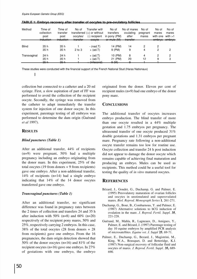
8/12/2019 Monograph Series No. 1 - 1st Meeting of European Equine Gamete Group on Reproduction
http://slidepdf.com/reader/full/monograph-series-no-1-1st-meeting-of-european-equine-gamete-group-on-reproduction 61/85
50
Equine European Gamete Group (EEG)
TABLE 1: Embryo recovery after transfer of oocytes to pre-ovulatory follicles
Method Time of Time of No of Transfer with No of No of mares No of No of No ofcollection transfer transferred (+) or without transfers ovulating pregnant mares mares
post post oocytes/ (-) recipient in pony (PM) after mares with one with >1induction induction transfer oocyte or mule (M) transfer embryo embryo
Blind 35 h 35 h 1 - (nad.T) 14 (PM) 14 2 2
35 h 35 h 2 to 3 + (ad.T) 9 (PM) 9 4 2 2
Transvaginal 24 h 24 h 1 + (ad.T) 10 (PM) 8 4 2 235 h 35 h 1 + (ad.T) 21 (PM) 20 12 9 335 h 35 h 1 + (infecond) 9 (M) 9 1 1
These studies were conducted with the financial support of the French National Stud (Haras Nationaux)
collection but connected to a catheter and a 20 mlsyringe. First, a slow aspiration of part of FF wasperformed to avoid the collection of the recipientoocyte. Secondly, the syringe was removed fromthe catheter to adapt immediately the transfersystem for injection of one donor oocyte. In thisexperiment, parentage testing of all embryos wasperformed to determine the dam origin (Guérandet al 1997).
RESULTS
Blind punctures (Table 1)
After an additional transfer, 44% of recipients(n=9) were pregnant, 50% had a multiplepregnancy including an embryo originating fromthe donor mare. In this experiment, 25% of thetotal oocytes (19 from donors + 9 from recipients)gave one embryo. After a non-additional transfer,14% of recipients (n=14) had a single embryoindicating that 14% of the 14 donor oocytestransferred gave one embryo.
Transvaginal punctures (Table 1)
After an additional transfer, no significantdifference was found in pregnancy rates betweenthe 2 times of collection and transfers 24 and 35 hafter induction with 50% (n=8) and 60% (n=20)respectively of the recipient pony mares, 50% and25%, respectively carrying 2 embryos. In this case,38% of the total oocytes (28 from donors + 28from recipients) gave one embryo. From the 16pregnancies, the dam origin detection showed that
50% of the donor oocytes (n=16) and 81% of therecipient oocytes (n=16) gave one embryo. In 27%of gestations with one embryo, the embryo
originated from the donor. Eleven per cent of recipient mules (n=9) had one embryo of the donorpony mare.
CONCLUSIONS
The additional transfer of oocytes increasesembryo production. The blind transfer of morethan one oocyte resulted in a 44% multiplegestation and 1.75 embryos per pregnancy. Theultrasound transfer of one oocyte produced 31%double gestations and 1.31 embryos per pregnantmare. Pregnancy rate following a non-additional
oocyte transfer remains too low for routine use.Oocyte collection and transfer 24 h post inductiondid not appear to damage the donor oocyte whichremains capable of achieving final maturation andproducing an embryo. Mules can be used asrecipients. This method could be a useful tool fortesting the quality of in vitro matured oocytes.
REFERENCES
Bézard, J., Goudet, G., Duchamp, G. and Palmer, E.(1995) Preovulatory maturation of ovarian folliclesand oocytes in unstimulated and superovulatedmares. Biol. Reprod . Monograph Series 1, 261-271.
Duchamp, G., Bour, B., Combarnous, Y. and Palmer, E.(1987) Alternative solutions to hCG induction of ovulation in the mare. J. Reprod. Fertil. Suppl. 35,221-228.
Guérand, M., Mahla, R., Lagneaux, D., Amigues, Y.,Palmer, E. and Bézard, J. (1997) Parentage testing of day 10 equine embryos by amplified PCR analysisof microsatellites. Equine vet. J. Suppl 25, 69-71.
Palmer, E., Duchamp, G., Bezard, J., Magistrini, M.,King, W.A., Bousquet, D. and Betteridge, K.J.
(1987) Non-surgical recovery of follicular fluid andoocytes of mares. J. Reprod. Fertil. Suppl. 35, 689-690.

8/12/2019 Monograph Series No. 1 - 1st Meeting of European Equine Gamete Group on Reproduction
http://slidepdf.com/reader/full/monograph-series-no-1-1st-meeting-of-european-equine-gamete-group-on-reproduction 62/85
51
Havemeyer Foundation Monograph Series No. 1
INTRACYTOPLASMIC SPERM INJECTION (ICSI) OFHORSE OOCYTES
X. Li, L. H. A. Morris and W. R. Allen
University of Cambridge, Department of Clinical Veterinary Medicine; Equine Fert ility Unit, Mertoun Paddocks, Woodditton Road, Newmarket, Suffolk CB8 9BH, UK
INTRODUCTION
The difficulty of inducing decondensation of thestallion spermatozoon during ICSI of horseoocytes may relate to the maturation status of theoocyte and/or the activation treatment after sperminjection. Oocytes matured in vivo becomefertilisable because of normal, synchronisedmaturation of the nucleus and ooplasm. Oocytescollected from ovarian follicles in vivo, or ovariesobtained at a slaughter house and cultured in vitro,can release the first polar body of metaphase II(MII) during 24–40 h of in vitro maturation
(Zhang et al.1989; Goudet et al.1997). However,although these MII oocytes have completednuclear maturation, it remains difficult to fertilisethem by ICSI and induce their development duringsubsequent culture, either in vitro or in vivo
(Hamano et al. 1999; Li et al.1999). This suggeststhat MII oocytes obtained from in vitro maturationmay be deficient in ‘fertilisation and/or earlydevelopment factors’, which prevents maturationof the ooplasm (Sutovsky and Schatten 1997). Inthis study, the actions of glutathione (GSH),
buthionine sulfoximine (BSO) and 4-dimethylamino-pyridine (DMAP) on ooplasmmaturation were assessed by adding themindividually to IVM medium and noting theresponse of the matured oocytes to ICSI.
MATERIALS AND METHODS
Oocyte collection and in vitro maturation
Cumulus-oocyte complexes (COCs) were
recovered from slaughter house ovaries by scrapingthe follicle wall, and aspirated from ovarian folliclesin vivo by transvaginal ultrasound guided follicle
puncture. The basic medium for in vitro maturationconsisted of TCM199 with 20% FBS, 10 mg/mlFSH, 5 mg/ml LH and 1 mg/ml oestradiol-17ß;
culture was carried out at 38°C in 5% CO2-in-air.Four groups of COCs were treated by adding: a) 15mM GSH to the basic medium for 24 h followed byfurther culture in basic medium for 12–16 h; b) 10mM BSO to the basic medium for 24 h followed byfurther culture in basic medium for 12–16 h; c) 1.9mM DMAP to the basic medium for 24 h followedby further culture in basic medium for 12–16 h; andd) continuous culture in basic medium for 36–40 h.At the end of these maturation periods the cumuluscells were separated from the COCs by pipetting the
latter in medium containing hyaluronidase. Theproportion of oocytes that had reached MII stagewas recorded before selection for ICSI.
Preparation of spermatozoa for ICSI
Frozen semen in 0.25 ml plastic straws was thawedat 35°C and washed twice with STALP solutioncontaining 0.3 mg/ml BSA by centrifugation at300 g for 5 min. The spermatozoa were then keptin EBSS+20% FBS at 38°C until used for ICSI.
ICSI and activation treatment of the oocytes
ICSI was carried out in 20 mM Hepes bufferedEBSS-20% FBS medium at 30°C. Between 5 and10 MII oocytes were placed in the ICSI medium anda spermatozoon injected into the cytoplasm of eachone. The spermatozoa were exposed to the EBSS-20% FBS containing 10% PVP during the operationand the oocytes were activated by immersion inSTALP solution containing 50-400 µM thimerosal
for 5 min before injection of the spermatozoon.After activation, they were transferred to G1.2medium for development culture.

8/12/2019 Monograph Series No. 1 - 1st Meeting of European Equine Gamete Group on Reproduction
http://slidepdf.com/reader/full/monograph-series-no-1-1st-meeting-of-european-equine-gamete-group-on-reproduction 63/85
52
Equine European Gamete Group (EEG)
TABLE 1: Nuclear maturation in oocytesderived from living mares versus abattoirovaries*
Oocytes**
No. treated MII(%) ≠MII Degenerate
Living mares 5 4 (80) 0 1Abattoirovaries† 104 48 (46) 50 6
* Basic culture medium (TCM199 with 20% v:v FBS; 10 µg/mlFSH; 5 µg/ml LH; and 1 µg/ml oestradiol-17ß) used for bothtypes of oocytes** Nuclear classification of cultured oocytes, MII = metaphaseII; ≠MII = includes the germinal vesicle (GV) and is still atmetaphase I; Degenerate = oocytes with a heterologouscytoplasm or showing vacuolisation)†The ovaries were transported from the abattoir at 12–26°C
TABLE 2: Nuclear maturation of oocytes culturedin medium added with GSH, BSO or DMAP
Treatment* Oocytes
No. treated MII(%) ≠MII Degenerate
15 mM GSG 10 4 (40) 4 210 mM BSO 16 7 (43) 5 41 mM DMAP 8 2 (25) 4 2
*After culture for 24 h in TCM199 medium with added 15 mMGSH, 10 mM BSO or 2.0 mM DMAP, the oocytes were culturedfor a further 12–16 h in basic medium
TABLE 3: Oocyte activation and spermdecondensation after injection
Culture medium
Basic +GSH +BSO +DMAP
medium
No. treated 6 8 7 2
MII 1 3 4 2
Oocyte AII-TII 3 4 3 0
FPN 2 1 0 0
Condensed 4 3 7 2
Spermatozoon
Decondensed 2 5 0 0
MPN 0 0 0 0
AII-TII = anaphase II-telophase II; FPM - female pronocleus;MPN = male pronucleus
The results were assessed, either after 18–20 hby staining the oocytes with Orcein to observeactivation and sperm transformation status, or after24–30 h by staining them with Hoechst 33342 toobserve cleavage and normality of development
CONCLUSIONS
The rate of nuclear maturation was higher in horse
oocytes derived from living mares than fromslaughter house ovaries (80% vs 46%), and it wasnot improved by adding GSH (40%), BSO (44%)or DMAP (25%) to the culture medium. AddingBSO to the maturation medium blockeddecondensation of the spermatozoon injected intothe cytoplasm, which may relate to GSHsynthesised by oocytes cultured in vitro. Further,oocytes exposed to BSO in the maturation mediumshowed a lower activation rate than the othergroups (43% vs 83%). Finally, cleavage rates in
oocytes subjected to ICSI increased when thethimerosal concentration in the activation mediumwas increased from 50 to 200 µM (17% to 75%).
REFERENCES
Goudet, G., Bezard, J., Duchamp, G., Gerard, N. andPalmer, E. (1997) Equine oocyte competence fornuclear and cytoplasmic in vitro maturation Biol.
Reprod. 57, 232-245.Hamano, K., Li, X., Qian, X., Funauchi, K., Furudate, M.
and Minato, Y. (1999) Gender preselection in cattlewith intracytoplasmically injected, flow cyto-metrically sorted sperm heads Biol. Reprod. 60,1194-1197.
Li, X., Hamano, K., Qian, X, Funauchi, K, Furudate, Mand Minato, Y. (1999) Oocyte activation andparthenogenetic development of bovine oocytesfollowing intracytoplasmic sperm injection Zygote
7, 233-237.Sutovsky, P. and Schatten, G. (1997) Depletion of
glutathione during bovine oocyte maturationreversibly blocks the decondensation of the malepronucleus and pronuclear apposition during
fertilization Biol. Reprod . 56, 1503-1512.Zhang, J.J., Boyle, M.S. and Allen, W.R. (1989) Recentstudies on in vitro fertilisation of in vitro maturedhorse oocytes Equine vet. J. Suppl. 8, 101-104.
TABLE 4: Fertilisation of oocytes following ICSIand thimerosal treatment for oocyte activation
Oocyte development Concentration of
thimerosal (µm)
50 100 200 400
No. oocytes 12 23 1 8
No. oocytes 2 (17) 11 (48) 12 (75) 0
cleaved (%)
No (%) cleaved oocytes showing
Normal fertlisation 0 7 (64) 10 (83) 0
Parthenogenesis 2 (100) 3 (27) 2 (17) 0Abnormal cleavage 0 1 (9) 0 0
* Oocytes were harvested from abattoir ovaries and cultured inTCM199 with 20% v.v FBS for 36-40 h to induce maturation.
8/12/2019 Monograph Series No. 1 - 1st Meeting of European Equine Gamete Group on Reproduction
http://slidepdf.com/reader/full/monograph-series-no-1-1st-meeting-of-european-equine-gamete-group-on-reproduction 64/85
53
Havemeyer Foundation Monograph Series No. 1
Embryo preservationChairman: T. Kat ila
SESSION VI:

8/12/2019 Monograph Series No. 1 - 1st Meeting of European Equine Gamete Group on Reproduction
http://slidepdf.com/reader/full/monograph-series-no-1-1st-meeting-of-european-equine-gamete-group-on-reproduction 65/85
54
Equine European Gamete Group (EEG)

8/12/2019 Monograph Series No. 1 - 1st Meeting of European Equine Gamete Group on Reproduction
http://slidepdf.com/reader/full/monograph-series-no-1-1st-meeting-of-european-equine-gamete-group-on-reproduction 66/85
55
Havemeyer Foundation Monograph Series No. 1
CONFOCAL MICROSCOPIC ANALYSIS OFMICROFILAMENT DAMAGE INDUCED BY CRYOPRESERVATION OF EQUINE EMBRYOS
M. Huhtinen and J. Paranko
MTT, Equines, Ypäjä, Finland; Department of Biology, Laboratory of Animal Physiology, University of
Turku, Turku, Finland
INTRODUCTION
Cleavage of the embryonic cells involves
2 cytoskeleton dependent processes. Karyokinesis,the mitotic division of the nucleus, is coordinatedby the mitotic microtubule spindle formation. Thesecond process is cytokinesis. The mechanicalforce driving the cell division is provided by thecontractile ring of actin microfilaments. If cytokinesis is disrupted, embryonic developmentceases.
Cryopreservation has been shown to have anadverse effect upon the cellular actin cortex andcytoplasmic network of human embryos. The actincortex was totally disrupted and microfilamentsshowed widespread ooplasmic dispersion. Majorvariation was seen within a cohort of embryos, butdifferential temperature sensitivity appeared to belimited to the cytoskeleton (Levy et al. 1998).
Confocal fluorescence microscopy is an idealtool for analysis of difficult biological specimenssuch as fluorescently labelled multicellularembryos. The aim of the present work was to studythe applicability of confocal microscopy for theanalysis of cryodamage in the early uterine stageequine embryos. The number of dead cells wasrecorded by DAPI-labelling under epifluorescencemicroscopy. Double labelling for nuclearchromatin and microfilamentous cytoskeleton wasanalysed by confocal microscopy.
MATERIALS AND METHODS
Thirty-seven embryos, 140 to 190 mm in diameter,were allocated into 2 different freezing protocols
as follows:1) Embryos (n=18) selected for freezing in
glycerol were washed 3 times in PBS + 10%
FCS. Glycerol and 50 mM glutamine werethen added in 5 min steps in RT to giveglycerol concentrations of 2.5%, 5%, 7.5%,
and finally 10%. The embryos were loadedinto 0.25 ml plastic straws and frozenconventionally in a programmable freezer.
2) Embryos (n=19) to be frozen in ethyleneglycol were washed 3 times in Emcare holdingmedium. They were then placed into 1.5 Methylene glycol in Emcare for 10 min in RT.Within the next 10 min, the embryos wereloaded into straws and frozen like embryos inthe glycerol group.
All embryos were thawed by holding the straws inthe air for 10 sec and then in a water bath at 30°Cfor 20 s. In the glycerol group, cryoprotectant wasremoved in 6 steps with sucrose (Huhtinen et al.1997). Half of the embryos in each treatmentgroup were stained with DAPI and transferred,half the remaining were fixed for confocalmicroscopic analysis.
Embryos (n=9) in the glycerol group werestained with DAPI (1 µ g/ml) in PBS + 10% FCSfor 15 min in RT and then washed in PBS.
Embryos (n=10) in the ethylene glycol wereplaced for 15 min into Emcare holding mediumcontaining DAPI. They were then washed in freshEmcare holding medium. Embryos were evaluatedunder epifluorescence microscopy for <10 s, andthe image was recorded on a videotape. AfterDAPI-staining, the embryos were transferred non-surgically into the uteri of recipient mares that hadovulated 4 days before transfer.
Five additional fresh embryos were fixed forconfocal microscopy to serve as controls for
cryopreservation.For confocal microscopy, embryos were fixed
in 3.7% formaldehyde for 15 min at RT. After
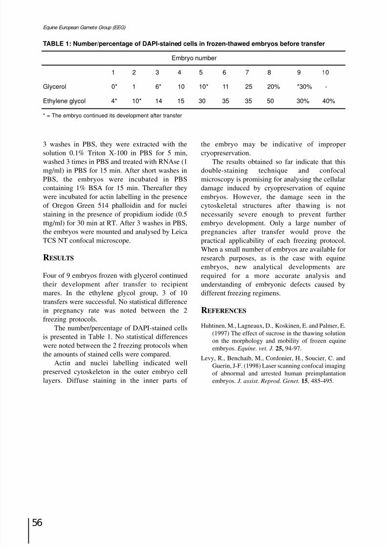
8/12/2019 Monograph Series No. 1 - 1st Meeting of European Equine Gamete Group on Reproduction
http://slidepdf.com/reader/full/monograph-series-no-1-1st-meeting-of-european-equine-gamete-group-on-reproduction 67/85
8/12/2019 Monograph Series No. 1 - 1st Meeting of European Equine Gamete Group on Reproduction
http://slidepdf.com/reader/full/monograph-series-no-1-1st-meeting-of-european-equine-gamete-group-on-reproduction 68/85
57
Havemeyer Foundation Monograph Series No. 1
Commercial developmentin equine embryo transfer
Chairman: M . S. Boyle
SESSION VII:

8/12/2019 Monograph Series No. 1 - 1st Meeting of European Equine Gamete Group on Reproduction
http://slidepdf.com/reader/full/monograph-series-no-1-1st-meeting-of-european-equine-gamete-group-on-reproduction 69/85
58
Equine European Gamete Group (EEG)
8/12/2019 Monograph Series No. 1 - 1st Meeting of European Equine Gamete Group on Reproduction
http://slidepdf.com/reader/full/monograph-series-no-1-1st-meeting-of-european-equine-gamete-group-on-reproduction 70/85
59
Havemeyer Foundation Monograph Series No. 1
COMMERCIAL DEVELOPMENT OF AN EQUINE
EMBRYO TRANSFER CENTRE IN SPAIN
G. Formiguera
CREA, Hípica El Montanyà, Seva, Spain
This paper describes the development of a centrewhich is intended to provide financial andpractical advantages for the clients, the providers
of recipient mares and ourselves.Throughout the paper, donor owners are
referred to as ‘clients’ and recipient owners as‘breeders’.
Two years ago we decided to set up the firstequine embryo transfer centre in Spain. The aimwas to develop and promote this technique and feltthat there was potentially a large market. Theintention was to provide a low-cost quality productto encourage clients to participate.
The major economic factor encountered was
that of the recipient mares. This is because:
a) The centre needed a large number of recipientmares before the breeding season started.Because the exact number of embryos totransfer is not known in advance, more maresare bought than are likely to be required.
b) Once bought, they have to be kept by thecentre until they are pregnant.
c) Once pregnant, the mares have to be bought bythe client.
Other problems included:
a) finding suitable brood mares;
b) clients being saddled with mares they have nouse for after weaning; or
c) clients sending these mares back to the centrefor further embryo transfer treatment usingtheir donor mare embryos. This leads to theadditional difficulty of synchronising thecycles of these 2 particular mares.
The centre is in Seva - 50 km north of Barcelonaand 50 km south of the Catalan Pyrenees.Barcelona provides the technological and transport
support for the programme and, in the Pyrenees,we found the solution to some of our problems.
For centuries Catalan hill-farmers have bred
horses to work on the land and as a meat supply.The predominant system is one of commongrazing land. Over the years, the mares of thesestrong, warm-blooded Hispanic-Breton horseshave been selected, above all, to be good mothers,producing a large foal every year and raising itwithout human intervention. Costs for the farmerare therefore minimal - both in terms of upkeep of the herd and raising of new livestock.
This represented a potential source of good,reliable mares which could be used as recipients in
our breeding programme. They would have beenrelatively cheap to buy, but this would still leave uswith some of the problems outlined above.Therefore, we considered a system of rentingmares from the farmers, which would drasticallyreduce the pre-breeding season costs. In addition,if we left the mares in situ during pregnancy andlactation, we would reduce upkeep costs. Theclient, in turn, would not need to buy a recipientmare.
We approached the farmers in November
1997. Given their traditional approach to farming,it was no surprise that, initially, they had no faithin the artificial nature of an embryo transferprogramme and, following the first meeting, only2 breeders requested further information. Themajor attraction of the scheme for them wasfinancial - by renting out their mares they couldearn almost double the amount that they wouldearn from breeding and selling the foals in thetraditional way.
A further meeting was held with these 2
farmers to discuss and clarify terms andconditions. The initial contract, which was verbal,included the following:

8/12/2019 Monograph Series No. 1 - 1st Meeting of European Equine Gamete Group on Reproduction
http://slidepdf.com/reader/full/monograph-series-no-1-1st-meeting-of-european-equine-gamete-group-on-reproduction 71/85
60
Equine European Gamete Group (EEG)
i) The breeder (farmer) is paid by the client(through the Centre) in 2 installments; the firstwhen the mare is 45 days pregnant and thesecond when the foal is weaned. The firstpayment is non-refundable;
ii) Breeders do no more or less than they already
do in terms of the care and upkeep of mare andfoal;
iii) The breeders cannot sell the mare or foalduring the rental period;
iv) They contact the Centre immediately if anyhealth or other problem with the mare and/orfoal is detected, such as spontaneous abortion,accident, illness or death;
v) They relinquish responsibility of care of mareor foal should there be a problem or suspected
problem;vi) The client pays all extra costs (eg vaccines);
vii) In case of the death of mare or foal, the clientis released from the second payment.
In May 1998, we had our first pregnancy - a 9-year-old show-jumper mare was inseminated withfresh semen from a young show-jumping stallion.A week later, the embryo was transferred to a 2-year-old Hispanic-Breton filly, rented by theclient.
By the end of the 1998 breeding season we had5 more pregnancies by embryo transfer. Of these,3 donor mares were show-jumpers and the other 2competed in endurance riding. Further interestfrom other owners suggested that the technique isworth exploiting. This programme shares theadvantages of other embryo transfers with theadded benefit that clients do not have to buy arecipient mare nor are they left with it afterweaning. This suits mare owners who have no landor who keep their mares in livery.
As an indication of the saving that can beachieved by renting, rather than buying the mares,in Spain it would cost about 1,000 euros to buy anacceptable brood mare. If the mare was later sold,it would be very difficult to recover the cost. Thereare also minimal upkeep costs of about 800 eurosper year. Conversely, renting an Hispanic-Breton
mare living in the Pyrenees costs, in total, about650 euros. If there is no foal or the foal has anykind of defect, the client only loses 50% of this asthe first payment to the breeder.
Since its inception, we have changed andrefined many aspects of the initial programme. For
example, the contracts are written and arebecoming more detailed and specific.
The initially unconvinced breeders are nowwanting to become involved in the programmewhich means that a ready and reliable source of good recipient mares is available. Given thatclients are not tied to using a particular recipientmare, we can match recipient and donor maresaccording to their ovulation which makes theprocess easier, quicker and cheaper. Planning forfuture breeding programmes can be flexible with
no need to balance the number of recipient anddonor mares.
In summary, the system works as follows:
i) Pyrenean farmers who wish to rent out theirmares contact the centre;
ii) The centre takes a reproductive history of eachmare and conducts a physical and reproductiveexamination;
iii) The mares are transported to the centre onemonth before the breeding season;
iv) Clients contact the centre with breedingrequirements;
v) Donor mares are transported to the centre;
vi) Recipient and donor mares are synchronised;
vii) Embryo transfer is carried out;
vii) Pregnant recipient mares stay at the centreuntil at least Day 45 of pregnancy;
viii)The recipient mares are returned to either thePyrenees or the clients’ premises;
ix) After successful birth and weaning of foals,the rental agreement comes to an end.
This programme suits the needs of all parties,relies on the abundance of good breeding mares inclose proximity to the centre and makes viable thecommercial development of an equine embryotransfer centre.
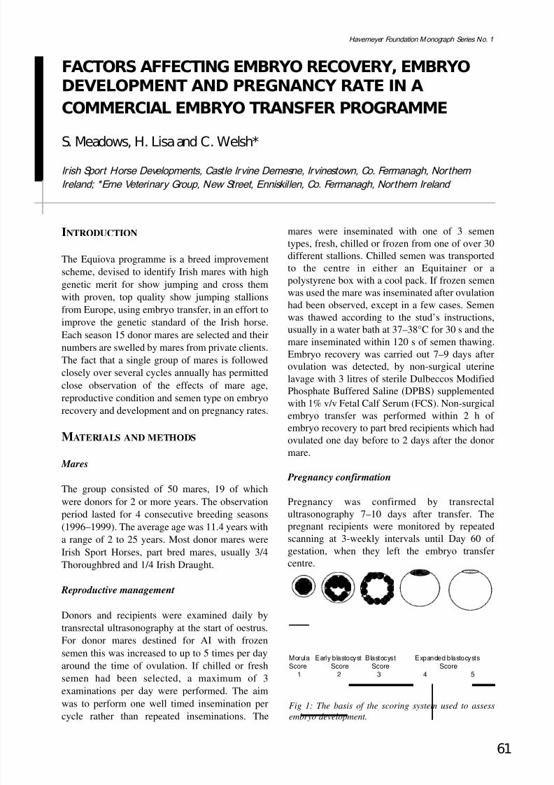
8/12/2019 Monograph Series No. 1 - 1st Meeting of European Equine Gamete Group on Reproduction
http://slidepdf.com/reader/full/monograph-series-no-1-1st-meeting-of-european-equine-gamete-group-on-reproduction 72/85
INTRODUCTION
The Equiova programme is a breed improvement
scheme, devised to identify Irish mares with highgenetic merit for show jumping and cross themwith proven, top quality show jumping stallionsfrom Europe, using embryo transfer, in an effort toimprove the genetic standard of the Irish horse.Each season 15 donor mares are selected and theirnumbers are swelled by mares from private clients.The fact that a single group of mares is followedclosely over several cycles annually has permittedclose observation of the effects of mare age,reproductive condition and semen type on embryo
recovery and development and on pregnancy rates.
MATERIALS AND METHODS
Mares
The group consisted of 50 mares, 19 of whichwere donors for 2 or more years. The observationperiod lasted for 4 consecutive breeding seasons(1996–1999). The average age was 11.4 years witha range of 2 to 25 years. Most donor mares were
Irish Sport Horses, part bred mares, usually 3/4Thoroughbred and 1/4 Irish Draught.
Reproductive management
Donors and recipients were examined daily bytransrectal ultrasonography at the start of oestrus.For donor mares destined for AI with frozensemen this was increased to up to 5 times per dayaround the time of ovulation. If chilled or freshsemen had been selected, a maximum of 3
examinations per day were performed. The aimwas to perform one well timed insemination percycle rather than repeated inseminations. The
mares were inseminated with one of 3 sementypes, fresh, chilled or frozen from one of over 30different stallions. Chilled semen was transportedto the centre in either an Equitainer or apolystyrene box with a cool pack. If frozen semenwas used the mare was inseminated after ovulationhad been observed, except in a few cases. Semenwas thawed according to the stud’s instructions,usually in a water bath at 37–38°C for 30 s and themare inseminated within 120 s of semen thawing.Embryo recovery was carried out 7–9 days afterovulation was detected, by non-surgical uterinelavage with 3 litres of sterile Dulbeccos ModifiedPhosphate Buffered Saline (DPBS) supplementedwith 1% v/v Fetal Calf Serum (FCS). Non-surgical
embryo transfer was performed within 2 h of embryo recovery to part bred recipients which hadovulated one day before to 2 days after the donormare.
Pregnancy confirmation
Pregnancy was confirmed by transrectalultrasonography 7–10 days after transfer. Thepregnant recipients were monitored by repeatedscanning at 3-weekly intervals until Day 60 of
gestation, when they left the embryo transfercentre.
61
Havemeyer Foundation Monograph Series No. 1
FACTORS AFFECTING EMBRYO RECOVERY, EMBRYODEVELOPMENT AND PREGNANCY RATE IN A
COMMERCIAL EMBRYO TRANSFER PROGRAMME
S. Meadows, H. Lisa and C. Welsh*
Irish Sport Horse Developments, Castle Irvine Demesne, Irvinestown, Co. Fermanagh, Northern Ireland; *Erne Veterinary Group, New Street, Enniskillen, Co. Fermanagh, Northern Ireland
Morula Early blastocyst Blastocyst Expanded blastocystsScore Score Score Score
1 2 3 4 5
Fig 1: The basis of the scoring system used to assess
embryo development.
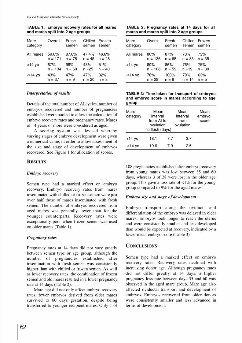
8/12/2019 Monograph Series No. 1 - 1st Meeting of European Equine Gamete Group on Reproduction
http://slidepdf.com/reader/full/monograph-series-no-1-1st-meeting-of-european-equine-gamete-group-on-reproduction 73/85
62
Equine European Gamete Group (EEG)
Interpretation of results
Details of the total number of AI cycles, number of embryos recovered and number of pregnanciesestablished were pooled to allow the calculation of
embryo recovery rates and pregnancy rates. Maresof 14 years or more were considered as aged.
A scoring system was devised wherebyvarying stages of embryo development were givena numerical value, in order to allow assessment of the size and stage of development of embryosrecovered. See Figure 1 for allocation of scores.
RESULTS
Embryo recovery
Semen type had a marked effect on embryorecovery. Embryo recovery rates from maresinseminated with chilled or frozen semen were justover half those of mares inseminated with freshsemen. The number of embryos recovered fromaged mares was generally lower than for theyounger counterparts. Recovery rates wereexceptionally poor when frozen semen was usedon older mares (Table 1).
Pregnancy rates
Pregnancy rates at 14 days did not vary greatlybetween semen type or age group, although thenumber of pregnancies established afterinsemination with fresh semen was consistentlyhigher than with chilled or frozen semen. As wellas lower recovery rates, the combination of frozensemen and old mares resulted in a lower pregnancyrate at 14 days (Table 2).
Mare age did not only affect embryo recoveryrates, fewer embryos derived from older mares
survived to 60 days gestation, despite beingtransferred to younger recipient mares. Only 1 of
108 pregnancies established after embryo recoveryfrom young mares was lost between 35 and 60days, whereas 3 of 28 were lost in the older agegroup. This gave a loss rate of <1% for the younggroup compared to 9% for the aged mares.
Embryo size and stage of development
Embryo transport along the oviducts anddifferentiation of the embryo was delayed in oldermares. Embryos took longer to reach the uterusand were consistently smaller and less developedthan would be expected at recovery, indicated by alower mean embryo score (Table 3).
CONCLUSIONS
Semen type had a marked effect on embryorecovery rates. Recovery rates declined withincreasing donor age. Although pregnancy ratesdid not differ greatly at 14 days, a higherpregnancy loss rate between days 35 and 60 wasobserved in the aged mare group. Mare age alsoaffected oviductal transport and development of embryos. Embryos recovered from older donors
were consistently smaller and less advanced interms of development.
TABLE 2: Pregnancy rates at 14 days for allmares and mares split into 2 age groups
Mare Overall Fresh Chilled Frozencategory semen semen semen
All mares 80% 87% 73% 73%
n = 136 n = 68 n = 33 n = 35<14 yo 80% 86% 76% 75%
n = 108 n = 59 n =19 n = 30
14 yo 76% 100% 70% 63%n = 28 n = 9 n = 14 n = 5
TABLE 1: Embryo recovery rates for all maresand mares split into 2 age groups
Mare Overall Fresh Chilled Frozencategory semen semen semen
All mares 59.6% 87.6% 47.4% 46.6%
n = 171 n = 78 n = 45 n = 48<14 yo 67% 98% 48% 51%
n = 134 n = 69 n =25 n = 40
14 yo 43% 47% 47% 32%n = 37 n = 9 n = 20 n = 8
TABLE 3: Time taken for transport of embryosand embryo score in mares according to agegroup
Mare Mean Mean Meancategory interval interval embryo
from AI to from scoreovulation ovulation
to flush (days)
<14 yo 18.1 7.7 3.7
14 yo 19.6 7.9 2.5
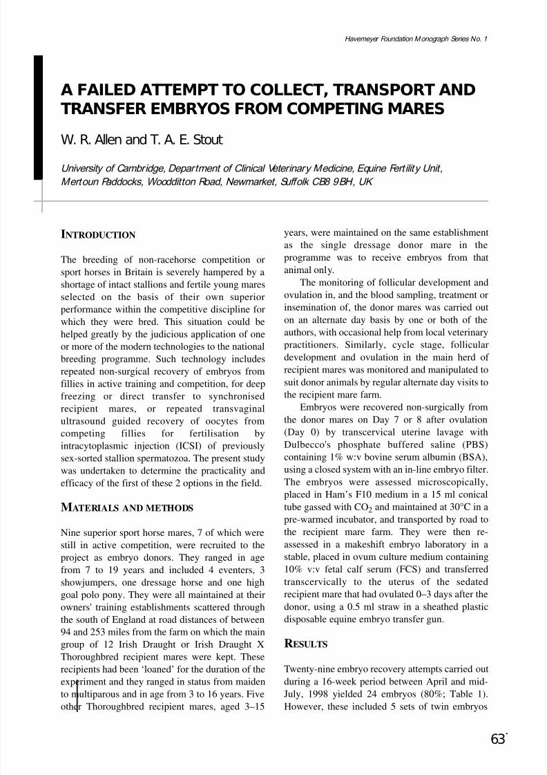
8/12/2019 Monograph Series No. 1 - 1st Meeting of European Equine Gamete Group on Reproduction
http://slidepdf.com/reader/full/monograph-series-no-1-1st-meeting-of-european-equine-gamete-group-on-reproduction 74/85
63
Havemeyer Foundation Monograph Series No. 1
A FAILED ATTEMPT TO COLLECT, TRANSPORT ANDTRANSFER EMBRYOS FROM COMPETING MARES
W. R. Allen and T. A. E. Stout
University of Cambridge, Department of Clinical Veterinary Medicine, Equine Fert ility Unit,Mertoun Paddocks, Woodditton Road, Newmarket, Suffolk CB8 9BH, UK
INTRODUCTION
The breeding of non-racehorse competition or
sport horses in Britain is severely hampered by ashortage of intact stallions and fertile young maresselected on the basis of their own superiorperformance within the competitive discipline forwhich they were bred. This situation could behelped greatly by the judicious application of oneor more of the modern technologies to the nationalbreeding programme. Such technology includesrepeated non-surgical recovery of embryos fromfillies in active training and competition, for deepfreezing or direct transfer to synchronised
recipient mares, or repeated transvaginalultrasound guided recovery of oocytes fromcompeting fillies for fertilisation byintracytoplasmic injection (ICSI) of previouslysex-sorted stallion spermatozoa. The present studywas undertaken to determine the practicality andefficacy of the first of these 2 options in the field.
MATERIALS AND METHODS
Nine superior sport horse mares, 7 of which were
still in active competition, were recruited to theproject as embryo donors. They ranged in agefrom 7 to 19 years and included 4 eventers, 3showjumpers, one dressage horse and one highgoal polo pony. They were all maintained at theirowners' training establishments scattered throughthe south of England at road distances of between94 and 253 miles from the farm on which the maingroup of 12 Irish Draught or Irish Draught XThoroughbred recipient mares were kept. Theserecipients had been ‘loaned’ for the duration of the
experiment and they ranged in status from maidento multiparous and in age from 3 to 16 years. Fiveother Thoroughbred recipient mares, aged 3–15
years, were maintained on the same establishmentas the single dressage donor mare in theprogramme was to receive embryos from that
animal only.The monitoring of follicular development andovulation in, and the blood sampling, treatment orinsemination of, the donor mares was carried outon an alternate day basis by one or both of theauthors, with occasional help from local veterinarypractitioners. Similarly, cycle stage, folliculardevelopment and ovulation in the main herd of recipient mares was monitored and manipulated tosuit donor animals by regular alternate day visits tothe recipient mare farm.
Embryos were recovered non-surgically fromthe donor mares on Day 7 or 8 after ovulation(Day 0) by transcervical uterine lavage withDulbecco's phosphate buffered saline (PBS)containing 1% w:v bovine serum albumin (BSA),using a closed system with an in-line embryo filter.The embryos were assessed microscopically,placed in Ham’s F10 medium in a 15 ml conicaltube gassed with CO2 and maintained at 30°C in apre-warmed incubator, and transported by road tothe recipient mare farm. They were then re-
assessed in a makeshift embryo laboratory in astable, placed in ovum culture medium containing10% v:v fetal calf serum (FCS) and transferredtranscervically to the uterus of the sedatedrecipient mare that had ovulated 0–3 days after thedonor, using a 0.5 ml straw in a sheathed plasticdisposable equine embryo transfer gun.
RESULTS
Twenty-nine embryo recovery attempts carried out
during a 16-week period between April and mid-July, 1998 yielded 24 embryos (80%; Table 1).However, these included 5 sets of twin embryos
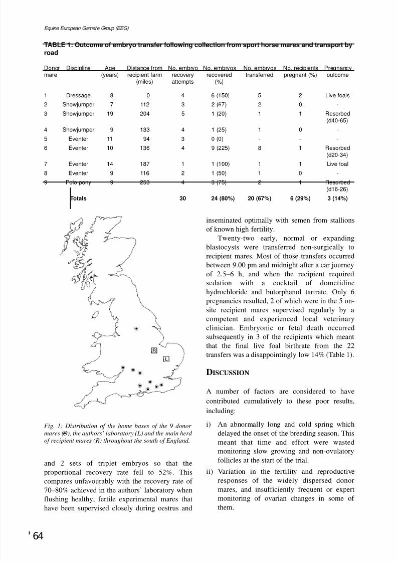
8/12/2019 Monograph Series No. 1 - 1st Meeting of European Equine Gamete Group on Reproduction
http://slidepdf.com/reader/full/monograph-series-no-1-1st-meeting-of-european-equine-gamete-group-on-reproduction 75/85
64
Equine European Gamete Group (EEG)
and 2 sets of triplet embryos so that theproportional recovery rate fell to 52%. Thiscompares unfavourably with the recovery rate of
70–80% achieved in the authors’ laboratory whenflushing healthy, fertile experimental mares thathave been supervised closely during oestrus and
inseminated optimally with semen from stallionsof known high fertility.
Twenty-two early, normal or expandingblastocysts were transferred non-surgically torecipient mares. Most of those transfers occurredbetween 9.00 pm and midnight after a car journeyof 2.5–6 h, and when the recipient requiredsedation with a cocktail of dometidine
hydrochloride and butorphanol tartrate. Only 6pregnancies resulted, 2 of which were in the 5 on-site recipient mares supervised regularly by acompetent and experienced local veterinaryclinician. Embryonic or fetal death occurredsubsequently in 3 of the recipients which meantthat the final live foal birthrate from the 22transfers was a disappointingly low 14% (Table 1).
DISCUSSION
A number of factors are considered to havecontributed cumulatively to these poor results,including:
i) An abnormally long and cold spring whichdelayed the onset of the breeding season. Thismeant that time and effort were wastedmonitoring slow growing and non-ovulatoryfollicles at the start of the trial.
ii) Variation in the fertility and reproductiveresponses of the widely dispersed donor
mares, and insufficiently frequent or expertmonitoring of ovarian changes in some of them.
TABLE 1: Outcome of embryo transfer following collection from sport horse mares and transport by
road
Donor Discipline Age Distance from No. embryo No. embryos No. embryos No. recipients Pregnancymare (years) recipient farm recovery recovered transferred pregnant (%) outcome
(miles) attempts (%)
1 Dressage 8 0 4 6 (150) 5 2 Live foals2 Showjumper 7 112 3 2 (67) 2 0 -
3 Showjumper 19 204 5 1 (20) 1 1 Resorbed(d40-65)
4 Showjumper 9 133 4 1 (25) 1 0 -
5 Eventer 11 94 3 0 (0) - - -
6 Eventer 10 136 4 9 (225) 8 1 Resorbed
(d20-34)
7 Eventer 14 187 1 1 (100) 1 1 Live foal
8 Eventer 9 116 2 1 (50) 1 0 -
9 Polo pony 9 253 4 3 (75) 2 1 Resorbed(d16-26)
Totals 30 24 (80%) 20 (67%) 6 (29%) 3 (14%)
Fig. 1: Distribution of the home bases of the 9 donor
mares (•O), the authors’ laboratory (L) and the main herd of recipient mares (R) throughout the south of England.
8/12/2019 Monograph Series No. 1 - 1st Meeting of European Equine Gamete Group on Reproduction
http://slidepdf.com/reader/full/monograph-series-no-1-1st-meeting-of-european-equine-gamete-group-on-reproduction 76/85
65
Havemeyer Foundation Monograph Series No. 1
iii) Difficulties relating to the fertility and/orhandling of the semen used to inseminate thedonor mares.
iv) Selection of the recipient mares for size, ratherthan temperament and reproductiveparameters, so that many of the multiparousanimals were difficult to handle and hadtortuous and difficult cervices for non-surgicaltransfer.
v) Transfer facilities that were not ideal. Mosttransfers were performed at night to sedatedmares and most of the embryos had beentransported at 30°C for 3–6 h before transfer.
Although the results of the study weredisappointing, some valuable lessons werelearned:
i) Donor mares should be temporarily resident ata well organised equine reproduction facility
around the time of insemination and ovulation.ii) Stallions should be selected on the basis of
their fertility, as well as their geneticexcellence and/or athletic prowess.
iii) Recipient mares with any suspicion of subfertility, behaviour problems or difficultcervices must be eliminated at the outset.

8/12/2019 Monograph Series No. 1 - 1st Meeting of European Equine Gamete Group on Reproduction
http://slidepdf.com/reader/full/monograph-series-no-1-1st-meeting-of-european-equine-gamete-group-on-reproduction 77/85
66
Equine European Gamete Group (EEG)

8/12/2019 Monograph Series No. 1 - 1st Meeting of European Equine Gamete Group on Reproduction
http://slidepdf.com/reader/full/monograph-series-no-1-1st-meeting-of-european-equine-gamete-group-on-reproduction 78/85
67
Havemeyer Foundation Monograph Series No. 1
WORKSHOP SUMMARY

8/12/2019 Monograph Series No. 1 - 1st Meeting of European Equine Gamete Group on Reproduction
http://slidepdf.com/reader/full/monograph-series-no-1-1st-meeting-of-european-equine-gamete-group-on-reproduction 79/85
68
Equine European Gamete Group (EEG)
8/12/2019 Monograph Series No. 1 - 1st Meeting of European Equine Gamete Group on Reproduction
http://slidepdf.com/reader/full/monograph-series-no-1-1st-meeting-of-european-equine-gamete-group-on-reproduction 80/85
69
Havemeyer Foundation Monograph Series No. 1
WORKSHOP SUMMARY
R. H. F. Hunter
Faculty of Veterinary Medicine, University of Cambridge, Madingley Road, Cambridge CB3 0ES, UK
First, if I may, I am going to take this opportunityon behalf of all the speakers to reiterate our thanksto the organisers of the meeting, not only to
Professors Twink Allen and Marian Tischner forinitiating and focusing our days of discussion, butto all of the Tischner family and to their colleaguesfor the special arrangements and wonderfulhospitality here in Lopuszna. Particular thanks areextended to Jan Wade for being so effective andhelpful an organiser and coordinator behind thescenes, and for producing the excellent record of matters so far – the book containing theprogramme, the list of participants and a completerecord of abstracts.
Now to the speakers themselves: throughoutthe Workshop, we have had well-prepared,consistently good lectures with splendid modernslides, and everyone has been agreeably concise –a point worth emphasising. No-one has droned onand on, thank goodness. So, if and when thequestion arises as to whether another Workshopwould be appropriate in due course, the responsemust surely be ‘yes’ if it meets the high standardsof the present one. Perhaps you remember theinitial letter from Twink along the lines that “… he
and Marian Tischner being the autocrats that theyare, have decided to arrange …”. Well, if thesestimulating days together here in Lopuszna havebeen the fruits of autocracy, then long livebenevolent dictatorships. We need more of them.
Now for 10 minutes or so of scientificcomment without mentioning names or indeedspecifically covering every topic or presentation.My own impression is that the Workshop has dealtvery largely with applied research and I am not sure
just how much of the underlying science has been
clarified – or remains to be clarified – in yourchosen species of animal. Research with anapplication in view is doubtless of special appeal to
the Havemeyer Foundation, but the techniques andtherapies to be used in the future, say 10 years fromnow, must stem in part from fundamental studies.
Reproductive physiology is still a relatively youngdiscipline, so wisdom would caution not too heavyan emphasis on applied research with the prospectof immediate application.
Of course, the opening paper from Utrecht diddeal with fundamental changes – changes in themembranes of stallion spermatozoa as an approachto gaining a more complete understanding of theprocess of capacitation. Certainly excellent workwhich started the Workshop off at an impressiveintellectual level. However, one of the problems
remains to define which of the 2 currently mootedmodels of capacitation applies to stallionspermatozoa. Is there a continuous availability of sub-populations of capacitated sperm in the femaletract after mating, ie. a series of curves of ripeningand decay, or do stallion spermatozoa respond tothe ovarian control model, only achieving fullcapacitation at the time of ovulation? I suggest thatwe still do not have an answer, but would proposethat the second of the 2 models operates in thisspecies with a prolonged pre-ovulatory interval.
A step back from sperm cell interactions withthe female tract concerns their interactions withthe epididymal duct. How much is known aboutsperm surface modifications within the epididymisand the transfer of macromolecules from theepithelium in particular portions of the duct?Indeed, how much do we know about androgenbinding protein and its target tissues in thestallion? Almost certainly not nearly as much as inthe ram, bull, boar, rabbit or rat. If correct, onefeels strongly that such fundamental studies need
to proceed simultaneously with attempts to exploitsuspensions of epididymal spermatozoa obtainedat autopsy or during elective castration.
8/12/2019 Monograph Series No. 1 - 1st Meeting of European Equine Gamete Group on Reproduction
http://slidepdf.com/reader/full/monograph-series-no-1-1st-meeting-of-european-equine-gamete-group-on-reproduction 81/85
70
Equine European Gamete Group (EEG)
Keeping on the subject of sperm cells a littlelonger, does everyone here working withcryopreserved stallion spermatozoa recall thatstudies with frozen-thawed boar spermatozoaindicated that sperm cells from many – perhaps amajority – of boars under test were extremely
vulnerable to the uterine environment whenthawed and extended in insemination medium?Expressed in another way, introduction of frozen-thawed boar spermatozoa into the uterine lumendamaged a high proportion of such gametes, andthe greater the extent of exposure to the uterinetissues (eg intra-cervical insemination), the greaterthe resultant damage and the lower the conceptionrate. By contrast, insemination at the utero-tubal
junction or just into the Fallopian tube isthmus wasfound to be highly beneficial. If we accept for a
moment that this may also be the case for frozen-thawed stallion spermatozoa (and bearing in mindthat both species have intra-uterine accumulationof semen at mating), then endoscopic techniquesof deep insemination close to or onto the utero-tubal junction, already shown to be valuable inanother context, assume an even greaterimportance. Moreover, although technicallychallenging, a means of insemination into theisthmus would be a very specific advance towardsexploiting low numbers of cryopreserved stallion
spermatozoa. Coincident with such appliedstudies, further work on the relationships betweenstructural modifications in preserved spermatozoaand subsequent fertility of the inseminate isdefinitely to be encouraged.
A few more words concerning spermatozoa.There is preliminary evidence from cattle that re-suspending the spermatozoa of low fertility bullsin seminal plasma derived from high fertility bullsmay act to improve the conception rates of inseminations from poor bulls. These are early
days: this work is recent and the results areconfounded from several points of view, but it doesseem important enough for practitioners of equineinsemination to be alert to future possibilities. Thecellular physiology underlying a seemingly crudeapproach may have a rational explanation:substitution of beneficial male duct proteins onand in the sperm plasma membrane may facilitateboth sperm survival in the female tract andsubsequent sperm-egg interactions. Perhaps amore timely acrosome reaction or a more
coordinated vesiculation of plasma and outeracrosomal membranes is a consequence of thisapproach.
Finally, the interactions between stress, seasonof the year and sperm quality came to light inseveral presentations and made a strong impressionon this reviewer. Without any exaggeration, suchinteractions seem of immense importance and yetwill be difficult to unravel without extensive multi-
centre collaborations. Here, the experiments of theHungarian-French teams offered one example of aninspired step forward, focusing especially on aputative role for perturbed glucocorticoid secretionin syndromes of stress leading to low fertility.Presumably a variety of mechanisms of action willneed to be invoked, and the scope for furtherstudies is therefore considerable.
Turning now to ovarian follicles and tooocytes, and as one with a research background inother large domestic species, I was struck forcibly
by the limited number of oocytes that can beobtained by means of follicular aspiration,puncture or maceration techniques, frequentlyonly 4–5 oocytes per ovary or per mare. This isdramatically different from the situation in sheep,cows or pigs. Why should it be so? You must havereflected on the problem. Assuming that theexplanation is not primarily due to the initialcomplement of oogonia and oocytes in the fetalgonad, nor to massive waves of follicular atresiabefore puberty, then there would seem to be scope
here too for imaginative new approaches. Notleast, these could involve a combination of enzymatic treatment and mild sonication of ovarian tissues. If the problem resides primarily inthe nature of attachment of the cumulus-oocytecomplex to the wall of the Graafian follicle, thensuch approaches might be beneficial. If theproblem is associated with the number of developing vesicular follicles, then there may beroom for a form of controlled and predictableovarian stimulation. The special morphology of
the equine ovary does not appear as anoverwhelming block to progress in this field. So letus not be discouraged but rather more inventive.
Related points to which some members of theWorkshop may have devoted serious thought are: (1)whether morphologically good Graafian folliclescan shed defective oocytes at ovulation and (2)whether presumptively poor follicles can yieldviable oocytes? Do the latest staining techniquesreally enable us to make such vital distinctions?Remaining with the theme of oocytes and especially
of ovulated eggs, do we actually know what theviable lifespan of the mare’s egg is in the Fallopiantube? Indeed, is this concept meaningful in the sense
8/12/2019 Monograph Series No. 1 - 1st Meeting of European Equine Gamete Group on Reproduction
http://slidepdf.com/reader/full/monograph-series-no-1-1st-meeting-of-european-equine-gamete-group-on-reproduction 82/85
71
Havemeyer Foundation Monograph Series No. 1
of a viable lifespan of say 6–8 hours, or 10–12 hoursafter ovulation, essentially constant within andbetween animals and season, or is there substantialvariation? Bearing in mind that we are dealing withonly one egg liberated per cycle in contrast tomillions of spermatozoa introduced, the egg is
clearly more vulnerable from a quantitative point of view and possibly also from a structural angle withits large cytoplasmic component. In any event, itseems a research topic of considerable significanceto clarify this lifespan and to see if it is open tomodification. This point is made even though thepopulation of spermatozoa should ideally beestablished in the Fallopian tube(s) before ovulation– and would be so under biological conditions.However, procedures of artificial insemination mayalter these relationships and the shortened lifespan of
frozen-thawed spermatozoa might dictate a peri- orpost ovulatory insemination. In brief, gametelifespans cannot be taken for granted and coulddefinitely benefit from a further bout of well focusedresearch in meaningful numbers of animals.
As to the zona pellucida, we heard of excellentstudies on this envelope around the mare’s oocyte.In view of the recent advances in understandingglycoproteins of the zona, especially those referredto as ZP3 and ZP2, this was extremely topicalwork. Even so, one must question what the
phenomenon of so-called zona hardening actuallytells us about oocyte or embryo quality and askwhat is, approximately, the maximum number of spermatozoa that can bind to the zona pellucidabefore completion of hardening. Do we know?With regard to the second of these questions, onehas the impression that it is rather low and also thatthe incidence of polyspermy – multiple spermpenetration of the vitellus – is again low in thehorse egg. But impressions are not enough.Careful experiments are needed that yield answersin vivo, not conveniently in a culture dish.
Moving from gametes to embryos, and inrelation to the detailed reports on transplantationtrials, presumably the only rigorous test of viability is generation of a full-term fetus. Of course, useful indicators of potential viability maybe developed but it is not easy to suppose that wecan ever, or would ever wish to, avoid the ultimatetest of competence. Predictive tests of embryonicviability may be refined in due course but theiraccuracy will almost certainly be for populations
(ie large numbers) of embryos rather than for anindividual embryo about to be transplanted.Nonetheless, the reports on transplantation studies
and indeed on neonatal loss have been invaluableto all members of the Workshop.
Various new techniques were described, andthese seem to have potential in more ways thanhighlighted. The work on protocols for in vivo
oocyte recovery or in vivo replacement or de novo
introduction of oocytes into follicles are excitingapproaches in their own right. In reality, their scopemay extend from studies of oocyte and follicularatresia through aspects of the actual process of ovulation to developing a model for the early stagesof ectopic pregnancy. And the microsamplingapproach to the composition of follicular fluid,somewhat reminiscent of the Göttingen techniquefor microsampling secretions of the corpus luteum,although a far more difficult surgical preparation,should not be limited to withdrawal of
microdroplets of follicular fluid. It would alsoenable accurate local testing of a number of gonadotrophin, prostaglandin and peptide growthfactor treatments, and might enable some dissectionof the interplay between thecal and granulosa cellsynthesis and secretion of steroid molecules. Sohere is scope for the very best sort of fundamentalresearch, and it is much to be encouraged.
Intra-cytoplasmic sperm injection (ICSI),confocal microscopy and multiple embryo transfercould all be commented on in similar detail. Theyare approaches that offer much mileage!Undoubtedly, we will be hearing stimulatingreports on their extensive application at futuremeetings, so I shall discipline myself not tospeculate on their potential.
There is a suspicion that I have spoken foralmost too long. If so, my sincere apologies andsimilarly so should anyone feel that I have notmade sufficient reference to the work of theirlaboratory. There remains the hope that in some
manner, perhaps almost cryptic, it has beenembraced by the above remarks. As noted at theoutset, everyone has done good work, everyonehas participated actively and in an open spirit, andtogether we have had a stimulating and superbWorkshop. We must hope for another. Such aninvestment would be more than justified bothintellectually and in a practical context of equinebreeding programmes and procedures. My ownthanks go to all members of the Workshop and our
joint thanks go to the Havemeyer Foundation for
their imaginative and generous support. R.H.F. Hunter
September 1999

8/12/2019 Monograph Series No. 1 - 1st Meeting of European Equine Gamete Group on Reproduction
http://slidepdf.com/reader/full/monograph-series-no-1-1st-meeting-of-european-equine-gamete-group-on-reproduction 83/85
72
Equine European Gamete Group (EEG)
8/12/2019 Monograph Series No. 1 - 1st Meeting of European Equine Gamete Group on Reproduction
http://slidepdf.com/reader/full/monograph-series-no-1-1st-meeting-of-european-equine-gamete-group-on-reproduction 84/85

8/12/2019 Monograph Series No. 1 - 1st Meeting of European Equine Gamete Group on Reproduction
http://slidepdf.com/reader/full/monograph-series-no-1-1st-meeting-of-european-equine-gamete-group-on-reproduction 85/85
Equine European Gamete Group (EEG)