Embed Size (px)
Citation preview
J Clin Pathol 1988;41:753-758
Monoclonal antibody (Y1/82A) with specificitytowards peripheral blood monocytes and tissuemacrophagesF R DAVEY,* JACQUELINE L CORDELL, WENDY N ERBER,KAREN A F PULFORD, K C GATTER, D Y MASON
From the *Department ofPathology, SUNY Health Science Center, Syracuse, New York, USA, and theNuffield Department ofPathology, John Radeliffe Hospital, Oxford
SUMMARY A new monoclonal antibody, Y1/82A, was raised against phytohaemagglutinin activatedperipheral blood mononuclear cells. Using an immunohistochemical technique it was shown that YI/82A reacts against peripheral blood and bone marrow monocytes and resident macrophages fromessentially all human tissues. Y1/82A bound to determinants present in leukaemic cells from patientswith acute myelomonocytic leukaemia and acute monocytic leukaemia, but not to neoplastic cellsfrom patients with malignant lymphoproliferative disorders or malignant epithelial tumours. YI /82Afailed to react with other cell types, with the exception of osteoclasts and megakaryocytes. Analysis byWestern blotting showed that the antigen detected by antibody Y1/82A was associated withintracellular granules in macrophages.Monoclonal antibody Y1/82A may be useful in the diagnosis of monocytic leukaemias and
histiocytic neoplasms and in the identification of macrophages in tissues from various inflammatoryand neoplastic conditions.
The monocyte-macrophage system is regarded as anetwork of specialised phagocytic cells which arewidely scattered throughout the body.' These cellsinclude peripheral blood monocytes, Kupffer cells ofthe liver, microglial cells of the brain, macrophages oflymph node and spleen, and fixed tissue macrophagesin various other human tissues. Cells of the monocyte-macrophage system, however, do not include non-lymphoid mononuclear cells known as dendritic cells.2There is evidence that peripheral blood monocytesoriginate from bone marrow precursor cells3 and thattissue macrophages are derived from circulating mon-ocytes.4 Bone marrow precursor cells therefore serveas the cells of origin for peripheral blood monocytesand tissue macrophages.
Cells of the monocyte-macrophage system areheterogeneous in terms of their morphology, cyto-chemistry, function and surface determinants.'56 Aspromonocytes mature into monocytes and then intotissue macrophages they increase in size. The ratio ofthe nucleus to that of the cytoplasm decreases and thenumber of lysosomes and IgG receptors increases.Functionally, mature macrophages have greater
Accepted for publication 15 February 1988
ability to undergo phagocytosis and lymphocyteinteraction than the less mature monocyte.' Further-more, populations of resident macrophages in lym-phoid tissues appear to differ from each other: residentmacrophages thought to be accessory cells of the T cellimmune response can be differentiated from accessorycells of the B cell immune response on the basis ofdifferent cellular determinants.5
Various morphological, cytochemical, and im-munological techniques have been used to identifycells of the monocyte-macrophage system.7 Amongthese techniques immunocytochemical labelling withmonoclonal antibodies is one of the most sensitive andspecific for identifying monocytes and tissue macro-phages in non-neoplastic and neoplastic disorders.'0Numerous monoclonal antibodies have been raisedagainst cells of the monocyte-macrophage system"'5but most of these react only with subpopulations ofcells in the monocyte-macrophage system. Further-more, many monoclonal antibodies raised againstcells of the monocyte-macrophage system also reactwith determinants found on cells belonging to othercellular systems.'"
In this respect we describe our experience with a newmonoclonal antibody (Yl /28A) raised against phyto-
753
on 13 July 2018 by guest. Protected by copyright.
http://jcp.bmj.com
/J C
lin Pathol: first published as 10.1136/jcp.41.7.753 on 1 July 1988. D
ownloaded from

754hemagglutinin activated peripheral blood mononu-clear cells. Using an immunohistochemical technique,we showed that this monoclonal antibody reacts withperipheral blood monocytes and a range of tissuemacrophages from patients with non-neoplastic andneoplastic disorders.
Material and methods
Blocks of fresh tissue were obtained from thehistopathology department of the John RadcliffeHospital at necropsy or at the time of surgicalresection and were immediately snap frozen in liquidnitrogen and stored at - 70°C until use. Tissuessampled from patients without neoplastic disordersincluded three lymph nodes with reactive hyperplasia,two sections of liver, and one section each of thymus,tonsil, spleen, liver, colon, kidney, heart, brain,striated muscle, thyroid, lung and skin. Tissues sam-pled from patients with neoplastic diseases includedeight cases of non-Hodgkin's lymphoma, five cases ofHodgkin's lymphoma, six cases of squamous cellcarcinoma of the bronchus, and one case of adenocar-cinoma of the breast.
Cryostat sections (6 pm) were prepared from thesefrozen tissues and stored (wrapped in aluminium foil)at - 20°C until staining.
Samples ofperipheral blood and bone marrow wereobtained from patients attending the haematologydepartment of the John Radcliffe Hospital. Thesamples were drawn into ethylene diamine tetra-aceticacid or heparin anticoagulant and either push films orcytocentrifuge films ofmononuclear cells separated onTriosil-Ficoll were prepared.'6 Ten peripheral bloodand four bone marrow samples were obtained fromsubjects without a haematological malignancy, andperipheral blood smears, as well as one of bonemarrow film were received from those with eitheracute myelomonocytic leukaemia or acute monocyticleukaemia.
PREPARATION OF ANTIGENPeripheral blood from a healthy subject was collectedin heparinised tubes. The mononuclear cells wereseparated from other blood elements by centrifu-gation on Triosil-Ficoll (Lymphoprep-Nyegaard andCompany, Oslo). Mononuclear cells were removedfrom the interphase, washed, and resuspended inRPMI-1640 tissue culture medium at a concentrationof I x 106/Ul. Mononuclear cells were then incubatedwith a 1/200 dilution (one phial reconstituted with5 ml ofdistilled water) ofphytohaemagglutinin (PHA)(Flow Laboratories). After three daysPHA stimulatedblasts were washed once in phosphate buffered saline(PBS) and then resuspended in PBS at appropriateconcentrations.
PRODUCTION OF MONOCLONAL ANTIBODY
A BALB/c mouse was immunised with an
Davey, Cordell, Erber, Pulford, Gatter, Mason
intraperitoneal injection of I x 106 PHA stimulatedblasts. This was followed by an intraperitoneal injec-tion of 2 x 10⁶ and 5 x 10⁶ PHA stimulated blastsafter intervals of 10 days and four weeks, respectively.Three days after an intravenous injection of 1 5 x 10⁶PHA stimulated blasts spleen cells from the mouse (1 8x 10') were fused with 1-8 x I07 cells from the NS-1myeloma cell line.'7 After 14 days the cultures wereinspected for growth and the supematant fluid wasscreened for specific reactivity on cryostat sections oftonsil using the APAAP immunoalkaline phosphatasetechnique.'8 The clone Y1/28A, which selectivelystained germinal centre macrophages, was thenisolated by standard cloning techniques and estab-lished in long term tissue culture. In all experimentsantibody Y1/82A was used in the form of spent tissueculture supernatant.
IMMUNOCHEMICAL LABELLINGFrozen tissue sections (6 pm) and blood and bonemarrow films were labelled using the APAAP im-munoalkaline phosphatase technique.'8 '9 Slides werefixed before APAAP staining in acetone (sections andcytocentrifuged preparations) or acetone:methanol.'
IMMUNOFLUORESCENCE ANALYSISPeripheral blood monocytes, granulocytes, and lym-phocytes were analysed for expression of the Y1/82Adeterminant by indirect immunofluorescence usingflow cytometry. Aliquots of normal peripheral bloodmononuclear cells were incubated for 30 minutes withantibody Y1/82A at doubling dilutions in PBS from1/5 through 1/80. Lymphocytes, monocytes, andgranulocytes were analysed separately by gating onforward and right (900) angle light scatter.
BIOCHEMICAL ANALYSIS OF THE DETERMINANTRECOGNISED BY Y l /82ANormal human spleen tissue was solubilised in asolution of 10% Brij (2 parts Brij 99:1 part 96, SigmaChemical Co) as described by Dalchau and Fabre.20The lysate was then centrifuged at 70 000 x g for 30minutes at 4°C. Samples of both the supernatant andthe pellet were dotted on to nitrocellulose paper andallowed to dry. Aliquots of the supematant were thenrun under both reducing and non-reducing conditionsin a 10% polyacrylamide slab gel in the presence ofsodium dodecyl sulphate.2' Proteins were blottedovernight on to nitrocellulose membranes aspreviously described.' After blocking with 3% bovineserum albumin (BSA) both the dot blots and theWestern blots were immunostained using the APAAPtechnique.'8 The molecular weights of the immuno-stained bands were established by comparison withmolecular weight standards stained with fast green.23
on 13 July 2018 by guest. Protected by copyright.
http://jcp.bmj.com
/J C
lin Pathol: first published as 10.1136/jcp.41.7.753 on 1 July 1988. D
ownloaded from
Monocyte monoclonal antibodyResults
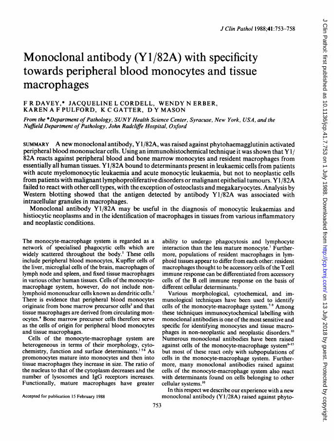
SAMPLES FROM PATIENTS WITHOUT NEOPLASTICDISORDERSExamination of lymphoid tissue showed a positivereaction in many types of tissue macrophages (table 1).In lymph nodes this included macrophages in thesubcapsular and medullary sinuses, macrophages ingerminal centres, mantle zones, and interfollicularareas (fig 1). Lymphocytes, endothelial cells, anddendritic reticulum cells were unreactive. In thethymus, cortical and medullary macrophages werepositive, but cortical and medullary thymocytes andthymic epithelial cells were negative. There was anintense staining reaction of the macrophages of thesplenic cords (fig 2). A few macrophages were presentin germinal centres as well as around small arteries andcapillaries.
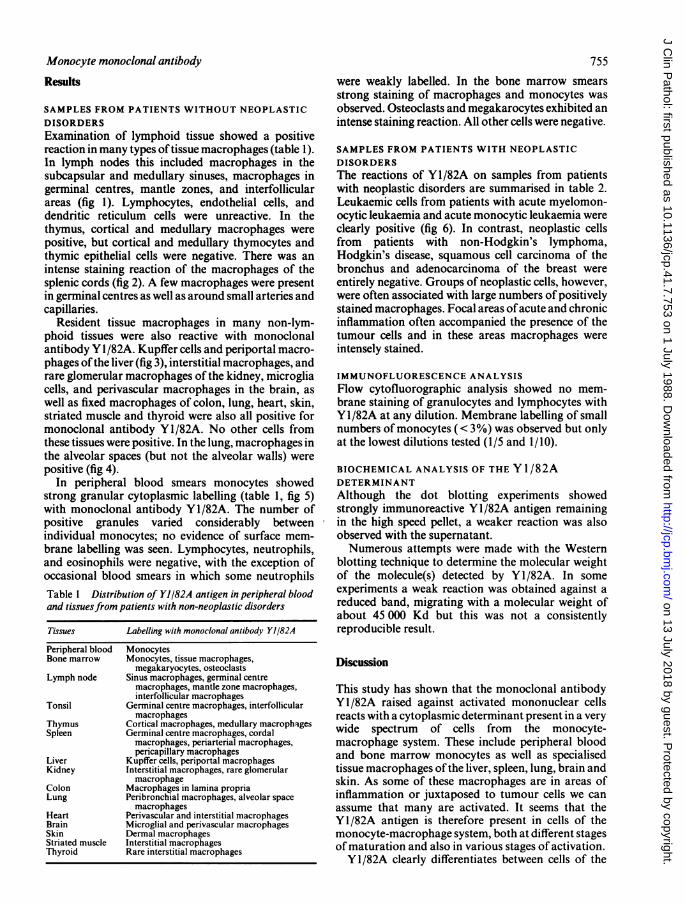
Resident tissue macrophages in many non-lym-phoid tissues were also reactive with monoclonalantibody Y1/82A. Kupffer cells and periportal macro-phages of the liver (fig 3), interstitial macrophages, andrare glomerular macrophages of the kidney, microgliacells, and perivascular macrophages in the brain, aswell as fixed macrophages of colon, lung, heart, skin,striated muscle and thyroid were also all positive formonoclonal antibody Yl/82A. No other cells fromthese tissues were positive. In the lung, macrophages inthe alveolar spaces (but not the alveolar walls) werepositive (fig 4).
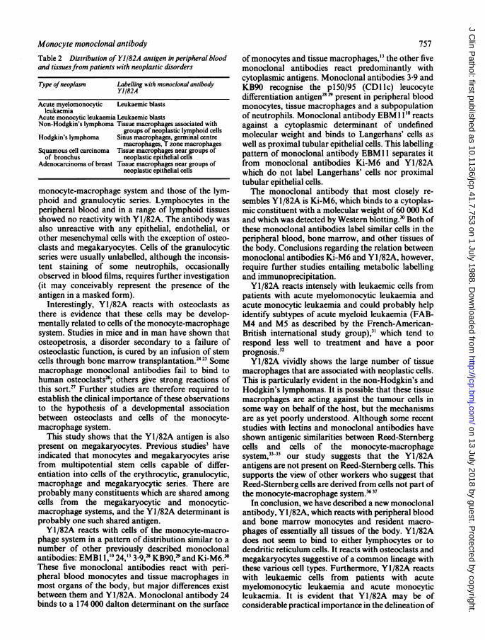
In peripheral blood smears monocytes showedstrong granular cytoplasmic labelling (table 1, fig 5)with monoclonal antibody Yl/82A. The number ofpositive granules varied considerably betweenindividual monocytes; no evidence of surface mem-brane labelling was seen. Lymphocytes, neutrophils,and eosinophils were negative, with the exception ofoccasional blood smears in which some neutrophilsTable 1 Distribution of Y1/82A antigen in peripheral bloodand tissuesfrom patients with non-neoplastic disorders
Tissues Labelling with monoclonal antibody Y1/82A
Peripheral blood MonocytesBone marrow Monocytes, tissue macrophages,
megakaryocytes, osteoclastsLymph node Sinus macrophages, germinal centre
macrophages, mantle zone macrophages,interfollicular macrophages
Tonsil Germinal centre macrophages, interfollicularmacrophages
Thymus Cortical macrophages, medullary macrophagesSpleen Germinal centre macrophages, cordal
macrophages, periarterial macrophages,pericapillary macrophages
Liver Kupffer cells, periportal macrophagesKidney Interstitial macrophages, rare glomerular
macrophageColon Macrophages in lamina propriaLung Peribronchial macrophages, alveolar space
macrophagesHeart Perivascular and interstitial macrophagesBrain Microglial and perivascular macrophagesSkin Dermal macrophagesStriated muscle Interstitial macrophagesThyroid Rare interstitial macrophages
755were weakly labelled. In the bone marrow smearsstrong staining of macrophages and monocytes wasobserved. Osteoclasts and megakarocytes exhibited anintense staining reaction. All other cells were negative.
SAMPLES FROM PATIENTS WITH NEOPLASTICDISORDERSThe reactions of Y1/82A on samples from patientswith neoplastic disorders are summarised in table 2.Leukaemic cells from patients with acute myelomon-ocytic leukaemia and acute monocytic leukaemia wereclearly positive (fig 6). In contrast, neoplastic cellsfrom patients with non-Hodgkin's lymphoma,Hodgkin's disease, squamous cell carcinoma of thebronchus and adenocarcinoma of the breast wereentirely negative. Groups of neoplastic cells, however,were often associated with large numbers of positivelystained macrophages. Focal areas ofacute and chronicinflammation often accompanied the presence of thetumour cells and in these areas macrophages wereintensely stained.
IMMUNOFLUORESCENCE ANALYSISFlow cytofluorographic analysis showed no mem-brane staining of granulocytes and lymphocytes withY1/82A at any dilution. Membrane labelling of smallnumbers of monocytes ( < 3%) was observed but onlyat the lowest dilutions tested (1/5 and 1/10).
BIOCHEMICAL ANALYSIS OF THE Y1/82ADETERMINANTAlthough the dot blotting experiments showedstrongly immunoreactive Y1/82A antigen remainingin the high speed pellet, a weaker reaction was alsoobserved with the supernatant.Numerous attempts were made with the Western
blotting technique to determine the molecular weightof the molecule(s) detected by Y1/82A. In someexperiments a weak reaction was obtained against areduced band, migrating with a molecular weight ofabout 45 000 Kd but this was not a consistentlyreproducible result.
Discussion
This study has shown that the monoclonal antibodyY1/82A raised against activated mononuclear cellsreacts with a cytoplasmic determinant present in a verywide spectrum of cells from the monocyte-macrophage system. These include peripheral bloodand bone marrow monocytes as well as specialisedtissue macrophages ofthe liver, spleen, lung, brain andskin. As some of these macrophages are in areas ofinflammation or juxtaposed to tumour cells we canassume that many are activated. It seems that theY1/82A antigen is therefore present in cells of themonocyte-macrophage system, both at different stagesof maturation and also in various stages of activation.Y1/82A clearly differentiates between cells of the
on 13 July 2018 by guest. Protected by copyright.
http://jcp.bmj.com
/J C
lin Pathol: first published as 10.1136/jcp.41.7.753 on 1 July 1988. D
ownloaded from

Davey, Cordell, Erber, Pulford, Gatter, Mason
I *
/
Ia
iS
4 )
t
< 3.K~~
d~~~~. ~~A
Fig 1 APAAPstaining ofreactive lymph node (cryostat section) with antibody Yl/82A. (a) Low power view shows B cellfollicles (arrowed) containing Yl/82A positive germinal centre macrophages. (b) Higher power view ofB cellfollicle. Notescattered Yl/82A positive cells in extrafollicular tissue (T cell area). (c) High power view showing Y1/82A positive germinalcentre macrophages.
Fig 2 A.PAAP staining ofhuman spleen (cryostat section) showing strong YJ/82A positive staining ofmacrophages in spleniccords (contrasting with unstained splenic sinusoids).
Fig 3 APAAP staining ofhuman liver (cryostat section) with antibody Y1/82A showing intense staining ofKupffer cells, butnot ofhepatocytes.
Fig 4 APAAP staining ofhuman lung (cryostat section) showing Yl/82A positive alveolar macrophages.
Fig 5 APAAP stainedperipheral bloodsmear showing two Y1/82A positive monocytes, contrasting with unstainedneutrophils and red cells.
Fig 6 APAAP staining ofperipheral bloodfrom patient with acute myelomonocytic leukaemia showing strong positivelabelling ofleukaemic cells with antibody Y1182A.
756
4w,
;'2
on 13 July 2018 by guest. Protected by copyright.
http://jcp.bmj.com
/J C
lin Pathol: first published as 10.1136/jcp.41.7.753 on 1 July 1988. D
ownloaded from
Monocyte monoclonal antibodyTable 2 Distribution of Yl/82A antigen in peripheral bloodand tissuesfrom patients with neoplastic disorders
Type ofneoplasm Labelling with monoclonal antibodyYl/82A
Acute myelomonocytic Leukaemic blastsleukaemia
Acute monocytic leukaemia Leukaemic blastsNon-Hodgkin's lymphoma Tissue macrophages associated with
groups of neoplastic lymphoid cellsHodgkin's lymphoma Sinus macrophages, germinal centre
macrophages, T zone macrophagesSquamous cell carcinoma Tissue macrophages near groups of
of bronchus neoplastic epithelial cellsAdenocarcinoma of breast Tissue macrophages near groups of
neoplastic epithelial cells
monocyte-macrophage system and those of the lym-phoid and granulocytic series. Lymphocytes in theperipheral blood and in a range of lymphoid tissuesshowed no reactivity with Yl/82A. The antibody wasalso unreactive with any epithelial, endothelial, orother mesenchymal cells with the exception of osteo-clasts and megakaryocytes. Cells of the granulocyticseries were usually unlabelled, although the inconsis-tent staining of some neutrophils, occasionallyobserved in blood films, requires further investigation(it may conceivably represent the presence of theantigen in a masked form).
Interestingly, Y1/82A reacts with osteoclasts asthere is evidence that these cells may be develop-mentally related to cells of the monocyte-macrophagesystem. Studies in mice and in man have shown thatosteopetrosis, a disorder secondary to a failure ofosteoclastic function, is cured by an infusion of stemcells through bone marrow transplantation.2425 Somemacrophage monoclonal antibodies fail to bind tohuman osteoclasts26; others give strong reactions ofthis sort.2" Further studies are therefore required toestablish the clinical importance of these observationsto the hypothesis of a developmental associationbetween osteoclasts and cells of the monocyte-macrophage system.
This study shows that the Y1/82A antigen is alsopresent on megakaryocytes. Previous studies3 haveindicated that monocytes and megakaryocytes arisefrom multipotential stem cells capable of differ-entiation into cells of the erythrocytic, granulocytic,macrophage and megakaryocytic series. There areprobably many constituents which are shared amongcells from the megakaryocytic and monocytic-macrophage systems, and the Yl/82A determinant isprobably one such shared antigen.Y1/82A reacts with cells of the monocyte-macro-
phage system in a pattern of distribution similar to anumber of other previously described monoclonalantibodies: EMB1 1,'° 24,'3 3.9,28 KB90,28 and Ki-M6.3°These five monoclonal antibodies react with peri-pheral blood monocytes and tissue macrophages inmost organs of the body, but major differences existbetween them and Y1/82A. Monoclonal antibody 24binds to a 174 000 dalton determinant on the surface
757
of monocytes and tissue macrophages,'3 the other fivemonoclonal antibodies react predominantly withcytoplasmic antigens. Monoclonal antibodies 3 9 andKB90 recognise the p150/95 (CD1 1c) leucocytedifferentiation antigen28"' present in peripheral bloodmonocytes, tissue macrophages and a subpopulationof neutrophils. Monoclonal antibody EBM 110' reactsagainst a cytoplasmic determinant of undefinedmolecular weight and binds to Langerhans' cells aswell as proximal tubular epithelial cells. This labellingpattern of monoclonal antibody EBM 11 separates itfrom monoclonal antibodies Ki-M6 and Y1/82Awhich do not label Langerhans' cells nor proximaltubular epithelial cells.The monoclonal antibody that most closely re-
sembles Y1/82A is Ki-M6, which binds to a cytoplas-mic constituent with a molecular weight of 60 000 Kdand which was detected by Western blotting.3' Both ofthese monoclonal antibodies label similar cells in theperipheral blood, bone marrow, and other tissues ofthe body. Conclusions regarding the relation betweenmonoclonal antibodies Ki-M6 and Y1/82A, however,require further studies entailing metabolic labellingand immunoprecipitation.Y1/82A reacts intensely with leukaemic cells from
patients with acute myelomonocytic leukaemia andacute monocytic leukaemia and could probably helpidentify subtypes of acute myeloid leukaemia (FAB-M4 and M5 as described by the French-American-British international study group),3' which tend torespond less well to treatment and have a poorprognosis."Y1/82A vividly shows the large number of tissue
macrophages that are associated with neoplastic cells.This is particularly evident in the non-Hodgkin's andHodgkin's lymphomas. It is possible that these tissuemacrophages are acting against the tumour cells insome way on behalf of the host, but the mechanismsare as yet poorly understood. Although some recentstudies with lectins and monoclonal antibodies haveshown antigenic similarities between Reed-Sternbergcells and cells of the monocyte-macrophagesystem,33-35 our study suggests that the Yl/82Aantigens are not present on Reed-Sternberg cells. Thissupports the view of other workers who suggest thatReed-Sternberg cells are derived from cells not part ofthe monocyte-macrophage system.?637
In conclusion, we have described a new monoclonalantibody, Y1/82A, which reacts with peripheral bloodand bone marrow monocytes and resident macro-phages of essentially all tissues of the body. Y1/82Adoes not seem to bind to either lymphocytes or todendritic reticulum cells. It reacts with osteoclasts andmegakaryocytes suggestive of a common lineage withthese various cell types. Furthermore, Y1/82A reactswith leukaemic cells from patients with acutemyelomonocytic leukaemia and acute monocyticleukaemia. It is evident that Y1/82A may be ofconsiderable practical importance in the delineation of
on 13 July 2018 by guest. Protected by copyright.
http://jcp.bmj.com
/J C
lin Pathol: first published as 10.1136/jcp.41.7.753 on 1 July 1988. D
ownloaded from

758 Davey, Cordell, Erber, Pulford, Gatter, Masonmonocytic leukaemias and histiocytic malignancies.
This work was supported by grants from theLeukaemia Research Fund. KCG is a WellcomeSenior Research Fellow in clinical service.
References
I Cline MJ, Lehrer RI, Territo MC, Golde DW. Monocytes andmacrophages: Functions and diseases. Ann Intern Med1978;88:78-88.
2 Steinman RM, Nussenzweig MC. Dendritic cells: features andfunctions. Immunol Rev 1980;53:127-47.
3 Quesenberry P, Levitt L. Hematopoietic stem cells. N Engi J Med1979;301:755-60,819-23,868-72.
4 Gale RP, Sparkes RS, Golde DW. Bone marrow origin of hepaticmacrophages (Kupffer cells) in humans. Science 1978;201:937-8.
5 Radzun HJ, Parwaresch MR. Differential immunohistochemicalresolution of the human mononuclear phagocyte system. CellImmunol 1983;82:174-83.
6 Zwadlo G, Brocker EB, von Bassewitz DB, Feige U, Sorg C. Amonoclonal antibody to a differentiation antigen present onmature human macrophages and absent from monocytes.J Immunol 1985;134:1487-92.
7 Bainton DF, Golde DW. Differentiation of macrophages fromnormal human bone marrow in liquid culture. Electron micro-scopy and cytochemistry. J Clin Invest 1978;61:1555-69.
8 Hancock WW, Zola H, Atkins RC. Antigenic heterogeneity ofhuman mononuclear phagocytes: Immunohistologic analysisusing mononuclear antibodies. Blood 1983;62:1271-9.
9 Yam LT, Li CY, Crosby WH. Cytochemical identification ofmonocytes and granulocytes. Am J Clin Pathol 1971 ;55:283-90.
10 Franklin WA, Mason DY, Pulford K, et al. Immunohistologicalanalysis ofhuman mononuclear phagocytes and dendritic cellsby using monoclonal antibodies. Lab Invest 1986;54:322-35.
11 Buckley PJ, Dickson SA, Walker WS. Tissue distribution ofmacrophage cell surface antigens in human spleen. Lab Invest1984;S0:8A.
12 Dimitriu-Bona A, Burmester GR, Waters SJ, Winchester RJ.Human mononuclear phagocytes differentiation antigens. I.Patterns of antigenic expression on the surface of humanmonocytes and macrophages defined by monoclonalantibodies. J Immunol 1983;130:145-52.
13 Hogg N, Selvendran Y. An anti-human monocyte/macrophagemonoclonal antibody, reacting most strongly with macro-phages in lymphoid tissue. Cell Immunol 1985;92:247-53.
14 Knowles DM II, Tolijain B, Marboe D, D'Agati V, Grimes M,Chess L. Monoclonal antihuman monocyte antibodies OKM 1and OKM5 possess distinctive tissue distributions includingdifferential reactivity with vascular endothelium. J Immunol1984;132:2170-3.
15 RaffHV, Picker LJ, Stobo JD. Macrophage heterogeneity in man.A subpopulation of HLA-DR-bearing macrophages requiredfor antigen-induced T cell activation contains stimulators forautologous-reactive T cells. J Exp Med 1980;152:581-93.
16 Erber WN, Mynheer LC, Mason DY. APAAP labelling of bloodand bone marrow samples for phenotyping leukaemia. Lancet1986;i:761-5.
17 Mason DY, Cordell JL, Pulford KAF. Production ofmonoclonalantibodies for immunocytochemical use. In: Bullock GR,Petrusz P, eds. Techniques in immunocytochemistry. London:Academic Press, 1983:175.
18 Cordell JL, Falini B, Erber WN, et al. Immunoenzymatic labelingof monoclonal antibodies using immune complexes of alkalinephosphatase and monoclonal anti-alkaline phosphatase(APAAP complexes). J Histochem Cytochem 1984;32:219-29.
19 Mason DY, Erber WN, Falini B, Stein H, Gatter KC. Immuno-enzymatic labelling ofhaematological samples with monoclonal
antibodies. In: Beverley PCL, ed. Methods in hematology:monoclonal antibodies. Edinburgh: Churchill Livingstone,1986:145-81.
20 Dalchau R, Fabre JW. The purification of antigens and otherstudies with monoclonal antibody affinity columns: thecomplementary new dimension of monoclonal antibodies. In:McMichael AJ, Fabre JW, eds. Monoclonal antibodies in clinicalmedicine. London: Academic Press, 1982:519-56.
21 Laemli UK. Clearage ofstructural proteins during the assembly ofthe head of bacteriophage T4. Nature 1970;227:680-5.
22 Towbin H, Staehelin T, Gordon J. Electrophoretic transfer ofproteins from polyacrylamide gels to nitrocellulose sheets:Procedure and some applications. Proc Natil Acad Sci USA1979;76:4350-4.
23 Woodcock-Mitchell J, Eichner R, Nelson WG, Sun TT. Immuno-localization of keratin polypeptides in human epidermis usingmonoclonal antibodies. J Cell Biol 1982;95:580-8.
24 Coccia PF, Krivit W, Cervenka J, et al. Successful bone marrowtransplantation for infantile malignant osteopetrosis. N Engl JMed 1980;302:701-8.
25 Walker DG. Control ofbone marrow resorption by hematopoietictissue. The induction and reversal of congenital osteopetrosis inmice through use ofbone marrow and splenic transplants. J ExpMed 1975;142:651-63.
26 Horton MA, Lewis D, McNulty K, Pringle JAS, Chambers TJ.Monoclonal antibodies to osteoclastomas (giant cell bonetumors): defiinition of osteoclast-specific cellular antigens.Cancer Res 1985;45:5663-9.
27 Athanasou NA, Heryet A, Quinn J, Gatter KC, Mason DY,McGee JOD. Osteoclasts contain macrophage and mega-karyocyte antigens. J Pathol 1986;150:239-46.
28 Hogg N, Takas L, Palmer DG, Selvendran Y, Allan C. Thep150,95 molecule is a marker of human mononuclearphagocytes: Comparison with expression of class II molecules.Eur J Immunol 1986;16:240-8.
29 MacDonald SM, Pulford K, Falini B, Micklem K, Mason DY. Amonoclonal antibody recognizing the p150/95 leucocytedifferentiation antigen. Immunol 1986;59:427-3 1.
30 Parwaresch MR, Radzun HJ, Kreipe H, Hansmann ML, Barth J.Monocyte/macrophage-reactive monoclonal antibody Ki-M6recognizes an intracytoplasmic antigen. Am J Pathol1986;124:141-51.
31 Bennett JM, Catovsky D, Daniel MT, et al. Proposals for theclassification of the acute leukaemias. Br J Haematol1976;33:451-8.
32 Griffin JD, Davis R, Nelson DA, et al. Use of surface markeranalysis to predict outcome of adult acute myeloblasticleukemia. Blood 1986;68:1232-41.
33 Hsu SM, Jaffe ES. Leu Ml and peanut agglutinin stain in theneoplastic cells of Hodgkin's disease. Am J Clin Pathol1984;82:29-32.
34 Hsu SM, Yang K, Jaffe ES. Phenotypic expression of Hodgkin'sand Reed-Sternberg cells in Hodgkin's disease. Am J Pathol1985;118:209-17.
35 Strauchen JA. Lectin receptors as markers of lymphoma cells. II.Reed-Stemnberg cells share lectin-binding properties ofmonocyte macrophages. Am J Pathol 1984;116:370-6.
36 Stein H, Mason DY, Gerdes J, et al. The expression of Hodgkin'sdisease associated antigen Ki-l in reactive and neoplasticlymphoid tissue: Evidence that Reed-Sternberg cells andhistiocytic malignancies are derived from activated lymphoidcells and histiocytic malignancies are derived from activatedlymphoid cells. Blood 1985;66:848-58.
37 Strauchen JA, Dimitriu-Bona A. Immunopathology ofHodgkin'sdisease. Characterization of Reed-Stemnberg cells with mono-clonal antibodies. Am J Pathol 1986;123:293-300.
Requests for reprints to: Dr F R Davey, Division of ClinicalPathology, SUNY Health Science Center, 750 East AdamsStreet, Syracuse, New York, 13210, USA.
on 13 July 2018 by guest. Protected by copyright.
http://jcp.bmj.com
/J C
lin Pathol: first published as 10.1136/jcp.41.7.753 on 1 July 1988. D
ownloaded from


![THE INFLUENCE OF THE INFRASOUND ON THE … · 3 Infrasound and immunological properties of rats blood 247 present [6]. The granulocytes and monocytes have a special capability to](https://img.dokumen.tips/doc/110x75/5f92cfcba1f94e71fb5c6fcd/the-influence-of-the-infrasound-on-the-3-infrasound-and-immunological-properties.jpg)