Embed Size (px)
Citation preview

Genitourin Med 1985;61:1-6
Monoclonal antibodies to Treponema pallidum:recognition of a major polypeptide antigenROBERT W THORNBURG, JANICE MORRISON-PLUMMER, ANDJOEL B BASEMANFrom the Department of Microbiology, The University of Texas Health Science Center, San Antonio,Texas, USA
SUMMARY Hybridomas secreting monoclonal antibodies that reacted with a 45 000 daltonsurface polypeptide and major immunogen of Tpallidum were produced. This polypeptide wasalso found in Tpertenue but not in T hyodysenteriae or Tphagedenis biotype Reiter.
Introduction
Although recent reports have described biochemicaland immunological properties of Treponemapallidum, 1-25 the causative agent of venereal syphilisand non-venereal endemic syphilis or bejel, muchremains unknown about the biology of the disease.The humoral immune response appears to play animportant part in the pathophysiology of syphiliticinfections, and specific surface treponemal proteinshave been identified as major immunogens.3 4 6-8To further characterise treponemal antigens, we
used hybridoma technology to produce antibodies toTpallidum. We describe monoclonal antibodies thatbind to a 45 000 dalton surface protein of thetreponeme.
Materials and methods
ORGANISMSTpallidum (Nichols strain) and Tpertenue (Gautierstrain) were provided by the Center for DiseaseControl, Atlanta, Georgia and were maintained byroutine passage in rabbits. Adult New Zealand whiterabbits (3-4 kg), obtained locally, were inoculatedintratesticularly with 50 x 106 treponemes andtreated with 6 mg/kg cortisone acetate (Merck Sharpeand Dohm, West Point, Pennsylvania, USA) on days3-7 after infection. Peak orchitis occurred at days10-14 for Tpallidum and days 24-30 for Tpertenue.Stock solutions were prepared from infected rabbittestes as described previously.2 Tphagedenis biotypeReiter was grown in Spirolate broth supplemented
Address for reprints: Dr J B Baseman, Department of Microbiology,University of Texas Health Science Center, 7703 Floyd Curl Drive,San Antonio, Texas 78284, USA
Accepted for publication 3 March 1984
with 0'6% glucose and 10% heat inactivated rabbitserum.26 T hyodysenteriae was a gift of Dr HankHarris, University of Iowa, Iowa, and was grown inanaerobically reduced media.27 (T hyodysenteriaecultures produce large quantities of hydrogen gas.Care was therefore taken to ventilate these closedcultures to prevent the build up of explosivepressures.)
RADIOLABELLING OF ORGANISMST pallidum harvested from rabbit testes at peakorchitis were concentrated by centrifugation(12 000 x g for 20 minutes) in sterile tubes.Treponemes were resuspended in .2 ml of minimalessential medium without methionine (Gibco, GrandIsland, New York, USA) but containing 10%odialysed fetal calf serum and 0'5-1 mCi35S-methionine and were incubated aerobicallyovernight. Then the treponemes were washed threetimes in 0'05 mol/l TRIS and 0'2 mol/l sodiumchloride (pH 8'0), pelleted, and frozen at -20°Cuntil used.
IMMUNISATIONTo generate activated mouse spleen cells, freshlyextracted and purified T pallidum (300 x 106Tpallidum in 0 5 ml saline) were emulsified 1/1(vol/vol) in Freund complete adjuvant, and injected-intramuscularly (0' 1 ml), subcutaneously (0' 1 ml),or intraperitoneally (0 3 ml) into BALB/c femalemice (3-6 weeks old). On days 7 and 21 the mice weresimilarly boosted with treponemes enmulsified inFreund incomplete adjuvant.
CELL HYBRIDISATIONMonoclonal antibodies were produced by modifi-cation of the procedure of Oi and Herzenberg.28Spleen cells from immunised mice and SP2/0-Agl4
on 5 June 2018 by guest. Protected by copyright.
http://sti.bmj.com
/G
enitourin Med: first published as 10.1136/sti.61.1.1 on 1 F
ebruary 1985. Dow
nloaded from
R W Thornburg, J Morrison-Plummer, and J B Baseman
BALB/c myeloma cells were washed separately inDulbecco's modified Eagle's medium (DME) free ofserum (MA Bioproducts, Walkersville, Maryland,USA) and then combined in a ratio of 7:1. The basicmethod of cell fusion was as described by us earlier.29A volume of 50,u of the fused cell preparation wasaliquoted into each well of 96 well microtitre tissueculture plates (Bellco, Vineland, New Jersey, USA)containing0 1 x 10⁶ normal BALB/c spleen feedercells in 50 pl of DME-HAT (containing 0 2 mmol/lhypoxanthine, 0 7 mmol/l aminopterin, and 32mmol/l thymidine)30 plus glycine. One day afterfusion, 100 M1 of DME-HAT plus glycine were addedto each well. Plates were incubated at 37°C in 7%carbon dioxide for seven to 10 days before beingscreened by an enzyme linked immunosorbent assay(ELISA). Selected hybrid cells were cloned bylimiting dilution in 96 well microtitre tissue cultureplates containing 0v 1 x 10⁶ BALB/c spleen feedercells/well. Clones were expanded for analysis byELISA.
ATTACHMENT OF ANTIGEN TOPOLYVINYLCHLORIDE MICROTITRE PLATESIndividual pellets of treponemes (T pallidum, Tpertenue, T hyodysenteriae, and T phagedenis bio-type Reiter) were resuspended in coating buffer3l andsonicated using six 15 second bursts with 45 secondintermittent incubations on ice (Sonifier CellDisruptor Model W140D, Heat Systems-Ultrasonics,Plainview, New York, USA). The protein concentra-tion of each treponemal preparation was measuredby the Lowry method.32 Aliquots of 100 MI ofI 0 mg/ml treponemal protein in coating bufferwere distributed into each well of Immunlon 2 strips(Dynatech, Alexandria, Virginia, USA) and incubatedovernight at 4°C. The plates were then filled with 1%bovine serum albumin in phosphate buffered saline(BSA/PBS) and incubated for two hours at 37°C.
Purified T pallidum proteins were isolated frompolyacrylamide gels33 and diluted to I mg/ml incoating buffer,3' aliquoted (50 ng/well) intoImmulon 2 strips, and incubated overnight at 4°C. Ifnot used immediately, plates were washed once withPBS, filled with 1% BSA/PBS, and stored at- 200C.
ENZYME LINKED IMMUNOSORBENT ASSAY(ELISA)A modified ELISA as previously reported by US29 33was used to detect antibodies to sonicated and puri-fied treponemal proteins.
SERUM SAMPLESNormal and immune mouse serum was collected byretro-orbital bleeding of BALB/c mice before andafter inoculating them with Tpallidum. Normal and
syphilitic rabbit serum was obtained before or 47 daysafter infection with Tpallidum. Normal and syphilitichuman serum was provided by Dr J Jorgenson atMedical Center Hospital, San Antonio, Texas. Allserum samples were stored at - 20°C before use.
SODIUM DODECYL SULPHATE (SDS)POLYACRYLAMIDE GEL ELECTROPHORESISTreponemal proteins were electrophoresed on 7* 5%polyacrylamide slab gels in the discontinuous TRIS-glycine system described by Laemmli.34After onedimensional electrophoresis, gels were fixed inmethanol, acetic acid, and water in a ratio of45:45:10 and either processed for fluorography withpreparations labelled with 35S-methionine35 or driedimmediately and exposed to x ray film for sampleslabelled with radioactive iodine (1251).
IODINATION OF PROTEIN AUsing the lactoperoxidase labelling method ofMarchalonis et aP6 100 mg protein A was labelledwith 1 mCi radioactive iodine for 30 minutes to give1251-protein A. After radiolabelling, iodinatedprotein A was purified from unreactive radioactiveiodine and denatured protein A by passage over a1 ml column of IgG-Sepharose, which was preparedby coupling IgG purified by protein Achromatography to Sepharose 4B activated bycyanogen bromide.37 Fractions of 1 ml of the PBSeluate were collected until the column backgroundwas reduced to less than 5000 counts per minute(cpm)/10,ul aliquot. The bound 125I-protein A waseluted with pH 2 8 glycine hydrochloride andseparated from the low molecular weight glycine bysubsequent passage over a 0 8 x 25 cm column ofBiogel P-10 in PBS.
WESTERN BLOTSAliquots (20-40g protein) of unlabelled treponemeswere solubilised in 0-063 mol/l TRIS containing 2%sodium dodecyl sulphate (SDS), 2% fl-mercapto-ethanol, and 10% glycerol, and were electrophoresedon a 7 5% polyacrylamide slab gel. The proteinswere then electrophoretically transferred for 16 hoursat 380 mA to nitrocellulose paper (BioRad,Richmond, California, USA) using the technique ofTowbin et al.38 One lane was stained with amidoblack to show whether both high and low molecularweight proteins were successfully transferred to thenitrocellulose sheets. Non-specific protein bindingsites were blocked by incubation in 3% BSA/PBS forsix hours. The nitrocellulose strips were thenincubated overnight with constant rocking in a 1/50dilution of antiserum in 3% BSA/PBS or withmonoclonal ascites purified antibody. The stripswere rinsed in either PBS plus 0'01%7o SDS or PBS
2
on 5 June 2018 by guest. Protected by copyright.
http://sti.bmj.com
/G
enitourin Med: first published as 10.1136/sti.61.1.1 on 1 F
ebruary 1985. Dow
nloaded from
Monoclonal antibodies to Treponema pallidum: recognition of a major polypeptide antigen
alone and incubated with 37o BSA/PBS containing'25I-protein A. The strips were finally rinsed asbefore, dried, and exposed to x ray film (KodakXR5) with an enhancing screen.39
PURIFICATION OF MONOCLONAL ANTIBODIESON PROTEIN A SEPHAROSEMonoclonal antibodies were generated as ascites inpristine primed BALB/c mice.28 The ascites fluid wasdiluted to 20% in 0 01 mol/l phosphate buffer (pH7 2) and passed over a 15 x 8 cm proteinA-Sepharose column. The column was eluted with0 01 mol/l phosphate buffer (pH 7 - 2) until the back-ground absorbance (A280) was less than 0 02,followed by elution with 0 1 mol/l glycine (pH 2 8).The fractions containing protein as determined byA280 were pooled and dialysed in a two step process,first against 1000 volumes of pH 5 5 citratephosphate buffer (0 05 mol/l citric acid, 0 05 mol/ldisodium hydrogen phosphate, and 0 145 mol/lsodium chloride; pH adjusted with 6 mol/l sodiumhydroxide) and finally against 1000 volumes of PBS.The protein concentrations were measured, and themonoclonal IgG was diluted to 1 g/l with PBS.
Results
Hybridoma culture supernatants were screened forreactivity to whole T pallidum organisms fixed inethanol. Of about 1200 hybridomas tested, 54 clonesbound selectively to T pallidum. To analyse thespecificity of the binding sites of these monoclonalantibodies, culture supernatants were screened byELISA using gel eluted T pallidum proteins.33Treponemal proteins corresponding to specificregions of SDS-polyacrylamide gels were purifiedand pooled as follows: high molecular weight(HMW) (proteins over 100 000 daltons); P5,6
TABLE I Reactivity of normal and immune mouse serumand monoclonal antibodies 1IF2, 13F3, and 23C9 to Tpallidum proteins eluted from sodium dodecyl sulphatepolyacrylamide gels
Mean (SD)* reactivity to T pallidum proteins
Dilution HMW (OD) P5,6 (OD) P1,2,3,4 (OD)
NMS 1:50 0 049 (0 006) 0 059 (0 002) 0-061 (0 004)IMS 1:50 0 072 (0-015) 0 375 (0-024) 0-355 (0-115)
1:100 0 033 (0 004) 0-357 (0-021) 0-237 (0-097)IIF2 1:100 0 005 (0-034) 0-138 (0 047) 0-034 (0-019)13F3 1:100 0 003 (0-005) 0 260 (0 075) 0-001 (0 004)23C9 1:100 0 002 (0 003) 0 244 (0 033) 0 030 (0 024)
* Mean (SD) of quadruplicate samples. PBS control00017.HMW = heavy molecular weight (>100 000 daltons); P1 (89 500daltons), P2 (29 500 daltons), P3 (25 500 daltons), P4 (20 000daltons), P5 (59 000 daltons), P6 (42 500 daltons).For meanings of other abbreviations see table II.
(59 000 and 42 500); and P1,2,3,4 (89 500, 29 500,25 500, 20 000 daltons). These protein designations(P1-P6) are consistent with previous reports.3 4 6- Asshown in table I, immune mouse serum (IMS)obtained just before killing for the hybridoma fusionreacted predominantly with protein pools P1-4 andP5,6; there was low reactivity towards the highmolecular weight antigen group. Normal mouseserum (NMS) showed poor reactivity to all antigengroups examined.
A tt..:.::. ......... :
.. : :...:
4...
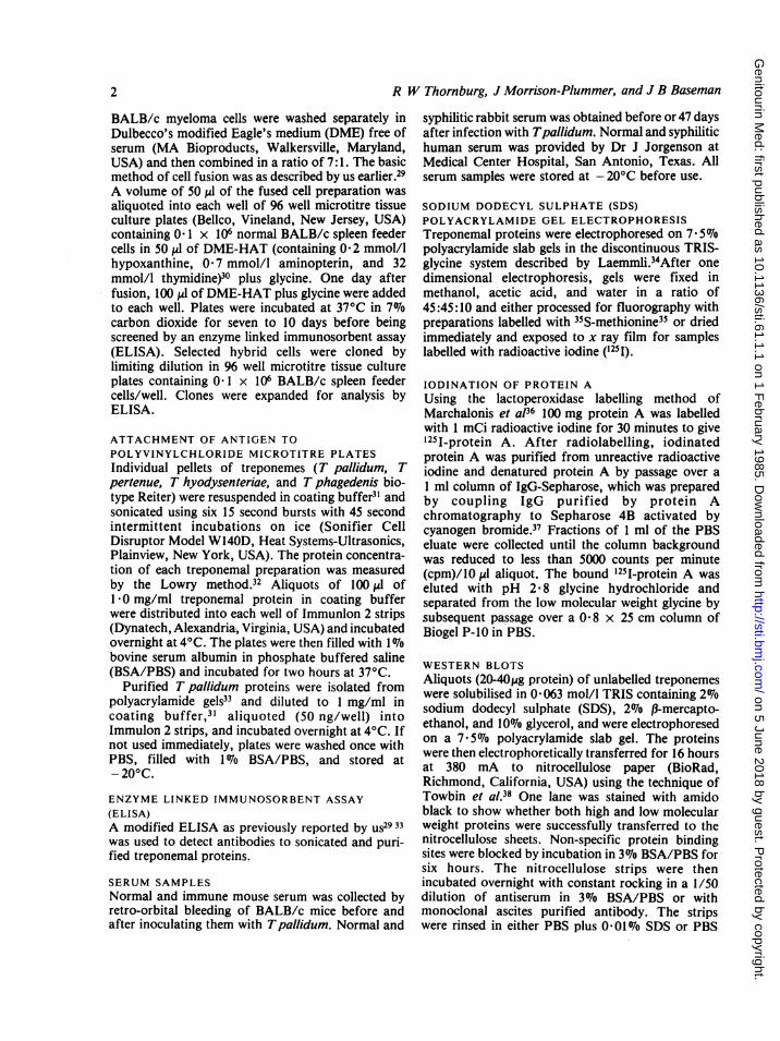
IFIG 1 Western blot analysis of: normal human serum(lane A); syphilitic human serum (lane B); syphiliticrabbit serum (lane C); monoclonal antibody HF2 (laneD); monoclonal antibody 13F3 (lane E); and monoclonalantibody 23C9 (lane F) against Tpallidum antigens.
3
< P6
on 5 June 2018 by guest. Protected by copyright.
http://sti.bmj.com
/G
enitourin Med: first published as 10.1136/sti.61.1.1 on 1 F
ebruary 1985. Dow
nloaded from

R W Thornburg, J Morrison-Plummer, and J B Baseman
1 2 3 4 1 2 3 4 1 2 3 4
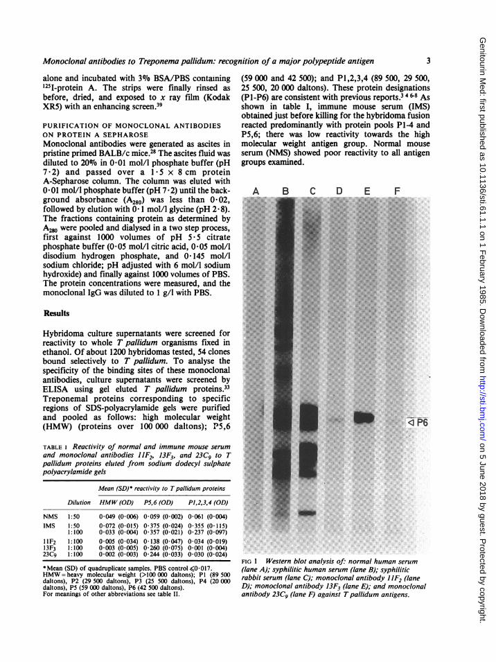
FIG 2 Western blot analysis of (a) syphilitic rabbit serum, (b) monoclonal antibody 13F3, and (c) normal rabbitserum against: T pallidum (lane 1); T pertenue (lane 2); T hyodysenteriae (lane 3); and T phagedenis biotype Reiter(lane 4) solubilised and electrophoresed on a 7S5% polyacrylamide slab gel.
From the original 54 hybridomas identified in thewhole cell ELISA, three hybridomes, (1iF2, 13F3,and 23C9) were selected because of their increasedreactivity towards the protein pool of P5,6 (table I).Western blot analysis of these monoclonal antibodiesconfirmed the ELISA results. As shown in fig 1, allthree monoclonal antibodies were specific for a45 000 molecular weight antigen (P6) of Tpallidum(lanes D-F), with monoclonal hybridoma 13F3 (laneE) showing the strongest intensity. Human (lane B)and rabbit (lane C) syphilitic serum also showed thismajor antigen, which was not present in poolednormal human serum (lane A).The immunological cross reactivity of specific
Treponema species was then examined. As shown in
table II, immune mouse serum was strongly crossreactive with T pallidum and T pertenue antigensand, to a lesser extent, with T hyodysenteriae and Tphagedenis indicating shared or cross reactiveantigens. Normal mouse serum showed low reactivityto T pallidum and T pertenue, and somewhatincreased reactivity to T hyodysenteriae and Tphagedenis. To further examine the cross reactivityof these species, Western blot analysis was performedusing both normal and syphilitic rabbit serum andmonoclonal antibody 13F3. ELISA typing of anti-body 13F3 showed the isotype to be IgG2a, x lightchain. Only one single heavy and light chain wasobtained following SDS-polyacrylamide gel electro-phoresis of the antibody. As shown in fig 2, syphilitic
TABLE ii Reactivity of normal and immune mouse serum to various treponemal species
Mean (SD)* reactivity to sonicated treponemal antigenst
Serum Dilution T pallidum (OD) T pertenue (OD) T hyodysenteriae (OD) T phagedenis (OD)
NMS 1:50 0-091 (0*002) 0-058 (0 008) 0 157 (0-019) 0-259 (0 027)IMS 1:50 1-167 (0-035) 1-121 (0-110) 0 507 (0 146) 0-916 (0 070)
1:100 1-179(0-072) 0-967 (0-018) 0-314 (0-026) 0-761 (0-031)1:500 1-825 (0 086) 0-763 (0-064) 0 059 (0-012) 0 450 (0 004)
Mean (SD) of quadruplicate samples. PBS control0 -0017.tTreponemes were prepared and sonicated as described in Materials and methods. Treponemal proteins were coated on to microtitre strips ata concentration of I pg protein, per well.OD = optical density at 405 nm; NMS = normal mouse serum; IMS = immune mouse serum.
4
on 5 June 2018 by guest. Protected by copyright.
http://sti.bmj.com
/G
enitourin Med: first published as 10.1136/sti.61.1.1 on 1 F
ebruary 1985. Dow
nloaded from

Monoclonal antibodies to Treponema pallidum: recognition of a major polypeptide antigen
rabbit serum (a) showed cross reactive antigensamong all four treponemal species. Normal rabbitserum (c) did not react with Tpallidum (lane 1) or Tpertenue (lane 2) but low level reaction occurred withT hyodysenteriae (lane 3) and Tphagedenis biotypeReiter (lane 4). Monoclonal antibody 13F3(b) washighly selective for the 45 000 dalton protein antigen(P6) found in Tpalidum3 and Tpertenue.9 No crossreactive protein was shown by 13F3 in Thyodysenteriae or Tphagedenis.
Discussion
A group of 54 monoclonal antibodies reactive with Tpallidum were produced. Three hybridoma cloneswere selected for further study because of theirunique specificity for a 45 000 dalton molecularweight polypeptide, P6. Antibodies to this proteinhave been shown to appear early (day 10) in rabbitsexperimentally infected with T pallidum.15 Thisprotein also exhibits high immunogenicity in infectedrabbits and man.346Monoclonal antibody 13F3 was purified by protein
A-Sepharose chromatography and used to examinecross reactivity among four species of treponemes(fig 2). In contrast to polyclonal antiserum raisedagainst T pallidum, which shows strong reactivitywith all four species of treponemes, monoclonal 13F3reacted only with Tpallidum and Tpertenue. Thesedata further emphasise the close relation betweenthese two treponemes,9 40 and reinforce the biologicaldifferences between the treponemes pathogenic andnon-pathogenic to man.The immune response to syphilis is extremely
complex and includes both humoral and cell-mediated immunity. Monoclonal antibody probesoffer the opportunity to evaluate better the role ofspecific treponemal antigens in the pathophysiologyof the disease.94' 42 The availability of monoclonalprobes also permits further biochemical and immuno-chemical dissection of important antigens and enablesthe development of rational diagnostic reagents andvaccines.
We thank Mary Devadoss for her excellent secretarial helpand Diane Jones and Andrew Bird for their technical help.This work was supported by Public Health Service GrantAI-19566 from the National Institute of Allergy andInfectious Diseases and by Cistron Technology Incorporated.
References
1. Hayes NS, Muse KE, Collier AM, Baseman JB. Parasitism byvirulent Treponema pallidum of host cell surfaces. InfectImmun 1977; 17:174-86.
2. Alderete JF, Baseman JB. Surface-associated host proteins onvirulent Treponemna pallidum. Infect Immun 1979; 26:1048-56.
3. Alderete JF, Baseman JB. Surface characterization of virulentTreponema pallidum. Infect Immun 1980; 30:814-23.
4. Baseman JB, Hayes EC. Molecular characterization of receptorbinding proteins and immunogens of virulent Treponemapallidum. J Exp Med 1980; 151: 573-86.
5. Fitzgerald TJ. Pathogenesis and immunology of Treponemapallidum. Annu Rev Microbiol 1981;35:29-54,
6. Baseman JB, AldereteJF. The parasitic strategies of Treponemapallidum. In: Schell RF, Musher DM, eds. Pathogenesis andimmunology of treponemal infection. New York: MarcelDekker, 1983;229-39.
7. Peterson, KM, Baseman JB, Alderete JF. Treponemapallidumreceptor binding proteins interact with fibronectin. J Exp Med1983; 157:1958-70.
8. Baseman JB, Alderete JF. The biology of Treponemapallidumand syphilis. Clinical Microbiology Newsletter 1983;5:157-9.
9. Thornburg RW, Baseman JB. Comparison of major proteinantigens and protein profiles of Treponema pallidum andTreponema pertenue. Infect Immun 1983; 42:623-7.
10. Friedmann PS, Turk JL. A spectrum of lymphocyte responsive-ness in human syphilis. Clin Exp Immunol 1975;21:59-64.
11. Weiser RS, Erickson D, Perine PL, Pearsall NN. Immunity tosyphilis: passive transfer in rabbits using serial doses ofimmune serum. Infect Immun 1976; 13:1402-7.
12. Bishop NH, Miller JN. Humoral immunity in experimentalsyphilis. I. The demonstration of resistance conferred by passiveimmunization. J Immunol 1976; 117:191-6.
13. Bishop NH, Miller JN. Humoral immunity in experimentalsyphilis. II. The relationship of neutralizing factors in immuneserum to acquired resistance. J Immunol 1976; 117:197-207.
14. Pavia CS, Folds JD, Baseman JB. Cell-mediated immunityduring syphilis. British Journal of Venereal Diseases1977; 53: 144-50.
15. Alderete JF, Baseman JB. Analysis of serum IgG againstTreponema pallidum protein antigens in experimentallyinfected rabbits. British Journal of Venereal Diseases1981; 57: 302-8.
16. Strandberg-Pedersen N, Axelsen NH, Petersen CS. Antigenicanalysis of T pallidum: cross reaction between individualantigens of T pallidum and T Reiter. Scand J Immunol 1981;13:143-50.
17. Sell S, Gambosa D, Baker-Zander SA, Lukehart SA, MillerJN. Host response to Treponema paliidum in intradermallyinfected rabbits. Evidence for persistence of infection at localand distant sites. J Invest Dermatol 1980;75:470-5.
18. Lukehart SA, Baker-Zander SA, Gubish ER. Identification ofTreponema pallidum antigens: comparison with a non-patho-genic treponeme. J Immunol 1982; 129:833-8.
19. Baughn RE, Adams CB, Musher DM. Circulating immunecomplexes in experimental syphilis: identification of treponemalantigens and specific antibodies to treponemal antigens inisolated complexes. Infect Immun 1983; 42:585-93.
20. Baughn RE. Immunoregulatory effects in experimentalsyphilis. In: Schell RF, Musher DM, eds. Pathogenesis andimmunology of treponemal infection. New York: MarcelDekker, 1983;271-95.
21. Bishop NH, Miller JN. Humoral immune mechanisms inacquired syphilis. In: Schell RF, Musher DM, eds. Pathogenesisand immunology of treponemal infection. New York: MarcelDekker, 1983;241-69.
22. Folds JD. Cell-mediated immunity. In: Schell RF, Musher DM,eds. Pathogenesis and immunology of treponemal infection.New York: Marcel Dekker, 1983;315-30.
23. Lukehart SA. Macrophages and host resistance. In: Schell RF,Musher DM, eds. Pathogenesis and immunology oftreponemalinfection. New York: Marcel Dekker, 1983; 349-64.
24. Schell RF, Chan JK, Lefrock JL. In: Schell RF, Musher DM,eds. Pathogenesis and immunology of treponemal infection.New York: Marcel Dekker, 1983;331-48.
25. Sell S. Histopathology and immunopathology of experimentalsyphilis. In: Schell RF, Musher DM, eds. Pathogenesis andimmunology of treponemal infection. New York: MarcelDekker, 1983;297-313.
26. Christiansen AH. The Reiter strain of Treponema pallidum:origin, cultivation, and use in immunization of rabbits. ActaPathol Microbiol Scand 1963;57:81-6.
27. Kinyon JM, Harris DL. Growth of Treponema hyodysenteriaein a liquid medium. Vet Rec 1974;95:219-20.
28. Oi VT, Herzenberg LA. Immunoglobulin-producing hybridcell lines. In: Mishell BB, Shiigi SM, eds. Selected methods incellular immunology. San Francisco: WH Freeman, 1980;35 1-72.
5
on 5 June 2018 by guest. Protected by copyright.
http://sti.bmj.com
/G
enitourin Med: first published as 10.1136/sti.61.1.1 on 1 F
ebruary 1985. Dow
nloaded from

R W Thornburg, J Morrison-Plummer, and J B Baseman
29. Morrison-Plummer J, Jones DH, Baseman JB. An ELISA todetect monoclonal antibodies specific for lipid determinants ofMycoplasmapneumoniae. JImmunolMethods 1983; 64: 165-78.
30. Kennett RH. In: Kennett RH, McKearn TJ, Bechtol KB, eds.Monoclonal antibodies hybridomas: a new dimension inbiological analyses. New York: Plenum Press, 1980; 365-7.
31. Voller A, Bidwell D, Bartlett A. Microplate enzyme immuno-assays for the immunodiagnosis of virus infections. In: RoseNR, Friedman H, eds. Manual of clinical immunology.Washington DC: American Society for Microbiology, 1976;506-12.
32. Lowry OH, Rosebrough NJ, Farr AL, Randall RJ. Proteinmeasurement with the folin phenol reagent. J Biol Chem1951; 193:265-75.
33. Morrison-Plummer J, Alderete JF, Baseman JB. Enzyme-linked immunosorbent assay for the detection of serum anti-body to outer membrane proteins of Treponema pallidum.British Journal of Venereal Diseases 1983;59:75-9.
34. Laemmli UK. Cleavage of structural proteins during theassembly of the head of bacteriophage T4. Nature 1970;227:680-5.
35. Bonner WM, Laskey RA. A film detection method for tritium-labeled proteins and nucleic acids in polyacrylamide gels. EurJBiochem 1974;46:83-8.
36. Marchalonis JJ, Cone RE, Santer V. Enzymatic iodination: aprobe for accessible surface proteins of normal and neoplasticlymphocytes. Biochem J 1971; 124:921-7.
37. March SC, Parikh I, Cuatrecasas P. A simplified method forcyanogen bromide activation of agarose for affinity chromato-graphy. Anal Biochem 1974;60:149-52.
38. Towbin H, Staehelin T, Gordon J. Electrophoretic transfer ofproteins from polyacrylamide gels to nitrocellulose sheets:procedure and some applications. Proc Natl Acad Sci USA1979;76:4350-4.
39. Swainstrom R, Shank PR. X-ray intensifying screens greatlyenhance the detection by autoradiography of radioactiveisotopes 32P and 125I. Anal Biochem 1978; 86:184-92.
40. Miao RM, Fieldsteel AH. Genetic relationship betweenTreponema pallidum and Treponema pertenue, two non-cultivable human pathogens. J Bacteriol 1980; 141:427-9.
41. Robertson SM, Kettman JR, Miller JN, Norgard MV. Murinemonoclonal antibodies specific for virulent Treponemapallidum (Nichols). Infect Immun 1982;36:1076-85.
42. Saunders JM, Folds JD. Development of monclonal antibodiesthat recognize Treponema pallidum. Infect Immun1983;41:844-7.
6
on 5 June 2018 by guest. Protected by copyright.
http://sti.bmj.com
/G
enitourin Med: first published as 10.1136/sti.61.1.1 on 1 F
ebruary 1985. Dow
nloaded from


![Recent advances in graphene-based biosensor technology ... · pg/mL [41] InuenzaAvirus Grapheneoxide ‑ MB–chitosan Electrochemical Monoclonalantibodies(H5N1 orH1N1) Covalentandcrosslinkedvia](https://img.dokumen.tips/doc/110x75/5f732b8b4e7e4d3dd00723fb/recent-advances-in-graphene-based-biosensor-technology-pgml-41-inuenzaavirus.jpg)