Embed Size (px)
Citation preview

doi:10.1152/ajpheart.01291.2007 294:1036-1047, 2008. First published Dec 21, 2007;Am J Physiol Heart Circ Physiol
Dale Abel and Martin E. Young Sheldon E. Litwin, Paul E. Hardin, Chi-Wing Chow, Margaret P. Chandler, E. Dongfang Zhang, Andreas Rohrwasser, Julie H. Rennison, Jason R. B. Dyck,M. Zanquetta, David J. Durgan, William J. Jeong, Ju-Yun Tsai, Heiko Bugger, Molly S. Bray, Chad A. Shaw, Michael W. S. Moore, Rodrigo A. P. Garcia, Melissa
You might find this additional information useful...
for this article can be found at: Supplemental material http://ajpheart.physiology.org/cgi/content/full/01291.2007/DC1
46 articles, 24 of which you can access free at: This article cites http://ajpheart.physiology.org/cgi/content/full/294/2/H1036#BIBL
including high-resolution figures, can be found at: Updated information and services http://ajpheart.physiology.org/cgi/content/full/294/2/H1036
can be found at: AJP - Heart and Circulatory Physiologyabout Additional material and information http://www.the-aps.org/publications/ajpheart
This information is current as of March 5, 2008 .
http://www.the-aps.org/.ISSN: 0363-6135, ESSN: 1522-1539. Visit our website at Physiological Society, 9650 Rockville Pike, Bethesda MD 20814-3991. Copyright © 2005 by the American Physiological Society. intact animal to the cellular, subcellular, and molecular levels. It is published 12 times a year (monthly) by the Americanlymphatics, including experimental and theoretical studies of cardiovascular function at all levels of organization ranging from the
publishes original investigations on the physiology of the heart, blood vessels, andAJP - Heart and Circulatory Physiology
on March 5, 2008
ajpheart.physiology.orgD
ownloaded from

Disruption of the circadian clock within the cardiomyocyte influencesmyocardial contractile function, metabolism, and gene expression
Molly S. Bray,1 Chad A. Shaw,2 Michael W. S. Moore,1 Rodrigo A. P. Garcia,1,3 Melissa M. Zanquetta,1
David J. Durgan,1 William J. Jeong,1 Ju-Yun Tsai,1 Heiko Bugger,4 Dongfang Zhang,5
Andreas Rohrwasser,6 Julie H. Rennison,7 Jason R. B. Dyck,8 Sheldon E. Litwin,5 Paul E. Hardin,9
Chi-Wing Chow,10 Margaret P. Chandler,7 E. Dale Abel,4 and Martin E. Young1
1US Department of Agriculture/Agricultural Research Service Children’s Nutrition Research Center, Departmentof Pediatrics, and 2Department of Molecular and Human Genetics, Baylor College of Medicine, Houston, Texas;3Department of Physiology and Biophysics, Institute of Biomedical Sciences, University of Sao Paulo, Sao Paulo, Brazil;4Division of Endocrinology, Metabolism and Diabetes and Program in Human Molecular Biology, 5Division of Cardiology,and 6Department of Human Genetics, University of Utah, Salt Lake City, Utah; 7Department of Physiology and Biophysics,Case Western Reserve University, Cleveland, Ohio; 8Cardiovascular Research Group, Department of Pediatrics andPharmacology, Faculty of Medicine and Dentistry, University of Alberta, Edmonton, Alberta, Canada; 9Departmentof Biology, Texas A&M University, College Station, Texas; and 10Department of Molecular Pharmacology, Albert EinsteinCollege of Medicine, Bronx, New York
Submitted 2 November 2007; accepted in final form 17 December 2007
Bray MS, Shaw CA, Moore MW, Garcia RA, ZanquettaMM, Durgan DJ, Jeong WJ, Tsai JY, Bugger H, Zhang D,Rohrwasser A, Rennison JH, Dyck JR, Litwin SE, Hardin PE,Chow CW, Chandler MP, Abel ED, Young ME. Disruption ofthe circadian clock within the cardiomyocyte influences myocar-dial contractile function, metabolism, and gene expression. Am JPhysiol Heart Circ Physiol 294: H1036–H1047, 2008. First pub-lished December 21, 2007; doi:10.1152/ajpheart.01291.2007.—Virtually every mammalian cell, including cardiomyocytes, possesses anintrinsic circadian clock. The role of this transcriptionally based molec-ular mechanism in cardiovascular biology is poorly understood. Wehypothesized that the circadian clock within the cardiomyocyte influ-ences diurnal variations in myocardial biology. We, therefore, generateda cardiomyocyte-specific circadian clock mutant (CCM) mouse to testthis hypothesis. At 12 wk of age, CCM mice exhibit normal myocardialcontractile function in vivo, as assessed by echocardiography. Radio-telemetry studies reveal attenuation of heart rate diurnal variations andbradycardia in CCM mice (in the absence of conduction system abnor-malities). Reduced heart rate persisted in CCM hearts perfused ex vivo in theworking mode, highlighting the intrinsic nature of this phenotype. Wild-type,but not CCM, hearts exhibited a marked diurnal variation in responsivenessto an elevation in workload (80 mmHg plus 1 �M epinephrine) ex vivo, witha greater increase in cardiac power and efficiency during the dark (active)phase vs. the light (inactive) phase. Moreover, myocardial oxygen consump-tion and fatty acid oxidation rates were increased, whereas cardiac efficiencywas decreased, in CCM hearts. These observations were associated with noalterations in mitochondrial content or structure and modest mitochon-drial dysfunction in CCM hearts. Gene expression microarray analysisidentified 548 and 176 genes in atria and ventricles, respectively, whosenormal diurnal expression patterns were altered in CCM mice. Thesestudies suggest that the cardiomyocyte circadian clock influences myo-cardial contractile function, metabolism, and gene expression.
bradycardia; carbohydrate; chronobiology; epinephrine; fatty acids
OVER THE COURSE OF A NORMAL 24-h day, the environment of,and the demands placed on, cells, organs, and organisms
fluctuate dramatically. Many organisms have, therefore, evolvedcircadian clocks, which are intracellular molecular mechanismsthat allow individual cells to perceive the time of day (11). Indoing so, circadian clocks confer the selective advantage ofanticipation, enabling both rapid and appropriate responses toenvironmental stimuli upon their onset. Almost every mamma-lian cell possesses an intrinsic circadian clock, a transcription-ally based molecular mechanism capable of regulating multiplecellular functions. In terms of the cardiovascular system, cir-cadian clocks have been characterized within multiple celltypes, including cardiomyocytes (7, 42); however, little isknown regarding the influence of intramyocellular circadianclocks on cardiovascular physiology and pathophysiology.This is particularly relevant, given the possibility that impair-ment of the circadian clock within the cardiomyocyte maysignificantly alter cardiac function, pathogenesis of cardiovas-cular disease (CVD), and/or treatment strategies during CVDstates (e.g., chronopharmacology) (42).
To date, experiments investigating the influence of alteredcircadian clock function in vivo have primarily involved ma-nipulation of the light-dark cycle and/or genetic ablation ofcircadian clocks in a ubiquitous manner (24, 36). Experimentalstrategies using such global manipulations influence both cen-tral and peripheral circadian clocks within the organism, aswell as virtually every neurohumoral factor investigated to date(24, 36, 40). The most extensively characterized of thesemodels is the Clock mutant mouse, in which chemical mu-tagenesis-induced truncation of the native Clock gene resultedin a dominant-negative CLOCK (dnCLOCK) mutant proteinlacking the transactivation domain of this transcription factor(termed CLOCK�19, due to loss of exon 19). We recentlygenerated a cardiomyocyte-specific circadian clock mutant(CCM) mouse, through MHC-� promoter-driven expression ofthe CLOCK�19 protein within cardiomyocytes (9). We em-ployed this dominant-negative approach, in contrast to a tissue-
Address for reprint requests and other correspondence: M. E. Young,USDA/ARS Children’s Nutrition Research Center, Baylor College of Medi-cine, Dept. of Pediatrics, 1100 Bates St., Houston, TX 77030 (e-mail:[email protected]).
The costs of publication of this article were defrayed in part by the paymentof page charges. The article must therefore be hereby marked “advertisement”in accordance with 18 U.S.C. Section 1734 solely to indicate this fact.
Am J Physiol Heart Circ Physiol 294: H1036–H1047, 2008.First published December 21, 2007; doi:10.1152/ajpheart.01291.2007.
http://www.ajpheart.orgH1036
on March 5, 2008
ajpheart.physiology.orgD
ownloaded from

specific CLOCK knockout approach, due to the marked redun-dancy within the mammalian circadian clock mechanism.Ubiquitous CLOCK null mice exhibit normal circadianrhythms at multiple biological levels, due to compensation bythe CLOCK homolog neuronal Per-Arnt-Sim domain protein 2(6). In our CCM mice, the circadian clock within the heart isseverely attenuated in a sex-independent manner, as demon-strated by attenuation of clock component and output genecircadian oscillations, without affecting circadian clocks withinextracardiac tissues (9). Using this model, our laboratory hasrecently shown that the circadian clock within the cardiomyocyteis essential for responsiveness of the heart to fatty acids (9).
A global understanding of the role of the circadian clockwithin the cardiomyocyte is currently lacking. As such, thepurpose of the present study was to characterize cardiac func-tion, metabolism, and gene expression in CCM mice, therebyidentifying novel potential roles for the circadian clock withinthe cardiomyocyte. We find that disruption of this molecularmechanism within the cardiomyocyte influences diurnal vari-ations in heart rate, responsiveness of the heart to changes inworkload, myocardial metabolism, as well as a plethora ofmyocardial genes known to influence transport, transcription,signal transduction, protein turnover, and metabolism.
MATERIALS AND METHODS
Animals. Male CCM (on the FVB/N background) and littermatecontrols were housed at the Centers for Comparative Medicine atBaylor College of Medicine (ex vivo heart perfusion, mitochondrialDNA content, and gene expression studies), at the University of Utah(echocardiographic, radiotelemetric, and electron microscopy stud-ies), or at Case Western Reserve University (isolated mitochondrialand enzymatic studies). All studies were approved by the respectivelocal Institutional Animal Care and Use Committees. All mice werehoused under controlled conditions (23 � 1°C; 12:12-h light-darkcycle), and received standard laboratory chow and water ad libitum.At least 21 days before they were killed, mice were housed in pairs ina separate environment-controlled room, within which a strict 12:12-hlight-dark cycle regime was enforced [lights on at 7 AM; zeitgebertime (ZT) 0]. The light-dark cycle was maintained throughout thesestudies, facilitating elucidation of the potential roles for the cardio-myocyte circadian clock under physiological conditions. As such,diurnal variations were investigated.
Echocardiographic and radiotelemetric studies. In vivo myocar-dial contractile function was assessed in anesthetized mice (isoflu-rane) using echocardiography, as described previously (21). Ambula-tory heart rate and blood pressure, as well as activity, were continuouslyassessed by radiotelemetry, as described previously (3). Radiotelemetricdevices were surgically implanted in 11-wk-old wild-type and CCMmice, which were allowed to recover from this intervention for 1 wkbefore a 2-wk data collection period (i.e., 12–14 wk of age). ECGtelemetry was performed in a similar manner, in which radiotelemetricdevices were implanted in 11-wk-old mice 1 wk before initiation ofdata collection.
Isolated working mouse heart perfusions. Isolated working mouseheart perfusions were utilized to investigate diurnal variations inmyocardial metabolism and contractile function ex vivo, as describedpreviously (10). Hearts were perfused in the working mode in anon-recirculating manner with Krebs-Henseleit bicarbonate buffercontaining 5 mM glucose, 0.4 mM oleate conjugated to 3% BSA(fraction V, fatty acid free; dialyzed), 10 �U/ml insulin, and traceramounts of [U-14C]glucose (40 �Ci/l) and [9,10-3H]oleate (60 �Ci/l).After 30 min of normal workload perfusion conditions (11.5 mmHgpreload, 50 mmHg afterload), hearts were perfused for an additional30 min under high workload conditions (11.5 mmHg H2O preload, 80
mmHg afterload plus 1 �M epinephrine). Cardiac power, efficiency,heart rate, developed pressure, and rate pressure product, as well asrates of oxygen consumption [myocardial oxygen consumption(MVO2)], exogenous oleate oxidation, exogenous glucose oxidation,net lactate release (i.e., lactate derived from both extracellular glu-cose, as well as intracellular glycogen), 14C-labeled lactate release,and endogenously derived lactate release were determined as de-scribed previously (1, 8, 10). Data presented for this study are thoseobtained during the last 10 min of both the low and high workloadconditions. These steady-state rates expose the intrinsic properties ofthe myocardium in the absence of acute influences by extrinsic (i.e.,neurohumoral) factors (44).
Mitochondrial studies. Mitochondrial DNA content was measuredthrough PCR of mitochondrial (cytochrome b) and nuclear (�-actin)specific genes, as described previously (2). Mitochondrial number andvolume density were determined from electron micrographs, as de-scribed previously (2). Citrate synthase and medium chain acyl-CoAdehydrogenase (MCAD) activities were measured spectrophotometri-cally, as described previously (27). For determination of mitochon-drial function, cardiac subsarcolemmal (SSM) and intramyofibrillar(IFM) mitochondria were isolated from wild-type and CCM heartsusing the procedure of Palmer et al. (26), except that a modifiedChappell-Perry buffer (containing 100 mM KCl, 50 mM MOPS, 1mM EGTA, 5 mM MgSO4 �7H2O, and 1 mM ATP, pH 7.4 at 4°C)was used for isolation of both mitochondrial populations. Mice wereanesthetized (isoflurane), and the left ventricle was excised andsubsequently placed in KME (100 mM KCl, 50 mM MOPS, internalsalt, and 0.5 mM EGTA). The IFM were harvested following treat-ment of skinned fibers with 5 mg/g wet wt trypsin for 10 min at 4°C(22). Mitochondrial protein concentration was determined by theLowry method using bovine serum albumin as a standard.
Oxygen consumption in SSM and IFM was measured using aClark-type oxygen electrode (Strathkelvin) at 30°C (35). Mitochon-dria were incubated in a solution consisting of 80 mM KCl, 50 mMMOPS, 1 mM EGTA, 5 mM KH2PO4, and 1 mg/ml bovine serumalbumin at pH 7.4. The rate of oxidative phosphorylation was mea-sured using the complex-specific substrates glutamate (complexes I,III, and IV), succinate (complexes II, III, and IV), durohydroquinone(complexes III and IV), and N,N,N�,N�-tetramethyl-p-phenylenedi-amine-ascorbate (complex IV) (29, 30); durohydroquinone was a kindgift from Dr. Charles Hoppel (Case Western Reserve University).Mitochondrial respiration was also measured using pyruvate plusmalate and acetylcarnitine plus malate, which assess pyruvate dehy-drogenase and carnitine-acetyltransferase activities, respectively (29).Lipid substrates carnitine-dependent octanoyl-CoA plus malate, car-nitine-dependent palmitoyl-CoA plus malate, and palmitoylcarnitineplus malate were used to assess carnitine palmitoyltransferase I,palmitoyltransferase II, and carnitine-acylcarnitine translocase activ-ities (17). State 3 (ADP stimulated) respiration, state 4 (ADP limited)respiration, respiratory control ratio (RCR) (state 3/state 4, an indica-tor of mitochondrial coupling), and ADP/O ratio (ratio of oxygenconsumed following addition of known amount of ADP) were deter-mined, as described previously (4, 12). Uncoupled respiration, initi-ated by addition of the uncoupler dinitrophenol, was used to assess thephosphorylation apparatus, which is composed of adenine nucleotidetranslocase, F1F0 ATPase, and the pyruvate transporter (29).
Microarray analysis. Microarray gene expression studies wereperformed using mouse Ref-6 BeadChips and the BeadStation systemfrom Illumina (San Diego, CA). Atrial and ventricular tissue sampleswere collected from wild-type and CCM animals every 3 h for aperiod of 24 h (n � 4 samples per time point; 128 samples total), andRNA was subsequently extracted from each sample using standardprocedures (5). Microarrays were performed according to the manu-facturer’s guidelines. Total RNA was converted to cDNA by reversetranscription using ArrayScript reverse transcriptase and T7-(dT)24
primers, followed by second-strand synthesis to generate double-stranded cDNA (Ambion). After purification, the cDNA was con-
H1037THE CIRCADIAN CLOCK WITHIN THE CARDIOMYOCYTE
AJP-Heart Circ Physiol • VOL 294 • FEBRUARY 2008 • www.ajpheart.org
on March 5, 2008
ajpheart.physiology.orgD
ownloaded from

verted to biotin-labeled cRNA, hybridized to the BeadChip (Illumina),and stained with strepavidin-Cy3 for visualization. The mouse Ref-6BeadChips contain sequences representing �46,000 curated genesand putative expressed sequence tags, with an average of 30 replicatesfor each gene/transcript on each BeadChip. Quality standards forhybridization, labeling, staining, background signal, and basal level ofhousekeeping gene expression were verified for all BeadChips.
After the probe arrays were scanned, the resulting images were firstanalyzed using the BeadStudio software (Illumina, San Diego, CA),which calculates the mean fluorescence signal across all replicates ofeach gene/transcript, along with a probability that the mean signal foreach gene/transcript on the chip is greater than background (i.e.,detection score). Genes/transcripts were defined as being significantlyexpressed above background when the 75th percentile of each gene’sdetection score met or exceeded 0.9. The expression data were thennormalized within centiles of the distribution of gene expressionvalues. Identification of genes exhibiting significant oscillations inexpression was next performed using the method described byStraume (33). Separate analyses were performed for the atrium andventricle tissues. Rhythmicity of gene expression for each Illuminaprobe set was evaluated by fitting the nonlinear cosine function, A �cos[(2 t/T) �] C (where C is the mean expression of the genesacross all time, A is the amplitude of oscillation, � is a phase shift, tis time and T is the period of oscillation). In addition, Fisher’s Gstatistic for spectral power density was calculated for each gene usingthe time point means to make the calculation. Fits were performed inthe R statistical programming environment using the nonlinear leastsquares (NLS) method to identify the best amplitude, period, andphase shift parameters and to minimize the sum of squared deviationsabout the fitted curve for each probe set. Before NLS estimation of theperiodic curve, the gene mean C was calculated as the mean of thetime point means, and this value was subtracted from the data. Thecentered data were then subject to analysis using the R implementa-tion of the NLS fitting procedure; this procedure searches for theglobal optimum of the least squares criterion. The search is accom-plished by Newton-Raphson gradient-based method, which is com-putationally efficient compared with a more grid-based approach. Toconsider the significance of the oscillatory fits, we implemented asimulation procedure to parallel that which is described in the well-accepted COSOPT procedure. For each Illumina probe set, we deter-mined the standard error of the mean at each time point. We thenperformed 1,000 random permutations of the time point means, onwhich we superimposed independent Gaussian noise with mean 0 andwith variance determined by the time point standard error. The NLSbest fit for each permuted data set was then calculated. The ensembleof 1,000 parameter fits for the permuted data were then compared withthe true data fits to determine which genes showed significant patternsof oscillation. A gene (probe set) was considered to have a significantcircadian oscillation if it fit a cosine wave function for p multiplemeasures corrected � (MMC�) � 0.05 and Fisher G � 0.1, with aperiodicity of between 20 and 28 h. The P values for both the MMC�parameter and Fisher G were determined using the simulation-basedprocedure. This criterion was evaluated through the use of 10 knowncircadian clock guide genes, all of which oscillated significantly inwild-type atria and ventricles, as assessed by this analytic strategy.Subsequently, a two-way ANOVA was performed to identify thoseoscillating genes identified as differentially expressed in CCM atriaand ventricles. The two-way ANOVA was used to identify genes withshifts in mean expression (interpreted as a main effect of genotype)and with genotype-time interaction (interpreted as a phase shift inexpression associated with genotype). Ontology and pathway analyseswere performed to group genes from the final lists using the Onto-Tools package of ontology and pathway analysis from Wayne StateUniversity (http://vortex.cs.wayne.edu/projects.htm). Normalizeddata have been submitted to GEO archive and are available athttp://www.ncbi.nlm.nih.gov/geo/.
Quantitative RT-PCR. Quantitative RT-PCR was performed usingpreviously described methods (14, 16). Specific quantitative assayswere designed from mouse sequences available in GenBank. Primerand probe sequences for bmal1, dbp, per3, dgat2, and adpn Taqmanassays have been published previously (9, 45), whereas those for dec1,e4bp4, mcip1, pbef1, pik3r1, and prkar1a are presented in Supple-mental Table 1. (The online version of this article contains supple-mental data.) Standard RNA was made for all assays by the T7polymerase method (Ambion, Austin, TX), using total RNA isolatedfrom mouse hearts; the use of standard RNA allows absolute quanti-fication of gene expression. The correlation between the thresholdcycle (the number of PCR cycles required for the fluorescent signal toreach a detection threshold) and the amount of standard was linearover at least a five-log range of RNA for all assays (data not shown).Quantitative RT-PCR data are represented as mRNA molecules pernanogram total RNA.
Statistical analysis of nonmicroarray data. Telemetry data wereanalyzed using a one-way ANOVA with repeated measures. One-wayANOVA was used to compare peak and trough values by genotype.For analyses of main effects of group (e.g., genotype, workload) andtime, two-way ANOVA was performed, followed by post hoc pair-wise comparisons of time points by genotype. Stata version 8.0 (Stata,San Antonio, TX) was used to perform two-way ANOVA to inves-tigate the main effects of intervention and time. A full model,including second-order interactions, was conducted for each experi-ment. Significant differences were determined using Type III sumsof squares. The null hypothesis of no model effects was rejected atP � 0.05.
RESULTS
In vivo assessment of cardiovascular function. In vivo car-diac function was evaluated at ZT6 in 12-wk-old anaesthetizedwild-type and CCM mice by echocardiography (Table 1). Ofthe parameters measured, fractional shortening, left ventricularejection fraction, and biventricular mass were significantlyaltered (all elevated) in CCM mice. Next, radiotelemetricstudies were performed, for continuous measurement of phys-ical activity, systolic blood pressure (SBP), diastolic bloodpressure (DBP), mean arterial pressure (MAP), and heart ratein conscious mice. Relative to wild-type mice, CCM miceexhibited a lower heart rate during both the light and dark
Table 1. Echocardiographic analysis of anesthetizedwild-type and CCM (12 wk of age) mice at zeitgeber time 6(i.e., middle of the light phase)
Wild Type CCM
LVDd, cm 0.400�0.014 0.395�0.011LVDs, cm 0.235�0.011 0.208�0.009IVDs, cm 0.082�0.004 0.097�0.005LVPWd, cm 0.085�0.002 0.100�0.007FS, % 42.34�1.75 47.32�1.48*LVEF 0.797�0.015 0.853�0.013*LVOTvti, cm 3.67�0.18 4.43�0.18HR, beats/min 462�32 399�16CO, ml/min 13.22�0.89 13.85�0.78Mass, g 0.127�0.006 0.153�0.009*
Data are shown as means � SE for 6 separate mice per group. CCM,cardiomyocyte-specific circadian clock mutant; LVDd, left ventricular diam-eter in diastole; LVDs, left ventricular diameter in systole; IVDs, interventric-ular septum thickness in systole; LVPWd, left ventricular posterior wallthickness in diastole; FS, fractional shortening; LVEF, left ventricular ejectionfraction; LVOTvti, left ventricular outflow tract velocity time integral (strokevolume index); HR, heart rate; CO, cardiac output. *P � 0.05, wild-typevs. CCM.
H1038 THE CIRCADIAN CLOCK WITHIN THE CARDIOMYOCYTE
AJP-Heart Circ Physiol • VOL 294 • FEBRUARY 2008 • www.ajpheart.org
on March 5, 2008
ajpheart.physiology.orgD
ownloaded from

phases (Fig. 1Aa). The decrease in heart rate was greater duringthe dark phase (Fig. 1Ab), such that CCM mice exhibit atten-uated diurnal variations in this parameter (Fig. 1Ac). Despitethe lower heart rate observed in CCM mice, comparablediurnal variations in physical activity between wild-type andCCM mice were observed; both groups exhibit increasedactivity during the dark phase relative to the light phase
(Supplemental Fig. 1A). Increased physical activity during thedark phase was associated with increased SBP, DBP, MAP(Supplemental Fig. 1, B, C, and D, respectively), and heart rate(Fig. 1A) in both wild-type and CCM mice. Similar to physicalactivity, no significant differences were observed for troughand peak values of SBP, DBP, or MAP between wild-type andCCM mice.
Fig. 1. Heart rate (A) and ECG (B) radiotelem-etric analysis of wild-type (WT) vs. cardiomyo-cyte-specific circadian clock mutant (CCM)mice. Radiotelemetric devices were implanted inmice at 11 wk of age, and data collection wasinitiated at 12 wk of age and continued contin-uously for a 2-wk period. Aa: data across a 24-hperiod, at 0.5-h intervals. As indicated, signifi-cant main effects of genotype and time wereobserved (P � 0.05). Ab: trough [average ofzeitgeber time (ZT) 10 through ZT12] and peak(average of ZT18 through ZT20) values.Ac: trough-to-peak differences (i.e., amplitude ofoscillation). B: representative ECG tracings forWT and CCM mice. Data are shown as means �SE for 4 mice per group. BPM, beats/min. Forcomparisons of peak/trough values: *P � 0.05,WT vs. CCM; @P � 0.05, trough vs. peak.
H1039THE CIRCADIAN CLOCK WITHIN THE CARDIOMYOCYTE
AJP-Heart Circ Physiol • VOL 294 • FEBRUARY 2008 • www.ajpheart.org
on March 5, 2008
ajpheart.physiology.orgD
ownloaded from

Conscious ECG telemetry was performed to determine thebasis for the bradycardia that develops in CCM mice. Table 2and Fig. 1B show that the only abnormality observed was anincrease in the R-R interval, which is consistent with sinusbradycardia. All other intervals were normal, effectively rulingout significant conduction system abnormalities.
Ex vivo assessment of myocardial contractile function atnormal and high workloads. We utilized the isolated workingmouse heart preparation to investigate diurnal variations inheart rate for wild-type vs. CCM littermates (11 wk of age),thereby eliminating the acute influence of neurohumoral fac-tors. Figure 2Aa shows that CCM hearts exhibit decreasedheart rate, relative to wild-type hearts, when perfused ex vivounder normal workload conditions, independent of the time ofday. These experiments suggest that the circadian clock withinthe cardiomyocytes of the sinoatrial node may influence pace-maker function.
The isolated working mouse heart preparation was utilizedfurther to reveal differences in diurnal contractile functionbetween wild-type and CCM hearts, in the absence of acuteneurohumoral influences. Developed pressure and rate pressureproduct do not exhibit a significant time of day dependence inwild-type or CCM mouse hearts perfused under normal work-load conditions (Fig. 2, Ba and Ca); however, rate pressureproduct is significantly reduced in CCM hearts (compared withwild-type hearts), independent of the time of day (Fig. 2Ca),consistent with the observed bradycardia (Fig. 2Aa). Similar topreviously published in vivo studies (25, 39), Fig. 2Da showsthat wild-type hearts perfused under normal workload condi-tions exhibit a significant diurnal variation in cardiac power (aderivative of cardiac output), peaking during the dark (active)phase (i.e., 1.3-fold higher at ZT18 vs. ZT0). In contrast, CCMhearts do not exhibit a diurnal variation in cardiac power (Fig.2Da). MVO2 did not exhibit a significant diurnal variation ineither wild-type or CCM hearts during normal workload con-ditions (Fig. 2Ea). However, independent of the time of day,CCM hearts display higher rates of oxygen consumption com-pared with wild-type hearts (Fig. 2Ea). Wild-type, but notCCM hearts, exhibit increased cardiac efficiency during thedark (active) phase (i.e., 1.3-fold higher at ZT18 vs. ZT6; Fig.2Fa). CCM hearts have lower cardiac efficiency relative towild-type littermate hearts (independent of the time of day;Fig. 2Fa).
We next tested the hypothesis that the circadian clock withinthe cardiomyocyte allows the heart to anticipate circadianrhythms in workload (which is usually increased during the
dark phase for the rodent). To model increased physical activ-ity, we simultaneously stimulated the heart with epinephrineand increased the afterload ex vivo, as described previously(15) (note that ex vivo perfused hearts are in permanentparasympathetic withdrawal). Figure 2Ab shows a persistenceof bradycardia in CCM mice at this high workload. Developedpressure and rate pressure product did not exhibit a significantdiurnal variation in wild-type (or CCM) mouse hearts duringhigh workload conditions (Fig. 2, Bb and Cb). Figure 2Dbshows a marked diurnal variation in the responsiveness ofwild-type hearts to an increased workload, in terms of cardiacpower, with greatest responsiveness observed during the dark(active) phase (e.g., 1.7-fold higher at ZT18 vs. ZT12). Thisrhythm in responsiveness to increased workload was absent forCCM hearts. Neither wild-type nor CCM hearts exhibited asignificant diurnal variation in MVO2 during high workloadconditions (Fig. 2Eb). However, cardiac efficiency exhibited adiurnal variation in wild-type, but not CCM, mouse hearts,peaking during the dark (active) phase (i.e., ZT18; Fig. 2Fb).
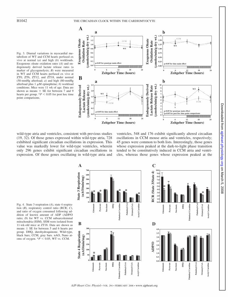
Ex vivo assessment of myocardial metabolism at normal andhigh workloads. Due to known close relationships betweenmyocardial contractile function and metabolism (34), as wellas the suggestion that peripheral circadian clocks likely influ-ence cellular metabolism (41, 42), myocardial metabolic fluxeswere measured in wild-type and CCM hearts ex vivo, at bothnormal and high workloads. When perfused at a normal work-load, neither rates of exogenous oleate oxidation, exogenousglucose oxidation, nor lactate release rates (net, 14C-labeled,endogenously derived) exhibited a time-of-day dependence inwild-type hearts under normal workload conditions (Fig. 3, Aaand Ba, and Supplemental Fig. 2, Ai, Bi, and Ci). However,consistent with increased MVO2 (Fig. 2Ea), exogenous oleateoxidation rates are constitutively elevated in CCM vs. wild-type hearts (Fig. 3Aa), independent of the time of day.
Neither exogenous oleate nor glucose oxidation rates exhib-ited significant differences between wild-type and CCM heartsunder high workload conditions (Fig. 3Ab and SupplementalFig. 2Aii). In contrast, lactate release rates (net, 14C-labeled,and endogenously derived) exhibited biphasic oscillations inwild-type hearts, which were absent in CCM hearts (Fig. 3Bband Supplemental Fig. 2, Bii and Cii). During high workloadconditions, CCM hearts had time-of-day independent rates oflactate release that were comparable to the lowest rates (i.e., atZT12) observed in wild-type hearts (Fig. 3Bb and Supplemen-tal Fig. 2, Bii and Cii), consistent with decreased responsive-ness of CCM hearts to the workload challenge at the level ofcontractile function (Fig. 2).
Mitochondrial number, structure, and function in CCM vs.wild-type hearts. Given the marked differences in MVO2, fattyacid oxidation, lactate release, and efficiency between wild-type and CCM hearts, as well as responsiveness to increasedworkload, we investigated whether alterations in mitochondriapotentially contribute to the phenotypes observed in CCMhearts. Mitochondrial DNA content, number, and volume den-sity did not differ between wild-type and CCM hearts, nor didtotal citrate synthase activity (Supplemental Table 2). In addi-tion, gross mitochondrial morphology was not altered in CCM(vs. wild-type) hearts (as qualitatively assessed by electronmicroscopy; Supplemental Fig. 3). Because SSM and IFMhave been shown to respond differentially to pathophysiolog-ical conditions, we isolated both mitochondrial populations
Table 2. Electrocardiographic analysis of wild-type andCCM (12 wk of age) at zeitgeber time 6 (i.e., middle of thelight phase) through radiotelemetry
Wild Type CCM
PQ, s 0.019�0.001 0.020�0.001PR, s 0.034�0.001 0.034�0.000QRS, s 0.018�0.001 0.019�0.000QT, s 0.029�0.000 0.031�0.000QTcB, s 0.099�0.001 0.103�0.001R-R, s 0.083�0.001 0.089�0.001*Heart rate, beats/min 725�12 672�8*
Data are means � SE for 4 separate mice per group. QTcB, QT intervalcorrected using Bazett’s formula. *P � 0.05, wild-type vs. CCM.
H1040 THE CIRCADIAN CLOCK WITHIN THE CARDIOMYOCYTE
AJP-Heart Circ Physiol • VOL 294 • FEBRUARY 2008 • www.ajpheart.org
on March 5, 2008
ajpheart.physiology.orgD
ownloaded from

from nonperfused wild-type and CCM hearts (11 wk old).Neither total mitochondrial (i.e., SSM plus IFM) nor IFMprotein yields significantly differed between CCM and wild-type hearts (Supplemental Table 2). However, SSM proteinyields were significantly decreased for CCM hearts comparedwith wild-type hearts (Supplemental Table 2), whereas IFMprotein yields tended to be increased.
Respiratory function was investigated for SSM and IFMisolated from wild-type and CCM mouse hearts. State 3 respi-ration, state 4 respiration, RCR, and ADP/O were not altered inthe IFM population of CCM hearts compared with wild-typehearts, using any of the substrates investigated (see Supple-mental Fig. 4). In the SSM of CCM hearts, state 3 respirationwas decreased using glutamate, pyruvate, palmitoycarnitine,palmitoyl-CoA, and acetyl carnitine (Fig. 4A), as well asN,N,N�,N�-tetramethyl-p-phenylenediamine-ascorbate (872 �31 vs. 627 � 67 nano atoms of oxygen (nAO) �min 1 �mgmitochondrial protein 1 for wild-type vs. CCM SSM; P �0.05). Depressed state 3 respiration rates in the SSM popula-tion of CCM hearts were not relieved by the addition of theuncoupler dinitrophenol (191 � 33 vs. 115 � 17nAO �min 1 �mg mitochondrial protein 1 for wild-type vs.CCM SSM, with glutamate as the sole substrate; P � 0.05).State 4 respiration was increased in the SSM population of
CCM hearts when glutamate was used as a substrate, but wasnot altered with any other substrates (Fig. 4B). RCR wasdecreased in CCM heart SSM only when glutamate or oc-tanoyl-CoA were used as substrates (Fig. 4C). The ADP/Oratio did not differ in the SSM of CCM hearts with anysubstrates investigated (Fig. 4D). Consistent with decreasedstate 3 respiration rates with lipid substrates, a slight, butsignificant, decrease in MCAD activity was observed in CCMvs. wild-type hearts (Supplemental Table 2).
Disruption of gene expression oscillations in CCM vs. wild-type hearts. Although the modest defects in mitochondrialfunction (e.g., decreased state 3 respiration in CCM heart SSM)may limit energetic reserve at high workloads, they do notreadily explain the differences in myocardial metabolism (e.g.,increased oleate oxidation rates, decreased lactate release rates)observed between wild-type and CCM hearts. Because thecircadian clock is a transcriptionally based mechanism, and theprimary intervention in our CCM model is cell-type-specificexpression of a dominant-negative transcription factor, diurnalvariations in gene expression were next investigated in atriaand ventricles isolated from 8-wk-old wild-type and CCMmice, through microarray analyses.
Of the approximate 46,000 genes/transcripts interrogatedthrough microarray analysis, over 15,000 were expressed in
PP
Fig. 2. Diurnal variations in contractile function of WT and CCM hearts perfused ex vivo at normal (a) and high (b) workloads. Heart rate (A), developed pressure(B), rate pressure product (C), cardiac power (CP; D), myocardial oxygen consumption (MVO2; E), and efficiency (F) were measured in WT and CCM heartsperfused ex vivo at ZT0, ZT6, ZT12, and ZT18, under normal (50-mmHg afterload; a) and high (80-mmHg afterload plus 1 �M epinephrine; b) workloadconditions. Mice were 11 wk of age. Data are shown as means � SE for between 7 and 9 hearts per group. *P � 0.05 for post hoc time point comparisons;#P � 0.05, peak vs. trough post hoc comparison for WT hearts.
H1041THE CIRCADIAN CLOCK WITHIN THE CARDIOMYOCYTE
AJP-Heart Circ Physiol • VOL 294 • FEBRUARY 2008 • www.ajpheart.org
on March 5, 2008
ajpheart.physiology.orgD
ownloaded from
wild-type atria and ventricles, consistent with previous studies(19, 32). Of those genes expressed within wild-type atria, 728exhibited significant circadian oscillations in expression. Thisvalue was markedly lower for wild-type ventricles, whereinonly 296 genes exhibit significant circadian oscillations inexpression. Of those genes oscillating in wild-type atria and
ventricles, 548 and 176 exhibit significantly altered circadianoscillations in CCM mouse atria and ventricles, respectively;45 genes were common to both lists. Interestingly, those geneswhose expression peaked at the dark-to-light phase transitiontended to be constitutively induced in CCM atria and ventri-cles, whereas those genes whose expression peaked at the
Fig. 3. Diurnal variations in myocardial me-tabolism of WT and CCM hearts perfused exvivo at normal (a) and high (b) workloads.Exogenous oleate oxidation rates (A) and en-dogenously derived lactate release rates (amarker of glycogenolysis; B) were measuredin WT and CCM hearts perfused ex vivo atZT0, ZT6, ZT12, and ZT18, under normal(50-mmHg afterload; a) and high (80-mmHgafterload plus 1 �M epinephrine; b) workloadconditions. Mice were 11 wk of age. Data areshown as means � SE for between 7 and 9hearts per group. *P � 0.05 for post hoc timepoint comparisons.
AO
Fig. 4. State 3 respiration (A), state 4 respira-tion (B), respiratory control ratio (RCR; C),and ratio of oxygen consumed following ad-dition of known amount of ADP (ADP/Oratio; D) for WT vs. CCM subsarcolemmalmitochondria (SSM). SSM were isolated from11-wk-old mice at ZT18. Data are shown asmeans � SE for between 5 and 6 hearts pergroup. DHQ, durohydroquinone. Wild-type,black bars; CCM, gray bars. nAO, Nano at-oms of oxygen. *P � 0.05, WT vs. CCM.
H1042 THE CIRCADIAN CLOCK WITHIN THE CARDIOMYOCYTE
AJP-Heart Circ Physiol • VOL 294 • FEBRUARY 2008 • www.ajpheart.org
on March 5, 2008
ajpheart.physiology.orgD
ownloaded from

light-to-dark phase transition tended to be constitutively re-pressed in CCM atria and ventricles (Fig. 5). The complete listof genes is included in Supplemental Table 3.
For validation purposes, a subset of genes identified by themicroarray analysis as being altered in CCM hearts was con-firmed by quantitative RT-PCR (Supplemental Fig. 5). Elevengenes were chosen to represent a spectrum of amplitudes andphases in gene expression oscillations for wild-type hearts, aswell as examples of induction and repression in CCM hearts. Inaddition, of the 11 genes investigated through RT-PCR anal-ysis, 5 are known core circadian clock components or outputgenes. These included bmal1, per3, dec1, e4bp4, and dbp. Themicroarray analysis identified additional clock componentand output genes (e.g., clock and tef), although RT-PCR wasnot performed for these. We also confirmed that adpn,dgat2, mcip1, pbef1, pik3r1, and prkar1a exhibit a markeddiminution in their circadian oscillation in expression forCCM hearts.
Gene ontology analysis was employed as a means to identifypotential biological and molecular functions for the circadianclock within the cardiomyocyte. Figure 6 shows an enrichmentof genes in multiple gene ontology categories that would beexpected to influence myocardial contractile function and me-tabolism. These include genes directly involved in transport,transcription, signal transduction, protein turnover, and metab-olism. In an attempt to understand further the potential roles ofthe circadian clock within the cardiomyocyte, Kegg pathwayanalysis was performed on this novel microarray data set.Supplemental Table 4 shows pathway enrichment for atria andventricles. Circadian rhythm was identified as a highly en-riched pathway in both atria and ventricles, validating therobustness of our array. Consistent with our gene ontologyanalysis, several signal transduction pathways were identifiedby the Kegg analysis, including the focal adhesion cascade(Supplemental Table 4). Because many of the identified genesmay be influenced by common transcriptional elements, apromoter analysis was performed to identify candidate tran-scription factors potentially linking the circadian clock to targetgenes. Supplemental Table 5 shows that multiple commontranscriptional elements were identified, including aryl hydro-carbon receptor nuclear translocator and retinoid-related or-phan receptor-�, two elements known to be directly regulatedby the circadian clock (13, 38).
DISCUSSION
The purpose of the present study was to determine whetherdisruption of the circadian clock within the cardiomyocyteinfluences myocardial contractile function, metabolism, and/orgene expression, in an attempt to delineate the potential rolesof this molecular mechanism in the heart. We find that disrup-tion of the circadian clock within the cardiomyocyte is asso-
.
Fig. 5. Diurnal variations in expression of genes in atria (A) and ventricles (B)isolated from WT and CCM mice. Blue represents low level of expression;yellow represents high level of expression. “Heat maps” were generated bycentering microarray data relative to the mean normalized expression acrossthe time course investigated, followed by scaling each gene, such that thevalues displayed have unit variance. The relative ordering for the rows wasgenerated according to the phase information determined for the WT data; thesame phase ordering was then applied to the CCM data. Mice were 8 wk ofage. Data are shown as the mean for 4 separate observations per group.
H1043THE CIRCADIAN CLOCK WITHIN THE CARDIOMYOCYTE
AJP-Heart Circ Physiol • VOL 294 • FEBRUARY 2008 • www.ajpheart.org
on March 5, 2008
ajpheart.physiology.orgD
ownloaded from

ciated with 1) decreased diurnal variations in heart rate in vivo;2) sinus bradycardia both in vivo and ex vivo; 3) loss of diurnalvariations in cardiac power ex vivo; 4) increased fatty acidoxidation and decreased cardiac efficiency ex vivo; 5) loss ofdiurnal variations in the responsiveness of the heart to changesin workload ex vivo; 6) population-specific mitochondrialdefects; and 7) alterations in the expression of an array ofmyocardial genes influencing transport, transcription, signaltransduction, protein turnover, and metabolism. These novelobservations support the idea that the circadian clock withinthe cardiomyocyte enables the heart to anticipate circadianrhythms in extracellular stimuli (such as workload), thereforeallowing both a rapid and temporally appropriate response.
The circadian clock within the cardiomyocyte influencescardiovascular function. Circadian rhythms in myocardialphysiology and pathophysiology have classically been ex-plained in terms of extracardiac influences (23, 28, 31, 37).Changes in autonomic, sympathetic, and adrenergic stimula-tion, nutrients (e.g., glucose, fatty acids, lipoproteins), circu-lating hormones (e.g., insulin, cortisol, adipokines), as well asvascular resistance and afterload, have all been suggested toinfluence circadian rhythms in myocardial gene expression,metabolism, and contractile function (42). The present studyhighlights the existence of an intrinsic molecular mechanismwithin the cardiomyocyte that likely augments or modulatesmyocardial biology. Data presented within Figs. 1 and 2Astrongly suggest that the circadian clock within the cardiomy-ocyte influences heart rate. Diurnal variations in heart rate areblunted in CCM mice in vivo (Fig. 1Ac). In addition, CCMhearts exhibit bradycardia both in vivo and ex vivo, in theabsence of conduction defects (Figs. 1 and 2A). Taken to-gether, these observations provide strong evidence supportingthe hypothesis that the circadian clock within the cardiomyo-cyte directly regulates heart rate, potentially allowing the heartto anticipate increased activity during the awake (dark) phase.The observation that bradycardia persists for CCM heartsex vivo, in the absence of acute neurohumoral influences,suggests potential intrinsic regulation of heart rate by thecircadian clock within cardiomyocytes of the sinoatrial node.
Diurnal variations in cardiac power persisted in wild-typemouse hearts ex vivo, peaking in the middle of the dark
(awake) phase (Fig. 2Da). These circadian rhythms were ab-sent in CCM hearts, suggesting that the circadian clock withinthe cardiomyocyte contributes toward increased cardiac outputat this time of day. We subsequently hypothesized that thecircadian clock within the cardiomyocyte would facilitate arapid response to increased workload during the waking pe-riod. Consistent with this hypothesis, our studies ex vivo (Fig.2Db) show that wild-type hearts exhibit the greatest respon-siveness to an acute increase in workload during the active(dark) phase, in terms of cardiac power. These data provideevidence that increased cardiac output observed in vivo duringthe dark phase is not solely a function of increased stimulation(e.g., sympathetic and adrenergic stimulation) during the activephase, but that increased intrinsic responsiveness of the myo-cardium to the stimulus at this time contributes to this phe-nomenon. The fact that CCM hearts lose diurnal variations inresponsiveness to the workload challenge suggests that in-creased responsiveness of the heart to an episode of elevatedworkload during the dark phase is mediated by the circadianclock within the cardiomyocyte.
Data presented in Fig. 2 cannot dissociate the relativecontributions of adrenergic stimulation and increased afterloadon steady-state myocardial function. However, in the presentstudy, hearts were challenged with epinephrine 1 min beforethe increase in afterload (to mimic the “fight-or-flight” re-sponse). For a subset of hearts, developed pressure and heartrate data were collected during this 1-min interval. Data pre-sented within Supplemental Fig. 6 indicate that wild-typehearts exhibit a diurnal variation in their responsiveness toadrenergic stimulation, and that this is mediated by the circa-dian clock within the cardiomyocyte. Consistent with theseobservations, multiple G-protein-coupled receptor signalingcascade components were identified by our microarray analysis(e.g., prkar1a, the regulatory subunit of PKA, was constitu-tively elevated in CCM hearts; Supplemental Fig. 5K), raisingthe possibility that the circadian clock mediates diurnal varia-tions in the sensitivity of the cardiomyocyte to adrenergicstimulation. Given the large number of signaling componentsaltered in CCM hearts, time-of-day-dependent alterations inthe phosphorylation status of contractile proteins represent anattractive hypothesis, warranting future investigation.
The circadian clock within the cardiomyocyte potentiallyinfluences myocardial metabolism. Myocardial contractilefunction and metabolism are inextricably interlinked. Alter-ations in contractile function influence myocardial metabolism(and vice versa). Consistent with circadian rhythms in contrac-tile function, our laboratory has previously reported that iso-lated working rat hearts exhibit marked diurnal variations inboth oxidative and nonoxidative metabolism (8, 44). Some-what surprisingly, wild-type mouse hearts perfused under nor-mal workload conditions did not exhibit diurnal variations inthe parameters of myocardial metabolism investigated (Fig. 3and Supplemental Fig. 2). However, independent of the time ofday, CCM hearts perfused under normal workload conditionsexhibited increased rates of oxygen consumption and fatty acidoxidation (relative to wild-type hearts; Figs. 2Ea and 3Aa).Consistent with increased rates of myocardial oleate oxidationand oxygen consumption, a number of genes promoting �-ox-idation of fatty acids, as well as general mitochondrial oxida-tive metabolism, were identified as circadian clock-regulatedgenes that are constitutively induced in CCM atria and ventri-
Fig. 6. Gene ontology analysis of genes identified as being potentially regu-lated by the circadian clock within atrial and ventricular cardiomyocytes.
H1044 THE CIRCADIAN CLOCK WITHIN THE CARDIOMYOCYTE
AJP-Heart Circ Physiol • VOL 294 • FEBRUARY 2008 • www.ajpheart.org
on March 5, 2008
ajpheart.physiology.orgD
ownloaded from

cles. These include slc27a1 (fatty acid transport protein 1) andndufa3/b3 (a3 and b3 subunits of NADH dehydrogenase)(Supplemental Table 3). However, decreased state 3 respirationin CCM heart SSM (Fig. 4A), as well as decreased MCADactivity in CCM hearts (Supplemental Table 2), suggest thatincreased rates of oxidative metabolism in perfused CCMhearts may be driven by influences upstream of the electrontransport chain. Future studies are required to address thispossibility.
During high workload conditions, rates of lactate release(net, 14C-labeled, and endogenously derived) exhibit a biphasicrhythm in wild-type hearts that is absent in CCM hearts (Fig.3Bb and Supplemental Fig. 2, Bii and Cii). In the case ofendogenously derived lactate (likely derived from glycogen),we speculate that decreased responsiveness of CCM hearts toadrenergic stimulation may have attenuated glycogen break-down.
Disruption of the circadian clock within the cardiomyocyteinfluences myocardial gene expression. Consistent with previ-ously published studies, we report that the expression of a largenumber of genes exhibit significant circadian oscillations inwild-type hearts. For example, Martino et al. (19) reported that13% of the genes expressed in the mouse heart oscillate in acircadian manner. A similar value (10%) was reported byStorch et al. (32). Consistent with these observations, a higherlevel of stringency for our microarray analysis (which willeliminate “weaker” oscillations) reveals that �5% of atrialgenes exhibit significant circadian oscillations in expression.Somewhat surprisingly, we find that this value was markedlylower for ventricular genes, being only 2% of expressed genes.
Circadian oscillations in peripheral tissue gene expressionare mediated by extracellular (e.g., neurohumoral factors) orintracellular (e.g., circadian clock) influences. Previous studieshave investigated circadian oscillations of gene expression intissues isolated from mice constitutively expressing theCLOCK�19 protein in a ubiquitous manner (i.e., Clock mutantmice) (20, 24). However, given the global nature of this mutantmodel, difficulties arise when addressing the roles of cell-type-specific circadian clocks. Indeed, Clock mutant mice exhibitabnormal circadian oscillations in virtually every biologicalparameter investigated to date, including behavior (sleep-wakecycles, feeding-fasting cycles), neurohumoral factors, as wellas organ function and gene expression (24, 36, 40). Using theCCM mouse, in which extracardiac clocks are unaltered (9),the present study was designed to identify genes regulated bythe circadian clock within the cardiomyocyte. We find that, ofthe 728 and 296 genes oscillating in atria and ventricles(respectively) for wild-type mice, 548 and 176 demonstratedsignificantly altered oscillations in CCM mice, the majority ofwhich are disrupted at the level of amplitude (as opposed tophase; Supplemental Table 3). Gene ontology analysis of thismicroarray data set suggests that the circadian clock within thecardiomyocyte likely influences a plethora of myocardial func-tions, including transport, transcription, signal transduction,protein turnover, and metabolism. Clearly, additional studiesare required to investigate whether these alterations in genesexpression manifest into subsequent alterations in myocardialbiology.
Perspectives, study limitations, and future directions. Onelimitation of the present study is the use of a dominant-negativeapproach to disrupt the circadian clock mechanism. A domi-
nant-negative approach was taken due to marked redundancyof the circadian clock mechanism [e.g., knocking out the Clockgene has no effect on the clock mechanism (6)], and that thesame dnCLOCK protein is expressed ubiquitously in Clockmutant mice (40). However, the possibility remains that thednCLOCK protein may interfere with CLOCK/brain and mus-cle ARNT-like-1-independent processes. For this reason, greatcare has been taken to focus attention on those processes (e.g.,contractile function, gene expression, responsiveness to work-load challenge) that exhibit a diurnal variation in wild-type, butnot CCM, hearts. A second limitation of the model is theconstitutive expression of the dnCLOCK protein, which willundoubtedly result in activation of secondary adaptation. Forthis reason, studies focused on relatively young mice (between8 and 14 wk of age). An additional potential concern of thepresent model is that the cardiomyocyte circadian clock maynot be completely inactive for CCM hearts; indeed, oscillationof multiple clock components was only attenuated, as opposedto completely abolished, in CCM hearts (Supplemental Fig. 5).However, for these studies, total RNA was isolated from wholeventricles and atria, which are composed of multiple cell types(cardiomyocytes, vascular smooth muscle cells, endothelialcells, fibroblasts), all of which possess cell autonomous clocks.As such, persistence of attenuated circadian clock gene oscil-lations in CCM hearts not only is expected, but is essential ifone is to state that this model is truly cardiomyocyte specific.The present study has investigated diurnal variations (i.e., inthe presence of light entrainment), as opposed to classic circa-dian rhythms (i.e., in the absence of light entrainment). Thiswas to allow interrogation of the potential roles of the circadianclock within the cardiomyocyte under normal physiologicalconditions. Finally, the present study has not fully character-ized the pathophysiological consequences of long-term (i.e.,�14 wk) disruption of the circadian clock within the cardio-myocyte on myocardial function. Long-term (i.e., aging) stud-ies, as well as interventions designed to stress CCM hearts(e.g., aortic constriction, myocardial infarctions), are the focusof studies in progress.
Conclusions. This is the first study to assess global physio-logical consequences of disruption of the circadian clock spe-cifically within the cardiomyocyte. We report that, in additionto regulating a plethora of myocardial genes, this transcription-ally based mechanism potentially regulates myocardial con-tractile function and metabolism. Importantly, we provideevidence that the cardiomyocyte circadian clock influencesboth heart rate and the responsiveness of the heart to increasedworkload. As such, both intracellular and extracellular factorsshould be considered as important mediators of circadianrhythms in cardiovascular function. We speculate that impair-ment of this molecular mechanism may contribute towardmyocardial contractile dysfunction associated with pressureoverload, diabetes mellitus, myocardial infarction, and/orshift work (conditions in which the circadian clock withinthe heart is known to be altered) (9, 18, 43, 45, 46). Futurestudies should focus on addressing this important hypo-thesis.
ACKNOWLEDGMENTS
We thank Elaine Hillas and Alfred McQueen (University of Utah) fortechnical assistance in the radiotelemetry and echocardiography studies, re-spectively.
H1045THE CIRCADIAN CLOCK WITHIN THE CARDIOMYOCYTE
AJP-Heart Circ Physiol • VOL 294 • FEBRUARY 2008 • www.ajpheart.org
on March 5, 2008
ajpheart.physiology.orgD
ownloaded from

GRANTS
This work was supported by the National Heart, Lung, and Blood Institute(HL-074259 and HL-070070; M. E. Young and E. D. Abel), the AlbertaHeritage Foundation for Medical Research (J. R. B. Dyck), the CanadianInstitutes of Health Research (J. R. B. Dyck), Conselho Nacional de Desen-volvimento Cientıfico e Tecnologico (202127/20060 and 200166/20069;R. A. P. Garcia and M. M. Zanquetta), the US Department of Agriculture/Agricultural Research Service (6250-51000-044 and 6250-51000-046; M. E.Young and M. S. Bray), the Deutsche Forschungsgemeinschaft Foundation (H.Bugger), and the American Heart Association (E. D. Abel).
REFERENCES
1. Belke DD, Betuing S, Tuttle MJ, Graveleau C, Young ME, Pham M,Zhang D, Cooksey RC, McClain DA, Litwin SE, Taegtmeyer H,Severson D, Kahn CR, Abel ED. Insulin signaling coordinately regulatescardiac size, metabolism, and contractile protein isoform expression.J Clin Invest 109: 629–639, 2002.
2. Boudina S, Sena S, Theobald H, Sheng X, Wright JJ, Hu XX, Aziz S,Johnson JI, Bugger H, Zaha VG, Abel ED. Mitochondrial energetics inthe heart in obesity-related diabetes: direct evidence for increased uncou-pled respiration and activation of uncoupling proteins. Diabetes 56:2457–2466, 2007.
3. Carlson SH, Wyss JM. Long-term telemetric recording of arterial pres-sure and heart rate in mice fed basal and high NaCl diets. Hypertension 35:E1–E5, 2000.
4. Chance B, Williams GR. Respiratory enzymes in oxidative phosphory-lation. III. The steady state. J Biol Chem 217: 409–427, 1955.
5. Chomczynski P, Sacchi N. Single-step method of RNA isolation by acidguanidium thiocyanate-phenol-chloroform extraction. Anal Biochem 162:159–169, 1987.
6. Debruyne JP, Noton E, Lambert CM, Maywood ES, Weaver DR,Reppert SM. A clock shock: mouse CLOCK is not required for circadianoscillator function. Neuron 50: 465–477, 2006.
7. Durgan DJ, Hotze MA, Tomlin TM, Egbejimi O, Graveleau C, AbelED, Shaw CA, Bray MS, Hardin PE, Young ME. The intrinsic circa-dian clock within the cardiomyocyte. Am J Physiol Heart Circ Physiol289: H1530–H1541, 2005.
8. Durgan DJ, Moore MW, Ha NP, Egbejimi O, Fields A, Mbawuike U,Egbejimi A, Shaw CA, Bray MS, Nannegari V, Hickson-Bick DL,Heird WC, Dyck JR, Chandler MP, Young ME. Circadian rhythms inmyocardial metabolism and contractile function: influence of workloadand oleate. Am J Physiol Heart Circ Physiol 293: H2385–H2393, 2007.
9. Durgan DJ, Trexler NA, Egbejimi O, McElfresh TA, Suk HY, Petter-son LE, Shaw CA, Hardin PE, Bray MS, Chandler MP, Chow CW,Young ME. The circadian clock within the cardiomyocyte is essential forresponsiveness of the heart to fatty acids. J Biol Chem 281: 24254–24269,2006.
10. Dyck JR, Hopkins TA, Bonnet S, Michelakis ED, Young ME, Wa-tanabe M, Kawase Y, Jishage K, Lopaschuk GD. Absence of malonylconenzyme A decarboxylase in mice increases glucose oxidation andprotects the heart from ischemic injury. Circulation 114: 1721–1728,2006.
11. Edery I. Circadian rhythms in a nutshell. Physiol Genomics 3: 59–74,2000.
12. Estabrook R. Mitochondrial respiratory control and the polarigraphicmeasurement of ADP:O ratios. Methods Enzymol 10: 41–47, 1967.
13. Gekakis N, Staknis D, Nguyen HB, Davis FC, Wilsbacher LD, KingDP, Takahashi JS, Weitz CJ. Role of the CLOCK protein in themammalian circadian mechanism. Science 280: 1564–1569, 1998.
14. Gibson UEM, Heid CA, Williams PM. A novel method for real timequantitative RT-PCR. Genome Res 6: 995–1001, 1996.
15. Goodwin GW, Taegtmeyer H. Improved energy homeostasis of the heartin the metabolic state of exercise. Am J Physiol Heart Circ Physiol 279:H1490–H1501, 2000.
16. Heid CA, Stevens J, Livak KJ, Williams PM. Real time quantitativePCR. Genome Res 6: 986–994, 1996.
17. Kerner J, Turkaly PJ, Minkler PE, Hoppel CL. Aging skeletal musclemitochondria in the rat: decreased uncoupling protein-3 content. Am JPhysiol Endocrinol Metab 281: E1054–E1062, 2001.
18. Kung TA, Egbejimi O, Cui J, Ha NP, Durgan DJ, Essop MF, BrayMS, Shaw CA, Hardin PE, Stanley WC, Young ME. Rapid attenuationof circadian clock gene oscillations in the rat heart following ischemia-reperfusion. J Mol Cell Cardiol 43: 744–753, 2007.
19. Martino T, Arab S, Straume M, Belsham DD, Tata N, Cai F, Liu P,Trivieri M, Ralph M, Sole MJ. Day/night rhythms in gene expression ofthe normal murine heart. J Mol Med 82: 256–264, 2004.
20. McCarthy JJ, Andrews JL, McDearmon EL, Campbell KS, BarberBK, Miller BH, Walker JR, Hogenesch JB, Takahashi JS, Esser KA.Identification of the circadian transcriptome in adult mouse skeletalmuscle. Physiol Genomics 31: 86–95, 2007.
21. McQeen AP, Zhang D, Hu P, Swenson L, Yang Y, Zaha VG, HoffmanJL, Yun UJ, Chakrabarti G, Wang Z, Albertine KH, Abel ED, LitwinSE. Contractile dysfunction in hypertrophied hearts with deficient insulinreceptor signaling: possible role of reduced capillary density. J Mol CellCardiol 39: 882–892, 2005.
22. Moghaddas S, Stoll MS, Minkler PE, Salomon RG, Hoppel CL,Lesnefsky EJ. Preservation of cardiolipin content during aging in rat heartinterfibrillar mitochondria. J Gerontol A Biol Sci Med Sci 57: B22–B28,2002.
23. Muller JE, Tofler GH, Stone PH. Circadian variation and triggersof onset of acute cardiovascular disease. Circulation 79: 733–743,1989.
24. Oishi K, Miyazaki K, Kadota K, Kikuno R, Nagase T, Atsumi GI,Ohkura N, Azama T, Mesaki M, Yukimasa S, Kobayashi H, Iitaka C,Umehara T, Horikoshi M, Kudo T, Shimizu Y, Yano M, Monden M,Machida K, Matsuda J, Shuichi H, Todo T, Ishida N. Genome-wideexpression analysis of mouse liver reveals CLOCK-regulated circadianoutput genes. J Biol Chem 278: 41519–41527, 2003.
25. Oosting J, Struijker-Boudier HA, Janssen BJ. Circadian and ultradiancontrol of cardiac output in spontaneous hypertension in rats. Am J PhysiolHeart Circ Physiol 273: H66–H75, 1997.
26. Palmer J, Tandler B, Hoppel C. Biochemical properties of subsarcolem-mal and interfibrillar mitochondria isolated from rat cardiac muscle. J BiolChem 252: 8731–8739, 1977.
27. Panchal AR, Stanley WC, Kerner J, Sabbah HN. Beta-receptor block-ade decreases carnitine palmitoyl transferase I activity in dogs with heartfailure. J Card Fail 4: 121–126, 1998.
28. Prinz PN, Halter J, Benedetti C, Raskind M. Circadian variation ofplasma catecholamines in young and old men: relation to rapid eyemovement and slow wave sleep. J Clin Endocrinol Metab 49: 300–304,1979.
29. Puchowicz MA, Varnes ME, Cohen BH, Friedman NR, Kerr DS,Hoppel CL. Oxidative phosphorylation analysis: assessing the integratedfunctional activity of human skeletal muscle mitochondria–case studies.Mitochondrion 4: 377–385, 2004.
30. Rennison JH, McElfresh TA, Okere IC, Vazquez EJ, Patel HV,Foster AB, Patel KK, Chen Q, Hoit BD, Tserng KY, Hassan MO,Hoppel CL, Chandler MP. High-fat diet postinfarction enhancesmitochondrial function and does not exacerbate left ventriculardysfunction. Am J Physiol Heart Circ Physiol 292: H1498–H1506,2007.
31. Richards AM, Nicholls MG, Espiner EA, Ikram H, Cullens M, HintonD. Diurnal patterns of blood pressure, heart rate and vasoactive hormonesin normal man. Clin Exp Hypertens 8: 153–166, 1986.
32. Storch KF, Lipan O, Leykin I, Viswanathan N, Davis FC, Wong WH,Weitz CJ. Extensive and divergent circadian gene expression in liver andheart. Nature 417: 78–83, 2002.
33. Straume M. DNA microarray time series analysis: automated statisticalassessment of circadian rhythms in gene expression patterning. MethodsEnzymol 383: 149–166, 2004.
34. Taegtmeyer H. Metabolism–the lost child of cardiology. J Am CollCardiol 36: 1386–1388, 2000.
35. Tomec R, Hoppel C. Carnitine palmitoyltransferase in bovine fetal heartmitochondria. Arch Biochem Biophys 170: 716–723, 1975.
36. Turek FW, Joshu C, Kohsaka A, Lin E, Ivanova G, McDearmon EL,Laposky A, Losee-Olson S, Easton A, Jensen DR, Eckel RH, Taka-hashi JS, Bass J. Obesity and metabolic syndrome in Clock mutant mice.Science 308: 1043–1045, 2005.
37. Turton MB, Deegan T. Circadian variations of plasma catecholamine,cortisol and immunoreactive insulin concentrations in supine subjects.Clin Chim Acta 55: 389–397, 1974.
38. Ueda HR, Chen W, Adachi A, Wakamatsu H, Hayashi S, Takasugi T,Nagano M, Nakahama K, Suzuki Y, Sugano S, Iino M, Shigeyoshi Y,Hashimoto S. A transcription factor response element for gene expressionduring the circadian night. Nature 418: 534–539, 2002.
H1046 THE CIRCADIAN CLOCK WITHIN THE CARDIOMYOCYTE
AJP-Heart Circ Physiol • VOL 294 • FEBRUARY 2008 • www.ajpheart.org
on March 5, 2008
ajpheart.physiology.orgD
ownloaded from

39. van den Buuse M. Circadian rhythms of blood pressure and heart rate inconscious rats: effects of light cycle shift and timed feeding. PhysiolBehav 68: 9–15, 1999.
40. Vitaterna MH, King DP, Chang AM, Kornhauser JM, Lowrey PL,McDonald JD, Dove WF, Pinto LH, Turek FW, Takahashi JS. Mu-tagenesis and mapping of a mouse gene, Clock, essential for circadianbehavior. Science 264: 719–725, 1994.
41. Wijnen H, Young MW. Interplay of circadian clocks and metabolicrhythms. Annu Rev Genet 40: 409–448, 2006.
42. Young ME. The circadian clock within the heart: potential influence onmyocardial gene expression, metabolism, and function. Am J PhysiolHeart Circ Physiol 290: H1–H16, 2006.
43. Young ME, Bray MS. Potential role for peripheral circadian clockdyssynchrony in the pathogenesis of cardiovascular dysfunction. SleepMed 8: 656–667, 2007.
44. Young ME, Razeghi P, Cedars AM, Guthrie PH, Taegtmeyer H.Intrinsic diurnal variations in cardiac metabolism and contractile function.Circ Res 89: 1199–1208, 2001.
45. Young ME, Razeghi P, Taegtmeyer H. Clock genes in the heart:characterization and attenuation with hypertrophy. Circ Res 88: 1142–1150, 2001.
46. Young ME, Wilson CR, Razeghi P, Guthrie PH, Taegtmeyer H.Alterations of the circadian clock in the heart by streptozotocin-induceddiabetes. J Mol Cell Cardiol 34: 223–231, 2002.
H1047THE CIRCADIAN CLOCK WITHIN THE CARDIOMYOCYTE
AJP-Heart Circ Physiol • VOL 294 • FEBRUARY 2008 • www.ajpheart.org
on March 5, 2008
ajpheart.physiology.orgD
ownloaded from