Embed Size (px)
Citation preview
Molecular Surveillance of Dengue in Semarang,Indonesia Revealed the Circulation of an Old Genotypeof Dengue Virus Serotype-1Sukmal Fahri1,2,3., Benediktus Yohan1., Hidayat Trimarsanto1,4, S. Sayono3,5, Suharyo Hadisaputro3,
Edi Dharmana3, Din Syafruddin1, R. Tedjo Sasmono1*
1 Eijkman Institute for Molecular Biology, Jakarta, Indonesia, 2 Health Polytechnic, Jambi Provincial Health Office, Ministry of Health of the Republic of Indonesia, Kotabaru,
Jambi, Indonesia, 3 Graduate School in Medicine and Health, Faculty of Medicine, Universitas Diponegoro, Semarang, Indonesia, 4 The Agency for the Assessment and
Application of Technology, Ministry of Research and Technology of the Republic of Indonesia, Jakarta, Indonesia, 5 Faculty of Public Health, Universitas Muhammadiyah
Semarang, Semarang, Indonesia
Abstract
Dengue disease is currently a major health problem in Indonesia and affects all provinces in the country, includingSemarang Municipality, Central Java province. While dengue is endemic in this region, only limited data on the diseaseepidemiology is available. To understand the dynamics of dengue in Semarang, we conducted clinical, virological, anddemographical surveillance of dengue in Semarang and its surrounding regions in 2012. Dengue cases were detected inboth urban and rural areas located in various geographical features, including the coastal and highland areas. During aneight months’ study, a total of 120 febrile patients were recruited, of which 66 were serologically confirmed for dengueinfection using IgG/IgM ELISA and/or NS1 tests. The cases occurred both in dry and wet seasons. Majority of patients wereunder 10 years old. Most patients were diagnosed as dengue hemorrhagic fever, followed by dengue shock syndrome anddengue fever. Serotyping was performed in 31 patients, and we observed the co-circulation of all four dengue virus (DENV)serotypes. When the serotypes were correlated with the severity of the disease, no direct correlation was observed.Phylogenetic analysis of DENV based on Envelope gene sequence revealed the circulation of DENV-2 Cosmopolitangenotype and DENV-3 Genotype I. A striking finding was observed for DENV-1, in which we found the co-circulation ofGenotype I with an old Genotype II. The Genotype II was represented by a virus strain that has a very slow mutation rate andis very closely related to the DENV strain from Thailand, isolated in 1964 and never reported in other countries in the lastthree decades. Moreover, this virus was discovered in a cool highland area with an elevation of 1,001 meters above the sealevel. The discovery of this old DENV strain may suggest the silent circulation of old virus strains in Indonesia.
Citation: Fahri S, Yohan B, Trimarsanto H, Sayono S, Hadisaputro S, et al. (2013) Molecular Surveillance of Dengue in Semarang, Indonesia Revealed theCirculation of an Old Genotype of Dengue Virus Serotype-1. PLoS Negl Trop Dis 7(8): e2354. doi:10.1371/journal.pntd.0002354
Editor: Remi Charrel, Universite de la Mediterranee, France
Received April 24, 2013; Accepted June 17, 2013; Published August 8, 2013
Copyright: � 2013 Fahri et al. This is an open-access article distributed under the terms of the Creative Commons Attribution License, which permitsunrestricted use, distribution, and reproduction in any medium, provided the original author and source are credited.
Funding: This work is partially funded by SINAS 2012 grant from the Indonesia Ministry of Research and Technology to RTS. SF and SS are doctoral students atthe post-graduate program, Faculty of Medicine, Diponegoro University Semarang, Indonesia. The funders had no role in study design, data collection andanalysis, decision to publish, or preparation of the manuscript.
Competing Interests: The authors have declared that no competing interests exist.
* E-mail: [email protected]
. These authors contributed equally to this work.
Introduction
Dengue is one of the most important arthropod-borne viral
diseases with large global burden. The disease is caused by dengue
virus (DENV), a member of Flaviviridae family, with four distinct
serotypes (DENV-1, -2, -3, and -4) circulating in tropical and
subtropical regions in the world. DENV is transmitted to human
by Aedes mosquitoes as vector [1]. Dengue clinical manifestations
vary from asymptomatic or mild flu-like syndrome known as
classic Dengue Fever (DF) to more severe form known as Dengue
Hemorrhagic Fever (DHF) and the potentially fatal Dengue Shock
Syndrome (DSS) [2]. DENV genome consists of ,10.7 kb single-
stranded positive-sense RNA genome encoding 3 structural (C,
prM/M, E) and 7 non-structural (NS1, NS2A, NS2B, NS3, NS4A,
NS4B, NS5) proteins [3]. Similar to other RNA viruses, DENV
possess diverse genetic characteristics as shown by the presence of
various genotypes within serotypes [4].
Dengue was first reported in Indonesia in 1968 in Jakarta and
Surabaya [5]. Up to now, dengue afflicts all the 33 provinces of the
vast Indonesian archipelago [6] and become a public health
problem annually while periodic major outbreaks occurred such as
those in 1998 [7] and 2004 [8]. Nearly 60% of Indonesian people
reside in Java island where most of them living in urban areas of
big cities where dengue is a problem. However, it has been
reported that the disease has also influenced people living in rural
areas which probably due to intense people movement [6].
Semarang municipality is a region located in Central Java that is
routinely affected by the disease. The region contributes 1.15% of
Central Java province with 373.7 km2 of areas, divided into
coastal and inland areas with various topographical features. The
city was inhabited by more than 1.5 million residents. Semarang is
listed as top 5 of population number in Central Java with
population density of 4,133 per km2. In the year of 2011,
PLOS Neglected Tropical Diseases | www.plosntds.org 1 August 2013 | Volume 7 | Issue 8 | e2354

Semarang region has reported 1,303 dengue cases with 10
fatalities (Profil Kesehatan Kota Semarang 2011).
Despite of annual dengue incidence in the region, no detail data
of the epidemiology of the disease is present. To fully understand
the dengue disease in Semarang municipality, we performed
comprehensive dengue surveillance study in Semarang regions,
including the Semarang district and Salatiga City, investigating the
clinical, virological, and demographical aspects of the disease. The
clinical and demographical data of patients were recorded, and
the geographical distribution was studied by monitoring the
dengue cases both in urban/coastal and rural/highland areas.
DENVs were isolated from patients’ sera, their serotypes were
determined, and their genetic aspects were studied by using
phylogenetic and comparative genomic analyses. Correlation
between clinical manifestation and virological aspects was also
studied.
Materials and Methods
Ethics statementEthical clearance for this study was obtained from the Dr.
Kariadi hospital and Diponegoro University Medical Research
Ethics Committees. Dengue-suspected patients from hospitals and
primary health care centers were invited to participate in the study
and enrolled after written informed consents were obtained from
all participants. For minors/children participants, written in-
formed consents were sought from their parent/legal guardians.
Study site, patient recruitment, and samples collectionThe study was performed in three regions around Semarang
municipality, the capital city of Central Java province, Indonesia
during December 2011 until July 2012. The study sites were
encompassing both coastal and urban area (Semarang municipal-
ity) and rural/highland areas (Semarang district and Salatiga City)
with altitude ranged from 0–1,500 meters above sea level (masl).
Patients recruitments were conducted at Dr. Kariadi hospital, Dr.
Adhyatma hospital, Semarang City hospital, Roemani Muham-
madiyah Semarang hospital, Ungaran hospital, Ambarawa
hospital, Salatiga City hospital, and primary health care centers
(Puskesmas), namely Sumowono and Kedungmundu. The geo-
graphic coordinates of each patient were recorded using handheld
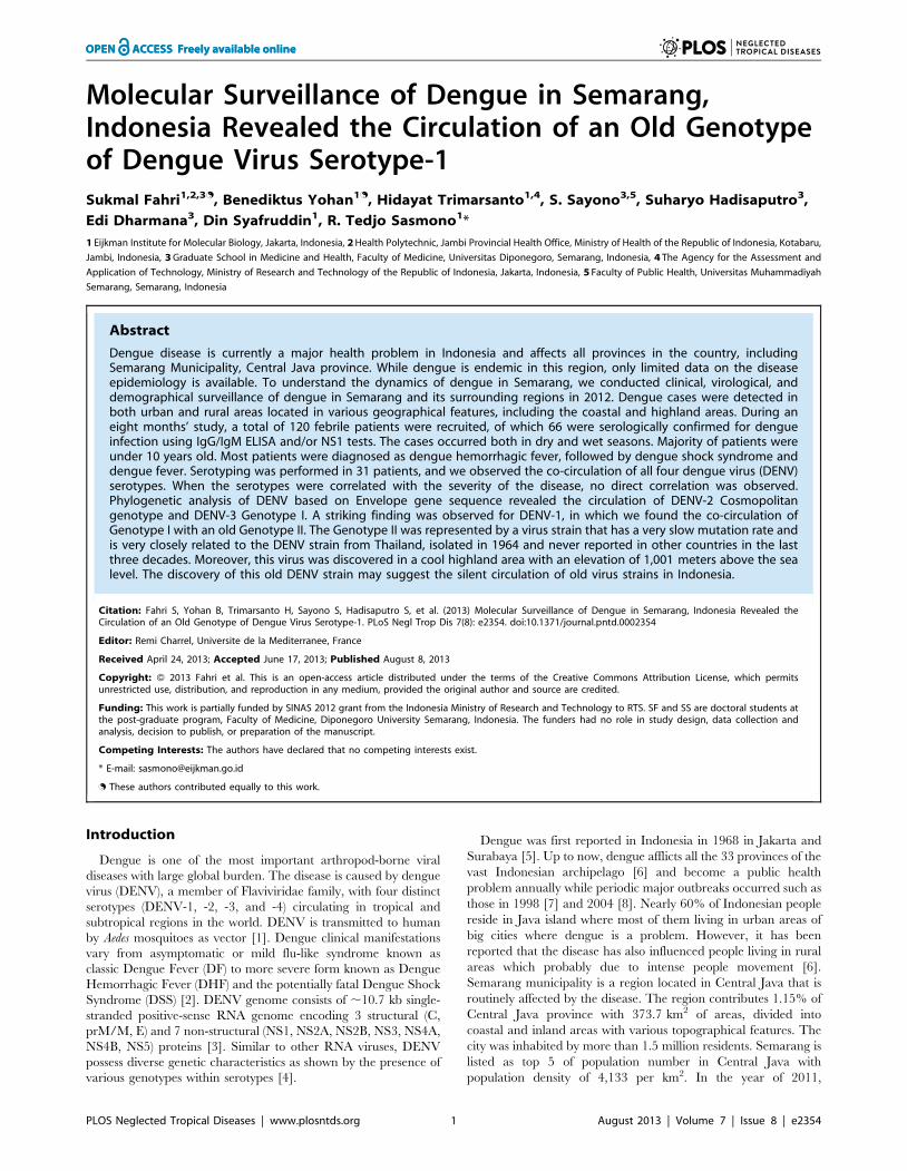
GPS Garmin 72H and mapped (Figure 1).
Clinical and laboratory examinationSera from dengue-suspected patients were subjected to serology
tests. Serological analysis was performed in sample collection sites
using Panbio Dengue Duo Cassette (Alere, Brisbane, Australia) to
detect anti-dengue IgG and IgM. Serology test results were
confirmed using Panbio Dengue Duo ELISA (Alere) in a
laboratory setting, which was also used to determine the
primary/secondary infection status of patients. Detection of
DENV NS1 antigen was done using Panbio Dengue Early Rapid
kit (Alere). Clinical symptoms were documented and laboratory
tests including thrombocyte count and hematocrit level were
performed as a routine procedure in the hospital. Dengue
classification was based on WHO/TDR 2009 guidelines [9].
RNA extraction and reverse transcriptase-polymerasechain reaction (RT-PCR)
RT-PCR confirmations were performed to detect the presence
of DENV in 66 NS1 and/or IgM-positive samples. Virus RNA was
extracted from serum samples using QiaAmp Viral RNA Mini kit
(Qiagen, Hilden, Germany) according to manufacturer’s instruc-
tions. DENV nucleic acid detection and serotyping were performed
using two steps conventional RT-PCR according to protocol
previously described by Lanciotti, et al. [10]. Detection and
serotyping results were confirmed by quantitative real-time RT-
PCR using Simplexa Dengue Molecular Assay performed in 3M
Integrated Cycler machine (Focus Diagnostic, Cypress, CA). Detail
method of the Simplexa Dengue Molecular Assay was as described
by the manufacturer. To determine the positivity of the samples,
the cycle threshold (Ct) cut off value of 38 (instead of 40) was used to
ensure the strict detection and serotyping of the DENV.
Virus isolation using cell cultureSerum samples with serological or RT-PCR-positive result were
subjected to a maximum of two passages of inoculation in C6/36
(Aedes albopictus, mid gut) cell line [11]. Briefly, monolayer of cells
was inoculated with 200 ml of sera in 2 ml of 16RPMI medium
supplemented with 2% of Fetal Bovine Serum (FBS), 2 mM of l-
glutamine, 100 U/ml of Penicillin, and 100 mg/ml of Streptomy-
cin (all from Gibco-Life Technologies, Carlsbad, CA). Flasks were
incubated for 1 hour at 28uC to allow virus attachment. Following
the incubation period, inoculation medium was discarded and the
medium was replenished with 3 ml of fresh medium. Infected cells
were incubated at 28uC for up to 14 days.
DENV Envelope gene sequencingDENV genotyping was performed using Envelope (E) gene
sequence (1,485 nt in length). DENV RNA was reverse-tran-
scribed into cDNA using Superscript III reverse transcriptase (RT)
(Invitrogen-Life Technologies) and DENV-specific primers de-
scribed elsewhere [12]. The resulting cDNA was then used as
template for PCR amplification using Pfu Turbo Polymerase
(Stratagene-Agilent Technologies, La Jolla, CA). PCR products
were purified from 0.8% agarose gel using QIAquick gel
extraction kit (Qiagen) and used in cycle sequencing reaction
performed using 6 overlapping primers [12] from both strands and
BigDye Dideoxy Terminator sequencing kits v3.1 (Applied
Biosystems-Life Technologies), following manufacturer’s instruc-
tions. Purified DNA was subjected to capillary sequencing
performed on 3130xl Genetic Analyzer (Applied Biosystems) at
the Eijkman Institute sequencing facility. Primers used in
genotyping were described elsewhere [12]. Sequence reads were
assembled using SeqScape v.2.5 software (Applied Biosystems)
with additional manual adjustment performed when manual
inspection of the assembly showed some discrepancies. The E
protein gene sequences obtained in this study have been deposited
in GenBank with accession number KC589008–KC589013
(Supplementary Table S1).
Author Summary
We studied dengue disease in Semarang municipality,Central Java, one of the endemic regions in Indonesia. Thedisease occurred in wide geographical regions whichinclude urban, rural, coastal, and highland areas. All fourdengue virus serotypes were found. The infecting sero-types were not associated with disease severities. We alsodetermined the genotype of the circulating viruses. One ofthe interesting findings was the presence of an oldgenotype of DENV-1 which has never been reported inthe last three decades, which may suggest the silentcirculation of this particular genotype in Semarang. Thesefindings offer the first information of the clinical, virolog-ical and demographical aspects of the dengue disease inSemarang, Indonesia.
Dengue in Semarang, Indonesia
PLOS Neglected Tropical Diseases | www.plosntds.org 2 August 2013 | Volume 7 | Issue 8 | e2354
Figure 1. Study area around the Semarang region. Colored areas represent the three districts involved in this study. Black dots represent thelocations were the cases occurred.doi:10.1371/journal.pntd.0002354.g001
Dengue in Semarang, Indonesia
PLOS Neglected Tropical Diseases | www.plosntds.org 3 August 2013 | Volume 7 | Issue 8 | e2354

DENV genotype analysisFor genotype classification, we grouped the isolate sequences
with the relevant reference sequences based on classifications by
Goncalvez et al. [13], Twiddy et al. [14], and Lanciotti et al. [15]
for DENV-1, -2 and -3, respectively. MrBayes was used to
construct Bayesian inference phylogenetic trees with mixed model
across GTR space model and gamma rates for one million
generations with 4 chains, sampled every 1,000 iterations. For
evolution studies, we downloaded all publicly available DNA
sequences of DENV-1, -2 and -3 from NCBI GenBank as of 12
December 2012. Sample sequences were combined with the
downloaded GenBank sequences according to sample’s serotypes
to create dataset for each dengue serotype. Sequence clustering
was performed on each dataset using USEARCH [16]. Multiple
alignments resulted from sequence clustering from each cluster
containing sample sequences were trimmed to obtain only the
alignment representing E protein segment. Sequences without E
protein segment were removed from the alignment. We built
phylogenetic tree using FastTree [17] for fast approximation with
GTR and gamma rate evolution model. We selected 20 closest
public sequences from each isolate sequence based on the patristic
distance of the FastTree’s phylogenetic trees. The multiple
alignment of the selected sequences along with the sequence
samples were used for phylogenetic reconstruction using Bayesian
MCMC method as implemented in BEAST v 1.7.2 [18] using
GTR+C4 model with codon model, relaxed uncorrelated lognor-
mal molecular clock and Bayesian skyline prior, with 60 million
generations and sampled for every 1000th iteration.
Statistical analysisStatistical analysis was performed using SPSS Statistics software
version 11.5 (SPSS Inc., Chicago, IL). Pearson chi-square test was
used to correlate the clinical manifestations and DENV serotypes.
One-way ANOVA test was used to compare groups of laboratory
tests results and DENV serotypes. A probability value of p,0.05
was considered statistically significant.
Results
Dengue prevalence, demography, and topographyOne hundred and twenty febrile patients were recruited during
the course of the study. The dengue cases were equally distributed
throughout the month of January–July 2012 with the most cases
observed in May (Figure 2A). Of 120 patients, 66 (55%) patients
were serologically positive for dengue as determined by IgM/IgG
and/or NS1 tests. RT-PCR confirmations were performed to
detect the presence of virus on those 66 serum samples and 31
(47%) samples were dengue positive. Of the 66 serologically
positive dengue cases, most cases (n = 51 or 77%) were secondary
infections, as determined by IgM and IgG ELISA according to
kit’s manual. The male to female ratio was 1.0 with an average age
of 15.98612.16 (CI 95% 12.99–18.98). Cases occurred predom-
inantly in children aged below 10 year old (42%) (Figure 2B).
Among serologically-positive patients, most of them are school
children/student (41.7%), followed by working adult (33.3%) and
toddler/pre-school children/unemployed adults (25%) (data not
shown).
Of the 31 RT-PCR-confirmed dengue cases, serotyping
revealed the predominance of DENV-1 (35.5%), followed by
DENV-2 (12.9%), DENV-3 (12.9%), and DENV-4 (9.7%). We
also observed the presence of multiple infections of different
serotypes (29%), of which DENV-1 involved in most of the
multiple infections (Figure 3A). These multiple infections were
confirmed by Simplexa Dengue Molecular Assay.
Dengue vector distribution is influenced by the local temper-
ature where the mosquitoes circulating. To determine whether
topological aspect give account to the dengue cases in Semarang,
we performed dengue surveillance encompassing areas with
various topological and geographical features. We obtained
dengue cases in either coastal or highland areas. Most cases were
found in areas with the altitude of 0–250 masl. However, a single
case was also found in area with the altitude of 1,001 masl, in
which the local temperature in daytime was approximately 24–
26uC (Figure 1 and 3B).
Clinical manifestationAll dengue-confirmed patients underwent clinical examination
and laboratory tests at enrollment. The severity of their clinical
manifestations was categorized based on WHO 2009 guidelines
grading criteria. Of the 66 dengue-confirmed patients, most of
them (n = 56 or 84%) were classified as DHF. To assess whether
there is correlation between serotype and clinical manifestation,
we compiled the patients’ data and summarized them in Table 1.
Of the 31 cases confirmed by RT-PCR detection, 23 (74%) cases
were DHF, and 8 (26%) cases were DSS (Table 1). We did not
find any statistically significant correlation between clinical
manifestations and medical laboratory examination results of
the patients with the infecting DENV serotypes.
DENV genotypes and evolution distributionIn order to study the circulating DENV genotypes in Semarang,
we performed genotyping analysis based on E gene sequences.
The DENV E gene sequences were aligned with reference
sequences to generate genotype classifications in each serotype.
The resulting phylogenetic trees for the genotype grouping are
described in Supplementary Figures.
Of 11 isolates that were serotyped as DENV-1, three viruses
were successfully PCR-amplified for their E genes after a single
passage in C6/36 cell line. Phylogenetic analysis revealed the
grouping of those isolates into two different genotypes, Genotype I
(SMG-SE058 and SMG-SE059) and Genotype II (SMG-SE003)
based on Goncalvez classification [13] (Supplementary Figure S1).
The grouping of Semarang DENV-1 into Genotype II is a new
information given this genotype never been found in Indonesia
previously. Meanwhile, the Genotype I of DENV-1 is quite
commonly found in Indonesia [12,19].
We further analyzed the evolutionary history and rate of the
DENV-1 from Semarang. Of 20 closely-related sequences of each
Semarang isolates from published sequences in GenBank, a total
of 56 non-redundant, unique sequences were used for further
analysis consisting of mainly isolates from Singapore, China, and
imported cases in Taiwan. Because the lack of public sequences for
Genotype II, the 20 closely-related sequences for the Genotype II
isolate (SMG-SE003) included other genotypes such as Genotype
I, III and IV. As shown in Figure 4, this particular isolate was
closely related to DENV-1 isolated in Thailand in 1964 [20],
which was used as a strain for vaccine development [21]. The
mean evolutionary rate of DENV-1 as calculated by BEAST was
2.7261024 subs/site/year [95% Highest Posterior Density/HPD:
1.24–4.5461024].
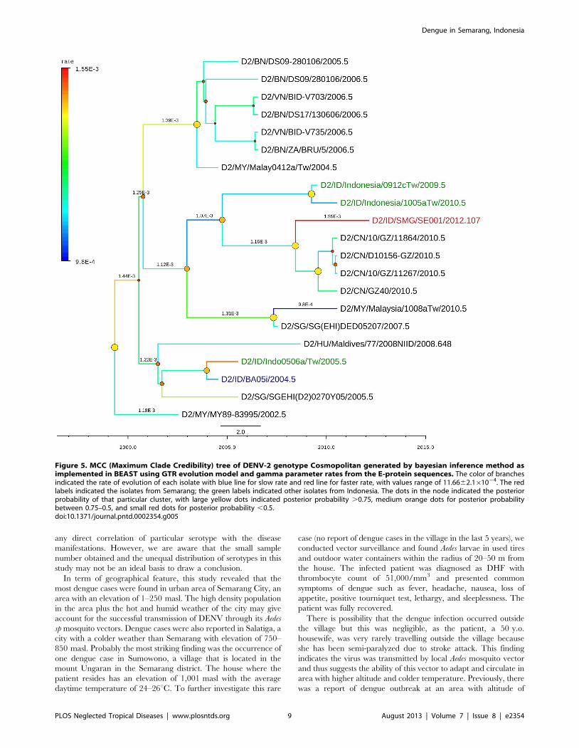
For DENV-2, we managed to genotype one isolate which was
grouped into Cosmopolitan genotype according to Twiddy
classification [14] (Supplementary Figure S2). This genotype is
commonly found in Indonesia and surrounding countries. The
collected 20 closely-related sequences of the Semarang isolate
consisted of sequences from Indonesia and neighboring countries
such as Singapore, Malaysia and Brunei, as well as other Asian
countries such as China, Taiwan, and Vietnam. The Semarang
Dengue in Semarang, Indonesia
PLOS Neglected Tropical Diseases | www.plosntds.org 4 August 2013 | Volume 7 | Issue 8 | e2354

DENV-2 isolate clustered together with Jakarta DENV-2 isolated
in 2004 as indicated in Figure 5. The mean rate of evolution of E-
protein in this data set was 11.6661024 subs/site/year [95%
HPD: 6.04–17.6861024].
We also successfully genotyped two isolates of DENV-3 from
Semarang, which were grouped into Genotype I based on
Lanciotti classification [15] (Supplementary Figure S3). Figure 6
showed that the majority of 20 non-redundant, unique closely-
related sequences from these isolates were Indonesian isolates,
either collected previously in other regions in Indonesia such as
Yogyakarta (in 1988), Palembang (in 1998) [7], and Jakarta (in
2004) [12] or collected as imported cases such as from Queens-
land, Australia (of Bali origin in 2010) [22] and from Taiwan. This
suggests the endemicity of this genotype in the area. Other isolates
from Asian countries such as Singapore, Malaysia and China were
also included. The mean evolutionary rate of DENV-3 was
7.2461024 subs/site/year [95% HPD: 5.08–9.5061024].
In this study, we also detected three DENV-4 infections
(Figure 3A), however, we were not able to PCR-amplify the E
gene from the isolates to be used for genotyping.
Figure 2. Dengue cases monthly distribution (A) and patients’ age (B). All serologically positive dengue patients were grouped according totheir hospital admission date and ages.doi:10.1371/journal.pntd.0002354.g002
Dengue in Semarang, Indonesia
PLOS Neglected Tropical Diseases | www.plosntds.org 5 August 2013 | Volume 7 | Issue 8 | e2354

Discussion
We described here the clinical, virological, and demographical
features of dengue in Semarang, the capital city of Central Java
province, and its surrounding regions. The study is somewhat
unique as it involved the survey of those features in various regions
with different topography, encompassing the coastal, urban/inner
city and rural/inland areas, as well as highland areas with the
elevation of more than 1,000 m above sea level. Our surveillance
was conducted from December 2011 until July 2012. This
duration encompasses the rainy and dry season periods, as in
Indonesia the wet season is commonly occurred during October–
April while the dry season occurred in April–October. The dengue
cases were equally detected throughout January to July with the
recorded peak in May. Data from Semarang Meteorology Bureau
indicated that May to June 2012 was the transition months
Figure 3. Dengue virus serotype distribution (A) and the altitude of dengue cases (B). Serotypes were determined using bothconventional RT-PCR and real-time RT-PCR as described in the Method section. The altitudes of the locations where the cases occurred were recordedusing altimeter.doi:10.1371/journal.pntd.0002354.g003
Dengue in Semarang, Indonesia
PLOS Neglected Tropical Diseases | www.plosntds.org 6 August 2013 | Volume 7 | Issue 8 | e2354

between wet and dry seasons (data not shown); therefore the high
dengue incidence in this month is understandable. The dengue
mosquito vectors might still actively breed soon after the decrease
of rainfall, since heavy rainfall may wash out the breeding sites
while lower rain intensity will maintain the breeding sites.
A total of 120 dengue-suspected patients were recruited in this
study. Of this, 66 (55%) patients were serologically confirmed for
dengue infection, suggesting that dengue places a considerable
burden in the community. Molecular detection revealed the
presence of DENV in 31 (47%) patients’ sera. Results from
serotyping identified the presence of all DENV serotypes in
Semarang, with DENV-1 as the predominant serotype, followed
by DENV-2, -3, and -4 (Figure 3A). Using a real-time quantitative
RT-PCR detection system with a strict standard for detecting the
presence of DENV genomes, we observed the presence of multiple
DENV infections in nine (29%) out of 31 samples, with DENV-1
involved in most of the multiple infections. This finding further
supports the hyper-endemicity of the disease and the predomi-
nance of DENV-1. The DENV-1 predominance is currently in
place in other cities in Indonesia including in Surabaya [19] and
Makassar (Sasmono et al, 2013, submitted for publication).
However, historical data of DENV serotype distribution in
Indonesia reported the predominance of DENV-2 and -3 in
several cities [6,12]. As there has been a very limited data on
dengue serotype distribution in Semarang, we were not able to
conclude whether the current serotype replaced the previous
serotype predominance.
On the clinical aspect of dengue in Semarang, we documented
the clinical symptoms and medical laboratory tests results and
grouped the clinical manifestation according to WHO 2009
Table 1. Characteristics of 31 RT-PCR-confirmed dengue patients involved in this study.
Parameters N DENV-1 DENV-2 DENV-3 DENV-4 Mix infection p valuec
Gender 0.258
Male 14 5 0 3 1 5
Female 17 6 4 1 2 4
Infection type 0.919
Primary 6 2 1 1 0 2
Secondary 25 9 3 3 3 7
NS1 antigen detection 0.231
Positive 25 10 3 3 1 8
Negative 6 1 1 1 2 1
Severity 0.486
DHF 23 9 3 4 2 5
DSS 8 2 1 0 1 4
Clinical/lab features
Fever.37uC 31 11 4 4 3 9 NA
Headache 29 10 4 4 3 8 0.877
Retro-orbital pain 28 9 4 4 3 8 0.716
Myalgia 24 7 3 4 2 8 0.517
Arthralgia 20 6 3 2 2 7 0.784
Nausea 23 7 2 3 3 8 0.417
Loss of appetite 25 9 3 2 3 8 0.461
Rash 13 5 1 2 0 5 0.480
Bleeding 8 2 1 1 0 4 0.556
Leucopenia 14 5 2 2 0 5 0.568
Tourniquet test positive 14 6 0 3 0 5 0.092
Dyspnea 2 1 0 0 0 1 0.877
Abdominal pain 25 8 3 4 3 7 0.900
Mucosal bleeding 10 3 1 1 1 4 0.920
Lethargy 24 8 3 4 3 6 0.594
Restlessness 18 7 0 4 1 6 0.048
Drowsiness 16 4 2 3 2 5 0.093
Allergy 2 0 0 2 0 0 0.006
Thrombocyte counta NA 77,818630,619 66,750622,081 71,000623,338 69,666610,016 55,111628,162 0.484
Hematocrit levelb NA 33.368.6 35.068.4 31.369.9 33.769.5 32.669.4 0.982
aThrombocyte count in Mean cells/mm3 6 SD.bHematocrit level in Mean % 6 SD.cPearson’s Chi-squared test, except for thrombocyte count and hematocrit level by One-way ANOVA.doi:10.1371/journal.pntd.0002354.t001
Dengue in Semarang, Indonesia
PLOS Neglected Tropical Diseases | www.plosntds.org 7 August 2013 | Volume 7 | Issue 8 | e2354

guideline. In our study, we observed the occurrence of DF, DHF
and DSS in dengue-confirmed patients involved in this study.
Most of the cases were manifested as DHF, followed by DSS and
DF. This finding is understandable as the surveillance was
conducted in either health care center or hospital. Based on
serological data, most patients (77%) were secondary infection.
This indicates sustained disease intensity over a number of years
and the endemicity of dengue in the region. There have been
studies reporting the association of DENV serotype with clinical
manifestation [23–25], in which particular serotypes have been
correlated with the severity of the disease. To understand the role
of each serotype in influencing the clinical outcomes of the disease,
we compared the clinical findings of 31 serotyped dengue cases
against each serotype. As shown in the Table 1, we did not observe
Figure 4. MCC (Maximum Clade Credibility) tree of DENV-1 genotype I and II generated by bayesian inference method asimplemented in BEAST using GTR evolution model and gamma parameter rates from the E-protein sequences. The color of branchesindicated the rate of evolution of each isolate with blue line for slow rate and red line for faster rate, with values range of 2.7262.161024. The redlabels indicated the isolates from Semarang; the green labels indicated other isolates from Indonesia. The dots in the node indicated the posteriorprobability of that particular cluster, with large yellow dots indicated posterior probability .0.75, medium orange dots for posterior probabilitybetween 0.75–0.5, and small red dots for posterior probability ,0.5.doi:10.1371/journal.pntd.0002354.g004
Dengue in Semarang, Indonesia
PLOS Neglected Tropical Diseases | www.plosntds.org 8 August 2013 | Volume 7 | Issue 8 | e2354
any direct correlation of particular serotype with the disease
manifestations. However, we are aware that the small sample
number obtained and the unequal distribution of serotypes in this
study may not be an ideal basis to draw a conclusion.
In term of geographical feature, this study revealed that the
most dengue cases were found in urban area of Semarang City, an
area with an elevation of 1–250 masl. The high density population
in the area plus the hot and humid weather of the city may give
account for the successful transmission of DENV through its Aedes
sp mosquito vectors. Dengue cases were also reported in Salatiga, a
city with a colder weather than Semarang with elevation of 750–
850 masl. Probably the most striking finding was the occurrence of
one dengue case in Sumowono, a village that is located in the
mount Ungaran in the Semarang district. The house where the
patient resides has an elevation of 1,001 masl with the average
daytime temperature of 24–26uC. To further investigate this rare
case (no report of dengue cases in the village in the last 5 years), we
conducted vector surveillance and found Aedes larvae in used tires
and outdoor water containers within the radius of 20–50 m from
the house. The infected patient was diagnosed as DHF with
thrombocyte count of 51,000/mm3 and presented common
symptoms of dengue such as fever, headache, nausea, loss of
appetite, positive tourniquet test, lethargy, and sleeplessness. The
patient was fully recovered.
There is possibility that the dengue infection occurred outside
the village but this was negligible, as the patient, a 50 y.o.
housewife, was very rarely travelling outside the village because
she has been semi-paralyzed due to stroke attack. This finding
indicates the virus was transmitted by local Aedes mosquito vector
and thus suggests the ability of this vector to adapt and circulate in
area with higher altitude and colder temperature. Previously, there
was a report of dengue outbreak at an area with altitude of
Figure 5. MCC (Maximum Clade Credibility) tree of DENV-2 genotype Cosmopolitan generated by bayesian inference method asimplemented in BEAST using GTR evolution model and gamma parameter rates from the E-protein sequences. The color of branchesindicated the rate of evolution of each isolate with blue line for slow rate and red line for faster rate, with values range of 11.6662.161024. The redlabels indicated the isolates from Semarang; the green labels indicated other isolates from Indonesia. The dots in the node indicated the posteriorprobability of that particular cluster, with large yellow dots indicated posterior probability .0.75, medium orange dots for posterior probabilitybetween 0.75–0.5, and small red dots for posterior probability ,0.5.doi:10.1371/journal.pntd.0002354.g005
Dengue in Semarang, Indonesia
PLOS Neglected Tropical Diseases | www.plosntds.org 9 August 2013 | Volume 7 | Issue 8 | e2354

1,700 masl in Guerrero State, Mexico, in 1988 [26]. Therefore the
presence of this vector in high altitude area is not impossible.
Nevertheless, the occurrence of the dengue cases in this highland
area, to the best of our knowledge, represents the first report in
Indonesia. A more detail data on the vector surveillance in the
study area will be described elsewhere (Fahri et al, 2013,
unpublished results).
The genotype of DENV circulating in Semarang was
determined by phylogenetic analysis of the E gene of the DENV.
For the DENV-1, based on classification by Goncalvez [13], we
observed the presence of Genotype I circulating in the region. The
SMG-SE058 isolate was clustered with the Singaporean samples
isolated in 2008 [27], and had TMRCA (time to most recent
common ancestor) around year 1999. The SMG-SE059 isolate
was clustered together with Taiwan isolate (0705aTw) in 2007
originated from Indonesia as stated imported case [28], and
Korean isolate (DenKor-11) in 2008 from a traveler who visited
Indonesia. The TMRCA for this clade is around year 2002. This
indicates that strains from these DENV-1 Genotype I clades are
likely to have been circulating in Semarang more than a decade.
This genotype is currently predominant and common in Indonesia
and has been reported to replace the previously predominant
Genotype IV [12,19]. In this study, we did not find the Genotype
IV in Semarang area, which may be present but not sampled and
genotyped.
The other genotype of DENV-1 that was discovered was the
Genotype II, based on Goncalvez classification [13]. This is a
novel DENV-1 genotype in Indonesia, as it has never been
reported before. This genotype also has never been spotted in
other countries in the last three decades. This genotype was
Figure 6. MCC (Maximum Clade Credibility) tree of DENV-3 genotype I generated by bayesian inference method as implemented inBEAST using GTR evolution model and gamma parameter rates from the E-protein sequences. The color of branches indicated the rateof evolution of each isolate with blue line for slow rate and red line for faster rate. The red labels indicated the isolates from Semarang; the greenlabels indicated other isolates from Indonesia. The dots in the node indicated the posterior probability of that particular cluster, with large yellowdots indicated posterior probability .0.75, medium orange dots for posterior probability between 0.75–0.5, and small red dots for posteriorprobability ,0.5.doi:10.1371/journal.pntd.0002354.g006
Dengue in Semarang, Indonesia
PLOS Neglected Tropical Diseases | www.plosntds.org 10 August 2013 | Volume 7 | Issue 8 | e2354

isolated from the patient resides in Sumowono village (elevation
1,001 masl) described above. From all publicly available DENV-1
sequences in GenBank, this isolate was found to be very closely
related with DENV-1 strain 16007 isolated from patient in
Thailand having dengue hemorrhagic fever and shock in 1964
[20], as well as a virus strain which has been undergone serial
passages from the parental 16007 isolate (strain 16007(PDK-13))
produced during vaccine development [21]. The evolution rate of
Indonesian SMG-SE003 was very slow (6.9261025 mutation/
site/year) compared to the general rate of DENV-1 in this analysis
(2.7261024 mutation/site/year), as indicated by the blue branch
line in Figure 4. The high mutation rate of strain 16007(PDK-13),
indicated by bright red branch line, was most likely the effect of
serial passages process. Another interesting finding related to this
strain was that the dengue NS1 antigen test performed in patient’s
serum was negative, which may raise question if this virus escaped
from detection by NS1 diagnostic test. However, the IgM/IgG
ELISA confirmed the dengue infection for this sample. Further
study is needed to fully understand this finding.
On the origin of this Genotype II strain in Indonesia, one may
suspect a possibility of DENV contamination from lab strains
during the process of culture, PCR and sequencing. However, this
is very unlikely since our lab has never handled and manipulated
DENV-1 strains from outside Indonesia (except DENV-1 refer-
ence strains Nauru-Western Pacific and Hawaii-USA which do
not belong to this genotype). Therefore, this finding reflects the
reappearance of an old and unique strain of DENV-1 in Central
Java, Indonesia. Considering that SMG-SE003 isolate is closely
related to a strain from Thailand isolated in 1964 and has very
slow mutation rate, there is a possibility that either this strain has
been actually present for a long time and is maintained in nature
in a low circulation and infection rate but only sampled now, or
that this strain has been dormant and recently emerged into
circulation. We are not sure whether this strain will behave like the
Thailand strain which caused DHF if it actively re-infecting
humans. We are also not sure if it will be spreading and causing
epidemic in the region, but given the presence of Aedes vector
breeding sites in the proximity of isolate origin, the spreading of
this strain is possible. Further surveillance is needed to monitor the
activity of this strain.
The genotype of DENV-2 circulating in Semarang region was
the Cosmopolitan genotype. This genotype is quite common in the
region and is widely circulated in India, South East Asia, Africa,
the Middle East, and Australia [14]. In Indonesia, this genotype
has been circulating since a long time ago and causing outbreaks in
1998 and 2004 [12]. The phylogenetic analysis of the SMG-SE001
indicated that the isolate share common ancestor with some
isolates circulating in Taiwan, as imported cases from Indonesia
[28,29], and Guangzhou, China. Semarang is one of the regions in
Indonesia that have been supplying workers for countries such as
Taiwan; hence dengue cases found in those particular countries
might be brought by those workers. Semarang is also one of the
regions with high population of Chinese-descendant, with frequent
direct flights between Semarang and Guangzhou, China.
The genotypes of DENV-3 isolates from Semarang were
clustered in genotype I, which is also common in South East Asia
regions. The DENV-3 isolates from Semarang had common
ancestor with the isolates from imported cases in Taiwan, which is
similar situation as DENV-2 isolate. In particular, the cluster
containing these strains had been circulating in Indonesia for more
than a decade.
The mean evolution rates of the DENV in this study were
within the range observed by other studies, which were in the
range of 4.6–11.661024 subs/site/year [30]. With the exception
of DENV-1 Genotype II isolate SMG-SE003, all rates of
individual isolates were within the boundary of the 95% HPD of
each corresponding serotypes. This is another indication that this
Genotype II isolate warrants a further investigation.
In this study, we were unable to obtain any DENV-4 sequences
from Semarang cases. This might be attributed to low viral titers
which can only be detected by real-time RT-PCR method, but not
sufficient enough for conventional PCR amplification for sequenc-
ing purposes. We are aware that there are methods that utilize
shorter fragment of E gene that could be used in determining the
genotype of our DENV-4, and we might apply this in the future to
better understand the genetic aspects of DENV-4 in Semarang.
In conclusion, we have described the clinical, virological, and
demographical features of dengue in Semarang in which all
serotypes are circulating and highlighted the presence of an old
genotype of DENV-1. We also observed the occurrence of dengue in
area with high altitude. Altogether, the study suggests the importance
of continuous virus surveillance in dengue endemic regions such as
Indonesia to better understand the dynamic of the disease.
Supporting Information
Figure S1 Summary tree of DENV-1 genotype grouping
generated by bayesian inference method as implemented in
MrBayes from the E-protein sequences. The Semarang isolates
(red font) were grouped into genotype I (SMG-SE058 and SMG-
SE059) and genotype II (SMG-SE003) based on classification by
Goncalves [13]. The posterior probabilities of the clades, indicated
as numbers in the node labels, were shown only for major clades.
(PDF)
Figure S2 Summary tree of DENV-2 genotype grouping
generated by bayesian inference method as implemented in
MrBayes from the E-protein sequences. The Semarang isolate
(SMG-SE001) was grouped into Cosmopolitan genotype, based on
classification by Twiddy [14]. The posterior probabilities of the
clades, indicated as numbers in the node labels, were shown only
for major clades.
(PDF)
Figure S3 Summary tree of DENV-3 genotype grouping
generated by bayesian inference method as implemented in MrBayes
from E-protein sequences. The Semarang isolates (SMG-SE005 and
SMG-SE052) were grouped into genotype I, based on classification
by Lanciotti [15]. The posterior probabilities of the clades, indicated
as numbers in the node labels, were shown only for major clades.
(PDF)
Table S1 Sequenced samples information with the correspond-
ing GenBank accession numbers.
(DOCX)
Acknowledgments
Eijkman Institute is under the auspices of Ministry of Research and
Technology of the Republic of Indonesia. The authors wish to thank the
people who have consented to take a part in this surveillance study, nurses
and staff at the health centers for their assistance and access to the medical
records. We would like to thank Sanofi Pasteur for the donation of
Simplexa Dengue Molecular Assay system. Technical help from T. Yuli
Setianingsih and Febrina Meutiawati is highly appreciated.
Author Contributions
Conceived and designed the experiments: SF BY SH ED DS RTS.
Performed the experiments: SF BY HT. Analyzed the data: SF BY HT SS
RTS. Contributed reagents/materials/analysis tools: SF BY HT SS RTS.
Wrote the paper: SF BY HT RTS.
Dengue in Semarang, Indonesia
PLOS Neglected Tropical Diseases | www.plosntds.org 11 August 2013 | Volume 7 | Issue 8 | e2354

References
1. Simmons CP, Farrar JJ, Nguyen van VC, Wills B (2012) Dengue. N Engl J Med
366: 1423–1432. doi:10.1056/NEJMra1110265.
2. Martina BEE, Koraka P, Osterhaus ADME (2009) Dengue virus pathogenesis:
an integrated view. Clin Microbiol Rev 22: 564–581. doi:10.1128/CMR.00035-
09.
3. Guzman MG, Halstead SB, Artsob H, Buchy P, Farrar J, et al. (2010) Dengue: a
continuing global threat. Nat Rev Microbiol 8: S7–16. doi:10.1038/nrmi
cro2460.
4. Holmes EC (2009) RNA virus genomics: a world of possibilities. J Clin Invest
119: 2488–2495. doi:10.1172/JCI38050.
5. Sumarmo (1987) Dengue haemorrhagic fever in Indonesia. Southeast
Asian J Trop Med Public Health 18: 269–274.
6. Setiati TE, Wagenaar JF, De Kruif MD, Mairuhu AT, Van Gorp EC, et al.
(2006) Changing epidemiology of dengue haemorrhagic fever in Indonesia. Bull
WHO 30: 1–14.
7. Corwin AL, Larasati RP, Bangs MJ, Wuryadi S, Arjoso S, et al. (2001) Epidemic
dengue transmission in southern Sumatra, Indonesia. Trans R Soc Trop Med
Hyg 95: 257–265.
8. Suwandono A, Kosasih H, Nurhayati, Kusriastuti R, Harun S, et al. (2006) Four
dengue virus serotypes found circulating during an outbreak of dengue fever and
dengue haemorrhagic fever in Jakarta, Indonesia, during 2004. Trans R Soc
Trop Med Hyg 100: 855–862.
9. TDR/WHO (2009) Dengue: guidelines for diagnosis, treatment, prevention and
control. Geneva: World Health Organization.
10. Lanciotti RS, Calisher CH, Gubler DJ, Chang GJ, Vorndam AV (1992)
Rapid detection and typing of dengue viruses from clinical samples by
using reverse transcriptase-polymerase chain reaction. J Clin Microbiol 30:
545–551.
11. Igarashi A (1979) Characteristics of Aedes albopictus cells persistently infected
with dengue viruses. Nature 280: 690–691.
12. Ong SH, Yip JT, Chen YL, Liu W, Harun S, et al. (2008) Periodic re-emergence
of endemic strains with strong epidemic potential-a proposed explanation for the
2004 Indonesian dengue epidemic. Infect Genet Evol 8: 191–204.
13. Goncalvez AP, Escalante AA, Pujol FH, Ludert JE, Tovar D, et al. (2002)
Diversity and evolution of the envelope gene of dengue virus type 1. Virology
303: 110–119.
14. Twiddy SS, Farrar JJ, Vinh Chau N, Wills B, Gould EA, et al. (2002)
Phylogenetic relationships and differential selection pressures among genotypes
of dengue-2 virus. Virology 298: 63–72.
15. Lanciotti RS, Lewis JG, Gubler DJ, Trent DW (1994) Molecular evolution and
epidemiology of dengue-3 viruses. J Gen Virol 75 (Pt 1): 65–75.
16. Edgar RC (2010) Search and clustering orders of magnitude faster than BLAST.
Bioinformatics 26: 2460–2461. doi:10.1093/bioinformatics/btq461.
17. Price MN, Dehal PS, Arkin AP (2010) FastTree 2–approximately maximum-likelihood trees for large alignments. PLoS ONE 5: e9490. doi:10.1371/
journal.pone.0009490.18. Drummond AJ, Rambaut A (2007) BEAST: Bayesian evolutionary analysis by
sampling trees. BMC Evol Biol 7: 214.19. Yamanaka A, Mulyatno KC, Susilowati H, Hendrianto E, Ginting AP, et al.
(2011) Displacement of the predominant dengue virus from type 2 to type 1 with
a subsequent genotype shift from IV to I in Surabaya, Indonesia 2008–2010.PLoS ONE 6: e27322. doi:10.1371/journal.pone.0027322.
20. Halstead SB, Simasthien P (1970) Observations related to the pathogenesis ofdengue hemorrhagic fever. II. Antigenic and biologic properties of dengue
viruses and their association with disease response in the host. Yale J Biol Med
42: 276–292.21. Halstead SB, Marchette NJ (2003) Biologic Properties of Dengue Viruses
Following Serial Passage in Primary Dog Kidney Cells: Studies at the Universityof Hawaii. Am J Trop Med Hyg 69: 5–11.
22. Warrilow D, Northill JA, Pyke AT (2012) Sources of dengue viruses imported
into Queensland, australia, 2002–2010. Emerging Infect Dis 18: 1850–1857.doi:10.3201/eid1811.120014.
23. Balmaseda A, Hammond SN, Perez L, Tellez Y, Saborio SI, et al. (2006)Serotype-specific differences in clinical manifestations of dengue. Am J Trop
Med Hyg 74: 449–456.24. Fried JR, Gibbons RV, Kalayanarooj S, Thomas SJ, Srikiatkhachorn A, et al.
(2010) Serotype-specific differences in the risk of dengue hemorrhagic fever: an
analysis of data collected in Bangkok, Thailand from 1994 to 2006. PLoS NeglTrop Dis 4: e617. doi:10.1371/journal.pntd.0000617.
25. Nisalak A, Endy TP, Nimmannitya S, Kalayanarooj S, Thisayakorn U, et al.(2003) Serotype-specific dengue virus circulation and dengue disease in Bangkok,
Thailand from 1973 to 1999. Am J Trop Med Hyg 68: 191–202.
26. Herrera-Basto E, Prevots DR, Zarate ML, Silva JL, Sepulveda-Amor J (1992)First reported outbreak of classical dengue fever at 1,700 meters above sea level
in Guerrero State, Mexico, June 1988. Am J Trop Med Hyg 46: 649–653.27. Lee KS, Lai YL, Lo S, Barkham T, Aw P, et al. (2010) Dengue virus surveillance
for early warning, Singapore. Emerging Infect Dis 16: 847–849. doi:10.3201/eid1605.091006.
28. Shu P-Y, Su C-L, Liao T-L, Yang C-F, Chang S-F, et al. (2009) Molecular
characterization of dengue viruses imported into Taiwan during 2003–2007:geographic distribution and genotype shift. Am J Trop Med Hyg 80: 1039–1046.
29. Huang J-H, Su C-L, Yang C-F, Liao T-L, Hsu T-C, et al. (2012) Molecularcharacterization and phylogenetic analysis of dengue viruses imported into
Taiwan during 2008–2010. Am J Trop Med Hyg 87: 349–358. doi:10.4269/
ajtmh.2012.11-0666.30. Chen R, Vasilakis N (2011) Dengue–quo tu et quo vadis? Viruses 3: 1562–1608.
doi:10.3390/v3091562.
Dengue in Semarang, Indonesia
PLOS Neglected Tropical Diseases | www.plosntds.org 12 August 2013 | Volume 7 | Issue 8 | e2354


![Sentinel versus passive surveillance for measuring changes ... · PDF file19.02.2016 · 3 79 Although laboratory-based sentinel surveillance has been recommended for dengue [9], 80](https://img.dokumen.tips/doc/110x75/5a9e1ab47f8b9ad2298d5060/sentinel-versus-passive-surveillance-for-measuring-changes-79-although-laboratory-based.jpg)