Embed Size (px)
Citation preview

Molecular Recognition of Aldehydes by AldehydeDehydrogenase and Mechanism of Nucleophile ActivationTroy Wymore,1* John Hempel,2 Samuel S. Cho,3 Alexander D. MacKerell, Jr.,3 Hugh B. Nicholas, Jr.,1 andDavid W. Deerfield II1
1Pittsburgh Supercomputing Center, Biomedical Initiative Group, Pittsburgh, Pennsylvania2Department of Biological Sciences, University of Pittsburgh, Pittsburgh, Pennsylvania3University of Maryland, School of Pharmacy, Baltimore, Maryland
ABSTRACT Experimental structural data onthe state of substrates bound to class 3 AldehydeDehydrogenases (ALDH3A1) is currently unknown.We have utilized molecular mechanics (MM) simula-tions, in conjunction with new force field parame-ters for aldehydes, to study the atomic details ofbenzaldehyde binding to ALDH3A1. Our results indi-cate that while the nucleophilic Cys243 must be inthe neutral state to form what are commonly callednear-attack conformers (NACs), these structures donot correlate with increased complexation energycalculated with the MM-Generalized Born Molecu-lar Volume (GBMV) method. The negatively chargedCys243 (thiolate form) of ALDH3A1 also binds benz-aldehyde in a stable conformation but in this com-plex the sulfur of Cys243 is oriented away frombenzaldehyde yet yields the most favorable MM-GBMV complexation energy. The identity of thegeneral base, Glu209 or Glu333, in ALDHs remainsuncertain. The MM simulations reveal structuraland possible functional roles for both Glu209 andGlu333. Structures from the MM simulations thatwould support either glutamate residue as the gen-eral base were further examined with Hybrid Quan-tum Mechanical (QM)/MM simulations. These simu-lations show that, with the PM3/OPLS potential,Glu209 must go through a step-wise mechanism toactivate Cys243 through an intervening water mole-cule while Glu333 can go through a more favorableconcerted mechanism for the same activation pro-cess. Proteins 2004;57:758–771. © 2004 Wiley-Liss, Inc.
Key words: enzyme mechanism; thiolate; aldehydes;general base; molecular dynamics simu-lation; molecular mechanics; hybridQM/MM potentials; force field param-eterization
INTRODUCTION
Detoxification of aldehydes by Aldehyde Dehydroge-nases (ALDHs) is a critical function in healthy cells sincealdehydes are implicated in cytotoxicity, mutagenicity,genotoxicity, and carcinogenesis.1 ALDHs occur in a widevariety of phyla and most organisms have multiple ALDHsthat act on different sets of substrates oxidizing them totheir corresponding carboxylic acid. Human class 3 ALDH(ALDH3A1; referred to as ALDH3 in this text) has been
shown to detoxify a metabolite of the cancer chemothera-peutic agent cyclophosphoamide (CP), leading to drugresistance.2 Therefore, inhibitors of ALDH3 taken with CPdrugs may lead to more effective treatments for thoseexperiencing drug resistance to CP. Designing inhibitorsof ALDH3 should be based on a thorough knowledge of thereaction mechanism. Several insights into the ALDHmechanism of converting benzaldehyde to benzoic acidhave been provided by the crystal structure,3 site-directedmutagenesis,4–6 and NMR.7 These studies provide thebasis by which to carry out advanced molecular modelingstudies on the enzyme.
ALDH3 is a homodimeric enzyme; each subunit containsa nicotinamide adenine dinucleotide (NAD)-binding do-main, a catalytic domain, and an oligomerization domain.3
A 15-Å-long funnel-shaped passage formed between thesedomains leads to the active site. NAD binds to the face ofthe dimer opposite of the funnel entrance. The catalyticcysteine (Cys243) is located in a loop structure connectinghelix-9 with beta strand-7. This loop structure starts withAsn238 and ends with Asp247. Two water molecules liewithin 5.0 Å of the cysteine sulfur atom and allow thepossibility of hydrogen bonds with the backbone amideprotons of Cys243 and Val244. The side-chain amideprotons of Asn114 also may favorably interact with Cys243.There do not appear to be any obvious direct interactionsthat would stabilize the thiolate form of Cys243. Twohighly conserved acidic residues are located near thecatalytic cysteine, Glu209 and Glu333. Either one of theseresidues could serve the function as a general base toactivate Cys243. The side-chain of Glu209 is directedsomewhat away from Cys243 yet still has two watermolecules within 4.0 Å of both carboxylate oxygen atoms.These water molecules are separated from the sulfur of
The Supplementary Materials referred to in this article can be foundat http://www.interscience.wiley.com/jpages/0887-3585/suppmat/index.html
Grant sponsor: NIH-NIAAA; Grant number: AA-12753; Grant spon-sor: NIH-NCRR; Grant number: RR06009; Grant sponsor: NIH; Grantnumber: GM51501.
*Correspondence to: Troy Wymore, Pittsburgh SupercomputingCenter, Biomedical Initiative Group, 4400 Fifth Avenue, Pittsburgh,PA 15213. E-mail: [email protected]
Received 3 November 2003; Accepted 8 June 2004
Published online 10 August 2004 in Wiley InterScience(www.interscience.wiley.com). DOI: 10.1002/prot.20256
PROTEINS: Structure, Function, and Bioinformatics 57:758–771 (2004)
© 2004 WILEY-LISS, INC.
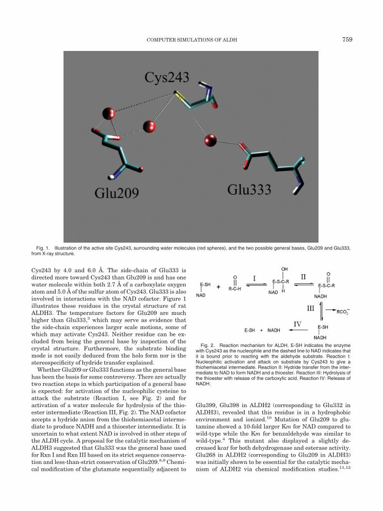
Cys243 by 4.0 and 6.0 Å. The side-chain of Glu333 isdirected more toward Cys243 than Glu209 is and has onewater molecule within both 2.7 Å of a carboxylate oxygenatom and 5.0 Å of the sulfur atom of Cys243. Glu333 is alsoinvolved in interactions with the NAD cofactor. Figure 1illustrates these residues in the crystal structure of ratALDH3. The temperature factors for Glu209 are muchhigher than Glu333,3 which may serve as evidence thatthe side-chain experiences larger scale motions, some ofwhich may activate Cys243. Neither residue can be ex-cluded from being the general base by inspection of thecrystal structure. Furthermore, the substrate bindingmode is not easily deduced from the holo form nor is thestereospecificity of hydride transfer explained.
Whether Glu209 or Glu333 functions as the general basehas been the basis for some controversy. There are actuallytwo reaction steps in which participation of a general baseis expected: for activation of the nucleophilic cysteine toattack the substrate (Reaction I, see Fig. 2) and foractivation of a water molecule for hydrolysis of the thio-ester intermediate (Reaction III, Fig. 2). The NAD cofactoraccepts a hydride anion from the thiohemiacetal interme-diate to produce NADH and a thioester intermediate. It isuncertain to what extent NAD is involved in other steps ofthe ALDH cycle. A proposal for the catalytic mechanism ofALDH3 suggested that Glu333 was the general base usedfor Rxn I and Rxn III based on its strict sequence conserva-tion and less-than-strict conservation of Glu209.8,9 Chemi-cal modification of the glutamate sequentially adjacent to
Glu399, Glu398 in ALDH2 (corresponding to Glu332 inALDH3), revealed that this residue is in a hydrophobicenvironment and ionized.10 Mutation of Glu209 to glu-tamine showed a 10-fold larger Km for NAD compared towild-type while the Km for benzaldehyde was similar towild-type.4 This mutant also displayed a slightly de-creased kcat for both dehydrogenase and esterase activity.Glu268 in ALDH2 (corresponding to Glu209 in ALDH3)was initially shown to be essential for the catalytic mecha-nism of ALDH2 via chemical modification studies.11,12
Fig. 1. Illustration of the active site Cys243, surrounding water molecules (red spheres), and the two possible general bases, Glu209 and Glu333,from X-ray structure.
Fig. 2. Reaction mechanism for ALDH. E-SH indicates the enzymewith Cys243 as the nucleophile and the dashed line to NAD indicates thatit is bound prior to reacting with the aldehyde substrate. Reaction I:Nucleophilic activation and attack on substrate by Cys243 to give athiohemiacetal intermediate. Reaction II: Hydride transfer from the inter-mediate to NAD to form NADH and a thioester. Reaction III: Hydrolysis ofthe thioester with release of the carboxylic acid. Reaction IV: Release ofNADH.
COMPUTER SIMULATIONS OF ALDH 759

Later, Wang and Weiner13 found further support thatGlu268 in ALDH2 served as the general base since bothdehydrogenase and esterase activity were abolished whenthis residue was changed to an aspartate, glutamine, orlysine residue and no pre-steady state burst of NADH wasfound in either reaction of these mutants. Therefore,Glu268 was ascribed the role of general base in both Rxn1and Rxn2. Yet, it was also shown that E399Q in ALDH2showed no pre-steady state burst of NADH either, while itskcat was reduced 10-fold.14 Mann and Weiner5 laterpresented experimental evidence that Glu333 served asthe general base in ALDH3. Since this evidence was basedon the fact that the E333Q mutant destabilized the proteinas well as the results mentioned above, the general basemay be different depending on the form of ALDH. Oneargument against this would be the high degree of se-quence and structural similarity between differentisozymes (ALDH2 vs. ALDH3, for example).15
In this report, we detail our efforts using molecularmodeling methods to characterize the initial events in thecatalytic cycle of ALDH3. For these studies, we consideredseveral possibilities for modeling the Michaelis complex ofbenzaldehyde (BA) with ALDH3. A major challenge in thisprocess is assigning protonation states to ionizable resi-dues, since the ionization states remain constant duringan MD simulation using a molecular mechanics (MM)force field. While continuum methods exist for calculatingpKa’s in proteins,16 these methods are best at detectinglarge shifts in pKa. Their accuracy suffers at modelingsmall shifts in pKa. Thus, in the present study an ap-proach is applied in which MD simulations were per-formed with different ionization states for Cys243 andGlu333 (see Methods). These simulations and the subse-quent post-processing of the trajectories were used todetermine the most energetically favorable conditions forsubstrate binding. The ligand-binding energies were calcu-lated with the MM-Generalized Born Molecular Volume(GBMV) method.17 Burgi and Dunitz have previouslyshown that there is a preferred orientation between nitro-gen- and oxygen-nucleophiles and the plane of the electro-philic carbonyl group.18,19 This Burgi-Dunitz approachtrajectory (BDat) can be defined as an angle of 109°between nucleophile-carbonyl carbon-carbonyl oxygen.Therefore, we examined the orientation of the Cys243sulfur nucleophile with respect to the plane of the sub-strate carbonyl group to determine if this preferred orien-tation was present in ALDH3 as well. A study byChakrabarti and Pal20 of this nucleophile-electrophileinteraction within cysteine residues of metalloproteinstructures has been reported. Structures that lie some-what closer to covalent bonding along the BDat have beencalled near-attack conformers (NACs)21 and we examinedtheir occurrence in the simulations as well. A full assess-ment of any NAC effect would require simulations toestimate the free energy of moving from the bound state toa NAC as well as the free energy profile for formation of thethiohemiacetal intermediate from the NAC and also thereference solution reactions.22,23 A complete account of the
cysteine nucleophilic attack on benzaldehyde is forthcom-ing.
We also employed a hybrid quantum mechanical (QM)/molecular mechanical (MM) potential as the energy func-tion24,25 to simulate possible Cys243 activation mecha-nisms. In the QM/MM approach, atoms making up theactive site are treated explicitly by a QM method and,therefore, can be used to model bond breaking, bondformation, and charge transfer while the surroundings arerepresented by a MM force field. A term in the hybridpotential is used to couple the two regions so that the QMregion is affected by the enzyme environment. This meth-odology has been used in several investigations of enzymereactions.26,27
METHODSOptimization of the Aldehyde Parameters
Parameters were developed for a set of common, simplealdehydes: acetaldehyde, propionaldehyde, chloroacetalde-hyde, and benzaldehyde. The CHARMM potential energyfunction, consisting of external (nonbonded) and internal(bonded) interaction terms, has the general form28:
E � �bonds
Kb(b�b0)2� �angles
K�(���0)2
� �torsions
K��1 � cos�n� � �� � �U�B
KUB�S � S0�2
� �impropers
K� � 0�2
� �nonbonded pairs
�εij��Rmin,ij
rij�12
� 2�Rmin,ij
rij�6� �
qiqj4�Drij
�where Kb, KUB, K�, K�, and Kimp are the bond, Urey-Bradley, angle, dihedral angle, and improper dihedralangle force constants, respectively; b, S, �, �, and are thebond length, Urey-Bradley 1,3-distance, bond angle, dihe-dral angle, and improper torsion angle, respectively, withthe subscript zero representing the equilibrium values forthe individual terms. Coulomb and Lennard-Jones 6–12terms contribute to the nonbonded interactions; is theLennard-Jones well depth and Rmin is the distance at theLennard-Jones minimum, qi is the partial atomic charge,D is the effective dielectric constant, and rij is the distancebetween atoms i and j. The Lennard-Jones parametersbetween pairs of different atoms are obtained from theLorentz-Berthelodt combination rules, in which ij valuesare based on the geometric mean of i and j and Rmin,ij
values are based on the arithmetic mean between Rmin,i
and Rmin,j.The optimization of the force field for the aldehydes was
performed using the same iterative approach previouslyemployed for proteins, nucleic acids, lipids, and otherbiomolecules in the CHARMM force field29–31 insuringtheir compatibility. All of the data obtained for parameteroptimization were determined either via QM calculationsor, when appropriate, from already existing parameters.Initially, the nonbonded interaction parameters were opti-
760 T. WYMORE ET AL.

mized, followed by the internal parameters for the bonds,angles, and torsions. Details of the parameter optimiza-tion are included in the Supplemental Material.
Molecular Dynamics Simulation Using a MMForce Field
The starting coordinates for the rat ALDH3 (1ad33)were taken from the RCSB Protein Data Bank.32 Thisstructure was solved to a resolution of 2.6 Å and containsthe position of the NAD cofactors. CHARMM33 was usedfor all MD simulations using a MM force field. We em-ployed the all-atom force field in CHARMM that containsparameters for the protein29 and NAD.34 The MM param-eterization for benzaldehyde is described in the Supplemen-tal Material. Hydrogens were added to the protein andNAD using the HBUILD algorithm.35 All histidine resi-dues were assigned to be neutral with their tautomericstate determined by the proximity of a hydrogen bondacceptor to either N or N� of histidine. Cys243 wasassigned to its neutral state while Glu209 and Glu333were negatively charged. All other residues were repre-sented in their standard protonation states. The protein-cofactor system was then solvated by TIP3P water mol-ecules and the system was minimized for 2,000 steps withharmonic restraints on all solute heavy atoms. Sodiumions were placed uniformly around the protein with atleast 3 water molecules between each ion and the proteinto neutralize the system. The system contained 13,914protein atoms, 140 NAD atoms (2 NAD molecules, one permonomeric unit), 41,538 water atoms, and 14 sodiumcounterions for a total of 55,606 atoms. SHAKE36 re-straints were used for bonds to hydrogen, which removesthis high-frequency stretching motion. Periodic boundaryconditions were used with the system simulated in theNVT (constant volume, constant temperature) ensemble.The minimization was followed by 30 picoseconds (ps) ofMD in which the velocities were reassigned every 3 ps tocorrespond to a temperature that started at 10 K andended at 310 K. A 1-femtosecond (fs) timestep was used atthis stage. The remainder of all simulations were per-formed in the NPT ensemble, a 2-fs timestep and the use ofparticle mesh Ewald (PME)37 for calculating long-rangeelectrostatic interactions. During the final part of equilibra-tion (last 120 ps of 200 ps) and during the entire produc-tion run, the harmonic restraints on the protein/NAD
solute for the holo form were released. The average dimen-sions of the simulation cell were 104.8 � 71.6 � 72.4 Å.
Our benzaldehyde (BA) model was placed in the activesite of the solvated ALDH system with the followingrestraints that are consistent with observations fromX-ray models of substrates complexed with other ALDHfamily members.38,39 In all simulations, the carbonylcarbon of BA was restrained to be within 2–4 Å of thesulfur of Cys243 and the carbonyl oxygen of BA wasrestrained to be within 2–4 Å of the sidechain nitrogen ofAsn-114 during the first 83 ps of each simulation andremoved during the production phase. Two simulationswere performed with BA in the active site with a neutralCys243 and ionized Glu333. Of these, one simulation hadBA oriented for nucleophilic attack in which the productwould be a thiohemiacetal in the R-configuration whereasthe other simulation was oriented for production of athiohemiacetal in the S-configuration (see Fig. 3). Twoadditional simulations were performed with BA in theactive site; each with Cys243 in the negatively chargedthiolate form. Of these, one had Glu333 negatively charged(labeled S-1-1) and the other had a neutral protonatedform (labeled S-1-0) as shown in Figure 4. The simulationswere performed for 2.2 nanoseconds (ns) for the holo formand 1.063 ns each for the four possible Michaelis complexforms. Analysis of substrate-bound conformations wasperformed in two ways. First, we wanted to determine ifthe enzyme would orient the substrate along the BDat fornucleophilic addition to the carbonyl group and, if so, thenwhat percentage of these structures could be classified as aNAC. A NAC is defined here as a cysteine sulfur–BAcarbonyl carbon distance of 3.5 Å or less and a sulfur-carbonyl carbon (BA)-carbonyl oxygen (BA) of 109.5 �15°.40 In principle, these geometric criteria could be metwith the cysteine sulfur interacting with the aldehydicproton. This would represent an in-plane approach of thenucleophile, which is unfavorable. All NACs identifiedwere out of the aldehyde plane and thus were trulyoriented for reaction.
Next, to assess the relevance of forming structures alongthe BDat and within the NAC region, we divided thetrajectory structures into complex (ALDH, NAD, and BA),receptor (ALDH and NAD), and substrate BA systems. Wethen calculated substrate-binding energies with the MM-GBMV analytical method17 with an added term that
Fig. 3. Two possibilities are shown for benzaldehyde binding in the ALDH3 active site, one in which the Cys243 attack on benzaldehyde would lead toa R-thiohemiacetal (labeled R, left) and one in which the attack would lead to a S-thiohemiacetal (labeled S, right)
COMPUTER SIMULATIONS OF ALDH 761

calculates the non-polar contribution to solvation throughthe solvent-accessible surface area (available in CHARMMversion c30b1). This was done over the last 600 ps (6,200structures) of each trajectory. The �GMM-GBMV energieswere then calculated in the standard way by subtractingthe average ligand and receptor energy from the averagecomplex energy. GBMV solvation energies have beenshown to highly correlate with results from the Poisson-Boltzmann (PB) method.17 The resulting energies are notabsolute binding energies because they neglect the effect ofprotein relaxation on loss of substrate, loss of rotationaland translation motion to the ligand, and other effects.41
Yet, these effects should cancel since we are not looking atdifferent ligands, just slightly different protonation statesof active site residue(s). Hydrogen bond analysis wasperformed with the CORREL module and the hydrogen-bonding parameters in CHARMM.
Modeling Reactions With QM/MM potential
Snapshots from the MM simulations in the presence ofsubstrate that showed the thiol proton of Cys243 pointingin the direction of a carboxylate oxygen of Glu333 orGlu209 that would most support either residue as thegeneral base were used as starting points for simulationswith the QM/MM potential. Each structure had the thiolproton hydrogen bonded to an intervening water molecule,which was hydrogen bonded to either Glu, respectively. Inaddition, the conformations had the substrate carbonylcarbon within 3.5 Å of the sulfur atom of Cys243 along theBDat.18,19 The selected snapshots appeared most relevantfor the reactions we studied but we cannot rule out thatother structures with similar features but with different“second-sphere” geometries would give different results.Water molecules outside 30 Å of the sulfur atom of Cys243and all sodium ions were deleted. We divided up thesystem into the following regions for both simulations: (1)the QM region consisted of the side-chains of Cys243 andGlu333 or Glu209, the BA substrate, and 6 or 7 surround-ing water molecules (46 or 49 QM atoms); (2) the inner MMregion in which atoms that were allowed to move freelywere those residues with one or more atoms within 20 Å ofthe sulfur atom of Cys243; and (3) the outer MM regionthat remained fixed included atoms in residues with noatoms within 20 Å of the sulfur atom of Cys-243. Thesesimulations were performed with the program DYNAMO.42
In the QM/MM simulations, we used the OPLS-AA forcefield43 for the MM partition with charges for NAD from theCHARMM distribution34 and the PM3 Hamiltonian forthe QM atoms.44 The PM3 method was chosen because it
gives the correct orientation for the water dimer andsolvent structures are critical for the reactions studied.The heat of formation, �Hf, is underestimated for theglutamate side-chain in the AM1 method. The �Hf for thehydroxide anion and hydronium ion are underestimated inboth AM1 and PM3.44 So the magnitude of the energydifferences for these reactions will not be accurate. Wecould make them more accurate by the use of reaction-specific parameters.45 However, since our goal was toinvestigate the difference between two likely mechanisms,it is unlikely that the qualitative conclusions would changeby making the potential more accurate. The use of reaction-specific parameters also can change the magnitude ofinteractions that were parameterized correctly.46 To dealwith the case where a covalent bond traverses the QM/MMboundary, a hydrogen link-atom approximation was used.42
Since periodic boundary conditions are not used in theQM/MM simulations, nonbonding interactions were calcu-lated using an atom-based switching function with innerand outer cutoffs of 9.5 and 13.5 Å, respectively, which hasshown to give results comparable to no-cutoff methods.42
An appropriate reaction coordinate, q1, for the protontransfer between the intervening water molecule andglutamate (atom type of donor and acceptor are the same)is the symmetric stretch:
q1 � rOwH�rOGH
where rOWH and rOGH are the distances of the proton fromthe donor water molecule and the acceptor carboxylateoxygen of each respective glutamate. The minimum struc-ture for the hydroxide anion and protonated glutamate canbe used as the starting point for the second proton transferfrom Cys243 to the hydroxide anion. An appropriatereaction coordinate, q2, for the proton transfer betweenthese two residues (atom type of donor and acceptor aredifferent) is the asymmetric stretch:
q2�[1/(ms � mo)]�msrSH �morOH)
where rSH and rOH are the distances of the proton from thedonor sulfur and the acceptor oxygen, respectively, withms and mo as their masses.47 Umbrella sampling molecu-lar dynamics was performed with 100 windows harmoni-cally constrained to reaction coordinate values from �1.00to 1.00 with spacing of 0.02 Šfor the first reaction (q1). Aforce constant of 2,000 kJ mol�1 �2 was used for theumbrella potentials. Each window was minimized for2,000 steps. Then 2 ps of equilibration was performed withthe velocity Verlet algorithm followed by 10 ps of sam-
Fig. 4. Protonation state of Cys243 and Glu333 shown for the S-1-1 and S-1-0 simulation.
762 T. WYMORE ET AL.

pling. A 1-fs time-step is used in all QM/MM simulations.Therefore, each free energy profile consisted of 1.2 ns ofsimulation. The sampled values of q were used with theweighted histogram analysis method (WHAM) to derivethe potential of mean force (free energies) along thereaction coordinate.48
Simulations along the q1 reaction coordinate for produc-ing a hydroxide anion and a protonated Glu333 resulted ina spontaneous proton transfer from Cys243 to the hydrox-ide anion. This result suggested a possible lower energyconcerted mechanism to activate Cys243. Therefore, wesimulated the 2D free energy profile (q1 � q2). Umbrellasampling MD was performed with 672 windows (32 win-dows in q1 and 21 windows in q2) harmonically con-strained to reaction coordinate values from �1.00 to 1.00with spacing of 0.0625 Å in q1 and reaction coordinatevalues of 0.10 to 1.40 with spacing of 0.065 Å in q2. Eachwindow was simulated as in the 1D simulation totaling 8ns of simulation. The activation of Cys243 through Glu209indicated a step-wise mechanism; therefore, we performedumbrella sampling MD along q2 (no q1 coupling) rangingfrom �0.32 to 1.40 with spacing of 0.02 Å totaling 960 ps ofsimulation. The free energies for the 2D profile weredetermined from Alan Grossfield’s 2D-WHAM program(freely available on his web site, dasher.wustl.edu/alan).PSI-PLOT49 version 7.0 was used to display the freeenergy surface. VMD50 was used extensively to view thetrajectories and create Figures 1 and 11.
RESULTS AND DISCUSSIONMM Simulations of Holo Form ALDH3
The results showing that our simulation protocol pro-duces structures along the trajectory that only deviateslightly from the crystal structure and that the dynamicsappear reasonable have been reported in a preliminarypublication.51 Briefly, the NAD-binding domains (209 resi-dues each) average C� root-mean-square-coordinate devia-tions (RMSCD) from the X-ray structure of 1.23 and 0.97 Åwhile the catalytic domains (169 residues each) average0.97 Å for both subunits over the last 2 ns of simulation.Since active sites are often made up of residues within loopstructures and the side-chains themselves can undergoconformational changes, a single experimental or simula-tion structure may not provide a full account of theinteractions between residues. Several conformationalstates should be examined for relevance to the enzymemechanism. This 2-ns MD simulation is too short in lengthto definitively determine whether or not we have sampledall the relevant conformations. Still, the consistency be-tween the results of the simulations suggests that someexperimentally relevant conformations have been identi-fied.
A charged glutamate side-chain buried in the active siteshould participate in stabilizing intramolecular interac-tions or intermolecular interactions with water moleculesor the NAD cofactor. The MD simulation of the holo formALDH3 protein reveals that Glu209 has one carboxylateoxygen atom in hydrogen bonding contact with the C-terminal end of a segment forming a characteristic loop
structure in all ALDHs (residues Met409-Gly410-Ala411,the U-turn15). This hydrogen bonding with the amidebackbone hydrogens is shown in Figure 5 with averagedistances and fluctuations of these values. In the A sub-unit, the hydrogen bond between the Ala411 amide protonand the carboxylate oxygen of Glu209 is broken at approxi-mataely 500 ps and remains broken for the rest of thesimulation. In the B subunit, these hydrogen-bondinginteractions are also present but at approximately 400 ps awater molecule wedges in between these hydrogen-bondedpartners and thus they are separated a bit more. The othercarboxylate oxygen atom of Glu209 is solvated by approxi-mately 5 water molecules and is directed toward thenucleophilic Cys243. The average distance of this atomfrom the sulfur of Cys243 is 11.4 and 9.3 Å in subunit Aand B, respectively. Distances ranging from 6.9 to 7.0 Åare observed in the B subunit throughout the trajectory.These distances in the crystal structure are 7.6 and 8.1 Å,respectively. Furthermore, most of these also have thethiol proton pointing in the direction of Glu209 forming ahydrogen bond bridge with Glu209 through a water mole-cule. Other nearby water molecules help fill the active-sitefunnel.
The only significant direct interaction of Glu333 side-chain with the rest of the system comes from one carboxy-late oxygen atom with a hydroxyl group on the NAD riboseas shown in Figure 6. This interaction is formed near the500-ps mark and remains for the rest of the simulationwith an average distance of 1.8 Å. The crystal structureshows this carboxylate oxygen bifurcating the two ribosehydroxyl groups with distances of approximately 5.5 Å forboth subunits. This binding mode is observed in theunoccupied subunit of the ALDH3 dimer for some sub-
Fig. 5. Hydrogen-bonding interactions between one carboxylate oxy-gen atom of Glu209 and protein backbone amide protons of Met409-Ala411. The average distances and rms fluctuations in parentheses areshown for the A subunit (top) and for the B subunit (bottom).
COMPUTER SIMULATIONS OF ALDH 763

strate-bound simulations. The distance from the othercarboxylate oxygen atom of Glu333 to the sulfur of Cys243averages 7.3 and 8.6 Å for subunit A and B, respectively,compared with the crystal structure distance of 7.1 Å. Bothof these average distances are shorter than the averagesfor Glu209. Furthermore, the hydrogen-bonded bridgefrom Cys243 to Glu333 running through a water moleculeis also seen several times in the trajectory. As with the holoform crystal structure solved at medium resolution, it isdifficult to determine the identity of the general base fromthe holo form simulation.
MM Simulations of Substrate Bound Forms ofALDH3Thiol forms of Cys243
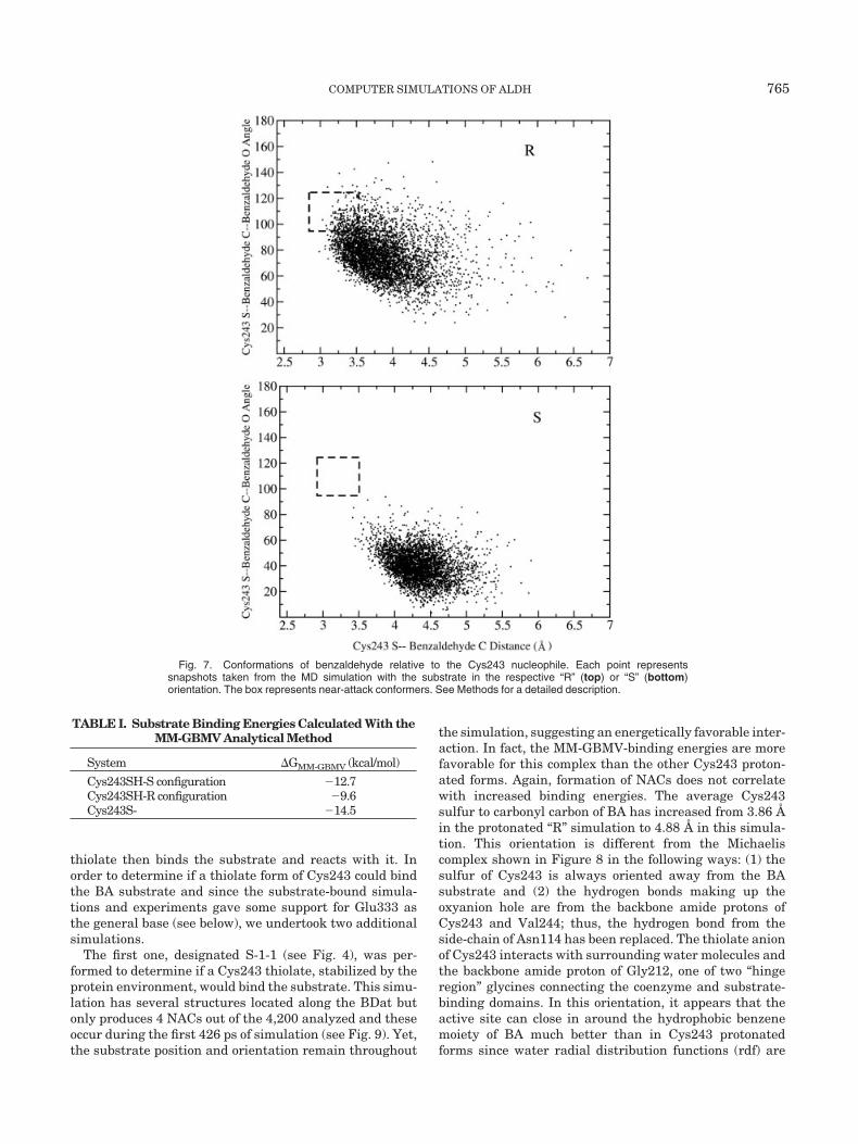
Benzaldehyde (BA) can be docked into the active site intwo ways such that the carbonyl oxygen of BA wouldinteract with the highly conserved Asn114 while the sulfurof Cys243 is oriented to attack the carbonyl carbon of BA.One of these orientations would lead to a thiohemiacetalintermediate in the R-configuration with the other in theS-configuration (see Fig. 3). The R-configuration binds inthe active site forming structures located along the BDatand more NACs (4.0 vs. 0.0%) than the S-configuration asshown in Figure 7. Yet this ensemble of structures shows aless favorable MM-GBMV binding energy by 3.1 kcal/mol(see Table I). The distributions are also very different withthe R-configuration average much closer to the NACregion than the S-configuration. Interestingly, these distri-butions do not change throughout the simulation andconversions between the R- and S-configuration do notoccur. Both simulations show fluctuations where the car-bonyl carbon of BA is 6–7 Å from the sulfur of Cys243indicating that the substrate is not locked into the activesite. These results suggest that either BA configurationcould be present in the active site and that some non-negligible barrier exists preventing conversion of oneconfiguration to the other. The S-configuration places thecarbonyl oxygen atom of the substrate in close contact withCys243. This configuration is held in the active site byenhanced hydrogen bonding from the thiol of Cys243
compared to the R-configuration where this interaction isminimal and sporadic. Therefore, despite favorable bind-ing characteristics this hydrogen bonding may preventCys243 from being activated by a general base. Thebackbone amide proton of Cys243 also interacts stronglywith the substrate in the S-configuration. A recent crystalstructure of ALDH2 shows crotonaldehyde in the activesite with the nucleophilic Cys302 aligned to form a thiohe-miacetal in the R-configuration.38 Finally, BA in theR-configuration places the hydride of BA closer to the C-4of NAD than in the S-configuration. Whether this closerplacement is favorable for hydride transfer, consideringthe dynamic movements of the NAD cofactor, will beinvestigated in our future studies.
The Michaelis complex from the MM simulation isdrawn in Figure 8. This complex shows that the ALDH3active site forms a hydrophobic region into which thebenzene moiety of BA fits and a polar region made up ofhydrogen bonds to the aldehyde moiety. The hydrophobicpocket consists of Val244, Leu361, and Ile394 below theBA and Tyr115 above. There is likely a �-stacking interac-tion between Tyr115 and BA as well, although this cannotbe considered to be a determinant of the aromatic sub-strate specificity of ALDH3 since this residue type (Tyrand Phe) is highly conserved throughout the family. Thesecalculations suggest that there would be a significantentropic effect to substrate binding since this bindingwould displace some water molecules occupying the hydro-phobic pocket. Hydrogen bonds to the carbonyl oxygen ofBA are primarily donated by the side-chain of Asn114 andthe backbone amide of Cys243. Energy analysis of theseinteractions shows the backbone amide on average hydro-gen bonding more to the substrate than Asn114. This isdue to the fact that during the 400–800-ps section of thetrajectory, the Asn114 hydrogen bond is lost before return-ing during the latter part of the simulation. In fact, thereare 190 NACs during the last 85 ps of this simulation out ofa total 369 produced during the 1-ns production simula-tion. Therefore, 22% of the conformations (860 analyzed)were NACs in the latter part of the simulation compared to2% (8,340 analyzed) in the first 1 ns of the simulation. Yetduring this time, the running average of the MM-GBMV-binding energy decreases slightly from �9.9 kcal/mol to�9.6 kcal/mol. Clearly, there is some enhancement ofNACs that coincides with the greater contribution ofAsn114 side-chain hydrogen bonding to BA but it does notcorrelate with more favorable MM-GBMV-binding ener-gies. The thiol proton of Cys243 hydrogen bonds to thesubstrate only in a few instances, and even then is not asenergetically favorable as the other two donors. Thesehydrogen-bond donors make up what is commonly calledthe oxyanion hole since they are positioned to stabilize adeveloping charge on the carbonyl oxygen atom of thethiohemiacetal intermediate.52
Thiolate forms of Cys243
It is commonly assumed in several enzyme systemscontaining cysteine nucleophiles that the protein environ-ment stabilizes the thiolate form of this nucleophile. This
Fig. 6. Hydrogen bonding interactions between Glu333 to ribosehydroxyl group of NAD and to Cys243 through an intervening watermolecule.
764 T. WYMORE ET AL.
thiolate then binds the substrate and reacts with it. Inorder to determine if a thiolate form of Cys243 could bindthe BA substrate and since the substrate-bound simula-tions and experiments gave some support for Glu333 asthe general base (see below), we undertook two additionalsimulations.
The first one, designated S-1-1 (see Fig. 4), was per-formed to determine if a Cys243 thiolate, stabilized by theprotein environment, would bind the substrate. This simu-lation has several structures located along the BDat butonly produces 4 NACs out of the 4,200 analyzed and theseoccur during the first 426 ps of simulation (see Fig. 9). Yet,the substrate position and orientation remain throughout
the simulation, suggesting an energetically favorable inter-action. In fact, the MM-GBMV-binding energies are morefavorable for this complex than the other Cys243 proton-ated forms. Again, formation of NACs does not correlatewith increased binding energies. The average Cys243sulfur to carbonyl carbon of BA has increased from 3.86 Åin the protonated “R” simulation to 4.88 Å in this simula-tion. This orientation is different from the Michaeliscomplex shown in Figure 8 in the following ways: (1) thesulfur of Cys243 is always oriented away from the BAsubstrate and (2) the hydrogen bonds making up theoxyanion hole are from the backbone amide protons ofCys243 and Val244; thus, the hydrogen bond from theside-chain of Asn114 has been replaced. The thiolate anionof Cys243 interacts with surrounding water molecules andthe backbone amide proton of Gly212, one of two “hingeregion” glycines connecting the coenzyme and substrate-binding domains. In this orientation, it appears that theactive site can close in around the hydrophobic benzenemoiety of BA much better than in Cys243 protonatedforms since water radial distribution functions (rdf) are
Fig. 7. Conformations of benzaldehyde relative to the Cys243 nucleophile. Each point representssnapshots taken from the MD simulation with the substrate in the respective “R” (top) or “S” (bottom)orientation. The box represents near-attack conformers. See Methods for a detailed description.
TABLE I. Substrate Binding Energies Calculated With theMM-GBMV Analytical Method
System �GMM-GBMV (kcal/mol)
Cys243SH-S configuration �12.7Cys243SH-R configuration �9.6Cys243S- �14.5
COMPUTER SIMULATIONS OF ALDH 765

decreased in this form (see Supplementary Material for rdfgraphs). The cysteine side-chain just needs to rotate aboutits C�-C� bond to bring the thiolate anion closer to thecarbonyl carbon to react and form the thiohemiacetalintermediate. Bringing this thiolate within 3.5 Å of thecarbonyl carbon of BA costs approximately 5.4 kcal/mol(unpublished results). We are currently examining theenergetics of forming the thiohemiacetal intermediatefrom this conformation.
The second simulation designated S-1-0 (see Fig. 4)represents a state in which Glu333 activates Cys243 priorto substrate binding by transfer of a proton. Surprisingly,this resulted in the substrate becoming expelled from theactive site during the simulation. Large shifts occur nearthe 500-ps mark, which suggests that this state is onlymarginally stable. It is likely that the enzyme requiredsome small collective reorientations before the substratecould exit the active site. The aldehyde moiety of BA
Fig. 8. Schematic representation of Michaelis complex showing both hydrophobic and polar interactions that contribute to substrate binding for thiolform of Cys243 (left) and thiolate form of Cys243 (right).
Fig. 9. Conformations of benzaldehyde relative to the Cys243 nucleophile. Each point represents snapshots taken from the S-1-1 (top left) andS-1-0 (top right) MD simulation with the substrate. The box represents near-attack conformers. Distance plots between the sulfur of Cys243 and thecarbonyl carbon of BA are shown for the S-1-1 (bottom left) and S-1-0 (bottom right) simulation.
766 T. WYMORE ET AL.
actually rotates around to face away from the active siteand is solvent separated from Cys243. The few NACs thatare formed in the initial part of the simulation show thecarbonyl oxygen atom of BA interacting primarily with theamide group of Asn114 and the amide group of NAD. Thus,the aldehyde has left the binding region observed in theother stable substrate-bound forms. The thiolate hydrogenbonds almost exclusively with the protonated Glu333throughout the entire trajectory with some interactionwith the face of the NAD ring. The position of the thiolateand its interaction partners appear crucial to binding thesubstrate if indeed Cys243 does bind BA as a thiolate. It isinteresting to note that the E333Q mutant causes seriousstability problems with ALDH3.5 Perhaps it is importantfor this position to have two hydrogen bond acceptor sites,and if Glu333 is the general base, it is important that ithave a mechanism for fast removal of the transferredproton.
Glu209 Versus Glu333 in Substrate-Bound MMSimulation
As shown above in the holo form simulation, Glu209makes significant interactions with the three backbone amideprotons of Met409-Gly410-Ala411 in the substrate-boundsimulation (the simulation that produced the most NACs).The hydrogen bond between Glu209 and Ala411 is alsobroken, this time near the 600-ps mark (plots shown inSupplemental Material). In contrast to the holo form simula-tion, this hydrogen-bond breaking leads to a slightly greaterseparation from this loop structure (5–6 Å) and allows theother carboxylate oxygen to move closer to the thiol proton ofCys243. Indeed, during the latter part of the simulation, thisdistance gets as close as 6.1 Å. Interestingly, the substrate-bound form does not block a possible route for Glu209 to actas a general base as was once proposed.8 In the unoccupiedunit of the dimer, the hydrogen bonds to the backbone loopremain for the entire simulation. The distance from thecarboxylate oxygen to Cys243 sulfur averages 10.3 Å andonly gets as close as 7.7 Å.
As in the holo form simulation, one carboxylate oxygenatom of Glu333 hydrogen bonds with the hydroxyl group of
the NAD ribose in the substrate bound simulation. Thisinteraction is formed at the 650-ps mark (plots shown inSupplemental Material). The other carboxylate oxygenatom makes significant interactions with the thiol protonof Cys243. During the 420–580-ps section, Cys243 andGlu333 are directly hydrogen bonded while at other timesthey are solvent separated. Some of the larger distancesabove 7 Å are the result of both solvent separation and thethiol proton not being directly oriented toward Glu333.Several conformations show a strong hydrogen-bond bridgebetween Glu333 and Cys243 through an intervening watermolecule. Many of these conformations also have thesubstrate positioned for attack by the sulfur of Cys243.
QM/MM Simulations of Nucleophilic CysteineActivation
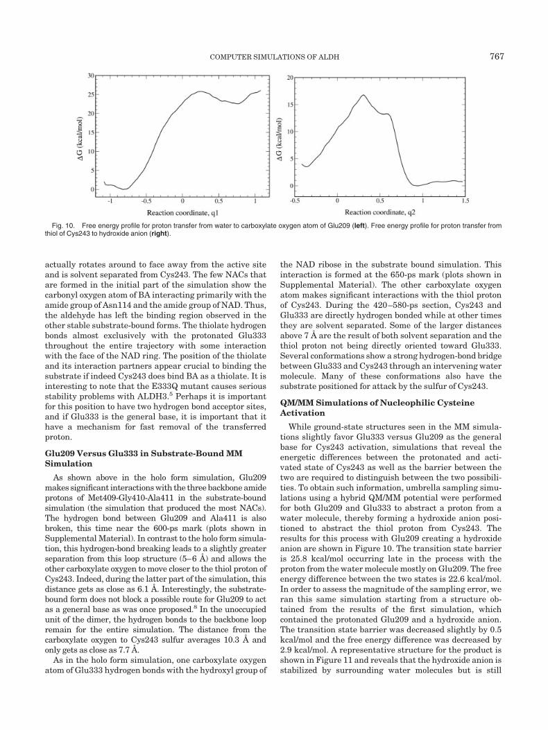
While ground-state structures seen in the MM simula-tions slightly favor Glu333 versus Glu209 as the generalbase for Cys243 activation, simulations that reveal theenergetic differences between the protonated and acti-vated state of Cys243 as well as the barrier between thetwo are required to distinguish between the two possibili-ties. To obtain such information, umbrella sampling simu-lations using a hybrid QM/MM potential were performedfor both Glu209 and Glu333 to abstract a proton from awater molecule, thereby forming a hydroxide anion posi-tioned to abstract the thiol proton from Cys243. Theresults for this process with Glu209 creating a hydroxideanion are shown in Figure 10. The transition state barrieris 25.8 kcal/mol occurring late in the process with theproton from the water molecule mostly on Glu209. The freeenergy difference between the two states is 22.6 kcal/mol.In order to assess the magnitude of the sampling error, weran this same simulation starting from a structure ob-tained from the results of the first simulation, whichcontained the protonated Glu209 and a hydroxide anion.The transition state barrier was decreased slightly by 0.5kcal/mol and the free energy difference was decreased by2.9 kcal/mol. A representative structure for the product isshown in Figure 11 and reveals that the hydroxide anion isstabilized by surrounding water molecules but is still
Fig. 10. Free energy profile for proton transfer from water to carboxylate oxygen atom of Glu209 (left). Free energy profile for proton transfer fromthiol of Cys243 to hydroxide anion (right).
COMPUTER SIMULATIONS OF ALDH 767

separated from Cys243. Thus, to activate Cys243 thishydroxide anion must either diffuse a few Ångstroms toabstract the thiol proton or abstract a proton from anintervening water molecule with the resulting hydroxideanion abstracting the thiol proton. Either way, the processrequires traversing another energetic barrier due to rear-rangement of the water molecules surrounding the hydrox-
ide anion. Indeed, our calculations along the q2 reactioncoordinate reveal that the barrier leading to the activatedCys243 and the protonated Glu209 is 13.3 kcal/mol with afree energy difference of �3.6 kcal/mol. Thus, the overallfree energy difference (�G � �Gq1 � �Gq2) is in the rangeof 17.1–19.0 kcal/mol. The reverse barrier leading back tocharged Glu209 and water from the protonated Glu209
Fig. 12. 2D Free energy surface for transferring a proton from Cys243 to Glu333 through an intervening water molecule. The lowest free energypathway from reactants (bottom left, blue graph) to the product (top right, green graph) is along the diagonal of the surface.
Fig. 11. The product structure of the proton transfer from water to carboxylate oxygen atom of Glu209showing the stabilizing interactions with the hydroxide anion.
768 T. WYMORE ET AL.
and hydroxide anion is only 3.2–6.1 kcal/mol. Therefore,the intermediate state is more likely to return to reactantsthan to proceed to the product. It should be noted that inthis case, q2 describes both hydroxide anion diffusionalong the S-H bond vector of Cys243 and the asymmetricstretch that transfers the proton. This reaction coordinatemay not be optimal for describing the small diffusion partof the reaction. While a more descriptive reaction coordi-nate would lower the energy barrier, the difference fromthe results obtained here is anticipated to be small.
In contrast, if the same reaction coordinate (q1) isfollowed with Glu333 abstracting a proton from an inter-vening water molecule to create a hydroxide anion, it leadsdirectly to an activated Cys243 without any additionalsteps. This result suggested that another reaction coordi-nate may be more applicable to this process, one thatwould go through a concerted mechanism. Therefore, wesimulated the 2D reaction profile. The results (see Fig. 12)do indeed show a concerted mechanism as being the lowestenergy pathway for activation of Cys243 by Glu333 throughan intervening water molecule. The transition state bar-rier is 29.2 kcal/mol and occurs very close to where bothprotons are halfway between the donor and acceptor atomsin mass-weighted coordinates. The free energy differencebetween the two states is 14.7 kcal/mol. The samplingerror for this reaction is likely similar to the one calculatedfor activation of Cys243 by Glu209. The surface also showsa barrier-less downhill path from the hydroxide anion/protonated Glu333 state to the activated Cys243. Alongthis path, the carbonyl carbon of benzaldehyde to sulfur ofCys243 fluctuates between distances of 2.9 to 4.5 Å andremains along the BDat. Obviously, this 2D free energyprofile is dependent on the placement of benzaldehydefrom the zone of the reaction, yet the placement of BA isvery similar in both simulations.
CONCLUSIONS
We have used several different MM and QM/MM simula-tions to model the initial stages of the ALDH3 catalyticcycle. Since it was not known a priori how the substrate isbound in the active site, we performed several long MMsimulations. These MM simulations allowed the BA sub-strate to exit the active site if placed in an energeticallyunfavorable location or protein state. Simulations shorterthan about 500 ps would have resulted in different conclu-sions. Furthermore, due to a large favorable solvationenergy of methyl thiolate (�73.7 kcal/mol),53 we ran thesimulations with different protonation states for Cys243.In these, we hypothesized that the thiolate forms incontact with the aldehyde would result in large unfavor-able forces and expulsion of the substrate from the activesite. Indeed, when modeling Cys243 as a thiolate, theaverage distance between the Cys243 sulfur atom and thecarbonyl carbon of BA was increased by 1.0 Å over theneutral form. Based on this analysis, it would appear thata neutral Cys243 is required to bind BA. Yet, we discov-ered a stable conformation of BA in the S-1-1 simulationwith the thiolate pointing away from the substrate. Fur-thermore, our MM-GBMV analysis revealed this to be the
most favorable binding mode by 4.9 kcal/mol due primarilyto more expulsion of water around the benzene moiety ofBA and shielding of the thiolate anion by a dihedralrotation. A rotation about the C�-C� bond would bring thethiolate in contact with the carbonyl carbon of BA formingthe thiohemiacetal intermediate. These calculations donot reveal a favored protonation state of Cys243 in thepresence of BA but they do reveal the relevant configura-tions one should use in calculations of this property. Suchcalculations represent an enormous challenge in thisfield.54
Both BA-bound conformations exhibit hydrogen bondingfrom the backbone amide protons of Cys243 and Val244and side-chain amide protons of Asn114, though to differ-ent extents. The thiol form prefers hydrogen bonding toAsn114 versus Val244 for the thiolate form. Both exhibit aconformation such that if Cys243 were to attack thecarbonyl carbon of BA, a thiohemiacetal of the R-configuration would be formed. This assumes that there isnot a configuration change upon activation of Cys243 bythe general base. Indeed, the QM/MM simulations of thisactivation process from either Glu209 or Glu333 did notresult in any changes in the BA orientation with respect toCys243. The R-configuration also brings the hydride of thethiohemiacetal intermediate closer to the NAD cofactorthan if it were to form the S-configuration. Both simula-tions contained structures of the electrophilic BA sub-strate located along the BDat, which suggests that thestructure of ALDH3 is pre-organized for the nucleophilicattack. How the structure changes to catalyze other reac-tions in the catalytic cycle will be of interest to determine.Hydride transfer is the next step in the catalytic cycle afterthiohemiacetal formation and has been shown to be ste-reospecific.7 Our simulations do not offer any obvious cluesas to why hydride transfer is stereospecific. Since this stepis rate-limiting in ALDH3,4 it is possible that some proteinreorganization takes place before this step is favorable.Also, the protein reorganization may only take place oncethe thiohemiacetal intermediate is formed, which was notsimulated here.
While the productive substrate-bound MM simulationwith Cys243 in thiol form favored Glu333 over Glu209 asthe general base due to closer contacts and several morestructures that were hydrogen bonded through a watermolecule to Cys243, the differences still appeared minor.This led us to speculate that if Cys243 required activation,either the free energy barrier or possibly the free energydifference for the respective activation mechanisms weredeciding factors. Therefore, we simulated the Cys243activation mechanism using a hybrid QM/MM potential todistinguish between the two possibilities. While we cannotstrictly compare the energetics of the two reactions sincethey use slightly different QM mechanical zones, theresults show two different pathways for activating Cys243through an intervening water molecule. Very differentquantum regions can be used if one first calibrates refer-ence solution reactions as demonstrated by the Warshelgroup.55 The activation of Cys243 by Glu209 occurs step-wise through a hydroxide anion that is stabilized by
COMPUTER SIMULATIONS OF ALDH 769

several surrounding water molecules. This hydroxide an-ion must then traverse another significant barrier toabstract the thiol proton. In contrast, a concerted mecha-nism was found for Glu333 in which, as the proton fromthe intervening water molecule donates a proton to Glu333,the thiol proton of Cys243 transfers to the water molecule.The barrier for the concerted mechanism did not differgreatly from the stepwise mechanism yet the hydroxideanion was not a minimum on q2 with Glu333.
Since PM3 underestimates the �Hf of the hydroxideanion and the hydronium cation, we might expect thestability of structures along the diagonal of the 2D surfaceto be underestimated to a greater extent than along thestep-wise pathways. Furthermore, this transition statestructure is very delocalized and may require the use ofhigh level ab initio methods or an extensive reparameter-ization of PM3 for accurate results. Given these caveats,the fact that Glu333 can activate Cys243 through anintervening water molecule in a concerted manner may beconsiderably more favorable than a step-wise mechanismand, hence, be crucial to the function of Glu333 as thegeneral base in ALDH3. The results also support the viewthat small changes in the active site could easily shift theidentity of the general base to Glu209. This appears to bethe case with ALDH2 where Glu268 (Glu209 in ALDH3)has been identified as the general base.
This study supports a research plan in which MMsimulations and subsequent postprocessing of the trajec-tory through MM-GBMV calculations are used to deter-mine favorable substrate-bound conformations. Some ofthese structures may be oriented in a manner that wouldlead to a chemical reaction. These structures can then befurther examined by a QM/MM potential to determinetheir relevance to the enzyme mechanism. Finally, thesesimulations support the importance of examining differentionization states for active-site cysteine nucleophiles whenmodeling the substrate bound form.
ACKNOWLEDGMENTS
The calculations were done in part on the NationalScience Foundation Terascale Computing System at thePittsburgh Supercomputing Center with an allocationfrom the National Resource Advisory Committee. A.D.M.gratefully acknowledges support from NIH (GM51501).We also thank Martin J. Field for several useful discus-sions.
REFERENCES
1. Lindahl R. Aldehyde dehydrogenases and their role in carcinogen-esis. Crit Rev Biochem Mol Biol 1992;27:283–335.
2. Sladek NE. Aldehyde dehydrogenase-mediated cellular relativeinsensitivity to the oxazaphosphorines. Curr Pharm Des 1999;5:607–625.
3. Liu Z, Sun Y, Rose J, Chung Y, Hsiao C, Chang W, Kuo I, PerozichJ, Lindahl R, Hempel J, Wang B-C. The first structure of analdehyde dehydrogenase reveals novel interactions between NADand the Rossman fold. Nat Struct Biol 1997;4:317–326.
4. Hempel J, Kuo I, Perozich J, Wang B-C, Lindahl R, Nicholas H.ALDH: maintaining critical active site geometry at motif 8 in theclass 3 enzyme. Eur J Biochem 2001;268:722–726.
5. Mann CJ, Weiner H. Differences in the roles of conserved glutamicacid residues in the active site of human class 3 and class 2aldehyde dehydrogenases. Prot Sci 1999;8:1922–1929.
6. DeLaurenzi V, Rogers GR, Hamrock DL, Marekov LN, SteinertPM, Compton JG, Marekova N, Rizzo WB. Sjogren-Larsson syn-drome is caused by mutations in the fatty acid aldehyde dehydro-genase gene. Nature Genet 1996;12:52–57.
7. Jones KH, Lindahl R, Baker DC, Timkovich R. Hydride transferstereospecificity of rat liver aldehyde dehydrogenases. J BiolChem 1987;262:10911–10913.
8. Hempel J, Perozich J, Chapman T, Rose J, Boesch JS, Liu Z-J,Lindahl R, Wang BC. Aldehyde Dehydrogenase catalytic mecha-nism: a proposal. In: Weiner H, editor. Enzymology and molecularbiology of carbonyl metabolism 7. New York: Plenum Press; 1999.p. 53–59.
9. Hempel J, Nicholas H, Lindahl R. Aldehyde dehydrogenases:widespread structural and functional diversity within a sharedframework. Prot Sci 1993;2:1890–1900.
10. Dryjanski M, Kosley LL, Pietruszko R. N-Tosyl-L-phenylalaninechloromethyl ketone, a serine protease inhibitor, identifies gluta-mate 398 at the coenzyme-binding site of human aldehyde dehy-drogenase. Evidence for a second “naked anion” at the active site.Biochemistry 1998;37:14151–14156.
11. MacKerell AD Jr, MacWright RS, Pietruszko R. Bromoacetophe-none as an affinity reagent for human liver aldehyde dehydroge-nase. Biochemistry 1986;25:5182–5189.
12. Abriola DP, Fields R, Stein S, MacKerell AD Jr, Pietruszko R.Active site of human liver aldehyde dehydrogenase. Biochemistry1987;26:5679–5684.
13. Wang, X, Weiner H. Involvement of glutamate 268 in the activesite of human liver mitochondrial (class 2) aldehyde dehydroge-nase as probed by site-directed mutagenesis. Biochemistry 1995;34:237–243.
14. Ni L, Sheikh S, Weiner H. Involvement of Glutamate 399 andLysine 192 in the mechanism of human liver mitochondrialaldehyde dehydrogenase. J Biol Chem 1997;272:18823–18826.
15. Perozich J, Nicholas H, Wang B, Lindahl R, Hempel J. Relation-ships within the aldehyde dehydrogenase extended family. ProtSci 1999;8:137–146.
16. Yang A, Gunner M, Sampogna R, Sharp K, Honig B. On thecalculation of pKas in proteins. Proteins 1993;15:252–265.
17. Feig M, Onufriev A, Lee MS, Im W, Case DA, Brooks III CL.Performance comparison of generalized born and poisson methodsin the calculation of electrostatic solvation energies for proteinstructures. J Comp Chem 2004;25:265–284.
18. Burgi HB, Dunitz JD, Shefter EJ. Geometrical reaction coordi-nates. II. Nucleophilic addition to a carbonyl group. J Am ChemSoc 1973;95:5065–5067.
19. Burgi HB, Dunitz JD, Shefter E. Chemical reaction paths. IV.Aspects of O—C�O interactions in crystals. Acta Cryst B 1974;30:1517–1527.
20. Chakrabarti P, Pal D. An electrophile-nucleophile interaction inmetalloprotein structures. Prot Sci 1997;6:851–859.
21. Bruice TC, Benkovic SJ. Chemical basis for enzyme catalyis.Biochemistry 2000;39:6267–6274.
22. Shurki A, Strajbl M , Villa J, Warshel A. How much do enzymesreally gain by restraining their reacting fragments? J Am ChemSoc 2002;124:4097–4107.
23. Strajbl M, Shurki A, Kato M, Warshel A. Apparent NAC effect inchorismate mutase reflects electrostatic transition state stabiliza-tion. J Am Chem Soc 2003;125;10228–10237.
24. Warshel A, Levitt M. Theoretical studies of enzymic reactions:dielectric, electrostatic and steric stabilization of the carboniumion in the reaction of lysozyme. J Mol Biol 1976;103:227–249.
25. Field MJ. Simulating enzyme reactions: challenges and perspec-tives. J Comput Chem 2002;23:48–58.
26. Gao J, Truhlar DG. Quantum mechanical methods for enzymekinetics. Annu Rev Phys Chem 2002;53:467–505.
27. Warshel A. Computer simulations of enzyme catalysis:methods,progress, and insights. Ann Rev Biophys Biomol Struct 2003;32:425–443.
28. MacKerell, AD Jr, Banavali NB, Foloppe N. Development andcurrent status of the CHARMM force field for nucleic acids.Biopolymers 2001;56:257–265.
29. MacKerell AD Jr, Bashford D, Bellott M, Dunbrack Jr. RL,Evanseck JD, Field MJ, Fischer S, Gao J, Guo H, Ha S, Joseph-McCarthy D, Kuchnir L, Kuczera K, Lau FTK, Mattos C, MichnickS, Ngo T, Nguyen DT, Prodhom B, Reiher III WE, Roux B,Schlenkrich M, Smith JC, Stote R, Straub J, Watanabe M,Wiorkiewicz-Kuczera J, Yin D, Karplus M. All-atom empirical
770 T. WYMORE ET AL.

potential for molecular modeling and dynamics studies of pro-teins. J Phys Chem B 1998;102:3586–3616.
30. Foloppe N, MacKerell AD Jr. All-atom empirical force field fornucleic acids: 1. Parameter optimization based on small moleculeand condensed phase macromolecular target data. J Comp Chem2000;21:86–104.
31. Feller SE, MacKerell AD Jr. An improved empirical potentialenergy function for molecular simulations of phospholipids. JPhys Chem B 2000;104:7510–7515.
32. Berman HM, Westbrook J, Feng Z, Gilliland G, Bhat TN, WeissigH, Shindyalov IN, Bourne PE. The Protein Data Bank. NucleicAcids Res 2000;28:235–242.
33. Brooks BR, Bruccoleri RE, Olafson BD, States DJ, SwaminathanS, Karplus M. CHARMM: a program for macromolecular energy,minimization, and dynamics calculations. J Comput Chem 1983;4:187–217.
34. Pavelites JJ, Bash PA, Gao J, MacKerell AD Jr. Molecularmechanics force field for NAD�, NADH, and the pyrophosphategroups of nucleotides. J Comp Chem 1997;18:221–239.
35. Brunger AT, Karplus M. Polar hydrogen positions in proteins:empirical energy placement and neutron diffraction comparison.Proteins 1988;4:148–156.
36. Ryckaert J-P, Cicotti G, Berendsen HJC. Numerical integration ofthe cartesian equations of motion of a system with constraints:molecular dynamics of n-alkanes. J Comput Phys 1977;23:327–341.
37. Darden T, York D, Pedersen L. Particle mesh ewald: an N � log(N)method for ewald sums in large systems. J Chem Phys 1993;98:10089–10092.
38. Perez-Miller SJ, Hurley TD. Coenzyme isomerization is integralto catalysis in aldehyde dehydeogenase. Biochemistry 2003;42:7100–7109.
39. Cobessi D, Tete-Favier F, Marchal S, Branlant G, Aubry A.Structural and biochemical investigations of the catalytic mecha-nism of an NADP-dependent aldehyde dehydrogenase from strep-tococcus mutans. J Mol Biol 2000;300:141–152.
40. Reddy SY, Kahn K, Zheng Y, Bruice TC. Protein engineering ofnitrile hydratase activity of papain: molecular dynamics study of amutant and wild-type enzyme. J Am Chem Soc 2002;124:12979–12990.
41. Swanson JMJ, Henchman RH, McCammon JA. Revisiting freeenergy calculations: a theoretical connection to MM/PBSA anddirect calculation of the association free energy. Biophys J 2004;86:67–74.
42. Field MJ, Albe M, Bret C, Proust-De Martin F, Thomas A. TheDYNAMO library for molecular simulations using hybrid quan-tum mechanical and molecular mechanical potentials. J ComputChem 2000;21:1088–1100
43. Jorgensen WL, Maxwell DS, Tirado-Rives J. Development andtesting of the OPLS all-atom force field on conformational energet-ics and properties of organic liquids. J Am Chem Soc 1996;118:11225–11236.
44. Stewart JJP. Optimization of parameters for semiempirical meth-ods. I. Methods. J Comp Chem 1989;10:209–220.
45. Gonzalez-Lafront A, Truong TN, Truhlar DG. Direct dynamicscalculations with neglect of diatomic differential overlap molecu-lar orbital theory with specific reaction parameters. J Phys Chem1991;95:4618–4627.
46. Repasky MP, Chandrasekhar J, Jorgensen WL. PDDG/PM3 andPDDG/MNDO: improved semiemperical methods. J Comput Chem2002;23:1601–1622.
47. Karplus M. Aspects of protein reaction dynamics: deviations fromsimple behavior. J Phys Chem B 2000;104:11–27.
48. Kumar S, Bouzida D, Swendsen RH, Kollman PA, Rosenberg JM.The weighted histogram analysis method for free energy calcula-tions on biomolecules. I. The method. J Comp Chem 1992;13:1011–1021.
49. PSI-Plot, Version 7.0. New York: Poly Software International;2002.
50. Humphrey W, Dalke A, Schulten K. VMD: visual moleculardynamics. J Mol Graph 1996;14:33–38.
51. Wymore T, Deerfield II DW, Field MJ, Nicholas HB, HempelJ. Initial events in class 3 aldehyde dehydrogenase: MM andQM/MM simulations. Chem Biol Int 2003;143–144:75–84.
52. Kraut DA, Carroll KS, Herschlag D. Challenges in enzymemechanism and energetics. Ann Rev Biochem 2003;72:517–571.
53. Pliego Jr. J, Riveros JM. Gibbs energy of solvation of organic ionsin aqueous and dimethyl sulfoxide solutions. Phys Chem ChemPhys 2002;4:1622–1627.
54. Olsson MHM, Hong G, Warshel A. Frozen density functional freeenergy simulations of redox proteins: computational studies of thereduction potential of plastocyanin and rusticyanin. J Am ChemSoc 2003;125:5025–5039.
55. Strajbl M, Florian J, Warshel A. Ab initio evaluation of the freeenergy surfaces for the general base/acid catalyzed thiolysis offormamide and the hydrolysis of methyl thiolformate: a referencesolution reaction for studies of cysteine proteases. J Phys Chem B2001;105:4471–4484.
COMPUTER SIMULATIONS OF ALDH 771