Embed Size (px)
Citation preview

REVIEW
Molecular orchestration of differentiationand function of regulatory T cells
Li-Fan Lu2 and Alexander Rudensky1
Howard Hughes Medical Institute and Immunology Program, Memorial Sloan-Kettering Cancer Center, New York, New York10065, USA
During the last decade, a unique mechanism of negativeregulation of immune responses and inflammation bya dedicated population of so-called regulatory T cells(Treg) has become a focus of intensive investigation.Through the discovery of transcription factor Foxp3 asa central molecular determinant of differentiation andfunction of Treg cells, the complex biology of these cells,including maintenance of immunological tolerance to‘‘self’’ and regulation of immune responses to pathogens,commensals, and tumors, has become amenable to mech-anistic studies. In this review, we discuss the molecularaspects of Treg cell lineage commitment, maintenance,and function.
The cornerstone of the adaptive immune system is therandom generation of antigen-specific receptors in theprocess of somatic gene rearrangement. The immensediversity of this anticipatory recognition system providesan efficient counterbalance to rapidly evolving patho-gens. Two major classes of lymphocytes, key cellularactors of the adaptive immune system, rely on differentprinciples of antigen recognition: Immunoglobulin (Ig)receptors displayed by B lymphocytes recognize intactforms of antigens, whereas T-cell receptors (TCR) recog-nize antigens in the form of short peptides bound to theproducts of the major histocompatibility gene complex(MHC). The peptide–MHC complexes are displayed forT-cell recognition on the surface of antigen-presentingcells (APCs), among which dendritic cells (DCs) areunique in their ability to process and present antigensto T cells and to initiate the immune response. APCexploit the proteolytic machinery of intracellular proteinturnover to generate antigenic peptides for T-cell recog-nition. MHC molecules sample the peptide content ofmain sites of intracellular proteolysis, the cytosol andthe endocytic compartment, without discriminating be-tween ‘‘self’’ and ‘‘foreign’’—i.e., pathogen-derived—pep-tides. CD8 T cells recognize MHC class I moleculesbound to peptides generated in the cytosol, whereas
CD4 T cells recognize MHC class II molecules bound topeptides generated in the endosomes and lysosomes,although some important exceptions to this rule exist(Trombetta and Mellman 2005).
Sequence analyses of peptides displayed by MHCmolecules revealed that the vast majority of peptidesbound to MHC class I and class II molecules is derivedfrom self-proteins. Furthermore, TCR have an intrinsicbias toward recognition of MHC molecules, and ‘‘tick-ling’’ of TCR through certain low-affinity interactionswith self-peptide–MHC complexes displayed by thymiccortical epithelial cells is prerequisite for thymic T-cellprecursors to pass a critical differentiation checkpointdubbed positive selection. In the periphery, analogousinteractions facilitate the long-term maintenance ofmature CD4 and CD8 T cells and further shape TCRrepertoire. Thus, this ‘‘self-referential’’ nature of T-cellrecognition presents a tremendous problem of establish-ing T-cell tolerance to genome-encoded self-antigens, andto limiting reactivity to antigens derived from commen-sal microbiota, food, and environmental factors. In thisreview, we call ‘‘self’’ a sum of the aforementionedantigens, unrestrained reactivity to which causes diverseimmune-mediated pathologies including autoimmunity,allergy, and asthma.
Dominant and recessive tolerance
Numerous mechanisms of tolerance described during thelast several decades can be divided into two major groups.Cell-intrinsic mechanisms, also known as recessive tol-erance, leading to physical elimination or functionalinactivation of a given ‘‘self’’ reactive T-cell clone, operateboth in the thymus and in the periphery and are likelyresponsible for neutralization of the majority of high-affinity T cells recognizing self. Immature CD4 and CD8double-positive (DP) and single-positive (SP) thymocytesundergo apoptosis, or ‘‘negative selection,’’ upon high-affinity TCR engagement by self-antigen displayed bythymic DCs and thymic medullary epithelial cells. Thoseself-reactive thymocytes that escape clonal deletion be-come ‘‘anergic’’ or incapable of efficient proliferation inresponse to a ‘‘self’’ antigen and differentiation intofunctional effector cytokine-producing or cytotoxic Tcells (for review, see Starr et al. 2003).
[Keywords: T lymphocytes; tolerance; differentiation; transcriptionalregulation]Correspondence.1E-MAIL [email protected]; FAX (646) 888-3160.2E-MAIL [email protected]; FAX (646) 888-3160.Article is online at http://www.genesdev.org/cgi/doi/10.1101/gad.1791009.
1270 GENES & DEVELOPMENT 23:1270–1282 � 2009 by Cold Spring Harbor Laboratory Press ISSN 0890-9369/09; www.genesdev.org
Cold Spring Harbor Laboratory Press on July 10, 2018 - Published by genesdev.cshlp.orgDownloaded from

Beyond thymus, chronic engagement of TCR displayedby peripheral T cells by ‘‘self’’ antigens can also lead toactivation-induced cell death or anergy induction. Pe-ripheral anergy induction is reinforced by the two-signalrequirement for initiation of the naive T-cell responses.This requirement is fulfilled upon simultaneous engage-ment of TCR and T-cell costimulatory receptor CD28 bycognate peptide–MHC complex and inducible CD28ligands CD80 and CD86 displayed on the surface ofAPC (Lenschow et al. 1996). High levels of CD80 andCD86 expression are induced upon activation of a diverseset of dedicated sensors directly in response to microbialor viral products or indirectly by metabolic or biochem-ical changes they induce.
However, the recessive tolerance operating in a cell-intrinsic manner and the two-signal requirement forthe induction of productive immune response appearinsufficient to counter the threat of immune-mediatedpathology without dominant tolerance, a unique immu-nological mechanism that involves the suppression of‘‘self’’-reactivity by specialized cells acting in a dominantfashion. The initial experimental evidence in support ofthymic generation of cells capable of preventing autoim-munity came from a surprising finding of spontaneousT-cell-dependent autoimmune lesions in a variety oforgans observed in mice subjected to thymectomy on day3 of life (d3Tx) (Nishizuka and Sakakura 1969; Sakaguchiet al. 1982; Bonomo et al. 1995; Asano et al. 1996) andtolerance observed in chicken–quail chimeras (Ohki et al.1987). Since adoptive transfers of T cells from euthymicmice prevented autoimmunity in d3Tx mice, these earlyobservations suggested paucity of T cells capable of pre-venting autoimmunity in neonatal mice and, thus, laida foundation for the concept of dominant tolerance.
CD25+CD4+ T cells as mediators of dominant tolerance
Long-lasting labors to define the cell type-mediating do-minant tolerance resulted in the landmark discovery ofa subset of CD4 T cells, constitutively expressing highamounts of the interleukin-2 receptor a-chain (CD25), asbeing highly enriched in suppressor activity (Sakaguchiet al. 1995). CD25+CD4+ Tcells, dubbed regulatory T cellsor Treg cells, were shown to be able to prevent autoim-munity upon transfer into d3Tx mice. Their potentsuppressive capacity has been further demonstrated inmultiple experimental models of autoimmunity, trans-plant rejection, and tumor immunity (for reviews, seeShevach 2000; Fehervari and Sakaguchi 2004). These cellsmake up ;10%–15% of lymph node and splenic CD4 Tcells in mice and ;2% of peripheral blood CD4 T cells inhumans. In the thymus, a subset of CD25+CD4+ SPthymocytes with suppressive activity is present, butthese cells are inconspicuous within the first few daysafter birth in agreement with d3Tx experiments (Asanoet al. 1996; Fontenot et al. 2005a). These results indicatedthat CD25+ Treg cells serve as mediators of dominanttolerance and that these cells differentiate in the thymusin a developmentally controlled manner. Although iden-tification of CD25 allowed for functional analyses of
Treg cells isolated from nonimmune animals, its utilityas a definitive Treg cell marker was limited due up-regulation of CD25 in all activated T cells. The inabilityto discriminate between protective and inflammation-promoting cells during the immune response impededfurther understanding of dominant tolerance, especiallyits mechanistic aspects. Moreover, it was proposed thatTreg cells are only conventional activated CD4 T cellsthat down-modulate immune responses by competing forinterleukin-2. Thus, a search for a specific molecularmarker of Treg cells and a genetic mechanism underlyingthe differentiation and function of these cells ensued.
Foxp3: a key to dominant tolerance
The key to discovery of the genetic basis of dominanttolerance was provided by identification of mutations inX-chromosome-encoded transcription factor Foxp3 inmice with a spontaneous scurfy mutation and in patientswith the IPEX (immune dysregulation, polyendocrinop-athy, enteropathy, X-linked) syndrome (Chatila et al.2000; Bennett et al. 2001; Wildin et al. 2001). In bothhumans and mice, loss-of-function Foxp3 mutations re-sult in a fatal early-onset T-cell-dependent autoimmunitymanifested by diabetes, thyroiditis, hemolytic anemia,hyper-IgE syndrome, exfoliative dermatitis, splenomeg-aly, lymphadenopathy, and ‘‘cytokine storm’’ (for review,see Gambineri et al. 2003). Importantly, the diseaseaffected only hemizygous Foxp3 mutant males. In con-trast, heterozygous female carriers of Foxp3 mutationswere spared from the disease, suggesting that T cellsexpressing a wild-type Foxp3 allele were able to restrainactivation of T cells (Godfrey et al. 1994). An implicitassumption in this line of reasoning—namely, that Foxp3mutations do not affect random X-chromosome inactiva-tion in T cells—was subsequently confirmed by analysisof Foxp3 reporter mice (Fontenot et al. 2005c; Gavin et al.2007), as discussed below. This consideration along withsystemic nature of immune inflammatory lesions affect-ing multiple tissues in Foxp3 mutant individuals promp-ted several groups to examine expression of Foxp3 inCD25+CD4+ Treg cells in mice. These studies revealedthat Foxp3 is expressed at a high level in CD25+CD4+
Treg cells, but it is not present in significant amounts innaı̈ve CD25-CD4+ T cells or in antigen-stimulated CD4+
T cells, which acquired CD25 expression upon activation(Fontenot et al. 2003; Hori et al. 2003; Khattri et al. 2003).Analysis of Foxp3 knockout mice showed that T cells inthese mice become activated very early after birth,whereas the numbers of CD25+CD4+ thymocytes aresignificantly diminished. Although these experimentsstrongly suggested that Foxp3 is required for differentia-tion of Treg cells, a caveat remained that early-onsetautoimmune disease and T-cell activation potentiallyadversely affected the size or properties of the CD25+
Treg cell subset. This caveat was addressed, however, byexamination of CD25+CD4+ cell populations in thethymus and peripheral lymphoid organs of mixed bonemarrow chimeras lacking lymphoproliferative diseaseand immune-mediated tissue lesions. In these studies,
Molecular regulation in Treg cells
GENES & DEVELOPMENT 1271
Cold Spring Harbor Laboratory Press on July 10, 2018 - Published by genesdev.cshlp.orgDownloaded from
only Foxp3-sufficient, but not Foxp3-deficient hemato-poietic stem cells were able to give rise to CD25+ Tregcells upon reconstitution of lethally irradiated recipientmice with Foxp3null and allelically marked Foxp3wt
bone marrow (Fontenot et al. 2003). These experimentsshowed an absolute requirement for Foxp3 for Tregdifferentiation in the thymus. Furthermore, forced ex-pression of Foxp3 in peripheral CD25-CD4+ T cells usingretroviral expression vectors results in acquisition ofCD25 expression and suppressor function (Fontenotet al. 2003; Hori et al. 2003). In addition, expression ofa Foxp3 transgene confers suppressive properties to CD8Tcells (Khattri et al. 2003). Finally, Cre-mediated ablationof a conditional Foxp3 allele in mature Treg cells resultsin a loss over time of suppressor function and character-istic Treg surface markers (Williams and Rudensky 2007).Together, these studies showed that Foxp3 is essential forTreg differentiation and suppressor function. However,they did not answer the question as to whether Treg celldeficiency alone is responsible for the fatal disease inFoxp3-deficient mice and men, or Foxp3 function in otherimmune cell types or in nonhematopoietic cells contrib-utes to the disease.
Foxp3-expressing Treg cells are vital for immunehomeostasis
Analysis of Foxp3 reporter mice generated upon insertioninto the Foxp3 locus of a DNA sequence encoding a GFP-Foxp3 fusion protein or a GFP or RFP coding sequenceequipped with an IRES showed a surprisingly restrictedexpression of Foxp3 protein expression to a subset ofCD4 T cells (Fontenot et al. 2005c; Wan and Flavell2005; Bettelli et al. 2006; Lin et al. 2007). In addition tothe prevalent CD25hiFoxp3+CD4+ T-cell subset, someCD4+CD25� or CD4+CD25lo T cells also express Foxp3(Fontenot et al. 2005c). The potent suppressive capacityof all Foxp3+ T-cell subsets and lack of detectable highamounts of Foxp3 expression in other hematopoietic cellsor nonlymphoid tissues were consistent with the notionof a dedicated function of Foxp3 in Treg cell differentia-tion. Although low levels of Foxp3 expression outside theregulatory T-cell subset was reported (Zuo et al. 2007a,b),additional experimentation is needed to further confirmits physiologic significance.
Initial evidence that the paucity of Treg cells accountsfor fatal autoimmunity resulting from the Foxp3 de-ficiency was provided by studies where adoptive transfersof Treg cells rescued disease in neonatal Foxp3-deficientmice (Fontenot et al. 2003). Furthermore, mice with theT-cell-specific and germline ablation of the Foxp3 genewere indistinguishable in the progression and severity ofthe autoimmune lesions (Fontenot et al. 2005c). Incontrast, deletion of a conditional Foxp3 allele in thymicepithelial cells or DCs, which shape the repertoire ofdeveloping T-cell precursors, did not result in any dis-cernable immune dysregulation or alteration in the T-celldifferentiation (Liston et al. 2007; L Williams and ARudensky, unpubl.). These genetic studies implicateddeficiency of Foxp3 within the T-cell lineage as a cause
of the disease. Importantly, Foxp3 gene inactivation didnot affect the sensitivity and two-signal requirement forT-cell activation, levels of cytokine production, and clonalexpansion in peripheral nonregulatory T cells (Fontenotet al. 2003, 2005c; Chen et al. 2005; Hsieh et al. 2006).These findings together with a failure to detect a cell-intrinsic role for the Foxp3 gene in regulation of recessivetolerance pointed to a lack of Treg cells as the cause of thedisease associated with Foxp3 deficiency (Fontenot et al.2003, 2005c; Chen et al. 2005; Hsieh et al. 2006).
Notwithstanding breakthroughs that came from theanalyses of Foxp3-deficient mice, the question remainedwhether Treg function is critical during the developmentof the immune system or after the immune system isfully developed. This major outstanding issue wasaddressed through the analysis of Foxp3DTR knock-inand Foxp3-DTR BAC transgenic mice harboring an‘‘ablatable’’ Treg population (Kim et al. 2007; Lahl et al.2007). Chronic ablation of Treg cells in adult healthymice results in their death within 3 wk and demonstratesthat Treg cell-mediated suppression is indispensable forpreventing immune pathology throughout the lifespan ofnormal mice (Kim et al. 2007).
Considering a central role for Foxp3 in Treg biology,analysis of regulation of Foxp3 expression in the thymusand periphery and of Foxp3-dependent transcriptional andfunctional programs provides a key to mechanistic un-derstanding of differentiation of Treg cells and of suppres-sion they mediate. Furthermore, restricted expression ofFoxp3 in Treg cells allows for development of powerfultools for in vivo genetic manipulation of various aspectsof Treg biology. In the remaining sections of this review,we focus on recent progress in studies of moleculardeterminants of Treg cell differentiation and function.
Regulation of Foxp3 expression
In addition to CD25, Treg cells are known to stablyexpress high levels of two negative regulators of TCRsignaling and T-cell activation CD5 and CTLA4 (forreview, see Sakaguchi 2004). These observations sug-gested that Treg cells themselves might express TCRwith an increased affinity for ‘‘self’’ peptide–MHC com-plexes as compared with conventional ‘‘nonregulatory’’T cells and that TCR specificity might guide Treg celldifferentiation. The latter notion was supported by obser-vations that in mice where all T cells express a singletransgene-encoded TCR specific for a self-protein, myelinbasic protein (MBP), a severe immune-mediated in-flammatory disease allergic encephalomyelitis (EAE)commences (Lafaille et al. 1994). In contrast, mice ex-pressing the very same TCR transgene, but capable ofendogenous TCR gene rearrangement, were disease-freedue to the presence of protective Treg cells expressingendogenous TCR chains. Furthermore, adoptive transferof these cells prevented disease in mice with monoclonalMBP-specific T cells present (Olivares-Villagomez et al.1998). A further support for an essential role of TCRsignaling in Treg differentiation came from studies wheretransgenic TCR coexpressed with a transgene-encoded
Lu and Rudensky
1272 GENES & DEVELOPMENT
Cold Spring Harbor Laboratory Press on July 10, 2018 - Published by genesdev.cshlp.orgDownloaded from

corresponding TCR ligand in the thymus or chronicallyexposed to TCR ligand in the periphery facilitated differ-entiation of Treg cells (Jordan et al. 2001; Apostolou et al.2002). Numerous transcription factors activated down-stream from TCR were implicated in regulation of Foxp3at a transcriptional level. These factors, including NFAT,AP1, CREB, and ATF, were shown to bind to the promoterregion or to an intronic Foxp3 element proposed to serveas an enhancer (Mantel et al. 2006; Kim and Leonard2007). In addition, TCR-induced NF-kB pathway wasimplicated in Treg differentiation (Schmidt-Supprianet al. 2004; Gupta et al. 2008). Besides TCR, CD28 signalsplay a cell-intrinsic role in Treg cell generation andmaintenance. This notion is supported by greatly reducednumbers of Foxp3+ thymocytes and peripheral T cells inthe absence of CD28 or its ligands CD80 and CD86(Salomon et al. 2000; Tai et al. 2005). However, molecularmechanisms of CD28 involvement in Foxp3 inductionare not clear since in vitro studies showed that extendedactivation of Akt, a main target of the CD28 signalingpathway, impaired generation of Foxp3+ cells. Con-versely, inhibition of PI3K activation and, therefore, ofAkt activation facilitated Foxp3 induction (Haxhinastoet al. 2008; Sauer et al. 2008). These recent findings werein agreement with an earlier observation of a twofoldincrease in numbers of thymic Foxp3+ cells, yet signifi-cant decrease in splenic Foxp3+Treg cells in mice harbor-ing an inactive form of p100d isoform of PI3K (Pattonet al. 2006).
TCR/CD28 signals alone appear to be insufficient toinduce Foxp3 expression. Indirect evidence in supportof this notion came from observations of a delayedappearance of Foxp3+ thymocytes after birth, whereasCD25+Foxp3� CD4 SP thymocytes were readily detect-able (Fontenot et al. 2005a). These findings were ascribedto a requirement of a second signal for Foxp3 inductionlikely emanating from the IL-2 receptor (Bayer et al. 2005;Fontenot et al. 2005a). Mice lacking IL-2 or IL-2R sufferfrom severe lymphoproliferative syndrome, and diseasein the latter mice can be prevented upon provision of IL-2R-sufficient Treg cells (Furtado et al. 2002; Malek et al.2002). Two recent studies suggested a two-step model forTreg cell differentiation where, first, a TCR signal of anincreased strength result in up-regulation of CD25, mak-ing Treg precursor competent to receive IL-2 signal,which then would induce Foxp3 expression in a STAT5-dependent manner (Burchill et al. 2008; Lio and Hsieh2008). A major role for IL-2 signaling in differentiation ofTreg cells is emphasized by diminished numbers ofFoxp3+ cells in mice lacking IL-2 or IL-2Ra or IL2Rb
chains, the latter shared by IL2 and IL15 receptors.Furthermore, a complete lack of Foxp3+ cells is found inmice lacking common g-chain (gc), a shared signalingsubunit of all gc family cytokines, or three key membersof the family, IL-2, IL-15, and IL-7 (Fontenot et al. 2005b;Burchill et al. 2007; Malek et al. 2008; Vang et al. 2008).Since STAT5 is a key transcription factor activateddownstream from gc cytokine signaling, it was proposedthat STAT5 plays a pivotal role in induction of Foxp3expression. STAT5-binding sites were found in the pro-
moter and a conserved intronic region of the Foxp3 gene(Zorn et al. 2006; Burchill et al. 2007; Yao et al. 2007).Furthermore, in mice subjected to deletion of a conditionalSTAT5 allele at the DP stage of thymocyte differentiation,all Foxp3+ T cells originate from cells, which escapeddeletion and continued to express STAT5, whereasSTAT5-deficient Foxp3+ Treg cells are lacking (Burchillet al. 2007; Yao et al. 2007). Furthermore, constitutivelyactive form of STAT5 leads to expansion of CD25+CD4+
Treg cells and is capable of rescue a defect in Treg cellgeneration in the absence of IL2, but also in the absence ofCD28 (Burchill et al. 2003, 2008). Although in aggregatethese studies appear to make a compelling case for anessential role for STAT5 in induction of Foxp3 geneexpression, important caveats remain. Specifically, it isunknown as to what extent the recruitment of STAT5, andlikewise of other transcription factors implicated in Foxp3regulation so far, promotes Foxp3 locus opening, Foxp3transcription, or survival of Foxp3+ Treg cells and theirprecursors and how nonredundant these functions mightbe. This concern is supported by observations of rescueddifferentiation of STAT5-deficient Foxp3+ Treg cells uponforced expression of Bcl2 and of a high level of Foxp3expression upon ablation of STAT5 restricted to Treg cells(S Malin and M Busslinger, pers. comm.; Y Zheng and ARudensky, unpubl.). Thus, further gene targeting and bio-chemical studies are needed to elucidate the cooperativitybetween, and redundancy of, numerous putative transcrip-tional regulators of Foxp3 expression recruited down-stream from TCR and IL-2R as well as of additionalsignaling pathways; for example, Notch/RBP-J (Fig. 1;Samon et al. 2008; Ou-Yang et al. 2009).
In addition to IL-2 and other gc cytokines, TGF-b,jointly with IL-2, was implicated in Foxp3 induction inperipheral T cells and more recently, in developingthymocytes (Chen et al. 2003; Zheng et al. 2004; David-son et al. 2007; Horwitz et al. 2008; Liu et al. 2008). Thelatter notion is based on a profound, but transient,decrease in numbers of thymic Foxp3+ cells during thefirst week of life of mice subjected to thymocyte-specificTGF-b receptor ablation (Liu et al. 2008). In agreementwith this idea, a conserved intronic Smad/NFAT-bindingFoxp3 element with an enhancer activity has beensuggested to play a central role in induction of the Foxp3expression in the thymus and in the periphery (Tone et al.2008). It is possible, however, that during thymic differ-entiation TGFb signaling affects survival of newly gen-erated Foxp3+ thymocytes or their precursors rather theninduction of Foxp3 expression. These findings implydistinct mechanistic requirements for thymic and periph-eral Foxp3 induction and suggest distinct biological rolesfor Treg cells generated in the thymus and in theperiphery (Fig. 1).
Foxp3 expression in human T cells
It is noteworthy that although Foxp3 is necessary for Tregdevelopment in both mice and humans, differences inFoxp3 expression patterns exist between these two spe-cies (Ziegler 2006). First, Foxp3 induction is observed
Molecular regulation in Treg cells
GENES & DEVELOPMENT 1273
Cold Spring Harbor Laboratory Press on July 10, 2018 - Published by genesdev.cshlp.orgDownloaded from
upon TCR stimulation of human T cells in the absence ofexogenously supplied TGF-b (Walker et al. 2003; Allanet al. 2005; Morgan et al. 2005; Gavin et al. 2006).However, this up-regulation is dependent on TGF-b pro-duced by activated T cells and present in the serum (DUnutmaz, pers. comm.). Furthermore, Foxp3 up-regulationupon human T-cell activation is transient and results inlevels of Foxp3 markedly lower than those in human Tregcells (Gavin et al. 2006; Wang et al. 2007). Consequently,this transient Foxp3 up-regulation does not result inacquisition of suppressor function in agreement withobservations in mice that decreased Foxp3 expressionin Treg cells results in impaired suppressive function(Wan and Flavell 2007). In addition, another difference inFoxp3 expression between humans and mice is genera-tion of a human-specific Foxp3 splicing variant (Allanet al. 2005). This Foxp3 isoform is missing exon 2 and,therefore, lacks ability to bind and repress activity ofRORgt, a transcription factor critical for Th17 effector T-cell differentiation (L Zhou et al. 2008). Both splicevariants are capable of imparting suppressor function totransduced human non-Treg cells, and their differentialexpression has not been reported so far. Thus, a physio-logic role of the human-specific Foxp3 splice isoformremains to be defined.
Determinants of stability of Foxp3 expression
Adoptive transfers of Treg cells into Treg-deficient orlymphopenic recipients as well as studies employing Treg
genetic tagging provide a compelling evidence of impres-sive Treg lineage stability (Komatsu et al. 2009; YRubstov and A Rudensky, unpubl.). Recent analyses ofmolecular features of TGFb-dependent induction ofFoxp3 expression upon activation of peripheral T cellsrevealed important differences in epigenetic marks of theFoxp3 locus in these cells compared with ex vivo isolatedFoxp3+ Treg cells. Specifically, it was found that in thelatter cells an intronic CpG island within a conservednoncoding sequence element is demethylated consistentwith the active Foxp3 locus, whereas it remains largelymethylated in TGFb-induced Foxp3+ T cells as in Foxp3�
T cells (Baron et al. 2007; Floess et al. 2007; Polansky et al.2008). Instability of TGF-b-mediated Foxp3 induction inhuman and mouse T cells, and transient, relatively lowlevel up-regulation of Foxp3 upon activation of humannonregulatory T cells is consistent with continuousmethylation of this CpG-containing element and dimin-ished CREB binding to a site overlapping with thiselement (Fig. 1; Kim and Leonard 2007). In support ofthis idea, pharmacologic inhibition of DNA methyltrans-ferase activity and knocking down or ablating Dnmt1gene markedly increase efficiency of induction and stabil-ity of Foxp3 expression (Kim and Leonard 2007; Polanskyet al. 2008; Josefowitz et al. 2009). Despite these advan-ces, the transcriptional and epigenetic mechanisms ofheritable maintenance of Foxp3 expression in dividingTreg cells, including, but not limited to those, enabled bydemethylation of the intronic CpG island, remain largelyunknown. In this regard, Foxp3 protein itself appears tobe necessary for heritable Foxp3 expression, but not forFoxp3 transcriptional activity in nondividing cells (Gavinet al. 2007). These observations suggest a simple feed-forward mechanism of Treg lineage stability (Fig. 1).
Molecular mechanisms of Treg-mediated suppression
Despite the rapidly accumulating knowledge of Treg cellinvolvement in immune regulation, the understanding ofmolecular mechanisms of suppression is still limited.Several groups employed DNA microarray analyses touncover molecular mechanisms facilitating suppressorfunction of Treg cells. As we briefly discuss below, theseefforts were very productive.
Transcriptional profiling of Treg cells versus naive oractivated T cells revealed a substantial number of func-tionally important genes including cell surface moleculesand secreted proteins including well-known makers ofTreg cells like CD25 and CTLA-4 and new players likeTNF receptor family member GITR and a neuronalguidance protein neuropilin-1 (Nrp-1) (Gavin et al. 2002;McHugh et al. 2002; Bruder et al. 2004; Herman et al.2004; Fontenot et al. 2005b). Although an indispensablerole of IL2R signaling in Treg homeostasis has been wellestablished (Fontenot et al. 2005b; Malek 2008), a pro-posed role for GITR as a negative regulator of Treg-mediated suppression has been controversial (McHughet al. 2002; Shimizu et al. 2002; Ronchetti et al. 2004;Stephens et al. 2004). In addition to elevated expressionon Treg cells, GITR is up-regulated on activated effector
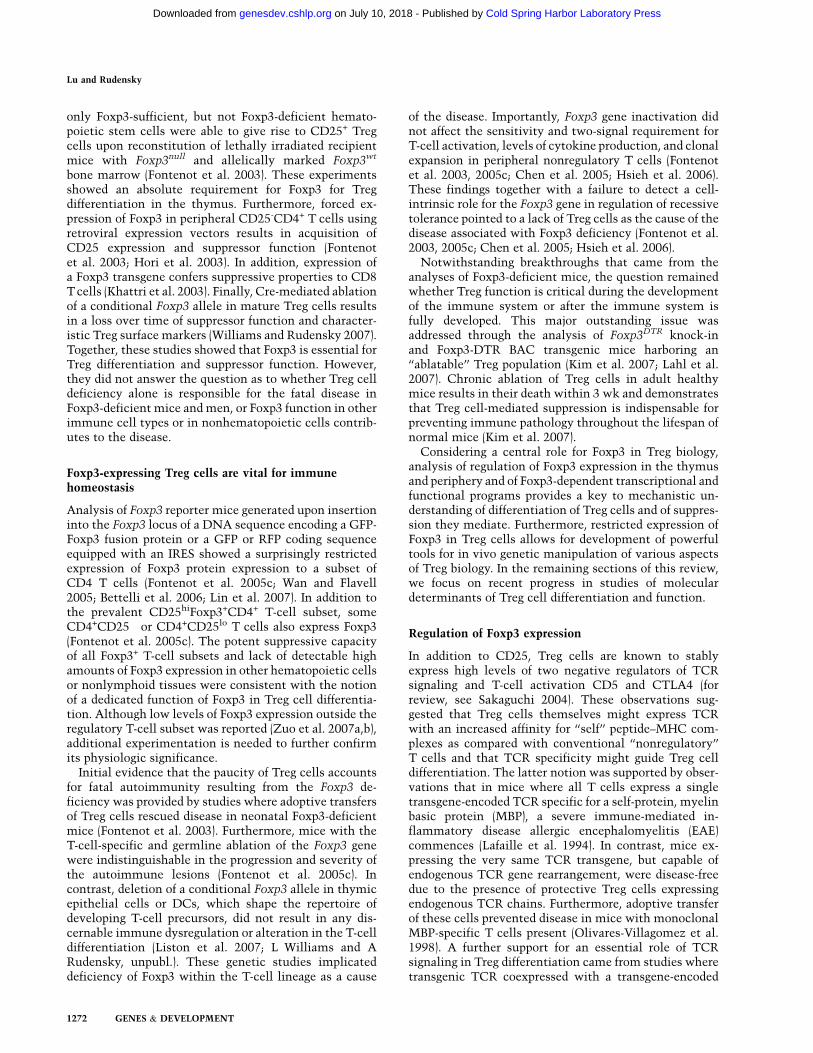
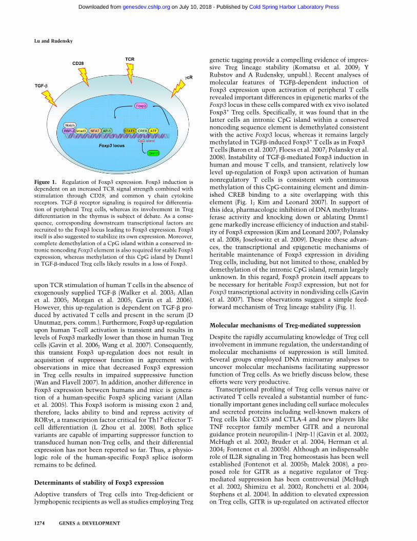
Figure 1. Regulation of Foxp3 expression. Foxp3 induction isdependent on an increased TCR signal strength combined withstimulation through CD28, and common g chain cytokinereceptors. TGF-b receptor signaling is required for differentia-tion of peripheral Treg cells, whereas its involvement in Tregdifferentiation in the thymus is subject of debate. As a conse-quence, corresponding downstream transcriptional factors arerecruited to the Foxp3 locus leading to Foxp3 expression. Foxp3itself is also suggested to stabilize its own expression. Moreover,complete demethylation of a CpG island within a conserved in-tronic noncoding Foxp3 element is also required for stable Foxp3expression, whereas methylation of this CpG island by Dnmt1in TGF-b-induced Treg cells likely results in a loss of Foxp3.
Lu and Rudensky
1274 GENES & DEVELOPMENT
Cold Spring Harbor Laboratory Press on July 10, 2018 - Published by genesdev.cshlp.orgDownloaded from
T cells, where along with other TNFR family membersOX40, 41BB and TNFRII, it serves as a potent costimu-latory and survival molecule. It seems more likely thatengagement of GITR on the surface of effector T cellsenhances their responses, effectively releasing these cellsfrom the Treg-mediated suppression (Ronchetti et al.2004; Stephens et al. 2004). Consistent with these results,GITR deficiency does not result in noticeable aberrationsin immune homeostasis or tolerance (Ronchetti et al.2002), yet GITR may serve as a promising target forcancer immunotherapy.
In contrast to GITR, CTLA-4, which like GITR is up-regulated on effector T cells and is constitutively ex-pressed at a high level on Treg cells, is indispensable forimmune homeostasis. At 3 wk of age, CTLA-4-deficientmice succumb to a highly aggressive immune lesionsmanifested in splenomegaly, lymphadenopathy, myocar-ditis, and pancreatitis (Tivol et al. 1995). Although CTLA-4 plays an important cell-intrinsic role in limitingresponses of activated T cells, a surprising observationthat the presence of CTLA-4-sufficient T cells preventsexcessive activation of CTLA-4-deficient T cells pointedto an important function of CTLA-4 in Treg-mediatedsuppression (Bachmann et al. 1999). Building on earlierreports using cell transfer and antibody blockade (Readet al. 2000, 2006; Takahashi et al. 2000), a role of CTLA-4in Treg cells has been reassessed recently in geneticstudies, where the mice with a selective loss of CTLA-4in Treg cells were analyzed (Wing et al. 2008; Friedlineet al. 2009; Y Rubstov and A Rudensky, in prep.). In thesestudies, selective CTLA-4 deficiency resulted in greatlyincreased numbers and activation of Treg cells underinflammatory conditions, yet the suppressive activityof CTLA-4-deficient Treg cells was impaired. It wassuggested that in BALB/c mice, known for their suscep-tibility to a variety of immune-mediated disorders, thereduced suppression capacity of CTLA-4-deficient Tregcells is due to their inability of down-regulating CD80and CD86 expression on and expansion of DCs (Winget al. 2008). These results were consistent with a pro-nounced expansion and activation of DCs observed veryearly upon acute Treg cell ablation (Kim et al. 2007).Unexpectedly, however, Treg-specific ablation of CTLA-4on a less autoimmune-prone genetic background ofC57Bl6 mice does not cause an increase in DC numbersor CD80/CD86 expression, yet Treg cell suppressorcapacity was impaired albeit only partially (Y Rubstovand A Rudensky, in prep.). Thus, CTLA-4 likely hasadditional, yet to be discovered functions in facilitatingTreg suppression.
These results also raise a possibility that in addition toCTLA-4, other surface molecules highly expressed byTreg cells might contribute to suppression of DC activa-tion, making this aspect of CTLA-4 function in Treg cellsredundant in a genetic background less susceptible toautoimmunity. The latter possibility is supported bya recent discovery of a novel Ig family member TIGITexpressed, like CTLA-4, at a very high level on Tregcells and on activated conventional T cells. It appearsthat upon Treg interactions with DCs TIGIT induces
production of immunosuppressive cytokines IL-10 andTGFb by DCs (Yu et al. 2009). Long-lasting Treg cellinteractions with DCs were convincingly documented byrecent studies employing intravital microscopy (Tadokoroet al. 2006; Tang et al. 2006). These interactions arefacilitated by Nrp-1 highly expressed by the majority ofTreg cells, and Nrp-1 blockade interferes with suppressionmediated by Treg cells (Sarris et al. 2008).
In addition to functionally important transmembranemolecules, a number of secreted proteins identified ingene expression analyses, including granzyme B, IL-9, IL-10, and IL-35, have also been implicated in Treg cell-mediated suppression function. Finally, elaboration ofadenosine facilitated by ectoenzymes CD73 and CD39,highly expressed on Treg cells, and extrusion of cAMPlikely contribute to Treg-mediated suppression (Kobieet al. 2006; Bopp et al. 2007; Borsellino et al. 2007; Deaglioet al. 2007).
It is important to note that none of the aforementionedmechanisms of suppression can singly account for Treg-mediated control of immunity. Most likely, distinctsuppressor mechanisms prominently feature in particulartissue and inflammatory settings. In this regard, studiesusing experimental skin transplantation models sug-gested that Treg-derived granzyme B and IL-9 contributeto long-lived transplanted graft survival (Lu et al. 2006;Gondek et al. 2008), whereas IL-10 and IL-35 secretedby Treg cells likely limit inflammation in the colon(Collison et al. 2007; Rubtsov et al. 2008).
A role of Foxp3 in Treg transcriptionaland functional programs
The aforementioned central role of Foxp3 in defining Tregcell lineage raised a question as to what degree that Treg-specific genomic program is directly controlled by Foxp3.Based on comparison of gene expression profiles in Foxp3+
T cells generated under different conditions (Sugimotoet al. 2006; Hill et al. 2007), it was suggested that thedistinct Foxp3-independent features of Treg transcrip-tional program precede and are established in parallelwith the Foxp3-dependent transcriptional program. Adirect investigation of Foxp3 contribution to transcrip-tional and functional features of Treg cells was madepossible through the analysis of mice harboring a Foxp3reporter-null allele (Foxp3GFPKO) generated upon insertionof a GFP coding sequence into the Foxp3 locus witha concomitant ablation of the Foxp3 protein expression(Gavin et al. 2007). Cells expressing Foxp3GFPKO can bearguably considered an equivalent of Treg precursor cells.These cells exhibit some of phenotypic and molecularcharacteristics of Foxp3+Treg cells, including an inabilityto proliferate and produce IL-2 in response to TCRstimulation, expression of low amounts of IL7Ra chain,and elevated CD25, CTLA-4, and GITR, albeit at signif-icantly lower levels in comparison of Treg cells. However,unlike Treg cells, GFP+Foxp3GFPKO T cells produce im-mune response promoting Th2 and Th17 cytokines IL-4and IL-17, and the block in their autonomous proliferativeactivity is less severe as their in vitro proliferation can be
Molecular regulation in Treg cells
GENES & DEVELOPMENT 1275
Cold Spring Harbor Laboratory Press on July 10, 2018 - Published by genesdev.cshlp.orgDownloaded from

readily restored by limited TCR/CD28 costimulation.The Foxp3-mediated repression of IL-17 production,a characteristic feature of Th17 cells, is likely due toa modulation of transcriptional activity of orphan nuclearreceptors RORg and RORa through direct interaction ofFoxp3 (L Zhou et al. 2008). RORg serves a role of a keyTh17 lineage specifying factor (Ivanov et al. 2006). Inaddition, GFP+Foxp3GFPKO T-cell population were quies-cent, whereas Foxp3+ Treg cell subset exhibits impressiveproliferative activity in vivo in the absence of inflamma-tion. Most importantly, GFP+Foxp3GFPKO T cells werecompletely devoid of suppressor activity (Gavin et al.2007). Similar results were obtained by Chatila andcolleagues (Lin et al. 2007) using analogous knock-instrategy, although notable differences in the phenotypeof T cells expressing dysfunctional Foxp3 allele wereobserved in the latter study, most likely due to thepresence of Foxp3 protein lacking DNA-binding domain,yet capable of protein–protein interactions (Lin et al.2007), in contrast to a complete Foxp3 ablation in theformer study (Gavin et al. 2007). Together these studiesshowed that Foxp3 is absolutely required for suppressorfunction, proliferative potential, and metabolic fitness ofTreg cells. In addition, Foxp3 prevents differentiation ofTreg precursor cells into effector T-cell lineages. Charac-teristically, Foxp3 amplifies and stabilizes expression ofa number of genes transiently up-regulated in activatednonregulatory T cells. At the same time, Foxp3 enforcesrepression of immune response promoting genes normallyinduced in naı̈ve and effector T cells upon TCR stimula-tion. Thus, Foxp3 controls Treg cell differentiation bypotentiating or consolidating the beneficial features andat the same time correcting the disadvantageous featuresof precursor cells.
The analyses of Foxp3-dependent transcriptional pro-gram invited a question as to how many genes are directlyregulated by Foxp3. Recently, a systemic examination ofFoxp3 target genes was undertaken using a combinationof chromatin immunoprecipitation (ChIP) with mousegenome tiling array or promoter array analyses (Marsonet al. 2007; Zheng et al. 2007). A cross-comparison of thedata sets of Foxp3-bound genes and genes differentiallyexpressed in Foxp3+Treg cells versus GFP+ Foxp3GFPKO Tcells revealed that ;10% of Foxp3-dependent genes aredirectly regulated by Foxp3 (Zheng et al. 2007). How-ever, the direct Foxp3 target genes include a number ofsequence-specific transcription factors and miRNAs,playing important roles in Treg biology and contributingto differential mRNA and protein expression in Treg cells(Fig. 2; Zheng et al. 2007). The analysis of Foxp3-bindinggenes also showed that in contrast to early notion thatFoxp3 acts as a transcriptional repressor (Schubert et al.2001; Bettelli et al. 2005; Grant et al. 2006; Lopes et al.2006), more Foxp3-bound genes are up-regulated, thanrepressed, in Treg cells (Zheng et al. 2007). Thus, Foxp3acts as both transcriptional activator and repressor.Furthermore, Foxp3 binding correlates with markedenrichment in permissive (H3K4me3) and inhibitory(H3K27me3) histone modifications associated with itsbinding sites in activated and repressed genes, respec-
tively (Zheng et al. 2007). These results suggest thatFoxp3 imparts epigenetic marks on its target genes and,thereby, establishes a heritable transcriptional programduring Treg differentiation. However, ablation of a condi-tional Foxp3 allele in mature Treg cells over time resultedin a loss of characteristic gene expression, suppressorfunction, and the acquisition of effector T-cell function(Williams and Rudensky 2007). These results indicatethat heritable maintenance of developmentally estab-lished Treg transcriptional and functional program re-quires continuous expression of Foxp3. Subsequent studydemonstrated a similar requirement for Pax5 for themaintenance of the B-cell lineage identity (Cobaledaet al. 2007). These observations suggest that this is likelya common feature of late cellular differentiation.
Treg cell-mediated suppression of distinct classesof the immune response
Foxp3-dependent suppressor program implemented byTreg cells keeps in check Th1, Th2, and Th17 types ofeffector immune responses to ‘‘self’’ antigens and patho-gens. Until very recently it was not clear whether Tregcells implement a universal hard-wired program to limitdifferent types of immunity or modular programs ofsuppression tailored to inhibit a particular class of theimmune response. Experimental support for the latternotion comes from a recent analysis of a role for tran-scription factor IRF4 in regulatory T cells. In naı̈ve non-regulatory CD4 T cells, IRF4 is expressed at a very lowlevel, but it is up-regulated upon their activation and itsexpression is required for Th2 differentiation (Lohoff et al.2002; Rengarajan et al. 2002). Unlike conventional Tcells, Treg cells constitutively express high level ofIRF4, which serves as a direct target of Foxp3 (Zhenget al. 2007). Ablation of a conditional IRF4 allele in Tregcells led to a selective dysregulation of unprovokedpathogenic Th2 responses; i.e., increased production ofTh2 cytokines, IL-4-dependent Ig isotype production, and
Figure 2. Foxp3-dependent transcriptional program. Foxp3 actsas both transcriptional activator and repressor. Whereas moregenes are up-regulated by Foxp3 (green) in Treg cells, a numberof genes are down-regulated (red) in a Foxp3-dependent manner.Genome-wide analysis of Foxp3-binding sites revealed thata relatively small fraction (;6%–10%) of Foxp3-dependentgenes are direct targets of Foxp3. In addition, Foxp3 controlsTreg cell transcriptional program indirectly through modulatingexpression of a set of genes encoding transcription factors andmiRNA.
Lu and Rudensky
1276 GENES & DEVELOPMENT
Cold Spring Harbor Laboratory Press on July 10, 2018 - Published by genesdev.cshlp.orgDownloaded from

pronounced plasma cell infiltration (Zheng et al. 2009).Thus, IRF4, a transcriptional factor required for differen-tiation of Th2 effector cells, is expressed in Treg cells ina Foxp3-dependent manner and endows Treg cells withthe ability to suppress Th2 responses. Furthermore, IRF4forms complexes with Foxp3 and in a cooperative mannerregulates at least some of the genes in Treg cells; e.g., Icos,encoding an essential costimulatory molecule. Thesefindings suggest Treg cells might hijack certain compo-nents of cell transcriptional machinery guiding differen-tiation of a particular effector T-cell type to efficientlycontrol the corresponding type of the immune response.
Foxp3 and its interaction partners
The aforementioned interactions of IRF4 and RORg withFoxp3 highlight the importance of understanding thecomposition of Foxp3 transcriptional complexes anddifferent modalities afforded by Foxp3-interacting part-ners. Recent mass-spectrometric analysis of Foxp3 pro-tein complexes isolated by immunoprecipitation revealednumerous regulators of gene expression, including factorsinvolved in chromatin remodeling (BRG1, Ku70/Ku80,and MBD3), acetyltransferase TIP60 and histone deace-tylase HDAC7, and sequence-specific transcription fac-tors (Li and Greene 2007). It was also proposed that therecruitment of TIP60 into Foxp3 transcriptional com-plexes results in Foxp3 acetylation, whereas HDAC7deacetylates Foxp3, and that the ensuing changes in theFoxp3 acetylation state modulate Foxp3 activity ina manner analogous to c-myc (Patel et al. 2004; Li et al.2007). Among sequence-specific transcription factorsserving as Foxp3 interaction partners, NFAT and Runtdomain transcription factor Runx1 were proposed to beindispensable for establishing Treg transcriptional andfunctional programs (Wu et al. 2006; Ono et al. 2007). Themolecular details of NFAT–Foxp3 interactions were de-duced from crystallographic analysis of DNA-boundNFAT and the forkhead domain of Foxp2, a close relativeof Foxp3 (Wu et al. 2006). DNA template-dependentinteractions of Foxp3 with NFAT are thought to preventformation of NFAT–AP-1 complexes, required for theexpression of immune response-promoting genes in ef-fector T cells, thereby ensuring their repression in Tregcells. NFAT:Foxp3 cooperation is able to drive the geno-mic program required for Treg cell differentiation andfunction (Wu et al. 2006). In addition, it has beensuggested that Foxp3 might inhibit AP-1 functionthrough direct association with the activated AP-1 pro-tein (Lee et al. 2008). Site-directed mutagenesis of pre-dicted NFAT interactions sites in the DNA-bindingdomain of the Foxp3 protein resulted in a loss of itsability to impose Treg gene signature and suppressorfunction (Wu et al. 2006). A similar loss of function wasobserved upon introduction of mutations disruptingFoxp3 interactions with Runx1, initially identified ina yeast two-hybrid screen (Ono et al. 2007). As a note ofcaution, a considerable caveat to the site-mutagenesisapproach is that introduced mutations might lead to a lossof additional interacting partners besides those under
study. Considering the complexity of the Foxp3 ‘‘inter-actome,’’ further biochemical, genetic, and functionalstudies of components of Foxp3 transcriptional com-plexes are warranted.
Role of miRNA in Foxp3+ Treg cells
MicroRNA (miRNA) are small untranslated RNA spe-cies, which have been implicated in the regulation of geneexpression essential for organ development, cellular dif-ferentiation, homeostasis, and functioning through targetmRNA degradation or translational interference (Bartel2004). Although miRNA-mediated gene regulation iscritical during B-cell differentiation (O’Carroll et al. 2007;Koralov et al. 2008), depletion of miRNA in developingthymocytes does not result in a gross perturbation of T-celldifferentiation (Cobb et al. 2005; Muljo et al. 2005).However, ablation of either Dicer or Drosha, two RNaseIII enzymes critical for the generation of mature miRNAs,at a stage of the DP thymocyte differentiation results inreduced numbers of Foxp3+ thymocytes and peripheralTreg cells and immune-mediated lesions developing at 6mo of age (Cobb et al. 2006; Chong et al. 2008). Likewise,a reduction in the efficiency of Foxp3 induction is alsofound upon stimulation of naı̈ve Dicer-deficient T cells inthe presence of TGF-b (Cobb et al. 2006). Despite a role ofmiRNA in the generation of Foxp3+ Treg cells both in thethymus and in the periphery, it seems unlikely miRNAsare involved in the regulation of Foxp3 amounts. Threestudies found no evidence of reduced Foxp3 amounts inTreg cells in the mice with a T-cell-specific or Treg-specificDicer or Drosha deletion (Cobb et al. 2006; Chong et al.
Figure 3. miRNA-dependent gene regulation preserves Tregsuppressor function in inflammatory settings. miRNA depletiondue to ablation of Dicer in Treg cells (green) results in impairedhomeostasis and attenuated suppressor function in a healthyheterozygous Dicer fl/flFoxp3cre/+ female mice cohabited byDicer-deficient (green) Dicer-sufficient Treg cells (blue). (Middle
panel) Nevertheless, Dicer-deficient Treg cells (green) are stillable to suppress self-reactive effector T cells (red), albeit lessefficiently than their wild-type counterparts. (Left panel) Con-sequently, the mice remain healthy and show no sign of in-flammation, similar to their wild-type littermates (Dicer fl/+
Foxp3cre/cre). (Right panel) In Dicer fl/flFoxp3cre mice, lack ofDicer-sufficient Treg cells results in an inflammation anda complete loss of suppressor capacity of Dicer-deficient Tregcells. This failure of suppressor function in face of inflammationultimately leads to fatal immune lesions indistinguishable fromthat in Treg-deficient mice.
Molecular regulation in Treg cells
GENES & DEVELOPMENT 1277
Cold Spring Harbor Laboratory Press on July 10, 2018 - Published by genesdev.cshlp.orgDownloaded from

2008; Liston et al. 2008). However, upon deletion of aconditional Dicer allele mediated by a Foxp3 BAC trans-gene encoding YFP-Cre, a significant proportion of Dicer-deficient Treg cells became Foxp3 negative, implicatingmiRNA in Treg lineage stabilization (X Zhou et al. 2008).Finally, miRNA pathway also promotes survival and pro-liferative potential of Treg cells similar to its role in T- andB-cell lineages (Liston et al. 2008).
In addition to its role in Treg development and homeo-stasis, miRNA-dependent regulation of gene expression isalso critical to controlling Treg cell function. Depletionof miRNA within the Treg cell lineage resulted ina fatal autoimmunity indistinguishable from that inTreg-deficient mice (Chong et al. 2008; Liston et al.2008; X Zhou et al. 2008). Importantly, the suppressorcapacity of Dicer-deficient Treg cells was maintained,albeit at a markedly reduced level, under noninflamma-tory conditions. However, in inflammatory settings,these cells entirely lost the suppressive capacity despitea marked increase in their numbers and activation (Listonet al. 2008). These observations implicate miRNA as keyguardians of a stable Treg suppressor program under in-flammatory conditions (Fig. 3).
Treg-specific Dicer and Drosha ablation studies raiseda question as to the identity of specific miRNAs regulat-ing distinct aspects of Treg biology. Differential miRNAsexpression in Foxp3+ Treg cells was first demonstrated byCobb et al. (2006). Furthermore, many of Treg-specificmiRNAs are expressed in Treg cells in a Foxp3-dependentmanner. Among Foxp3-dependent miRNAs, miR-155 hasbeen shown to be directly regulated by Foxp3 (Marsonet al. 2007; Zheng et al. 2007; Lu et al. 2009). Althoughdispensable for Treg differentiation and suppressor func-tion, Foxp3-driven miR-155 up-regulation is critical forheightened responsiveness of Treg cells to their keysurvival and growth factor, IL-2. At a mechanistic level,miR-155-mediated control of Treg cell homeostasis ismediated through targeting SOCS1, a negative regulatorof IL-2R signaling. Thus, constitutively high expression of
miR-155 driven by Foxp3 ensures efficient STAT5 phos-phorylation in the presence of limiting amounts of IL-2and, thus, fitness of Treg cell subset in a competitiveenvironment (Fig. 4; Lu et al. 2009). This finding not onlyprovides the first example of a single miRNA controllingTreg homeostasis but also demonstrates that differentmiRNAs affect distinct facets of Treg biology.
Concluding remarks
The discovery of Treg cells and of their key controller,Foxp3, has revitalized the field of immunological toler-ance. The regulation of Foxp3 expression, identificationof its interaction partners and downstream gene targetsprovide essential mechanistic insights into highly com-plex and intricate regulation of immune homeostasismediated by Treg cells. Recent advances have made Tregbiology a fascinating research field amenable to mecha-nistic dissection. From a general biological perspective,Foxp3 and Treg cells provide a powerful model to dissectthe molecular orchestration of cellular differentiation,function, and homeostasis. From an immunological pointof view, in-depth exploration of Treg biology is funda-mental to understanding the origin and mechanisms ofdominant tolerance, a vital and unique means of negativeregulation in the immune system. On a practical level,Treg manipulation offers promise for developing noveltherapeutic approaches in cancer, autoimmune and in-fectious diseases.
Acknowledgments
We thank Meinrad Busslinger for sharing with us unpublishedobservations. This work was supported by NIH (A.Y.R.). L.L.F. isa Leukemia and Lymphoma Society Fellow. A.Y.R. is a HowardHughes Medical Institute investigator.
References
Allan SE, Passerini L, Bacchetta R, Crellin N, Dai M, Orban PC,Ziegler SF, Roncarolo MG, Levings MK. 2005. The role of 2FOXP3 isoforms in the generation of human CD4+ Tregs.J Clin Invest 115: 3276–3284.
Apostolou I, Sarukhan A, Klein L, von Boehmer H. 2002. Originof regulatory T cells with known specificity for antigen. NatImmunol 3: 756–763.
Asano M, Toda M, Sakaguchi N, Sakaguchi S. 1996. Autoim-mune disease as a consequence of developmental abnormal-ity of a T cell subpopulation. J Exp Med 184: 387–396.
Bachmann MF, Kohler G, Ecabert B, Mak TW, Kopf M. 1999.Cutting edge: Lymphoproliferative disease in the absence ofCTLA-4 is not T cell autonomous. J Immunol 163: 1128–1131.
Baron U, Floess S, Wieczorek G, Baumann K, Grutzkau A, DongJ, Thiel A, Boeld TJ, Hoffmann P, Edinger M, et al. 2007.DNA demethylation in the human FOXP3 locus discrim-inates regulatory T cells from activated FOXP3+ conven-tional T cells. Eur J Immunol 37: 2378–2389.
Bartel DP. 2004. MicroRNAs: Genomics, biogenesis, mecha-nism, and function. Cell 116: 281–297.
Bayer AL, Yu A, Adeegbe D, Malek TR. 2005. Essential role forinterleukin-2 for CD4+CD25+ T regulatory cell developmentduring the neonatal period. J Exp Med 201: 769–777.
Figure 4. miR-155-dependent regulation of Treg cell homeo-stasis. In addition to CD25, Foxp3 induces high level of miR-155expression to ensure increased IL-2 responsiveness through miR-155-mediated down-regulation of the SOCS1 protein (left panel).In the absence of miR-155 (right panel), increased amounts ofthe SOCS1 protein attenuate IL-2R signaling, leading to di-minished STAT5 phosphorylation and diminished competitivefitness.
Lu and Rudensky
1278 GENES & DEVELOPMENT
Cold Spring Harbor Laboratory Press on July 10, 2018 - Published by genesdev.cshlp.orgDownloaded from

Bennett CL, Christie J, Ramsdell F, Brunkow ME, Ferguson PJ,Whitesell L, Kelly TE, Saulsbury FT, Chance PF, Ochs HD.2001. The immune dysregulation, polyendocrinopathy, en-teropathy, X-linked syndrome (IPEX) is caused by mutationsof FOXP3. Nat Genet 27: 20–21.
Bettelli E, Dastrange M, Oukka M. 2005. Foxp3 interacts withnuclear factor of activated T cells and NF-kB to represscytokine gene expression and effector functions of T helpercells. Proc Natl Acad Sci 102: 5138–5143.
Bettelli E, Carrier Y, Gao W, Korn T, Strom TB, Oukka M,Weiner HL, Kuchroo VK. 2006. Reciprocal developmentalpathways for the generation of pathogenic effector TH17 andregulatory T cells. Nature 441: 235–238.
Bonomo A, Kehn PJ, Payer E, Rizzo L, Cheever AW, Shevach EM.1995. Pathogenesis of post-thymectomy autoimmunity. Roleof syngeneic MLR-reactive T cells. J Immunol 154: 6602–6611.
Bopp T, Becker C, Klein M, Klein-Hessling S, Palmetshofer A,Serfling E, Heib V, Becker M, Kubach J, Schmitt S, et al. 2007.Cyclic adenosine monophosphate is a key component ofregulatory T cell-mediated suppression. J Exp Med 204: 1303–1310.
Borsellino G, Kleinewietfeld M, Di Mitri D, Sternjak A,Diamantini A, Giometto R, Hopner S, Centonze D, BernardiG, Dell’Acqua ML, et al. 2007. Expression of ectonucleoti-dase CD39 by Foxp3+ Treg cells: Hydrolysis of extracellularATP and immune suppression. Blood 110: 1225–1232.
Bruder D, Probst-Kepper M, Westendorf AM, Geffers R, BeissertS, Loser K, von Boehmer H, Buer J, Hansen W. 2004. Neuro-pilin-1: A surface marker of regulatory T cells. Eur J Immunol
34: 623–630.Burchill MA, Goetz CA, Prlic M, O’Neil JJ, Harmon IR, Ben-
singer SJ, Turka LA, Brennan P, Jameson SC, Farrar MA.2003. Distinct effects of STAT5 activation on CD4+ andCD8+ T cell homeostasis: Development of CD4+CD25+
regulatory T cells versus CD8+ memory T cells. J Immunol171: 5853–5864.
Burchill MA, Yang J, Vogtenhuber C, Blazar BR, Farrar MA.2007. IL-2 receptor b-dependent STAT5 activation is requiredfor the development of Foxp3+ regulatory T cells. J Immunol178: 280–290.
Burchill MA, Yang J, Vang KB, Moon JJ, Chu HH, Lio CW, VegoeAL, Hsieh CS, Jenkins MK, Farrar MA. 2008. Linked T cellreceptor and cytokine signaling govern the development ofthe regulatory T cell repertoire. Immunity 28: 112–121.
Chatila TA, Blaeser F, Ho N, Lederman HM, Voulgaropoulos C,Helms C, Bowcock AM. 2000. JM2, encoding a fork head-related protein, is mutated in X-linked autoimmunity-allergic disregulation syndrome. J Clin Invest 106: R75–R81. doi: 10.1172/JCI11679.
Chen W, Jin W, Hardegen N, Lei KJ, Li L, Marinos N, McGradyG, Wahl SM. 2003. Conversion of peripheral CD4+CD25-naive T cells to CD4+CD25+ regulatory T cells by TGF-binduction of transcription factor Foxp3. J Exp Med 198: 1875–1886.
Chen Z, Benoist C, Mathis D. 2005. How defects in centraltolerance impinge on a deficiency in regulatory T cells. Proc
Natl Acad Sci 102: 14735–14740.Chong MM, Rasmussen JP, Rudensky AY, Littman DR. 2008.
The RNAseIII enzyme Drosha is critical in T cells forpreventing lethal inflammatory disease. J Exp Med 205:2005–2017.
Cobaleda C, Jochum W, Busslinger M. 2007. Conversion ofmature B cells into T cells by dedifferentiation to uncom-mitted progenitors. Nature 449: 473–477.
Cobb BS, Nesterova TB, Thompson E, Hertweck A, O’Connor E,Godwin J, Wilson CB, Brockdorff N, Fisher AG, Smale ST,
et al. 2005. T cell lineage choice and differentiation in theabsence of the RNase III enzyme Dicer. J Exp Med 201: 1367–1373.
Cobb BS, Hertweck A, Smith J, O’Connor E, Graf D, Cook T,Smale ST, Sakaguchi S, Livesey FJ, Fisher AG, et al. 2006. Arole for Dicer in immune regulation. J Exp Med 203: 2519–2527.
Collison LW, Workman CJ, Kuo TT, Boyd K, Wang Y, VignaliKM, Cross R, Sehy D, Blumberg RS, Vignali DA. 2007. Theinhibitory cytokine IL-35 contributes to regulatory T-cellfunction. Nature 450: 566–569.
Davidson TS, DiPaolo RJ, Andersson J, Shevach EM. 2007.Cutting edge: IL-2 is essential for TGF-b-mediated inductionof Foxp3+ T regulatory cells. J Immunol 178: 4022–4026.
Deaglio S, Dwyer KM, Gao W, Friedman D, Usheva A, Erat A,Chen JF, Enjyoji K, Linden J, Oukka M, et al. 2007. Adenosinegeneration catalyzed by CD39 and CD73 expressed on reg-ulatory T cells mediates immune suppression. J Exp Med
204: 1257–1265.Fehervari Z, Sakaguchi S. 2004. CD4+ Tregs and immune
control. J Clin Invest 114: 1209–1217.Floess S, Freyer J, Siewert C, Baron U, Olek S, Polansky J,
Schlawe K, Chang HD, Bopp T, Schmitt E, et al. 2007.Epigenetic control of the foxp3 locus in regulatory T cells.PLoS Biol 5: e38. doi: 10.1371/journal.pbio.0050038.
Fontenot JD, Gavin MA, Rudensky AY. 2003. Foxp3 programsthe development and function of CD4+CD25+ regulatory Tcells. Nat Immunol 4: 330–336.
Fontenot JD, Dooley JL, Farr AG, Rudensky AY. 2005a. De-velopmental regulation of Foxp3 expression during ontogeny.J Exp Med 202: 901–906.
Fontenot JD, Rasmussen JP, Gavin MA, Rudensky AY. 2005b. Afunction for interleukin 2 in Foxp3-expressing regulatory Tcells. Nat Immunol 6: 1142–1151.
Fontenot JD, Rasmussen JP, Williams LM, Dooley JL, Farr AG,Rudensky AY. 2005c. Regulatory T cell lineage specificationby the forkhead transcription factor foxp3. Immunity 22:329–341.
Friedline RH, Brown DS, Nguyen H, Kornfeld H, Lee J, Zhang Y,Appleby M, Der SD, Kang J, Chambers CA. 2009. CD4+
regulatory T cells require CTLA-4 for the maintenance ofsystemic tolerance. J Exp Med 16: 421–434.
Furtado GC, Curotto de Lafaille MA, Kutchukhidze N, LafailleJJ. 2002. Interleukin 2 signaling is required for CD4+ regula-tory T cell function. J Exp Med 196: 851–857.
Gambineri E, Torgerson TR, Ochs HD. 2003. Immune dysregu-lation, polyendocrinopathy, enteropathy, and X-linked in-heritance (IPEX), a syndrome of systemic autoimmunitycaused by mutations of FOXP3, a critical regulator of T-cellhomeostasis. Curr Opin Rheumatol 15: 430–435.
Gavin MA, Clarke SR, Negrou E, Gallegos A, Rudensky A. 2002.Homeostasis and anergy of CD4+CD25+ suppressor T cells invivo. Nat Immunol 3: 33–41.
Gavin MA, Torgerson TR, Houston E, DeRoos P, Ho WY,Stray-Pedersen A, Ocheltree EL, Greenberg PD, Ochs HD,Rudensky AY. 2006. Single-cell analysis of normal andFOXP3-mutant human T cells: FOXP3 expression withoutregulatory T cell development. Proc Natl Acad Sci 103:6659–6664.
Gavin MA, Rasmussen JP, Fontenot JD, Vasta V, ManganielloVC, Beavo JA, Rudensky AY. 2007. Foxp3-dependent pro-gramme of regulatory T-cell differentiation. Nature 445: 771–775.
Godfrey VL, Rouse BT, Wilkinson JE. 1994. Transplantation of Tcell-mediated, lymphoreticular disease from the scurfy (sf)mouse. Am J Pathol 145: 281–286.
Molecular regulation in Treg cells
GENES & DEVELOPMENT 1279
Cold Spring Harbor Laboratory Press on July 10, 2018 - Published by genesdev.cshlp.orgDownloaded from

Gondek DC, Devries V, Nowak EC, Lu LF, Bennett KA, ScottZA, Noelle RJ. 2008. Transplantation survival is maintainedby granzyme B+ regulatory cells and adaptive regulatory Tcells. J Immunol 181: 4752–4760.
Grant C, Oh U, Fugo K, Takenouchi N, Griffith C, Yao K,Newhook TE, Ratner L, Jacobson S. 2006. Foxp3 repressesretroviral transcription by targeting both NF-kB and CREBpathways. PLoS Pathog 2: e33. doi: 10.1371/journal.ppat.0020033.
Gupta S, Manicassamy S, Vasu C, Kumar A, Shang W, Sun Z.2008. Differential requirement of PKC-u in the developmentand function of natural regulatory T cells. Mol Immunol 46:213–224.
Haxhinasto S, Mathis D, Benoist C. 2008. The AKT–mTOR axisregulates de novo differentiation of CD4+Foxp3+ cells. J Exp
Med 205: 565–574.Herman AE, Freeman GJ, Mathis D, Benoist C. 2004.
CD4+CD25+ T regulatory cells dependent on ICOS promoteregulation of effector cells in the prediabetic lesion. J Exp
Med 199: 1479–1489.Hill JA, Feuerer M, Tash K, Haxhinasto S, Perez J, Melamed R,
Mathis D, Benoist C. 2007. Foxp3 transcription-factor-dependent and -independent regulation of the regulatory Tcell transcriptional signature. Immunity 27: 786–800.
Hori S, Nomura T, Sakaguchi S. 2003. Control of regulatory Tcell development by the transcription factor Foxp3. Science
299: 1057–1061.Horwitz DA, Zheng SG, Wang J, Gray JD. 2008. Critical role of
IL-2 and TGF-b in generation, function and stabilization ofFoxp3+CD4+ Treg. Eur J Immunol 38: 912–915.
Hsieh CS, Zheng Y, Liang Y, Fontenot JD, Rudensky AY. 2006.An intersection between the self-reactive regulatory andnonregulatory T cell receptor repertoires. Nat Immunol 7:401–410.
Ivanov II, McKenzie BS, Zhou L, Tadokoro CE, Lepelley A,Lafaille JJ, Cua DJ, Littman DR. 2006. The orphan nuclearreceptor RORgt directs the differentiation program of proin-flammatory IL-17+ T helper cells. Cell 126: 1121–1133.
Jordan MS, Boesteanu A, Reed AJ, Petrone AL, Holenbeck AE,Lerman MA, Naji A, Caton AJ. 2001. Thymic selection ofCD4+CD25+ regulatory T cells induced by an agonist self-peptide. Nat Immunol 2: 301–306.
Josefowicz SZ, Wilson CB, Rudensky AY. 2009. TCR stimulationis sufficient for induction of Foxp3 expression in the absenceof DNA methyltransferase I. J Immunol (in press).
Khattri R, Cox T, Yasayko SA, Ramsdell F. 2003. An essential rolefor Scurfin in CD4+CD25+ T regulatory cells. Nat Immunol
4: 337–342.Kim HP, Leonard WJ. 2007. CREB/ATF-dependent T cell
receptor-induced FoxP3 gene expression: A role for DNAmethylation. J Exp Med 204: 1543–1551.
Kim JM, Rasmussen JP, Rudensky AY. 2007. Regulatory T cellsprevent catastrophic autoimmunity throughout the lifespanof mice. Nat Immunol 8: 191–197.
Kobie JJ, Shah PR, Yang L, Rebhahn JA, Fowell DJ, MosmannTR. 2006. T regulatory and primed uncommitted CD4 T cellsexpress CD73, which suppresses effector CD4 T cells byconverting 59-adenosine monophosphate to adenosine. J
Immunol 177: 6780–6786.Komatsu N, Mariotti-Ferrandiz ME, Wang Y, Malissen B,
Waldmann H, Hori S. 2009. Heterogeneity of natural Foxp3+
T cells: A committed regulatory T-cell lineage and an un-committed minor population retaining plasticity. Proc Natl
Acad Sci 106: 1903–1908.Koralov SB, Muljo SA, Galler GR, Krek A, Chakraborty T,
Kanellopoulou C, Jensen K, Cobb BS, Merkenschlager M,
Rajewsky N, et al. 2008. Dicer ablation affects antibodydiversity and cell survival in the B lymphocyte lineage. Cell132: 860–874.
Lafaille JJ, Nagashima K, Katsuki M, Tonegawa S. 1994. Highincidence of spontaneous autoimmune encephalomyelitis inimmunodeficient anti-myelin basic protein T cell receptortransgenic mice. Cell 78: 399–408.
Lahl K, Loddenkemper C, Drouin C, Freyer J, Arnason J, Eberl G,Hamann A, Wagner H, Huehn J, Sparwasser T. 2007. Selec-tive depletion of Foxp3+ regulatory T cells induces a scurfy-like disease. J Exp Med 204: 57–63.
Lee SM, Gao B, Fang D. 2008. FoxP3 maintains Treg unrespon-siveness by selectively inhibiting the promoter DNA-bindingactivity of AP-1. Blood 111: 3599–3606.
Lenschow DJ, Walunas TL, Bluestone JA. 1996. CD28/B7 systemof T cell costimulation. Annu Rev Immunol 14: 233–258.
Li B, Greene MI. 2007. FOXP3 actively represses transcriptionby recruiting the HAT/HDAC complex. Cell Cycle 6: 1432–1436.
Li B, Samanta A, Song X, Iacono KT, Bembas K, Tao R, Basu S,Riley JL, Hancock WW, Shen Y, et al. 2007. FOXP3 inter-actions with histone acetyltransferase and class II histonedeacetylases are required for repression. Proc Natl Acad Sci
104: 4571–4576.Lin W, Haribhai D, Relland LM, Truong N, Carlson MR, Williams
CB, Chatila TA. 2007. Regulatory T cell development in theabsence of functional Foxp3. Nat Immunol 8: 359–368.
Lio CW, Hsieh CS. 2008. A two-step process for thymicregulatory T cell development. Immunity 28: 100–111.
Liston A, Farr AG, Chen Z, Benoist C, Mathis D, Manley NR,Rudensky AY. 2007. Lack of Foxp3 function and expressionin the thymic epithelium. J Exp Med 204: 475–480.
Liston A, Lu LF, O’Carroll D, Tarakhovsky A, Rudensky AY.2008. Dicer-dependent microRNA pathway safeguards regu-latory T cell function. J Exp Med 205: 1993–2004.
Liu Y, Zhang P, Li J, Kulkarni AB, Perruche S, Chen W. 2008. Acritical function for TGF-b signaling in the development ofnatural CD4+CD25+Foxp3+ regulatory T cells. Nat Immunol
9: 632–640.Lohoff M, Mittrucker HW, Prechtl S, Bischof S, Sommer F, Kock
S, Ferrick DA, Duncan GS, Gessner A, Mak TW. 2002.Dysregulated T helper cell differentiation in the absence ofinterferon regulatory factor 4. Proc Natl Acad Sci 99: 11808–11812.
Lopes JE, Torgerson TR, Schubert LA, Anover SD, Ocheltree EL,Ochs HD, Ziegler SF. 2006. Analysis of FOXP3 revealsmultiple domains required for its function as a transcriptionalrepressor. J Immunol 177: 3133–3142.
Lu LF, Lind EF, Gondek DC, Bennett KA, Gleeson MW, Pino-Lagos K, Scott ZA, Coyle AJ, Reed JL, Van Snick J, et al. 2006.Mast cells are essential intermediaries in regulatory T-celltolerance. Nature 442: 997–1002.
Lu LF, Thai TH, Calado DP, Chaudhry A, Kubo M, Tanaka K,Loeb GB, Lee H, Yoshimura A, Rajewsky K, et al. 2009.Foxp3-dependent microRNA155 confers competitive fitnessto regulatory T cells by targeting SOCS1 protein. Immunity
30: 80–91.Malek TR. 2008. The biology of interleukin-2. Annu Rev
Immunol 26: 453–479.Malek TR, Yu A, Vincek V, Scibelli P, Kong L. 2002. CD4
regulatory T cells prevent lethal autoimmunity in IL-2Rb-deficient mice. Implications for the nonredundant functionof IL-2. Immunity 17: 167–178.
Malek TR, Yu A, Zhu L, Matsutani T, Adeegbe D, Bayer AL.2008. IL-2 family of cytokines in T regulatory cell develop-ment and homeostasis. J Clin Immunol 28: 635–639.
Lu and Rudensky
1280 GENES & DEVELOPMENT
Cold Spring Harbor Laboratory Press on July 10, 2018 - Published by genesdev.cshlp.orgDownloaded from

Mantel PY, Ouaked N, Ruckert B, Karagiannidis C, Welz R,Blaser K, Schmidt-Weber CB. 2006. Molecular mechanismsunderlying FOXP3 induction in human T cells. J Immunol
176: 3593–3602.Marson A, Kretschmer K, Frampton GM, Jacobsen ES, Polansky
JK, MacIsaac KD, Levine SS, Fraenkel E, von Boehmer H,Young RA. 2007. Foxp3 occupancy and regulation ofkey target genes during T-cell stimulation. Nature 445:931–935.
McHugh RS, Whitters MJ, Piccirillo CA, Young DA, ShevachEM, Collins M, Byrne MC. 2002. CD4+CD25+ immunoreg-ulatory T cells: Gene expression analysis reveals a functionalrole for the glucocorticoid-induced TNF receptor. Immunity16: 311–323.
Morgan ME, van Bilsen JH, Bakker AM, Heemskerk B, SchilhamMW, Hartgers FC, Elferink BG, van der Zanden L, de VriesRR, Huizinga TW, et al. 2005. Expression of FOXP3 mRNA isnot confined to CD4+CD25+ T regulatory cells in humans.Hum Immunol 66: 13–20.
Muljo SA, Ansel KM, Kanellopoulou C, Livingston DM, Rao A,Rajewsky K. 2005. Aberrant T cell differentiation in theabsence of Dicer. J Exp Med 202: 261–269.
Nishizuka Y, Sakakura T. 1969. Thymus and reproduction: Sex-linked dysgenesia of the gonad after neonatal thymectomy inmice. Science 166: 753–755.
O’Carroll D, Mecklenbrauker I, Das PP, Santana A, Koenig U,Enright AJ, Miska EA, Tarakhovsky A. 2007. A Slicer-independent role for Argonaute 2 in hematopoiesis and themicroRNA pathway. Genes & Dev 21: 1999–2004.
Ohki H, Martin C, Corbel C, Coltey M, Le Douarin NM. 1987.Tolerance induced by thymic epithelial grafts in birds.Science 237: 1032–1035.
Olivares-Villagomez D, Wang Y, Lafaille JJ. 1998. RegulatoryCD4+ T cells expressing endogenous T cell receptor chainsprotect myelin basic protein-specific transgenic mice fromspontaneous autoimmune encephalomyelitis. J Exp Med 188:1883–1894.
Ono M, Yaguchi H, Ohkura N, Kitabayashi I, Nagamura Y,Nomura T, Miyachi Y, Tsukada T, Sakaguchi S. 2007. Foxp3controls regulatory T-cell function by interacting withAML1/Runx1. Nature 446: 685–689.
Ou-Yang HF, Zhang HW, Wu CG, Zhang P, Zhang J, Li JC, HouLH, He F, Ti XY, Song LQ, et al. 2009. Notch signal-ing regulates the FOXP3 promoter through RBP-J- andHes1-dependent mechanisms. Mol Cell Biochem 320: 109–114.
Patel JH, Du Y, Ard PG, Phillips C, Carella B, Chen CJ,Rakowski C, Chatterjee C, Lieberman PM, Lane WS, et al.2004. The c-MYC oncoprotein is a substrate of the acetyl-transferases hGCN5/PCAF and TIP60. Mol Cell Biol 24:10826–10834.
Patton DT, Garden OA, Pearce WP, Clough LE, Monk CR, LeungE, Rowan WC, Sancho S, Walker LS, Vanhaesebroeck B, et al.2006. Cutting edge: The phosphoinositide 3-kinase p110 d iscritical for the function of CD4+CD25+Foxp3+ regulatory Tcells. J Immunol 177: 6598–6602.
Polansky JK, Kretschmer K, Freyer J, Floess S, Garbe A, Baron U,Olek S, Hamann A, von Boehmer H, Huehn J. 2008. DNAmethylation controls Foxp3 gene expression. Eur J Immunol
38: 1654–1663.Read S, Malmstrom V, Powrie F. 2000. Cytotoxic T lymphocyte-
associated antigen 4 plays an essential role in the function ofCD25+CD4+ regulatory cells that control intestinal inflam-mation. J Exp Med 192: 295–302.
Read S, Greenwald R, Izcue A, Robinson N, Mandelbrot D,Francisco L, Sharpe AH, Powrie F. 2006. Blockade of CTLA-4
on CD4+CD25+ regulatory T cells abrogates their function invivo. J Immunol 177: 4376–4383.
Rengarajan J, Mowen KA, McBride KD, Smith ED, Singh H,Glimcher LH. 2002. Interferon regulatory factor 4 (IRF4)interacts with NFATc2 to modulate interleukin 4 geneexpression. J Exp Med 195: 1003–1012.
Ronchetti S, Nocentini G, Riccardi C, Pandolfi PP. 2002. Role ofGITR in activation response of T lymphocytes. Blood 100:350–352.
Ronchetti S, Zollo O, Bruscoli S, Agostini M, Bianchini R,Nocentini G, Ayroldi E, Riccardi C. 2004. GITR, a memberof the TNF receptor superfamily, is costimulatory tomouse T lymphocyte subpopulations. Eur J Immunol 34:613–622.
Rubtsov YP, Rasmussen JP, Chi EY, Fontenot J, Castell L, Ye X,Treuting P, Siewe L, Roers A, Henderson WRJ, et al. 2008. IL-10 produced by regulatory T cells contributes to theirsuppressor function by limiting inflammation at environ-mental interfaces. Immunity 28: 546–558.
Sakaguchi S. 2004. Naturally arising CD4+ regulatory T cells forimmunologic self-tolerance and negative control of immuneresponses. Annu Rev Immunol 22: 531–562.
Sakaguchi S, Takahashi T, Nishizuka Y. 1982. Study on cellularevents in postthymectomy autoimmune oophoritis in mice.I. Requirement of Lyt-1 effector cells for oocytes damage afteradoptive transfer. J Exp Med 156: 1565–1576.
Sakaguchi S, Sakaguchi N, Asano M, Itoh M, Toda M. 1995.Immunologic self-tolerance maintained by activated T cellsexpressing IL-2 receptor a-chains (CD25). Breakdown ofa single mechanism of self-tolerance causes various autoim-mune diseases. J Immunol 155: 1151–1164.
Salomon B, Lenschow DJ, Rhee L, Ashourian N, Singh B, SharpeA, Bluestone JA. 2000. B7/CD28 costimulation is essentialfor the homeostasis of the CD4+CD25+ immunoregulatory Tcells that control autoimmune diabetes. Immunity 12: 431–440.
Samon JB, Champhekar A, Minter LM, Telfer JC, Miele L, FauqA, Das P, Golde TE, Osborne BA. 2008. Notch1 and TGFb1cooperatively regulate Foxp3 expression and the mainte-nance of peripheral regulatory T cells. Blood 112: 1813–1821.
Sarris M, Andersen KG, Randow F, Mayr L, Betz AG. 2008.Neuropilin-1 expression on regulatory T cells enhances theirinteractions with dendritic cells during antigen recognition.Immunity 28: 402–413.
Sauer S, Bruno L, Hertweck A, Finlay D, Leleu M, Spivakov M,Knight ZA, Cobb BS, Cantrell D, O’Connor E, et al. 2008. Tcell receptor signaling controls Foxp3 expression via PI3K,Akt, and mTOR. Proc Natl Acad Sci 105: 7797–7802.
Schmidt-Supprian M, Tian J, Grant EP, Pasparakis M, Maehr R,Ovaa H, Ploegh HL, Coyle AJ, Rajewsky K. 2004. Differentialdependence of CD4+CD25+ regulatory and natural killer-likeT cells on signals leading to NF-kB activation. Proc Natl
Acad Sci 101: 4566–4571.Schubert LA, Jeffery E, Zhang Y, Ramsdell F, Ziegler SF. 2001.
Scurfin (FOXP3) acts as a repressor of transcription andregulates T cell activation. J Biol Chem 276: 37672–37679.
Shevach EM. 2000. Regulatory T cells in autoimmmunity. AnnuRev Immunol 18: 423–449.
Shimizu J, Yamazaki S, Takahashi T, Ishida Y, Sakaguchi S.2002. Stimulation of CD25+CD4+ regulatory T cells throughGITR breaks immunological self-tolerance. Nat Immunol 3:135–142.
Starr TK, Jameson SC, Hogquist KA. 2003. Positive and negativeselection of T cells. Annu Rev Immunol 21: 139–176.
Stephens GL, McHugh RS, Whitters MJ, Young DA, LuxenbergD, Carreno BM, Collins M, Shevach EM. 2004. Engagement
Molecular regulation in Treg cells
GENES & DEVELOPMENT 1281
Cold Spring Harbor Laboratory Press on July 10, 2018 - Published by genesdev.cshlp.orgDownloaded from

of glucocorticoid-induced TNFR family-related receptor oneffector T cells by its ligand mediates resistance to suppres-sion by CD4+CD25+ T cells. J Immunol 173: 5008–5020.
Sugimoto N, Oida T, Hirota K, Nakamura K, Nomura T,Uchiyama T, Sakaguchi S. 2006. Foxp3-dependent and -in-dependent molecules specific for CD25+CD4+ natural re-gulatory T cells revealed by DNA microarray analysis. Int
Immunol 18: 1197–1209.Tadokoro CE, Shakhar G, Shen S, Ding Y, Lino AC, Maraver A,
Lafaille JJ, Dustin ML. 2006. Regulatory T cells inhibit stablecontacts between CD4+ T cells and dendritic cells in vivo. J
Exp Med 203: 505–511.Tai X, Cowan M, Feigenbaum L, Singer A. 2005. CD28 cos-
timulation of developing thymocytes induces Foxp3 expres-sion and regulatory T cell differentiation independently ofinterleukin 2. Nat Immunol 6: 152–162.
Takahashi T, Tagami T, Yamazaki S, Uede T, Shimizu J,Sakaguchi N, Mak TW, Sakaguchi S. 2000. Immunologicself-tolerance maintained by CD25+CD4+ regulatory T cellsconstitutively expressing cytotoxic T lymphocyte-associatedantigen 4. J Exp Med 192: 303–310.
Tang Q, Adams JY, Tooley AJ, Bi M, Fife BT, Serra P, SantamariaP, Locksley RM, Krummel MF, Bluestone JA. 2006. Visualiz-ing regulatory T cell control of autoimmune responses innonobese diabetic mice. Nat Immunol 7: 83–92.
Tivol EA, Borriello F, Schweitzer AN, Lynch WP, Bluestone JA,Sharpe AH. 1995. Loss of CTLA-4 leads to massive lympho-proliferation and fatal multiorgan tissue destruction, reveal-ing a critical negative regulatory role of CTLA-4. Immunity
3: 541–547.Tone Y, Furuuchi K, Kojima Y, Tykocinski ML, Greene MI, Tone
M. 2008. Smad3 and NFAT cooperate to induce Foxp3expression through its enhancer. Nat Immunol 9: 194–202.
Trombetta ES, Mellman I. 2005. Cell biology of antigen process-ing in vitro and in vivo. Annu Rev Immunol 23: 975–1028.
Vang KB, Yang J, Mahmud SA, Burchill MA, Vegoe AL, FarrarMA. 2008. IL-2, -7, and -15, but not thymic stromal lympho-poeitin, redundantly govern CD4+Foxp3+ regulatory T celldevelopment. J Immunol 181: 3285–3290.
Walker MR, Kasprowicz DJ, Gersuk VH, Benard A, Van Land-eghen M, Buckner JH, Ziegler SF. 2003. Induction of FoxP3and acquisition of T regulatory activity by stimulated humanCD4+CD25� T cells. J Clin Invest 112: 1437–1443.
Wan YY, Flavell RA. 2005. Identifying Foxp3-expressing sup-pressor T cells with a bicistronic reporter. Proc Natl Acad Sci
102: 5126–5131.Wan YY, Flavell RA. 2007. Regulatory T-cell functions are
subverted and converted owing to attenuated Foxp3 expres-sion. Nature 445: 766–770.
Wang J, Ioan-Facsinay A, van der Voort EI, Huizinga TW, ToesRE. 2007. Transient expression of FOXP3 in human activatednonregulatory CD4+ T cells. Eur J Immunol 37: 129–138.
Wildin RS, Ramsdell F, Peake J, Faravelli F, Casanova JL, BuistN, Levy-Lahad E, Mazzella M, Goulet O, Perroni L, et al.2001. X-linked neonatal diabetes mellitus, enteropathy andendocrinopathy syndrome is the human equivalent of mousescurfy. Nat Genet 27: 18–20.
Williams LM, Rudensky AY. 2007. Maintenance of the Foxp3-dependent developmental program in mature regulatory Tcells requires continued expression of Foxp3. Nat Immunol
8: 277–284.Wing K, Onishi Y, Prieto-Martin P, Yamaguchi T, Miyara M,
Fehervari Z, Nomura T, Sakaguchi S. 2008. CTLA-4 controlover Foxp3+ regulatory T cell function. Science 322: 271–275.
Wu Y, Borde M, Heissmeyer V, Feuerer M, Lapan AD, Stroud JC,Bates DL, Guo L, Han A, Ziegler SF, et al. 2006. FOXP3
controls regulatory T cell function through cooperation withNFAT. Cell 126: 375–387.
Yao Z, Kanno Y, Kerenyi M, Stephens G, Durant L, Watford WT,Laurence A, Robinson GW, Shevach EM, Moriggl R, et al.2007. Nonredundant roles for Stat5a/b in directly regulatingFoxp3. Blood 109: 4368–4375.
Yu X, Harden K, Gonzalez LC, Francesco M, Chiang E, Irving B,Tom I, Ivelja S, Refino CJ, Clark H, et al. 2009. The surfaceprotein TIGIT suppresses T cell activation by promoting thegeneration of mature immunoregulatory dendritic cells. Nat
Immunol 10: 48–57.Zheng SG, Wang JH, Gray JD, Soucier H, Horwitz DA. 2004.
Natural and induced CD4+CD25+ cells educate CD4+CD25�
cells to develop suppressive activity: The role of IL-2, TGF-b,and IL-10. J Immunol 172: 5213–5221.
Zheng Y, Josefowicz SZ, Kas A, Chu TT, Gavin MA, RudenskyAY. 2007. Genome-wide analysis of Foxp3 target genes indeveloping and mature regulatory T cells. Nature 445: 936–940.
Zheng Y, Chaudhry A, Kas A, Deroos P, Kim JM, Chu TT,Corcoran L, Treuting P, Klein U, Rudensky AY. 2009. Reg-ulatory T-cell suppressor program co-opts transcription fac-tor IRF4 to control T(H)2 responses. Nature 458: 351–356.
Zhou L, Lopes JE, Chong MM, Ivanov II, Min R, Victora GD,Shen Y, Du J, Rubtsov YP, Rudensky AY, et al. 2008. TGF-b-induced Foxp3 inhibits T(H)17 cell differentiation by antag-onizing RORgt function. Nature 453: 236–240.
Zhou X, Jeker LT, Fife BT, Zhu S, Anderson MS, McManus MT,Bluestone JA. 2008. Selective miRNA disruption in T regcells leads to uncontrolled autoimmunity. J Exp Med 205:1983–1991.
Ziegler SF. 2006. FOXP3: Of mice and men. Annu Rev Immunol
24: 209–226.Zorn E, Nelson EA, Mohseni M, Porcheray F, Kim H, Litsa D,
Bellucci R, Raderschall E, Canning C, Soiffer RJ, et al. 2006.IL-2 regulates FOXP3 expression in human CD4+CD25+
regulatory T cells through a STAT-dependent mechanismand induces the expansion of these cells in vivo. Blood 108:1571–1579.
Zuo T, Liu R, Zhang H, Chang X, Liu Y, Wang L, Zheng P. 2007a.FOXP3 is a novel transcriptional repressor for the breastcancer oncogene SKP2. J Clin Invest 117: 3765–3773.
Zuo T, Wang L, Morrison C, Chang X, Zhang H, Li W, Liu Y,Wang Y, Liu X, Chan MW, et al. 2007b. FOXP3 is an X-linkedbreast cancer suppressor gene and an important repressor ofthe HER-2/ErbB2 oncogene. Cell 129: 1275–1286.
Lu and Rudensky
1282 GENES & DEVELOPMENT
Cold Spring Harbor Laboratory Press on July 10, 2018 - Published by genesdev.cshlp.orgDownloaded from

10.1101/gad.1791009Access the most recent version at doi: 23:2009, Genes Dev.
Li-Fan Lu and Alexander Rudensky cellsMolecular orchestration of differentiation and function of regulatory T
References
http://genesdev.cshlp.org/content/23/11/1270.full.html#ref-list-1
This article cites 136 articles, 61 of which can be accessed free at:
License
ServiceEmail Alerting
click here.right corner of the article or
Receive free email alerts when new articles cite this article - sign up in the box at the top
Copyright © 2009 by Cold Spring Harbor Laboratory Press
Cold Spring Harbor Laboratory Press on July 10, 2018 - Published by genesdev.cshlp.orgDownloaded from