Embed Size (px)
Citation preview

KURS 4 – MOLECULAR MEDICINE
Molecular Medicine – A journey into our body
An introduction to our course
Felix Graw
At the opening weekend in June we first metand got to know each other.
We – that are our course leaders Annette, Sa-bine, Elisabeth and Xin and ten members ofthe molecular medicine course who travelled toAdelsheim from all over Baden-Wurttemberg.
After a short introduction, we started off witha very funny, but also very difficult experi-ment. In groups of four we had the task tobuild a cover for an egg which had to be pro-tected, because the egg should be thrown outof the window of the first floor.
The problem was that we only got a piece ofpaperboard, a balloon, a string and a pair ofscissors.
Surprisingly, it was possible to build such acover as two groups were successful and theegg didn’t break. After this entertaining start,we began to plan the summer academy.
The course leaders informed us about the maintopics, handed out some material and every-body chose a topic, which he/she was going topresent in August.
Our course was held in English because fourChinese students were also taking part in theprogramme.
Of course, the Chinese members couldn’t cometo the opening weekend. So everybody wasvery curious at the beginning of our academyin August to finally meet them. They quicklyintegrated into the group and we soon becamefriends.
During the two weeks of the academy, ourcourse examined a lot of topics concerning thehuman body.
We started with anatomy and had a closer lookat different organ systems of our body, so theurinary system and the digestive system. We
learned how the organs of these systems func-tion, e.g. the kidneys, the liver and the pan-creas. Furthermore, we talked about the vas-cular system with the heart and the blood andabout the brain.
But we did not only work theoretically, we alsohad very interesting work experiences. So ourcourse leaders organized pig organs and we dis-sected them.
For most of us this was a new but very inter-esting experience.
We analyzed histological slides of the discussedorgans under the microscope and learned a lotabout tissues and cell structures. So we hadproceeded to the histological level of our body.
In the second week, we heard about the basicsof the hormone system and its function in thehuman body.
Finally, we got onto the molecular level of ourbody and were introduced to the DNA, itsreplication, transcription and translation. Wegot to know the processes when a cell is divid-ing and what happens, if this is not properlyregulated: cancer.
In order to see how research actually lookslike, our course went to Heidelberg to the Ger-man Cancer Research Center (DKFZ). A sci-entist told us about her work, which deals withmechanisms of cancer formation resulting froma wrong hormone system, and showed us thelaboratory.
As you can see from this report, we had twowonderful and interesting weeks at Adelsheimand we thank our course leaders very much forintroducing us to this new world of molecularmedicine...
73

KURS 4 – MOLECULAR MEDICINE
In the back from left to right:
Hulya· Always good-humored· Turkey is her home· Table tennis queen of Adelsheim
Dimi(tri)· Speaks nonstop (even when nobody is
listening to him)· Black-haired half-Greek· The table tennis king of Adelsheim
Yannik· “The Clicker”during our PowerPoint pre-
sentations· Got the gist immediately· Paramedic in his school
Florian· Guitarist with a fixed finger nail· Some call him Flori, others Flo or Flocke· Very nice curly hair
Felix· Life-Science-Lab
· A passionate photographer· Team worker
Elisabeth· Camera-shy but nevertheless photogenic· Our hormone expert· Our tall student mentor
Paul· Knows all Chinese table tennis players
by heart· Open-minded· Liked German food and chocolate
In the front from left to right:
Xin· Superb Power Point presentations· Smiles all the time· Speaks very good English
Sabine· Always prepared to take risks. As a re-
sult she is often injured· Huge amount of general knowledge
74

KURS 4 – MOLECULAR MEDICINE
· Remains calm no matter how difficultthe situation is
Natalie· Has a lovely voice· Plays tennis (very well!)· Our guide in Heidelberg
Ailıs· Plays the piano very well· Native speaker· Her explanation of the heart made our
hearts beat faster
Sandra· Loves playing tennis with Natalie· Smiles all the time· Drinks a whole Vittel bottle during each
lesson
Sarah· Is very musical· Played “Fußbodenheizung” with Ailıs· Got on very well with our Chinese friends
Janina· Stagestruck· Freezing all the time· Loves eating fruit and vegetables
Lily· Sang the DNA song ALL the time· Liked German dancing· A very outgoing person
Lily· Polite and friendly· A calm personality· Keeps contact with the German partici-
pants
Helen· Loves the DNA song· Stabbed Dimitri’s finger 5 times· Played badminton very well
Annette· Laughs a lot· Studies Molecular Medicine in Erlangen· Makes a lot of jokes
Heart to Heart
Ailıs Hunt-Haney
We have all watched a film or a series, dur-ing which at some point a person is broughtto hospital, severely injured. Well known ex-amples of series are “Emergency Room” and“Grey’s Anatomy”. The ambulance arrives atthe hospital, the patient is quickly broughtinto the Intensive Care Unit and the surgeonsstart their work. But sometimes even theycome too late. The heart monitor, also calledelectrocardiogram or ECG, shows a straightline, a long sound is to be heard. The personis dead.
Usually, however, the heart monitor shows aline resembling a row of mountains. It showsthe electrical activity of the heart. This is oneof the topics related to the heart I am goingto explain to you in the following text. Enjoyyourselves!
Cross my heart and tell the truth
My heart is located in the thoracic cavity be-tween the lungs.
This area is called the mediastinum. The tipof the heart, where the strongest sound may beheard, is just above the diaphragm and pointsleft of the midline. Hence people have the im-pression that their heart is on the left side oftheir upper body.
Just as the whole body has an outer layer,which is the skin, the heart also possesses anouter membrane. This skin consists of threelayers called the pericardial membranes (seeFigure 1). Under these three layers you findthe heart muscle which is called myocardium.The last layer of the heart wall is called en-docardium. It is very smooth, which is a ne-cessity for the heart – a rough surface wouldcause blood clotting.
My heart is my home
The heart can be compared to a house withfour rooms, connecting doors, and doors exit-ing. The heart has four chambers and valves
75

KURS 4 – MOLECULAR MEDICINE
Figure 1: Layers of the wall of the heart and
the pericardial membranes.
connecting the chambers of the heart; othervalves allow for blood to exit the heart.
However, not only does the heart have fourdifferent chambers, but it is also divided intotwo halves: into the right half and into theleft half. The upper level of the heart givesthe right and the left atria (singular: atrium)and the lower level represents the ventricles,as you can see in Figure 2.
I’m stirring up my blood!
Blood flows from the inferior and superior venacava into the right atrium; when the pressureof the blood flowing from the veins into theatrium is high enough, the tricuspid valve isforced open. The tricuspid valve, which is alsocalled the right atrioventricular valve (rightAV valve), is found between the right atriumand the right ventricle, and prevents backflowof the blood. All this can be followed by look-ing at Figure 2.
Two thirds of the accumulated blood flowspassively into the right ventricle; then the atri-um contracts to pump the remaining bloodinto the ventricle. This contraction is calledthe systole. After the systole comes the dias-
Figure 2: Frontal section of the heart in
anterior view, showing internal structures
and the blood flow through the heart.
tole, i.e. the relaxation phase begins. At thistime, when the atrium is relaxing, the ven-tricle begins to contract and starts its systole.The blood forces the tricuspid valve closed andopens the pulmonary semilunar valve. Thepulmonary semilunar valve is found betweenthe right ventricle and the pulmonary artery.The pulmonary semilunar valve then opensand blood flows into the pulmonary artery, ex-iting the heart.
The blood flow from the veins into the righthalf of the heart and out of the heart is shownas a blue arrow in the diagram. Blood thenflows to the lungs to be oxygenated, flowingthen back to the heart, entering it throughthe left and right pulmonary veins. The proce-dure which takes place in the right half of theheart is repeated in the left half of the heart;the blood flows into the atrium, then passivelyruns into the ventricle and the atrium con-tracts to pump the remaining blood into theatrium. Due to the pressure in the ventriclethe mitral valve (the left AV valve) is forcedclosed and the aortic valve, leading to the aorta,is opened. This blood flow is also shown inFigure 2; the red arrow symbolises the oxy-genated blood running through the heart. Theblood now flows through the aorta into the restof the body and supplies it with oxygen.
Follow your heart!
How is my heartbeat caused? We asked our-selves this question as part of our course. Theanswer is pretty simple: you might be able
76
KURS 4 – MOLECULAR MEDICINE
to recall that your heartbeat consists of twobeats, a first, strong one and a second, lightone. The first beat, the longest one, is causedby ventricular systole closing the left and rightAV valves. The second one is caused by theclosure of the aortic and pulmonary semilunarvalves.
You can define the cardiac cycle as the se-quence of events occurring during one heart-beat.
Where does my heartbeat come from?
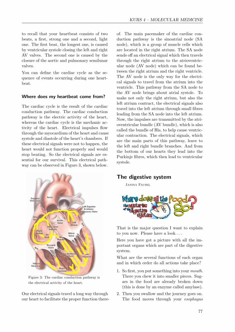
The cardiac cycle is the result of the cardiacconduction pathway. The cardiac conductionpathway is the electric activity of the heart,whereas the cardiac cycle is the mechanic ac-tivity of the heart. Electrical impulses flowthrough the myocardium of the heart and causesystole and diastole of the heart’s chambers. Ifthese electrical signals were not to happen, theheart would not function properly and wouldstop beating. So the electrical signals are es-sential for our survival. This electrical path-way can be observed in Figure 3, shown below.
Figure 3: The cardiac conduction pathway is
the electrical activity of the heart.
Our electrical signals travel a long way throughour heart to facilitate the proper function there-
of. The main pacemaker of the cardiac con-duction pathway is the sinoatrial node (SAnode), which is a group of muscle cells whichare located in the right atrium. The SA nodesends off an electrical signal which then travelsthrough the right atrium to the atrioventric-ular node (AV node) which can be found be-tween the right atrium and the right ventricle.The AV node is the only way for the electri-cal signals to travel from the atrium into theventricle. This pathway from the SA node tothe AV node brings about atrial systole. Tomake not only the right atrium, but also theleft atrium contract, the electrical signals alsotravel into the left atrium through small fibresleading from the SA node into the left atrium.Now, the impulses are transmitted by the atri-oventricular bundle (AV bundle), which is alsocalled the bundle of His, to help cause ventric-ular contraction. The electrical signals, whichare the main parts of this pathway, leave tothe left and right bundle branches. And fromthe bottom of our hearts they lead into thePurkinje fibres, which then lead to ventricularsystole.
The digestive systemJanina Fausel
That is the major question I want to explainto you now. Please have a look. . . .
Here you have got a picture with all the im-portant organs which are part of the digestivesystem.
What are the several functions of each organand in which order do all actions take place?
1. So first, you put something into your mouth.There you chew it into smaller pieces. Sug-ars in the food are already broken down(this is done by an enzyme called amylase).
2. Then you swallow and the journey goes on.The food moves through your esophagus
77

KURS 4 – MOLECULAR MEDICINE
The digestive system
into your. . .
3. Stomach. There, all the proteins containedin the food lose their structure. You have toimagine that each protein is a long chain ofamino acids. Normally this chain is foldedquite complicatedly in a way, that the pro-tein structure is compressed. In the stom-ach this structure is lost and the amino acidchains are unfolded.
Amino acid chain
4. After that, the food is transported into thesmall intestine. It is the main organ to di-gest food and absorb nutrients.
It is also the largest part of the digestivesystem and is divided into 3 parts:
- the duodenum
- the jejunum
- the ileum
There the chains of amino acids are split upinto the single amino acids.
5. The following large intestine is the last partof our digestive system and it is divided intofive parts.
6. Finally, the food (or better the remainders)leaves your body through the anus.
This is the way our food moves through ourbody.
But two other important organs are also partof the digestive system.
One of them is the liver.
Its main function concerning digestion is theproduction of bile which breaks fat pieces intosmaller ones.
The second is the pancreas.
It produces different enzymes which supporte.g. the splitting of the protein chains intotheir components.
Histology
Now, after having explained to you the func-tions of the organs, we will just have a shortcloser look at the layer of our small intestine,which gets in contact with the food: the mu-cosa.
First, you can see a picture of it. . . .
Diagram of the mucosa of the small intestine
In this picture you can see the structure of themucosa of our small intestine.
The structures above the large lymph vesselsare called villi.
And the glands, which are below the largelymph vessels, are called crypts.
The gland you can see at the bottom of thepicture produces enzymes, which support thedigestion of the food. These enzymes are giveninto the small intestine where they are needed.This action is called secretion.
78
KURS 4 – MOLECULAR MEDICINE
The nutrients from the food are absorbed atthe surface of the villi and get into the bloodvessels of the mucosa.
This way we are provided with the energy with-out which we could not move or do anything,without which we can not survive.
After we had learned so much theory withinour course, we also had the chance to preparereal pig organs:
- the small intestine
- the liver
First, we all were a little bit scared about it,because no one of us has ever seen a real organbefore.
After we watched how Annette, our courseteacher, cut it into pieces and explained us theseveral parts, we could make our own experi-ence with the organs.
It was a very strange feeling, very much bloodand at first we could not distinguish the dif-ferent parts, everything looked the same, butthen we learned it and it was a lot of fun!
Now we have talked about the digestive sys-tem, but you have already noticed, that thissystem just takes up nutrients and other sub-stances from our food.
Maybe you are wondering what happens withfluids we drink, after they are absorbed, orwhat happens with metabolic end productsour body can not use anymore.
You will learn something about this by Hulyain the next part.
The Urinary SystemHulya Erbil
As we learned, the digestive system is respon-sible for the food we eat. However, my topic,the urinary system, is very important for theprocess of transforming fluids we drink andsubstances in our blood into urine.
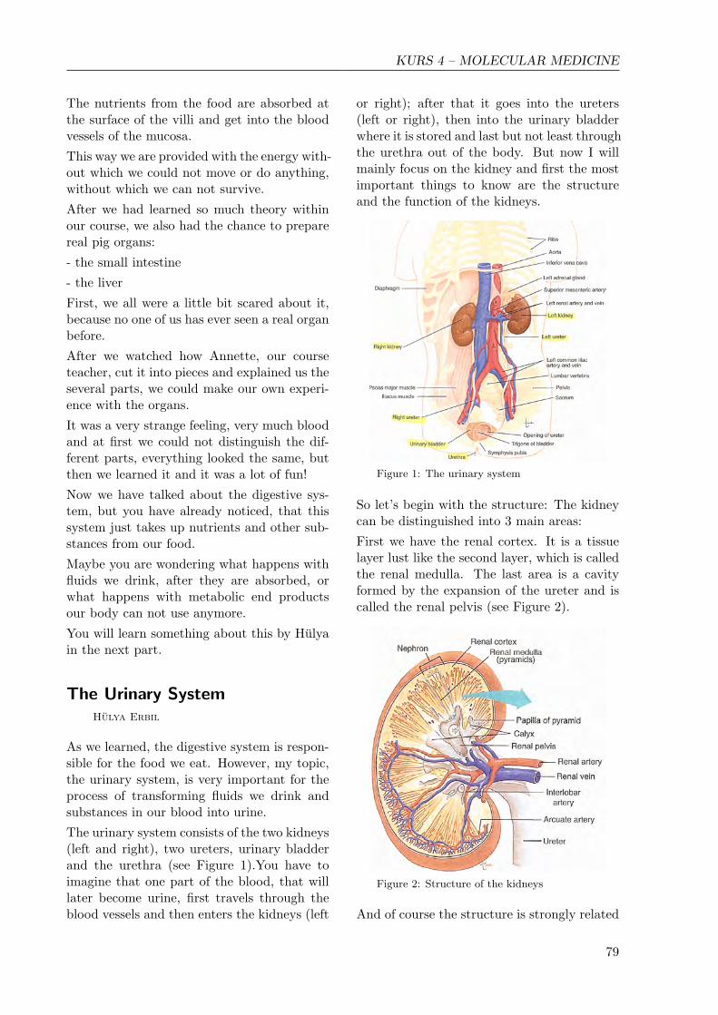
The urinary system consists of the two kidneys(left and right), two ureters, urinary bladderand the urethra (see Figure 1).You have toimagine that one part of the blood, that willlater become urine, first travels through theblood vessels and then enters the kidneys (left
or right); after that it goes into the ureters(left or right), then into the urinary bladderwhere it is stored and last but not least throughthe urethra out of the body. But now I willmainly focus on the kidney and first the mostimportant things to know are the structureand the function of the kidneys.
Figure 1: The urinary system
So let’s begin with the structure: The kidneycan be distinguished into 3 main areas:
First we have the renal cortex. It is a tissuelayer lust like the second layer, which is calledthe renal medulla. The last area is a cavityformed by the expansion of the ureter and iscalled the renal pelvis (see Figure 2).
Figure 2: Structure of the kidneys
And of course the structure is strongly related
79

KURS 4 – MOLECULAR MEDICINE
to the function. So the kidney is responsiblefor the production of urine, for the excretionof metabolic end products. Another functionis the monitoring of our blood’s pH, body’swater and salt. Furthermore, it has to controlour blood pressure. On top of that, hormonesare produced and secreted here.
Figure 3: Structure of the nephron
Looking at Figure 3, you can see the struc-tural and functional unit of the kidney: Thenephron. Each kidney consists of about 1 mil-lion nephrons. Having a deeper look onto thepicture we can see that a nephron consists of2 main parts: Renal corpuscle and the renaltubules (Figure 3). The renal corpuscle itselfconsists of the glomerulus and the Bowman’scapsule.
You have to imagine that the blood comesfrom the afferent arteriole (see Figure 4) andgoes to the Bowman’s capsule where it is fil-tered. The filtrate is no longer blood, but itis called renal filtrate and will later becomeurine. Then this renal filtrate goes out of therenal corpuscle and into the second part of thenephron, namely through different tubules.
The so called renal tubules continue from theBowman’s capsule and consist of: the proxi-mal convoluted tubule, the loop of Henle, thedistal convoluted tubule and the collecting tu-bule. Every tubule has very thin walls whichprovide an efficient exchange of materials andare surrounded by peritubular capillaries (tinyblood vessels). Their function is to receive the
material that is reabsorbed into the blood.
The tubular system
But how is urine formed?
The answer to this is simple: There are threemajor actions happening in the kidney.
The first action is in the glomerulus. Highblood pressure forces plasma, dissolved ma-terials and small proteins into the Bowman’scapsule and produces the renal filtrate.
The second action, the tubular reabsorption,takes place from the renal tubules into the per-itubular capillaries. In other words: In oneday the kidneys form about 150-180 litres re-nal filtrate, but we only excrete 1-2 litres ofurine, because 99% of the renal filtrate aregiven back to the peritubular capillaries.
The last action is the tubular secretion. Itensures that substances, e.g. substances thatare toxic for our organism, are secreted fromthe peritubular capillaries into the renal fil-trate and so it changes the composition of theurine.
So one could say that the tubular reabsorp-tion and the tubular secretion act in oppositedirections.
And in the end a funny picture!!
80

KURS 4 – MOLECULAR MEDICINE
The Brain
Sarah Walther
Besides the heart, the digestive system andthe urinary system, we also talked about thebrain.
The brain is the control center of the centralnervous system, responsible not just for be-havior, but also for movement and muscle co-ordination. In mammals, the brain is locatedin the head, protected by the skull and alsoincludes the primary sensory apparatus of vi-sion, hearing, balance, the sense of taste andsmell.
The human brain weighs about three poundsor 1.5 kg. In its natural state it is very soft,having approximately the consistency of pud-ding. When alive the brain is pinkish on theoutside and mostly white on the inside.
Anatomy
The brain of the human body consists of fiveparts: The cerebrum and the cerebellum, theinterbrain, the midbrain and the afterbrain.
The cerebrum is divided into the right and lefthemisphere. Everything we feel, touch, see,hear, smell, taste, but also what we think isprocessed in the cerebral cortex. For the dif-ferent perceptions, different regions in the cor-tex are responsible:
1. Frontal lobe: It is used for reasoning, emo-tions, judgment and voluntary movement.
2. Parietal lobe: It contains important sen-sory centers (only for the sense of touch).
3. Occipital lobe: It processes sensual infor-mation from the eyes.
4. Temporal lobe: It contains centers of hear-ing and memory.
The cerebellum also consists of two parts. It isresponsible for the coordination of movements.If the cerebrum, for example, says: “Walk!”to the leg muscles, this command is first sentto the cerebellum, which then coordinates theperformance of the movement. The functionof the cerebellum will be reduced by the con-sumption of alcohol, so a drunken person isunable to walk straight along.
The midbrain, the interbrain and the after-brain make up the brain stem. It has gota smooth surface and it is the connection tothe spinal cord. Here, essential processes aresteered, independently of our consciousness.The brain stem is the center for the rhythmof breathing and of our gut, for vomiting andit controls our heart beat.
Histology of the brain
The brain is composed of two classes of cells,neurons and glia. Neurons are cells in the ner-vous system that process and transmit infor-mation by chemical signals within the neuron.They are the core components of the brain.Glial cells actually outnumber the neurons byabout 10 to 1. Glial cells provide support andprotection for neurons. The four main func-tions of glial cells are to surround neurons andhold them in place, to supply neurons withnutrients and oxygen to insulate one neuronfrom another, and to destroy pathogens andremove dead neurons.
81

KURS 4 – MOLECULAR MEDICINE
Neuron general structure
In our course we had a look at the brain cellsunder the microscope, which was very inter-esting. First it was very difficult to find every-thing, but after some time it became clearer.We saw the cortex with lots of cell bodies andalso the pituitary gland. The pituitary glandis located at the base of the brain and se-cretes hormones regulating homeostasis of thebody. We also had a look at the hypotha-lamus, which is located just above the brainstem. It is the control center of many auto-nomic regulatory activities of the body. Inthe hypothalamus we could see the axons ofthe neurons, which looked like long fibers.
Dissection of the brain
Like the heart, the liver and the kidneys, wedissected the brain of a pig, which is very sim-ilar to the human brain. Before dissection, Iwas very excited, because I had never donesomething like this before. I have never seen“real” organs in detail and so I did not knowhow my reactions would be. Fortunately, ev-erything came up roses and my fear whetherI would maybe topple down did not becomereality.
In general, the pig brain was very interest-ing. You could see the interesting structureand even the brain skin. I mostly liked thecerebellum, which was bordered clearly of therest of the cortex. The cerebellum looked likea leaf of a tree with all its grey nerves.
For me it is unbelievable that such a small or-gan is steering all the relevant functions andthe behavior of the whole body. Furthermore
the whole knowledge, memories and feelingsare stored in it. Therefore it was very inter-esting for me to have a look at the brain andI still marvel lots of times about it.
Dissection
Above I have already talked about hormones,which can be produced by some parts of thebrain, as for example the pituitary gland. Butnow Sandra will explain to you more in detailhow hormones exactly work.
HormonesSandra Salvasohn
Hormones are chemical messenger substancesin our body which transport signals from onecell to another. So they can influence growth,development, function of many organs, bodytemperature, salt and water balance, coordi-nation of metabolism and so on. . .
The hormone system is also called endocrinesystem. It consists of glands located in thewhole body that are able to secrete hormones.
Different glands of the hormone system
82

KURS 4 – MOLECULAR MEDICINE
After the hormones are secreted, they are re-leased directly into the bloodstream. Theyflow through it and when they reach a cellwhich has got a suitable receptor they bindto this receptor and cause an effect. A hor-mone is like a key that fits only into its properkeyhole. It depends on the target cell and itsexpression of receptors whether the cell reactsto a hormone.
In most cases these receptors are located onthe cell membrane. But some hormones (e.g.the steroid hormones) are able to pass throughthe cell membrane and bind to a receptor, inthe cytoplasm or the nucleus. When the hor-mone has bound to its receptor this activatesbiochemical reactions inside the cell (the socalled primary response).
Hormones can cause one or more effects in thetarget cell. And in different cells, a certaintype of hormone can also have different or evenopposite effects. That means: in one musclecell for example, the hormone can cause relax-ation and in another cell the same hormonecan cause contraction of blood vessels.
Already very little amounts of hormones cancause a big effect. And therefore it is impor-tant that hormone secretion is regulated. Thisis the task of so called control cycles. Thereare a lot of different control cycles. Via thesecontrol cycles the hormone production can becharged or reduced and so the amount of hor-mones can be influenced.
One important control cycle is the so-calledhypothalamus-pituitary axis. This control cy-cle consists of more than only one step:
The hypothalamus is the superior inceptor. Atfirst, it releases the so called releasing-hormonethat causes the secretion of another hormonein the pituitary gland.
This hormone causes a peripheral endocrinegland to produce a third hormone. This thirdhormone generates the main effect.
The concentration of this hormone in the bloodis permanently controlled. The hypothalamusand pituitary gland get a feedback and so theycan increase or reduce the hormone secretion(positive/negative feedback).
Now to the pancreas:
Hypothalamus-pituitary axis
The two hormones insulin and glucagon pro-duced by the pancreas both regulate the bloodglucose level, just with opposite effects. Theyare antagonists. Insulin is responsible to keepup the nominal value during the day. For ex-ample when we eat something and our bloodglucose increases, insulin is released into theblood. The effect: The glucose is absorbedby the tissue cells, the blood glucose level de-creases. Later, when the glucose level reachedthe nominal value, the insulin production isreduced, too and so the balance can be keptup.
Diabetes
If this system does not work well, one will de-velop diabetes.
Diabetes is a metabolic disease. The mainsymptom is the increased concentration of glu-cose in the blood. The reason for this is eitherabsence of insulin or insensitivity to insulin.Depending on the cause there are two differ-ent types of diabetes (Type I and Type II).The frequency in Germany is about 550.000cases of type I and about 5 millions of type II.Type I has its onset especially among childrenand teenagers, but there is no age limit. TypeII affects adults from about 40 years on.
83

KURS 4 – MOLECULAR MEDICINE
In general: Transport of information by thehormone system is not as fast as by the ner-vous system. That means: When the hor-mones are released from a gland, it can takea few seconds (e.g. adrenalin) up to hours oreven days to cause an effect.
After we had looked at many different systemsin our body so far, for example the hormonesystem, we went on to the molecular level, tothe DNA and its elements.
DNA – The Genetic Information
Florian Goser
The DNA (deoxyribonucleic acid) is a moleculewhich is located in the nucleus of every singlecell. This molecule contains the genetic infor-mation to make all the proteins of our humanbody.
A eukaryotic cell
The structure of the DNA
The DNA has a double helix structure and ismade up of 2 single-strands.
A DNA strand consists of nucleotides. Theyare composed of a base, a sugar and a phos-phate.
In the DNA we can find four different typesof bases: Adenine (A), Thymine (T), Guanine(G) and Cytosine (C).
Hydrogen bonds between the bases of the twoDNA strands make the DNA strands stick to-gether.
Adenine always binds to Thymine and Gua-nine always binds to Cytosine.
The duplication of the DNA
If a cell divides, the DNA also has to be copied.Therefore the two strands first separate fromeach other and are then used as templates fornew DNA strands. That means that to eachbase of the two original strands binds a nu-cleotide with the correct “partner-base”.
So to every nucleotide with an A base will binda T base and to every nucleotide with a C basewill bind a G base. The bound nucleotidesform the new DNA strands.
DNA replication
The messenger RNA (m-RNA)
If a cell has to build a protein, the genetic in-formation of the DNA in the nucleus is needed.
BUT that is a big problem, because the pro-teins are built in the ribosome, which is lo-cated in the cytoplasm, and the DNA withthe needed genetic information is in the nu-cleus and cannot leave it. So there has to bea link between the DNA in the nucleus andthe ribosome. This link is called the messen-ger RNA. The messenger RNA is a copy of theDNA, which can be carried out of the nucleusto the ribosomes. The difference between theDNA and the messenger RNA is that the mes-senger RNA is single stranded and has uracilbases instead of thymine bases.
mRNA, transcription and translation
How is a protein actually built?
84

KURS 4 – MOLECULAR MEDICINE
In a process known as transcription a molecu-lar machine first unwinds a section of the DNAhelix to expose the genetic instructions neededto assemble a specific protein molecule. Thenthese instructions are copied and a moleculeknown as messenger RNA is formed. Whenthe transcription is complete the slender RNAstrand carries the genetic information throughthe nucleus pore complex into the cytoplasm.
The messenger RNA strand is directed to atwo part molecular factory called a ribosome.In the ribosome the process of translation be-gins: inside the ribosome the messenger RNAgets translated into many amino acids whichwill make up the protein. The sequential ar-rangement of amino acids determines the typeof the protein.
When the chain of amino acids is finished, it ismoved from the ribosome to a machine whichfolds this chain properly. Then the protein iscomplete and ready for usage.
mRNA, transcription and translation
DNA in bacteria
One difference between the DNA in an animalcell and in a bacterium is that the genetic in-formation in a bacterium just lies loose in the
cytoplasm. This ring of DNA in a bacteriumis called a plasmid.
The plasmid lies in the cytoplasm of the cell, incontrast to an animal cell, there is no nuclearmembrane in a bacterium, and the plasmid iseasy to manipulate. That is why it is oftenused for genetic engineering.
The manipulation of the plasmid in a bac-terium can for example be used to produceproteins, e.g. insulin. Therefore, scientists re-move a piece of the plasmid DNA strand andput the genetic information for insulin produc-tion into the plasmid.
The DNA system looks quite safe, but some-times there are also exceptions to that.
If the so called cell cycle does not work cor-rectly there is a potential of getting one of theworst diseases known in our world – cancer.
Cell Cycle and cancer
Dimitri Nothdurft
To understand how cancer really works, youfirst have to know what the cell cycle exactlyis:
The cell cycle is the cell’s daily schedule, thatmeans every cell goes through this scheduleonce a day.
During the 4 phases of this schedule, namelythe Gap phase 1 (=G1), the Synthesis phase(=S), the Gap phase 2 (=G2) and the Mitosisphase (=M), the cell is divided.
In each of the phases mentioned above some-thing happens:
During G1 the cell organelles and proteins aredoubled, so that they can be transferred to the2 daughter cells later.
During S Phase the DNA is doubled.
During G2 the cell is prepared for the M Phase,during which the cell is finally divided into twodaughter cells.
In order to avoid unwanted mutations in thecells’ genomes, the cell cycle has several check-points at which the result of each phase ischecked.
85

KURS 4 – MOLECULAR MEDICINE
The 3 main checkpoints are the G1, the G2and the metaphase checkpoints.
The G1 checkpoint controls, whether doublingof organelles and proteins has taken place prop-erly.
At G2 checkpoint the replicated DNA is con-trolled for mutations.
The cell cycle
Then in the middle of the Mitosis Phase thecomplete cell division is controlled. To con-tinue with the next phase after a checkpoint,there has to be a special signal. This signal isonly sent if the product of the phase does nothave any mistakes. If there are mistakes likefor example mutations in the DNA, the celltries to solve the problem via its cell repairmechanisms.
If it is not possible the cell kills itself (Apop-tosis).
But how is this complex cycle controlled? In-terestingly it is controlled by two types of pro-teins: the CDKs (cyclin dependent kinases)and the cyclin proteins.
Cdks are enzymes which control cell activityby adding phosphate groups to certain pro-teins. But they can only work if they are di-rectly bound to cyclins. So the time when theCDKs are active can be controlled.
After we had learned all this, we thought thecell cycle was a quite safe way of doubling cells.
But despite this save system there can be ir-regularities in the cell cycle. These can causeproblems like disease cancer, a highly dreadeddisease which was our next topic.
Cancer cells are cells which have unlimitedpotential regarding growth and reproduction.This means they can double as often as theywant. Together they make up a tumor, a largeaccumulation of cancer cells. Such tumors canbe built up nearly everywhere in the humanbody.
But how exactly is cancer caused?
Normal cells are influenced by factors in theenvironment, called carcinogens. Carcinogenscan be, for example, UV radiation, differentchemicals in food or polluted air. These car-cinogens work as so called mutagens. Becausethey can change the cell’s DNA so that essen-tial cell functions can be affected.
For example, a mutagen can change the genefor encoding a certain hormone receptor sothat this receptor binds to other hormones.
Through this change the production of cellgrowth factors can be increased.
That could lead to cancer formation.
These mutated genes which can trigger uncon-trolled cell growth are called oncogenes.
Surprisingly that is not the only way of induc-ing cancer cells:
There are also some viruses which have onco-genes integrated into their genome.
When docking onto a host cell, the RNA ofthe virus modifies the normal cell’s DNA andadds these oncogenes. As a result of that thehost cell eventually grows uncontrolled.
After we had studied the development of thecancer cells we went over to their abilities.
We talked about the scientific publication“Hall-marks of Cancer”, by Robert Weinberg, whichdescribes six acquired abilities of cancer cellsby which you can characterize and recognizethem. (The single hallmarks are shown in thepicture below.)
As we first heard these complicated names wewere unable to deal with them, but their mean-ing was explained to us shortly afterwards:
Self-sufficiency in growth signals
The cell is able to produce its own growth sig-nals and does not depend on signals from the
86

KURS 4 – MOLECULAR MEDICINE
Cancer hallmarks
outside. Therefore it grows limitless.
Insensitivity to antigrowth signals
The cell does not react to signals from the out-side which make a normal cell stop growing.
Limitless replicative potential
The cell can divide as often as it wants (normalcells have a limitation).
Evading apoptosis
The cells are able to inactivate their apoptosisfunction, which means that the cell cannot killitself when it detects a mutation in the DNA.
Sustained angiogenesis
The cancer cells can induce connections fromnormal blood vessels to a tumor. Through thisability the tumor never has any problems withgetting nutrients.
Tissue invasion and metastasis
At a certain stage the cells of a tumor cantravel to another part of the body via the bloodvessels and build up a tumor there. So if a tu-mor is removed from your body, there is stilla chance of getting cancer a second time.
Finally I can say that the high amount of newinformation has helped us moving on to a to-tally new perspective on cancer and the cellcycle.
After all the theory on the molecular level inthe last lessons, it was our turn again to doalso some practical things.
Another Way of Blood DonationYannik Laich
This lesson we wanted to make an experimentwith our blood. At first, we got the descriptionof the experiment which was called the bloodsmear experiment. After reading and posingsome questions, we started with cleaning themicroscope slides with water and after thatwith alcohol. Then, we took our gloves andwe discussed who should be stabbed into thefingertip. The stabbing was a little bit painful(especially for Dimitri who was stabbed a lotof times). After several trials, we succeededto smear the blood drops onto the microscopeslides. So we had to make two microscopeslides with a blood smear.
The Staining of our slides
But the two smeared blood drops had no realcolour so that we still had to do the Pappen-
87
KURS 4 – MOLECULAR MEDICINE
heim staining. This staining gives the bloodcells some colour so that we can distinguishthem under the microscope. When the twopreparations had been air-dried, we coveredthem with some May-Grunwald solution andwaited for 3 minutes. After that, we had toadd some drops of distilled water and 1 minutelater we poured of the May-Grunwald solu-tion. After cleaning the slides carefully withdistilled water, we had to cover the prepara-tion with another toxic solution: The Giemsasolution. After that, we waited for fifteen min-utes, which was a good possibility to talk toeach other. Then we had to clean the prepara-tions again with distilled water and the glassundersides with alcohol.
There were lots of stairs to the castle
As the preparations were dried, we put a dropof oil on them because we used a special micro-scope which only worked with oil. Under themicroscope we were able to see our blood cells,especially the erythrocytes and the leucocytes,which had different colours and forms. It wasvery interesting to have a look on your ownblood with the blood cells, but the experiencehow to make a microscope slide was also verygood. So all in all it was a very interestinglesson.
The excursionYannik Laich
On Monday, we had our excursion to the Ger-man Cancer Research Center in Heidelberg sothat we were able to get an insight into realresearch. We had to get up early in the morn-ing and after a short breakfast we went to the
train station by car. The ride on the trainwas a good possibility for sleeping and relax-ing some time.
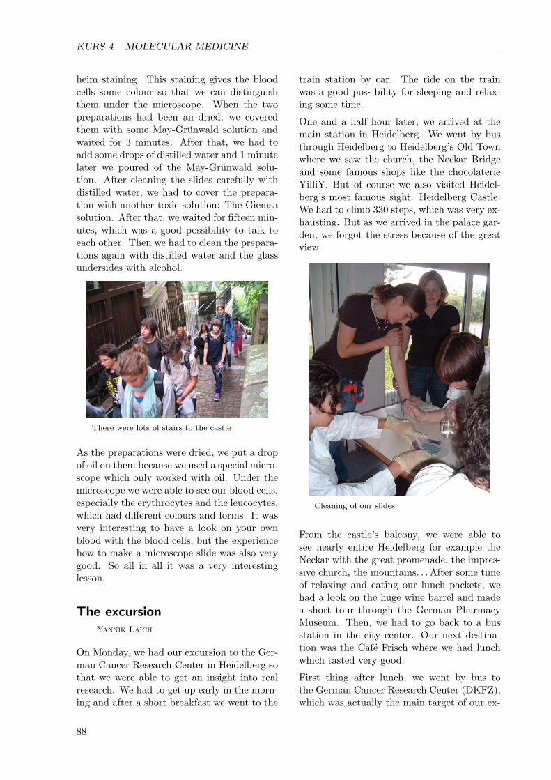
One and a half hour later, we arrived at themain station in Heidelberg. We went by busthrough Heidelberg to Heidelberg’s Old Townwhere we saw the church, the Neckar Bridgeand some famous shops like the chocolaterieYilliY. But of course we also visited Heidel-berg’s most famous sight: Heidelberg Castle.We had to climb 330 steps, which was very ex-hausting. But as we arrived in the palace gar-den, we forgot the stress because of the greatview.
Cleaning of our slides
From the castle’s balcony, we were able tosee nearly entire Heidelberg for example theNeckar with the great promenade, the impres-sive church, the mountains. . . After some timeof relaxing and eating our lunch packets, wehad a look on the huge wine barrel and madea short tour through the German PharmacyMuseum. Then, we had to go back to a busstation in the city center. Our next destina-tion was the Cafe Frisch where we had lunchwhich tasted very good.
First thing after lunch, we went by bus tothe German Cancer Research Center (DKFZ),which was actually the main target of our ex-
88

KURS 4 – MOLECULAR MEDICINE
cursion.
There, at 2 pm, we met Professor Doris Mayer,a scientist who researches in the DKFZ. Atthe beginning of her presentation, she wantedto check our knowledge on hormones. Then,she talked about the basics of the endocrinesystem and how insulin and estrogen are ableto influence cancer: These two hormones canstimulate the cell growth or even cause can-cer. After a short break, she talked abouther own research, which deals among otherswith a vicious cycle with insulin and estro-gen in cancer development. In the end, wehad the possibility to look into a real labora-tory, where she showed us the most importantequipment which is used by her students andher researchers.
Doris Mayer explained how to use a pipette
After this very interesting visit and a shorttravel through Heidelberg, we arrived in a Chi-nese restaurant where we had made a reserva-tion for dinner. The buffet was very rich anddelicious and we had a lot of fun during thedinner.
The return journey in the train was also amus-ing. But unfortunately we did not have enoughspace in the cars to transport all people andso some people had to walk from the train sta-tion back to the LSZU. The walk was a littlebit exhausting after a day like that and so we
arrived totally tired at 10 pm.
All in all, the excursion was very good for theatmosphere in our course and we got to knoweach other much better.
An essay on our courseatmosphere
Natalie Plewig
Nobody in our course had a real idea of whatthe work in the course would look like. It couldbe like in school, with a formal atmosphere, adefined structure and where the single indi-vidual is not that important. Or it might be agroup, which can be compared to a workshop,where you achieve not a feeling of hierarchy, sosee the group as a whole and where you reachsomething through teamwork. So there weresome conceptions, but nobody really knew.
But what we then experienced during the open-ing weekend surprised the majority. Our teach-ers often changed their presentations and useda variety of media. This factor and the il-lustration with pictures, videos and everydaylife examples, made it easier for us to under-stand. That is why we got a good impressionof molecular medicine, about which we wouldlearn in detail during the academy. Experi-ments that were conducted right at the begin-ning made us think that practical work wouldmake up an important part of our work, whatwas different to school lessons. One was ableto do something and reason by oneself. Be-cause this aspect was pushed strongly by thecourse teachers, they clarified, that we shouldask questions to show them that we under-stood the topic and that we dealt with thesubject. Nevertheless, at the beginning, no-body was brave enough to do this. We wantedto leave a good first impression.
At the end of the opening weekend we weretold the topics of our presentations. We hadto choose between topics that dealt with thebrain and the nervous system, the digestivesystem, the urinary system and the heart. Af-ter we had decided, we got the correspondinginformation material. Then we were told toamplify the contact with the German partici-
89

KURS 4 – MOLECULAR MEDICINE
pants and to get in touch with the Chinese par-ticipants for the first time, by reporting abouthow the work for the presentations went on.The anticipation for the academy grew withthe contact with the course participants, withthe preparation of our presentations and withreceiving the script that contained the basicprinciples of molecular medicine. And mean-while the time until the academy passed by. . .
There we were then: the beginning of the acad-emy. Now everything would really start, al-though we had experienced a lot and collecteddifferent impressions so far. In that situation,it was comforting to spend some time in thecommon environment of the course. It wasnow, that we met the Chinese participantsfor the first time and before we started withMolecular Medicine, we got an interesting in-sight to Chinese culture. That delivered newtopics of conversation with the Chinese par-ticipants.
The following days our course teachers and we,the course participants, changed into present-ing and working on the course topics. Withthat we talked through all the topics bit bybit. Here, the practical part was not neglected.We had a closer look at pig organs and inves-tigated histological slides through the micro-scope. During our work in the course we grewtogether more and more and the special feelingof us as a whole group rose increasingly. Ev-eryone got in contact with everyone and stepby step friendships were built – not only on onenation – level. Of course, the course teach-ers were included in our group, too. Therewas no teacher-pupil-connection between us,but a peer-to-peer-relationship, where every-one faced the other one with respect.
This especially became obvious during the ro-tation. In small groups of three, we worked outself-chosen topics, whereat we were supportedoptimally by the course teachers. Material,for example, that they had used in their pre-sentations so far, could be used by us withoutany exception, what made it ways easier for usto prepare. But the time to finish was quiteshort and that is why some of us were a bitstressed. But also in this situation, the courseteachers acted understandingly, which calmed
us down. Eased and satisfied, as everybodywas after the rotation, personal conversationswere conducted, that dealt with the own pre-sentation or the rotation on the one hand andwith our own person on the other hand. Thatenforced the feeling as a unity, because the sin-gle person felt understood and encouraged toopen up a bit more.
Our course in the Chinese restaurant
With this knowledge, our behaviour changed.We finally asked more and more, and evenwanted to know things that transferred whatwe discussed to other topics, for example. Wealways admitted that we sometimes did notunderstand. In fact, some days before, ourcourse teachers had recognised a certain dis-quiet, after that, they discussed the reason forthis with us and started to change their wayof presenting immediately. Fortunately, ev-erything got resolved fast and improved verymuch.
Now everyone was looking forward to our ex-cursion to Heidelberg which was an uniquehighlight. Not only because of the informativecontent, but also because we grew togetheragain. After the effective trip, the final spurtstarted: We all aimed for the comprehensiveclosing presentations that contained a lot ofwork. Luckily, in this phase, we were sup-ported by our course teachers again. Thisrich processing step represented the perfectforerun to the ending, because during the lastmoments and the farewell, we were extremelyhappy with what we achieved since our arrivalin Adelsheim. On our way home we lookedback onto the experiences we collected andswore to never forget the great time we spentin our course at Science Academy 2008.
90