Embed Size (px)
Citation preview

TURUN YLIOPISTON JULKAISUJAANNALES UNIVERSITATIS TURKUENSIS
SARJA - SER. D OSA - TOM. 880
MEDICA - ODONTOLOGICA
TURUN YLIOPISTOUNIVERSITY OF TURKU
Turku 2009
MOLECULAR MARKERS OFORAL LICHEN PLANUS
by
Riikka Mattila

From the Department of Oral Pathology and Radiology, Institute of Dentistry and Department of Pathology, Institute of Microbiology and Pathology, Faculty of Medicine, University of Turku, Turku, Finland
Supervised by Professor Stina Syrjänen, DDS, PhDDepartment of Oral Pathology and RadiologyInstitute of DentistryFaculty of MedicineUniversity of TurkuTurku, Finland
Docent Kalle Alanen, MD, PhDDepartment of PathologyFaculty of MedicineUniversity of TurkuTurku, Finland
Reviewed by Professor Mats Jontell, DDS, PhD, FDSRCSEdDepartment of Oral MedicineInstitute of OdontologySahlgrenska AcademyGöteborg University, Sweden Professor Timo Paavonen, MD, PhDDepartment of PathologyFaculty of MedicineUniversity of TampereTampere, Finland
Dissertation opponent Professor Yrjö T. Konttinen, MD, PhDDepartment of MedicineInstitute of Clinical MedicineBiomedicum HelsinkiUniversity of HelsinkiHelsinki, Finland
ISBN 978-951-29-4103-2 (PRINT)ISBN 978-951-29-4104-9 (PDF)ISSN 0355-9483Painosalama Oy – Turku, Finland 2009

To Verna, Sanni and Tommi
4 Abstract
ABSTRACT
Riikka MattilaMolecular Markers of Oral Lichen Planus
Department of Oral Pathology and Radiology, Institute of Dentistry and Department of Pathology, Institute of Microbiology and Pathology, Faculty of Medicine, University of Turku, Finland. Annales Universitatis Turkuensis. Sarja – Ser D Medica – Odontologica, Tom. 880, Painosalama Oy, Turku, Finland, 2009.
Oral lichen planus (OLP) is a chronic inflammatory mucosal disease and is detected in between 0.5% - 2.2% of the population. WHO has defined OLP as a potentially precancerous disorder, representing a generalized state associated with a significantly increased risk of cancer. However, only 0.5 – 2.9% of OLP lesions will progress to cancer. Currently, there are no prognostic markers to identify the lesions at increased risk for malignant transformation. The main aim of these studies was to identify cellular and molecular markers in order to understand the pathogenesis of atrophic OLP and its progression towards malignancy. Selected markers for cell proliferation, adhesion, apoptosis, and lymphocytic infiltration were assessed by immunohistochemistry in addition to static cytometry analyses for DNA content.
DNA quantification of epithelial cells in 82 biopsy samples derived from atrophic lichen planus showed altered DNA content in 41% of the samples. DNA content was associated with proliferation activity, topoisomerase IIα, desmocollin-1 and infection with human papillomavirus. CD27+ and CD38+ lymphocytes were detected in inflammatory cell infiltrate, indicating an abnormal homing of B cells from blood circulation to tissue. Physiologic cell death, apoptosis, is frequently seen in OLP, but its pathways are unknown. Here it was shown that caspases 2 and 12 were up-regulated in OLP, indicating that intracellular apoptosis, rather than an external causal factor, is triggering apoptosis. However, this thesis could not identify any singular prognostic marker of malignancy in OLP. Thus, every OLP patient should receive regular follow-up care to identify cancer risk patients at an early stage.
Key words: oral lichen planus, risk of malignancy, immunohistochemistry, atrophy, apoptosis, inflammation, cell cycle, DNA content, topoisomerase IIα, Ki-67, Ck-19, E-cadherin, desmocollin-1, cdk-1, Rad-51, caspases, CD5, CD20, CD27, CD38

Tiivistelmä 5
TIIVISTELMÄ
Riikka MattilaMolekulaariset merkit suun limakalvon lichen planuksessa
Suupatologian ja -radiologian osasto, Hammaslääketieteen laitos, Patologian osasto, Kliinis-teoreettinen laitos, Lääketieteellinen tiedekunta, Turun yliopisto, Suomi. Annales Universitatis Turkuensis. Sarja – Ser D Medica – Odontologica, Tom. 880, Painosalama Oy, Turku, Suomi, 2009.
Suun lichen planus eli punajäkälätauti on krooninen tulehduksellinen limakalvo-muutos, jota esiintyy 0.5 – 2.2 %:lla väestöstä. WHO:n määritelmän mukaan se on yleistila, jossa syöpävaara on lisääntynyt. Kuitenkin vain 0.5 – 2.9 % suun lichen planuksista muuttuu syöväksi. Tällä hetkellä ei ole menetelmiä, jolla voidaan erottaa syöpäriskissä olevat muutokset harmittomista kroonisista muutoksista. Tämän väitöskirjatyön tarkoituksena oli tutkia niitä solu- ja molekyylitason tekijöitä, jotka selventäisivät suun limakalvon atrofisen lichen planuksen taudinkuvaa sekä merkkejä sellaisista solutason muutoksista, jotka voisivat olla yhteydessä syöpäriskiin. Immunohistokemian avulla määritettiin lichen planuksen epiteelisoluista valikoituja solujakautumista, solujen kiinnittymistä, solukuolemaa sekä lymfosyyttejä kuvaavia merkkiaineita, ja lisäksi analysoitiin staattisen sytometrian avulla solujen DNA pitoisuutta.
Ensin selvitettiin 82:sta suun limakalvon atrofisen lichen planuksen histologisista näytteistä epiteelisolujen DNA-pitoisuutta. Tulokset osoittavat, että 41 %:ssa atrofisista lichen-näytteistä epiteelisoluissa voidaan havaita poikkeavia DNA-määriä, mikä on merkkinä syöpäsoluille tyypillisestä muutoksesta. Samalla myös näytteiden epiteeli-solujen jakautuminen osoitettuna topoisomeraasi IIα -vasta-aineella, epiteelisolujen kiinnittymistä kuvaava desmocollin-1 sekä papilloomavirusinfektio olivat yhteydessä näytteen DNA-pitoisuuden muutokseen. Tutkimuksessa todettiin myös CD27+ ja CD38+ B-lymfosyyttisolujen esiintyvän suun lichen planuksen T-soluvoittoisella tulehdussolualueella, mikä on merkkinä tulehdussolujen epänormaalista hakeutu-misesta kudokseen. Itseohjautuvaa solukuolemaa eli apoptoosia kartoittavan tutki-muksen tulokset osoittivat, että atrofisessa lichenissä apoptoosin kaspaasireiteistä sisäisen reitin osat, kaspaasit 2 ja 12, ovat korostuneet, mikä viittaa lichenissä apoptoosiin johtavan tekijän olevan ennemmin solun sisäinen kuin solun ulkoinen aiheuttaja. Kuitenkaan yksittäistä selvää syöpään johtavaa tekijää ei tässä työssä voitu osoittaa, joten kaikkia lichen-potilaita on syytä pitää tiiviissä seurannassa mahdollisen syöpäriskin varhaisen todentamisen vuoksi.
Avainsanat: lichen planus, syöpäriski, immunohistokemia, atrofia, apoptoosi, tulehdusreaktio, solusykli, DNA-pitoisuus, topoisomeraasi IIα, Ki-67, Ck-19, E-cadherin, desmocollin-1, cdk-1, Rad-51, kaspaasit, CD5, CD20, CD27, CD38

6 Table of Contents
TABLE OF CONTENTSABBREVIATIONS ........................................................................................................8
LIST OF ORIGINAL PUBLICATIONS ...................................................................10
1. INTRODUCTION ..................................................................................................11
2. REVIEW OF THE LITERATURE .......................................................................122.1 Structure of the healthy oral mucosa ................................................................122.2. Oral lichen planus (OLP) ..................................................................................13
2.2.1. Epidemiology .........................................................................................142.2.2. Clinical features......................................................................................14
2.2.2.1. Oral, mucosal and skin lesions .................................................152.2.3. Histology ................................................................................................16
2.2.3.1. Lymphocyte infiltration in OLP ................................................172.2.3.2. Lichen / lichenoid lesions .........................................................17
2.2.4. Associations with systemic disease ........................................................182.2.5. Differential diagnosis .............................................................................202.2.6. Treatment................................................................................................20
2.3. Etiopathogenesis of OLP ..................................................................................212.3.1. Immunopathogenesis of OLP .................................................................212.3.2. Molecular markers and OLP ..................................................................222.3.3. Cell cycle and proliferation ....................................................................23
2.3.3.1. Cell cycle markers ....................................................................232.3.3.2. Cell proliferation markers .........................................................25
2.3.4. DNA damage and cell death ...................................................................272.3.4.1. Apoptosis pathways, caspases ..................................................28
2.3.5. Cell-to-cell adhesion ..............................................................................302.4. Malignant transformation of OLP ....................................................................32
2.4.1. Lichenoid dysplasia ................................................................................34
3. AIMS OF THE STUDY ..........................................................................................36
4. MATERIALS AND METHODS ............................................................................374.1. Tissue samples ..................................................................................................374.2. Histological examination ..................................................................................374.3. Methods ............................................................................................................38
4.3.1. Static cytometry (I).................................................................................384.3.1.1. DNA staining ............................................................................384.3.1.2. Static cytometry ........................................................................384.3.1.3. Parameters measured ................................................................38
4.3.2. Immunohistochemistry (II – V) ..............................................................384.3.2.1. Grading of the expression .........................................................40
4.3.3. Detection and genotyping of human papillomavirus DNA ....................414.3.4. Statistical methods (I-V) ........................................................................42

Table of Contents 7
5. RESULTS.................................................................................................................445.1. DNA content in samples from atrophic OLP (I) ...............................................445.2. Expression of proliferation and DNA repairing markers topoisomerase IIα,
Ki-67, cdk-1 and Rad-51 in atrophic OLP (II, III) ...........................................455.3. Expression of intercellular adhesion markers desmocollin-1 and E-cadherin
and cytoskeleton marker keratin 19 in atrophic OLP (II, III) ...........................455.4. Apoptosis and caspase expression in atrophic OLP (V) ...................................465.5. Expression of lymphocytes CD5, CD20, CD27 and CD38 in atrophic OLP
(IV) ...................................................................................................................475.6. Results of OLP which progressed to squamous cell carcinoma (I-V) ..............485.7. HPV detection and type distribution in OLP (unpublished data) .....................48
6. DISCUSSION ..........................................................................................................496.1. DNA content in oral lichen planus (I) ..............................................................506.2. Expression of proliferation and DNA repairing markers topoisomerase IIα,
Ki-67, cdk-1 and Rad-51 in atrophic OLP (II, III) ...........................................516.3. Expression of cytoskeleton marker keratin 19 and intercellular adhesion
markers E-cadherin and desmocollin-1 in atrophic OLP (II, III) .....................536.4. Apoptosis and caspase expression in atrophic OLP (V) ...................................556.5. Expression of lymphocytes CD5, CD20, CD27 and CD38 in atrophic OLP
(IV) ..................................................................................................................576.6. HPV and cancer development in OLP ..............................................................59
7. CONCLUSIONS .....................................................................................................60
ACKNOWLEDGEMENTS ........................................................................................61
REFERENCES .............................................................................................................63
ORIGINAL PUBLICATIONS ....................................................................................73

8 Abbreviations
ABBREVIATIONS
ANOVA analysis of varianceAUC area under ROC curveBAFF B cell-activating factor belonging to the TNF familyBM basement membraneCAM cell adhesion moleculeCk cytokeratinCdk cyclin dependent kinaseCI confidence intervalsCTL cytotoxic T lymphocyteDNA deoxyribonucleic acidFADD Fas-associated death domainGVHD graft-versus-host diseaseEBV Epstein-Barr virusER endoplasmic reticulumER exceeding rateHBV hepatitis B virusHHV human herpes virusHLA human leukocyte antigenHPV human papilloma virusHR-HPV high risk for cancer HPVICH immunohistochemistryIFP intermediate filament proteinIL interleukinIOD integrated optical densityLC Langerhans cellLD lichenoid dysplasiaLP lichen planusLR likelihood ratioLR-HPV low risk for cancer HPVMCH major histocompatibility complexMMP matrix metalloproteinasesNOM normal oral mucosaNPV negative predictive valueOLL oral lichenoid lesionOLP oral lichen planusOLR oral lichenoid reactionOR odds ratioPCNA proliferating cell nuclear antigen

Abbreviations 9
PCR polymerase chain reactionPI proliferation indexPPV positive predictive valueRANTE regulated on activation, normal T-cell expressed and secretedSCC squamous cell carcinomaSE sensitivitySLE systemic lupus erythematosusSP specificitySPSS statistical package for the social sciencesTh-1 T cells helper-1TIL tissue infiltrating lymphocyteTNF tumour necrosis factorTopo IIα topoisomerase IIalpha
DEFINITIONS
2.5c / 5.0cERThe percentage of the measured cells exceeding the 2.5c / 5.0c value on the DNA scale
PloidyThe mean value of the G1 fraction position of measured cells on the DNA scale
Proliferation index (PI)The sum of S and G2/M phase fractions
10 List of Original Publications
LIST OF ORIGINAL PUBLICATIONSThe thesis is based on the following original publications, which are referred to in the text by the Roman numerals I-V. In addition, some unpublished data are presented.
I Mattila R, Alanen K, Syrjänen S. (2004) DNA Content as a Prognostic Marker of Oral Lichen Planus with Risk of Cancer Development. Anal Quant Cytol Histol 26:278-284
II Mattila R, Alanen K, Syrjänen S. (2007) Immunohistochemical study on topoisomerase IIalpha, Ki-67 and cytokeratin-19 in oral lichen planus lesions. Arch Dermatol Res. Jan;298(8):381-388
III Mattila R, Alanen K, Syrjänen S. (2008) Desmocollin Expression in Oral Atrophic Lichen Planus Correlates with Clinical Behaviour and DNA Content. J Cutan Pathol 35:832-838
IV Mattila R, Ahlfors E, Syrjänen S. CD27 and CD38 Lymphocytes Are Detected in Oral Lichen Planus Lesions. Manuscript submitted
V Mattila R, Syrjänen S. Caspase Cascade Pathways in Apoptosis of Oral Lichen Planus. Manuscript submitted
The original publications have been reproduced with the permission of the copyright holders.
Introduction 11
1. INTRODUCTIONIn 1978, the World Health Organization defined oral lichen planus (OLP) as a potentially precancerous condition, representing a generalized state associated with a significantly increased risk of cancer. OLP is a relatively common chronic inflammatory disease of oral mucosa with a prevalence rate of 0.5% and 2.2% of the population. Clinically, OLP may assume a variety of morphological changes. The most prevalent type is the reticular form characterized with interlacing white lines that are usually bilaterally distributed on the buccal mucosa and sometimes on the tongue. Other types of OLP are papular, plaque-like, atrophic, erosive and bullous forms. OLP typically affects middle-aged or elderly women, although it can be detected also in younger men, but rarely in children. Associations of OLP with simultaneous presence of lichen lesions also in the skin and genital mucosa have been described.
The histology of OLP is characterized by a band-like lymphocytic infiltrate in juxtaepithelial lamina propria. In addition, there is hyperkeratinization, acanthosis, liquefaction degeneration of the basal cells, colloid bodies, saw-tooth appearance of rete pegs and distribution of the epithelial basement membrane (BM). Despite these well-characterized histological features of OLP, inter- and intra-observer reproducibility to diagnose OLP is modest, however. The etiology of OLP is still unknown. The previous studies support the view that cell-mediated mechanisms are involved in the initiation and the progression of the disease. Also, localized autoimmunity has been suggested as playing a role in the pathogenesis of OLP. Therefore, lacking a known causative factor, there is no specific cure for OLP.
Despite the WHO definition of OLP as a precancerous condition, the premalignant potential of OLP is still debatable. Malignant transformation has been estimated to occur in 0.5 – 2.9% of the OLP patients. Currently, there are no prognostic markers to identify which chronic OLP lesions are at a higher risk for progression. Thus, every OLP patient should be monitored carefully to detect early cancer development.
To understand the etiopathogenesis of OLP, it is important to identify the key molecules in this disease. In the present series of studies, molecular markers for cell proliferation, apoptosis, adhesion and inflammatory cell infiltrates have been studied to characterize the molecular phenotypes of OLP more closely and to estimate their progression toward malignancy.
12 Review of the Literature
2. REVIEW OF THE LITERATURE
2.1 Structure of the healthy oral mucosaThe oral cavity is lined by a mucous membrane that forms a barrier between the environment and the body. The oral mucosa is classified into keratinized and non-keratinized oral mucosa. In the hard palate and marginal gingiva, the epithelium of the mucosa is ortho- or parakeratinized to resist hard mechanical trauma caused by mastication forces. The dorsum of the tongue and vermilion border of the lips consist of both keratinized and non-keratinized epithelium. In the buccal mucosa, alveolar mucosa, the floor of the mouth, the ventral tongue, the soft palate and the lips, the epithelium is non-keratinized and thus soft and flexible to accommodate chewing, speech or swallowing of a bolus (Dale et al. 1990; Presland and Dale 2000; Squier and Kremer 2001; Presland and Jurevic 2002).
Oral mucosa consists of two distinct layers: the stratified squamous cell epithelium and the lamina propria, which contain a layer of loose fatty or glandular connective tissue containing the major blood vessels and nerves. This tissue separates the oral mucosa from underlying bone or muscle. The epithelium and the lamina propria are distinguished by the basement membrane (BM) (Bhaskar 1980; Cate 1989; Squier and Kremer 2001).
Oral epithelium is composed of stratified squamous epithelium (Mackenzie and Fusenig 1983), which consists of multiple layers of keratinocytes which are proliferating in the basal cell layer (Dale et al. 1990). A small population of progenitor cells is considered to represent stem cells, which produce basal cells and hereby maintain the proliferation potential of the epithelium (Squier and Kremer 2001). With keratinocytes, some 10% of the epithelium represents a variety of different cell types, including melanocytes, Langerhans’ cells, Merkel cells and infiltrating inflammatory cells such as lymphocytes.
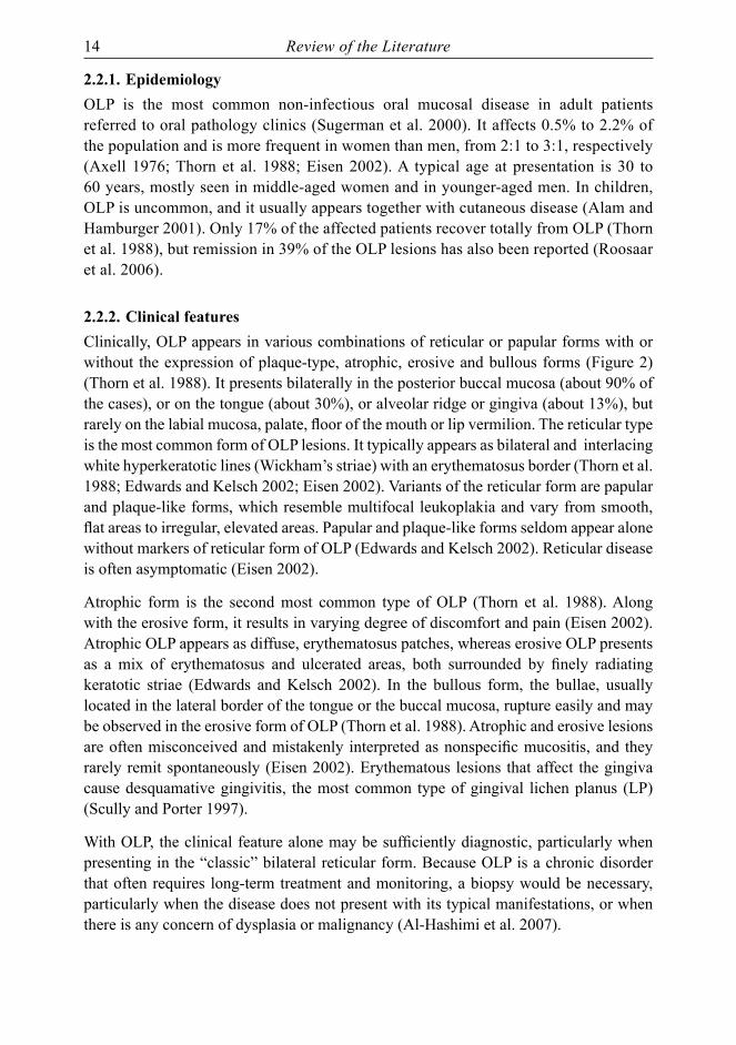
Keratinized and non-keratinized oral epithelia show two principal patterns of differentiation (Figure 1). In keratinized oral epithelia, differentiation leads to production of the ortho- or parakeratinized layer. As the cells leave the basal layer and enter into differentiation to the intermediate layer, keratinocytes become larger and begin to flatten. In the orthokeratinized layer, keratinocytes accumulate cytoplasmic protein filaments, representing cytokeratins. In the end, the intracellular organelles and the nuclei are extruded into the extracellular space, and the cells are fulfilled with cytokeratin filaments (Mackenzie and Fusenig 1983; Squier and Kremer 2001). In orthokeratinization, the keratinization process is complete and the nuclei are lost. In contrast, in parakeratinized cells, small pyknotic nuclei remain. The superficial layer is an essential part of the epithelial barrier of the keratinizing oral epithelia, consisting of cross-linked proteins and lipids in a 15nm thick layer (Presland and Jurevic 2002). In non-keratinizing epithelia, the keratinocytes contain less lipids and cytokeratins, and morphological changes are minor compared to those in keratinizing epithelia. The absence of the organized lipid lamellae in the intercellular spaces accounts for the higher permeability of this tissue (Squier and Kremer 2001).

Review of the Literature 13
Figure 1. Schematic illustration of the keratinized and non-keratinized oral mucosa
Cytokeratins (Ck) are the main cytoskeletal component of stratified keratinizing epithelia. Their expressions differ in keratinized and non-keratinized epithelia (Dale et al. 1990; Moll et al. 2008). There are type I (acidic) and type II (basic to neutral) keratins. Cytokeratins can constitute their filamentous stage only by heteropolymeric pair formation of type I and type II molecules (Moll et al. 2008). In the basal cell layer, all stratified epithelia express Ck-5, Ck-14 and Ck-15, while in the suprabasal cells, cytokeratin pairs Ck-4/-13 represent non-keratinized epithelia and pairs Ck-1/-10 represent keratinized epithelia.
Desmosomes and hemidesmosomes are involved to maintain the epithelial intercellular and keratinocytes-basement membrane adhesion, respectively. In keratinized epithelium, about 50% of the intercellular space of the superficial layer is occupied by desmosomes (Presland and Jurevic 2002). Desmosomes consists of two principal groups of proteins: desmosomal cadherins, the desmocleins and desmocollins, and a large group of plaque-associated proteins. In addition, epithelial cells are connected also with gap and tight junctions.
2.2. Oral lichen planus (OLP)Oral lichen planus (OLP) is a chronic inflammatory disease of oral mucosa. The World Health Organization (WHO) has defined OLP as a potentially precancerous disorder, representing a generalized state associated with a significantly increased risk of cancer (WHO 1978; van der Waal 2009). Etiopathogenesis is unknown, but several molecular hypotheses have been presented which will be discussed later (chapter 2.3.).
14 Review of the Literature
2.2.1. EpidemiologyOLP is the most common non-infectious oral mucosal disease in adult patients referred to oral pathology clinics (Sugerman et al. 2000). It affects 0.5% to 2.2% of the population and is more frequent in women than men, from 2:1 to 3:1, respectively (Axell 1976; Thorn et al. 1988; Eisen 2002). A typical age at presentation is 30 to 60 years, mostly seen in middle-aged women and in younger-aged men. In children, OLP is uncommon, and it usually appears together with cutaneous disease (Alam and Hamburger 2001). Only 17% of the affected patients recover totally from OLP (Thorn et al. 1988), but remission in 39% of the OLP lesions has also been reported (Roosaar et al. 2006).
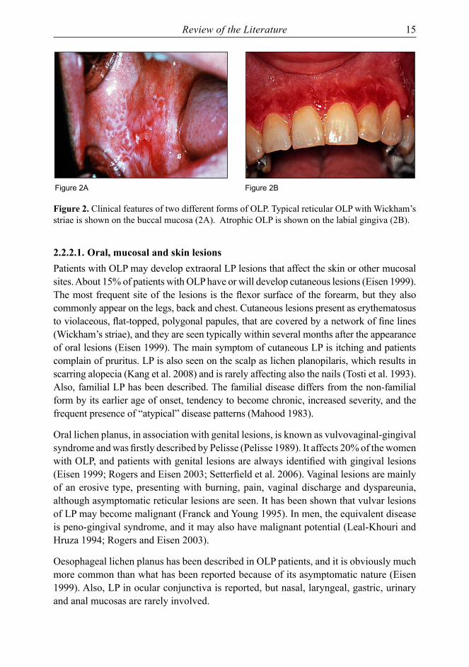
2.2.2. Clinical featuresClinically, OLP appears in various combinations of reticular or papular forms with or without the expression of plaque-type, atrophic, erosive and bullous forms (Figure 2) (Thorn et al. 1988). It presents bilaterally in the posterior buccal mucosa (about 90% of the cases), or on the tongue (about 30%), or alveolar ridge or gingiva (about 13%), but rarely on the labial mucosa, palate, floor of the mouth or lip vermilion. The reticular type is the most common form of OLP lesions. It typically appears as bilateral and interlacing white hyperkeratotic lines (Wickham’s striae) with an erythematosus border (Thorn et al. 1988; Edwards and Kelsch 2002; Eisen 2002). Variants of the reticular form are papular and plaque-like forms, which resemble multifocal leukoplakia and vary from smooth, flat areas to irregular, elevated areas. Papular and plaque-like forms seldom appear alone without markers of reticular form of OLP (Edwards and Kelsch 2002). Reticular disease is often asymptomatic (Eisen 2002).
Atrophic form is the second most common type of OLP (Thorn et al. 1988). Along with the erosive form, it results in varying degree of discomfort and pain (Eisen 2002). Atrophic OLP appears as diffuse, erythematosus patches, whereas erosive OLP presents as a mix of erythematosus and ulcerated areas, both surrounded by finely radiating keratotic striae (Edwards and Kelsch 2002). In the bullous form, the bullae, usually located in the lateral border of the tongue or the buccal mucosa, rupture easily and may be observed in the erosive form of OLP (Thorn et al. 1988). Atrophic and erosive lesions are often misconceived and mistakenly interpreted as nonspecific mucositis, and they rarely remit spontaneously (Eisen 2002). Erythematous lesions that affect the gingiva cause desquamative gingivitis, the most common type of gingival lichen planus (LP) (Scully and Porter 1997).
With OLP, the clinical feature alone may be sufficiently diagnostic, particularly when presenting in the “classic” bilateral reticular form. Because OLP is a chronic disorder that often requires long-term treatment and monitoring, a biopsy would be necessary, particularly when the disease does not present with its typical manifestations, or when there is any concern of dysplasia or malignancy (Al-Hashimi et al. 2007).
Review of the Literature 15
Figure 2A Figure 2B
Figure 2. Clinical features of two different forms of OLP. Typical reticular OLP with Wickham’s striae is shown on the buccal mucosa (2A). Atrophic OLP is shown on the labial gingiva (2B).
2.2.2.1. Oral, mucosal and skin lesionsPatients with OLP may develop extraoral LP lesions that affect the skin or other mucosal sites. About 15% of patients with OLP have or will develop cutaneous lesions (Eisen 1999). The most frequent site of the lesions is the flexor surface of the forearm, but they also commonly appear on the legs, back and chest. Cutaneous lesions present as erythematosus to violaceous, flat-topped, polygonal papules, that are covered by a network of fine lines (Wickham’s striae), and they are seen typically within several months after the appearance of oral lesions (Eisen 1999). The main symptom of cutaneous LP is itching and patients complain of pruritus. LP is also seen on the scalp as lichen planopilaris, which results in scarring alopecia (Kang et al. 2008) and is rarely affecting also the nails (Tosti et al. 1993). Also, familial LP has been described. The familial disease differs from the non-familial form by its earlier age of onset, tendency to become chronic, increased severity, and the frequent presence of “atypical” disease patterns (Mahood 1983).
Oral lichen planus, in association with genital lesions, is known as vulvovaginal-gingival syndrome and was firstly described by Pelisse (Pelisse 1989). It affects 20% of the women with OLP, and patients with genital lesions are always identified with gingival lesions (Eisen 1999; Rogers and Eisen 2003; Setterfield et al. 2006). Vaginal lesions are mainly of an erosive type, presenting with burning, pain, vaginal discharge and dyspareunia, although asymptomatic reticular lesions are seen. It has been shown that vulvar lesions of LP may become malignant (Franck and Young 1995). In men, the equivalent disease is peno-gingival syndrome, and it may also have malignant potential (Leal-Khouri and Hruza 1994; Rogers and Eisen 2003).
Oesophageal lichen planus has been described in OLP patients, and it is obviously much more common than what has been reported because of its asymptomatic nature (Eisen 1999). Also, LP in ocular conjunctiva is reported, but nasal, laryngeal, gastric, urinary and anal mucosas are rarely involved.
16 Review of the Literature
2.2.3. HistologyThe classic histopathologic features of OLP are shown in Table 1. It includes the dense, band-like subepithelial inflammatory infiltrate consisting of lymphocytes beneath the basement membrane, increased number of intraepithelial lymphocytes and liquefactive degeneration of basal keratinocytes (WHO 1978; Eisenberg 2000; Epstein et al. 2003). Eosinophilic colloid bodies (Civatte bodies) are formed by degenerating basal keratinocytes and immunocomplexes, and they are often identified in the supra-basal epithelial area (Griffin et al. 1980). The ultrastructure of these colloid bodies suggest that they are apoptotic keratinocytes, which is shown by demonstrating of DNA and nuclear fragmentation and immunoglobulins, especially IgM in these cells (WHO 1978; Pihlman et al. 1985; Dekker et al. 1997; Bloor et al. 1999; Sugerman et al. 2002). Early ultrastructural changes in OLP include widening of epithelial intercellular spaces and degenerative changes of basal cells (Jungell et al. 1989a). Also, disruption of anchoring elements of the basal cells such as hemidesmosomes, filaments and fibrils are seen (Haapalainen et al. 1995). In addition, alterations in basement membrane, such as breaks, branches and patch-like thickenings have been reported, reflecting the severity of the lesion (Jungell et al. 1987). Thus, these disorders in the basal cells and BM lead to interface between the epithelium and the lamina propria, which may result in histological cleft formation (Max-Joseph space) and in clinical blistering of oral mucosa, as rarely seen in bullous OLP (Sugerman et al. 2002). Hyperkeratosis as ortho- or parakeratosis is seen in the epithelium appearing clinically as Wickham’s striae and occasional areas of acantosis and “saw-tooth” rete ridges. In atrophic epithelium, rete ridges may be shortened and pointed or totally absent. Thus, hyperkeratinization is detected in every form of OLP. In general, the epithelium in OLP is thinner than in normal oral mucosa, which offers less protection against mechanical and chemical irritation and leads to oral discomfort for the patient (Karatsaidis et al. 2003).
The histologic diagnosis of OLP is based on the interpretation of the microscopic features described above. Both the interobserver and the intraobserver variability in OLP diagnosis shows that histopathological assessment of OLP, based on the available WHO definition, is rather subjective and an insufficiently reproducible process (van der Meij et al. 1999a). The interobserver agreement has been shown to vary from 0.20 (poor) to 0.51 (moderate), while the intraobserver agreement variation ranges from 0.50 (moderate) to 0.67 (substantial). Difficulties in histological diagnostics increase because of the variability of the histology of the lesions at the different sites of oral mucosa. Therefore, more strict diagnostic criteria are needed to improve the accuracy of diagnosis of OLP.
Table 1. Histologic criteria of OLP (WHO 1978)
Dense, band-like chronic lymphocytic infiltrate beneath and partly inside the basal cell layer Liquefactive degeneration of the basal cellsCivatte bodies (colloid bodies)Saw-tooth appearance of rete pegsAcanthosis or epithelial atrophyHyperkeratosis, parakeratosis

Review of the Literature 17
2.2.3.1.LymphocyteinfiltrationinOLPThe band-like subepithelial chronic lymphocyte infiltrate of OLP consist predominantly of T lymphocyte (CD3+). The relative proportion of helper-inducer (CD4+) and suppressor-cytotoxic (CD8+) T cells is variable (Ishii 1987; Jungell et al. 1989b; Hasseus et al. 2001). It has been stated that the composition of the inflammatory cell infiltrate is variable in different clinical forms of the disease. In the initial lesion of reticular OLP, the predominant lymphocyte subset is CD4+ cells, whereas in more advanced atrophic-erosive lesions, the number of CD8+ cells is increased substantially (Sugerman et al. 2002; Charazinska-Carewicz et al. 2008). CD4+ helper T cells are detected in the lamina propria while CD8+ cytotoxic T cells are in close proximity to the epithelial basement membrane. Most of the intraepithelial T cells are CD8+ cytotoxic lymphocytes, and the proportion of these lymphocytes is higher in the superficial than the deeper lamina propria (Jungell et al. 1989b; Walsh et al. 1990). Activated CD8+ T cells expressing human leukocyte antigen, (HLA)-DR surface antigen, is detected close to damaged epithelial cells and BM, and adjacent to areas of epithelial erosion (Kilpi, A. M. 1987; Kilpi, A. 1988).
The expression of B cells and plasma cells (about 5% of all inflammatory cells) are detected in OLP lesions, but until now it is thought that the proportion of these cells are relatively low (Malmstrom et al. 1989; Sugerman et al. 2002). By contrast, the proportion of Langerhans cells (LCs) (Hasseus et al. 2001) and mast cells (Jontell et al. 1986; Zhao et al. 2001; Zhao et al. 2002) is increased in OLP lesions when compared to that in normal oral mucosa. Degranulation of mast cells in OLP is 60% as compared to 20% found in normal mucosa (Zhao et al. 2001).
2.2.3.2. Lichen / lichenoid lesionsThere are various lesions that resemble LP, both clinically and histopathologically (Al-Hashimi et al. 2007). These lesions, usually referred to as oral lichenoid lesions (OLL) or reactions (OLR), have an identifiable etiology, and they may be unilateral, asymmetrical or occur in uncommon sites. The term OLL is used by some authors when several clinical or histological features are present but the diagnosis remains inconclusive (van der Meij and van der Waal 2003). Others consider OLL only when an association with dental materials has been established (Issa et al. 2005). Oral lichenoid lesions further encompass clinical settings such as 1) oral lichenoid contact lesions (OLCL) as a result of direct allergic contact of dental restorative materials e.g. amalgams and composite resins, 2) oral lichenoid drug reaction (OLDR) as a result of taking certain medication, e.g. oral hypoglycaemic agents, angiotensin-converting enzyme inhibitors, and nonsteroidal anti-inflammatory agents and 3) oral lichenoid lesions of graft-versus-host disease (OLL-GVHD) classified as acute or chronic GVHD (Issa et al. 2005; Imanguli et al. 2006; Al-Hashimi et al. 2007). As for differential diagnostics, the characteristics of the inflammatory cell infiltrate within the connective tissue seems to be more important than the epithelial changes to differentiate OLR from OLP. Thornhill et al. have identified four distinguishing features to discriminate between OLR and OLP (Thornhill et al. 2006). They suggest that these markers, which

18 Review of the Literature
may be present in OLR but are absent in OLP, are 1) an inflammatory infiltrate located deep to surface infiltrate in some or all areas, 2) focal perivascular infiltrate, 3) plasma cells in the connective tissue and 4) neutrophils in the connective tissue. OLDR lesions may develop months or even years after the patient has taken medication which exacerbates the etiology and thus the diagnosis (McCartan and McCreary 1997). Furthermore, OLR lesions caused by amalgam recur within three to six months after replacing the fillings with other restorative materials (Bratel et al. 1996; Thornhill et al. 2006). To summarize, it is difficult to distinguish OLP and OLL or OLR purely on histological grounds, and pathologists always need specific anamnestic data and a good clinical description of the lesion with medical history (Thornhill et al. 2006). Differential diagnoses with OLP and other diseases are presented in chapter 2.2.5.
2.2.4. Associations with systemic diseaseEven though OLP has been widely studied for decades, the etiology of the disease is still unknown. Likewise, it is unknown if lichen planus represents a single disease process or if there are several closely related entities with similar clinical presentations.
Autoimmune diseasesThe possible contribution of autoreactivity to the pathogenesis of OLP has been suggested and is based on several lines of evidence (Sugerman et al. 1993). In general, etiology of autoimmune diseases is unknown. However, genetic susceptibility in addition to external or internal triggers is usually needed (Davidson and Diamond 2001). OLP is sometimes detected in the same patient simultaneously with a known autoimmune disease, such as systemic lupus erythematosus and Sjögren’s syndrome, and it arises predominantly in older females, which is characteristic of autoimmune disease (Sugerman et al. 1993; Tanei et al. 1997; Davidson and Diamond 2001). Moreover, the human leukocyte antigens (HLA), especially HLA-DR3 which is associated with autoimmune diseases, are shown to increase in OLP lesions, suggesting an autoimmune component of the pathogenesis of OLP (Jontell et al. 1987; Farthing and Cruchley 1989; Karatsaidis et al. 2003). In contrast, MHC class II antigen HLA DP or DQ expression in OLP is not shown (Farthing and Cruchley 1989). Recently, evidence has been provided, that B cells have a role in autoimmunity and that the pathogenesis of autoimmune diseases cannot be attributed exclusively to T cells (Jonsson et al. 2001). The possible connection of OLP with autoimmunity has recently gained increasing interest.
HCVAn association between OLP and the hepatitis C virus (HCV) infection has been widely studied (Carrozzo and Gandolfo 2003; Lodi et al. 2005a). Epidemiological data suggest that OLP may be significantly associated with HCV infection, mainly in Southern Europe and Japan (Lodi et al. 2004; Carrozzo et al. 2005; Nagao et al. 2007). Conversely, in Northern European countries or the United States, such association has not been reported. Also in Egypt and Nigeria, where the prevalence of HCV infections is high, no increase in OLP lesions have been found (Daramola et al. 2002). It has been postulated that HCV-

Review of the Literature 19
infected patients may have an increased risk of developing OLP, or alternatively, patients with OLP have an enhanced risk of HCV infection (Lodi et al. 2005b; a; Carrozzo 2008). However, it is likely that the patients are first infected with HCV and only later develop OLP. This is probably a result of an abnormal immune-response with TNF-α characterized with HCV-infected patients (Carrozzo et al. 2007). Moreover, it has been shown that HCV-specific T cells can be found in the oral mucosa of the patients with chronic HCV infection and OLP (Pilli et al. 2002). This suggests that HCV-specific T cells may play a role in the pathogenesis of epithelial cell damage in OLP, which may be the result of a direct immune aggression of epithelial cells expressing HCV antigens, possibly sustained by a cytokine environment favourable to trigger and maintain the lichenoid reactions (Pilli et al. 2002).
HPV, herpes viruses and HIVHuman papillomavirus (HPV) DNA has been found in oral lichen planus lesions, but there is no evidence of HPV as a causative factor in OLP. Review of the literature from 1998 has shown the presence of HPV DNA in 23% (25/107) of OLP lesions (Syrjänen and Syrjänen 2000). Similar results have been found with human herpes viruses, such Herpes simplex 1 (HSV-1), Ebstein-Barr virus (EBV), Cytomegalovirus (CMV) and Herpes virus 6 (HHV-6) (Sand et al. 2002). In subjects infected with human immunodeficiency virus (HIV), oral lichenoid reactions have been found, but the lesions could not be associated either with the HIV or anti-viral therapy (Campisi et al. 2004; Giuliani et al. 2008).
Gingival plaque and calculusThe prevalence of isolated gingival lichen planus has been reported in approximately 8% of the OLP patients (Eisen 2002; Mignogna et al. 2005). Diagnosis of gingival OLP may be difficult due to its clinical appearance as desquamative gingivitis. Also, the histopathologic features of gingival lichen planus are often nondiagnostic, since they are altered by a superimposed gingivitis (Eisen 2002). Increased plaque and calculus deposits are associated with a significantly higher incidence of erythematosus and erosive gingival lesions. Patients with desquamative gingivitis frequently complain of pain and bleeding while performing daily dental hygiene, which results in poorer oral hygiene with the accumulation of plaque and calculus and thus worsening bleeding and pain (Holmstrup et al. 1990; Ramon-Fluixa et al. 1999; Eisen 2002). This may cause exacerbation of OLP lesions. Furthermore, disposing of microbial plaque is shown to heal particularly OLR lesions on the mucosal side of the lips (Backman and Jontell 2007). Thus, oral hygiene must be optimized in OLP patients by dentists or periodontologists to reduce the gingival inflammation and pain (Scully et al. 2000; Mignogna et al. 2005).
Candida albicansSeveral studies have shown that Candida infection is common in OLP lesions (Holmstrup and Dabelsteen 1974; Simon and Hornstein 1980; Lundstrom et al. 1984; Scully et al. 1998). Candida infection has been reported in the culture studies of 37% to 50% of the

20 Review of the Literature
OLP lesions, but nearly 17% in histological samples: the detection rate being actually the same as in the general population. The Candida infection, however, may be a secondary infection associated with the topical steroid treatment of OLP (Jainkittivong et al. 2007).
Psychological aspectsStress has widely been considered as a possible causative factor of OLP, but there is no firm evidence to support this concept, and the data are controversial (Rodstrom et al. 2001). Anxiety is shown to occur in OLP patients, but it could not be related to any specific clinical pattern of OLP (McCartan 1995). Chaudhary (Chaudhary 2004) has stated that psychological stressors such as stress, anxiety and depression play an important role in the initiation of OLP, similarly as these stressors are known to initiate various autoimmune reactions. Ivanovski et al. (Ivanovski et al. 2005) suggested that psychosocial and emotional stress is one possible factor that may precipitate reticular OLP to transform to the erosive form. The lack of educational materials increases the stress of the patients, because the chronic lesion with discomfort awakens concerns of the possibility of malignancy or transmission of the disease (Burkhart et al. 1997). This all may contribute to the activity and persistence of OLP lesions.
2.2.5. Differential diagnosisThere are a number of oral mucosal diseases which mimic OLP, making the differential diagnoses important and difficult (Dissemond 2004). The correct diagnosis of OLP is always based not only on the characteristic clinical appearance and typical histology but also the course of the disease (Mattsson et al. 2002; van der Meij et al. 2003). The term “lichenoid dysplasia” is used by some pathologists to describe a dysplastic epithelium accompanied by a band-like lymphocytic infiltrate in the subjacent lamina propria (Al-Hashimi et al. 2007). However, clinically similar diseases may show entirely different histology or vice versa, e.g. atypical lichenoid stomatitis (ALS) or lichenoid dysplasia (LD) (Eisenberg 2000).
2.2.6. TreatmentThere is no specific treatment for OLP, and thus other diseases should always be excluded. Therefore, treatment of OLP patients depends on symptoms, the extent of oral and extra-oral clinical involvement, medical history, and other factors (Chan et al. 2000; Scully and Carrozzo 2008). Asymptomatic reticular lesions usually do not require active treatment. All the mechanical irritants, such as non-fitting dentures and sharp or rough fillings, especially dental amalgam restorations, in teeth should be repaired, and as well calculus and gingivitis should be treated. Thus, excellent oral hygiene is mandatory. It is also important to exclude the possibility of candidiasis, since the medical components may aggravate Candida infection. Candidiasis itself causes soreness of the oral mucosa. Elderly patients with OLP might have poor nutrition with folate deficiency, even when they are not found to be anaemic (Thongprasom et al. 2001). Spicy or irritant food, tobacco and alcohol usage should be minimized. Furthermore, it should be remembered
Review of the Literature 21
that, together with medical treatment, the patient with stress-induced oral disease may benefit from psychological therapeutic intervention with OLP (Ivanovski et al. 2005). Overall, treatment is aimed primarily at reducing the length and severity of symptomatic outbreaks (Edwards and Kelsch 2002). However, OLP cannot be completely cured (Chan et al. 2000; Lodi et al. 2005b).
Drug treatmentThe most common treatment for OLP is topical corticosteroids, such as triamcinolone, potent fluorinated steroids such as fluocinolone acetonide and fuocinonide, and superpotent halogenated steroids such as clobetasol. These midpotency corticosteroids are effective in most patients when applied according to the instructions, i.e. a thin layer of the steroid (ointment, gel, spray or rinse) several times daily. Patients who suffer from severe OLP lesions, desquamative gingivitis, widespread oral disease or diffuse ulcerations may need more potent immunosuppressants (cyclosporine) or immunomodulatory agents, such as calcineurin inhibitors (tacrolimus or pimecrolimus) or retinoids (tretinoin) in topical formulations. Systemic treatment with corticosteroids are used for patients with severe atrophic and erosive OLP or multisite disease, where topical approaches have failed and skin, genital or scalp lesions are present. However, the use of systemic corticosteroids alone has been found less effective than topical corticosteroids or a combination of both of these treatment modes (Chan et al. 2000; Scully et al. 2000; Edwards and Kelsch 2002; Eisen et al. 2005; Lodi et al. 2005a; Scully and Carrozzo 2008).
Non-drug therapiesSurgery, as well as CO2 laser, has been used with isolated plaque-like or non-healing erosive lesions to cure localized lesions. Cryosurgery is shown to specifically heal gingival lichen planus. Photochemotherapy with 8-methoxypsoralen and long-wave ultraviolet light (PUVA) might also have therapeutic effects, especially on skin lesions (Kuusilehto 2001).
2.3. Etiopathogenesis of OLP
2.3.1. Immunopathogenesis of OLPAs stated before, the real cause of OLP is unknown. Thus, several hypotheses of etiopathogenesis have been presented. Some of the causal factors are considered as etiology of lichenoid reactions, such as dental restorative materials and drug reactions, as described in chapter 2.2.3.2. Described here is one hypothesis of OLP immuno-pathogenesis that is based on the studies and description of Sugerman et al. 2002.
Non-specificmechanismsinOLPAs discussed above, epithelial BM changes, such as breaks, branches and duplications, are common in OLP (Jungell et al. 1989a). Also, apoptosis is shown to be prevalent in OLP (Bloor et al. 1999; Neppelberg et al. 2001). It is hypothesized that distribution of the epithelial basement membrane is caused by mast cell proteases or T-cell MMP-9
22 Review of the Literature
(matrix metalloproteinases), which may trigger keratinocyte apoptosis in OLP (Sugerman et al. 2002). However, it is not known which event comes first in OLP; BM distribution or keratinocyte apoptosis. Both events have been found in OLP. Other non-specific mechanisms may be involved in the pathogenesis of OLP, including 1) mast cell chemotaxis and degranulation stimulated by T-cell RANTES (regulated on activation, normal T-cell expressed and secreted) 2) endothelial cell adhesion molecule expression stimulated by mast cell TNF-α (tumour necrosis factor), 3) T-cell MMP-9 activation by mast cell chymase, 4) intra-epithelial CD8+ T-cell migration through BM breaks, 5) inflammatory cell survival prolonged by T-cell RANTES and 6) non-specific T-cell recruitment by keratinocyte-derived chemokines (Sugerman et al. 2002; Lodi et al. 2005a).
AntigenspecificityinOLPGenerally, it is accepted that in OLP there is chronic, cell-mediated immune damage to basal keratinocytes in the oral mucosa that are recognized as being antigenically foreign or altered (Thornhill 2001). An early event in the development of an OLP lesion may be either the expression of antigen in keratinocytes together with MHC class I (major histocompatibility complex), or unmasking the exogenous agents (Sugerman et al. 2002). These target keratinocytes may express antigen and thus produce chemokines, which are presented to intraepithelial CD8+ cytotoxic T cells followed by their activation. However, most lymphocytes in the lamina propria of OLP are CD4+ helper-1 T (Th-1) cells. Thus, an early event in the development of OLP may be MHC class II antigen presentation to CD4+ helper T cells mediated by keratinocytes or Langerhans cells present in epithelium. CD4+ helper T cells are thus stimulated to secrete Th-1 cytokines, which activates CD8+ cytotoxic T cells. This activation may lead to apoptosis of keratinocytes. Together, CD4+ and CD8+ T cells are thought to produce and respond to a range of cytokines and inflammatory mediators and resulting also in variations in the clinical presentation of OLP. In summary, many antigen-specific mechanisms may be involved in the pathogenesis of OLP, including 1) MHC class I- and MHC class II-restricted antigen presentation by lesional keratinocytes, 2) activation of antigen-specific CD4+ helper T cells and CD8+ cytotoxic T cells, 3) clonal expansion of antigen-specific T cells and 4) keratinocyte apoptosis triggered by antigen-specific CD8+ cytotoxic T cells (Walsh et al. 1990; Zhao et al. 2002; Lodi et al. 2005a).
2.3.2. Molecular markers and OLPThe transformation of normal oral mucosa to lichen planus or other mucosal disease is a complex multistep process, which requires changes in normal keratinocyte DNA replication, cell division, cell death and cell-to-cell adhesion. The cell proliferation, differentiation, senescence, and apoptosis are closely linked to the cell cycle regulation. The expressions of various molecular markers have been studied in order to understand the molecular mechanisms involved in OLP. Next, an overview is given on the molecular markers thought to be important in the development of OLP and also in its progression toward malignancy. A database collecting information on human genes and their function is maintained at www.ncbi.nlm.nih.gov/sites/entrez.
Review of the Literature 23
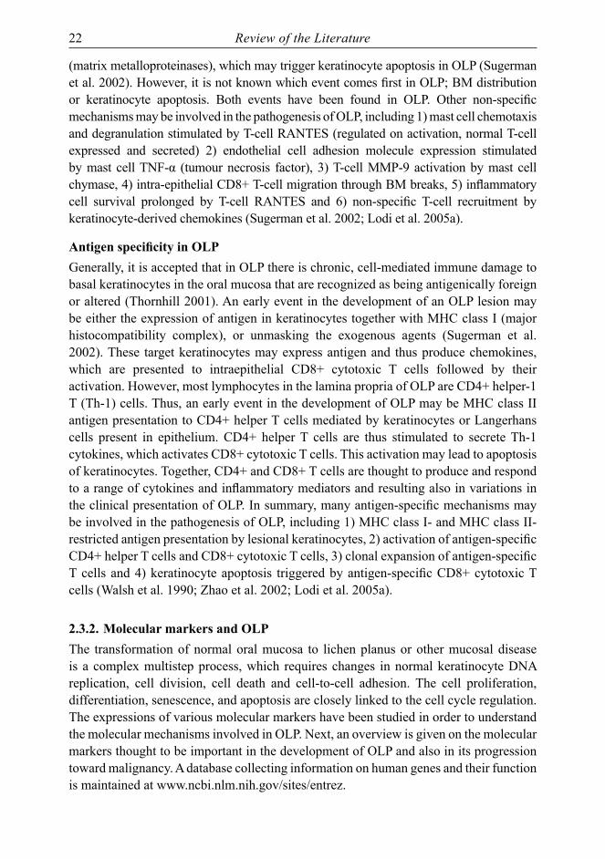
2.3.3. Cell cycle and proliferationThe cell cycle consists of the G1 phase, as the DNA replication initiation, the S phase with DNA synthesis, G2 with time lapse between completion of DNA synthesis, followed by cell division in mitosis M. After mitosis, the cell may exit the cell cycle for the stationary G0 phase. The cell cycle is carefully regulated and controlled by checkpoint genes in a normal cell. Checkpoint genes not only control transition between the phases of the cell cycle but also coordinate cell cycle progression with cell signals. They can also modulate regulation of the integrity of the genome in case of DNA damage and if the damage is such as to not immediately cause cell death. Increased proliferate activity and cell division may indicate early events in many pathological processes, which may end towards malignancy. Cell proliferation markers attempt to identify and measure the proportion of cells undergoing the particular phases of cell cycle (Alberts et al. 2008).
Figure 3. Schematic figure shows approximate expression times of individual protein during the cell cycle.
2.3.3.1. Cell cycle markersCdk The family of cyclin-dependent kinase complexes (Cdks) is well known for its role in the cell division cycle. Cyclin-dependent kinase 1 (cdk-1), formally called cell cycle controller (CDC2), is a catalytic subunit of a protein kinase complex, called the M-phase promoting factor, that induces entry into mitosis. Cdk-1 is responsible for controlling the transition from the G1 phase to the S phase and from the G2 phase to the M phase of the cell cycle. Cdks also participate in a subset of apoptosis programs. Cdk-1 plays a major role in the homeostatic control, defining the frontier between normal cellular replication, repair after damage, mitotic catastrophe and apoptosis (Castedo et al. 2002; Santamaria et al. 2007). Aberrant cdk-1 expression is present in cancer cells (Salh et al. 1999).

24 Review of the Literature
Recently, it has been found that cdk-1 mRNA was significantly upregulated in cell lines established from oral cancer which were developed from OLP (Ruutu et al. 2005). This finding, together with the known data on increased proliferation activity and apoptosis in OLP, make cdk-1 expression as a potential marker to be upregulated in OLP lesions, although this has not been studied earlier.
Rad-51 Rad-51 is an important enzyme in DNA homologous recombination. It is involved in the repair of DNA damage and genome continuation in mitosis and meiosis (Yamamoto et al. 1996). Rad-51 expression starts to appear late in the G1 phase and increases significantly in the S phase, remaining constantly at high levels during the G2 and M phases. Increased Rad-51 levels may protect tumour cells from undergoing apoptosis in response to DNA damage, and this may be associated with enhanced recombination and genomic instability. Thus, increased Rad-51 levels make cells more resistant to DNA damage, and there is a link between elevated Rad-51 protein levels, genome instability and tumour progression (Richardson et al. 2004; Richardson 2005; Klein 2008). Earlier, it has been shown that mRNA Rad-51 levels were upregulated in the cancer cell line originated from OLP (Ruutu et al. 2005), but the expression of Rad-51 has not been studied in OLP or oral cancer biopsy samples.
p53 The transcription factor p53 is an important nuclear protein, which responds to diverse cellular stresses to regulate many target genes that induce cell cycle arrest, apoptosis, senescence, DNA repair, or changes in metabolism. It is the guardian of human genome and thus widely studied in cell cycling and cancer development and progression as reviewed by Vousden and Lane (2007) and Olivier et al. (2009). In normal cells, wild-type p53 controls cell cycle by acting as a G1 checkpoint control. If a cell is damaged and repair is impossible (e.g. both strands of DNA are broken), p53 triggers apoptosis resulting in cell death. Activation of p53 after DNA damage or oncogenic signalling is an important protective mechanism which facilitates DNA repair and stimulates apoptosis of the target cells. (Toledo and Wahl 2006; Vousden and Lane 2007; Olivier et al. 2009) Activation and functions of p53 is shown in Table 2.
Table 2. Activation and functions of p53, modified from Vousden et al. (2007).
ACTIVATION in different types of stress FUNCTIONS and cellular responsesNutrient deprivation ApoptosisTelomere erosion Cell-cycle arrestHypoxia SurvivalDNA damage DNA repairRibosomal stress Genomic stabilityOncogene activation Senescence
In human cancers, p53 is frequently altered by gene mutation, which results in the expression of a mutated protein that differs from the wild type by a single amino-acid change. This mutant p53 has altered the DNA-binding capacity, which results in the loss of most of the normal functions of p53. In cancer induced by oncogenic viruses (e.g.
Review of the Literature 25
HPV, HBV, HHV-8, EBV), p53 is nearly always a wild type, but the function of p53 is blocked by viral oncoproteins (Lane and Lain 2002). Loss of p53 function in cells leads to uncontrolled proliferation and loss of antiproliferative activities including apoptosis and senescence, which all promote cancer development. Mutations in p53 occur in 50% of the human cancers (Toledo and Wahl 2006; Vousden and Lane 2007; Olivier et al. 2009). It has been shown that immunohistochemistry is not a good method for distinguishing wild-type p53 and mutant p53 gene (Taylor et al. 1999). In contrast, some authors have shown that anti-p53 antibody DO7 is a prior target to wild-type p53, and anti-p53 antibody pAb240 recognizes the mutated form of the p53 protein (Ahomadegbe et al. 1995; Gasco and Crook 2003; Gonzalez-Moles et al. 2008a).
There are numerous studies on p53 expression in OLP, and most of them have found increased expression of p53 in the basal and parabasal keratinocytes (Hirota et al. 2002; Acay et al. 2006; Gonzalez-Moles et al. 2006), especially in OLPs with dysplasia (Schifter et al. 1998). However, p53 expression in OLP could not be related to their further malignant progression (Schifter et al. 1998). No association between p53 and apoptosis markers: such caspase-3 and the terminal deoxinucleotidyl transferase (TdT)-mediated dUTP-biotin nick end-labeling method (TUNEL) have been found, which might suggest that p53 expression is rather associated with cell cycle arrest than apoptosis in OLP (Tanda et al. 2000; Gonzalez-Moles et al. 2006). Contradictory to that, a significant association has been found between p53 and the proliferation marker Ki-67 expression in OLP (Gonzalez-Moles et al. 2006). Only immunohistochemical studies with p53 expression in OLP have been done, but the presence of p53 mutations has not been confirmed by DNA sequencing.
2.3.3.2. Cell proliferation markersKi-67 (Mib-1) Ki-67 is a monoclonal antibody that reacts with a nuclear antigen expressed in proliferating cells but not in quiescent cells. Expression of this antigen occurs preferentially during late G1, S, G2, and M phases of the cell cycle, while in G0-phase cells the antigen cannot be detected. Mib-1 is a monoclonal antibody that has been proven to be equivalent to an anti-Ki-67, with the advantage that it can be used on paraffin sections. In normal, healthy oral epithelium, Ki-67 expression as a proliferation activity marker is detected mostly in the parabasal layer and not in the basal layer. Consequently, the antibody is used also in tumour pathology to assess the proliferation activity in neoplastic tissues (Brown and Gatter 1990; Gonzalez-Moles et al. 2000; Liu and Klein-Szanto 2000). Several studies have shown that Ki-67 expression is increased in OLP as compared to that found in normal oral mucosa (Hirota et al. 2002; Taniguchi et al. 2002; Acay et al. 2006; Gonzalez-Moles et al. 2006; Montebugnoli et al. 2006). The most intensive expression of Ki-67 has been detected in the erosive form of OLP (Pirkic et al. 2004). Thus, it has been proposed that proliferation activity in OLP may be a useful prognostic marker of malignant transformation.

26 Review of the Literature
Topoisomerase II alpha Topoisomerase II alpha (topo IIα) is an enzyme that can modify (isomerise) the tertiary structure of DNA without changing its primary structure, determined by the nucleotide sequence. It exerts an important role in DNA topology, repair and replication by breaking and rejoining the DNA double helix (Schoeffler and Berger 2005). Thus, topo IIα is a cell cycle-related protein and is expressed in normal as well as neoplastic cells in the S, G2 and M phases. The lowest level of expression is found in the G0 and G1 phases. Consequently, topo IIα is related to cell proliferation (Heck and Earnshaw 1986; Wells et al. 1995). Thus, topo IIα has been considered as a better proliferation marker than Ki-67, because it can be detected during the S, G2 and M phases, providing a better estimation of the proportion of actively cycling cells than Ki-67 does (Lynch et al. 1997). Previous studies have also shown that aberrant expression of topo IIα is associated with the induction of apoptosis and cell viability (McPherson and Goldenberg 1998; Durrieu et al. 2000; Yoshida et al. 2006). There are no earlier studies of OLP and topo IIα, but in oral precancer lesions and head and neck carcinomas, topo IIα is shown to be a valuable marker to assess proliferative activity of the lesion (Stathopoulos et al. 2000; Hafian et al. 2004; Segawa et al. 2008; Shamaa et al. 2008). Because topo IIα is involved in proliferation, DNA repair and apoptosis, it is a useful marker for cell stress and thus also a potential marker for OLP where all these aspects have been discussed to be important in its etiopathogenesis.
Proliferating cell nuclear antigen (PCNA) Proliferating cell nuclear antigen (PCNA) is a nuclear protein. Its expression increases during the G1 phase, peaks at the transition from the G1 to the S phase, and decreases through the G2 phase. Thus, PCNA is present in proliferating cells, and it is essential for cell replication (Zuber et al. 1989). In addition, PCNA has a central role in DNA repair synthesis (Essers et al. 2005). Previous studies have shown that PCNA expression is significantly increased in oral dysplasia and SCC and thus is a potential indicator of the malignant transformation of oral lesions (Pich et al. 2004). However, there are at least two intracellular forms of PCNA, the replication-associated and non-associated forms, in almost all cycling cells. That is why PCNA detects more cycling cells than Ki-67. In addition, the half time of PCNA is longer (around 20 h) resulting in detection of expression in cells which have already left the cell cycle. Thus, Ki-67 is thought to be a more reliable tool to assess proliferation activity (Liu and Klein-Szanto 2000). Similarly, as with Ki-67, PCNA expression is also shown to be increased in OLP lesions, especially in the atrophic form when compared to that found in normal oral mucosa (Tipoe et al. 1996; Schifter et al. 1998; Lee et al. 2005). To summarize, all data support the view that proliferation activity in OLP lesions is increased.
Cyclin D1 D-type cyclins, including cyclin D1, are the rate-limiting controllers of G1-phase progression and are expressed by cells in the G1 – S phases of the cell cycle in a manner dependent on Cdks. Cyclin D1 also has roles in cellular proliferation, metabolism, and cellular differentiation. Overexpression of cyclin D1 may result in the loss of cell cycle control with consequent increased cell proliferation. It also inhibits the size and activity of the cell’s mitochondria. The inhibition allows the cell to shift its biosynthetic

Review of the Literature 27
priorities; cyclin D1 thus integrates mitochondrial function and nuclear DNA synthesis following the cell proliferation (Sakamaki et al. 2006). Therefore, overexpression of cyclin D1 is known to correlate with the early onset of cancer and risk of tumour progression and metastases (Fu et al. 2004). So far, there is only one study on cyclin D1 expression in OLP which showed overexpression in the epithelial cells indicating an increase in cell proliferation activity (Hirota et al. 2002).
2.3.4. DNA damage and cell deathAs stated earlier, DNA replication is a carefully controlled event in the cell division. Normal non-dividing human cells contain 23 pairs of chromosomes, and this amount of DNA is called diploid. In dividing cells, the DNA content (ploidy) in the nucleus is doubled, and thus four pairs of chromosomes are called tetraploid. However, damages in the DNA repairing system may lead to uncontrolled DNA replication, which induces unbalanced representation of chromosomes. This abnormality in the number of the chromosomes in the cell is called aneuploidy, as the amount of chromosomes is not an exact diploid or tetraploid. Aneuploidy initiation is generated either by a carcinogen or spontaneously. It destabilizes the numbers and structures of chromosomes, because it unbalances the highly conserved teams of proteins, which segregate, synthesize and repair chromosomes. Aneuploidy is proposed as a primary cause of genomic instability in preneoplastic cells in human premalignant lesions and cancers (Sen 2000; Duesberg et al. 2004). Genetic instability, as measured by the number of chromosomal copy alterations, is stated to increase significantly at the transition from precursor lesions to invasive carcinomas and will continently increase in line with the tumour stage (Ried et al. 1999).
Static or image DNA cytometry allows the measurement of the DNA content in a cell population by comparing the integrated optical density (IOD) of the nuclei of interest with that of control nuclei, which may usually be lymphocytes or muscle cells in oral mucosal biopsy samples. This method provides information about the number of abnormal cells with a high DNA content (Haroske et al. 1998). Measurement of DNA content is a tool to assess the damaged DNA repairing and DNA aneuploidy, which both have become potential markers of malignant transformation of the lesion. However, it is not yet known whether this method is specific and sensitive enough to be used alone to assess the risk of oral mucosal lesions to progress toward malignancy (Reibel 2003; Bremmer et al. 2008).
There are a few previous studies on DNA content in OLP lesions (Biesterfeld et al. 1991; Femiano and Scully 2005; Maraki et al. 2006; Rode et al. 2006; Neppelberg and Johannessen 2007). The results are contradictory, while both diploid to aneuploid DNA content have been found, which partly was related to the method used and the clinical type of OLP lesions analyzed. Mostly, the reticular form of OLP was shown to be diploid and thus not expressing malignancy-associated changes in DNA content (Femiano and Scully 2005; Rode et al. 2006; Neppelberg and Johannessen 2007). In contrast to that,
28 Review of the Literature
atrophic or erosive forms of OLP seemed to have more aneuploid changes and DNA cytometry was suggested to be an appropriate screening method for OLP to detect the high risk lesions (Biesterfeld et al. 1991; Maraki et al. 2006).
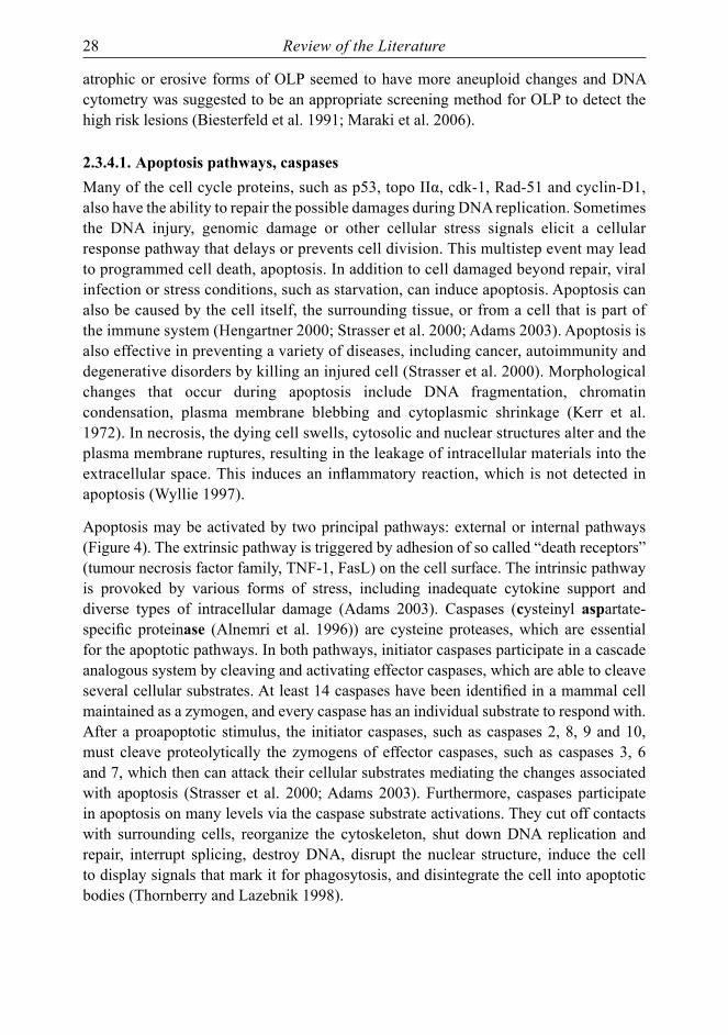
2.3.4.1. Apoptosis pathways, caspasesMany of the cell cycle proteins, such as p53, topo IIα, cdk-1, Rad-51 and cyclin-D1, also have the ability to repair the possible damages during DNA replication. Sometimes the DNA injury, genomic damage or other cellular stress signals elicit a cellular response pathway that delays or prevents cell division. This multistep event may lead to programmed cell death, apoptosis. In addition to cell damaged beyond repair, viral infection or stress conditions, such as starvation, can induce apoptosis. Apoptosis can also be caused by the cell itself, the surrounding tissue, or from a cell that is part of the immune system (Hengartner 2000; Strasser et al. 2000; Adams 2003). Apoptosis is also effective in preventing a variety of diseases, including cancer, autoimmunity and degenerative disorders by killing an injured cell (Strasser et al. 2000). Morphological changes that occur during apoptosis include DNA fragmentation, chromatin condensation, plasma membrane blebbing and cytoplasmic shrinkage (Kerr et al. 1972). In necrosis, the dying cell swells, cytosolic and nuclear structures alter and the plasma membrane ruptures, resulting in the leakage of intracellular materials into the extracellular space. This induces an inflammatory reaction, which is not detected in apoptosis (Wyllie 1997).
Apoptosis may be activated by two principal pathways: external or internal pathways (Figure 4). The extrinsic pathway is triggered by adhesion of so called “death receptors” (tumour necrosis factor family, TNF-1, FasL) on the cell surface. The intrinsic pathway is provoked by various forms of stress, including inadequate cytokine support and diverse types of intracellular damage (Adams 2003). Caspases (cysteinyl aspartate-specific proteinase (Alnemri et al. 1996)) are cysteine proteases, which are essential for the apoptotic pathways. In both pathways, initiator caspases participate in a cascade analogous system by cleaving and activating effector caspases, which are able to cleave several cellular substrates. At least 14 caspases have been identified in a mammal cell maintained as a zymogen, and every caspase has an individual substrate to respond with. After a proapoptotic stimulus, the initiator caspases, such as caspases 2, 8, 9 and 10, must cleave proteolytically the zymogens of effector caspases, such as caspases 3, 6 and 7, which then can attack their cellular substrates mediating the changes associated with apoptosis (Strasser et al. 2000; Adams 2003). Furthermore, caspases participate in apoptosis on many levels via the caspase substrate activations. They cut off contacts with surrounding cells, reorganize the cytoskeleton, shut down DNA replication and repair, interrupt splicing, destroy DNA, disrupt the nuclear structure, induce the cell to display signals that mark it for phagosytosis, and disintegrate the cell into apoptotic bodies (Thornberry and Lazebnik 1998).
Review of the Literature 29
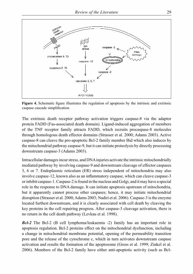
Figure 4. Schematic figure illustrates the regulation of apoptosis by the intrinsic and extrinsic caspase cascade simplification.
The extrinsic death receptor pathway activation triggers caspase-8 via the adaptor protein FADD (Fas-associated death domain). Ligand-induced aggregation of members of the TNF receptor family attracts FADD, which recruits procaspase-8 molecules through homologous death effector domains (Strasser et al. 2000; Adams 2003). Active caspase-8 can cleave the pro-apoptotic Bcl-2 family member Bid which also induces by the mitochondrial pathway caspase-9, but it can initiate proteolysis by directly processing downstream caspase-3 (Adams 2003).
Intracellular damages incur stress, and DNA injuries activate the intrinsic mitochondrially mediated pathway by involving caspase-9 and downstream cleavage of effector caspases 3, 6 or 7. Endoplasmic reticulum (ER) stress independent of mitochondria may also involve caspase-12, known also as an inflammatory caspase, which can cleave caspase-3 or inhibit caspase-1. Caspase-2 is found in the nucleus and Golgi, and it may have a special role in the response to DNA damage. It can initiate apoptosis upstream of mitochondria, but it apparently cannot process other caspases; hence, it may initiate mitochondrial disruption (Strasser et al. 2000; Adams 2003; Nadiri et al. 2006). Caspase-3 is the enzyme located furthest downstream, and it is clearly associated with cell death by cleaving the key proteins in the cell repairing progress. After caspase-3 cleavage activation, there is no return in the cell death pathway (Levkau et al. 1998).
Bcl-2 The Bcl-2 (B cell lymphoma/leukaemia -2) family has an important role in apoptosis regulation. Bcl-2 proteins effect on the mitochondrial dysfunction, including a change in mitochondrial membrane potential, opening of the permeability transition pore and the release of the cytochrome c, which in turn activates downstream caspase activation and results the formation of the apoptosome (Gross et al. 1999; Zinkel et al. 2006). Members of the Bcl-2 family have either anti-apoptotic activity (such as Bcl-

30 Review of the Literature
2, Bcl-XL, Bcl-W, Mcl-1 and A1) or they promote cell death (such as Bax, Bad, Bak, Bid, Bik, Blk, Bim and Bcl-XS). Anti-apoptotic members are initially integral membrane proteins found in the mitochondria, ER or nuclear membrane. Pro-apoptotic members localize to cytosol or cytoskeleton. The ratio of these two subsets in part determines the susceptibility of cells to a death signal (Gross et al. 1999). Thus, Bcl-2 normally blocks the activation of caspase-3. When Bcl-2 activity is blocked by Bax, caspase-3 activity is unchecked and apoptotic cell death proceeds.
Of the caspase markers, only the expression of the caspase-3 as an early and specific marker of apoptosis has been studied in OLP lesions (Tobon-Arroyave et al. 2004; Bascones et al. 2005; Bascones-Ilundain et al. 2006; Gonzalez-Moles et al. 2006; Bascones-Ilundain et al. 2008; Abdel-Latif et al. 2009). The results on caspase-3 expression in OLP lesions are conflicting. The expression was shown to vary from less than 10% to more than 50% of the basal cells to express caspase-3. There are several studies on Bcl-2 expression in OLP lesions (Dekker et al. 1997; Bloor et al. 1999; Tanda et al. 2000; Bascones et al. 2005; Abdel-Latif et al. 2009; Sousa et al. 2009), which have all shown only weak Bcl-2 expression in OLP keratinocytes, supporting the role of apoptosis in OLP.
2.3.5. Cell-to-cell adhesionCell adhesion molecules (CAMs) are found on the surfaces of all epithelial cells, where they bind to extracellular matrix molecules or to receptors on other cells: thus, they are essential for maintaining stable tissue structure. Moreover, CAMs function as signalling receptors, transducing signals initiated by cellular interactions which regulate many diverse processes, including cell division, migration, and differentiation. The expression of CAMs is normally tightly regulated, thereby controlling cell proliferation, mobility, differentiation, and survival. Altered expression of these molecules has been found in oral carcinoma, where loss of CAM expression is often seen in poorly differentiated lesions (Thomas and Speight 2001). In addition, both the junctional and the cytoskeletal components, such as Ck-19, E-cadherin and desmocollin-1, of the desmosome – cytokeratin network might be useful markers for the differentiation and behaviour of oral SCC (Shinohara et al. 1998).
Ck-19 Intermediate filament proteins (IFPs) form a major part of the cytoskeleton in eukaryotic cells (Moll et al. 2008). Cytokeratins (Cks) are the largest family of IFPs that are typically specific for epithelial cells (van der Velden et al. 1993). As a part of the epithelial cytoskeleton, keratins are important for the mechanical stability and integrity of epithelial cells and tissue. Cytokeratins are subdivided into two principal categories: type I as acidic and type II as basic to neutral keratins (Moll et al. 2008). IFPs are also important markers of tissue differentiation, which have shown to be useful in the characterization of malignant tumours (van der Velden et al. 1993). The type I keratin, Cytokeratin 19 (Ck-19) is an intermediated filament protein, which is found in most of the simple and non-keratinizing stratified epithelia. During pathological processes, the expression of Ck-19 may be induced in epithelia that normally lack this keratin (Moll

Review of the Literature 31
et al. 2008). Suprabasal expression of Ck-19 has been correlated to mucosal instability and thus could be a useful marker of cellular atypia in potentially pre-malignant lesions in oral mucosa (Lindberg and Rheinwald 1989). A sparse basal expression of Ck-19 has been detected in OLP lesions, and it has even been suggested that inflammation is inducing the Ck-19 expression (Bosch et al. 1989; van der Velden et al. 1999). Ck-19 is frequently expressed in both adenocarcinomas and SCC (Moll et al. 2008). Thus, changes in Ck-19 expression in OLP might be useful to assess which might reflect the inflammation or/and malignant transformation in the lesion.
E-cadherin Cadherins are a large family of single-pass transmembrane glycoproteins mainly involved in calcium-dependent cell adhesion. The cadherin molecules comprise three domains: the intracellular domain, the transmembrane domain and the extracellular domain. Hence, cadherins structure biologically essential extracellular and intracellular signalling process (Gooding et al. 2004; Halbleib and Nelson 2006). Epithelial (E)-cadherin is a member of the classic (type I) cadherin superfamily, expressed predominantly in epithelial tissue. It plays a major role in the maintenance of intercellular junctions of tissue architecture and integrity by controlling growth and development of cells (Gooding et al. 2004). In normal oral epithelium, E-cadherin is expressed in the lower parabasal layer and basal cell layer, but not at the basal cell surface of the basal cells. In human carcinomas, including oral cancer, the E-cadherin expression is reduced or lost, which correlates with the invasive and metastatic potential of these tumours (Downer and Speight 1993; Bankfalvi et al. 2002b; Diniz-Freitas et al. 2006). However, in the early stages of oral carcinogenesis there may be a transient increase in E-cadherin expression (Bankfalvi et al. 2002a). Furthermore, it has previously been shown that loss of E-cadherin mediated cell adhesion and triggered caspase-3 activation and thus
Figure 5. Schematic expression of Ck-19, E-cadherin and desmocollin-1 in cell-to-cell junction

32 Review of the Literature
apoptotic cell death (Peluso et al. 2001; Galaz et al. 2005). There are a few earlier reports presenting reduced E-cadherin expression in OLP lesions (Neppelberg and Johannessen 2007; Neppelberg et al. 2007; Ebrahimi et al. 2008). As the E-cadherin is also reduced in dysplastic and SCC lesions, the specificity of E-cadherin as a prognostic marker of cancer development in OLP patients could be usable.
Desmocollin-1 The desmosome is a complex adhesive structure that plays a fundamental role in maintaining the strength and integrity of epithelial tissues. Central to this role are transmembrane glycoproteins that mediate cell-to-cell adhesion at the extracellular surface and interact with the cytoskeleton, thus linking the intermediate filament networks of adjacent cells. Desmosomal glycoproteins constitute two distinct groups: the desmogleins and the desmocollins, both of which are members of the cadherin superfamily of Ca2+-dependent cell adhesion molecules (Garrod 1993; Presland and Dale 2000). In keratinized oral mucosa, desmosomal cadherin is lacking in desmosomes, but in contrast, it is detected in all the nucleated cells in the tongue and in normal non-keratinized oral mucosa (Shinohara et al. 1998; Donetti et al. 2005). Many studies have shown that expression of desmosomes is reduced in oral SCC especially in association with invasive and metastatic behaviour (Shinohara et al. 1998; Dusek et al. 2007). Expression of desmocollin-1 has not been studied in OLP lesions or in other oral premalignant conditions.
2.4. Malignant transformation of OLPAmong researchers, during the last few decades, there has been a large ongoing debate on the malignant transformation of OLP, and the results are still conflicting. According to the current definition, a precancerous lesion is a morphologically altered tissue in which cancer is more likely to occur than in its apparently normal counterpart (WHO 1978; Axell et al. 1996). Based on their own results and review on the literature, Krutchkoff et al. concluded, in 1978, that there is little evidence of OLP premalignancy (Krutchkoff et al. 1978). During the very same year, the WHO report stated that malignant transformation has been observed in as many as 2 – 3% of the patients included in several studies and defined. This is why the expert group nominated by WHO concluded that OLP is a potentially precancerous condition, representing a generalized state associated with a significantly increased risk of cancer (WHO 1978). Recently, WHO stated that the recommended term to be used is “potentially malignant disorder” (van der Waal 2009).
Ten years ago, in a review by van der Meij et al. (van der Meij et al. 1999b) reported that during the years 1977 – 1999, a total of 98 papers were published on OLP, and in 34% of them, there were sufficiently documented evidence of malignant transformation of OLP. In two recent reviews, the issue has been widely discussed based on the numerous prospective and retrospective studies as well as case reports (Mattsson et al. 2002; Gonzalez-Moles et al. 2008b). Both of these reports summarized that with the strict criteria of OLP, there is evidence of OLP malignization, but the question of continuous recall of all OLP patients is still controversial among these authors.
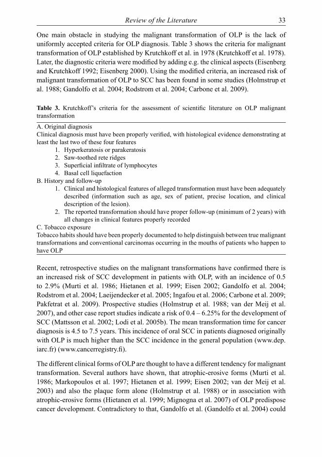
Review of the Literature 33
One main obstacle in studying the malignant transformation of OLP is the lack of uniformly accepted criteria for OLP diagnosis. Table 3 shows the criteria for malignant transformation of OLP established by Krutchkoff et al. in 1978 (Krutchkoff et al. 1978). Later, the diagnostic criteria were modified by adding e.g. the clinical aspects (Eisenberg and Krutchkoff 1992; Eisenberg 2000). Using the modified criteria, an increased risk of malignant transformation of OLP to SCC has been found in some studies (Holmstrup et al. 1988; Gandolfo et al. 2004; Rodstrom et al. 2004; Carbone et al. 2009).
Table 3. Krutchkoff’s criteria for the assessment of scientific literature on OLP malignant transformation
A. Original diagnosis Clinical diagnosis must have been properly verified, with histological evidence demonstrating at least the last two of these four features
1. Hyperkeratosis or parakeratosis2. Saw-toothed rete ridges3. Superficial infiltrate of lymphocytes4. Basal cell liquefaction
B. History and follow-up1. Clinical and histological features of alleged transformation must have been adequately
described (information such as age, sex of patient, precise location, and clinical description of the lesion).
2. The reported transformation should have proper follow-up (minimum of 2 years) with all changes in clinical features properly recorded
C. Tobacco exposureTobacco habits should have been properly documented to help distinguish between true malignant transformations and conventional carcinomas occurring in the mouths of patients who happen to have OLP
Recent, retrospective studies on the malignant transformations have confirmed there is an increased risk of SCC development in patients with OLP, with an incidence of 0.5 to 2.9% (Murti et al. 1986; Hietanen et al. 1999; Eisen 2002; Gandolfo et al. 2004; Rodstrom et al. 2004; Laeijendecker et al. 2005; Ingafou et al. 2006; Carbone et al. 2009; Pakfetrat et al. 2009). Prospective studies (Holmstrup et al. 1988; van der Meij et al. 2007), and other case report studies indicate a risk of 0.4 – 6.25% for the development of SCC (Mattsson et al. 2002; Lodi et al. 2005b). The mean transformation time for cancer diagnosis is 4.5 to 7.5 years. This incidence of oral SCC in patients diagnosed originally with OLP is much higher than the SCC incidence in the general population (www.dep.iarc.fr) (www.cancerregistry.fi).
The different clinical forms of OLP are thought to have a different tendency for malignant transformation. Several authors have shown, that atrophic-erosive forms (Murti et al. 1986; Markopoulos et al. 1997; Hietanen et al. 1999; Eisen 2002; van der Meij et al. 2003) and also the plaque form alone (Holmstrup et al. 1988) or in association with atrophic-erosive forms (Hietanen et al. 1999; Mignogna et al. 2007) of OLP predispose cancer development. Contradictory to that, Gandolfo et al. (Gandolfo et al. 2004) could

34 Review of the Literature
not find any association between different clinical forms and malignant transformation. In association with this conclusion, Mattsson et al. have stated that the similar proportion of malignant transformation in different OLP forms cannot explain the cancer development of the disease (Mattsson et al. 2002). The site of the OLP might be important while the tongue appears to be the preferred site for SCC emergence (Holmstrup et al. 1988; Markopoulos et al. 1997; Carbone et al. 2009). However, malignant transformation has also been reported in OLP lesions located in the buccal mucosa, midline of the palate, gingiva and lips (Mignogna et al. 2001).
The putative role of well-known risk factors of SCC, such as tobacco and alcohol, has been evaluated in OLP-related malignant transformation by Gandolfo et al. (Gandolfo et al. 2004). The results showed that alcohol and tobacco or their interaction cannot explain the excess risk of SCC in OLP. More likely, the immune activation and chronic inflammation in OLP might be the cause of the progression towards malignancy as shown to be evident in a variety of other epithelial malignancies, such as gastric cancer and colorectal adenocarcinoma (Mignogna et al. 2004). Furthermore, Mignogna et al. (Mignogna et al. 2007) have suggested that patients with OLP, where a wide area of mucosa is involved, are prone to develop multiple or multifocal neoplastic events in oral mucosa, a phenomenon which parallels the concept of field cancerization. This explains the appearance of multiple secondary tumours once a primary tumour is established.
In a retrospective study, it has been shown that patients with cutaneous LP, of unknown oral status, had a high incidence of oral and lip cancer, with a relative risk of 5,9% (Sigurgeirsson and Lindelof 1991). However, patients with cutaneous LP have no increased risk of SCC of the skin (Sigurgeirsson and Lindelof 1991).
2.4.1. Lichenoid dysplasiaThe term “lichenoid dysplasia” (LD) is defined by Krutchkoff and Eisenberg to describe lesions that resemble OLP but are dysplastic (Krutchkoff and Eisenberg 1985). According to this, any departure from normal epithelial maturation and growth excludes a diagnosis of OLP. Thus, the OLP lesions with epithelial dysplasia should not be included in studies of malignant transformation as misdiagnoses of OLP. Therefore, Eisenberg (Eisenberg 2000) suggested that essential and exclusionary histologic features must be present to make a definitive diagnosis of OLP. The term “lichenoid dysplasia” defines merely on the biopsy with histological findings, which is taken in a certain area of the diseases. Thus, it limits out the rest of the disease. It is disclosed that the real significance is related to the potential difference in the risk of transformation to cancer (Silverman 2000; Epstein et al. 2003). According to Gonzalez-Moles et al. (Gonzalez-Moles et al. 2008b), it cannot be ruled out that the epithelium in OLP may develop epithelial dysplasia during the process of carcinomatous transformation: hence, the exclusion of all lesions that resemble OLPs but exhibit epithelial dysplasia may lead to an underestimation of the rate of malignant transformation. This is supported also by the theory of cancer field cancerization phenomena (Mignogna et al. 2007).

Review of the Literature 35
Lodi et al. (Lodi et al. 2005b) propose that the existence of LD may correspond into two groups of conditions: lesions with clinical feature of OLP but with dysplasia; and lesions with lichenoid histological features (such as band-like inflammatory infiltration) but without the clinical features of classic OLP (such as bilateral distribution and reticular lesion). The former may represent an early phase in the malignant transformation of OLP, while the latter may represent various clinical conditions with lichenoid histopathology (such as OLR, leukoplakia, lupus erythematosus and proliferative verrucous leukoplakia).
36 Aims of the Study
3. AIMS OF THE STUDYThe main aim of this present study was to investigate the cell and molecular markers of atrophic oral lichen planus (OLP) in order to predict the prognosis of the disease progression as a potential premalignant disease. The etiology of OLP is still unknown, which makes finding a cure for the disease difficult. With an increased oral cancer risk, every patient suffering from OLP should be closely monitored in follow-up care, which is rather time-consuming and expensive. Therefore, the major goal of this study was to find such markers on a cellular level which distinguish aggressive OLP lesions from those latent OLP forms where malignant progression is improbable.
The specific aims of the study were:
1. To study the alterations of DNA content in the keratinocytes of atrophic OLP as a potential marker of malignant progression by static cytometry.
2. To investigate the proliferation activity and potential DNA repairing of the epithelial keratinocytes in atrophic OLP lesions.
3. To investigate the alterations in epithelial cell adhesion as a marker of tissue changes in OLP epithelium.
4. To study the expression of apoptosis and caspase cascade pathways as a marker of apoptosis rate in keratinocytes of OLP lesions.
5. To evaluate the lymphocyte cell infiltration with T and B cell markers to distinguish potential autoimmune OLP lesions from those with any other unknown etiological factor.
Materials and Methods 37
4. MATERIALS AND METHODS
4.1. Tissue samplesAll patients included in this study were sent by their dentists’ referral to the Department of Oral and Maxillofacial Surgery of Turku University Hospital. The mucosal diagnosis was given by the health care oral surgeon at the Turku University Hospital based on the pathologic-anatomical diagnosis given by the oral pathologist. Oral biopsy samples from consecutive patients diagnosed with atrophic-erosive OLP during the years 1991 to 2002 were retrieved from the files of the Department on Oral Pathology and Radiology at the University of Turku. All patients born before the year 1928 and those with any medication or suffering from any systemic disease were excluded. Also, the location of lesions in the oral mucosa was recorded. After collecting the biopsy sample from the files, all slides were re-examined again by an oral pathologist to confirm the histologic diagnosis of atrophic OLP. A total of 81 biopsy specimens taken from 70 patients were included. Of these patients, 53 (76%) were women with the mean age 53.3 years (range 29 years to 73 years), and 17 (24%) were men with a mean age of 52.7 years (range 36 years to 72 years). The patients represented thus a typical age and sex distribution of OLP patients. The mean follow-up time was 63.5 months (range from 7.2 to 117.4 months). Five of the OLP patients developed oral squamous cell carcinoma during the follow-up. (I, II, III)
For the studies IV and V, 11 samples were excluded due to the quality of the samples after several serial cuttings. Thus, there were only 70 biopsy specimens taken from 66 consecutive available patients. Of these 66 patients, 49 (74%) were women with the mean age of 53.9 years (range 35 years to 73 years), and 17 (26%) were men with the mean age of 51.0 years (range 36 years to 65 years). The mean follow-up time of the patients was 62.4 months (range from 7.2 to 117.4 months).
4.2. Histological examinationFor re-examination, new sections (6µm) were cut for haematoxylin and eosin staining. Re-examination of the slides confirmed the diagnosis of atrophic OLP in all samples. In addition to the confirmation of the diagnosis, a variety of morphological parameters were recorded in each lesion. These include the following: the presence and type of hyperkeratinization, atrophy, ulceration, basal cell hyperplasia and dysplasia. These parameters were recorded in area of the sections with the most severe changes. Dysplastic changes were found in 11 OLP lesions, of which 6 were mild and 5 moderate dysplasia, as also reported already in the original histopathologic statement of the lesion. During the histological examination, the samples were re-examined without knowing the outcome of the lesion. The same was true also when the immunologically stained sections were evaluated.
38 Materials and Methods
4.3. Methods
4.3.1. Static cytometry (I)
4.3.1.1. DNA stainingFor static cytometry (I), two sections (8µm) from the paraffin-embedded blocks were cut. The sections were stained with the modified Feulgen’s stain method as described in study I. In brief, the sections were first deparaffined and washed in distilled water followed by acid hydrolysis in 5M HCl at room temperature for one hour. After washing in distilled water, they were stained with Schiff’s reagent for 165 minutes at room temperature, rinsed in distilled water, treated three times for 10 minutes in fresh sodium thiosulphate (180ml distilled water, 10ml 1M HCl, 10ml 10% Na2S2O5) and rinsed for five minutes. The sections were then dehydrated with a series of graded alcohol and xylene and mounted.
4.3.1.2. Static cytometryStatic cytometry measurements were made with Ahren’s Cytometry Analysis system, version 5.02 (Institut für Meβ-Technik, Hamburg, Germany). This is computer software for DNA measurement. An Olympus BH2 microscope (Olympus Optical Co., Ltd., Tokyo, Japan) with 60 X objective and a CCD camera (Panasonic WV-CD 20, Matsushita Communication Industrial Co., Ltd., Osaka, Japan) were used with a green filter (550 nm, Olympus). To assess the position of the normal diploid 2c value (c is the haploid genome equivalent), 30 endothelial cells or muscle cells were measured in each section as a control. The DNA content of approximately 150 epithelial cells within the basal or parabasal area was measured. All other cells, e.g. lymphocytes frequently present in epithelium, were excluded from the measurements.
4.3.1.3. Parameters measuredThe following parameters were measured in the static cytometry: G1/G0, S-Phase, G2/M, G2/M exceeding rate, 2.5c exceeding rate, 5c exceeding rate, ploidy and proliferation index. The 2.5c exceeding rate (2.5cER) was defined as the percentage of the measured cells exceeding the 2.5c value on the DNA scale, and the 5c exceeding rate (5cER) exceeding the 5c value on the DNA scale. The ploidy is the mean value of the G1 fraction position of measured cells on the DNA scale. The histograms were classified as DNA diploid when the mean ploidy value was ≤2.5c, as DNA tetraploid when the mean ploidy value was 3.5<c≤4.5 and as DNA aneuploid when the mean ploidy value was 2.5<c≤3.5 or >4.5c. Proliferation index (PI) is the sum of the S and G2/M phase fractions. The cut-off values for lesions classified as diploid/aneuploid were also tested statistically. As the 2.5cER cut-off value was 15.4%, the sample was defined as aneuploid.
4.3.2. Immunohistochemistry (II – V)Table 4 summarizes the antibodies and their functional category as proliferation and DNA repair, intercellular adhesion and cytoskeleton markers, apoptosis and inflammatory cell

Materials and Methods 39
infiltration, pre-treatment methods for antigen retrieval and the positive controls used in immunohistochemical studies (II, III, IV, V).
For immunohistochemical (IHC) staining, consecutive 6-µm thick (II) and 4-µm thick (III-V) sections from formalin-fixed, paraffin-embedded tissue blocks were cut. Thus, it was possible to make the mutual correlation of different antigen expressions in every study. The DAKO TechMate™ 500 Plus automatic staining system was used in studies II, IV, V, while the manual routine IHC with the DAKO EnVision™ System and Alkaline Phosphatase in study III (DAKO Corporation, Carpinteria CA, USA) were used for immunohistochemical staining according to the instructions given by the provider. Negative controls were processed parallel to test samples by replacing the primary antibody with a buffer.
Table 4. The antibodies used to study protein expression, grouped according to their functional category
Antibody Clone Dilution Pre-treatment Positive control Manufacturer Study
Proliferation and DNA repairTopoisomerase IIα
M7186: clone Ki-S1 1:100 microwave for
2 x 5 minutesBreast carcinoma
DAKO, Glostrup, Denmark II
Ki-67 M7240: MIB-I ready-to-use
microwave for 2 x 5 minutes
Oral verruca vulgaris
DAKO, Glostrup, Denmark II
cdk-1 (p34cdc2) 1:200 microwave 10 minutes
Cancer gradus III
Sigma-Aldrich, St. Louis, Missouri, USA III
Rad-51 monoclonal 51RAD01 1:75 microwave 10
minutes TestisNeoMarkers, Lab Vision Corporation, Fremont, California, USA
III
Intercellular adhesion and cytoskeleton markersCytokeratine-19 on cytoskeleton
M888: RCK108 1:100 microwave for
2 x 5 minutesBreast carcinoma
DAKO, Glostrup, Denmark II
E-cadherin NCH-38 1:50 microwave 10 minutes
Oral cancer gradus I
DAKO, Glostrup, Denmark III
Desmocollin-1 monoclonal Dsc1-U100
ready-to-use
microwave 10 minutes
Dermis with hair follicle
Research Diagnostics Inc, Flanders, USA III
Apoptosis
Caspase-2 monoclonal 10C6 1:20 microwave for
10 + 7 minutes Testis Alexis Biochemicals, Lausen, Switzerland V
Caspase-3 monoclonal 3CSP03 1:50 microwave for
10 + 7 minutes TonsilsNeoMarkers, Lab Vision Corporation, Fremont, California, USA
V
Caspase-8 polyclonal FLICE(Ab-4) 1:300 microwave for
10 + 7 minutes StomachNeoMarkers, Lab Vision Corporation, Fremont, California, USA
V
Caspase-9 monoclonal 9CSP02 1:35 microwave for
10 + 7 minutes TonsilsNeoMarkers, Lab Vision Corporation, Fremont, California, USA
V
Caspase-12 polyclonal (IN) 1:800 microwave for 10 + 7 minutes Tonsils ProSci Incorporated, San
Diego, California, USA V
Inflammatorycellinfiltration
CD5 T-cell, clone 54/F6 1:50 microwave for
2 x 5 minutes Tonsil DAKO A/S, Glostrup, Denmark IV
CD20cy B-cell, clone L26 1:400 microwave for
2 x 5 minutes Tonsil DAKO A/S, Glostrup, Denmark IV
CD27 137B4 1:60 microwave for 2 x 5 minutes Tonsil Novocastra, Newcastle
upon Tyne, UK IV
CD38 SPC32 1:100 microwave for 2 x 5 minutes Tonsil Novocastra, Newcastle
upon Tyne, UK IV

40 Materials and Methods
4.3.2.1. Grading of the expression
Figure 6. The figure shows the schematic expression of the measured parameters in OLP histology samples (II-V). Cell cycle and proliferation markers with different types of staining are marked with red, adhesion molecules are marked with green, caspase proteins of apoptosis are marked with blue and lymphocytes are marked with black.
Proliferation and DNA repairing proteins (II, III)Proliferation and DNA repairing markers expression were present in the cell nucleus. In the basal and parabasal epithelial cell layer, 500 and 200 cells expressing topo IIα (II), Ki-67 (II), cdk-1 (III) and Rad-51 (III) were counted, respectively. The staining patterns of topo IIα and Ki-67 were graded as negative, diffuse staining (<50%), and strong staining. The intracellular cytoplasm expression of cdk-1 and Rad-51 in 200 cells in each section were counted as stained or not stained. The results were presented as a percentage of the counted cells.
Intercellular adhesion proteins and cytoskeleton keratin 19 (II, III)In the entire epithelium, the expressions of the following intercellular adhesion proteins were studied: desmocollin-1 (III) and E-cadherin (III) and cytoskeleton protein Ck-19 (II). The expressions of desmocollin-1 and E-cadherin were graded into three groups: 1) no staining, 2) partial staining: 10% or more, but less than 50% of the epithelia stained, 3) strong staining: 50% or more of the epithelia stained. The expression of Ck-19 was graded into four groups: 1) no staining, 2) only a few cells stained, 3) less than 50% of the cells stained, and 4) 50% or more of the basal cells stained.
Markers of apoptosis (V)The markers of caspase cascade are expressed in the whole epithelium. The expression of caspase-2 in 200 epithelial cells in each specimen was evaluated in epithelium and graded as 1) no staining, 2) moderate staining and 3) intense staining. The intense expression of caspase-2 was further subgraded as 1) 30% ≤ 50% of the epithelial cells
Materials and Methods 41
were intensively stained, 2) 50% ≤ 70% of the epithelial cells were intensively stained and 3) >70% of the epithelial cells were intensively stained.
The expression of caspase-3 was graded: 1) no staining, 2) moderate staining (only a few cells stained) or 3) intense staining (50% or more of the basal cells stained). The staining pattern of caspase-8 was graded into three groups: 1) no staining, 2) most of the basal cells stained, 3) all of the basal cells and some cell areas in the epithelium were stained. Expression of caspases 9 and 12 were graded as 1) no staining in epithelial cells and 2) all the epithelial cells are stained in the epithelium of OLP samples.
Inflammatorycellinfiltration(IV)The expressions of CD5, CD20, CD27 and CD38 were evaluated in the subepithelial inflammatory infiltrate. The expressions of CD5 and CD20 were graded into three grades: 1) no expression 2) less than 50% of the cells were positive or 3) 50% or more of the lymphocytes were positive. Also, the presence of CD5+ lymphocytes in epithelia was graded as present or not. The proportions of CD27+ or CD38+ cells were separately graded as 1) negative, 2) only a few scattered CD27+ or CD38+ lymphocytes, and 3) nearly the entire inflammatory infiltration contained CD27+ or CD38+ cells. The pattern of CD27+ or CD38+ lymphocytes among the inflammatory infiltrate was graded as 1) diffuse 2) band-like zone or 3) clusters.
Based on the proportion of the CD5 and CD20 expressions, the lesions were graded in three subtypes: 1) T cell-dominant, where CD5 expression was present in 50% or more of the lymphocytes and CD20 expression was negative, 2) B cell-dominant, where CD5 expression was found in 10% to less than 50% of the lymphocytes, and CD20 expression was present in 50% or more of the lymphocytes, or 3) mixed, where 50% or more of the lymphocytes expressed CD5, and CD20 expression was found in 10% to less than 50% of the lymphocytes, or 50% or more of the lymphocytes were CD20 positive. As CD5 expression was found in all samples, the dominance was based on the number of the lymphocytes expressing CD20 versus those with CD5 expression (IV).
4.3.3. Detection and genotyping of human papillomavirus DNADNA extractionFrom the paraffin-embedded samples, HPV DNA was extracted with the high salt method (Miller et al. 1988). For DNA extraction, sequential sections (approximately the size of 1cm2) were used. In brief, the sample was lysed with proteinase K at 37°C overnight. The proteins were then precipitated with saturated 6M NaCl followed by centrifugation. DNA was precipitated with absolute ethanol.
HPV genotypingHPV genotyping was made with Luminex-based assay that combines polymerase chain reaction amplification (PCR) with hybridization to fluorescence-labelled polystyrene bead microarrays (Luminex suspension array technology).
42 Materials and Methods
PCR For PCR, GP05+/bio-GP06+ primers targeting the 150-bp-long fragment of the viral L1 open reading frame were used. PCR was done in 50ml reaction containing PCR buffer, 20pmol of each of the GP5+ (5’-TTT GTT ACT GTG GTA GAT ACT AC-3’) and 5’-biotinylated GP6+ (5’-GAA AAA TAA ACT GTA AAT CAT ATT C-3’) primers, 200µM of each deoxynucleotide triphosphate and 1.25U AmpliTaq GoldR DNA polymerase (Perkin Elmer, New Jersey, USA) and 300ng of DNA. Amplification was started by initial denaturation at 95°C for 10 minutes, followed by 40 cycles denaturation at 95°C for 60 seconds, annealing at 40°C for 60 seconds and extension at 72°C for 90 seconds.
Controls for PCROnly eight study samples were processed at the same time. Additionally, every 8th sample for PCR contained no DNA. DNA dilution of SiHa cells was used as a positive control for HPV DNA detection. DNA extraction, master mix for PCR, and the addition of target DNA to the reaction mixture were all done in separate rooms.
Fluorescent bead arrayAfter HPV amplification, the biotinylated PCR products were HPV genotyped with the fluorescent bead array on 24 HPV types according to Schmitt et al. (Schmitt et al. 2006), using the Multimetrix kit (Multimetrix, Regensburg, Germany). The assay can detect the following 24 HPV types: LR-HPV: 6, 11, 42, 43, 44, 70; HR-HPV: 16, 18, 31, 33, 35, 39, 45, 51, 52, 56, 58, 59, 68, 73 and 82; and probably also HR-HPV types 26, 53, 66, and 70. The manufacturer’s instructions were followed, except the reactions were done in 50µl instead of 100µl. In the final step, the beads were washed three times with 100µl blocking buffer and finally resuspended in 100µl blocking buffer for 5 minutes on a shaker. Beads were than analyzed for internal bead colour and R-phycoerythrin reporter fluorescence on a Luminex 100 analyzer. The median reporter fluorescence intensity (MFI) of at least 100 beads was computed for each bead set in the sample. The cut-off value was defined for each HPV probe individually as follows: 1.5 times the background MFI + 5MFI.
4.3.4. Statistical methods (I-V)Statistical analyses were performed using the SPSS® and STATA software packages (SPSS for Windows, Versions 10.0 (I), 11.5 (II, III), 13.0 (IV), 15.0 (V) 16.0.2. (unpublished data), SPSS Inc., Chicago, USA and STATA/SE 10.2. Stata Corp., Texas, USA). Frequency tables were analyzed using the Chi-square test, with Pearson’s R or likelihood ratio (LR) being used to assess the significance of the correlation between the categorical variables. Differences in the means of continuous variables between the groups were analyzed using ANOVA (analysis of variance), when appropriate, or non-parametric tests (Mann–Whitney, Kruskal–Wallis). In study I-III, binary logistic regression models were used to analyze the power of different variables as predictors of dysplasia, using the stepwise backward approach.

Materials and Methods 43
Univariate survival analysis for the outcome measure (cancer endpoint) was based on the Kaplan-Meier method, where all categorical variables were tested (I). For continuous variables, the Cox proportional hazards regression model was used, in which all continuous variables were entered one by one (univariate mode). Variables shown to be significant in univariate analyses were entered in the multivariate Cox model, run in a backward stepwise mode with the log-likelihood ratio (L-R) significance test, using p = 0.10 as the probability for stepwise removal and p = 0.05 as the probability for stepwise entry.
In study III, Odds ratios (OR) and their 95% confidence intervals (CI) were calculated where appropriate. Univariate survival (life-table) analysis for the outcome measures (dysplasia and cancer endpoints) was based on Cox’s regression method (marker values treated as continuous variables; cdk-1 and Rad-51) or the Kaplan-Meier method (dichotomized variables; E-cadherin, desmocollin-1). The multivariate (Cox) proportional hazards method was used to assess the value of the markers as independent predictors of dysplasia and carcinoma, run as described above. The assumption of proportional hazards was controlled by log-minus-log (LML) plots.
Performance indicators: sensitivity (SE), specificity (SP), positive predictive value (PPV), negative predictive value (NPV), and their 95% Confidence Intervals (CI), of the markers in predicting biopsy-confirmed endpoints (dysplasia and SCC) were calculated using STATA/SE software and the algorithm introduced by Seed et al. (Seed and Tobias 2001), which also calculates the area under ROC curve (AUC) for each test. In all analyses, the probability values less than 0.05 were regarded as significant.

44 Results
5. RESULTS
5.1. DNA content in samples from atrophic OLP (I)Static cytometry was used to measure the DNA content in the OLP samples (Study I). First a regression analysis was performed to define the cut-off value for 2.5cER to detect aneuploidy in the samples. The analyses showed that the statistically significant value for 2.5cER, explaining the presence of DNA aneuploidy was 15.3% (p = 0.001). In 41% (34/81) of the OLP samples, the 2.5c exceeding rate (2.5cER) exceeded 15.3%, which indicates aneuploidy in the atrophic OLP lesions. Statistically significantly higher values of 2.5cER (p < 0.001) and proliferation index (p = 0.012) were found in the ulcerated lesions than in the non-ulcerated lesions. Also, the location of the lesion showed to be important; OLPs located in the tongue had significantly higher values of 2.5cER and proliferation index than lesions derived from other sites (p < 0.001 and p = 0.013). Not unexpectedly, the 2.5cER and ploidy index were statistically significantly higher in dysplastic lesions than in non-dysplastic lesions; p < 0.001 and p = 0.048, respectively. The criterion of an aneuploid lesion, according to Auer et al, (Auer et al. 1980) is 2.5cER more than 35% and 5cER values over 0, simultaneously. In study I, only four (5%) OLP samples taken from three patients fulfilled this criterion. All of these lesions located on the tongue and dysplastic changes were detected in three of four samples.
Univariate survival analysis showed that several variables used in the static cytometry were shown to be significant covariates of predicting the cancer development. However, when multivariate analysis was used, only the G2/M exceeding rate (G2/MER) proved to be a significant independent predictor of oral lichen planus to become malignant (OR 2.349; 95%CI 1.39-3.97, p = 0.001).
Figure 7. Two histograms of the atrophic lichen lesions measured. Figure 7A shows a normal histogram with no sign of aneuploidy. Figure 7B shows a histogram with typical non-diploidy changes for aneuploidy. This patient developed squamous cell carcinoma 17 months after the first diagnosis of atrophic lichen planus. y-axis: Number of measured cells, x-axis: DNA content

Results 45
5.2. Expression of proliferation and DNA repairing markers topoisomerase IIα,Ki-67,cdk-1andRad-51inatrophicOLP(II,III)
Topo IIα (II) was intensively expressed in epithelium in 11% of the 81 OLP specimens, while a diffuse expression was detected in 59% of the samples. In dysplastic lesions, there were cells which statistically significantly expressed topo IIα more strongly (p = 0.019) (mean 58.7 ±16.6 SD) and diffusely (p = 0.030) (mean 11.5 ±6.3 SD) than the cells in non-dysplastic lesions. Strong and diffuse expression of topo IIα was also related to basal cell hyperplasia (p = 0.005) (mean 14.0 ±5.4 SD) and ulceration (p = 0.008) (mean 48.4 ±18.9 SD), respectively.
When topo IIα expression was compared to the DNA content, it was seen that the proportion of cells expressing topo IIα was 65.9% (±31.8 SD). In those 34 samples, in which 2.5cER is higher than 15%, strong expressions of topo IIα were significantly more frequent than in those samples with 2.5cER below 15% (p = 0.013; Mann-Whitney). A similar finding in topo IIα was also found in samples with ploidy higher than 1.3 than those below 1.3 (p = 0.034; Mann-Whitney). However, there were only two samples with ploidy higher than 1.3 of the proportion of expressing cells was 20.4% (±2.3 SD). Strong topo IIα expression also correlated significantly also to G2/M fraction (p = 0.036).
Ki-67 was strongly expressed in 24% of the cells, while 12% of the cells were diffusely stained (II). The G2/M fraction was the only DNA parameter which was significantly associated either with diffuse expression of Ki-67 (p = 0.046) or with no expression (p = 0.024). No association was shown with Ki-67 and any histological parameters. Mutual correlations showed that topo IIα was statistically significantly correlated to a strong expression of Ki-67 (p = 0.000; Pearson Correlation), (p = 0.002; Spearman’s rho).
The number of cdk-1 positive cells varied from one (in eight specimens) to 18 cells (in one specimen) (III). The number of Rad-51 positive cells varied from two (in 57 specimens) to 12 cells (in 1% of the specimens) (III). Neither cdk-1 nor Rad-51 expression had any association with any of the histological parameters, location or DNA parameters of the lesion.
5.3. Expression of intercellular adhesion markers desmocollin-1 and E-cadherin and cytoskeleton marker keratin 19 in atrophic OLP (II, III)
Intercellular markers desmocollin-1 and E-cadherin expression was detected in 24.4% (20/82) of the samples (III). Of the positive samples, only eight specimens expressed both desmocollin-1 and E-cadherin. Desmocollin-1 and E-cadherin expression was detected in 8.5% (7/82) and 3.7% (3/82) of the samples, respectively. Either desmocollin-1 and/or E-cadherin expression was detected in 39% (32/82) of the samples. The expression of desmocollin-1 was significantly related to several histological parameters including ulceration (p < 0.001), mitotic counts in the epithelium (p < 0.001), loss of polarity of the basal cells (p < 0.001), reduction of cellular cohesion (p < 0.001), dyskeratotic changes
46 Results
(p < 0.001), and epithelial dysplasia (p < 0.001). Desmocollin-1 expression was also significantly related to the location of the lesion (p = 0.003). When the OLP location was used as a dichotomized variable (tongue vs. other sites), desmocollin-1 staining showed a significant predilection for tongue lesions, with OR = 11.86 (95% CI 3.1- 45.4). A similar analysis for the other sites was not feasible, because their staining patterns are very much alike. Expression of E-cadherin was not related to any of the histological parameters or location of the lesion.
Using the performance indicators, desmocollin-1 predicted dysplasia with the sensitivity of 87.5% (95%CI 47.3-99.7), specificity of 81.9% (95%CI 71.1-90.0), PPV 35.0% (95%CI 15.4-59.2), and NPV 98.3% (95%CI 91.1-100), giving the area under the ROC (receiver operating characteristics) curve (AUC) = 0.847 (95%CI 0.717-0.978). The corresponding figures for desmocollin-1 in predicting incident SCC were: sensitivity 60.0% (95%CI 14.7-94.7), specificity 77.9% (95%CI 67.0-86.6), PPV 15.0% (95%CI 3.2-37.9), and NPV 96.8% (95%CI 88.8-99.6), with the area under the ROC curve (AUC) = 0.690 (95%CI 0.445-0.934).
The sensitivity of E-cadherin as a predictor of dysplasia was 25.0% (95%CI 3.2-65.1), specificity was 76.4% (95%CI 64.9-85.6), PPV 10.5% (95%CI 1.3-33.1), and NPV 90.2% (95%CI 79.8-96.3), with the area under the ROC curve (AUC) = 0.507 (95%CI 0.339-0.675). The corresponding performance characteristics for E-cadherin as predictor of SCC are: sensitivity 100% (95%CI 47.8-100), specificity 80.5% (95%CI 69.9-88.7), PPV 25.0% (95%CI 8.66-49.1), and NPV 100% (95%CI 94.2-100), with the area under the ROC curve (AUC) = 0.903 (95%CI 0.858-0.947).
Only desmocollin-1 correlated with 5.0cER (p = 0.045 Kruskal-Wallis), but not with 2.5cER (p = 0.071). The other adhesion markers had no statistically significant association with DNA content.
Cytoskeleton marker Ck-19 expression was detected in 24 of the 81 biopsy samples (29%) (II). Of the positive samples, 11/24 were classified in the category of less than 50% of the cells expressed Ck-19, while only in four samples, were there 50% or more of the cells expressing Ck-19. Ck-19 expression was significantly more frequent in the samples derived from the tongue than from the other sites of oral mucosa (p = 0.047). Ck-19 had no significant associations with any of the DNA parameters tested.
5.4. Apoptosis and caspase expression in atrophic OLP (V)
Intrinsic pathways (caspases 2, 9 and 12) Apoptosis in OLP was monitored by using caspases as a marker. Caspase-2 expression was present in every specimen. For grading, 200 epithelial cells were counted. In the entire specimen, intense expression of caspase-2 was shown in more than 70% of the epithelial cells in 33% (23/70) of the samples. More than 50% but less than 70%, of the epithelial cells were expressed intensively caspase-2 in 46% (32/70) of the samples and

Results 47
more than 30%, but less or equal to 50%, of the epithelial cells were intensively stained in 21% (15/70) of the samples.
Caspase-9 expression was not found in any of the OLP specimens, while caspase-12 was intensively expressed in every specimen. More than 70% of the counted epithelial cells expressed caspase-12 in 84% (59/70) of the specimens. The expression of caspase-12 was lowest in the samples when half of the counted cells showed caspase-12 expression, and this was found in only two (3%) specimens. Caspases 2 and 12, when intensively expressed, were found in all epithelial layers. When the expression was sparse, it located to the basal cells.
Extrinsic pathway and downstream enzyme (caspases 8 and 3)Caspases 3 and 8 expressions were mostly located in basal and parabasal cells. No caspase-8 expression was found in 13% (9/70) of the specimens, while moderate expression was found in 61% (43/70) of the specimens. Intense expression of caspase-8 was present in 26% (18/70) of the OLP specimens.
No caspase-3 expression was found in 57% (40/70) of the samples, while moderate expression was detected in 39% (27/70) of the specimens. Intense expression of caspase-3 was present in epithelial cells in 4% (3/70) of the specimens.
5.5. Expression of lymphocytes CD5, CD20, CD27 and CD38 in atrophic OLP (IV)
OLP has been characterized to be a T-cell lesion. CD5+ cells (marker of T cells) were present in all samples, while 25.7% (18/70) of the samples were totally lacking CD20+ cells (marker of B cells). Over 50% of the lymphocytes in the infiltrate were CD5+ or CD20+ in 68.6% and 32.9% of OLP samples, respectively. In 85.7% (60/70) of the lesions, CD5+ cells have infiltrated into the epithelium. Of the OLP lesions, only 25.7% (18/70) were T cell-dominant, 7.1% (5/70) were B cell-dominant, and 67.1% of the lesions showed an infiltration with mixed T and B cells. In the T cell-dominant group, epithelial infiltration of CD5+ cells was detected in 78% (14/18) of the specimen.
CD27+ and CD38+ cells were found in 84.3% and 54.3% of the lesions, respectively. CD27+ cells were scattered among the inflammatory infiltration in 44.3% (31/70) of the biopsies, while a clustered pattern was found in 40% (28/70). The corresponding figures for CD38+ cells were 32.9% (23/70) and 24.7% (15/70), respectively.
In the T cell-dominant group, CD27+ lymphocytes were found in 67% (12/18) of the lesions. In the B cell-dominant group, 80% (4/5) of the biopsy samples were CD27+. CD27+ lymphocytes were the predominant cells in the mixed T- and B-cell lesions (91%, 43/47).
In the T cell-dominant group, 72% (13/18) of the specimen had no CD38+ cells. In the B cell-dominant group, only one of five samples had no CD38+ cells. CD38+ cells

48 Results
were present in 62% of the lesions, mixed both with B and T cells (29/47). CD38+, but not CD27+, lymphocytes were statistically significantly related to the inflammatory subgroups (p = 0.008; Fisher’s Exact Test).
5.6. Results of OLP which progressed to squamous cell carcinoma (I-V)During the follow-up, there was a progression of oral lichen lesions to squamous cell carcinoma in five patients. It’s important to note that only one of the patients had moderate dysplasia in the original biopsy sample from OLP. However, when the results of static cytometry are used, we could find that the 2.5cER, ploidy and proliferation index were already altered in the original OLP samples of every patient. In 4 of the biopsy samples, topo IIα was expressed in 75% of the counted cells. As comparing the expression of topo IIα in patients who developed oral SCC and in those who did not, there was a borderline significant difference of topo IIα expression between these two groups (Fisher’s exact test; p = 0.054). Another significant predictor of cancer was E-cadherin, with OR = 5.13 (95%CI 3.26-8.08) (p = 0.931). E-cadherin expression predicted progression to cancer (log-rank p = 0.0001) in the Kaplan-Meier analysis, while desmocollin-1 was a significant predictor of cancer using the log-rank (Mantel-Cox) test 4.561 (p = 0.033). The limited number of cases (n = 5) precluded desmocollin-1 expression reaching significance (p = 0.091) (OR = 5.29; 95%CI 0.81-34.29) as a prediction of progression to cancer. None of the other analyzed markers had a significant relationship with cancer progression in these five OLP lesions.
5.7. HPV detection and type distribution in OLP (unpublished data)Altogether, HPV DNA was detected in 16% (13/81) of the OLP samples. The following HPV types were found in atrophic OLP lesions: HPV type 6 in one sample, HPV11 in three samples, HPV16 in seven samples, HPV31 in one sample and HPV33 in one sample. One lesion had a triple infection with HPV types 16, 58 and 66, of which 16 and 58 are HR-HPV and 66 is probably HR-HPV. Of the 13 samples with HPV infection, only two samples expressed mild dysplasia: one was infected with HPV 16 and the other with HPV33.
There was a statistically significant correlation between HPV DNA detection and DNA content and ploidy markers. A statistically significant correlation was found between HPV DNA positivity and with the following parameters: G1/G0 (p = 0.000), S-phase (p = 0.000), G2/M-ER (p = 0.027), G1-ER (p = 0.000) 2.5cER (p = 0.009), proliferation index (p = 0.000), Tum.CV/Ref.CV (p = 0.021), S-Phase (p = 0.000) and G2-Exc (p = 0.010). Of the five patients who developed cancer in OLP lesions during the follow-up time, interestingly low-risk HPV was detected in two samples: one had HPV 6 and the other HPV 11. In the original biopsies, there was no dysplasia present.

Discussion 49
6. DISCUSSIONMolecular markers offer the possibility to identify patients with potentially malignant lesions which are in progression towards cancer before malignant cells are detectable histologically at the primary site (Partridge et al. 2005). Numerous potential markers have been identified, and their associations with early detection, progression and prognosis of oral SCC have also been discussed. Tumour markers can be allocated in four groups according to their function: 1) enhancement of tumour growth: cell cycle accelerations and proliferation, 2) tumour suppression and anti-tumour defence: immune responses and apoptosis, 3) angiogenesis, 4) tumour invasion and metastatic potential: adhesion molecules and matrix degradation (Schliephake 2003). Oral lichen planus has been defined as a potentially malignant disorder representing a generalized state associated with a significantly increased risk of cancer (WHO 1978; van der Waal 2009). Currently, there are no specific markers to identify the risk lesions from stabile disease. The general purpose of this thesis was to identify certain molecular markers to understand the molecular mechanism involved in etiopathogenesis of OLP and to predict the progression of the lesion towards malignancy.
The patients of this study were selected based on the histological diagnosis of atrophic OLP and not having any diseases or taking medication. Before inclusion of the patient in the study, the histology of the lesion was re-examined in the original slide and an oral pathologist confirmed this diagnosis. Thus, the final selection criterion was the histology of the lesion. The import aspect to consider is the inclusion of the OLP samples with dysplasia into the studies. Krutchkoff and Eisenberg (Krutchkoff and Eisenberg 1985) have stated that epithelial dysplasia is an exclusion criteria in OLP studies, and these lesions should be classified as lichenoid dysplasia. van der Meij et al. (van der Meij and van der Waal 2003; van der Meij et al. 2007) demanded similar determinations by revising diagnostic criteria of OLP and OLL. However, one can never know, based on the clinical picture, in which part of the mucosa dysplastic changes are present and if the biopsy has been taken from the most appropriate site. According to the routine, the sample with typical signs of OLP will be signed as OLP. Dysplasia, whenever visible in the sample, has to be recorded separately even if it would only be present in a minor part of the epithelia. Accordingly, the most severe diagnosis is signed out together with OLP. The clinician is trained to take the biopsy always from the mucosal lesion with the most severe clinical changes. However, as discussed above, the histology can not be predicted from the clinical point of view. For early detection of cancer, OLP with dysplasia has to be followed more carefully, both clinically and histologically, because no reliable markers for early malignant changes exist. The main aim of this thesis was to determine the expression pattern of the selected molecular markers in atrophic OLP diagnosed by histology. The few samples with dysplasia and those which later progressed to cancer allowed us to estimate the prognostic value of these markers. Identification of any prognostic markers could later be tested in prospective study settings with patients suffering from clinically different OLPs.

50 Discussion
6.1. DNA content in oral lichen planus (I)Abnormal nuclear DNA content (aneuploidy) is an indicator of chromosomal aberrations and is associated with malignant and premalignant lesions (Sen 2000). Evidence such as tumour-specific aneuploidy, presence of aneuploidy in various preneoplastic conditions, increased frequency of genetic instability in aneuploid cell lines compared with diploid cells, and maturation of mitotic checkpoint genes suggests that aneuploidy possibly plays an active role in carcinogenesis (Dey 2004). However, aneuploidy is not a necessity for malignancy, and it is most likely that it is involved in the development of a more aggressive phenotype in the cancer cell (Dey 2004). In oral SCC, only 52% of the lesions are shown to be aneuploid (Torres-Rendon et al. 2009). In oral precancer studies, DNA index measurement is thought to be more suitable for risk assessment of an identified pre-cancerous field, and of less value in early diagnoses despite the higher analytical sensitivity (Bremmer et al. 2008). Aneuploid dysplastic lesions are shown to develop SCC in a shorter period of time than diploid; thus, measurement of DNA index might be valuable to determine the time to progressions (Torres-Rendon et al. 2009).
The purpose of the study I was to determine the DNA content in atrophic-erosive OLP samples as a suspect premalignant lesion to identify possible alterations in OLP epithelial cells as a risk marker of cancer development. Based on earlier studies, 2.5cER and the proliferation index of DNA content are shown to be useful parameters in predicting malignant transformation (Brinck et al. 1999; Korabiowska et al. 2000; Ross et al. 2003). We have shown that the cut-off value for 2.5cER was 15.3% in detecting aneuploidy, and it was found in 41% of the atrophic OLP lesions. With more strict criteria defined by Auer et al. (Auer et al. 1980) where aneuploidy is classified as 2.5cER more than 35% and 5cER value over 0%, we found only four (5%) samples in the atrophic OLP lesions. Our results are compatible with the figures on the potential risk of cancer development in OLP (0.4 – 6.25%) (Mattsson et al. 2002; Lodi et al. 2005b). Importantly, we found that three of these four lesions found with aneuploidy also had dysplastic alterations in the primary biopsy and one with no dysplastic alterations. This would suggest that changes in the DNA content of the epithelial cell nuclei precede or arise similarly with the development of the morphologically identifiable signs of dysplasia.
Our results in study I support the view in which the atrophic-erosive form of OLP is more susceptible to malignant transformation than the other OLP forms (Hietanen et al. 1999; Eisen 2002; van der Meij et al. 2003). Moreover, ulceration in atrophic-erosive lesions appears to increase the risk for cancer development (Murti et al. 1986). Our findings, with statistically significantly higher 2.5cER and PI values in ulcerated atrophic-erosive OLP lesions than in non-ulcerated lesions and also congruent values with the lesions in the tongue compared to other sites of oral mucosa, sustain this opinion. In this manner, atrophic-erosive OLP lesions in the tongue with altered DNA content might own a higher risk in malignant transformation. Similar results, with higher risk of malignant transformation of OLP located in the tongue rather than in the other oral mucosal sites,
Discussion 51
have also been found earlier (Holmstrup et al. 1988; Markopoulos et al. 1997; Carbone et al. 2009).
In nuclear DNA content studies, many cellular parameters can be detected with static cytometry analysis. In study I, Cox univariate analysis proved several static cytometry variables which significantly predicted oral SCC development later in the five OLP patients. In multivariate analysis, only the G2/M exceeding rate independently prognosticates the malignant transformation in OLP lesions. Former studies show that in prostate and cervical carcinomas, the G2/M phase is a strong prognostic marker in cancer development (Konski et al. 1994; Muller et al. 1994). Our study confirms this finding also in oral SCC.
A few additional reports exist on epithelial DNA content measurements, including OLP biopsies and cytology (Biesterfeld et al. 1991; Femiano and Scully 2005; Maraki et al. 2006; Rode et al. 2006; Neppelberg and Johannessen 2007). The results are conflicting, and DNA content varies from diploid to aneuploid DNA content in OLP. The most important difference among these studies seems to be the method used in DNA content measurement. In four studies (Biesterfeld et al. 1991; Femiano and Scully 2005; Maraki et al. 2006; Rode et al. 2006; Neppelberg and Johannessen 2007), the cell separation technique was used for the image cytometry measurement. In one study, the exfoliative cytology samples from OLP lesions were used (Maraki et al. 2006). In contrast to those methods, in the present study, the method we used is more accurate in selecting the correct cells for DNA measurements in histological sections from OLP lesions. In static cytometry, both the morphology and exact location of the measured cells can be assessed simultaneously. Another distinct difference is that unlike the present study, most of the authors used the reticular form of OLP, which is the most unlikely form for malignant transformation. However, there is one previous study where few erosive OLP lesions were classified as aneuploid (Femiano and Scully 2005).
To conclude, our results support the view that OLP patients with the atrophic-erosive form of OLP, having the lesion in the tongue with ulceration and also with dysplastic changes at the same time, are at a significantly higher risk of developing an oral cancer. The study also shows the valuable use of 2.5cER to determine DNA content in OLP lesions in cancer development prediction. Thus, their OLP should, therefore, be classified as premalignant, and these patients should be closely monitored in follow-up care to identify malignant transformation at an early point.
6.2. Expression of proliferation and DNA repairing markers topoisomerase IIα,Ki-67,cdk-1andRad-51inatrophicOLP(II,III)
Enhanced expressions of topo IIα and Ki-67 are reflecting increased proliferation activity in a tissue. In study II, our results showed that topo IIα expression was frequent in OLP as detected in 70% of the counted (total 500) cells. Thus, these results indicate that atrophic lichen planus is a highly proliferative lesion of oral mucosa. It has been recently
52 Discussion
detected in previous studies that topo IIα is a confidential marker of cell proliferation in breast and vulva tumours and also in head and neck precancerous and cancerous lesions (Lynch et al. 1997; Stathopoulos et al. 2000; Brustmann and Naude 2002; Segawa et al. 2008; Shamaa et al. 2008). Studies on oral mucosa indicate that topo IIα expression is increasing along the disease progression from intraepithelial dysplasia to SCC. Similarly, an increased expression has been found in well or moderately differentiated SCC than in highly differentiated SCC (Stathopoulos et al. 2000; Segawa et al. 2008; Shamaa et al. 2008). This is also supported by our results, which showed significantly more topo IIα expression in dysplastic lesions than in non-dysplastic lesions. We were also able to find an association between topo IIα expression and basal cell hyperplasia and ulceration, also confirming the reliability of the method used in the quantification of the topo IIα expression.
We also found that Ki-67 was expressed much less in OLP lesions than topo IIα (36% versus 70%). However, strong staining of topo IIα correlated statistically significantly with Ki-67 (p = 0.002). In previous studies, topo IIα is considered as a better proliferation marker than Ki-67. This is stated by its shorter expression time during the cell cycle and thus giving a better estimation of the number of actively cycling cells than Ki-67 does (Lynch et al. 1997; Hafian et al. 2004). This is in accordance with our results and further confirmed with our results from static cytometry. Differences in expression of Ki-67 and topo IIα can also be explained by the repairing or apoptotic role of topo IIα, which Ki-67 does not have.
During cell proliferation, topo IIα not only repairs DNA damage but also plays a role in apoptosis by inducing apoptotic cell death (McPherson and Goldenberg 1998; Yoshida et al. 2006). The susceptibility of topo IIα to trigger apoptosis is found to be independent of the enzyme’s DNA cleavage activity, but yet required nuclear localization of topo IIα. Consequently, inappropriate expression of topo IIα confuses the temporal order of events that topo IIα mediates during cell division (McPherson and Goldenberg 1998). Our finding, that topo IIα correlated with the changes in DNA content, further support the view that there is a need for DNA repair in these lesions. Hence, failure in DNA repair due to the increased expression of topo IIα might be one key event leading to apoptosis of basal and parabasal cells. It is also shown that protein kinase C delta (PKCδ), which have an essential role in the genotoxic stress response, activates topo IIα to induce apoptotic cell death in response to DNA damage (Yoshida et al. 2006). Thus, based on these findings, it might be that increased expression of topo IIα in OLP is a marker not only for proliferation but also for unsuccessful DNA repair which finally will result in apoptosis.
We found that the G2/M fraction was the only DNA parameter in static cytometry which was significantly associated either with the diffuse expression of Ki-67 (p = 0.046) or with no expression (p = 0.024). This result on Ki-67 is slightly different from that with topo IIα where strong expression was statistically significantly correlated with the G2/M fraction (p = 0.036). It is known that Ki-67 accumulates during the S phase, and it rapidly
Discussion 53
disappears from post-mitotic cells (Brown and Gatter 1990). The rapid disappearance and short half-life of Ki-67 during the G2/M phase could be one explanation for the negative staining pattern of Ki-67 which we found. As a cell proliferation marker, however, our results on Ki-67 were in line with previous studies showing greater increased proliferation in OLP than in normal oral mucosa as detected with Ki-67 expression (Hirota et al. 2002; Taniguchi et al. 2002; Acay et al. 2006; Gonzalez-Moles et al. 2006; Montebugnoli et al. 2006).
From our group, Ruutu et al. (Ruutu et al. 2005) previously showed increased mRNA levels of cdk-1 and Rad-51 in cell lines established from oral cancers originally developed from OLP with microarray analysis. In tumour cells, overexpression of Rad-51 may protect the altered cells from apoptosis by increasing the resistance to DNA damage (Klein 2008). Additionally, elevated levels of Rad-51 provide chromosomal instability, which is associated with tumour progression. Moreover, deregulation of cdk-1 phosphatase activity is one of the common events in cancer and is associated with a poor prognosis. Inactivation of cdk-1 is also shown to increase the level of apoptosis (Castedo et al. 2002). Thus, it was relevant to examine the protein expression of these cell cycle regulators in OLP lesions. Nevertheless, in study III, we could not find any increased protein expression of cdk-1 or Rad-51 in OLP lesions. Both cdk-1 and Rad-51 expressions were weak in OLP epithelial cells, showing only a few positive cells per sample. However, these proteins might not be stable in formalin-fixed material or the half time is very short hindering their immunohistochemical detection. It might be postulated that increased apoptosis in OLP may reflect the low levels of cdk-1 and Rad-51. Thus, our results do not support the use of cdk-1 or Rad-51 as an early marker for malignant progression of OLP.
In conclusion, our study (II) showed that topoisomerase IIα was expressed in all OLP lesions; high expression being significantly associated with dysplasia, basal cell hyperplasia, and ulceration. Ki-67 was expressed in all samples but in a minority of the cells and showed no association with any histological features of OLP. Cdk-1 and Rad-51 show no use as predictors in OLP progression. Thus, topo IIα expression in association with altered DNA content could be useful in predicting OLP progression towards malignancy.
6.3. Expression of cytoskeleton marker keratin 19 and intercellular adhesion markers E-cadherin and desmocollin-1 in atrophic OLP (II, III)
Oral epithelia have a disposition to transmit the expression type and pattern of several adhesion-related gene products during cancer development. In malignant transformation, interactions with neighbouring cells and with extracellular cytoskeleton are altered.
Normal non-keratinized oral epithelia does express Ck-19, but in the normal keratinized addition and hyperplastic proliferation of keratinizing oral mucosa, no Ck-19 expression is found in any cell layer (Lindberg and Rheinwald 1989). OLP lesions are hyperkeratotic,

54 Discussion
and we were able to show that. Ck-19 was expressed in 29% of the OLP samples, but only four samples showed a strong expression (study II). Interestingly, Ck-19 expression was found more frequently in the tongue than in the other sites of oral mucosa (p = 0.047). Thus, the detection of Ck-19 expression in OLPs located in the tongue further supports our other results discussed earlier in chapter 6.2 (alterations in DNA content, increased proliferation and DNA repairing activity) that OLP lesions of the tongue are the high risk lesions as also suggested by others (Lindberg and Rheinwald 1989; Nie et al. 2002). Ck-19 expression may be considered as a marker of initial events during oral carcinogenesis. However, it has to be noted that a biopsy sample might not always cover the whole histological picture present in a clinically extensive lesion. Sometimes it might be difficult to target the correct biopsy site.
E-cadherin is expressed in normal oral mucosa, but the expression is lost in oral SCC (Downer and Speight 1993). The results in our study (III) showed E-cadherin expression in 24.4% of the OLP samples, while in 75.6% of the in OLP samples, the expression was reduced. However, there was no relation to E-cadherin expression and malignant transformation or other histological parameters or localization of OLP lesions. Bankfalvi et al. (Bankfalvi et al. 2002a) showed that, in the early stages of oral carcinogenesis, there is a general transient increase of E-cadherin expression, which finally turns into a loss of expression as the tumour acquires an invasive phenotype with significantly shortened survival. Thus, E-cadherin might inhibit the epidermal growth factor receptor to enhance the progression of oral cancer. Our results are in line with other studies showing focal loss of E-cadherin expression in the epithelium of OLP (Neppelberg and Johannessen 2007; Neppelberg et al. 2007; Ebrahimi et al. 2008), but the down-regulation could not be related to malignant transformation. One could speculate that the loss of E-cadherin may play a role in the pathogenesis of OLP by allowing inflammatory cells to penetrate more easily into the epithelia compartment which requires further studies.
We found that desmocollin-1 expression was reduced in 75.6% of the OLP samples (study III). In keratinized normal oral mucosa, the level of desmocollin-1 is low, which could reflect weakened adhesion in the epithelium (Donetti et al. 2005). It is speculated that this allows oral keratinocytes to undergo a faster transition through the living layers of the epithelium (Donetti et al. 2005). However, we found that desmocollin-1 expression was more prevalent in biopsy samples taken from the tongue than in other parts of oral mucosa, even though the inferior and lateral part of the tongue mucosa is non-keratinized and similar to that of buccal mucosa. Thus, the degree of keratinization as such cannot explain the desmocollin-1 expression.
Importantly, we also found that the expression of desmocollin-1 was a highly significant predictor of dysplasia and a marginally significant predictor of cancer. Desmocollin-1 retained its value as the only independent predictor of dysplasia in the multivariate (Cox) model, and in fact, increased its predictive power when positively confounded by the other markers. The significant correlation between the expression of desmocollin-1 and the aneuploid marker (5.0cER, p = 0.045 Kruskal-Wallis) supports the importance of
Discussion 55
this finding. Contradictory to our results, desmocollin-1 expression has been found to be decreased in many cancers, such as in SCCs in urinary bladder, cervix uteri, epidermis and oral mucosa (Garrod 1993; Shinohara et al. 1998).
Finally, when desmocollin-1 and E-cadherin were tested for their performance indicators as predictors of both dysplasia and incident SCC, two important observations were made: 1) desmocollin-1 is a significant predictor of dysplasia, with SE/SP balance or AUC = 0.847, translating to 87.5% sensitivity and 81.9% specificity, and 2) E-cadherin predicts incident SCC with AUC = 0.903, being 100% sensitive and 80.5% specific. These high AUC values favourably compete with several diagnostic tests in clinical use, e.g. Pap test in detecting high-grade CIN lesions, and suggest that these two markers might be of potential practical value as predictors of dysplasia and/or SCC in oral lichen planus.
To conclude, desmocollin-1 expression increased the risk of dysplasia 31.8-fold, while E-cadherin was significantly related to cancer. Both of these two markers showed high AUC values: desmocollin-1 in dysplasia and E-cadherin in oral cancer. In univariate survival analysis, desmocollin-1 was a significant predictor of both cancer and dysplasia, while E-cadherin predicted incident cancer. Importantly, desmocollin-1 retained its value as the only independent predictor of dysplasia in the multivariate (Cox) model. Ck-19 was found in 29% of the cases but did not show any association with the histological features of OLP. Thus, desmocollin-1 is a powerful predictor of the surrogate endpoint marker (dysplasia) of oral cancer emerging from OLP.
6.4. Apoptosis and caspase expression in atrophic OLP (V)An increased rate of apoptosis in the epithelium in OLP compared to normal mucosa is shown in previous studies, but the wide variation of the apoptotic cells in the different OLP lesions is found (Dekker et al. 1997; Bloor et al. 1999; Tanda et al. 2000; Neppelberg et al. 2001; Bascones et al. 2005; Kim et al. 2006; Bascones-Ilundain et al. 2008). In our study (V), the range between the caspase markers in different OLP lesions was remarkable and thus is in association with the results shown before. There are no earlier reports on caspase cascade pathways of apoptosis in OLP.
Study V showed a remarkably high expression of intrinsic apoptotic pathway by expressing caspases 2 and 12 in every OLP sample. Caspase-2 activation by cytotoxic stress, such as DNA damage, is required for the permeabilization of mitochondria, which, for one, release proteins that promote cell death (Lassus et al. 2002). Caspase-2 is also required in cell death receptor-mediated apoptosis by, among others, contributing to caspase-8 activation (Duan and Dixit 1997; Droin et al. 2001; Wagner et al. 2004). As our study established, the extrinsic pathway marker caspase-8 showed 87% expression of the samples, it could be thought that caspase-2 activation converge on caspase-8 expression. However, some extra cellular components may also cause ER stress and create damage into cellular DNA, which activates caspases 2 and 12 expression. This hypothesis of an outer activator for apoptosis in OLP is supported by the absence of caspase-9 expression
56 Discussion
which excludes mitochondrial activation in apoptosis in our study. Moreover, extensive expression of caspase-2 in the epithelial cells of OLP samples might not be explained only by DNA damage.
Like caspase-2, caspase-12 activation is linked to ER stress, resulting in increased cell death by intrinsic apoptotic pathway. It has been reported that caspase-12 is one of the microsomal components required in ER stress-induced apoptosis independent from mitochondria-dependent apoptosis activation (Rao et al. 2002). With other ER stress- induced molecules, caspase-12 is needed to activate caspase-9 during this process. However, the absence of caspase-9 expression and comparatively low caspase-3 expression in this study do not strongly support ER stress-induced apoptosis. In OLP lesions, high expression of caspase-12 may implicate intracellular disorders occurring in ER, such as protein synthesis and Ca2+ homeostasis. Furthermore, caspase-12 is a member of the inflammatory caspase family, which are essential proteases for processing and maturation of the inflammatory cytokines, such as interleukin 1 (IL-1) and interleukin 18 (IL-18) (Nadiri et al. 2006). Caspase-12 appears to inhibit caspase-1, another member of the inflammatory caspase family, resulting in the reduction of proinflammatory mediators IL-1 and IL-18 formation and release. Thus, caspase-12 plays a role in decreasing the macrophage-elicited Th-1 and -2 cytokine responses (Saleh et al. 2004; Nadiri et al. 2006). These findings may suggest that not only with apoptosis, caspase-12 might also have an important part in inflammatory process in OLP lesions.
Caspase-8 expression represents the extrinsic pathway of apoptosis through FADD activation. Once activated, caspase-8 can initiate proteolysis via direct interaction with caspase-3. Alternatively, it can also trigger caspase-9 activation by cleaving the pro-apoptotic Bcl-2 family member Bid, which can then promote mitochondrial permeabilization and thus mitochondrial pathway activation (Zou et al. 1999; Strasser et al. 2000; Adams 2003). This study showed moderate and intense (87%) staining of caspase-8 in OLP lesions, but relatively low (43%) caspase-3 expression and no caspase-9 expression. Thus, these findings of mitochondrial pathway activation in OLP could be excluded. In addition, there was no statistical correlation between caspases 3 and 8. There is only one earlier study of caspases 3 and 8 expressions in OLP lesions (Karatsaidis et al. 2007), in which the results show weak cytoplasmic expression of both caspase markers. This divergence between those results and ours findings, particularly with caspase-8 expression, may be associated with intricate activations of proteins in programmed cell death.
Downstream cascade caspase-3 expression in OLP has been studied previously (Tobon-Arroyave et al. 2004; Bascones et al. 2005; Bascones-Ilundain et al. 2008), and the results are in accordance with our findings of a 43% expression. Caspase-3 cleavage activation means no returning of cell death. As most of the caspase-3 expression cells are located in the basal cell area of the epithelium, wherein also the cell proliferation occurs, it may be assumed that dividing cells could be focused for elimination in OLP. Despite this, no correlation with apoptosis and cell proliferation in OLP has been reported earlier,
Discussion 57
as Bcl-2 and Bax were used as apoptosis marker and MIB-1 as a proliferation marker (Bloor et al. 1999).
To conclude, apoptosis is increased in atrophic OLP lesions. Expression of caspases 2 and 12 are detected in all OLP samples and caspase-8 in most of the OLP samples. These findings accentuate the importance of the intracellular state in OLP epithelium, such as ER stress in programmed cell death activation. Furthermore, the importance of caspase-12 expression as a member of the inflammatory caspase family cannot be excluded in OLP lesions. However, while both caspase-2 and -8 are also involved in apoptosis activated by extrinsic pathway, the activation of dead receptors in OLP can not be excluded.
6.5. Expression of lymphocytes CD5, CD20, CD27 and CD38 in atrophic OLP (IV)
The main aim in study IV was to detect the presence of B cells in atrophic OLP and also the presence of CD27+ cells to find any evidence of autoimmunity in OLP. CD27+ BAFF (the B cell-activating factor belonging to the TNF family) plays a central role in B-lineage cell biology; however, the regulation of BAFF-binding receptor (BBR) expression during B cell activation and differentiation is not completely understood (Darce et al. 2007). Autoimmune disease has been defined as a clinical syndrome caused by the activation of T cells or B cells, or both, in the absence of an ongoing infection or other discernible cause (Davidson and Diamond 2001). Evidence of autoimmunity in OLP has been described (Sugerman et al. 1993; Sugerman et al. 2002) indicating the simultaneous occurrence of a known autoimmune disease such as systemic lupus erythematosus (SLE), primary biliary cirrhosis, Sjögren’s syndrome and OLP in the same patient. Thus, it could be hypothesized that at least part of the OLP lesions may be related to autoimmunity. Also, one of the possible contributions of autoreactivity to OLP has been provided by studies of CD4+ helper-induced T cell subset in the peripheral blood and lesions of patients affected with OLP (Walsh et al. 1990). The present study (IV) shows CD27+ expression in nearly 85% of the cells in OLP lesions, and CD27+ cells were predominated in 40% of the OLP lesions. This finding indicates that the unknown target molecule of CD27+ cells is not in the epithelium of OLP, as the CD27+ lymphocytes are located rather deeper in lamina propria than in subepithelial area. Thus, the evidence of CD27+ homing molecule expression in OLP lesions could be in association with autoimmune diseases. However, CD27+ expression, also with CD8+ T cells, cannot be ruled out.
Likewise, the CD38+ expression in B cells is evidently connected with autoimmune diseases, such as type II diabetes mellitus, SLE and Sjögren’s syndrome (Briskin et al. 1997; Larsson et al. 2005). In intestinal mucosa, CD38+ targets at the mucosal addressing cell adhesion molecule-1 (MAdCAM-1), but oral mucosa and tonsils lack this expression (Briskin et al. 1997; Guilliano et al. 2001). Thus, the target of CD38+ in oral mucosa is

58 Discussion
also still unknown. This study showed CD38+ positive cells in 55% of the OLP lesions. CD38+ expression provides that the B cell invasion is more extensive than previously thought. Furthermore, these findings suggest the association with autoimmune diseases and OLP. However, like CD27+, the role of CD38+ with T cells should also be taken into account.
Our hypothesis of autoimmunity in OLP was supported by detecting CD20+ expressing B cells in 74.3 % (52/70) of the OLP lesions; this figure is remarkably higher than expected. This indicates a significant role of B cells in OLP lesions and widens the view of lymphocyte patterns detectable in OLP lesions. The presence of T cells in OLP as detected with CD5 expression was expected, even as the phenotyping of the inflammatory infiltrate in OLP resulted in a more controversial picture (Walton et al. 1998; Zhao et al. 2002). We could confirm the presence of CD5+ cells in every OLP lesion.
CD20 expression is detected in different stages of B-cell maturation from pre-B cells to immature and mature cells as well as in activated B cells, but not in plasma cells (Mason et al. 1987). Although the exact function of the CD20 gene is not clear, it has been the target gene in therapy of B-cell lymphomas and some autoimmunity disorders, such as rheumatoid arthritis (Kosmas et al. 2002; Martinez-Gamboa et al. 2006). By immunohistochemistry, it is not able to assess the activation or immature stage of the B cells. Instead, BAFF appears as a critical factor controlling B cell homeostasis in survival and maturation, but it does not affect proliferation (Ng et al. 2005). In BAFF transgenic mice, it has been shown that CD27+ B cells translocates from serum to cutaneous tissue and exocrine glands, causing similar changes as found in certain autoimmune diseases, like rheumatoid arthritis, SLE and Sjögren’s syndrome (Mackay et al. 1999; Larsson et al. 2005; Sutherland et al. 2005). Thus, the finding of CD20+ and CD27+ expressions in OLP lesions may propose the autoimmune nature of OLP. So far, the expression of BAFF in OLP has not been assessed.
In this study (IV), CD5 was selected as a marker for T lymphocytes. The main interest was to find any evidence favouring the concept that OLP, or at least some of the lesions, would be autoimmune related. Thus, the CD5 molecule has been implicated in the proliferative response of activated T cells and in T-cell helper function. It has been pointed out that autoimmune disorders may result from the disruption of inhibitory receptors, particularly in their conserved intracellular immunoreceptor tyrosine-based inhibitory motifs (ITIMs), which are sites for alternative phosphorylation, typically by a Src kinase, and dephosphorylation, either by the tyrosine phosphatase SHP1 or the inositol phosphatase SHIP, transducing signals to distinct pathways (Ravetch and Lanier 2000). Furthermore, it has been noted that CD5 has an ITIM that interacts with SHP1 and opposes activation mediated by the B cell receptor. Spour et al. have detected that CD3+CD5- cells represent a discrete small subset of mature T lymphocytes which are cytotoxic in nature (Spour et al. 1990). As it has been discussed in chapter 2.2.3.1, CD3 expression is well detected in OLP lesions. Thus, CD5 in this study setting is a more interesting marker for T cells than CD3.

Discussion 59
To conclude, tissue infiltrating lymphocytes (TILs) are an essential component of OLP lesions. In the present study, OLP lesions were graded as 1) T cell-dominant, 2) B cell-dominant or 3) a mixed-cell pattern, based on CD5+ and CD20+ cell counts of their TILs as follows: 26%, 7%, and 67%, respectively. Even though in OLP lesions 80% to 90% of the subepithelial lymphocyte infiltration is T cells, in 7% of the lesions the expression of CD5 cells could be found from 10% to less than 50%; thus these lesions were graded as B cell-dominant, as CD20 expression was present in 50% or more of the lymphocytes. In this way, it was possible to distinguish those lesions which may be related to autoimmunity. CD27+ and CD38+ lymphocytes represent abnormal mononuclear cell populations homing from the blood or lymphatic vessels to TILs in atrophic OLP lesions, being present in 84% and 54% of our OLP lesions, respectively. CD27+ cells were found in 67% of the T cell-dominant, in 80% of the B cell-dominant, and in 91% of the mixed-cell lesions; the corresponding figures for CD38+ cells being 72%, 80% and 62%. Because CD27+ or CD38+ lymphocytes were not present in all lesions, at least two forms of OLP might exist with different pathogenesis, despite similar histology and clinical behaviour. The targets of CD27+ and CD38+ TILs in oral mucosa are unknown and require further studies.
6.6. HPV and cancer development in OLPHPV infection has been linked with oral cancer, and it has been detected in approximately 23% of the OLPs. There are no earlier studies on the role of HPV as a prognostic marker of OLP. HPV was detected in 16% (13/81) of the samples, which is in line with earlier studies. Importantly, HR-HPVs were detected altogether in nine samples, and none of these lesions developed cancer during the follow-up time. Only two of five patients who developed cancer had LR-HPV infections (HPV 6 and 11). This indicates that malignant progression is initiated rather from OLP itself than from the viral infection.
Correlation with DNA content and ploidy markers were the only associations with HPV expression and the parameters we used in these studies. It is known that HPV alters the DNA of the target cell and its replication (Syrjänen and Syrjänen 2000), which is confirmed here in this study. However, alterations in DNA content in these OLP lesions are not explained by HPV due to the minor expression of HPV variants in OLP compared to expressions of DNA content alterations. Only two of five precancerous OLPs showed HPV infection detecting the low risk HPV types in both of these lesions. Thus, HPV is not a likely risk factor of OLP to develop towards malignancy. In conclusion, HPV expression in OLP lesions is rather more of a coincidence than a causative factor in cancer development.
60 Conclusions
7. CONCLUSIONSThis series of studies evaluated a panel of molecular markers in OLP as potential predictors of disease progression and eventual malignant transformation. The main findings and conclusions were:
• Alterations in DNA content were detected in atrophic OLP by static cytometry, including aneuploidy in 41% of the samples. Ulcerated atrophic-erosive OLPs of the tongue and those with dysplasia are at increased risk for cancer development. In predicting an increased risk of malignancy in OLP, 2.5cER and PI could be potentially useful markers.
• Topoisomerase IIα was expressed in all OLP lesions, high expression being significantly associated with dysplasia, basal cell hyperplasia, and ulceration. Ki-67 was expressed in all samples but in a minority of the cells, and showed no association with any histological features of OLP. Thus, topo IIα expression in association with altered DNA content could be useful in predicting OLP progression towards malignancy.
• In OLP lesions, desmocollin-1 expression increased the risk of dysplasia and cancer, while E-cadherin was significantly related only to cancer. Thus, desmocollin-1 is a powerful predictor of the surrogate endpoint marker (dysplasia) of oral cancer emerging from OLP.
• Apoptosis was increased in OLP lesions. Frequent detection of markers of the intrinsic apoptotic pathway (caspase-2 and caspase-12) indicates intracellular stress, e.g. DNA damage and endoplasmic reticulum (ER) stress, in epithelial cells of atrophic OLP lesions.
• Tissue infiltrating lymphocytes (TILs) are an essential component of OLP lesions. CD27+ and CD38+ lymphocytes represent abnormal mononuclear cell populations homing from the blood or lymphatic vessels to TILs in atrophic OLP lesions. Because CD27+ or CD38+ lymphocytes were not present in all lesions, at least two forms of OLP might exist with different pathogenesis, despite similar histology and clinical behaviour. A rather high expression of these two B cells may indicate an autoimmunity component in OLP.
It can be concluded that several molecular alterations implicating preneoplastic changes are detectable in atrophic OLP. Of all markers included in the present panel, desmocollin-1 was the only independent predictor of the surrogate endpoint (dysplasia) of oral cancer emerging from OLP, while E-cadherin was such a predictor in univariate analysis only. Furthermore, there is evidence of autoimmunity in the background of OLP. It is recommended that all OLP patients should be kept under regular surveillance to enable early identification of the patients at increased risk for developing oral cancer from their OLP lesions.

Acknowledgements 61
ACKNOWLEDGEMENTSThis study was carried out at the Department of Oral Pathology and Radiology at the Institute of Dentistry and Department of Pathology at Faculty of Medicine, University of Turku, during the years 2001-2009. I wish to express my sincere gratitude to everyone I have worked with and who have helped me during these years. I would especially like to acknowledge the following people:
Above all, my supervisor, Professor Stina Syrjänen. Stina, most of all I would like to thank you for this great opportunity to work with your optimistic and inspiriting guidance. With your encouragement, I have grown as a scientist, and I have learned so much about the science world of oral pathology and medicine.
My second supervisor Docent Kalle Alanen. Kalle, the discussions we had at the very beginning and in the end of my work have taught me how to ask and to answer to those questions in the science. Thank you for your support and interest in my work during these years.
I would like to warmly thank the official referees, Professor Mats Jontell and Professor Timo Paavonen, for their careful reading and valuable comments and criticism of this thesis.
I thank Professor Pekka Vallittu, the Dean of the Institute of Dentistry and the former Deans, Professor Pentti Kirveskari and Professor Jorma Tenovuo, for kindly providing the facilities to carry out of this work. I would also like to thank the Postgraduate School of Oral Sciences (PeGaSOS), in particular to Professor Juha Varrela, Docent Kaisu Pienihäkkinen and Docent Eva Söderling for their support during these years.
I also especially wish to thank Professor Kari Syrjänen for all the statistical work. In addition, Kari, thank you for holding your helping hand with the tricky questions in the scientific writing. I warmly thank my co-author, Docent Eva Ahlfors, for the valuable discussions we had concerning our publication. Your knowledge helped me to understand the multi-dimensional aspects of our work.
I wish to thank the staff of the Institute of Dentistry and MediCity Research Laboratory, especially Ms Marja-Riitta Uola, Ms Henna Sihvonen, Ms Katja Sampalahti and Ms Tatjana Peskova for your excellent assistance in all the laboratory work needed for this thesis. I warmly thank Ms Annikki Vaisto, my personal mail assistant, for sending me all those articles and materials and for all the practical help with the layout of this thesis. Also, secretaries Annukka Wallenius, Päivi Mäkimattila, Sirpa Laakso and Paula Appelblom are all acknowledged.
I would especially like to thank my dear friends and colleagues Katja Narva, Helena Yli-Urpo, Anna-Maria Le Bell-Rönnlöf and Arzu Tezvergil-Mutluay, my “doctoral bridesmaids”. Without you, girls, I would have no idea about the best sides of the scientific world. Thank you for your company and friendship during lunch and coffee breaks, the conference excursions: you all made them unforgettable. Each of you, thank you for your positive attitude towards life and all the encouraging words which carried

62 Acknowledgements
me on to continue with this lonely work. As a peer group, you made me believe in myself. Also, I want to thank Jaana Willberg for your company and long talks about life during these years. I thank Kaisa Kurvinen and Merja Ruutu, my room-mates, for keeping me company in the same department. I am greatful to Pia Meurman, Anna Haukioja, Marina Merne, Panu Peitsaro and Merja Pöllänen for their company, and I also thank Tapio Tammisalo, Kimmo Vähätalo, Tero Soukka and Juhani Laine for the clinical guidance in the Department of Oral and Maxillofacial Surgery.
Also, Professor Pekka Vallittu and Professor Timo Närhi are acknowledged for your inspiring spirit of science over the Departments in the Institute of Dentistry.
I warmly thank my friend Erin Kärkkäinen for the excellent revision of the language of this thesis. Thank you so much for your schedule flexibility. Timo Kattelus at Kuvapalvelut is warmly thanked for helping with the figures.
My special thanks go to all the people at the private practice in Salo, especially my dental nurses Milja and also Eila in Tampere, for your understanding of my work. My colleagues Eija Piippo and Mikko Piippo and all the great colleagues and the people in the private practice in Tampere are all specially thanked for your flexibility in my working hours and your support during these last few years. I thank my dental nurses Leila and Noora for making the clinical work enjoyable.
Finally, I would like to thank my family. Above all, I would like to thank my mother Eila and father Pekka Jokinen for your love and support. Parhaimmat kiitokseni äidille ja isälle tuestanne ja etenkin tyttöjen hyvästä hoidosta tämän kirjoitustyön aikana. Ilman apuanne etenkin viikonloppuisin kevään 2009 aikana tämä väitöskirja tuskin olisi vielä valmis. I also want to thank my dear sister, Eija, for your great faith in my skills and being my career guide since comprehensive school. I express my gratitude for my parents-in-law, Pirjo and Juhani Mattila, for using the “Turku office” as a free hostel and your never-failing help with the children.
I owe my deepest thanks to my dear husband Tommi. Thank you, Tommi, for your help and being my personal assistant with the computer. Most of all, thank you for your endless faith in me and your love during everyday life. You never doubted that this work would end, even though you wished it would have happened a little earlier. And to our beloved daughters, Verna and Sanni: you both are my precious girls and the true joy in my life!
This study was financially supported in the form of personal grants from the Finnish Dental Society Apollonia, Hilkka Brusiin’s Foundation, Turun Lääkärit ry, Suomen Naishammaslääkärit ry, the Emil Aaltonen Foundation, Finnish cultural Foundation (Pirkanmaa Regional Found) and Turun Hammaslääkäriseura ry, which all are gratefully acknowledged.
Tampere, October 29th 2009
Riikka Mattila

References 63
REFERENCESAbdel-Latif AM, Abuel-Ela HA, El-Shourbagy SH.
(2009) Increased caspase-3 and altered expression of apoptosis-associated proteins, Bcl-2 and Bax in lichen planus. Clin Exp Dermatol 34(3):390-395.
Acay RR, Felizzola CR, de Araujo N, de Sousa SO. (2006) Evaluation of proliferative potential in oral lichen planus and oral lichenoid lesions using immunohistochemical expression of p53 and Ki67. Oral Oncol 42(5):475-480.
Adams JM. (2003) Ways of dying: multiple pathways to apoptosis. Genes Dev 17(20):2481-2495.
Ahomadegbe JC, Barrois M, Fogel S, Le Bihan ML, Douc-Rasy S, Duvillard P, Armand JP, Riou G. (1995) High incidence of p53 alterations (mutation, deletion, overexpression) in head and neck primary tumors and metastases; absence of correlation with clinical outcome. Frequent protein overexpression in normal epithelium and in early non-invasive lesions. Oncogene 10(6):1217-1227.
Alam F, Hamburger J. (2001) Oral mucosal lichen planus in children. Int J Paediatr Dent 11(3):209-214.
Alberts B, Bray D, Lewis J, M R, Roberts K, Watson J (2008). Molecular Biology of the Cell. New York - Abingdon, Garland Science.
Al-Hashimi I, Schifter M, Lockhart PB, Wray D, Brennan M, Migliorati CA, Axell T, Bruce AJ, Carpenter W, Eisenberg E, Epstein JB, Holmstrup P, Jontell M, Lozada-Nur F, Nair R, Silverman B, Thongprasom K, Thornhill M, Warnakulasuriya S, van der Waal I. (2007) Oral lichen planus and oral lichenoid lesions: diagnostic and therapeutic considerations. Oral Surg Oral Med Oral Pathol Oral Radiol Endod 103 Suppl:S25 e21-12.
Alnemri ES, Livingston DJ, Nicholson DW, Salvesen G, Thornberry NA, Wong WW, Yuan J. (1996) Human ICE/CED-3 protease nomenclature. Cell 87(2):171.
Auer GU, Caspersson TO, Wallgren AS. (1980) DNA content and survival in mammary carcinoma. Anal Quant Cytol 2(3):161-165.
Axell T. (1976) A prevalence study of oral mucosal lesions in an adult Swedish population. Odontol Revy Suppl 36:1-103.
Axell T, Pindborg JJ, Smith CJ, van der Waal I. (1996) Oral white lesions with special reference to precancerous and tobacco- related lesions: conclusions of an international symposium held in Uppsala, Sweden, May 18-21 1994. International Collaborative Group on Oral White Lesions. J Oral Pathol Med 25(2):49-54.
Backman K, Jontell M. (2007) Microbial-associated oral lichenoid reactions. Oral Dis 13(4):402-406.
Bankfalvi A, Krassort M, Buchwalow IB, Vegh A, Felszeghy E, Piffko J. (2002a) Gains and losses of adhesion molecules (CD44, E-cadherin, and beta-catenin) during oral carcinogenesis and tumour progression. J Pathol 198(3):343-351.
Bankfalvi A, Krassort M, Vegh A, Felszeghy E, Piffko J. (2002b) Deranged expression of the E-cadherin/beta-catenin complex and the epidermal growth factor receptor in the clinical evolution and progression of oral squamous cell carcinomas. J Oral Pathol Med 31(8):450-457.
Bascones C, Gonzalez-Moles MA, Esparza G, Bravo M, Acevedo A, Gil-Montoya JA, Bascones A. (2005) Apoptosis and cell cycle arrest in oral lichen planus Hypothesis on their possible influence on its malignant transformation. Arch Oral Biol 50(10):873-881.
Bascones-Ilundain C, Gonzalez-Moles MA, Campo-Trapero J, Gil-Montoya JA, Esparza-Gomez GC, Cano-Sanchez J, Bascones-Martinez A. (2008) No differences in caspase-3 and Bax expression in atrophic-erosive vs. reticular oral lichen planus. J Eur Acad Dermatol Venereol 22(2):204-212.
Bascones-Ilundain C, Gonzalez-Moles MA, Esparza-Gomez G, Gil-Montoya JA, Bascones-Martinez A. (2006) Importance of apoptotic mechanisms in inflammatory infiltrate of oral lichen planus lesions. Anticancer Res 26(1A):357-362.
Bhaskar SN (1980). Orban’s Oral histology and embryology. St. Louis - Toronto - London.
Biesterfeld S, Fuzesi L, Harle F, Bocking A. (1991) DNA-cytometric detection of euploid polyploidization in oral lichen ruber planus. Anal Quant Cytol Histol 13(1):7-10.
Bloor BK, Malik FK, Odell EW, Morgan PR. (1999) Quantitative assessment of apoptosis in oral lichen planus. Oral Surg Oral Med Oral Pathol Oral Radiol Endod 88(2):187-195.
Bosch FX, Ouhayoun JP, Bader BL, Collin C, Grund C, Lee I, Franke WW. (1989) Extensive changes in cytokeratin expression patterns in pathologically affected human gingiva. Virchows Arch B Cell Pathol Incl Mol Pathol 58(1):59-77.
Bratel J, Hakeberg M, Jontell M. (1996) Effect of replacement of dental amalgam on oral lichenoid reactions. J Dent 24(1-2):41-45.
Bremmer JF, Braakhuis BJ, Brink A, Broeckaert MA, Belien JA, Meijer GA, Kuik DJ, Leemans CR, Bloemena E, van der Waal I, Brakenhoff RH. (2008) Comparative evaluation of genetic assays

64 References
to identify oral pre-cancerous fields. J Oral Pathol Med 37(10):599-606.
Brinck U, Korabiowska M, Buschmann N, Stachura J, Ruschenburg I, Cordon-Cardo C, Schauer A. (1999) Relevance of ploidy related parameters for prognosis in malignant fibrous histiocytomas. Anticancer Res 19(6B):5211-5216.
Briskin M, Winsor-Hines D, Shyjan A, Cochran N, Bloom S, Wilson J, McEvoy LM, Butcher EC, Kassam N, Mackay CR, Newman W, Ringler DJ. (1997) Human mucosal addressin cell adhesion molecule-1 is preferentially expressed in intestinal tract and associated lymphoid tissue. Am J Pathol 151(1):97-110.
Brown DC, Gatter KC. (1990) Monoclonal antibody Ki-67: its use in histopathology. Histopathology 17(6):489-503.
Brustmann H, Naude S. (2002) Expression of topoisomerase IIalpha, Ki-67, proliferating cell nuclear antigen, p53, and argyrophilic nucleolar organizer regions in vulvar squamous lesions. Gynecol Oncol 86(2):192-199.
Burkhart NW, Burkes EJ, Burker EJ. (1997) Meeting the educational needs of patients with oral lichen planus. Gen Dent 45(2):126-132; quiz 143-124.
Campisi G, Di Fede O, Craxi A, Di Stefano R, Margiotta V. (2004) Oral lichen planus, hepatitis C virus, and HIV: no association in a cohort study from an area of high hepatitis C virus endemicity. J Am Acad Dermatol 51(3):364-370.
Carbone M, Arduino PG, Carrozzo M, Gandolfo S, Argiolas MR, Bertolusso G, Conrotto D, Pentenero M, Broccoletti R. (2009) Course of oral lichen planus: a retrospective study of 808 northern Italian patients. Oral Dis 15(3):235-243.
Carrozzo M. (2008) Oral diseases associated with hepatitis C virus infection. Part 2: lichen planus and other diseases. Oral Dis 14(3):217-228.
Carrozzo M, Brancatello F, Dametto E, Arduino P, Pentenero M, Rendine S, Porter SR, Lodi G, Scully C, Gandolfo S. (2005) Hepatitis C virus-associated oral lichen planus: is the geographical heterogeneity related to HLA-DR6? J Oral Pathol Med 34(4):204-208.
Carrozzo M, Dametto E, Fasano ME, Arduino P, Bertolusso G, Uboldi de Capei F, Rendine S, Amoroso A. (2007) Cytokine gene polymorphisms in hepatitis C virus-related oral lichen planus. Exp Dermatol 16(9):730-736.
Carrozzo M, Gandolfo S. (2003) Oral diseases possibly associated with hepatitis C virus. Crit Rev Oral Biol Med 14(2):115-127.
Castedo M, Perfettini JL, Roumier T, Kroemer G. (2002) Cyclin-dependent kinase-1: linking
apoptosis to cell cycle and mitotic catastrophe. Cell Death Differ 9(12):1287-1293.
Cate ART (1989). Oral Histology; Developement, Stucture, and Function. St. Louis - Baltimore - Toronto, The C.V. Mosby Company.
Chan ES, Thornhill M, Zakrzewska J. (2000) Interventions for treating oral lichen planus. Cochrane Database Syst Rev(2):CD001168.
Charazinska-Carewicz K, Ganowicz E, Krol M, Gorska R. (2008) Assessment of the peripheral immunocompetent cells in patients with reticular and atrophic-erosive lichen planus. Oral Surg Oral Med Oral Pathol Oral Radiol Endod 105(2):202-205.
Chaudhary S. (2004) Psychosocial stressors in oral lichen planus. Aust Dent J 49(4):192-195.
Dale BA, Salonen J, Jones AH. (1990) New approaches and concepts in the study of differentiation of oral epithelia. Crit Rev Oral Biol Med 1(3):167-190.
Daramola OO, George AO, Ogunbiyi AO. (2002) Hepatitis C virus and lichen planus in Nigerians: any relationship? Int J Dermatol 41(4):217-219.
Darce JR, Arendt BK, Wu X, Jelinek DF. (2007) Regulated expression of BAFF-binding receptors during human B cell differentiation. J Immunol 179(11):7276-7286.
Davidson A, Diamond B. (2001) Autoimmune diseases. N Engl J Med 345(5):340-350.
Dekker NP, Lozada-Nur F, Lagenaur LA, MacPhail LA, Bloom CY, Regezi JA. (1997) Apoptosis-associated markers in oral lichen planus. J Oral Pathol Med 26(4):170-175.
Dey P. (2004) Aneuploidy and malignancy: an unsolved equation. J Clin Pathol 57(12):1245-1249.
Diniz-Freitas M, Garcia-Caballero T, Antunez-Lopez J, Gandara-Rey JM, Garcia-Garcia A. (2006) Reduced E-cadherin expression is an indicator of unfavourable prognosis in oral squamous cell carcinoma. Oral Oncol 42(2):190-200.
Dissemond J. (2004) Oral lichen planus: an overview. J Dermatolog Treat 15(3):136-140.
Donetti E, Bedoni M, Boschini E, Dellavia C, Barajon I, Gagliano N. (2005) Desmocollin 1 and desmoglein 1 expression in human epidermis and keratinizing oral mucosa: a comparative immunohistochemical and molecular study. Arch Dermatol Res 297(1):31-38.
Downer CS, Speight PM. (1993) E-cadherin expression in normal, hyperplastic and malignant oral epithelium. Eur J Cancer B Oral Oncol 29B(4):303-305.

References 65
Droin N, Bichat F, Rebe C, Wotawa A, Sordet O, Hammann A, Bertrand R, Solary E. (2001) Involvement of caspase-2 long isoform in Fas-mediated cell death of human leukemic cells. Blood 97(6):1835-1844.
Duan H, Dixit VM. (1997) RAIDD is a new ‘death’ adaptor molecule. Nature 385(6611):86-89.
Duesberg P, Fabarius A, Hehlmann R. (2004) Aneuploidy, the primary cause of the multilateral genomic instability of neoplastic and preneoplastic cells. IUBMB Life 56(2):65-81.
Durrieu F, Samejima K, Fortune JM, Kandels-Lewis S, Osheroff N, Earnshaw WC. (2000) DNA topoisomerase IIalpha interacts with CAD nuclease and is involved in chromatin condensation during apoptotic execution. Curr Biol 10(15):923-926.
Dusek RL, Godsel LM, Green KJ. (2007) Discriminating roles of desmosomal cadherins: beyond desmosomal adhesion. J Dermatol Sci 45(1):7-21.
Ebrahimi M, Boldrup L, Wahlin YB, Coates PJ, Nylander K. (2008) Decreased expression of the p63 related proteins beta-catenin, E-cadherin and EGFR in oral lichen planus. Oral Oncol 44(7):634-638.
Edwards PC, Kelsch R. (2002) Oral lichen planus: clinical presentation and management. J Can Dent Assoc 68(8):494-499.
Eisen D. (1999) The evaluation of cutaneous, genital, scalp, nail, esophageal, and ocular involvement in patients with oral lichen planus. Oral Surg Oral Med Oral Pathol Oral Radiol Endod 88(4):431-436.
Eisen D. (2002) The clinical features, malignant potential, and systemic associations of oral lichen planus: a study of 723 patients. J Am Acad Dermatol 46(2):207-214.
Eisen D, Carrozzo M, Bagan Sebastian JV, Thongprasom K. (2005) Number V Oral lichen planus: clinical features and management. Oral Dis 11(6):338-349.
Eisenberg E. (2000) Oral lichen planus: a benign lesion. J Oral Maxillofac Surg 58(11):1278-1285.
Eisenberg E, Krutchkoff DJ. (1992) Lichenoid lesions of oral mucosa. Diagnostic criteria and their importance in the alleged relationship to oral cancer. Oral Surg Oral Med Oral Pathol 73(6):699-704.
Epstein JB, Wan LS, Gorsky M, Zhang L. (2003) Oral lichen planus: progress in understanding its malignant potential and the implications for clinical management. Oral Surg Oral Med Oral Pathol Oral Radiol Endod 96(1):32-37.
Essers J, Theil AF, Baldeyron C, van Cappellen WA, Houtsmuller AB, Kanaar R, Vermeulen W. (2005) Nuclear dynamics of PCNA in DNA replication and repair. Mol Cell Biol 25(21):9350-9359.
Farthing PM, Cruchley AT. (1989) Expression of MHC class II antigens (HLA DR, DP and DQ) by keratinocytes in oral lichen planus. J Oral Pathol Med 18(5):305-309.
Femiano F, Scully C. (2005) DNA cytometry of oral leukoplakia and oral lichen planus. Med Oral Patol Oral Cir Bucal 10 Suppl 1:E9-14.
Franck JM, Young AW, Jr. (1995) Squamous cell carcinoma in situ arising within lichen planus of the vulva. Dermatol Surg 21(10):890-894.
Fu M, Wang C, Li Z, Sakamaki T, Pestell RG. (2004) Minireview: Cyclin D1: normal and abnormal functions. Endocrinology 145(12):5439-5447.
Galaz S, Espada J, Stockert JC, Pacheco M, Sanz-Rodriguez F, Arranz R, Rello S, Canete M, Villanueva A, Esteller M, Juarranz A. (2005) Loss of E-cadherin mediated cell-cell adhesion as an early trigger of apoptosis induced by photodynamic treatment. J Cell Physiol 205(1):86-96.
Gandolfo S, Richiardi L, Carrozzo M, Broccoletti R, Carbone M, Pagano M, Vestita C, Rosso S, Merletti F. (2004) Risk of oral squamous cell carcinoma in 402 patients with oral lichen planus: a follow-up study in an Italian population. Oral Oncol 40(1):77-83.
Garrod DR. (1993) Desmosomes and hemidesmosomes. Curr Opin Cell Biol 5(1):30-40.
Gasco M, Crook T. (2003) The p53 network in head and neck cancer. Oral Oncol 39(3):222-231.
Giuliani M, Lajolo C, Sartorio A, Scivetti M, Capodiferro S, Tumbarello M. (2008) Oral lichenoid lesions in HIV-HCV-coinfected subjects during antiviral therapy: 2 cases and review of the literature. Am J Dermatopathol 30(5):466-471.
Gonzalez-Moles MA, Bascones-Ilundain C, Gil Montoya JA, Ruiz-Avila I, Delgado-Rodriguez M, Bascones-Martinez A. (2006) Cell cycle regulating mechanisms in oral lichen planus: molecular bases in epithelium predisposed to malignant transformation. Arch Oral Biol 51(12):1093-1103.
Gonzalez-Moles MA, Gil-Montoya JA, Ruiz-Avila I, Esteban F, Bascones-Martinez A. (2008a) Differences in the expression of p53 protein in oral lichen planus based on the use of monoclonal antibodies DO7 and pAb 240. Oral Oncol 44(5):496-503.
Gonzalez-Moles MA, Ruiz-Avila I, Rodriguez-Archilla A, Martinez-Lara I. (2000) Suprabasal expression of Ki-67 antigen as a marker for the
66 References
presence and severity of oral epithelial dysplasia. Head Neck 22(7):658-661.
Gonzalez-Moles MA, Scully C, Gil-Montoya JA. (2008b) Oral lichen planus: controversies surrounding malignant transformation. Oral Dis 14(3):229-243.
Gooding JM, Yap KL, Ikura M. (2004) The cadherin-catenin complex as a focal point of cell adhesion and signalling: new insights from three-dimensional structures. Bioessays 26(5):497-511.
Griffin CJ, Jolly M, Smythe JD. (1980) The fine structure of epithelial cells in normal and pathological buccal mucosa. II. Colloid body formation. Aust Dent J 25(1):12-19.
Gross A, McDonnell JM, Korsmeyer SJ. (1999) BCL-2 family members and the mitochondria in apoptosis. Genes Dev 13(15):1899-1911.
Guilliano MJ, Foxx-Orenstein AE, Lebman DA. (2001) The microenvironment of human Peyer‘s patches inhibits the increase in CD38 expression associated with the germinal center reaction. J Immunol 166(4):2179-2185.
Haapalainen T, Oksala O, Kallioinen M, Oikarinen A, Larjava H, Salo T. (1995) Destruction of the epithelial anchoring system in lichen planus. J Invest Dermatol 105(1):100-103.
Hafian H, Venteo L, Sukhanova A, Nabiev I, Lefevre B, Pluot M. (2004) Immunohistochemical study of DNA topoisomerase I, DNA topoisomerase II alpha, p53, and Ki-67 in oral preneoplastic lesions and oral squamous cell carcinomas. Hum Pathol 35(6):745-751.
Halbleib JM, Nelson WJ. (2006) Cadherins in development: cell adhesion, sorting, and tissue morphogenesis. Genes Dev 20(23):3199-3214.
Haroske G, Giroud F, Reith A, Bocking A. (1998) 1997 ESACP consensus report on diagnostic DNA image cytometry. Part I: basic considerations and recommendations for preparation, measurement and interpretation. European Society for Analytical Cellular Pathology. Anal Cell Pathol 17(4):189-200.
Hasseus B, Jontell M, Brune M, Johansson P, Dahlgren UI. (2001) Langerhans cells and T cells in oral graft versus host disease and oral lichen planus. Scand J Immunol 54(5):516-524.
Heck MM, Earnshaw WC. (1986) Topoisomerase II: A specific marker for cell proliferation. J Cell Biol 103(6 Pt 2):2569-2581.
Hengartner MO. (2000) The biochemistry of apoptosis. Nature 407(6805):770-776.
Hietanen J, Paasonen MR, Kuhlefelt M, Malmstrom M. (1999) A retrospective study of oral lichen planus patients with concurrent or subsequent
development of malignancy. Oral Oncol 35(3):278-282.
Hirota M, Ito T, Okudela K, Kawabe R, Yazawa T, Hayashi H, Nakatani Y, Fujita K, Kitamura H. (2002) Cell proliferation activity and the expression of cell cycle regulatory proteins in oral lichen planus. J Oral Pathol Med 31(4):204-212.
Holmstrup P, Dabelsteen E. (1974) The frequency of Candida in oral lichen planus. Scand J Dent Res 82(8):584-587.
Holmstrup P, Schiotz AW, Westergaard J. (1990) Effect of dental plaque control on gingival lichen planus. Oral Surg Oral Med Oral Pathol 69(5):585-590.
Holmstrup P, Thorn JJ, Rindum J, Pindborg JJ. (1988) Malignant development of lichen planus-affected oral mucosa. J Oral Pathol 17(5):219-225.
Imanguli MM, Pavletic SZ, Guadagnini JP, Brahim JS, Atkinson JC. (2006) Chronic graft versus host disease of oral mucosa: review of available therapies. Oral Surg Oral Med Oral Pathol Oral Radiol Endod 101(2):175-183.
Ingafou M, Leao JC, Porter SR, Scully C. (2006) Oral lichen planus: a retrospective study of 690 British patients. Oral Dis 12(5):463-468.
Ishii T. (1987) Immunohistochemical demonstration of T cell subsets and accessory cells in oral lichen planus. J Oral Pathol 16(7):356-361.
Issa Y, Duxbury AJ, Macfarlane TV, Brunton PA. (2005) Oral lichenoid lesions related to dental restorative materials. Br Dent J 198(6):361-366; disussion 549; quiz 372.
Ivanovski K, Nakova M, Warburton G, Pesevska S, Filipovska A, Nares S, Nunn ME, Angelova D, Angelov N. (2005) Psychological profile in oral lichen planus. J Clin Periodontol 32(10):1034-1040.
Jainkittivong A, Kuvatanasuchati J, Pipattanagovit P, Sinheng W. (2007) Candida in oral lichen planus patients undergoing topical steroid therapy. Oral Surg Oral Med Oral Pathol Oral Radiol Endod 104(1):61-66.
Jonsson R, Brokstad KA, Lipsky PE, Zouali M. (2001) B-lymphocyte selection and autoimmunity. Trends Immunol 22(12):653-654.
Jontell M, Hansson HA, Nygren H. (1986) Mast cells in oral lichen planus. J Oral Pathol 15(5):273-275.
Jontell M, Stahlblad PA, Rosdahl I, Lindblom B. (1987) HLA-DR3 antigens in erosive oral lichen planus, cutaneous lichen planus, and lichenoid reactions. Acta Odontol Scand 45(5):309-312.
References 67
Jungell P, Konttinen YT, Malmstrom M. (1989a) Basement membrane changes in oral lichen planus. Proc Finn Dent Soc 85(2):119-124.
Jungell P, Konttinen YT, Nortamo P, Malmstrom M. (1989b) Immunoelectron microscopic study of distribution of T cell subsets in oral lichen planus. Scand J Dent Res 97(4):361-367.
Jungell P, Malmstrom M, Wartiovaara J, Konttinen Y, Sane J. (1987) Ultrastructure of oral leukoplakia and lichen planus. I. Basal region and inflammatory cells. J Oral Pathol 16(4):170-178.
Kang H, Alzolibani AA, Otberg N, Shapiro J. (2008) Lichen planopilaris. Dermatol Ther 21(4):249-256.
Karatsaidis A, Hayashi K, Schreurs O, Helgeland K, Schenck K. (2007) Survival signalling in keratinocytes of erythematous oral lichen planus. J Oral Pathol Med 36(4):215-222.
Karatsaidis A, Schreurs O, Helgeland K, Axell T, Schenck K. (2003) Erythematous and reticular forms of oral lichen planus and oral lichenoid reactions differ in pathological features related to disease activity. J Oral Pathol Med 32(5):275-281.
Kerr JF, Wyllie AH, Currie AR. (1972) Apoptosis: a basic biological phenomenon with wide-ranging implications in tissue kinetics. Br J Cancer 26(4):239-257.
Kilpi A. (1988) Characterization of mononuclear cells of inflammatory infiltrates in oral tissues. A histochemical and immunohistochemical study of labial salivary glands in Sjogren‘s syndrome and of oral lesions in systemic lupus erythematosus and in lichen planus. Proc Finn Dent Soc 84 Suppl 3:1-93.
Kilpi AM. (1987) Activation marker analysis of mononuclear cell infiltrates of oral lichen planus in situ. Scand J Dent Res 95(2):174-180.
Kim SG, Chae CH, Cho BO, Kim HN, Kim HJ, Kim IS, Choi JY. (2006) Apoptosis of oral epithelial cells in oral lichen planus caused by upregulation of BMP-4. J Oral Pathol Med 35(1):37-45.
Klein HL. (2008) The consequences of Rad51 overexpression for normal and tumor cells. DNA Repair (Amst) 7(5):686-693.
Konski A, Domenico D, Irving D, Tyrkus M, Neisler J, Phibbs G, Bishop K, Mah J, Eggleston W. (1994) Flow cytometric DNA content analysis of paraffin-embedded tissue derived from cervical carcinoma. Int J Radiat Oncol Biol Phys 30(4):839-843.
Korabiowska M, Brinck U, Brinkmann U, Berger H, Ruschenburg I, Droese M. (2000) Prognostic significance of newly defined ploidy related parameters in melanoma. Anticancer Res 20(3A):1685-1690.
Kosmas C, Stamatopoulos K, Stavroyianni N, Tsavaris N, Papadaki T. (2002) Anti-CD20-based therapy of B cell lymphoma: state of the art. Leukemia 16(10):2004-2015.
Krutchkoff DJ, Cutler L, Laskowski S. (1978) Oral lichen planus: the evidence regarding potential malignant transformation. J Oral Pathol 7(1):1-7.
Krutchkoff DJ, Eisenberg E. (1985) Lichenoid dysplasia: a distinct histopathologic entity. Oral Surg Oral Med Oral Pathol 60(3):308-315.
Kuusilehto A (2001). PUVA of oral mucosa. Turku, Painosalama.
Laeijendecker R, van Joost T, Kuizinga MC, Tank B, Neumann HA. (2005) Premalignant nature of oral lichen planus. Acta Derm Venereol 85(6):516-520.
Lane DP, Lain S. (2002) Therapeutic exploitation of the p53 pathway. Trends Mol Med 8(4 Suppl):S38-42.
Larsson A, Bredberg A, Henriksson G, Manthorpe R, Sallmyr A. (2005) Immunohistochemistry of the B-cell component in lower lip salivary glands of Sjogren‘s syndrome and healthy subjects. Scand J Immunol 61(1):98-107.
Lassus P, Opitz-Araya X, Lazebnik Y. (2002) Requirement for caspase-2 in stress-induced apoptosis before mitochondrial permeabilization. Science 297(5585):1352-1354.
Leal-Khouri S, Hruza GJ. (1994) Squamous cell carcinoma developing within lichen planus of the penis. Treatment with Mohs micrographic surgery. J Dermatol Surg Oncol 20(4):272-276.
Lee JJ, Kuo MY, Cheng SJ, Chiang CP, Jeng JH, Chang HH, Kuo YS, Lan WH, Kok SH. (2005) Higher expressions of p53 and proliferating cell nuclear antigen (PCNA) in atrophic oral lichen planus and patients with areca quid chewing. Oral Surg Oral Med Oral Pathol Oral Radiol Endod 99(4):471-478.
Levkau B, Koyama H, Raines EW, Clurman BE, Herren B, Orth K, Roberts JM, Ross R. (1998) Cleavage of p21Cip1/Waf1 and p27Kip1 mediates apoptosis in endothelial cells through activation of Cdk2: role of a caspase cascade. Mol Cell 1(4):553-563.
Lindberg K, Rheinwald JG. (1989) Suprabasal 40 kd keratin (K19) expression as an immunohistologic marker of premalignancy in oral epithelium. Am J Pathol 134(1):89-98.
Liu SC, Klein-Szanto AJ. (2000) Markers of proliferation in normal and leukoplakic oral epithelia. Oral Oncol 36(2):145-151.
Lodi G, Giuliani M, Majorana A, Sardella A, Bez C, Demarosi F, Carrassi A. (2004) Lichen planus and
68 References
hepatitis C virus: a multicentre study of patients with oral lesions and a systematic review. Br J Dermatol 151(6):1172-1181.
Lodi G, Scully C, Carrozzo M, Griffiths M, Sugerman PB, Thongprasom K. (2005a) Current controversies in oral lichen planus: report of an international consensus meeting. Part 1. Viral infections and etiopathogenesis. Oral Surg Oral Med Oral Pathol Oral Radiol Endod 100(1):40-51.
Lodi G, Scully C, Carrozzo M, Griffiths M, Sugerman PB, Thongprasom K. (2005b) Current controversies in oral lichen planus: report of an international consensus meeting. Part 2. Clinical management and malignant transformation. Oral Surg Oral Med Oral Pathol Oral Radiol Endod 100(2):164-178.
Lundstrom IM, Anneroth GB, Holmberg K. (1984) Candida in patients with oral lichen planus. Int J Oral Surg 13(3):226-238.
Lynch BJ, Guinee DG, Jr., Holden JA. (1997) Human DNA topoisomerase II-alpha: a new marker of cell proliferation in invasive breast cancer. Hum Pathol 28(10):1180-1188.
Mackay F, Woodcock SA, Lawton P, Ambrose C, Baetscher M, Schneider P, Tschopp J, Browning JL. (1999) Mice transgenic for BAFF develop lymphocytic disorders along with autoimmune manifestations. J Exp Med 190(11):1697-1710.
Mackenzie IC, Fusenig NE. (1983) Regeneration of organized epithelial structure. J Invest Dermatol 81(1 Suppl):189s-194s.
Mahood JM. (1983) Familial lichen planus. A report of nine cases from four families with a brief review of the literature. Arch Dermatol 119(4):292-294.
Malmstrom M, Konttinen YT, Jungell P. (1989) Lymphocyte activation in oral lichen planus. Proc Finn Dent Soc 85(2):109-117.
Maraki D, Yalcinkaya S, Pomjanski N, Megahed M, Boecking A, Becker J. (2006) Cytologic and DNA-cytometric examination of oral lesions in lichen planus. J Oral Pathol Med 35(4):227-232.
Markopoulos AK, Antoniades D, Papanayotou P, Trigonidis G. (1997) Malignant potential of oral lichen planus; a follow-up study of 326 patients. Oral Oncol 33(4):263-269.
Martinez-Gamboa L, Brezinschek HP, Burmester GR, Dorner T. (2006) Immunopathologic role of B lymphocytes in rheumatoid arthritis: rationale of B cell-directed therapy. Autoimmun Rev 5(7):437-442.
Mason DY, Stein H, Gerdes J, Pulford KA, Ralfkiaer E, Falini B, Erber WN, Micklem K, Gatter KC. (1987) Value of monoclonal anti-CD22 (p135) antibodies for the detection of normal and
neoplastic B lymphoid cells. Blood 69(3):836-840.
Mattsson U, Jontell M, Holmstrup P. (2002) Oral lichen planus and malignant transformation: is a recall of patients justified? Crit Rev Oral Biol Med 13(5):390-396.
McCartan BE. (1995) Psychological factors associated with oral lichen planus. J Oral Pathol Med 24(6):273-275.
McCartan BE, McCreary CE. (1997) Oral lichenoid drug eruptions. Oral Dis 3(2):58-63.
McPherson JP, Goldenberg GJ. (1998) Induction of apoptosis by deregulated expression of DNA topoisomerase IIalpha. Cancer Res 58(20):4519-4524.
Mignogna MD, Fedele S, Lo Russo L, Lo Muzio L, Bucci E. (2004) Immune activation and chronic inflammation as the cause of malignancy in oral lichen planus: is there any evidence ? Oral Oncol 40(2):120-130.
Mignogna MD, Fedele S, Lo Russo L, Mignogna C, de Rosa G, Porter SR. (2007) Field cancerization in oral lichen planus. Eur J Surg Oncol 33(3):383-389.
Mignogna MD, Lo Muzio L, Lo Russo L, Fedele S, Ruoppo E, Bucci E. (2001) Clinical guidelines in early detection of oral squamous cell carcinoma arising in oral lichen planus: a 5-year experience. Oral Oncol 37(3):262-267.
Mignogna MD, Lo Russo L, Fedele S. (2005) Gingival involvement of oral lichen planus in a series of 700 patients. J Clin Periodontol 32(10):1029-1033.
Miller SA, Dykes DD, Polesky HF. (1988) A simple salting out procedure for extracting DNA from human nucleated cells. Nucleic Acids Res 16(3):1215.
Moll R, Divo M, Langbein L. (2008) The human keratins: biology and pathology. Histochem Cell Biol 129(6):705-733.
Montebugnoli L, Farnedi A, Marchetti C, Magrini E, Pession A, Foschini MP. (2006) High proliferative activity and chromosomal instability in oral lichen planus. Int J Oral Maxillofac Surg 35(12):1140-1144.
Muller JG, Demel S, Wirth MP, Manseck A, Frohmuller HG, Muller HA. (1994) DNA-ploidy, G2M-fractions and prognosis of stages B and C prostate carcinoma. Virchows Arch 424(6):647-651.
Murti PR, Daftary DK, Bhonsle RB, Gupta PC, Mehta FS, Pindborg JJ. (1986) Malignant potential of oral lichen planus: observations in 722 patients from India. J Oral Pathol 15(2):71-77.

References 69
Nadiri A, Wolinski MK, Saleh M. (2006) The inflammatory caspases: key players in the host response to pathogenic invasion and sepsis. J Immunol 177(7):4239-4245.
Nagao Y, Myoken Y, Katayama K, Tanaka J, Yoshizawa H, Sata M. (2007) Epidemiological survey of oral lichen planus among HCV-infected inhabitants in a town in Hiroshima Prefecture in Japan from 2000 to 2003. Oncol Rep 18(5):1177-1181.
Neppelberg E, Johannessen AC. (2007) DNA content, Cyclooxygenase-2 expression and loss of E-cadherin expression do not predict risk of malignant transformation in oral lichen planus. Eur Arch Otorhinolaryngol 264(10):1223-1230.
Neppelberg E, Johannessen AC, Jonsson R. (2001) Apoptosis in oral lichen planus. Eur J Oral Sci 109(5):361-364.
Neppelberg E, Loro LL, Oijordsbakken G, Johannessen AC. (2007) Altered CD40 and E-cadherin expression--putative role in oral lichen planus. J Oral Pathol Med 36(3):153-160.
Ng LG, Mackay CR, Mackay F. (2005) The BAFF/APRIL system: life beyond B lymphocytes. Mol Immunol 42(7):763-772.
Nie M, Zhong L, Zeng G, Li B. (2002) The changes of cytokeratin 19 during oral carcinogenesis. Zhonghua Kou Qiang Yi Xue Za Zhi 37(3):187-190.
Olivier M, Petitjean A, Marcel V, Petre A, Mounawar M, Plymoth A, de Fromentel CC, Hainaut P. (2009) Recent advances in p53 research: an interdisciplinary perspective. Cancer Gene Ther 16(1):1-12.
Pakfetrat A, Javadzadeh-Bolouri A, Javadzadeh A, Shabestari S, Falaki F. (2009) Oral lichen planus: a retrospective study of 420 Iranian patients. Med Oral Patol Oral Cir Bucal.
Partridge M, Gaballah K, Huang X. (2005) Molecular markers for diagnosis and prognosis. Cancer Metastasis Rev 24(1):71-85.
Pelisse M. (1989) The vulvo-vaginal-gingival syndrome. A new form of erosive lichen planus. Int J Dermatol 28(6):381-384.
Peluso JJ, Pappalardo A, Fernandez G. (2001) E-cadherin-mediated cell contact prevents apoptosis of spontaneously immortalized granulosa cells by regulating Akt kinase activity. Biol Reprod 64(4):1183-1190.
Pich A, Chiusa L, Navone R. (2004) Prognostic relevance of cell proliferation in head and neck tumors. Ann Oncol 15(9):1319-1329.
Pihlman K, Hietanen J, Linder E, Reunala T. (1985) Immunologic findings of oral lichen planus. Scand J Dent Res 93(4):336-342.
Pilli M, Penna A, Zerbini A, Vescovi P, Manfredi M, Negro F, Carrozzo M, Mori C, Giuberti T, Ferrari C, Missale G. (2002) Oral lichen planus pathogenesis: A role for the HCV-specific cellular immune response. Hepatology 36(6):1446-1452.
Pirkic A, Biocina-Lukenda D, Cekic-Arambasin A, Bukovic D, Habek M, Hojsak I. (2004) Tissue expression of proliferative antigens (PCNA and Ki-67) in oral lichen ruber related to clinical status. Coll Antropol 28(1):447-453.
Presland RB, Dale BA. (2000) Epithelial structural proteins of the skin and oral cavity: function in health and disease. Crit Rev Oral Biol Med 11(4):383-408.
Presland RB, Jurevic RJ. (2002) Making sense of the epithelial barrier: what molecular biology and genetics tell us about the functions of oral mucosal and epidermal tissues. J Dent Educ 66(4):564-574.
Ramon-Fluixa C, Bagan-Sebastian J, Milian-Masanet M, Scully C. (1999) Periodontal status in patients with oral lichen planus: a study of 90 cases. Oral Dis 5(4):303-306.
Rao RV, Castro-Obregon S, Frankowski H, Schuler M, Stoka V, del Rio G, Bredesen DE, Ellerby HM. (2002) Coupling endoplasmic reticulum stress to the cell death program. An Apaf-1-independent intrinsic pathway. J Biol Chem 277(24):21836-21842.
Ravetch JV, Lanier LL. (2000) Immune inhibitory receptors. Science 290(5489):84-89.
Reibel J. (2003) Prognosis of oral pre-malignant lesions: significance of clinical, histopathological, and molecular biological characteristics. Crit Rev Oral Biol Med 14(1):47-62.
Richardson C. (2005) RAD51, genomic stability, and tumorigenesis. Cancer Lett 218(2):127-139.
Richardson C, Stark JM, Ommundsen M, Jasin M. (2004) Rad51 overexpression promotes alternative double-strand break repair pathways and genome instability. Oncogene 23(2):546-553.
Ried T, Heselmeyer-Haddad K, Blegen H, Schrock E, Auer G. (1999) Genomic changes defining the genesis, progression, and malignancy potential in solid human tumors: a phenotype/genotype correlation. Genes Chromosomes Cancer 25(3):195-204.
Rode M, Flezar MS, Kogoj-Rode M, Us-Krasovec M. (2006) Image cytometric evaluation of nuclear texture features and DNA content of the reticular form of oral lichen planus. Anal Quant Cytol Histol 28(5):262-268.
70 References
Rodstrom PO, Jontell M, Hakeberg M, Berggren U, Lindstedt G. (2001) Erosive oral lichen planus and salivary cortisol. J Oral Pathol Med 30(5):257-263.
Rodstrom PO, Jontell M, Mattsson U, Holmberg E. (2004) Cancer and oral lichen planus in a Swedish population. Oral Oncol 40(2):131-138.
Rogers RS, 3rd, Eisen D. (2003) Erosive oral lichen planus with genital lesions: the vulvovaginal-gingival syndrome and the peno-gingival syndrome. Dermatol Clin 21(1):91-98, vi-vii.
Roosaar A, Yin L, Sandborgh-Englund G, Nyren O, Axell T. (2006) On the natural course of oral lichen lesions in a Swedish population-based sample. J Oral Pathol Med 35(5):257-261.
Ross JS, Linette GP, Stec J, Ross MS, Anwar S, Boguniewicz A. (2003) DNA ploidy and cell cycle analysis in breast cancer. Am J Clin Pathol 120 Suppl:S72-84.
Ruutu M, Johansson B, Grenman R, Syrjanen S. (2005) Two different global gene expression profiles in cancer cell lines established from etiologically different oral carcinomas. Oncol Rep 14(6):1511-1517.
Sakamaki T, Casimiro MC, Ju X, Quong AA, Katiyar S, Liu M, Jiao X, Li A, Zhang X, Lu Y, Wang C, Byers S, Nicholson R, Link T, Shemluck M, Yang J, Fricke ST, Novikoff PM, Papanikolaou A, Arnold A, Albanese C, Pestell R. (2006) Cyclin D1 determines mitochondrial function in vivo. Mol Cell Biol 26(14):5449-5469.
Saleh M, Vaillancourt JP, Graham RK, Huyck M, Srinivasula SM, Alnemri ES, Steinberg MH, Nolan V, Baldwin CT, Hotchkiss RS, Buchman TG, Zehnbauer BA, Hayden MR, Farrer LA, Roy S, Nicholson DW. (2004) Differential modulation of endotoxin responsiveness by human caspase-12 polymorphisms. Nature 429(6987):75-79.
Salh B, Bergman D, Marotta A, Pelech SL. (1999) Differential cyclin-dependent kinase expression and activation in human colon cancer. Anticancer Res 19(1B):741-748.
Sand LP, Jalouli J, Larsson PA, Hirsch JM. (2002) Prevalence of Epstein-Barr virus in oral squamous cell carcinoma, oral lichen planus, and normal oral mucosa. Oral Surg Oral Med Oral Pathol Oral Radiol Endod 93(5):586-592.
Santamaria D, Barriere C, Cerqueira A, Hunt S, Tardy C, Newton K, Caceres JF, Dubus P, Malumbres M, Barbacid M. (2007) Cdk1 is sufficient to drive the mammalian cell cycle. Nature 448(7155):811-815.
Schifter M, Jones AM, Walker DM. (1998) Epithelial p53 gene expression and mutational analysis,
combined with growth fraction assessment, in oral lichen planus. J Oral Pathol Med 27(7):318-324.
Schliephake H. (2003) Prognostic relevance of molecular markers of oral cancer--a review. Int J Oral Maxillofac Surg 32(3):233-245.
Schmitt M, Bravo IG, Snijders PJ, Gissmann L, Pawlita M, Waterboer T. (2006) Bead-based multiplex genotyping of human papillomaviruses. J Clin Microbiol 44(2):504-512.
Schoeffler AJ, Berger JM. (2005) Recent advances in understanding structure-function relationships in the type II topoisomerase mechanism. Biochem Soc Trans 33(Pt 6):1465-1470.
Scully C, Beyli M, Ferreiro MC, Ficarra G, Gill Y, Griffiths M, Holmstrup P, Mutlu S, Porter S, Wray D. (1998) Update on oral lichen planus: etiopathogenesis and management. Crit Rev Oral Biol Med 9(1):86-122.
Scully C, Carrozzo M. (2008) Oral mucosal disease: Lichen planus. Br J Oral Maxillofac Surg 46(1):15-21.
Scully C, Eisen D, Carrozzo M. (2000) Management of oral lichen planus. Am J Clin Dermatol 1(5):287-306.
Scully C, Porter SR. (1997) The clinical spectrum of desquamative gingivitis. Semin Cutan Med Surg 16(4):308-313.
Seed P, Tobias A. (2001) Summary statistics for diagnostic tests. Stata Techn Bull 59:9-12.
Segawa E, Sakurai K, Kishimoto H, Takaoka K, Noguchi K, Hashitani S, Hirota S, Urade M. (2008) Expression of cyclooxygenase-2 and DNA topoisomerase II alpha in precancerous and cancerous lesions of the oral mucosa. Oral Oncol 44(7):664-671.
Sen S. (2000) Aneuploidy and cancer. Curr Opin Oncol 12(1):82-88.
Setterfield JF, Neill S, Shirlaw PJ, Theron J, Vaughan R, Escudier M, Challacombe SJ, Black MM. (2006) The vulvovaginal gingival syndrome: a severe subgroup of lichen planus with characteristic clinical features and a novel association with the class II HLA DQB1*0201 allele. J Am Acad Dermatol 55(1):98-113.
Shamaa AA, Zyada MM, Wagner M, Awad SS, Osman MM, Azeem AA. (2008) The significance of Epstein Barr Virus (EBV) & DNA Topoisomerase II alpha (DNA-Topo II alpha) immunoreactivity in normal oral mucosa, Oral Epithelial Dysplasia (OED) and Oral Squamous Cell Carcinoma (OSCC). Diagn Pathol 3:45.
Shinohara M, Hiraki A, Ikebe T, Nakamura S, Kurahara S, Shirasuna K, Garrod DR. (1998) Immunohistochemical study of desmosomes in
References 71
oral squamous cell carcinoma: correlation with cytokeratin and E-cadherin staining, and with tumour behaviour. J Pathol 184(4):369-381.
Sigurgeirsson B, Lindelof B. (1991) Lichen planus and malignancy. An epidemiologic study of 2071 patients and a review of the literature. Arch Dermatol 127(11):1684-1688.
Silverman S, Jr. (2000) Oral lichen planus: a potentially premalignant lesion. J Oral Maxillofac Surg 58(11):1286-1288.
Simon M, Jr., Hornstein OP. (1980) Prevalence rate of Candida in the oral cavity of patients with oral Lichen planus. Arch Dermatol Res 267(3):317-318.
Sousa FA, Paradella TC, Carvalho YR, Rosa LE. (2009) Immunohistochemical expression of PCNA, p53, bax and bcl-2 in oral lichen planus and epithelial dysplasia. J Oral Sci 51(1):117-121.
Spour EF, Leemhuis T, Jenski L, Redmond R, Fillak D, Jansen J. (1990) Characterization of normal human CD3+ CD5- and gamma delta T cell receptor positive T lymphocytes. Clin Exp Immunol 80(1):114-121.
Squier CA, Kremer MJ. (2001) Biology of oral mucosa and esophagus. J Natl Cancer Inst Monogr(29):7-15.
Stathopoulos GP, Kapranos N, Manolopoulos L, Papadimitriou C, Adamopoulos G. (2000) Topoisomerase II alpha expression in squamous cell carcinomas of the head and neck. Anticancer Res 20(1A):177-182.
Strasser A, O’Connor L, Dixit VM. (2000) Apoptosis signaling. Annu Rev Biochem 69:217-245.
Sugerman PB, Savage NW, Walsh LJ, Seymour GJ. (1993) Disease mechanisms in oral lichen planus. A possible role for autoimmunity. Australas J Dermatol 34(2):63-69.
Sugerman PB, Savage NW, Walsh LJ, Zhao ZZ, Zhou XJ, Khan A, Seymour GJ, Bigby M. (2002) The pathogenesis of oral lichen planus. Crit Rev Oral Biol Med 13(4):350-365.
Sugerman PB, Savage NW, Zhou X, Walsh LJ, Bigby M. (2000) Oral lichen planus. Clin Dermatol 18(5):533-539.
Sutherland AP, Ng LG, Fletcher CA, Shum B, Newton RA, Grey ST, Rolph MS, Mackay F, Mackay CR. (2005) BAFF augments certain Th1-associated inflammatory responses. J Immunol 174(9):5537-5544.
Syrjänen K, Syrjänen S (2000). Papillomavirus Infections in Human Pathology. London, Wiley.
Tanda N, Mori S, Saito K, Ikawa K, Sakamoto S. (2000) Expression of apoptotic signaling proteins
in leukoplakia and oral lichen planus: quantitative and topographical studies. J Oral Pathol Med 29(8):385-393.
Tanei R, Ohta Y, Katsuoka K. (1997) Lichen planus and Sjogren-type sicca syndrome in a patient with chronic hepatitis C. J Dermatol 24(1):20-27.
Taniguchi Y, Nagao T, Maeda H, Kameyama Y, Warnakulasuriya KA. (2002) Epithelial cell proliferation in oral lichen planus. Cell Prolif 35 Suppl 1:103-109.
Taylor D, Koch WM, Zahurak M, Shah K, Sidransky D, Westra WH. (1999) Immunohistochemical detection of p53 protein accumulation in head and neck cancer: correlation with p53 gene alterations. Hum Pathol 30(10):1221-1225.
Thomas GJ, Speight PM. (2001) Cell adhesion molecules and oral cancer. Crit Rev Oral Biol Med 12(6):479-498.
Thongprasom K, Youngnak P, Aneksuk V. (2001) Folate and vitamin B12 levels in patients with oral lichen planus, stomatitis or glossitis. Southeast Asian J Trop Med Public Health 32(3):643-647.
Thorn JJ, Holmstrup P, Rindum J, Pindborg JJ. (1988) Course of various clinical forms of oral lichen planus. A prospective follow-up study of 611 patients. J Oral Pathol 17(5):213-218.
Thornberry NA, Lazebnik Y. (1998) Caspases: enemies within. Science 281(5381):1312-1316.
Thornhill MH. (2001) Immune mechanisms in oral lichen planus. Acta Odontol Scand 59(3):174-177.
Thornhill MH, Sankar V, Xu XJ, Barrett AW, High AS, Odell EW, Speight PM, Farthing PM. (2006) The role of histopathological characteristics in distinguishing amalgam-associated oral lichenoid reactions and oral lichen planus. J Oral Pathol Med 35(4):233-240.
Tipoe GL, Jin Y, White FH. (1996) The relationship between vascularity and cell proliferation in human normal and pathological lesions of the oral cheek epithelium. Eur J Cancer B Oral Oncol 32B(1):24-31.
Tobon-Arroyave SI, Villegas-Acosta FA, Ruiz-Restrepo SM, Vieco-Duran B, Restrepo-Misas M, Londono-Lopez ML. (2004) Expression of caspase-3 and structural changes associated with apoptotic cell death of keratinocytes in oral lichen planus. Oral Dis 10(3):173-178.
Toledo F, Wahl GM. (2006) Regulating the p53 pathway: in vitro hypotheses, in vivo veritas. Nat Rev Cancer 6(12):909-923.
Torres-Rendon A, Stewart R, Craig G, Wells M, Speight P. (2009) DNA ploidy analysis by image cytometry helps to identify oral epithelial dysplasias
72 References
with a high risk of malignant progression. Oral Oncol In Press.
Tosti A, Peluso AM, Fanti PA, Piraccini BM. (1993) Nail lichen planus: clinical and pathologic study of twenty-four patients. J Am Acad Dermatol 28(5 Pt 1):724-730.
Wagner KW, Engels IH, Deveraux QL. (2004) Caspase-2 can function upstream of bid cleavage in the TRAIL apoptosis pathway. J Biol Chem 279(33):35047-35052.
Walsh LJ, Savage NW, Ishii T, Seymour GJ. (1990) Immunopathogenesis of oral lichen planus. J Oral Pathol Med 19(9):389-396.
Walton LJ, Macey MG, Thornhill MH, Farthing PM. (1998) Intra-epithelial subpopulations of T lymphocytes and Langerhans cells in oral lichen planus. J Oral Pathol Med 27(3):116-123.
van der Meij EH, Mast H, van der Waal I. (2007) The possible premalignant character of oral lichen planus and oral lichenoid lesions: a prospective five-year follow-up study of 192 patients. Oral Oncol 43(8):742-748.
van der Meij EH, Reibel J, Slootweg PJ, van der Wal JE, de Jong WF, van der Waal I. (1999a) Interobserver and intraobserver variability in the histologic assessment of oral lichen planus. J Oral Pathol Med 28(6):274-277.
van der Meij EH, Schepman KP, Smeele LE, van der Wal JE, Bezemer PD, van der Waal I. (1999b) A review of the recent literature regarding malignant transformation of oral lichen planus. Oral Surg Oral Med Oral Pathol Oral Radiol Endod 88(3):307-310.
van der Meij EH, Schepman KP, van der Waal I. (2003) The possible premalignant character of oral lichen planus and oral lichenoid lesions: a prospective study. Oral Surg Oral Med Oral Pathol Oral Radiol Endod 96(2):164-171.
van der Meij EH, van der Waal I. (2003) Lack of clinicopathologic correlation in the diagnosis of oral lichen planus based on the presently available diagnostic criteria and suggestions for modifications. J Oral Pathol Med 32(9):507-512.
van der Waal I. (2009) Potentially malignant disorders of the oral and oropharyngeal mucosa; terminology, classification and present concepts of management. Oral Oncol 45(4-5):317-323.
van der Velden LA, Manni JJ, Ramaekers FC, Kuijpers W. (1999) Expression of intermediate filament proteins in benign lesions of the oral mucosa. Eur Arch Otorhinolaryngol 256(10):514-519.
van der Velden LA, Schaafsma HE, Manni JJ, Ramaekers FC, Kuijpers W. (1993) Cytokeratin expression in normal and (pre)malignant head and neck epithelia: an overview. Head Neck 15(2):133-146.
Wells NJ, Fry AM, Guano F, Norbury C, Hickson ID. (1995) Cell cycle phase-specific phosphorylation of human topoisomerase II alpha. Evidence of a role for protein kinase C. J Biol Chem 270(47):28357-28363.
WHO. (1978) World health organization collaborating centre for oral precancerous lesions, Definition of leukoplakia and related lesions; an aid to study precancer. Oral Surg Oral Med Oral Pathol 46:518-539.
Vousden KH, Lane DP. (2007) p53 in health and disease. Nat Rev Mol Cell Biol 8(4):275-283.
Wyllie AH. (1997) Apoptosis: an overview. Br Med Bull 53(3):451-465.
Yamamoto A, Taki T, Yagi H, Habu T, Yoshida K, Yoshimura Y, Yamamoto K, Matsushiro A, Nishimune Y, Morita T. (1996) Cell cycle-dependent expression of the mouse Rad51 gene in proliferating cells. Mol Gen Genet 251(1):1-12.
Yoshida K, Yamaguchi T, Shinagawa H, Taira N, Nakayama KI, Miki Y. (2006) Protein kinase C delta activates topoisomerase IIalpha to induce apoptotic cell death in response to DNA damage. Mol Cell Biol 26(9):3414-3431.
Zhao ZZ, Savage NW, Sugerman PB, Walsh LJ. (2002) Mast cell/T cell interactions in oral lichen planus. J Oral Pathol Med 31(4):189-195.
Zhao ZZ, Sugerman PB, Zhou XJ, Walsh LJ, Savage NW. (2001) Mast cell degranulation and the role of T cell RANTES in oral lichen planus. Oral Dis 7(4):246-251.
Zinkel S, Gross A, Yang E. (2006) BCL2 family in DNA damage and cell cycle control. Cell Death Differ 13(8):1351-1359.
Zou H, Li Y, Liu X, Wang X. (1999) An APAF-1.cytochrome c multimeric complex is a functional apoptosome that activates procaspase-9. J Biol Chem 274(17):11549-11556.
Zuber M, Tan EM, Ryoji M. (1989) Involvement of proliferating cell nuclear antigen (cyclin) in DNA replication in living cells. Mol Cell Biol 9(1):57-66.