Embed Size (px)
Citation preview

Molecular Interactions of a Soluble Gibberellin Receptor,GID1, with a Rice DELLA Protein, SLR1, and Gibberellin W
Miyako Ueguchi-Tanaka,a,1,2 Masatoshi Nakajima,b,1 Etsuko Katoh,c,1 Hiroko Ohmiya,a Kenji Asano,a Shoko Saji,c
Xiang Hongyu,c Motoyuki Ashikari,a Hidemi Kitano,a Isomaro Yamaguchi,d and Makoto Matsuokaa
a Bioscience and Biotechnology Center, Nagoya University, Nagoya 464-8601, Japanb Department of Applied Biological Chemistry, University of Tokyo, Tokyo 113-8657, Japanc Department of Biochemistry, National Institute of Agrobiological Sciences, Tsukuba 305-8602, Japand Department of Biotechnology, Maebashi Institute of Technology, Gunma 371-0816, Japan
GIBBERELLIN INSENSITIVE DWARF1 (GID1) encodes a soluble gibberellin (GA) receptor that shares sequence similarity
with a hormone-sensitive lipase (HSL). Previously, a yeast two-hybrid (Y2H) assay revealed that the GID1-GA complex di-
rectly interacts with SLENDER RICE1 (SLR1), a DELLA repressor protein in GA signaling. Here, we demonstrated, by pull-
down and bimolecular fluorescence complementation (BiFC) experiments, that the GA-dependent GID1–SLR1 interaction
also occurs in planta. GA4 was found to have the highest affinity to GID1 in Y2H assays and is the most effective form of GA
in planta. Domain analyses of SLR1 using Y2H, gel filtration, and BiFC methods revealed that the DELLA and TVHYNP do-
mains of SLR1 are required for the GID1–SLR1 interaction. To identify the important regions of GID1 for GA and SLR1
interactions, we used many different mutant versions of GID1, such as the spontaneous mutant GID1s, N- and C-terminal
truncated GID1s, and mutagenized GID1 proteins with conserved amino acids replaced with Ala. The amino acid residues
important for SLR1 interaction completely overlapped the residues required for GA binding that were scattered throughout
the GID1 molecule. When we plotted these residues on the GID1 structure predicted by analogy with HSL tertiary structure,
many residues were located at regions corresponding to the substrate binding pocket and lid. Furthermore, the GA–GID1
interaction was stabilized by SLR1. Based on these observations, we proposed a molecular model for interaction between
GA, GID1, and SLR1.
INTRODUCTION
Gibberellins (GAs) are a large family of tetracyclic diterpenoid plant
hormones that induce a wide range of plant growth responses,
including seed germination, stem elongation, leaf expansion, in-
duction of flowering, and pollen maturation (Davies, 1995).
One of the most important factors in GA signaling is the GA
receptor. Recently, a soluble GA receptor was isolated through
analysis of a rice (Oryza sativa) GA insensitive dwarf1 (gid1)
mutant (Ueguchi-Tanaka et al., 2005). The GID1 gene encodes
an unknown protein preferentially localized in the nucleus that
interacts with biologically active GAs in vitro with reasonable af-
finity. Nakajima et al. (2006) isolated three homologous genes in
Arabidopsis thaliana (At GID1a, At GID1b, and At GID1c) that
encode highly similar proteins to rice GID1. In vitro GA binding
experiments conducted with recombinant proteins confirmed
that these Arabidopsis proteins function similarly to rice GID1 in
terms of ligand selectivity and affinity. Recently, Griffiths et al.
(2006), Willige et al. (2007), and Iuchi et al. (2007) showed that
triple mutations in At GID1a, At GID1b, and At GID1c resulted in
a severe dwarf phenotype that was GA insensitive. These ob-
servations demonstrate that the GID1 proteins function in GA
signaling in Arabidopsis as in rice.
The overall primary structure of these GID1s is similar to that
of the hormone-sensitive lipase (HSL) family (Ueguchi-Tanaka
et al., 2005). HSL was first recognized as an enzyme involved
in lipid metabolism, including the hydrolysis of triacylglycerol
(Yeaman, 2004). Now some bacterial esterases are also classi-
fied in the HSL family, though they are not involved in lipid metab-
olism (Wei et al., 1999; De Simone et al., 2000, 2001). Proteins in
the HSL family form the a/b hydrolase fold that is composed of a
mostly parallel, eight-stranded b-sheet surrounded by a-helices
(Osterlund, 2001). Since the a/b hydrolase fold is constructed
of conserved motifs in the HSL family and is important for the
substrate–enzyme interactions of these proteins, this fold may
also mediate the interaction between GID1 and GA. However, the
GID1 protein lacks a single, well-conserved amino acid residue
essential for HSL activity and did not show hydrolase activity with
p-nitrophenyl acetate, a substrate often used to detect hydrolase
activity (Ueguchi-Tanaka et al., 2005).
Another important factor in GA signaling is the DELLA protein,
which acts as a repressor of GA signaling (Peng et al., 1997,
1999; Silverstone et al., 1998; Ikeda et al., 2001; Chandler et al.,
2002; Itoh et al., 2002). Rice has one DELLA protein, SLENDER
RICE1 (SLR1), whereas Arabidopsis has five (Repressor of ga1-3
[RGA], GA-INSENSITIVE [GAI], RGA-LIKE1 [RGL1], RGL2, and
RGL3). GA-dependent degradation of the DELLA protein is a key
event in GA signaling, and an F-box protein, referred to as GA
1 These authors contributed equally to this work.2 Address correspondence to [email protected] author responsible for distribution of materials integral to thefindings presented in this article in accordance with the policy describedin the Instructions for Authors (www.plantcell.org) is: Miyako Ueguchi-Tanaka ([email protected]).W Online version contains Web-only data.www.plantcell.org/cgi/doi/10.1105/tpc.106.043729
The Plant Cell, Vol. 19: 2140–2155, July 2007, www.plantcell.org ª 2007 American Society of Plant Biologists
INSENSITIVE2 (GID2) in rice and SLEEPY1 (SLY1) in Arabidop-
sis, is involved in its degradation as a component of the SCF E3
ubiquitin ligase (McGinnis et al., 2003; Sasaki et al., 2003).
The first evidence that the GID1 receptor is directly involved in
the DELLA-mediated GA signaling system was obtained from a
GA-dependent interaction between GID1 and SLR1 in a yeast
two-hybrid (Y2H) assay (Ueguchi-Tanaka et al., 2005). Recently,
three groups independently found GA-dependent interactions
between At GID1(s) and the Arabidopsis DELLA proteins (Griffiths
et al., 2006; Nakajima et al., 2006; Willige et al., 2007). Griffiths
et al. (2006) and Willige et al. (2007) further analyzed the inter-
acting domain(s) of the Arabidopsis DELLA proteins with At
GID1a using Y2H assays and arrived at different conclusions.
Griffiths et al. (2006) concluded that both the DELLA and VHYNP
domains of RGA are necessary for interaction with At GID1a,
while Willige et al. (2007) concluded that the VHYNP domain
is not essential for the interaction between GAI and At GID1a.
In this study, we confirmed the GA-dependent GID1–SLR1
interaction in vivo and compared the GA selectivity and GA dose
dependency estimated by the GID1–SLR1 interaction in Y2H
assays with those of in planta GA actions, such as leaf sheath
elongation and SLR1 degradation. These studies revealed that
GA-dependent events occurring in rice plants are reflective of the
characteristics of the GID1 molecule in yeast cells. Using Y2H
assays and analyses of the in vitro interaction between GID1 and
SLR1, we also identified amino acid residues of GID1 that are
important for GA and SLR1 interaction and regions of SLR1 that
are important for GID1 interaction. Based on these observations,
a molecular model for GA-dependent GID1-SLR1 complex for-
mation has been proposed.
RESULTS
The GA-Dependent Interaction between GID1 and
SLR1 in Planta
In the previous study, we found that the GA-dependent GID1–
SLR1 interaction occurs in yeast cells (Ueguchi-Tanaka et al.,
2005). This is a good experimental system for studying the
biochemical properties of GID1 binding to GA and GID1–SLR1
interaction, assuming that the interaction in yeast cells is reflec-
tive of the events in planta. Thus, in this study, we attempted to
confirm that the GA-dependent interaction really occurs in
planta. First, we performed an in vitro pull-down experiment
using thioredoxin (Trx)�His-GID1 and glutathione S-transferase
(GST)-SLR1 recombinant proteins produced in Escherichia coli.
GST-SLR1 copurified with Trx�His-GID1 in the presence of GA4
but not in the absence of GA4 (see Supplemental Figure 1 online),
demonstrating that GID1 binds to SLR1 in a GA-dependent
manner in vitro.
We also confirmed the in vivo GID1–SLR1 interaction using
rice callus lines that overproduce green fluorescent protein
(GFP)-GID1. Two independent transgenic lines, line 1 and line
2, were treated with or without GA4 for 5 min. The crude protein
fractions contained GFP-GID1 and SLR1, and the amount of
GFP-GID1 in line 1 was higher than line 2 (Figure 1A). When we
precipitated GFP-GID1 with a GFP antibody, the SLR1 protein
coimmunoprecipitated with GFP-GID1 only in the GA4-treated
extracts. The coprecipitated SLR1 consisted of two bands cor-
responding to nonphosphorylated and phosphorylated forms
(Figure 1A, shown by closed and open circles, respectively),
indicating that both forms of SLR1 interact with GID1 as previ-
ously reported (Itoh et al., 2005). This result supports the model
that GA-dependent interaction between SLR1 and GID1 occurs
in plant cells. However, this in vivo pull-down experiment con-
tained one limitation. For this experiment, we had to exogenously
add GA4 to the extraction buffer to stabilize the GID1-SLR1 com-
plex. On the other hand, when we added GA4 to a mixture
including GFP-GID1 and SLR1, both of which were indepen-
dently produced in E. coli, the GFP antibody–precipitated frac-
tion contained SLR1 (see Supplemental Figure 2 online). This
result suggests that the in vitro GID1–SLR1 interaction might
occur in the above in vivo pull-down experiment, even if the
GID1-SLR1 complex could not be formed in planta.
Thus, we attempted to observe in situ GA-dependent inter-
action between GID1 and SLR1 using the bimolecular fluores-
cence complementation (BiFC) method (Abe et al., 2005). Cell
suspensions of Agrobacterium tumefaciens carrying N-terminal
enhanced yellow fluorescent protein (N�EYFP)-GID1 and
C-terminal enhanced yellow fluorescent protein (C�EYFP)-SLR1
constructs were infiltrated into Nicotiana benthamiana leaf epi-
dermal cells. The YFP signal, caused by interaction between
N�EYFP-GID1 and C�EYFP-SLR1, was only detected in the leaf
treated with GA4 for 10 min (Figure 1B). We also tested the
requirement of the DELLA and TVHYNP domains for interacting
with GID1 in the in vivo BiFC experiment. Neither the C�EYFP-
DDELLA�SLR1 construct nor the C�EYFP-DTVHYNP�SLR1 con-
struct interacted with N�EYFP-GID1 to produce YFP signals in
the presence of GA4 (Figure 1B). Although the untreated cells
should contain endogenous GA, no fluorescence signals were
observed in leaves not treated with GA4. This is probably be-
cause the endogenous active GA level was too low to promote
sufficient interaction between the fluorescence-labeled GID1
and SLR1 proteins. These results clearly demonstrate that GID1
interacts with SLR1 in a GA-dependent manner in planta; there-
fore, the results of the Y2H assay reflected events that occur in
planta.
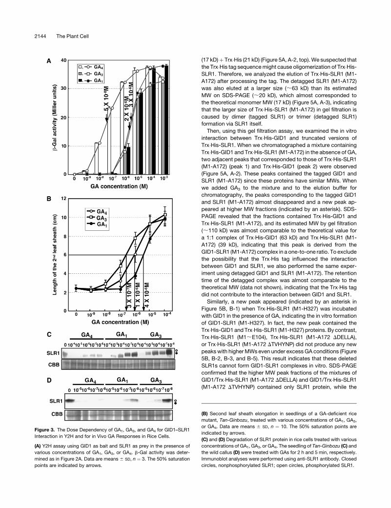
Comparison of GA Selectivity and GA Dose Dependency
Measured in Yeast and in Planta
Previously, we observed that GID1 preferentially binds to GA4
in vitro compared with GA3, although the physiological activity of
GA4 in leaf sheath elongation is lower than that of GA3 (Ueguchi-
Tanaka et al., 2005). The discrepancy between the in vitro GA
preference of GID1 and the physiological effectiveness of GAs
in planta suggests the presence of an unknown mechanism that
differentiates between in vitro and in planta events. To clarify this
point, we reexamined the GA preference of GID1 using Y2H
assays. Figure 2 shows the effects of various GAs (10�5 M) on the
GID1–SLR1 interaction in yeast cells (Figure 2A) and the dose
dependency of second leaf sheath elongation in seedlings of a
GA-deficient rice mutant, Tan-Ginbozu, after a one-drop treat-
ment of various GAs (Figure 2B). The one-drop treatment of GAs
on Tan-Ginbozu seedlings is a classic bioassay for estimation
Molecular Interaction of GID1 and SLR1 2141

of GA levels because Tan-Ginbozu seedlings sensitively and
quantitatively respond to the exogenously added GA (Nishijima
et al., 1993). The trend of effectiveness for various GAs in the
GID1–SLR1 interaction in yeast and for leaf sheath elongation of
Tan-Ginbozu was almost identical except for GA3 and GA9. For
example, GA4 and H2-GA4 showed the high activities in both
assays, that is, GA4 and H2-GA4 are the highest and the second
highest in yeast, respectively, and GA4 and H2-GA4 are the
second and third highest for leaf sheath elongation, respectively.
GA51 and GA4-Me showed the lowest activities in both assays.
On the other hand, GA3 was the most effective GA for leaf sheath
elongation but showed intermediate activity in the Y2H system,
whereas GA9 showed higher activity in the Y2H assay than in the
leaf sheath elongation experiment.
We also examined the dose dependency of the GID1–SLR1
interaction in yeast cells with respect to GA1, GA3, and GA4 (Fig-
ure 3A). GA4 showed the highest effectiveness of the three. The
interaction between GID1 and SLR1 was observed when GA4
was present at 10�9 M or higher concentrations, whereas the
interaction was observed at >10�7 M of GA1 and GA3. The strength
of the interaction increased as the GA concentration increased
and reached a plateau at ;10�5 M for GA4 and ;10�4 M for GA1
and GA3. The results indicate that the strength of the GID1–SLR1
interaction depends on the GA concentration. The 50% satura-
tion point of each GA was ;5 3 10�8 M for GA4, 2 3 10�6 M for
GA3, and 3 3 10�6 M for GA1. Then, we compared these GA dose
responses of the GID1–SLR1 interaction with those for leaf
sheath elongation of Tan-Ginbozu seedlings (Figure 3B). The
50% saturation in leaf sheath elongation was estimated to be
;1 3 10�6 M for GA4, 3 3 10�7 M for GA3, and 4 3 10�6 M for
GA1. Thus, in the case of GA4, the GA response of the GID1–
SLR1 interaction in yeast cells was more sensitive than the re-
sponse in planta. On the other hand, in the case of GA3, the
sensitivity of leaf sheath elongation was higher than that of the
GID1–SLR1 interaction, whereas both sensitivities to GA1 were
almost similar, with that of yeast being a little higher. Again, the
higher level of effectiveness of GA4 than GA3 in the GID1–SLR1
interaction in yeast cells is not consistent with lower physiolog-
ical activity of GA4 than GA3 in Tan-Ginbozu seedlings.
We predicted that the higher response in leaf sheath elonga-
tion to GA3 may be due to its stability in planta, whereas GA4 may
be rapidly inactivated in planta by GA-inactivating enzymes. To
test this prediction, we examined the GA dose response for SLR1
degradation in rice seedlings and callus, which is one of the
quickest events in response to GA treatment and therefore may
be less disturbed by GA-inactivating enzymes. Furthermore, we
observed that GA-inactivating enzymes are not expressed in rice
callus (M. Ueguchi-Tanaka, unpublished results). As we ex-
pected, the most effective GA in SLR1 degradation was GA4 in
both experiments (Figures 3C and 3D). The results clearly dem-
onstrate that GA4 is the most effective GA among GA1, GA3, and
GA4 in rice cells in the absence of GA-inactivating enzymes. The
reason why GA4 is more effective than GA3 in stimulating DELLA
Figure 1. GA-Dependent Interaction between GID1 and SLR1 in Vivo.
(A) SLR1 was coimmunoprecipitated with GFP-GID1 in a GA-dependent
manner by the GFP antibody. Each protein extract was prepared from
two independent lines of transgenic rice callus overproducing GFP-GID1
(line 1 and line 2) treated with (þ) or without (�) 10�5 M GA4 for 5 min.
Immunoblot analysis of the extract and a-GFP immunoprecipitates was
performed using anti-GFP or anti-SLR1 antibody. Closed circles, non-
phosphorylated SLR1; open circles, phosphorylated SLR1.
(B) BiFC analysis of in vivo interaction between GID1 and SLR1 in
N. benthamiana leaf epidermis (Abe et al., 2005). BF, blight-field image;
EYFP, EYFP fluorescence; DAPI, 49,6-diamidino-2-phenylindole; merge,
merge of EYFP and DAPI images; NY-GID1, expression of N�EYFP-GID1
alone; CY-SLR1, expression of C�EYFP-SLR1 alone; NY-GID1 and CY-
SLR1, coexpression of N�EYFP-GID1 and C�EYFP-SLR1; NY-GID1 and
CY-DDELLA, coexpression of N�EYFP-GID1 and C�EYFP-DDELLA�SLR1;
NYGID1 and CY-DTVHYNP, coexpression of N�EYFP-GID1 and C�EYFP-
DTVHYNP�SLR1. Leaves were sprayed with (þ) or without (�) 10�4 M GA4
10 min before observation of the signals. Bar ¼ 10 mm.
2142 The Plant Cell

protein degradation, even in the leaf sheath where GA3 is more
active than GA4, for its elongation can be explained as follows.
Exogenous GA application should cause the induction of some
GA-inactivating enzymes under the control of GA signaling, re-
sulting in enhanced GA degradation activity (Sakai et al., 2003). In
this context, GA4, which can be inactivated by GA-inactivating
enzymes, should be rapidly degraded in GA-treated cells; con-
sequently, its effect should be eased. However, as GA3 is not
inactivated by inactivating enzymes, GA3 can remain as an active
form in cells even after GA treatment.
These results also indicate that the GA selectivity of GID1, as
estimated by the GID1–SLR1 interaction in yeast cells, is almost
the same as that in rice cells, at least with regard to the most
important active GAs, GA1, GA3, and GA4. However, there is one
small discrepancy, that is, the relatively higher interacting activity
of GA9 in the Y2H system compared with its activity in leaf sheath
elongation, which must be due to some unknown mechanism. It
is possible that GA9 might be metabolized to form the active GA
in yeast.
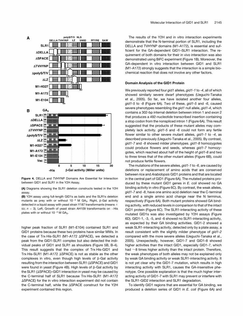
Domain Analysis of the SLR1 Protein
To investigate the GID1 binding domain of SLR1, we analyzed
SLR1 to identify essential domains for GID1 interaction in Y2H
assays. First, we used a deletion series of the SLR1 cDNA as
prey constructs (Figure 4A). Both plate and liquid assays showed
that deletion of either the DELLA (DDELLA) or the TVHYNP
(DTVHYNP) domain completely abolished the GA-dependent
GID1-interacting activity of SLR1, while deletion of the space
region (DSPACE) or the Ser/Thr-rich region (Dpoly S/T/V) did not
affect its activity (Figure 4B). Deletion of the Leu zipper domain
(DLZ) decreased the binding activity but did not diminish activity
completely. Deletion of the C-terminal half of SLR1 (M1-H327),
which included the most conserved regions of the GRAS family,
such as the VHIID, PFYRE, and SAW domains, did not cause a
complete loss of GID1 interaction; approximately one-third of the
activity of the full-length SLR1 was retained. Deletion of 1.4 kb of
the C-terminal region (M1-A172) led to an almost complete loss
of the b-galactosidase (b-Gal) activity in the liquid assay, but the
plate assay still showed that the GA-dependent interaction be-
tween the proteins occurred. Further deletion of the C-terminal
side, including the TVHYNP domain (M1-E104), abolished growth
on GA-containing plates. These results demonstrate that the
DELLA and TVHYNP domains are essential for interaction of
SLR1 with GID1, while the C-terminal side from the poly S/T/V
region is not.
Furthermore, we analyzed the in vitro formation of a GA-GID1-
SLR1 complex by gel filtration on Superdex-200 (Figure 5). For
this analysis, we first attempted to produce the full-length re-
combinant SLR1 with a Trx�His tag, but we could not produce
sufficient quantities of the full-length SLR1 protein. DSLR1s with
C-terminal truncations, such as Trx�His-SLR1 (M1-H327) and
Trx�His-SLR1 (M1-A172), were produced in sufficient quantities,
so we used these soluble, recombinant proteins for the in vitro
experiment. The internal deletions of SLR1, such as DDELLA,
DSPACE, and DTVHYNP, were produced from Trx�His-SLR1
(M1-A172).
Trx�His-GID1 was eluted from the column in the expected
fractions (;63 kD) corresponding to its molecular weight (MW)
estimated by SDS-PAGE (;65 kD) and the theoretical MW (GID1
[39 kD] þ Trx�His [21 kD]) (Figure 5A, A-1). On the other hand,
Trx�His-SLR1 (M1-A172) was eluted at a much larger size (;85
kD) than its estimated MW on SDS-PAGE (;40 kD), which
corresponded to the theoretical monomer SLR1 (M1-A172)
Figure 2. The Effect of Various GAs on GID1–SLR1 Interaction in Yeast
Cells or on Second Leaf Sheath Elongation.
(A) Y2H assay using GID1 as bait and SLR1 as prey in the presence of
10�5 M various GAs. b-Gal activity was determined by a liquid assay with
yeast strain Y187 transformants (means 6 SD; n ¼ 3). H2-GA4, 16,17-
dihydro-GA4; GA4-Me, GA4 methyl ester; BL, blank.
(B) Dose dependency of second leaf sheath elongation to various GAs
in seedlings of a GA-deficient rice mutant, Tan-Ginbozu, by one-drop
treatment (see Methods). The line colors correspond to those in (A). Data
are means 6 SD; n ¼ 10.
Molecular Interaction of GID1 and SLR1 2143
(17 kD)þ Trx�His (21 kD) (Figure 5A, A-2, top). We suspected that
the Trx�His tag sequence might cause oligomerization of Trx�His-
SLR1. Therefore, we analyzed the elution of Trx�His-SLR1 (M1-
A172) after processing the tag. The detagged SLR1 (M1-A172)
was also eluted at a larger size (;63 kD) than its estimated
MW on SDS-PAGE (;20 kD), which almost corresponded to
the theoretical monomer MW (17 kD) (Figure 5A, A-3), indicating
that the larger size of Trx�His-SLR1 (M1-A172) in gel filtration is
caused by dimer (tagged SLR1) or trimer (detagged SLR1)
formation via SLR1 itself.
Then, using this gel filtration assay, we examined the in vitro
interaction between Trx�His-GID1 and truncated versions of
Trx�His-SLR1. When we chromatographed a mixture containing
Trx�His-GID1 and Trx�His-SLR1 (M1-A172) in the absence of GA,
two adjacent peaks that corresponded to those of Trx�His-SLR1
(M1-A172) (peak 1) and Trx�His-GID1 (peak 2) were observed
(Figure 5A, A-2). These peaks contained the tagged GID1 and
SLR1 (M1-A172) since these proteins have similar MWs. When
we added GA3 to the mixture and to the elution buffer for
chromatography, the peaks corresponding to the tagged GID1
and SLR1 (M1-A172) almost disappeared and a new peak ap-
peared at higher MW fractions (indicated by an asterisk). SDS-
PAGE revealed that the fractions contained Trx�His-GID1 and
Trx�His-SLR1 (M1-A172), and its estimated MW by gel filtration
(;110 kD) was almost comparable to the theoretical value for
a 1:1 complex of Trx�His-GID1 (63 kD) and Trx�His-SLR1 (M1-
A172) (39 kD), indicating that this peak is derived from the
GID1-SLR1 (M1-A172) complex in a one-to-one ratio. To exclude
the possibility that the Trx�His tag influenced the interaction
between GID1 and SLR1, we also performed the same exper-
iment using detagged GID1 and SLR1 (M1-A172). The retention
time of the detagged complex was almost comparable to the
theoretical MW (data not shown), indicating that the Trx�His tag
did not contribute to the interaction between GID1 and SLR1.
Similarly, a new peak appeared (indicated by an asterisk in
Figure 5B, B-1) when Trx�His-SLR1 (M1-H327) was incubated
with GID1 in the presence of GA, indicating the in vitro formation
of GID1-SLR1 (M1-H327). In fact, the new peak contained the
Trx�His-GID1 and Trx�His-SLR1 (M1-H327) proteins. By contrast,
Trx�His-SLR1 (M1;E104), Trx�His-SLR1 (M1-A172 DDELLA),
or Trx�His-SLR1 (M1-A172 DTVHYNP) did not produce any new
peaks with higher MWs even under excess GA conditions (Figure
5B, B-2, B-3, and B-5). This result indicates that these deleted
SLR1s cannot form GID1-SLR1 complexes in vitro. SDS-PAGE
confirmed that the higher MW peak fractions of the mixtures of
GID1/Trx�His-SLR1 (M1-A172 DDELLA) and GID1/Trx�His-SLR1
(M1-A172 DTVHYNP) contained only SLR1 protein, while the
Figure 3. The Dose Dependency of GA1, GA3, and GA4 for GID1–SLR1
Interaction in Y2H and for in Vivo GA Responses in Rice Cells.
(A) Y2H assay using GID1 as bait and SLR1 as prey in the presence of
various concentrations of GA1, GA3, or GA4. b-Gal activity was deter-
mined as in Figure 2A. Data are means 6 SD, n ¼ 3. The 50% saturation
points are indicated by arrows.
(B) Second leaf sheath elongation in seedlings of a GA-deficient rice
mutant, Tan-Ginbozu, treated with various concentrations of GA1, GA3,
or GA4. Data are means 6 SD, n ¼ 10. The 50% saturation points are
indicated by arrows.
(C) and (D) Degradation of SLR1 protein in rice cells treated with various
concentrations of GA1, GA3, or GA4. The seedling of Tan-Ginbozu (C) and
the wild callus (D) were treated with GAs for 2 h and 5 min, respectively.
Immunoblot analyses were performed using anti-SLR1 antibody. Closed
circles, nonphosphorylated SLR1; open circles, phosphorylated SLR1.
2144 The Plant Cell
higher peak fraction of SLR1 (M1-E104) contained SLR1 and
GID1 proteins because these two proteins have similar MWs. In
the case of Trx�His-SLR1 (M1-A172 DSPACE), we detected a
peak from the GID1-SLR1 complex but also detected the indi-
vidual peaks of GID1 and SLR1 as shoulders (Figure 5B, B-4).
This result suggests that the complex of Trx�His-GID1 and
Trx�His-SLR1 (M1-A172 DSPACE) is not as stable as the other
complexes in vitro, even though high levels of b-Gal activity
resulting from the interaction between SLR1 (DSPACE) and GID1
were found in yeast (Figure 4B). High levels of b-Gal activity by
the SLR1 (DSPACE)–GID1 interaction in yeast may be caused by
the C-terminal half of SLR1 because Trx�His-SLR1 (M1-A172
DSPACE) for the in vitro interaction experiment did not contain
the C-terminal half, while the DSPACE construct for the Y2H
experiment contained this region.
The results of the Y2H and in vitro interaction experiments
demonstrate that the N-terminal portion of SLR1, including the
DELLA and TVHYNP domains (M1-A172), is essential and suf-
ficient for the GA-dependent GID1–SLR1 interaction. The re-
quirement of both domains for their in vivo interaction was also
demonstrated using BiFC experiment (Figure 1B). Moreover, the
GA-dependent in vitro interaction between GID1 and SLR1
(M1-A172) strongly suggests that the interaction is a simple bio-
chemical reaction that does not involve any other factors.
Domain Analysis of the GID1 Protein
We previously reported four gid1 alleles, gid1-1 to -4, all of which
showed similarly severe dwarf phenotypes (Ueguchi-Tanaka
et al., 2005). So far, we have isolated another four alleles,
gid1-5 to -8 (Figure 6A). Two of these, gid1-5 and -6, caused
severe phenotypes resembling the gid1 null allele, gid1-4, which
contains a 302-bp internal deletion between intron 1 and exon 2
that produces a 492-nucleotide transcribed insertion containing
a stop codon from the nonspliced intron 1 (Figure 6A). This result
suggested that the products of these mutant alleles may com-
pletely lack activity. gid1-5 and -6 could not form any fertile
flower similar to other severe mutant alleles, gid1-1 to -4, as
described previously (Ueguchi-Tanaka et al., 2005). By contrast,
gid1-7 and -8 showed milder phenotypes. gid1-8 homozygotes
could produce flowers and seeds, whereas gid1-7 homozy-
gotes, which reached about half of the height of gid1-8 and two
to three times that of the other mutant alleles (Figure 6B), could
not produce fertile flowers.
The mutations of the severe alleles, gid1-1 to -6, are caused by
deletions or replacement of amino acids that are conserved
between rice and Arabidopsis GID1 proteins and that are located
in the central part of GID1 (Figure 6A). The mutated proteins pro-
duced by these mutant GID1 genes in E. coli showed no GA
binding activity in vitro (Figure 6C). By contrast, the weak alleles,
gid1-7 and -8, have one amino acid deletion near the C-terminal
end and a single amino acid change near the N terminus,
respectively (Figure 6A). Both mutant proteins showed GA bind-
ing activity, with reduced levels in comparison to that of the intact
GID1 protein (Figure 6C). The SLR1-interacting activity of these
mutated GID1s was also investigated by Y2H assays (Figure
6D). GID1-1, -3, -5, and -6 showed no SLR1-interacting activity,
as expected by their GA binding activities. GID1-2 showed a
weak SLR1-interacting activity, detected only by a plate assay, a
result consistent with the slightly milder phenotype of gid1-2
compared with the more severe alleles (Ueguchi-Tanaka et al.,
2005). Unexpectedly, however, GID1-7 and GID1-8 showed
higher activities than the intact GID1, especially GID1-7, which
had ;8 times higher activity than the intact protein. Therefore,
the weak phenotypes of both alleles may not be explained only
by weak GA binding activity or weak SLR1-interacting activity. It
is not yet clear why the GID1-7 mutation, which results in high
interacting activity with SLR1, causes the GA-insensitive phe-
notype. One possible explanation is that the much higher inter-
acting activity of GID1-7 with SLR1 may prevent or interfere with
the SLR1-GID2 interaction and SLR1 degradation.
To identify GID1 regions that are essential for GA binding, we
produced a deletion series of GID1 in E. coli (Figure 6A) and
Figure 4. DELLA and TVHYNP Domains Are Essential for Interaction
between GID1 and SLR1 in the Y2H Assay.
(A) Diagrams showing the SLR1 deletion constructs tested in the Y2H
assay.
(B) Y2H assay using full-length GID1s as baits and the SLR1s deletion
mutants as prey with or without 10�4 M GA3. Right, b-Gal activity
detected in a liquid assay with yeast strain Y187 transformants (means 6
SD; n ¼ 3). Left, Growth of yeast strain AH109 transformants on �His
plates with or without 10�4 M GA3.
Molecular Interaction of GID1 and SLR1 2145

examined GA binding activities in vitro (Figure 6E). GA binding
was reduced to ;25% of the activity of the full-sized GID1
protein when the 15 N-terminal amino acids (DN1) were deleted.
Further deletion of 58 and 98 N-terminal residues (DN2 and DN3,
respectively) caused complete loss of GA binding activity, as did
the deletion of 30 C-terminal amino acids (DC). The SLR1-
interacting activity of these deleted GID1s was also investigated
by Y2H assays (Figure 6F). The results were essentially the same
as those of the in vitro GA binding experiment. These results
demonstrate that most parts of the GID1 protein, apart from the
15 N-terminal amino acids, are necessary for its activity. Other-
wise, the protein structure and conformation of almost all of GID1
is important for its activity.
Next, we performed an Ala scanning experiment. We selected
all conserved amino acid residues among the rice and three
Arabidopsis GID1 proteins (see Supplemental Figure 3 online)
and packed three or less contiguous conserved amino acids as
one block. Then, these conserved amino acids in each block
were exchanged with Ala residues. Consequently, we produced
94 mutagenized GID1 proteins carrying exchanged Ala residues
in each conserved block (Figure 7). We used these mutagenized
GID1s to analyze the GA binding and GA-dependent GID1–SLR1
interaction activities.
There were 12 blocks essential for GA binding activity (shown
by hatched boxes in Figure 7A). These blocks were scattered
throughout the GID1 molecule. This result is consistent with the
above prediction that almost the entire GID1 protein is important
for its GA binding activity. As previously mentioned, GID1 shares
sequence similarity with the consensus sequence for the HSL
family. When we draw attention to the residues shared with well-
conserved motifs in the HSL family, the HGG and GXSXG motifs
(indicated by HGG and GDSSG in Figure 7B, respectively) were
also important for GA binding of GID1, but the last two residues of
the GXSXG motif, S and G, were exchangeable with Ala without
loss of GA binding activity. Furthermore, one of the conserved
Figure 5. In Vitro Interaction between GID1 and SLR1 via DELLA and
TVHYNP Domains.
(A) Left: elution profiles of Trx�His-GID1, Trx�His-SLR1(M1-A172), the
mixture of Trx�His-GID1 and Trx�His-SLR1(M1-A172) in the absence or
presence of 10�4 M GA3, and detagged SLR1 (M1-A172) by Superdex-
200 gel filtration. Trx�His-GID1 was eluted as a monomer, while SLR1
(M1-A172) with or without the tag was eluted at much larger MW
fractions. The MW of the peak fractions was estimated from the following
molecular markers (M.M.): 25-kD chymotrypsinogen A, 43-kD ovalbu-
min, 67-kD albumin, and 134-kD albumin dimer, which are indicated by
arrowheads at the top. Dashed lines indicate the peak positions of
Trx�His-SLR1 (M1-A172) (;85 kD) and Trx�His-GID1 (;65 kD). Peaks
1 and 2 indicate overlapping peaks of the mixture of Trx�His-GID1 and
Trx�His-SLR1 (M1-A172) in the absence of GA3. The asterisk indicates a
new peak of the same mixture in the presence of GA3 with disappear-
ance of peaks 1 and 2. Each peak fraction was subjected to SDS-PAGE.
Right: SDS-PAGE of each peak fraction.
(B) Left: elution profiles of various kinds of mutant Trx�His-SLR1s and
their mixtures incubated with Trx�His-GID1 in the presence of 10�4 M
GA3 by Superdex-200 gel filtration. The tightly dashed line indicates the
peak position of Trx�His-GID1 (tagged GID1; ;63 kD). The roughly
dashed lines indicate the peak position of each Trx�His-SLR1 mutant
protein. Peaks shifted by incubation with GID1 and GA3 are indicated by
asterisks, and peaks not shifted by incubation are indicated by open
arrowheads. These peaks were subjected to SDS-PAGE. Right: SDS-
PAGE of each peak fraction. The molecular marker for gel filtration and
SDS-PAGE are the same as in (A).
2146 The Plant Cell

Figure 6. Eight gid1 Mutant Alleles and GA Binding and SLR1-Interacting Activities of Mutated GID1 Proteins.
(A) A schematic structure of GID1 represents the mutation positions of eight gid1 alleles and the positions of N-terminal (DN1, DN2, and DN3) and
C-terminal (DC) deletions. Amino acid residues shared with HSL, such as HGG and GXSXG, are presented within red boxes. The residues
corresponding to the catalytic triad of HSL, S, D, and V, are also presented by filled circles.
(B) Gross morphology of eight gid1 mutant alleles grown for 2 weeks. A wild-type plant is shown as a control. Bar ¼ 5 cm.
(C) Top: GA binding activities of Trx�His-GID1 (wild type) and the corresponding mutated gid1 alleles. Data are means 6 SD; n ¼ 3. Bottom: Coomassie
blue control. Approximately equal amounts of proteins (;3.2 mg) were used.
(D) Y2H assay using full-length GID1 and the corresponding mutated gid1 alleles as baits and the full-length SLR1 as prey in the presence (þ) and
absence (�) of 10�4 M GA3. The Y2H assay was performed the same as in Figure 4B. Top: b-Gal activity (means 6 SD; n¼ 3). Bottom: growth of yeast on
�His plates.
(E) Top: GA binding activities of Trx�His-GID1 and mutated Trx�His-GID1s with deletions in the N-terminal (DN1, DN2, and DN3) or C-terminal (DC)
regions. Data are means 6 SD; n ¼ 3. Bottom: Coomassie blue control. Approximately equal amounts of proteins (;3.2 mg) were used.
(F) Y2H assay using full-length GID1 and mutated GID1s with deletions in the N-terminal (DN1, DN2, and DN3) or C-terminal (DC) regions as baits and
the full-length SLR1 as prey in the presence (þ) and absence (�) of 10�4 M GA3. The Y2H assay was performed the same as in Figure 4B. Top: b-Gal
activity (means 6 SD; n ¼ 3). Bottom: growth of yeast on �His plates.

amino acid residues forming the catalytic triad of HSL, D296
(indicated by D in Figure 7B), was also exchangeable with Ala.
These results demonstrate that not all the conserved regions
shared with HSL proteins are important for GA binding activity.
There were 13 blocks important for GID1–SLR1 interaction
(shown by dotted boxes in Figure 7B). All regions essential for GA
binding were included in these blocks, confirming that GA
binding to GID1 is a prerequisite for GID1–SLR1 interaction. On
the other hand, there are eight blocks that carried GA binding but
not GID1–SLR1 interaction activity (shown by arrows in Figure
7B). These regions are considered to be involved in the interac-
tion with SLR1 but not necessary for GA binding. These eight
blocks are classified into two groups, that is, one is adjacent to
the region essential for GA binding (indicated by thick arrows),
and another is located independently from any other region
(indicated by narrow arrows). Such high proximity to the regions
essential for SLR1 interaction and GA binding suggests that GA
binding and SLR1 interaction may be overlapping on the GID1
molecule. Some Ala exchanges increased the GA binding activ-
ity. For example, exchange of Cys-270 caused ;4 times higher
GA binding activity relative to the intact GID1 (indicated by boxes
in Figure 7A). Some residues, whose replacement also caused
higher GA binding activity, such as Pro-114–Val-115, His-205,
and Val-246–Thr-247, are adjacent to regions essential for GA
binding (indicated by boxes in Figure 7A). Similarly, a few Ala
exchanges, such as Val-148–Val-150, Lys-287–Leu-289, and
Met-341 (indicated by arrowheads in Figure 7B), caused higher
GID1–SLR1 interaction, yet these replacements did not affect
their GA binding activity. This result indicates that higher GID1–
SLR1 interaction by these exchanges is not due to increased GA
binding but is due to increased interaction between GID1 and
SLR1. Such a phenomenon was also seen in the GID1-7 mutant
protein, namely, this mutant GID1 protein showed >8 times
higher interaction activity with SLR1 relative to the wild-type
GID1 without increasing its GA binding activity (Figures 6C and
6D). It is noteworthy that the mutation site of gid1-7 is located at
Glu-343, a one–amino acid deletion, whereas the Ala exchange
at Met-341 caused a >10 times increase in the GID1–SLR1
interaction. Taken together, it is possible that there is a hot spot
region around Met-341–Glu-343 involving the formation and/or
stability of the GID1-SLR1 complex.
Structure-Function Relationship of GID1
The Ala scanning experiment revealed that the amino acid
residues essential for these activities are scattered throughout
the GID1 molecule. We expected that such irregular placement
of the important residues is reflected in the stereostructure of
GID1. Therefore, we predicted the secondary structure of GID1
by the Jpred program (Cuff et al., 1998) (Figure 8A). As described
previously, the primary structure of GID1 resembles that of pro-
teins in the HSL group. To date, the secondary and tertiary struc-
tures of three bacterial esterases in the HSL family, Brefeldin A
esterase, Archaeoglobus fulgidus esterase (AFEST), and Alicy-
clobacillus acidocaldarius esterase 2 (EST2), have been char-
acterized by the x-ray diffraction analyses (Wei et al., 1999;
Figure 7. Ala Scanning Analysis of GID1 for Its GA Binding and SLR1-Interacting Activities.
(A) Specific GA binding activity of 94 mutated Trx�His-GID1s and wild-type Trx�His-GID1 (at the left). Mutated residues are indicated at the top of each
bar. Specific activity was calculated as radioactivity (dpm) per microgram of protein. Data are means 6 SD; n ¼ 3. Hatched boxes indicate the mutant
proteins showing no GA binding activity. The mutated proteins showing increased activity that are discussed in the text are boxed.
(B) SLR-interacting activity of 94 mutated GID1s and the wild-type GID1 (at the left). Y2H assay using 94 mutated and wild-type GID1s as baits and the
full-length SLR1 as prey in the presence of 10�4 M GA3. Each mutation of GID1 corresponds to (A). Dotted boxes indicate the mutant proteins showing
no b-Gal activity. Thick and narrow arrows indicate mutant proteins having GA binding activity but not SLR1-interacting activity, whose mutation points
are adjacent to the hatched box (thick) or independently located (narrow). The mutated proteins having increased SLR1-interacting activity that are
discussed in the text are indicated by arrowheads. HGG and GDSSG correspond to the substrate binding pocket of HSL, and D corresponds to one of
the residues in the catalytic triad in HSL.
2148 The Plant Cell

Figure 8. Prediction of the GID1 Secondary Structure, Location of Important Residues for GA Binding, and SLR1 Interaction Activity on the Predicted
Structure.
(A) The alignment of GID1 and a member of HSL, AFEST, whose special conformation was analyzed by x-ray crystallography. The line of AFEST 2nd
shows the secondary structure of AFEST analyzed by x-ray crystallography (De Simone et al., 2001). The predicted second structure of GID1 (GID1
Jpred) was calculated by a Jpred program (Cuff et al., 1998). a-helices and b-sheets are represented by a and b, respectively. Number of a-helices and
Molecular Interaction of GID1 and SLR1 2149

De Simone et al., 2000, 2001). The stereostructures of these
proteins are similar to each other; therefore, proteins belonging
to the HSL family have been considered to form a similar spatial
conformation. Thus, based on an analogy between GID1 and
AFEST, we compared a secondary structure of AFEST (AFEST
2nd in Figure 8A; De Simone et al., 2001) with the predicted
secondary structure of GID1 (GID1 Jpred in Figure 8A). The
structures of AFEST and the predicted GID1 are quite similar to
each other with the exception of the N-terminal region that cor-
responds to the lid region known as a variable region in proteins
of the HSL family. Most notably, the skeletal structure of HSL,
composed with the canonical a/b hydrolase fold consisting of a
parallel b-sheet surrounded on both sides by a-helices, is
completely shared with the predicted structure of GID1. Further-
more, the predicted structure of GID1 contains two a-helices
between b6 and b7 sheets (Figure 8B), corresponding to the right
side of the lid structure, whereas the left side of the lid corre-
sponds to the N-terminal region based on an analogy with the
HSL structure. Thus, it is very possible that GID1 has the canon-
ical a/b hydrolase fold with the lid structures similar to the HSL
group proteins (Figure 8B). For enzymatic activity of HSL, the
conserved HGG and catalytic triad, S, D, and H, are spatially lo-
cated in close proximity to form a substrate binding pocket and
catalytic site, respectively (Naridini and Dijkstra, 1999). In the
predicted structure of GID1, the amino acids corresponding to
these residues are located in close proximity at the upper side
loops, while one amino acid of the catalytic triad, H, in HSL is
replaced with V in GID1.
Then, we positioned the amino acid residues important for GA
binding (black dots and squares identified by Ala scanning and
spontaneous mutants, respectively) and SLR1-interacting activ-
ity (gray dots and squares identified by Ala scanning and spon-
taneous mutants, respectively) on this predicted structure of
GID1 (Figure 8B). Interestingly, many residues important for GA
binding and for SLR1 interaction are localized around the regions
corresponding to the substrate binding pocket and lid regions in
HSL. The lid structure of HSL is known to be involved in substrate
specificity since it is moveable (Naridini and Dijkstra, 1999).
Preferential localization of these amino acid residues in the sub-
strate binding pocket and lid regions leads us to speculate that
GID1 may interact with GA and SLR1 at this substrate binding
pocket region in collaboration with the lid region in a similar
fashion to HSL substrate interaction.
Stabilization of GA Binding to GID1 by SLR1
The possibility of overlapping SLR1-interacting and GA binding
domains on the GID1 molecule led us to speculate that the
binding properties between GID1 and GA may be affected by
SLR1. Therefore, we compared the GA binding activity of GID1
with and without SLR1. The GA binding activity of Trx�His-GID1
was enhanced approximately threefold by GST-SLR1, whereas
the GST tag alone was ineffective and GST-SLR1 alone did not
show any GA binding (Figure 9A). We also measured the as-
sociation and dissociation rates between Trx�His-GID1 and3H-16,17-dihydro-GA4 (Figures 9B and 9C). Although the half-
time of the association was not affected by GST-SLR1 (Figure 9B),
the dissociation rate markedly increased in the presence of
GST-SLR1 (Figure 9C). These results support the above idea that
the interaction between GID1 and GA is stabilized by SLR1.
These results also support the possibility that GA binding with
GID1 may induce the SLR1 interaction with GID1; in turn, the inter-
acting SLR1 covers the GA bound to GID1 and stabilizes the
GID1–GA interaction (see Discussion).
DISCUSSION
In this article, we have studied the molecular mechanism for
GA binding to GID1 and the mechanism of the GA-dependent
interaction between GID1 and SLR1 protein. For this purpose, we
used a Y2H assay because of its ease and reliability. To dem-
onstrate the validity of this Y2H assay, we examined the in vivo
GA-dependent GID1–SLR1 interaction by in vivo pull-down
(Figure 1A) and BiFC experiments (Figure 1B). We also compared
the GA selectivity of GID1 determined by the GID1–SLR1 inter-
action in yeast cells and the GA effectiveness in leaf sheath
elongation to SLR1 degradation in rice seedling and callus (Fig-
ures 2 and 3). These results confirmed that the GA-dependent
GID1–SLR1 interaction in yeast is reflective of the GA-dependent
events occurring in rice plants.
The consistency in GA selectivity and GA dose dependency
determined by the three different experiments, in vitro GA binding
of GID1 (Ueguchi-Tanaka et al., 2005), Y2H assay monitoring the
GID1–SLR1 interaction, and GA-dependent events occurring in
rice plants, indicates that GA actions in rice cells are quantitatively
and qualitatively reflected by the biochemical properties of the
GID1 molecule through its quantitative interaction with SLR1.
GA Preference of GID1
The GA selectivity of GID1 determined by Y2H and the GA ef-
fectiveness to SLR1 degradation in rice seedling and callus dem-
onstrated that GA4 is the most favored GA, although GA4 was not
the most effective GA for leaf sheath elongation probably be-
cause of its rapid inactivation by GA-catabolizing enzymes. Our
recent results demonstrate that the GA preference of three
Arabidopsis GA receptors, At GID1s, is similar to that of rice GID1
(Nakajima, et al., 2006). Similar GA preference between rice and
Arabidopsis GID1s suggests that the GID1 proteins both in
monocot and dicot plants, and therefore perhaps all GID1s, may
Figure 8. (continued).
b-sheets of the predicted GID1 was according to that of AFEST 2nd. Essential residues for GA binding (gray) and SLR1 interaction (black) were
determined by an Ala scanning experiment (dot) or spontaneous mutants, gid1-1 (G196/D), gid1-2 (R251/T), and gid1-5 (G169/E) (square). Three
amino acid residues corresponding to the catalytic triad of HSL are represented as asterisks.
(B) A topology diagram of GID1 written by the predicted second structure in (A). Symbols are the same as in (A). The regions of the lid and binding
pocket described in text are colored in light green and pink, respectively.
2150 The Plant Cell

have similar GA preferences. If so, it is curious that the major
bioactive GA is different in each plant species. For example, rice
uses GA1 as the major bioactive GA at the vegetative stage,
whereas Arabidopsis uses GA4. In the case of rice, the major
bioactive GA also depends on its developmental stages. In the
vegetative stage of rice, GA1 is the major GA, while, in the
reproductive stage, the amount of endogenous GA4 is precisely
regulated by the developmental stage (Luo et al., 2006; Zhu et al.,
2006). For example, in pollen, an extremely high amount of GA4
accumulates (Kobayashi et al., 1988). Since drastic GA-dependent
reactions, such as rapid elongation of the heading stem and
pollen tube, occur during the reproductive stage, rice may use
the most effective GA at this stage and use GA1 for ordinary GA-
dependent reactions at the vegetative stage. Further studies will
be necessary to explain this curious discrepancy between the
common GA preference of GID1 and the choice of major bioac-
tive GA in various plant species.
Domain Analysis of SLR1 Protein
The results of the Y2H assays (Figure 4) and in vitro interaction
experiments (Figure 5) clearly demonstrate that the N-terminal
portion of SLR1, including the DELLA and TVHYNP domains, is
essential and sufficient for the GA-dependent GID1–SLR1 inter-
action. Moreover, we showed by the BiFC experiment that these
domains are also necessary for in vivo GA-dependent interaction
between GID1 and SLR1 (Figure 1B). Recently, two groups
performed Y2H experiments using Arabidopsis DELLA proteins
and At GID1a, with inconsistent results (Griffiths et al., 2006;
Willige et al., 2007). Griffiths et al. (2006) reported that the DELLA
and VHYNP domains of RGA are both necessary for At GID1a
binding, whereas Willige et al. (2007) reported that the VHYNP
domain of GAI is not needed for GA-dependent At GID1a inter-
action and proposed that the DELLA domain functions as a
receiver domain for the GA receptor. Our results from using
truncated versions of SLR1 are consisted with the observations
of Griffiths et al. (2006) with RGA. It is an interesting possibility
that there might be two different types of DELLA proteins in
plants: one type, such as SLR1 and RGA, requiring both the
DELLA and TVHYNP/VHYNP domains for GID1 interaction, and
the other, including GAI, requiring only the DELLA domain.
Further studies, using not only Y2H assays but also in vitro and
in vivo interaction experiments, will be necessary to identify the
domain(s) of DELLA proteins that is essential for GID1 interaction
in Arabidopsis and other plants.
Domain Analysis of the GID1 Protein and a Model of the
GID1-GA-SLR1 complex
By comparing the Ala scanning analysis of GID1 and the muta-
tion analysis of gid1 alleles with the GID1 structure predicted by
analogy with the HSL tertiary structure, it appeared that many
residues important for GA binding and for SLR1 interaction are
localized around the regions corresponding to the substrate
binding pocket and lid regions in HSL. Based on these results, we
predicted the interacting model of the GID1-GA-SLR1 complex
(Figure 10). In this model, the structure of the GID1 receptor
resembles those of the HSL proteins. GID1 has the regions
Figure 9. GA Binding Activity of GID1 Is Enhanced by SLR1.
(A) GA binding activity of Trx�His-GID1 with (black) and without (white)
the full length of GST-SLR1 protein (means 6 SD; n ¼ 3). Approximately
equal amounts of proteins (;3.2 mg) of Trx�His-GID1, Trx�His vec, GST-
SLR1, and GST vector were used.
(B) Association kinetics of 3H-16,17-dihydro-GA4 and Trx�His-GID1 with
(closed symbols) and without (open symbols) the full length of GST-
SLR1, represented as percentage of the values reached after 60 min of
reaction. Data are means 6 SD; n ¼ 3.
(C) Dissociation kinetics of 3H-16,17-dihydro-GA4 and Trx�His-GID1 with
(closed symbols) or without (open symbols) GST-SLR1, represented as a
percentage of the value detected at 0 min. Data are means 6 SD; n ¼ 3.
Figure 10. Molecular Model for Formation of the GA-GID1-SLR1 Com-
plex.
See text for details.
Molecular Interaction of GID1 and SLR1 2151

corresponding to the substrate binding pocket and lid like the
bacterial HSLs. Similarly to the mechanism for substrate binding
in other HSLs, GID1 binds GA within the binding pocket with the
aid of the lid. SLR1 interacts with the GID1-GA complex at its
N-terminal region, including the DELLA and TVHYNP domains.
Both the lid and the binding pocket containing GA are necessary
for the SLR1 interaction, and as a result of SLR1 binding, the
GID1-GA complex is stabilized.
Our results showing the stabilization of the GA–GID1 interac-
tion by SLR1 (Figure 9C) strongly support this model and suggest
that the interaction of SLR1 with GA-GID1 might close the lid,
thereby ensuring that GA will be held in the substrate pocket. The
stabilized complex of GA, GID1, and SLR1 may be targeted by
GID2, an F-box protein, leading to its degradation by 26S pro-
teasomes through ubiquitination of the SCFGID2 complex. In fact,
recent results from Y3H assays indicated that the GA-GID1 com-
plex promoted the interaction between an Arabidopsis DELLA
protein, RGA, and an F-box protein, SLY1 (Griffiths et al., 2006).
Further studies, including a structural analysis of GID1, are
necessary to support this model.
METHODS
Plant Materials, Growth Conditions, and Screening of the gid1
Mutant Alleles
The new alleles of gid1 were obtained by screening three rice (Oryza
sativa) libraries mutagenized by g-irradiation, by cell culture followed by
regeneration, and by N-methyl-N-nitrosourea. We first screened GA-
related mutants showing dwarfism and wide dark-green leaves. Then,
GA-insensitive mutants were screened by the GA treatment. After se-
quencing the GID1 gene, we found four new alleles: gid1-5, from O. sativa
cv Nipponbare mutagenized by g-irradiation; gid1-6 and gid1-7 from
O. sativa cv Nipponbare mutagenized by cell culture; and gid1-8 from
O. sativa cv Taichung 65 mutagenized by N-methyl-N-nitrosourea. Rice
plants were grown in a greenhouse at 308C (day) and 248C (night).
GA Treatment of Rice Seedlings and Callus
For the GA response tests by a one-drop treatment, sterilized Tan-Ginbozu
seeds (Itoh et al., 2004) were germinated on solid half-strength Murashige
and Skoog medium. After 2 d, 1 mL of GA (10�6, 10�5, 10�4, and 10�3 M; in
50% acetone) was added to the joint between the coleoptile and leaf
sheath of each seedling. For the control, 1 mL of 50% acetone was added.
The length of the second leaf sheaths was measured 6 d later. For the GA
dose–response tests, sterilized seeds of Tan-Ginbozu (Itoh et al., 2004)
were germinated in various concentrations of GA solution and the length of
the second leaf sheaths was measured 8 d later. Young seedlings that had
been grown in the greenhouse for 2 weeks were used for the detection of
GA-dependent degradation of the SLR1 protein in shoots. Old leaves were
removed so that the seedling retained only the newest leaf blade and
sheath and the second newest leaf sheath. The seedlings were incubated
in various concentrations of GA solution containing 0.02% Tween 20 for
2 h at room temperature with gentle shaking. For the control, 0.1% ethanol
solution containing 0.02% Tween 20 was used. After 2 h, the seedlings
were collected and frozen at�808C until used for protein gel blot analysis.
Callus of wild-type rice (Taichung 65) was used for the detection of GA-
dependent degradation of the SLR1 protein in callus. The callus was
transferred to new N6D solid medium. After 3 d, the callus was treated with
various concentrations of GA solution, containing 0.02% Tween 20, for
5 min at room temperature. The callus was then immediately frozen at
�808C until it was used for protein gel blot analysis.
Plasmid Construction
Sequences of primers used in this study are listed in Supplemental Table
1 online. The PCR fragments were sequenced to confirm that no muta-
tions were induced. For the Y2H assay, pGADT7 (Clontech) and pGBKT7
(Clontech) were used as expression vectors. The mutated GID1 fragment
corresponding to gid1-1, -2, -3, -5, -6, -7, and -8 were produced by RT-
PCR using total RNA from seedlings of gid1 mutant alleles, with Y2H/
GID1f and Y2H/GID1r primers, and cloned into the NcoI-SmaI site of the
pGBKT7 vector. The fragments corresponding to the N-terminal and
C-terminal deletions of GID1 were produced by PCR with appropriate
primers having NcoI and SmaI sites and cloned into the NcoI-SmaI site of
the pGBKT7 vector. The fragments for internal deletions of SLR1, DDELLA,
DSPACE, DTVHYNP, DpolyS/T/V, and DLZ, were produced by PCR using
a corresponding deletion series of SLR1 cDNA, previously made in a
binary vector (Itoh et al., 2002), as templates with Y2H-SLR1f and Y2H-
SLR1r primers and cloned into the NdeI-EcoRI site of the pGADT7 vector.
The fragments for the C-terminal deletion of SLR1, M1-H327, M1-A172,
and M1-E104, were produced by PCR with Y2H-SLR1f and appropriate
reverse primers and cloned into the NdeI-EcoRI sites of pGADT7. A full-
length GID1 cDNA inserted into the pGBKT7 vector and a full-length SLR1
cDNA inserted in the pGADT7 vector were described previously (Ueguchi-
Tanaka et al., 2005).
To construct Trx�His tagged proteins, except for the Ala-exchanged
Trx�His -GID1 protein, we used the pDEST�Trx vector that is derived from
pET32a (Novagen) for the Gateway system (Tsunoda et al., 2005). The PCR
fragments, amplified with primers having a appropriate site for the Gateway
system were cloned into the entry vector of pENTR/SD/D-TOPO (Invitro-
gen) and recombined into the destination vector of pDEST�Trx according to
the Gateway instruction manual (Invitrogen). For the construction of the
mutated Trx�His-GID1s and the mutated GID1s in the pGBKT7 vector for
the Ala scanning experiment, a full-length wild-type GID1 was amplified
with GID1Alaf and GID1Alar primers and cloned into the pCR4 Blunt-TOPO
vector. Then, PCR was performed against the full-length GID1 cDNA with
one set of mutagenized primers corresponding to each mutation. The
parental methylated and hemimethylated DNA in the PCR reaction mixture
was digested with DpnI, and the mutated GID1 cDNA that could not be
digested with DpnI was transformed into Escherichia coli XL10-Gold
(Stratagene). After sequencing, each mutated GID1 cDNA was digested
with NcoI-EcoRI or BamHI-HindIII and cloned into pGBKT7 and the pET32a
vector, respectively. For construction of GST-SLR1, the cDNA was inserted
into the pGEX-4T vector (GE Healthcare) at the EcoRI target site. For the
construction of GFP-GID1 in the pActNos/Hm2 vector, the GFP fragment
was amplified using the GFP cDNA as a template and the GFP-f and GFP-r
primers and inserted into the GID1 cDNA at the NcoI target site, which is
located just before the initial ATG codon. The GFP-GID1 construct was
digested with SmaI and cloned into pActNos/Hm2 at the SmaI target site.
For the insertion of GFP-GID1 into the pET32a vector, the 2.0-kb GFP-GID1
fragment was amplified using the pActNos/Hm2 construct described
above as a template and the GFP-GID1-f and GFP-GID1-r primers. The
fragment was cloned into pCR4 Blunt-TOPO (Invitrogen), generating GFP-
GID1/pCR4 Blunt-TOPO. The GFP-GID1 fragment was then removed from
GFP-GID1/pCR4 Blunt-TOPO by digestion with BamHI and HindIII and
then inserted into the pET32a vector at the BamHI and HindIII target sites.
For constructs used in the BiFC experiment, the N-terminal half of the
EYFP clone without a stop codon and the C-terminal half of the EYFP
clone without a stop codon were kindly provided by T. Araki. To construct
N�EYFP-GID1 (NY-GID1), the 0.5-kb fragment of the N-terminal half of
EYFP was produced using the N-terminal half of the EYFP clone without a
stop codon as a template with BiFC-5 and BiFC-6 primers and cloned into
the XbaI-SmaI site of the pUC18 vector (Takara), generating NY/pUC18.
The 1.2-kb fragment of GID1 was produced using a full-length GID1 cDNA
as a template with BiFC-15 and BiFC-16 primers and cloned into pCR4
Blunt-TOPO (Invitrogen), generating BiFC-GID1. NY/pUC18 was digested
2152 The Plant Cell

with SmaI and SacI and ligated to a 1.2-kb SmaI-SacI fragment of
BiFC-GID1 to generate NY-GID1/pUC18. NY-GID1/pUC18 was digested
with XbaI and SacI, and the 1.7-kb XbaI-SacI fragment was inserted into
the XbaI-SacI site of pBI121 (Toyobo), generating NY-GID1. To construct the
C�EYFP-SLR1s (CY-SLR1s) containing full-length or truncated SLR1,
the 0.3-kb fragment of the C-terminal half of EYFP was produced using
the C-terminal half of the EYFP clone without a stop codon as a template
with BiFC-7 and BiFC-8 primers and cloned into the XbaI-SmaI site of the
pBluescript II SKþ vector (Stratagene), generating CY/pBS. The SLR1,
DDELLA-SLR1, and DTVHYNP-SLR1 fragments were amplified from a
full-length SLR1 cDNA, or corresponding truncated SLR1s in the pGADT7
vector, using the BiFC-13 and BiFC-14 primers. The fragments were
cloned into the pCR4 Blunt-TOPO vector, generating the BiFC-SLR1s.
CY/pBS was digested with SmaI, and the CY fragment was ligated to the
SmaI fragment of the BiFC-SLR1s in the correct orientation, generating
CY-SLR1s/pBS. pBI121 was digested with SacI, blunted with T4 DNA
polymerase (Takara), and digested with XbaI. XbaI-EcoRV fragments
from the CY-SLR1/pBS plasmids were inserted into pBI121 at the XbaI
and blunt-end SacI sites to generate the CY-SLR1s.
Production of Recombinant Protein
E. coli BL21 (DE3) pLysS Rosseta-gami 2 (Novagen) was used as a
host strain for the production of each recombinant protein. To produce
recombinant Trx�His-GIDs for use in GA binding assays, 5 mL of
precultured cells were added to 500 mL of Luria-Bertani (LB) medium in
a 2-liter flask, or 1.5 mL of precultured cells were added to 150 mL of LB
medium in a 500-mL flask for the Ala scanning experiment and cultured at
378C until OD600 0.4 to 0.6. Recombinant proteins were induced by the
addition of 0.01 mM isopropyl-b-D-thiogalactopyranoside (IPTG) and
further incubated at 188C for 18 h. Cells were harvested and resuspended
with buffer A containing 50 mM Tris-HCl, pH 8.0, 100 mM NaCl, 10 mM
imidazole, and 0.1% Triton X-100. The cells were lysed by sonication (20
kHz, 5 s 3 40 times). Or for the Ala scanning experiment, the cells were
lysed by addition of the detergent BugBuster protein extraction reagent
(Novagen) with benzonase nuclease (Benzon Pharma) according to the
instruction manual. Each lysate was centrifuged at 16,000g for 30 min,
and the supernatants were mixed usually with 400 or 100 mL (for the Ala
scanning experiment) of TALON metal affinity resin (Clontech) and rotated
for 2 h at 48C. The resin was washed five times with buffer A and eluted five
times with 400 mL usually or 100 mL (for the Ala scanning experiment) of
500 mM imidazole in buffer A. The five eluates were pooled and desalted
using a PD-10 column (GE Healthcare) equilibrated with buffer B contain-
ing 20 mM Tris-HCl, pH 7.5, 0.15 M NaCl, and 2 mM 2-mercaptoethanol
(2-ME) or dialyzed against 2 liters of buffer B (for the Ala scanning ex-
periment) for 18 h. For production of recombinant Trx�His-GIDs for use in
the gel filtration analyses, the cell culture and induction were performed
the same as the recombinant Trx�His-GIDs for the GA binding assays;
however, a different purification method was used because the gel fil-
tration analysis needed more highly purified proteins. Cells were harves-
ted and resuspended with buffer C containing 20 mM Tris-HCl, pH 7.5,
0.5 M NaCl, and 2 mM 2-ME. The cells were lysed by sonication (20 kHz,
10 s 3 20 times), the lysates were centrifuged at 16,000g for 30 min, and
the supernatants were loaded directly onto 5 mL of Ni-chelating HP (GE
Healthcare) equilibrated with buffer C and eluted by increasing the imid-
azole concentration to 0.5 M at a flow rate of 5 mL per min. The eluate was
dialyzed against buffer C and loaded onto a Hi-Load 26/60 Superdex 200
pg column (GE Healthcare). Purified recombinant proteins were analyzed
by immunoblotting. The production method for recombinant Trx�His-
SLR1s to use in gel filtration analyses was the same as for Trx�His-GID1
used for gel filtration analysis. Purified recombinant proteins were ana-
lyzed by immunoblotting and subjected to matrix-assisted laser desorp-
tion ionization–time of flight–mass spectrometry for measurement of MW.
For the production of recombinant GST and full-length GST-SLR1, the
cell culture and induction were performed the same as Trx�His-GID1 for
the GA binding experiment, except 0.4 mM IPTG was used instead of 0.01
mM IPTG. Cells were harvested and resuspended with buffer D contain-
ing 50 mM Tris-HCl, pH 8.0, 50 mM NaCl, 1 mM EDTA, and 1 mM DTT.
The cells were lysed by sonication (20 kHz, 5 s 3 40 times) with 1% Triton X-
100. The lysates were centrifuged at 16,000g for 30 min, and the super-
natants were mixed with 2 mL of Glutathione Sepharose 4B beads (GE
Healthcare) and rotated for 2 h at 48C. The beads were washed five times
with PBS containing 1% Triton X-100 and eluted five times with 400 mL of
20 mM glutathione in buffer D. The five eluates were pooled and desalted
using a PD-10 column (GE Healthcare) equilibrated with buffer E con-
taining 20 mM Tris-HCl, pH 7.5, and 2 mM 2-ME and further purified by a
MonoQ 5/50 GL (GE Healthcare) column equilibrated with buffer E and
eluted by increasing the NaCl concentration to 0.5 M at a flow rate of 0.5
mL per min. The peak fraction corresponding to full-length GST-SLR1
was collected, identified by immunoblotting, and used for in vitro binding
experiments.
GA Binding Assay
In vitro assays for GA binding were performed as described previously
except that Trx�His-GID1 and its derivatives were used instead of GST-
GID1 that was used in the previous article (Ueguchi-Tanaka et al., 2005).
The GA binding activity of Trx�His-GID1 was almost the same as that of
GST-GID1 in vitro (data not shown). In the case of the Ala scanning
experiment, 100 mL of each mutated Trx�His-GID1 in buffer B was added
to 300 mL of reaction mixture. The amount of each mutated protein was
determined from the band intensity of SDS-PAGE using 0.25, 1, and 4 mg
of BSA as a quantity standard (see Supplemental Figure 3 online), and the
GA binding activity was represented as a radioactivity (dpm) per micro-
gram of protein. For quantification of the protein from the SDS-PAGE
profile, NIH Image software was used.
The effect of SLR1 on GA binding of GID1 was studied by a GA binding
assay. Purified Trx�His-GID1 proteins were dissolved in 200 mL binding
buffer (20 mM Tris-HCl, pH 7.6, 0.1 M NaCl, and 2 mM 2-ME) and incu-
bated with 3H-16,17-dihydro-GA4 (6 pmol) either without excess unla-
beled GA4 for total binding or with an 833-fold excess of unlabeled GA4 for
nonspecific binding. The total volume of the reaction mixture was 300 mL.
After 15 min of incubation, 100 mL of GST-SLR1 proteins were added to
each reaction tube. After incubation for 45 min, radioactivity of the tri-
tiated 16,17-dihydro-GA4 bound to GST-GID1 was measured. As a
control, GST protein produced from the empty GST vector was added
instead of GST-SLR1.
To determine the association rate of GA binding, purified Trx�His-GID1
proteins (16 mg) were dissolved in 250 mL of binding buffer; 100 mL of the
purified GST-SLR1 (16 mg) or the vector control (GST) were added, and
the solution was incubated with 50 mL of 3H-16,17-dihydro-GA4 (6 pmol)
for various periods. For measurement of the dissociation rate of GA
binding, the association reaction was performed for 20 min before an 833-
fold excess of unlabeled GA4 was added, followed by incubation for
various periods. After incubation, radioactivity of the tritiated 16,17-
dihydro-GA4 bound to Trx�His-GID1 was measured.
Y2H Assay and Pull-Down Assay
The Y2H assay was performed as described previously (Ueguchi-Tanaka
et al., 2005). The yeast strain Y187 was used for detection of b-Gal activity
by liquid assay, and the strain AH109 was used for growth tests on �His
plates with or without GA. The basis for the pull-down assay was to detect
if SLR1-GST could bind Trx�His-GID1 that was immobilized on TALON
metal affinity resin. For the pull-down assay, 2 mg of Trx�His-GID1 was
incubated with 1 mg of GST-SLR1 at 308C for 15 min in 300 mL of binding
Molecular Interaction of GID1 and SLR1 2153

buffer (20 mM Tris-HCl, pH 7.6, 2.5 mM 2-ME, and 0.1 M NaCl) with or
without 10�4 M GA4. After incubation, 20 mL of TALON metal affinity resin
was added. After further incubation at 308C for 15 min, the resin was
washed five times with washing buffer (20 mM Tris-HCl, pH 7.5, 500 mM
NaCl, and 0.5% Tween 20). The washing buffer also contained 10�4 M
GA4 where appropriate. After washing, 10 mL of 23 SDS sample buffer
were added, the mixture was heat-denatured, and the sample was loaded
on a 7.5% SDS-PAGE gel, and proteins were visualized by Coomassie
Brilliant Blue staining. As negative controls, samples without GST-SLR1
or Trx�His-GID1 were prepared.
Coimmunoprecipitation
Transgenic rice calli overexpressing GFP-GID1 were treated with 10�5 M
GA4 in 0.1% ethanol and 0.2% Tween 20 or the same solvent as a control
for 5 min. The callus was harvested and ground in liquid nitrogen, and the
proteins were extracted by adding 500 mL of buffer F (20 mM Tris-HCl,
pH 8.0, 150 mM NaCl, 1 mM EDTA, 0.5% Tween 20, and 13 Complete
tablet [Roche]). The extracts were then centrifuged at 16,000g for 30 min
at 48C. For each pull-down assay, 20 mL of anti-GFP antibodies raised in
rabbit were added to 500 mL of the extract and rotated for 2 h at 48C
followed by the addition of 30 mL of equilibrated protein A agarose and
further rotated for 1 h. The protein A agarose was washed five times with
buffer G (20 mM Tris-HCl, pH 8.0, 500 mM NaCl, 1 mM EDTA, 0.5%
Tween 20, and 13 Complete tablet [Roche]), and 23 SDS sample buffer
was added and loaded on a 7.5% SDS-PAGE gel. The protein on the gel
was transferred to the membrane and analyzed by immunoblot analysis
using mouse anti-GFP antibodies or rabbit anti-SLR1 antibody. During all
procedures, 10�4 M GA4 was added to the assay mixture for GA4-treated
calli and was not added to the protein samples from calli not treated with
GA4. Coomassie Brilliant Blue staining was used to confirm equal loading.
Infiltration of Nicotiana benthamiana Leaf Epidermal Cells
N. benthamiana plants were grown under continuous light for ;1 month
at 268C. A. tumefaciens strain GV3101-pM90 containing an appropriate
construct was grown at 308C for 2 d in LB medium containing 50 mg/mL of
kanamycin and 50 mg/mL of rifampicin. One milliliter of cultured cells was
transferred to 10 mL of LB medium containing 15 mM acetosyringone, 50
mg/mL of kanamycin, and 50 mg/mL of rifampicin and further cultured at
308C to the early stationary phase. Bacterial cells were harvested by
centrifugation and suspended in 10 mM MgCl2 and 150 mM acetosy-
ringone in 10 mM MES buffer, pH 5.6, and left for 2 h at room temperature.
The bacterial suspension was infiltrated into the abaxial air spaces of a
leaf using a 1-mL syringe. Plants were kept for 2 d after infiltration in
continuous light conditions at 268C. EYFP and DAPI signals in leaf
epidermal cells were observed using fluorescence microscopy (Olympus
BX51) with U-MYFPHQ and U-MWU2 filter units, respectively.
Gel Filtration Analysis
Purified Trx�His-GID1 and Trx�His-SLR1 derivatives were mixed in a 1:1
ratio (mole/mole) estimated by each absorbance at 280 nm and incubated
at 48C for 30 min with or without 10�4 M GA3 in buffer F (20 mM Tris-HCl,
pH 7.5, 150 mM NaCl, and 2 mM 2-ME). One hundred microliters of the
reaction mixture (50 to 100 mg of total protein) was applied to a Superdex
200 10/300 GL column (GE Healthcare) equilibrated with buffer F and
eluted with the same buffer (610�4 M GA3) at a flow rate of 0.4 mL per
minute at 48C. Fractions (0.5 mL) were collected, and each peak fraction
was analyzed by SDS-PAGE. The column was calibrated using low
molecular weight markers (GE Healthcare): 25 kD chymotrypsinogen A,
43 kD ovalbumin, 67 kD albumin, and 134 kD albumin dimer. The MWs of
Trx�His-GID1 and Trx�His-SLR1 derivatives were estimated from the stan-
dard curve plotted from the elution volumes of these standard proteins.
Accession Numbers
GenBank/EMBL accession numbers and Arabidopsis Genome Initiative
locus identifiers for the genes mentioned in this article are as follows: GID1
(AB211399), SLR1 (AB262980), GID1a (At3g05120), GID1b (At3g63010),
and GID1c (At5g27320).
Supplemental Data
The following materials are available in the online version of this article.
Supplemental Figure 1. GID1 Interacts with SLR1 in a GA4-Dependent
Manner.
Supplemental Figure 2. Recombinant GFP-GID1 Interacts with GST-
SLR1 in a GA4-Dependent Manner in Vitro.
Supplemental Figure 3. Comparison of Amino Acid Sequences of
Rice and Arabidopsis.
Supplemental Figure 4. Loading Control for the GA Binding Exper-
iment Using Mutagenized GID1 Proteins with Conserved Amino Acids
Replaced with Ala.
Supplemental Table 1. Primers Used in This Study.
ACKNOWLEDGMENTS
We thank Yasufumi Daimon and Takashi Araki of Kyoto University for
providing the vector and technical advice about BiFC as well as Hirofumi
Yoshioka and Yoshihiro Kobae of Nagoya University for technical advice
about fluorescent microscopy and infiltration of N. benthamiana leaf
epidermal cells. We thank Ryoko Kigaku, Mayuko Kawamura, and
Hitomi Kihara for expert technical assistance. This work was supported
in part by the Ministry of Education, Culture, Sports, Science, and
Technology of Japan (Grant-in-Aid for Scientific Research [M.M., I.Y.,
M.N, M.U.-T., and H.K.] and Target Proteins Research Program [M.M.])
and by a grant from the Ministry of Agriculture, Forestry, and Fisheries of
Japan (Green Technology Project IP-1003; M.A. and M.M.).
Received April 26, 2007; revised June 28, 2007; accepted June 30, 2007;
published July 20, 2007.
REFERENCES
Abe, M., Kobayashi, Y., Yamamoto, S., Daimon, Y., Yamaguchi, A.,
Ikeda, Y., Ichinoki, H., Notaguchi, M., Goto, K., and Araki, T.
(2005). FD, a bZIP protein mediating signals from the floral pathway
integrator FT at the shoot apex. Science 309: 1052–1056.
Chandler, P.M., Marion-Poll, A., Ellis, M., and Gubler, F. (2002).
Mutants at the Slender1 locus of barley cv Himalaya: Molecular and
physiological characterization. Plant Physiol. 129: 181–190.
Cuff, J.A., Clamp, M.E., Siddiqui, A.S., Finlay, M., and Barton, G.J.
(1998). Jpred: A consensus secondary structure prediction server.
Bioinformatics 14: 892–893.
Davies, P.J. (1995). Plant Hormones. (Dordrecht, The Netherlands:
Kluwer Academic Publishers).
De Simone, G., Galdiero, S., Manco, G., Lang, D., Rossi, M., and
Pedone, C. (2000). A snapshot of a transition state analogue of a
novel thermophilic esterase belonging to the subfamily of mammalian
hormone-sensitive lipase. J. Mol. Biol. 303: 761–771.
De Simone, G., Menchise, V., Manco, G., Mandrich, L., Sorrentino,
N., Lang, D., Rossi, M., and Pedone, C. (2001). The crystal structure
2154 The Plant Cell
of a hyper-thermophilic carboxylesterase from the archaeon Archae-
oglobus fulgidus. J. Mol. Biol. 314: 507–518.
Griffiths, J., Murase, K., Rieu, I., Zentella, R., Zhang, Z.-L., Powers,
S.J., Gong, F., Phillips, A.L., Hedden, P., Sun, T.-P., and Thomas,
S.G. (2006). Genetic characterization and functional analysis of the
GID1 gibberellin receptors in Arabidopsis. Plant Cell 18: 3399–3414.
Ikeda, A., Ueguchi-Tanaka, M., Sonoda, Y., Kitano, H., Koshioka, M.,
Futsuhara, Y., Matsuoka, M., and Yamaguchi, J. (2001). slender
rice, a constitutive gibberellin response mutant is caused by a null
mutation of the SLR1 gene, an ortholog of the height-regulating gene
GAI/RGA/RHT/D8. Plant Cell 13: 999–1010.
Itoh, H., Sasaki, A., Ueguchi-Tanaka, M., Ishiyama, K., Kobayashi,
M., Hasegawa, Y., Minami, E., Ashikari, M., and Matsuoka, M.
(2005). Dissection of the phosphorylation of rice DELLA protein,
SLENDER RICE1. Plant Cell Physiol. 46: 1392–1399.
Itoh, H., Tatsumi, T., Sakamoto, T., Otomo, K., Toyomasu, T.,
Kitano, H., Ashikari, M., Ichihara, S., and Matsuoka, M. (2004). A
rice semi-dwarf gene, Tan-Ginbozu (D35), encodes the gibberellin
biosynthesis enzyme, ent-kaurene oxidase. Plant Mol. Biol. 54:
533–547.
Itoh, H., Ueguchi-Tanaka, M., Sato, Y., Ashikari, M., and Matsuoka,
M. (2002). The gibberellin signaling pathway is regulated by the
appearance and disappearance of SLENDER RICE1 in nuclei. Plant
Cell 14: 57–70.
Iuchi, S., Suzuki, H., Kim, Y.-C., Iuchi, A., Kuromori, T., Ueguchi-
Tanaka, M., Asami, T., Yamaguchi, I., Matsuoka, M., Kobayashi,
M., and Nakajima, M. (2007). Multiple loss-of-function of Arabidopsis
gibberellin receptor AtGID1s completely shuts down a gibberellin
signal. Plant J. 50: 958–966.
Kobayashi, M., Yamaguchi, I., Murofushi, N., Ota, Y., and Takahashi,
N. (1988). Fluctuation and localization of endogenous gibberellins in
rice. Agric. Biol. Chem. 52: 1189–1194.
Luo, A., et al. (2006). EUI1, encoding a putative cytochrome P450
monooxygenase, regulates internode elongation by modulating gib-
berellin responses in rice. Plant Cell Physiol. 47: 181–191.
McGinnis, K.M., Thomas, S.G., Soule, F.D., Strader, L.C., Zale, J.M.,
Sun, T.-p., and Steber, C.M. (2003). The Arabidopsis SLEEPY1 gene
encodes a putative F-box subunit of an SCF E3 ubiquitin ligase. Plant
Cell 15: 1120–1130.
Nakajima, M., et al. (2006). Identification and characterization of
Arabidopsis gibberellin receptors. Plant J. 46: 880–889.
Naridini, M., and Dijkstra, B.W. (1999). a/b Hydrolase fold enzymes:
The family keeps growing. Curr. Opin. Struct. Biol. 9: 732–737.
Nishijima, T., Koshioka, M., and Yamazaki, H. (1993). A highly-
sensitive rice seedling bioassay for the detection of femtomole quan-
tities of 3b-hydroxylated gibberellins. Plant Growth Regul. 13: 241–247.
Osterlund, T. (2001). Structure-function relationships of hormone-
sensitive lipase Eur. J. Biochem. (Tokyo) 268: 1899–1907.
Peng, J., Carol, P., Richards, D.E., King, K.E., Cowling, R.J., Murphy,
G.P., and Harberd, N.P. (1997). The Arabidopsis GAI gene defines a
signaling pathway that negatively regulates gibberellin responses. Genes
Dev. 11: 3194–3205.
Peng, J., et al. (1999). ‘Green revolution’ genes encode mutant gibber-
ellin response modulators. Nature 400: 256–261.
Sakai, M., Sakamoto, T., Saito, T., Matsuoka, M., Tanaka, H., and
Kobayashi, M. (2003). Expression of novel rice gibberellin 2-oxidase
gene is under homeostatic regulation by biologically active gibberellins.
J. Plant Res. 116: 161–164.
Sasaki, A., Itoh, H., Gomi, K., Ueguchi-Tanaka, M., Ishiyama, K.,
Kobayashi, M., Jeong, D.H., An, G., Kitano, H., Ashikari, M., and
Matsuoka, M. (2003). Accumulation of phosphorylated repressor for
gibberellin signaling in an F-box mutant. Science 299: 1896–1898.
Silverstone, A.L., Ciampaglio, C.N., and Sun, T.-p. (1998). The
Arabidopsis RGA gene encodes a transcriptional regulator repressing
the gibberellin signal transduction pathway. Plant Cell 10: 155–169.
Tsunoda, Y., Sakai, N., Kikuchi, K., Katoh, S., Akagi, K., Miura-O., J.,
Tashiro, Y., Murata, K., Shibuya, N., and Katoh, E. (2005). Improv-
ing expression and solubility of rice proteins produced as fusion
proteins in Escherichia coli. Protein Expr. Purif. 42: 267–277.
Ueguchi-Tanaka, M., Ashikari, M., Nakajima, M., Itoh, H., Katoh, E.,
Kobayashi, M., Chow, T.-Y., Hsing, Y.-i., Kitano, H., Yamaguchi, I.,
and Matsuoka, M. (2005). GIBBERELLIN INSENSITIVE DWARF1
encodes a soluble receptor for gibberellin. Nature 437: 693–698.
Wei, Y., Contreras, J.A., Sheffield, P., Osterlund, T., Derewenda, U.,
Kneusel, R.E., Matern, U., Holm, C., and Derewenda, Z.S. (1999).
Crystal structure of brefeldin A esterase, a bacterial homolog of the
mammalian hormone-sensitive lipase. Nat. Struct. Biol. 6: 340–345.
Willige,B.C., Ghosh, S.,Nill, C., Zourelidou,M.,Dohmann,E.M.N., Maier,
A., and Schwechheimer, C. (2007). The DELLA domain of GA INSEN-
SITIVE mediates the interaction with the GA INSENSITIVE DWARF1A
gibberellin receptor of Arabidopsis. Plant Cell 19: 1209–1220.
Yeaman, S.J. (2004). Hormone-sensitive lipase-new roles for an old
enzyme. Biochem. J. 379: 11–22.
Zhu, Y., et al. (2006). ELONGATED UPPERMOST INTERNODE encodes
a cytochrome P450 monooxygenase that epoxidizes gibberellins in a
novel deactivation reaction in rice. Plant Cell 18: 442–456.
Molecular Interaction of GID1 and SLR1 2155

DOI 10.1105/tpc.106.043729; originally published online July 20, 2007; 2007;19;2140-2155Plant Cell
Saji, Xiang Hongyu, Motoyuki Ashikari, Hidemi Kitano, Isomaro Yamaguchi and Makoto MatsuokaMiyako Ueguchi-Tanaka, Masatoshi Nakajima, Etsuko Katoh, Hiroko Ohmiya, Kenji Asano, Shoko
SLR1, and GibberellinMolecular Interactions of a Soluble Gibberellin Receptor, GID1, with a Rice DELLA Protein,
This information is current as of November 14, 2018
Supplemental Data /content/suppl/2007/07/03/tpc.106.043729.DC1.html
References /content/19/7/2140.full.html#ref-list-1
This article cites 29 articles, 11 of which can be accessed free at:
Permissions https://www.copyright.com/ccc/openurl.do?sid=pd_hw1532298X&issn=1532298X&WT.mc_id=pd_hw1532298X
eTOCs http://www.plantcell.org/cgi/alerts/ctmain
Sign up for eTOCs at:
CiteTrack Alerts http://www.plantcell.org/cgi/alerts/ctmain
Sign up for CiteTrack Alerts at:
Subscription Information http://www.aspb.org/publications/subscriptions.cfm
is available at:Plant Physiology and The Plant CellSubscription Information for
ADVANCING THE SCIENCE OF PLANT BIOLOGY © American Society of Plant Biologists