Embed Size (px)
DESCRIPTION
Molecular Hydrogen in Drinking Water Protects againstNeurodegenerative Changes Induced by Traumatic BrainInjury
Citation preview

Molecular Hydrogen in Drinking Water Protects againstNeurodegenerative Changes Induced by Traumatic BrainInjuryKenji Dohi1,2, Brian C. Kraemer1,3,4, Michelle A. Erickson1, Pamela J. McMillan4,5, Andrej Kovac1,3,
Zuzana Flachbartova6, Kim M. Hansen1,3, Gul N. Shah7, Nader Sheibani8, Therese Salameh1,3,
William A. Banks1,3*
1Geriatric Research Education and Clinical Center, Veterans Affairs Puget Sound Health Care System, Seattle, WA, United States of America, 2Department of Emergency
Medicine, The Jikei University School of Medicine, Tokyo, Japan, 3Division of Gerontology and Geriatric Medicine, Department of Medicine, University of Washington,
Seattle, WA, United States of America, 4Mental Illness Research Education and Clinical Center, Veterans Affairs Puget Sound Health Care System, Seattle, WA, United
States of America, 5Department of Psychiatry and Behavioral Sciences, University of Washington, Seattle, WA, United States of America, 6 Laboratory of Biomedical
Microbiology and Immunology, Department of Microbiology and Immunology, University of Veterinary Medicine and Pharmacy, Kosice, Slovakia, 7Division of
Endocrinology, Department of Internal Medicine, Saint Louis University School of Medicine, Edward A. Doisy Research Center, St. Louis, MO, United States of America,
8Opthalmology and Visual Sciences, University of Wisconsin School of Medicine and Public Health, Madison, WI, United States of America
Abstract
Traumatic brain injury (TBI) in its various forms has emerged as a major problem for modern society. Acute TBI cantransform into a chronic condition and be a risk factor for neurodegenerative diseases such as Alzheimer’s and Parkinson’sdiseases, probably through induction of oxidative stress and neuroinflammation. Here, we examined the ability of theantioxidant molecular hydrogen given in drinking water (molecular hydrogen water; mHW) to alter the acute changesinduced by controlled cortical impact (CCI), a commonly used experimental model of TBI. We found that mHW reversed CCI-induced edema by about half, completely blocked pathological tau expression, accentuated an early increase seen inseveral cytokines but attenuated that increase by day 7, reversed changes seen in the protein levels of aquaporin-4, HIF-1,MMP-2, and MMP-9, but not for amyloid beta peptide 1–40 or 1–42. Treatment with mHW also reversed the increase seen4 h after CCI in gene expression related to oxidation/carbohydrate metabolism, cytokine release, leukocyte or cell migration,cytokine transport, ATP and nucleotide binding. Finally, we found that mHW preserved or increased ATP levels and proposea new mechanism for mHW, that of ATP production through the Jagendorf reaction. These results show that molecularhydrogen given in drinking water reverses many of the sequelae of CCI and suggests that it could be an easily administered,highly effective treatment for TBI.
Citation: Dohi K, Kraemer BC, Erickson MA, McMillan PJ, Kovac A, et al. (2014) Molecular Hydrogen in Drinking Water Protects against NeurodegenerativeChanges Induced by Traumatic Brain Injury. PLoS ONE 9(9): e108034. doi:10.1371/journal.pone.0108034
Editor: Tsuneya Ikezu, Boston University School of Medicine, United States of America
Received May 23, 2014; Accepted August 18, 2014; Published September 24, 2014
This is an open-access article, free of all copyright, and may be freely reproduced, distributed, transmitted, modified, built upon, or otherwise used by anyone forany lawful purpose. The work is made available under the Creative Commons CC0 public domain dedication.
Data Availability: The authors confirm that all data underlying the findings are fully available without restriction. All relevant data are within the paper.
Funding: KD was supported by Kiban C grant number 23592683. WAB was supported by VA Merit Review; RO-1 AG029839. WAB and GNS was supported by RO-1 DK083485. BCK was supported by VA Merit Review; R0-1 NS064131. The funders had no role in study design, data collection and analysis, decision to publish, orpreparation of the manuscript.
Competing Interests: The authors have declared that no competing interests exist.
* Email: [email protected]
Introduction
Traumatic brain injury (TBI) has emerged as a signature injury
of the early 21st century. In addition to blast injuries in theaters of
war, TBI in the civilian population takes many forms with falls
recently supplanting automobile accidents as the most common
cause of TBI [1,2]. Injuries that penetrate the skull are more rare
but a very serious form of TBI [3]. Although TBI arises from a
variety of injuries, the clincial endpoints that result and the
mechanisms that drive the CNS towards those endpoints are
thought to be shared not only among TBI’s, but also by a host of
neurodegenerative diseases [4,5]. These endpoints and mecha-
nisms include brain edema, tauopathy, blood-brain barrier (BBB)
disruption and dysfunction, and neuroinflammation [6,7,8]. Once
set in motion, these processes often become self-reinforcing unless
the cycle can be disrupted. Thus, preventative and interventive
therapeutics are needed to treat TBI as early as possible.
Ideally, these treatments could be given immediately, even at
the time of injury. This would require the therapeutic to have low
toxicity and to be easily administered. One such potential
therapeutic is molecular hydrogen. First introduced by Ohsawa
et al in 2007 [9], molecular hydrogen acts at least in part as an
anti-oxidant, combining with hydroxyl ions that are produced by
CNS injuries, although other mechanisms may also exist [9]. As a
low molecular weight (2 Da), uncharged molecule, molecular
hydrogen has the ability to penetrate biological membranes. The
membranes penetrated would include those that form the BBB;
thus, molecular hydrogen should have unrestricted access to the
CNS, a characteristic shared with few potential therapeutics. This
high membrane penetrability explains why molecular hydrogen
PLOS ONE | www.plosone.org 1 September 2014 | Volume 9 | Issue 9 | e108034

has therapeutic effects when given by a variety of routes. Here, we
investigated the effects of molecular hydrogen given in drinking
water (mHW), a method of delivery that should be readily
translatable to clinical situations, on the brain edema, tau
pathology, neuroinflammation, and gene expression induced by
controlled cortical impact (CCI), an animal model of TBI.
The CCI model of TBI we used here is one of the gold standard
models and delivers a controlled, reproducible, and quantified
amount of mechanical force to the brain. According to the
classification of Cernak [3], it is a dynamic, direct impact,
penetrating injury model that results in direct contusion of brain
tissue. It reproduces many of the characteristics of TBI seen in
patients, including brain edema, alterations in cerebral blood flow,
and metabolic changes and is widely used both in the investiga-
tions of mechanisms of TBI as well as for drug development [4,10].
Materials and Methods
CCI ModelA stereotactic impactor (Leica Microsystems, Inc) was used to
induce CCI in 8 week old C57BL/6 male mice (Harlan Sprague
Dawley). All studies were conducted under protocols approved by
the local animal care and use committee at the Veterans Affairs
Puget Sound Institutional Animal Care and Use Committee, an
AAALAC approved facility, and all studies adhered to the NIH
Guide for the Care and use of Laboratory Animals. Anesthesia was
induced with isoflurane (2–4% at 0.5 l/min) and maintained by
administration with a nose cone. The head was shaved, the skin
overlying the skull infiltrated with a lidocaine/bupivicaine
mixture, and the mouse mounted in a stereotaxic apparatus that
is part of a Lighthall stroke-contained pneumatic compression
device. Using sterile technique, the skull was exposed through a
midline incision and skin and muscle retracted. A 4 mm diameter
opening was made in the skull 3 mm lateral and 2 mm caudal of
the bregma, thereby exposing the right parietotemporal cortex.
The bone was removed and the dura cut to expose the cortex. The
cortex was compressed by using the impactor device to deliver a
3 mm diameter flat tip weight at a velocity of 5.82 m/s to a depth
of 1.2 mm for a duration of 47 ms at a driving pressure of 73 psi.
The injured area was covered with a 4 mm diameter artificial dura
(GORE Peclude, WL Gore & Associates, Newark, NY) and a
5 mm diameter artificial bone plate made from dental cement (GC
Fuji I, GC Corporation, Tokyo, Japan), the skin placed back into
position and cemented, the isoflurane discontinued, and the mouse
placed in an incubator at 37uC until regaining consciousness
(return of righting reflex and mobility). Sham mice went through
the entire procedure except for delivery of impact.
Production and Administration of mHWThe mHW was prepared by dissolving molecular hydrogen (H2)
in water under high pressure (1.6–1.8 ppm) to a supersaturated
level using a hydrogen-water producing apparatus (Arega Co,
Aich Japan). The saturated mHW was kept in an aluminum bag
until use and freshly prepared each week and only water with a
minimum hydrogen content of 1.6 mM was used. Mice were
placed on mHW as their sole source of drinking water from 24 h
before CCI/sham surgery until study. The mHW drinking water
was given in glass water bottles having a stainless steel ball bearing;
the ball bearing prevents degassing of the molecular hydrogen. We
found that drinking, feeding, and grooming patterns did not differ
between CCI and sham mice and that neither onset of drinking
nor amount of water drank in 24 h was altered by CCI (data not
shown).
Brain EdemaBrains were harvested 24 h after sham surgery or 24 h, 3 days
(72 h) or 7 days after CCI (n = 4–5/group). The hemi-cortex
ipsilateral to CCI or sham surgery was weighed (wet weight), put in
metal canisters that were placed on a heating plate at 125uC for
24 h, reweighed (dry weight), and the percent of water determined
by the equation:
Water content~ wet weight { dry weightð Þ=wet weight
Additionally, the Edema Index of Keep et al [11] was applied to
the 24 h time point using the equation:
Edema Index~ wet weight { dry weightð Þ=dry weight
ImmunohistochemistryMice were examined by immunohistochemistry and the images
presented are representative of each group. To prepare tissue for
analysis, mice were anesthetized and fixed by transcardial
perfusion with 4% paraformaldehyde. Brains were removed,
paraffin embedded, and coronal sections were cut at 10 mmthickness and stored at 4uC until use. Sections were immuno-
stained with AT8 (1:250), Alz50 (1:25), GFAP (1:400), and IBA1
(1:1000). Sections were deparaffinized and rehydrated through
alcohols, and an antigen retrieval step consisting of heat
pretreatment by microwave or autoclave in DakoCytomation
Target Retrieval Solution (Vector, Burlingame, CA) was used.
Sections were treated for endogenous peroxidases with 3%
hydrogen peroxide in PBS (pH 7.4), blocked in 5% non-fat milk
in PBS, incubated with primary antibody overnight at 4uCfollowed by biotinylated secondary antibody for 45 minutes at
room temperature. Finally, sections were incubated in an avidin-
biotin complex (Vector’s Vectastain Elite ABC kit, Burlingame,
CA) and the reaction product was visualized with 0.05%
Figure 1. Effects of CCI and mHW on Brain Edema. Main figureshows that the percent of brain weight that was water increased 24 h(day 1) after CCI and that mHW significantly reduced the water content.Inset shows brain edema index calculated by the Keep method at 24 h;CCI increased the edema index, while mHW was protective. **p,0.01,***p,0.001.doi:10.1371/journal.pone.0108034.g001
Molecular Hydrogen and Traumatic Brain Injury
PLOS ONE | www.plosone.org 2 September 2014 | Volume 9 | Issue 9 | e108034
diaminobenzidine (DAB)/0.01% hydrogen peroxide in PBS.
Negative controls with secondary antibody alone did not
immunostain tissue sections (data not shown). Photomicrographs
were taken with a digital camera and imported into Adobe
Photoshop for mounting. To optimize visualization of staining,
photomicrographs were modified, when necessary, by adjusting
brightness and contrast.
mRNA StudiesMice were divided into 3 groups of 6 each. Group 1 underwent
sham surgery, group 2 underwent CCI, and group 3 underwent
CCI but was started on mHW 24 h prior to CCI. All mice were
sacrficed 4 h after CCI/sham surgery, and the hemicortex
ipsilateral to surgery collected. The RNA was extracted from
two pooled hemicortices using the MN nucleopsin II kit
(Macherey-Nagel Inc, Bethlehem, PA). mRNA-seq libraries were
constructed using the ScriptMiner kit (Epicentre, Madison, SI) and
sequenced with TruSeq v3 chemistry on a HiSeq 2500 (Illumina,
San Diego, Ca). Data was analyzed with GeneSifters software
filtering tools (Geospiza, Seattle, WA).
Brain HomogenateThe hemibrain ipsilateral to CCI/sham surgery was homoge-
nized in 1 ml of extraction buffer (0.01 M phosphate buffered
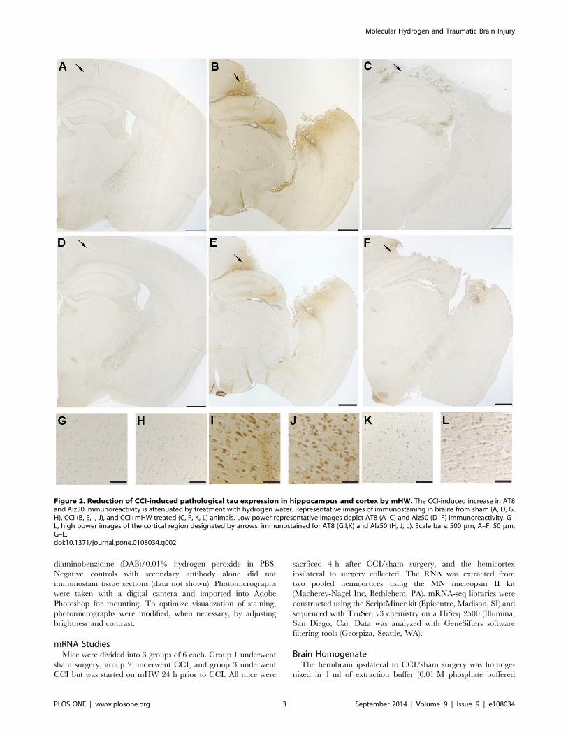
Figure 2. Reduction of CCI-induced pathological tau expression in hippocampus and cortex by mHW. The CCI-induced increase in AT8and Alz50 immunoreactivity is attenuated by treatment with hydrogen water. Representative images of immunostaining in brains from sham (A, D, G,H), CCI (B, E, I, J), and CCI+mHW treated (C, F, K, L) animals. Low power representative images depict AT8 (A–C) and Alz50 (D–F) immunoreactivity. G–L, high power images of the cortical region designated by arrows, immunostained for AT8 (G,I,K) and Alz50 (H, J, L). Scale bars: 500 mm, A–F; 50 mm,G–L.doi:10.1371/journal.pone.0108034.g002
Molecular Hydrogen and Traumatic Brain Injury
PLOS ONE | www.plosone.org 3 September 2014 | Volume 9 | Issue 9 | e108034

saline (PBS; 2.7 mM potassium chloride, 0.137 M sodium
chloride, pH 7.4) 2.7 mM, 1 mM EDTA, 1 mM PMSF, Protease
Inhibitor cocktail) using a mini beadbeater set at 4800 RPM for
30 sec to produce the PBS homogenate. Triton X-100 was added
to the PBS homogenate to a final concentration of 0.1%, and
vortexed vigorously. The homogenate was then centrifuged at
20,000 g for 10 min at 4uC. The resulting supernatant was used to
measure brain cytokines and amyloid beta peptide (Ab). Triton X-
100 was added to a third portion of PBS homogenate to a final
concentration of 1%, vortexed vigorously, and centrifuged at
20,000 g for 10 min at 4uC. The protein level of the resulting
supernatant was measured (BCA, Pierce, Rockford, IL) and
adjusted to 4 mg/ml with extraction buffer containing 1% Triton
X-100 and frozen at 280uC as the 1% Triton X-100 extraction.
Aliquots of this extraction were frozen and used for all Westerns
and dot blots except for pan-tau immunoblotting.
For total tau detection, mouse brains were dissected from
euthanized animals and rapidly snap frozen in liquid nitrogen
prior to extraction. Mouse brain hemispheres were homogenized
in high salt re-assembly buffer (RAB-High Salt [0.1 M MES,
1 mM EGTA, 0.5 mM MgSO4, 0.75 M NaCl, 0.02 M NaF,
0.5 mM PMSF, 0.1% protease inhibitor cocktail, pH 7.0]). For
semi-quantitative immunoblotting, we detected mouse and human
tau using the pan-tau antibody 17025 (provided as a generous gift
of Virginia Lee, see [12] for details) at a dilution of 1:3000 as
described previously [13] and anti-actin antibody at 1:1000
(DSHB). Rabbit polyclonal 17025 is a pan-tau antibody recog-
nizing total mouse and human tau raised against full length
recombinant tau.
Serum and Brain Cytokine MeasurementsAt 4 h, 24 h, and 7 days after CCI or sham surgery, mice were
anesthetized with urethane, the carotid artery exposed, severed,
and blood collected from it, and the hemibrain ipsilateral to CCI/
sham surgery was obtained and processed as above; the 0.1%
Triton X-100 extract was used to measure brain cytokines. The
arterial blood was centrifuged at 4500 g for 10 min at 4uC and
cytokine levels measured on the resulting serum. Brain cytokine
levels were measured at the 4 h, 24 h, and 7 day time points and
serum cytokines on the 24 h and 7 day time points. The multi-plex
kit for murine cytokines from BioRad was used and measures 23
cytokines: IL-1a, IL-1b, IL-2, IL-3, IL-4, IL-5, IL-6, IL-9, IL-10,IL-12(p40), IL-12(p70), IL-13, IL-17, Eotaxin (CCL11), G-CSF,
GM-CSF, IFN-c, KC (CXCL1), MCP-1 (CCL2), MIP-1a
Figure 3. Reduction of CCI-induced pathological tau by mHW in frontal cortex. CCI induced increases in AT8 and Alz50 immunoreactivityextend to the frontal cortex and are attenuated by treatment with hydrogen water. Low power representative images demonstrate increased AT8immunoreactivity in the frontal cortex of CCI (B) compared to sham treated (A) mice. C–F, high power images of the cortical region shown in B (arrow)demonstrate AT8 (C,D) and Alz50 (E,F) immunoreactivity in CCI (C,E) and HW treated (D,F) mice. Scale bars: 500 mm, A,B; 50 mm, D–F.doi:10.1371/journal.pone.0108034.g003
Molecular Hydrogen and Traumatic Brain Injury
PLOS ONE | www.plosone.org 4 September 2014 | Volume 9 | Issue 9 | e108034

(CCL3), MIP-1b (CCL4), RANTES (CCL5), and TNF-a. Brainlevels were reported relative to the protein content of the brain
sample and are reported as pg/mg.
Dot blots for Cyclophilin A and Amyloid PrecursorProtein (APP)Brains extracted in 1% Triton X-100 were analyzed for APP
and Cyclophilin A expression by dot blot as described previously
[14]. Briefly, 2 ug of protein in a volume of 250 ul PBS was loaded
in duplicate wells of a Bio-dot microfiltration apparatus (Biorad).
To normalize for antibody sensitivity, a standard curve was also
prepared using one of the control samples with protein amounts
per well ranging from 0.125 to 4 ug in a volume of 250 ul PBS and
loaded in duplicate. Samples and standards were bound to a
nitrocellulose membrane by vacuum, the membrane was washed,
blocked in 5% milk, and incubated overnight with APP antibody
(Epitomics, 1 mg/ml) or CypA antibody (Epitomics, 1 mg/ml) in
blocking buffer. The membrane was then washed in PBS, and
anti-rabbit secondary (Santa Cruz) prepared 1:5000 in 5% milk
and incubated with the membranes for 1 hour at room
temperature. After washing, signal was visualized with West Pico
chemiluminescent substrate. Images were captured using an
ImageQuant LAS4000, and IQTL software used to quantify spot
intensities. A standard curve was generated using spot intensities of
the standards with Graphpad Prism software, and the spot
intensity of each sample was normalized to the standard curve
prior to statistical analysis.
Ab AssaysSandwich ELISA kits which detect rodent Ab40 or Ab42
(Wako) were used to quantify Ab levels. 0.1% Triton X-100
extracts were diluted 1:1 in standard diluent provided in the kit,
and subsequently assayed according to kit instructions. We have
found that these kits specifically detect rodent Ab40 and Ab42 as
evidenced by absence of signal for both analytes in APP knockout
brains processed and assayed identically. Furthermore, this kit has
been used by other groups to quantify changes in rodent Ab in
aging [15] and blast injury [16]. In the latter study, it was reported
that the majority of rodent Ab detected by this assay is found in
the Triton-X fraction [16].
Brain Homogenates for Aquaporin-4 (AQP-4), Hypoxia-inducible factor-1 alpha (HIF-1), Matrix Metalloproteinase(MMP)-2 & -9The hemibrain ipsilateral to CCI/sham surgery was homoge-
nized and extracted in Tris-EDTA extraction buffer (20 mM
TRIS, 150 mM NaCl, 2 mM EDTA, 2% Triton X-100) with
protease inhibitors. The brain extracts were run on a 4–12% Bis-
Tris gels and transferred onto nitrocellulose membranes (Invitro-
gen, USA). The membranes were probed with a AQP-4 (Abcam,
USA), HIF-1a (Novus Biologicals, USA), MMP-2 (R&D systems,
USA) and MMP-9 (R&D systems, USA) primary antibodies,
followed by horseradish peroxidase-conjugated secondary anti-
bodies (Santa Cruz, USA). To verify uniformity of protein loading
membranes were stripped and reprobed with GAPDH or b-actinantibody (Cell Signaling Technology, USA). The enhanced
chemiluminescence western blot was digitalized with a LAS4000
CCD imaging system (GE Healthcare, USA) and analyzed by
ImageQuant TL software.
Figure 4. GFAP immunoreactivity is decreased in the hippo-campus (CA1 stratum radiatum) of CCI mice treated withhydrogen water. Representative images demonstrate similar levels of
hippocampal GFAP immunoreactivity in sham (A) and CCI (B) mice.Immunostaining for GFAP is greatly reduced in the hippocampus ofanimals treated with hydrogen water (C). Scale bars: 50 mm.doi:10.1371/journal.pone.0108034.g004
Molecular Hydrogen and Traumatic Brain Injury
PLOS ONE | www.plosone.org 5 September 2014 | Volume 9 | Issue 9 | e108034

Brain-to-blood Efflux of Ab and CSF ReabsorptionBrain-to-blood efflux of radioactively labeled Ab and albumin
were measured as previously described [17]. Murine Ab1–40 was
radioactively labeled with 125I and human serum albumin with131I by the chloramine-T method and purified on columns of
Sephadex G-10. Mice received an injection of 25,000 cpm of I-Aband 25,000 cpm of I-Albumin into the lateral ventricle of the brain
contralateral to CCI and 10 min later the brain was harvested.
Mice that had been dead between 10–30 min were used to
estimate the amount of radioactive ligand available for transport.
The amount of radioactive ligand transported was calculated as
the difference between the 10 min value in alive mice and the
10 min value in dead mice.
Effects of mHW on Cellular Respiration and ATPProductionConditionally immortalized mouse cerebral pericyte cells
(ImBPC) established as previously described [18] were cultured
in growth medium, which consisted of 5.5 mM glucose Dulbecco
minimal essential media (DMEM) (Sigma-Aldrich, St. Louis, MO)
supplemented with 10% fetal bovine serum (FBS), non-essential
amino acids (NEAA), interferon-c (44 U/ml; R&D Systems,
Minneapolis, MN), penicillin (100 U/mL), and streptomycin
(0.1 mg/mL). All cultures were maintained at 33uC under 5%
CO2. For pretreated cell experiments, media was freshly prepared
using mHW (1.6 mM) or deionized water with Dulbecco minimal
essential media powder (DMEM) (Sigma-Aldrich, St. Louis, MO)
supplemented with 10% fetal bovine serum (FBS), non-essential
amino acids (NEAA), interferon-c (44 U/ml; R&D Systems,
Minneapolis, MN), penicillin (100 U/mL) and streptomycin
(0.1 mg/mL). Cells for Seahorse analyzer experiments were
harvested using 16 Trypsin containing EDTA (Sigma-Aldrich,
St. Louis, MO) and plated into 24-well culture plates at a density
of 56104 cells/well. An n= 10 was used in each experiment and
each experiment was completed at least three times. Cells for ATP
assay were harvested and plated into 96-well culture plates at a
density of 56103 cells/well. An n= 8 was used in each experiment
and each experiment was completed three times.
A Seahorse Bioscience XF24 Extracellular Flux Analyzer
(Seahorse Bioscience, North Billerica, MA) was used to measure
the rate of oxidative metabolism of glucose (respiration). Under
typical in vitro cell culture conditions, the rate of oxygen
consumption (OCR) is an indicator of mitochondrial respiration
and the extracellular acidification rate (ECAR) is predominantly a
measure of lactic acid formed during glycolytic energy metabo-
Figure 5. The CCI induced increase in Iba1 immunoreactivity is attenuated by treatment with hydrogen water. Representative imagesare of Iba1 immunoreactivity in the cortex adjacent to the site of injury (A–C) and the hippocampus (D–F) of sham (A,D), CCI (B,E) and CCI/HW treated(C,F) animals. Scale bars: 50 mm, A–C; 100 mm, D–F.doi:10.1371/journal.pone.0108034.g005
Figure 6. Effects of CCI and mHW on Serum Cytokine Levels.Only G-CSF was elevated in blood after CCI and only at 24 h. There wasno statistically significant effect of mHW on serum G-CSF levels. *p,0.05; **p,0.01.doi:10.1371/journal.pone.0108034.g006
Molecular Hydrogen and Traumatic Brain Injury
PLOS ONE | www.plosone.org 6 September 2014 | Volume 9 | Issue 9 | e108034

Figure 7. CCI-induced Elevations in Six Cytokines in Brain Were Affected by mHW. IL-6 (not shown; see Table 1 for results) had a patternsimilar to G-CSF. Except for the day 7 IL-1a value, the statistically significant effects of mHW were to further increase brain cytokine levels. *p,0.05;**p,0.01; ***p,0.001.doi:10.1371/journal.pone.0108034.g007
Molecular Hydrogen and Traumatic Brain Injury
PLOS ONE | www.plosone.org 7 September 2014 | Volume 9 | Issue 9 | e108034

Table
1.Effect
ofCCIan
dCCI+mHW
onBrain
CytokineLevels.
4hPost
CCI
24hPost
CCI
7DaysPost
CCI
Sham
(n=6)
CCI(n
=5–6)
CCI+mHW
(n=5)
Sham
(n=7)
CCI(n
=7)
CCI+mHW
(n=7)
Sham
(n=4)
CCI(n
=6)
CCI+mHW
(n=4)
IL-1a
060
339646**
451644**#
3063
9462**
131612**##
3162
8668**
6566**#
IL-1b
595654
1692653**
1766697**
14610
244648**
4516105**#
262
121645
87640
IL-2
50613
64610
5866
965
361
1464
1467
1866
864
IL-3
6564
6563
6166
2662
5363**
6066**
2762
3963*
3761*
IL-4
11363
165611**
17868**
5063
6762**
7766**
4863
6664**
6061*
IL-5
13867
15366
157612
8765
9064
9167
8965
9865
8766
IL-6
7067
694656*
10036293**
2563
5756101*
11556271**#
2262
3663*
3462*
IL-10
487626
475637
482641
160619
186624
205626
143611
180617
182619
IL-12p40
847631
18976220**
22496183**
281623
698647**
616643**
307635
803692**
879676**
IL-12p70
37612
747676**
868675**
102623
269642*
293654*
92628
134620
130615
IL-13
2271692
21296145
23666235
510660
696662
840680**
512641
475631
454641
IL-17
595625
617613
562639
24268
208610
195621
224622
209612
212620
G-CSF
168632
48646762**
497461045**
5664
36506477**
62126765**##
5265
12568*
142635*
GM-CSF
587673
1821675**
1840665**
17617
221680
2326134
60660
060
060
KC
9436125
83906938**
982661332**
264652
18176346*
39506616**##
3636165
535680
538660
MCP-1
27156382
1083361305**
1407961822**
966676
1359361264**
1583561457**
10076110
29086327**
25166342**
MIP-1a
97628
62296474**
70396353**
119615
68246612**
1044561586**#
170644
14756148**
13516214**
MIP-1b
29046125
65076652**
84436629**#
10046132
20036119**
26286273**#
9546150
13776133
1200666
RANTES
6363
110614*
136617**
3865
160611**
145621**
73619
262628**
220624**
TNFa
101306511
85126463
78686828*
72596778
68286582
62436578
910961909
71886594
72096698
*p,0.05&**p,0.01forCCIvs
Sham
andforCCI+mHW
vsSh
am;#p,0.05&##
p,0.01forCCI+mHW
vsCCI.
Mean
6SE
(pg/m
gofbrain
protein).
doi:10.1371/journal.pone.0108034.t001
Molecular Hydrogen and Traumatic Brain Injury
PLOS ONE | www.plosone.org 8 September 2014 | Volume 9 | Issue 9 | e108034

Table 2. Changes in Brain Expression with CCI and mHW: Biological Processes.
Biological ProcessesPositive Z-score # Affected Genes # in Gene Set Z-Score
Positive regulation of cytokine transport 3 33 5.21
Positive regulation of hormone secretion 4 61 4.91
Positive Regulation of Secretion 7 170 4.72
Carbohydrate Homeostasis 4 67 4.62
Glucose Homeostatis 4 67 4.62
Regulation of Oxidoreductase Activity 3 41 4.56
Leukocyte Chemostasis 4 74 4.32
Regulation of Cytokine Secretion 3 45 4.30
Leukocyte Migration 5 114 4.17
Feeding Behavior 4 83 3.99
Cytokine Secretion 3 52 3.91
Positive Regulation of Protein Secretion 3 53 3.86
Regulation of Secretion 9 9 3.86
Cell Chemotaxis 4 89 3.80
Secretion by Cell 11 469 3.63
Categories listed consisted of a minimum of 6 genes, had at least 3 genes that were changed, and a Z-score of .3.63.doi:10.1371/journal.pone.0108034.t002
Table 3. Changes in Brain Expression with CCI and mHW: Cellular Component.
Cellular ComponentPositive Z-score # Affected Genes # in Gene Set Z-Score
Brush Border 3 62 3.60
Extracellular Region 27 1766 3.16
Cell-cell Junction 5 233 2.13
Negative Z-score
Intracellular 59 10670 24.79
Intracellular Part 59 10453 24.52
Organelle 50 8945 24.01
Intracellular Organelle 50 8919 23.98
Nucleus 20 4806 23.78
Membrane-bounded Organelle 45 7998 23.62
Cell 105 14900 23.61
Cell Part 105 14900 23.61
Intracellular Membrane-bounded Organelle 45 7982 23.60
Cytoplasm 45 7841 23.43
Intracellular Organelle Part 16 3899 23.36
Intracellular non-membrane-bounded Organelle 6 2328 23.33
Non-membrane-bounded Organelle 6 2328 23.33
Organelle Part 17 3978 23.27
Macromolecular Complex 14 3198 22.80
Cytoskeleton 3 1408 22.72
Protein Complex 12 2670 22.46
Cytosol 4 1421 22.45
Cytoplasmic Part 33 5320 22.19
Nuclear Part 7 1714 22.11
Membrane-enclosed Lumen 7 1658 22.11
Categories listed consisted of a minimum of 6 genes, had at least 3 genes that were changed, and a Z-score of $2.0.doi:10.1371/journal.pone.0108034.t003
Molecular Hydrogen and Traumatic Brain Injury
PLOS ONE | www.plosone.org 9 September 2014 | Volume 9 | Issue 9 | e108034

lism. ImBPC were seeded into a specialized Seahorse microplate
(V7; 0.32 cm2 growth area) at 56104 cells per well in 100 ml ofgrowth medium and incubated at 33uC in 5% CO2 for 2 h. An
additional 150 ml of medium was added after the cells had
adhered. On the following day, assays were initiated by replacing
the media with XF-DMEM (supplemented with 5 mM glucose
and 1 mM sodium pyruvate at pH 7.4) nonbuffered media and
incubating at 37uC for 60 min to allow the temperature and pH to
reach equilibrium. The microplate was then placed into the XF24
instrument to measure OCR and ECAR. Basal measurements of
OCR and ECAR were established, and inhibitors of mitochon-
drial respiration were injected sequentially as indicated to
determine mitochondrial function and glycolysis. The inhibitors
were used at the following concentrations: oligomycin- 3 mM;
carbonyl cyanide 4-(trifluoromethoxy)phenylhydrazone (FCCP)-
3 mM; rotenone- 3 mM; and antimycin A- 1.5 mM. ATP
production is measured as (basal respiration – oligomycin
respiration). Proton leak is measured as (oligomycin respiration –
rotenone/Antimycin A respiration). Maximal respiration is the
respiration measured after FCCP treatment. Reserve capacity is
measured as (FCCP respiration – basal respiration). Non-
mitochondrial respiration is the respiration that occurs after
rotenone/Antimycin A treatment.
ATP levels in ImBPC were measured using an ATPlite
detection assay (Perkin Elmer; Waltham, MA) according to
manufacturer’s protocol. Briefly, cells were lysed at various time
points in mammalian cell lysis solution followed by incubation in
substrate solution. After 10 min for dark adaption, luminescence
was measured using the Victor3 multi label plate reader (Perkin-
Elmer; Waltham, MA).
Table 4. Changes in Brain Expression with CCI and mHW: Molecular Function.
Molecular FunctionPositive Z-score # Affected Genes # in Gene Set Z-Score
Methyl Indole-3-acetate Esterase Activity 3 7 12.01
Methyl Jasmonate Esterase Activity 3 7 12.01
Methyl Salicylate Esterase Activity 3 7 12.01
Neuropeptide Receptor Binding 3 21 6.65
Hormone Activity 6 118 4.97
Carboxylesterase Activity 5 97 4.58
Serine Hydrolase Activity 7 203 4.00
G-protein-coupled Receptor Binding 6 183 3.55
Serine-type Peptidase Activity 6 200 3.28
Serine-type Endopeptidase Activity 5 155 3.19
Sodium Ion Transmembrane Transporter Activity 3 80 2.80
Cytokine Activity 5 183 2.75
Acyltransferase Activity 5 193 2.61
Transferace Activity, Transferring Acyl Groups Other Than Amino-acyl Groups 5 193 2.61
Transmembrane Transporter Activity 14 835 2.59
Channel Activity 8 388 2.58
Receptor Binding 16 999 2.58
Passive Transmembrane Transporter Activity 8 389 2.57
Transferase Activity, Transferring Acyl Groups 5 198 2.54
Transporter Activity 16 1026 2.47
Monovalent Inorganic Cation Transmembrane Transporter Activity 4 149 2.41
Ion Transmembrane Transporter Activity 11 654 2.30
Substrate-specific Transmembrane Transporter Activity 12 754 2.20
Substrate-specific Channel Activity 7 376 2.11
Substrate-specific Transporter Activity 13 863 2.09
Negative Z-score
Adenyl Nucleotide Binding 4 1446 22.49
Adenyl Ribonucleotide Binding 4 1439 22.48
ATP Binding 4 1414 22.44
Nucleotide Binding 9 2096 22.26
Nucleic Acid Binding 12 2510 22.22
Purine Ribonucleoside Triphosphate Binding 7 1718 22.13
Binding 77 10525 22.03
Categories listed consisted of a minimum of 6 genes, had at least 3 genes that were changed, and a Z-score of $2.0.doi:10.1371/journal.pone.0108034.t004
Molecular Hydrogen and Traumatic Brain Injury
PLOS ONE | www.plosone.org 10 September 2014 | Volume 9 | Issue 9 | e108034

Statistical AnalysisResults are reported at the mean +/2 the standard error of the
mean. Two means were compared by two-tailed t-test. More than
two means were compared by analysis of variance (ANOVA)
followed by Newman-Keuls post-test. Studies done using the XF24
Seahorse Analyzer were analyzed using AUC ANOVA software
provide by Seahorse Bioscience.
Results
Brain EdemaFigure 1 shows the results of CCI and treatment with mHW on
the percent of water content in the ipsilateral hemi-brain. A two
way ANOVA showed statistical significance for day
[F(3,30) = 30.4, p,,0.001], treatment [F(1,30) = 11.2, p,0.01],
and interaction [F(1,30) = 5.9, p,0.01] for % water. Tukey‘s post-
test showed that % water was significantly increased 24 h after
CCI (p,0.001), but was inhibited to a statistically significant
degree by mHW (p,0.001). Analysis of the 24 h data using the
Keep method [11] showed significant edema that was reversed by
mHW (inset).
Immunohistochemistry & ImmunoblottingStaining of phosphorylated tau with both AT8 and Alz 50 were
greatly increased in the cortex of CCI mice (Figures 2 & 3) with
extension into the hippocampal CA3 region (Figure 2). Total tau
as detected with the pan-tau antibody 17026 was not altered 12,
18, 24, or 48 h after CCI (data not shown). Treatment with mHW
abolished these increases in tau (Figures 2 & 3). CCI did not
produce a remarkable increase in GFAP staining, but CCI mice
treated with mHW had less GFAP staining that either sham or
CCI mice (Figure 4). CCI increased Iba1 immunostaining; mHW
decreased Iba1 immunostaining to levels lower than those seen in
sham mice. (Figure 5).
Serum and Brain Cytokine MeasurementsFor serum, only granulocyte-colony stimulating factor (G-CSF)
was significantly affected by CCI in comparison to sham surgery
and only at 24 h (Figure 6); mHW had no effect.
Because of an apparrent effect of sham surgery on brain
cytokine levels at 4 h, separate ANOVAs were conducted for the
4 h, 24 h, and 7 day results. Three cytokines were not detectable
in the brain sample (IL-9, eotaxin, INF-gamma). Table 1 shows
the effects of CCI and mHW on brain levels of the other 20
cytokines. Six of the detected cytokines were not affected by CCI
(IL-2, IL-5, IL-10, IL-13, IL-17, TNF). Of the remaining 14
cytokines, all had elevated brain levels at one or more time points
after CCI. Eight were elevated at all time points (IL-1a, IL-4, IL-6,IL-12p40, G-CSF, MCP-1, MIP-1a, RANTES), four at 4 h and
24 h but not at 7 days (IL-1b, IL-12p70, KC, MIP-1b), 1 at 4 h
only (GM-CSF) and one at 24 h and 7 days but not at 4 h (IL-3).
Seven cytokines had CCI+mHW levels at one or more time points
that differed significantly from CCI levels (Table 1 & Figure 7).
Except for the IL-1a 7 day value, the CCI+mHW values were
higher than those for CCI alone. These increases were statistically
significant at 4 h and 24 h for IL-1a and MIP-1a and at 4 h for
IL-1b, G-CSF, KC, and MIP1a (Figure 7). For many cytokines,
the CCI value tended to be higher at 7 days than the CCI+mHW
value, but only reached statistical significance for IL-1a. TNF
levels were lower in CCI+mHW than in sham mice 4 h after CCI
and IL-13 levels were higher in CCI+mHW than in sham mice
24 h after CCI.
Figure 8. Effects of CCI and mHW on AQP-4, HIF-1, and MMP-2 & -9 Levels. The upper left panel shows that CCI suppressed AQP-4expression at 24 h and that mHW blocked this decrease. HIF-1 (upper right panel) was increased at all times after CCI and mHW induced a partialrecovery. MMP-9 (lower left panel) showed elevations 24 h and 7 days after CCI that were blocked by mHW. MMP-2 was decreased 24 h after CCI butmHW increased levels at all time points.doi:10.1371/journal.pone.0108034.g008
Molecular Hydrogen and Traumatic Brain Injury
PLOS ONE | www.plosone.org 11 September 2014 | Volume 9 | Issue 9 | e108034

mRNA studiesAfter CCI, 246 genes had increased expression by 2 fold or
more and 38 genes had expression that was decreased by 50% or
more. mHW reduced by 50% or more 236 genes whose expression
was increased by 2 fold or more by CCI. We used GeneSifter to
analyze which categories of Biological Processes (Table 2),
Cellular Components (Table 3), and Molecular Functions (Ta-
ble 4) were most affected, restricting categories to those containing
a minimum of 6 genes and having at least 3 genes altered. Z-score
was used as a further filter with an absolute value of $3.5 for
Biological Processes and $2.0 for Cellular Components and
Molecular Functions. Of 15 significant categories for Biological
Processes, all Z-scores indicated positive enrichment and recurring
categories related to cytokine release (3 categories), oxidation/
carbohydrate metabolism (4 categories), and leukocyte or cell
migration/chemotaxis (3 categories). 8 categories involved trans-
port/secretion with most overlapping with an above category (e.g.,
cytokine transport). These findings suggest that mHW is very
effective at reversing CCI-activation of genes related to oxidation,
carbohydrate metabolism, and neuroinflammation. Molecular
Functions that were positively enriched included enzymatic genes,
receptor genes, and transporter, channel, and transmembrane
transporter genes. Molecular Functions was negatively enriched in
genes relating to ATP and nucleotide binding. Cellular Compo-
nents was positively enriched in those genes related to the
extracellular and cell membrane environment and negatively
enriched in those related to the intracellular, nuclear, and
cytoplasmic environments.
AQP-4, HIF-1, and MMP-2 &- 9 MeasuresAQP-4 was suppressed 24 h after CCI but had recovered to
baseline by 3 days (Figure 8, upper left panel). Treatment with
mHW induced a statistically significant increase in comparison to
CCI at 24 h and 3 days. HIF-1 was significantly increased by CCI
at all time points, peaking at 24 h; treatment with mHW
suppressed this increase to a statistically significant degree at day
3 and 7, although HIF-1 was still elevated in comparison to sham
treated mice (Figure 8, upper right panel). MMP-9 showed a
biphasic increase, being elevated at 24 h and 7 days but not at 3
days; mHW reversed these increases (Figure 8, lower left panel).
MMP-2 decreased 24 h after CCI; mHW induced a statistically
signifcant increase in MMP-2 at all time points in comparison both
to the sham and to the time-matched CCI groups (Figure 8, lower
right panel).
Measures of Cyclophilin A, APP, and AbCCI reduced CypA levels 7 days but not 24 h after CCI; mHW
further reduced CypA at 7 days after CCI (Figure 9, lower left
panel). Protein levels of APP were decreased by CCI at 7 days, but
not at 24 h (Figure 9, upper left panel). The CCI+mHW group
did not differ from the CCI group at either time point, but was
lower than sham mice at both 24 h and 7 days. Ab1–40 was not
affected at 24 h but was increased at 7 days in both the CCI and
CCI+mHW groups (Figure 9, upper right panel). Ab1–42 was
decreased 24 h after CCI and CCI+mHW but was not altered by
7 days; there were no differences at either time for CCI vs CCI+mHW (Figure 9, lower right panel). The Ab1–42/Ab1–40 ratios
Figure 9. Effects of CCI and mHW on Cyclophillin A, APP, and Amyloid Beta Peptide Levels. The upper left panel shows that protein levelsof APP were decreased 7 days after CCI and that mHW did not protect from CCI. Amyloid beta peptide1–40 (upper right panel) was increased on day 7and amyloid beta peptide1–42 (lower right panel) was decreased 24 h after CCI, but mHW did not alter these effects of CCI. CypA was decreased 7days after CCI and this decrease was enhanced by mHW (lower left panel).doi:10.1371/journal.pone.0108034.g009
Molecular Hydrogen and Traumatic Brain Injury
PLOS ONE | www.plosone.org 12 September 2014 | Volume 9 | Issue 9 | e108034

were lower at both 24 h and 7 days for both CCI and CCI+mHW
groups, but showed no differences between CCI vs CCI+mHW
(data not shown).
CSF Reabsorption and Brain-to-blood Efflux of AbNeither CSF reabsorption as measured by I-Albumin efflux nor
Ab efflux was affected 24 h, 3 days, or 7 days after CCI (data not
shown).
Effects on Cellular RespirationTo assess mitochondrial function that occurs in response to
mHW, a Seahorse analyzer assay was performed by exposing the
mHW-pretreated ImBPC to oligomycin, FCCP (carbonyl cyanide
p-trifluoromethoxyphenylhydrazone) and rotenone/antimycin A
in succession and measuring OCR (Figure 10A) and ECR
(Figure 10C). The results showed that molecular hydrogen
increased basal respiration, reserve capacity, and non-mitochon-
drial respiration, but not maximal respiration, proton leak, or the
oxygen-dependent production of ATP (Figure 10B). The direct
measure of ATP showed increased levels after 24 h of exposure to
mHW, but not after a 10 min or 6 h exposure.
In other ImBPCs (Figure 11), the ETC was shut off before
exposure to molecular hydrogen. OCR and ECAR were
unaffected by molecular hydrogen (Fig. 1 A–C) as measured for
up to 6 h. However, molecular hydrogen was associated with an
increase in ATP levels after 6 h of exposure (Figure 11D).
Discussion
TBI results in long-term injuries to the CNS that involve
immediate effects, such as brain edema, and more sustained
effects, such as neuroinflammation. Here, we found that many of
the immediate and sustained effects were modulated by treating
mice with mHW.
Edema is a significant clinical end point in the pathophysiology
of TBI. It occurs because of ongoing events, including BBB
disruption, and it leads to further damage to the brain from
multiple mechanisms that can include pressure, electrolyte
disturbances, and the toxins from BBB leakage. Edema peaked
at our 24 h point with the arithmetic increase seen at the third day
(72 h after CCI) not being statistically significant. mHW decreased
CCI-induced edema by about half at 24 h. Edema was also
reduced as measured by the Keep method. As the Keep method
Figure 10. Pretreatment with Molecular Hydrogen Enhances Mitochondrial Aerobic Metabolism. ImBPCs were subjected to amitochondrial stress test after treatment with molecular hydrogen water (red) or control (dI water; blue) for 24 h. Oxygen consumption rates (OCR)(Panel A) and extracellular acidification rates (ECAR) (Panel C) were measured using the Seahorse XF24 analyzer in the presence of the ATP synthaseinhibitor (oligomycin; 3 mM), the uncoupling agent (FCCP; 3 mM), the complex I inhibitor (rotenone; 3 mM), and the complex III inhibitor (antimycin A;1.5 mM). Analysis of OCR data showed molecular hydrogen increased basal respiration, reserve capacity, and non-mitochondrial (non-mt) respirationbut had no effects on ATP production rate as assessed indirectly by OCR analysis, proton leak, or maximal respiration (Panel B). Direct measurementof ATP levels (Panel D) in other cells not exposed to the metabolic stressors showed higher levels of ATP after 24 h but not after 10 min or 6 h ofexposure to molecular hydrogen. Results presented here are from one experiment completed, which is representative of the 3 experiments that weredone. Results are represented as a mean 6 SEM. ** is p,0.01 and *** is p,0.001.doi:10.1371/journal.pone.0108034.g010
Molecular Hydrogen and Traumatic Brain Injury
PLOS ONE | www.plosone.org 13 September 2014 | Volume 9 | Issue 9 | e108034

uses dry weight as the denominator, it is much less influenced by
either edema or tissue loss than the traditional method that uses
wet weight in the denominator. The results showed that water
content increased from being 4-fold greater than dry weight to 6.5-
fold greater with CCI and that mHW preserved the ratio at 4.7-
fold.
TBI, stroke, and hypoxia alter the expressions and functions of
AQP-4, HIF-1, and the MMP’s; these in turn affect edema
formation, BBB disruption, and alterations in brain interstitial
fluid circulations [19]. Cyclophilin A is associated with BBB
disruption, especially in humans or transgenic rodents that are
APOE4 positive, through its effects on MMP-9 [20,21]. CCI
induced changes at 24 h in AQP-4, HIF-1, and both MMP-2 and
-9. Changes were still present in comparison to shams at day 7 for
HIF-1 and MMP-9. In contrast, the effect of CCI on Cyclophilin
A was delayed, occurring at day 7 but without effect 24 h after
CCI. We found that mHW counteracted the CCI-induced
perturbance of each of these factors. These effects of mHW
countering the CCI-induced alterations for AQP-4, HIF-1, the
MMP’s, and Cyclophilin A provide mechanisms by which mHW
can protect against edema and BBB disruption.
Alterations in AQP-4, which others have also found is down
regulated in TBI, has been associated with alterations in clearance
of Ab through its effects on the circulation of interstitial fluid in the
brain. We, however, found that neither the brain-to-blood
clearance of Ab nor the reabsorption of cerebrospinal fluid back
into the circulation was altered by CCI at the time points
examined here. Although CCI did affect protein levels of APP and
the Ab’s, mHW did not alter the effects of CCI.
Treatment with mHW totally blocked the pathological phos-
phorylated tau changes induced by CCI. In contrast, total tau was
not altered at 12, 18, 24 or 48 h after CCI. TBI causes early
transient accumulation of abnormally phosphorylated tau and
repeated injury can result in chronic traumatic encephalopathy
which is classified as a tauopathy characterized by tau hyperpho-
sphorylation, and accumulation of conformationally altered or
aggregated pathological tau. Pathological tau changes are also a
hallmark of Alzheimer’s disease, ultimately resulting in neurofi-
brillary tangles, and is one of the findings that suggests TBI and
Alzheimer’s disease may share pathophysiological mechanisms.
Here, we found elevations of phosphorylated tau after CCI in both
hippocampal and cortical regions as detected by AT8 and Alz50,
but not in total tau. The AT8 antibody detects tau that is
phosphorylated at Ser202 and Thr205, a phosphorylation pattern
typical of the immature neurofibrillary tangles of Alzheimer’s
disease. Alz50 also recognizes a pre-tangle phosphorylation
pattern similar to one found in Alzheimer’s. Treatment with
Figure 11. Treatment with Molecular Hydrogen Does Not Rescue Respiration After ETC Inhibition. ImBPCs were subjected to amitochondrial stress test in the presence of the ATP synthase inhibitor (oligomycin; 3 mM), the uncoupling agent (FCCP; 3 mM), the complex I inhibitor(rotenone; 3 mM), and the complex III inhibitor (antimycin A; 1.5 mM). Once respiration was inhibited, ImBPCs were exposed to hydrogen water (red)or control (dI water; blue), and OCR was measured for 30 min (Panel A) or 6 h (Panel B). ECAR was also examined for 30 min (Panel C). ATP levels weremeasured after 10 min, 6 h, or 24 h of treatment with molecular hydrogen water. Results presented here are from one experiment completed, whichis representative of the 3 experiments that were done. Results are represented as a mean 6 SEM. ** is p,0.01 and *** is p,0.001.doi:10.1371/journal.pone.0108034.g011
Molecular Hydrogen and Traumatic Brain Injury
PLOS ONE | www.plosone.org 14 September 2014 | Volume 9 | Issue 9 | e108034

mHW totally blocked the detection of these CCI-induced forms of
phosphorylated tau.
Neuroinflammation is thought to be a significant driver of the
long-term pathology resulting from TBI. Specific agents of
inflammation, particularly cytokines, likely play roles in both
disease progression and recovery. Neuroinflammation is an early
event in CCI as seen here by elevations in cytokines and cytokine-
related genes. CCI-induced peripheral inflammation as assessed
by serum cytokines was, with the exception of an early rise in
serum G-CSF, unremarkable. The inflammatory drivers, then, are
likely centered in the CNS rather than as peripheral-to-brain
events. This is not surprising considering the CCI model of TBI
inflicts significant damage directly to the CNS, although it is
surprising given that neuroinflammation can influence the
peripheral immune system. Immunohistochemical staining sug-
gested that microglia, but not astrocytes, were activated by CCI.
Staining showed that microglia were activated in both the cortex
and hippocampus. The effect of mHW was profound on CCI-
induced microglial activation and seemed to have reduced levels of
activation lower than those seen in sham mice, especially in cortex.
Of the 14 cytokines whose levels were increased by CCI, 7 of them
had levels that were significantly affected by mHW at one or more
time points. However, mHW mostly induced a further increase in
brain cytokine levels, elevating them even higher than did CCI
alone. This disconnect between the effect of mHW on microglial
activation as detected by Iba1 staining and on brain cytokine levels
suggests that another cell type is responsible for cytokine
production. Both brain endothelial cells and pericytes are known
to secrete cytokines constitutively and in response to neuroimmune
stimuli [22,23,24]. Additionally, the transport rate of cytokines
across the BBB could be increased as shown to occur in other types
of CNS injury [25,26,27,28] or CCI could increase immune cell
trafficking into the CNS and their contribution to the pool of CNS
cytokines [29]. The results further suggest, given the therapeutic
effects of mHW, that the 7 cytokines which had further elevations
with mHW treatment may play a therapeutic role in recovery
from TBI rather than a pathological role in progression. Some
cytokines, such as TNF [30], are notorious for being either
beneficial or deleterious depending on concentration and context
and TBI is one such condition in which cytokines play both of
these roles [8].
Despite the paradoxic effect on brain cytokine protein levels, the
effects of mHW was to reverse CCI-induced increases in the
expression of genes related to cytokines. Treatment with mHW
affected genes whose proteins were active at the cell surface and
tended to not affect CCI-induced activated genes that affected
intracellular, nuclear, or ATP and nucleotide processes. The
surface genes affected were those that tended to relate to cytokine
release and secretion, oxidation and carbohydrate metabolism,
leukocyte trafficking, and receptors, channels, and transporters.
Recent work has shown that brain pericytes are critical to
protecting the BBB and the neurovascular unit from oxidative
stress, but are themselves very sensitive to oxidative stress,
especially as induced by mitochondrial respiration [18,31,32].
The work summarized in Table 2 shows that mHW reverses the
CCI-induced altered gene expression related to oxidation and
carbohydrate metabolism and, as such, brain pericytes are the
logical cell type to examine when assessing the protective effects of
a CNS anti-oxidant. Assessment of cellular respiration showed that
exposure of brain pericytes to molecular hydrogen for 24 h
increased basal respiration, reserve capacity, and non-mitochon-
drial respiration, where ‘‘non-mitochondrial respiration’’ is defined
as oxygen use that occurs despite shutdown of the electron
transport chain (Figure 10). In cells not exposed to the metabolic
stressors (Figure 10D), ATP production as assessed by oxygen
consumption was not increased but direct measurement of ATP
showed that it was, again indicating ATP production independent
of oxygen use. In pericytes in which the electron transport chain
was inactivated (Figure 11), molecular hydrogen was without effect
on oxygen consumption or ECAR, but ATP levels were found to
be elevated. Thus, the work in figures 10 and 11 provide evidence
that molecular hydrogen can support ATP production indepen-
dent of the electron transport chain. We propose that these
findings can all be explained by the Jagendorf reaction, defined as
ATP production that results from an inequality of hydrogen ion
electrochemical activity that has been produced independently of
the electron transport chain [33]. Under normal circumstances,
the electron transport chain establishes the proton gradient and
the gradient results in ATP production. Jagendorf, however,
showed that the electron transport chain could be bypassed and
ATP produced independently of it by establishing the hydrogen
gradient with an acid environment. We propose that molecular
hydrogen may also be producing a hydrogen gradient, thus
promoting mitochondrial ATP production independent of elec-
tron transport chain activity. Thus, molecular hydrogen may be
working in part by overcoming the deficit in ATP that cells
undergoing TBI, and so having mitochondrial damage [34],
experience.
Overall, these studies provide proof of principle that mHW can
potently reverse, block, or attenuate many of the effects of CCI.
These include effects on edema formation, tau pathology,
regulators of fluid and BBB functions, neuroinflammation, and
gene expression. These studies were limited in that they did not
evaluate mHW treatment without a pretreatment phase, but
suggest that mHW could be an important, nontoxic treatment for
acute TBI. Because of its nontoxicity and ease of administration,
mHW could be readily adapted for clinical, emergency, and even
first responder use.
Acknowledgments
We thank Peter Davies for the generous gift of Alz-50 antibody.
Author Contributions
Conceived and designed the experiments: KD BCK MAE AK GNS NS
TS WAB. Performed the experiments: KD BCK MAE PJM AK ZF KMH
GNS TS. Analyzed the data: KD BCK PJM AK ZF KMH NS TS WAB.
Contributed reagents/materials/analysis tools: NS BCK WAB. Contrib-
uted to the writing of the manuscript: KD BCK MAE PJM AK ZF KMH
GNS NS TS WAB.
References
1. Sayer NA (2012) Traumatic brain injury and its neuropsychiatric sequelae in
war veterans. Annu Rev Med 63: 405–419.
2. Wick JY (2012) Traumatic brain injury: special problem, special care. Consult
Pharm 27: 392–399.
3. Cernak I (2005) Animal models of head trauma. NeruoRx 2: 410–422.
4. Bazarian JJ, Cernak I, Noble-Haeusslein L, Potolicchio S, Temkin N (2009)
Long-term neurologic outcomes after traumatic brain injury. J Head Trauma
Rehabil 24: 439–451.
5. Deng Y, Thompson BM, GAo X, Hall ED (2007) Temporal relationship of
peroxynitrite-induced oxidative damage, calpain-mediated cytoskeletal degra-
dation and neurodegeneration after traumatic brain injury. Experimental
Neurology 205: 154–165.
6. Hawkins BE, Krishnamurthy S, Castillo-Carranza DL, Sengupta U, Prough DS,
et al. (2013) Rapid Accumulation of Endogenous Tau Oligomers in a Rat Model
of Traumatic Brain Injury: Possible Link Between TBI and Sporadic
Tauopathies. J Biol Chem.
Molecular Hydrogen and Traumatic Brain Injury
PLOS ONE | www.plosone.org 15 September 2014 | Volume 9 | Issue 9 | e108034

7. Johnson VE, Stewart W, Smith DH (2012) Widespread tau and amyloid-beta
pathology many years after a single traumatic brain injury in humans. BrainPathol 22: 142–149.
8. Morganti-Kossman MC, Lenzlinger PM, Hans V, Stahel P, Csuka E, et al.
(1997) Production of cytokines following brain injury: beneficial and deleteriousfor the damaged tissue. Molecular Psychiatry 2: 133–136.
9. Ohsawa I, Ishikawa M, Takahashi K, Watanabe M, Nishimaki K, et al. (2007)Hydrogen acts as a therapeutic antioxidant by selectively reducing cytotoxic
oxygen radicals. Nature Medicine 13: 688–694.
10. Miyamoto K, Tsumuraya T, Ohtaki H, Dohi K, Satoh K, et al. (2014)PACAP38 suppresses cortical damage in mice with traumatic brain injury by
enhancing antioxidant activity. J Mol Neurosci epub.11. Keep RF, Hua Y, Xi G (2012) Brain water content. A misunderstood
measurement? Transl Stroke Res 3: 263–265.12. Ishihara T, Hong M, Zhang B, Nakagawa Y, Lee MK, et al. (1999) Age-
dependent emergence and progression of a tauopathy in transgenic mice
overexpressing the shortest human tau isoform. Neuron 24: 751–762.13. Guthrie CR, Schellenberg GD, Kraemer BC (2009) SUT-2 potentiates tau-
induced neurotoxicity in Caenorhabditis elegans. Hum Mol Genet 18: 1825–1838.
14. Erickson MA, Niehoff ML, Farr SA, Morley JE, Dillman LA, et al. (2012)
Peripheral administration of antisense oligonucleotides targeting the amyloidbeta protein precursor reverses ABPP and LRP-1 overexpression in aged
SAMP8 mouse brain. J Alzheimer’s Dis 28: 951–960.15. Silverberg GD, Messier AA, Miller MC, Machan JT, Majmudar SS, et al. (2010)
Amyloid efflux transporter expression at the blood-brain barrier declines innormal aging. J Neuropathol Exp Neurol 69: 1034–1043.
16. De Gasperi R, Gama Sosa MA, Kim SH, Steele JW, Shaughness MC, et al.
(2012) Acute blast injury reduces brain abeta in two rodent species. Front Neurol3: 177.
17. Banks WA, Fasold MB, Kastin AJ (1997) Measurement of efflux rate from brainto blood. In: Irvine GB, Williams CH, editors. Methods in Molecular Biology:
Neuropeptides Protocols. Totowa, NJ: Humana Press. 353–360.
18. Shah GN, Price TO, Banks WA, Morofuji Y, Kovac A, et al. (2012)Pharmacological inhibition of mitochondrial carbonic anhydrases protects
mouse cerebral pericytes from high glucose-induced oxidative stress andapoptosis. J Pharmacol Exp Therap 344: 637–645.
19. Higashida T, Kreipke CW, Rafols JA, Peng C, Schafer S, et al. (2011) The roleof hypoxia-inducible factor-1alpha, aquaporin-4, and matrix metalloproteinase-
9 in blood-brain barrier disruption and brain edema after traumatic brain injury.
J Neurosurg 114: 92–101.20. Bell RD, Winkler EA, Singh I, Sagare AP, Deane R, et al. (2012) Apolipoprotein
E controls cerebrovascular integrity via cyclophilin A. Nature 485: 512–516.
21. Halliday MR, Pomara N, Sagare AP, Mack WJ, Frangione B, et al. (2013)
Relationship between cyclophilin A levels and matrix metalloproteinase 9
activity in cerebrospinal fluid of cognitively normal apolipoprotein E4 carriers
and blood-brain barrier breakdown. JAMA 70: 1198–1200.
22. Reyes TM, Fabry Z, Coe CL (1999) Brain endothelial cell production of a
neuroprotective cytokine, interleukin-6, in response to noxious stimuli. Brain
Research 851: 215–220.
23. Verma S, Nakaoke R, Dohgu S, Banks WA (2006) Release of cytokines by brain
endothelial cells: a polarized response to lipopolysaccharide. Brain, Behavior,
and Immunity 20: 449–455.
24. Kovac A, Erickson MA, Banks WA (2011) Brain microvascular pericytes are
immunoactive in culture: cytokine, chemokine, nitric oxide, and LRP-1
expression in response to lipopolysaccharide. Journal of Neuroinflammation 8:
139.
25. Pan W, Banks WA, Kennedy MK, Gutierrez EG, Kastin AJ (1996) Differential
permeability of the BBB in acute EAE: enhanced transport of TNF-alpha.
American Journal of Physiology 271: E636–E642.
26. Pan W, Kastin AJ (2001) Increase in TNF alpha transport after SCI is specific
for time, region, and type of lesion. Experimental Neurology 170: 357–363.
27. Pan W, Banks WA, Kastin AJ (1998) Permeability of the blood-brain barrier to
neurotrophins. Brain Research 788: 87–94.
28. Pan W, Cain C, Yu Y, Kastin AJ (2006) Receptor-mediated transport of LIF
across blood-spinal cord barrier is upregulated after spinal cord injury. Journal of
Neuroimmunology 174: 119–125.
29. JIn X, Ishii H, Bai Z, Itokazu T, Yamashita T (2012) Temporal changes in cell
marker expression and cellular infliltration in a controlled cortical impact model
in adult male C57BL/6 mice. PLoS One 7: e41892.
30. Pan W, Zadina JE, Harlan RE, Weber JT, Banks WA, et al. (1997) Tumor
necrosis factor-alpha: a neuromodulator in the CNS. Neuroscience and
Biobehavioral Reviews 21: 603–613.
31. Price TO, Eranki V, Banks WA, Ercal N, Shah GN (2012) Topiramate
treatment protects blood-brain barrier pericytes from hyperglycemia-induced
oxidative damage in diabetic mice. Endocrinology 153: 362–372.
32. Shah GN, Price TO, Banks WA, Morofuji Y, Kovac A, et al. (2013)
Pharmacological inhibition of mitochondrial carbonic anhydrases protects
mouse cerebral pericytes from high glucose-induced oxidative stress and
apoptosis. J Pharmacol Exp Therap 344: 637–645.
33. Jagendorf AT, Uribe E (1966) ATP formation caused by acid-base transition of
spinach chloroplasts. Proc Natl Acad Sci USA 55: 170–177.
34. Soustiel JF, Larisch S (2010) Mitochondrial damage: a target for new therapeutic
horizons. Neurotherapeutics 7: 13–21.
Molecular Hydrogen and Traumatic Brain Injury
PLOS ONE | www.plosone.org 16 September 2014 | Volume 9 | Issue 9 | e108034