Embed Size (px)
Citation preview

ORIGINAL RESEARCH Open Access
Molecular docking studies on InhA, MabAand PanK enzymes from Mycobacteriumtuberculosis of ellagic acid derivatives fromLudwigia adscendens and Trewia nudifloraJamil A. Shilpi1,2, Mohammad Tuhin Ali3, Sanjib Saha2, Shihab Hasan4,5, Alexander I. Gray1 and Véronique Seidel1*
Abstract
Purpose: There is an urgent need to discover and develop new drugs to combat Mycobacterium tuberculosis, thecausative agent of tuberculosis (TB) in humans. In recent years, there has been a renewed interest in the discoveryof new anti-TB agents from natural sources. In the present investigation, molecular docking studies were carriedout on two ellagic acid derivatives, namely pteleoellagic acid (1) isolated from Ludwigia adscendens, and3,3′-di-O-methyl ellagic acid 4-O-α-rhamnopyranoside (2) isolated from Trewia nudiflora, to investigate their bindingto two enzymes involved in M. tuberculosis cell wall biogenesis, namely 2-trans-enoyl-ACP reductase (InhA) andβ-ketoacyl-ACP reductase (MabA), and to pantothenate kinase (PanK type I) involved in the biosynthesis ofcoenzyme A, essential for the growth of M. tuberculosis.
Methods: Molecular docking experiments were performed using AutoDock Vina.The crystal structures of InhA, MabA and PanK were retrieved from the RCSB Protein Data Bank (PDB).Isonicotinic-acyl-NADH for InhA and MabA, and triazole inhibitory compound for PanK, were used as references.
Results: Pteleoellagic acid showed a high docking score, estimated binding free energy of −9.4 kcal/mol, for theMabA enzyme comparable to the reference compound isonicotinic-acyl-NADH.
Conclusions: Knowledge on the molecular interactions of ellagic acid derivatives with essential M. tuberculosistargets could prove a useful tool for the design and development of future anti-TB drugs.
Keywords: Mycobacterium tuberculosis, Ellagic acid derivatives, 2-trans-enoyl-ACP reductase (InhA), β-ketoacyl-ACPreductase (MabA), Pantothenate kinase (PanK), Ludwigia adscendens, Trewia nudiflora
BackgroundMycobacterium tuberculosis, the causative agent of tu-berculosis (TB) in humans, is the leading bacterialkiller worldwide. It led to 1.5 million deaths in 2013.TB rates are particularly high in developing countrieswhere, with HIV/AIDS and malaria, it creates a hugeburden on healthcare systems. The current recom-mended treatment for TB involves a prolonged courseof a combination of antibiotics with toxic side-effectsand is associated with poor patient compliance. This
has led to the emergence of multi-drug resistant(MDR) and extensively-drug resistant (XDR) strains ofM. tuberculosis (WHO 2014). The treatment of MDR-TB requires expensive second-line drugs whilst XDR-TB is often incurable. The number of anti-TB drugscurrently in the pipeline is insufficient to address thismajor health challenge. Therefore, there is an urgentneed to discover and develop new and efficient drugsagainst TB (Zumla et al. 2013). In addition to that,new antimycobacterial agents are needed to improvethe treatment of chronic infections caused by non-tuberculous mycobacteria which have become difficultto treat (Johnson and Odell 2014).
* Correspondence: [email protected] Products Research Laboratories, Strathclyde Institute of Pharmacyand Biomedical Sciences, University of Strathclyde, Glasgow, UKFull list of author information is available at the end of the article
© 2015 Shilpi et al. Open Access This article is distributed under the terms of the Creative Commons Attribution 4.0International License (http://creativecommons.org/licenses/by/4.0/), which permits unrestricted use, distribution, andreproduction in any medium, provided you give appropriate credit to the original author(s) and the source, provide a link tothe Creative Commons license, and indicate if changes were made.
Shilpi et al. In Silico Pharmacology (2015) 3:10 DOI 10.1186/s40203-015-0014-1

Several key enzymes involved in M. tuberculosis cellwall biogenesis and physiological functions have becomeattractive targets for the design of novel anti-TB agents(Jackson et al. 2013). Two of the target proteins of inter-est in this study, namely 2-trans-enoyl-ACP reductase(InhA) and β-ketoacyl-ACP reductase (MabA), belong tothe type-II fatty acid elongation system (FAS-II). The lat-ter is a complex group of enzymes responsible for theproduction of very long chain fatty acid derivatives thatare key precursors to mycolic acids, the main constitu-ents of M. tuberculosis cell wall (Marrakchi et al. 2000,2002; Takayama et al. 2005). Both enzymes are function-ally and structurally-related. They display the same spe-cificity for long chain substrates and are similarlyinhibited by the front-line anti-TB drug isoniazid(Quemard et al. 1995; Marrakchi et al. 2000, 2002;Ducasse-Cabanot et al. 2004). Another target for the de-velopment of novel anti-TB drugs is the enzyme panto-thenate kinase (PanK, type I) involved in thebiosynthesis of the cofactor Coenzyme A (CoA) frompantothenic acid, which is essential for the growth of M.tuberculosis (Bjorkelid et al. 2013).Natural sources represent a vast reservoir of
chemically-diverse molecules which can provide newtemplates for drug design. There has been a renewedinterest in recent years in the discovery of antimycobac-terial/anti-TB agents from natural sources (Guzmanet al. 2012; Dashti et al. 2014; Santhosh and Suriyanar-ayanan 2014). Among these natural products, ellagicacid derivatives are known to interfere with mycolic acidbiosynthesis (Kondo et al. 1979). Ludwigia adscendensand Trewia nudiflora were selected as part of a projecton the discovery of antimicrobial products from Bangla-deshi medicinal plants. We previously reported on the
phytochemical investigation of L. adscendens, leading tothe isolation of pteleoellagic acid (1) (Shilpi et al. 2010).In this work, we report on the isolation of compound 2from T. nudiflora and on molecular docking studies of 1and 2 on InhA, MabA and PanK enzymes from M.tuberculosis.
MethodsIsolation and characterisation of compound 2The plant Trewia nudiflora L. was collected in Rajshahi,Bangladesh, in May 2006 and a voucher specimen(DACB 34427) was deposited at the Bangladesh NationalHerbarium. The air-dried powdered stem bark (1.1 kg)was subjected to accelerated solvent extraction using anASE 100® system (Dionex, UK) successively with n-hex-ane, ethyl acetate and methanol. Operating conditionscomprised of four static cycles (one cycle = 8 min); oventemperature 100 °C, flush volume 60 %, purge time150 s, pressure 1400–1500 psi. The methanol extractwas successively partitioned with n-hexane, ethyl acetateand butanol. The butanol phase was further fractionatedby vacuum liquid chromatography using silica gel 60H(VWR International, UK). The fraction eluted with 35 %methanol in ethyle acetate was chromatographed on aC-18 silica column (10 g, Phenomenex, UK) using aFlash Master Personal® system (Biotage, UK). Elutionwith 100 % water, followed by gradual increases of acet-one, yielded compound (2) (46 mg) as a light brownamorphous solid. Characterisation work was performedby a combination of mass spectrometry and 1H and 13Cnuclear magnetic resonance spectroscopy experiments,acquired on a ThermoFinnigan LCQ- Orbitrap and aJEOL- 400 Lambda Delta instrument, respectively.
R1 R2 R3 R4
1 CH2 CH3 H
2 CH3 -L-Rhamnosyl CH3 H
Fig. 1 Chemical structures of ellagic acid derivatives
Shilpi et al. In Silico Pharmacology (2015) 3:10 Page 2 of 7

Molecular docking studiesLigand and protein preparationChemBio3D Ultra 12.0 (www.cambridgesoft.com) wasused to draw the structures of compounds 1 and 2(Fig. 1), optimise ligand geometry and run MM2 en-ergy minimisation of the 3D structures (Allinger1977). The structures of the experimental inhibitors,isonicotinic-acyl-NADH for InhA and MabA, and tri-azole inhibitory compound for PanK, were retrievedfrom the respective protein crystal structures (PDBID: 1ZID and PDB ID: 4BFT, respectively). All fileconversions required for the docking study were per-formed using the open source chemical toolbox Open
Babel version 2.3.2 (www.openbabel.org) (O’Boyleet al. 2011). All rotatable bonds present on the li-gands were treated as non-rotatable to perform therigid docking. The Gasteiger charge calculationmethod was used and partial charges were added tothe ligand atoms prior to docking (Gasteiger andMarsili 1980). The crystal structures of InhA (PDBID: 1BVR), MabA (PDB ID: 1UZN) and PanK (PDBID: 3AF3) were retrieved from the RCSB Protein DataBank (PDB) (www.rcsb.org/pdb/home/home.do). Allwater molecules and hetero atoms were removedfrom the crystal structures by using PyMOL molecu-lar graphic system, version 1.5.0.3 (www.pymol.org).
Fig. 2 Molecular interactions between compound 1 and InhA.a Docked pose of 1in the InhA binding site. The residues whichinteract with 1 are marked in a hot pink colour. b Interactionsbetween 1 and InhA with the H-bond distances generated byPyMOL. Dashed lines represent the H-bonds
Fig. 3 Molecular interactions between compound 2 and InhA.a Docked pose of 2 in the InhA binding site. The residues whichinteract with 2 are marked in a hot pink colour. b Interactionsbetween 2 and InhA with the H-bond distances generated byPyMOL. Dashed lines represent the H-bonds
Table 1 Binding site residues and grid box parameters selected for the target enzymes
Protein name Binding site residues Centre grid box (points) Size (points) Spacing (Å)
InhA Met103, Phe149, Met155, Tyr158, Met161, Ala198, Met199, Ala201, Ile202,Leu207, Ile215, Leu218, and Thr196.
12.832 × 16.388 × 6.306 20 × 20 × 20 1.0
MabA Gly22, Asn24, Ile27, Arg47, Asp61, Val62, Gly90, Asn88, Ser140, Ile138,Gly139, Tyr153, Ile186, and Lys157.
3.561 × 17.242 × 11.951 22 × 22 × 22 1.0
PanK Gly97, Ser98, Val99, Ala100, Val101, Gly102, Lys103, Ser104, His179,Tyr235, Arg238, Met242, Asn277
−40.278 × 34.674 × −5.52 20 × 20 × 20 1.0
Shilpi et al. In Silico Pharmacology (2015) 3:10 Page 3 of 7

Identification of binding site residuesThe binding site residues for InhA and MabA wereidentified from previous studies (Rozwarski et al.1999; Marrakchi et al. 2000; Rosado et al. 2012). Theactive site residues of PanK were retrieved from theanalysis of the crystal structures of PanK in complexwith pantothenate (PDB ID: 3AF3) and the triazoleinhibitory compound (PDB ID: 4BFT) and its se-quence annotation available in the Uniport database(Accession number: P9WPA7).
Grid box preparation and dockingDocking experiments were performed with compound 1,2 and the experimental control inhibitors against InhA,MabA and PanK proteins. Grid box parameters (Table 1)were set by using AutoDock Tools (ADT), a free graphicuser interface of MGL software packages (version1.5.6rc3) (Morris et al. 2009). The molecular dockingprogram AutoDock Vina (version 1.1.2) (Trott andOlson 2010) was employed to perform the docking ex-periment. The Lamarckian Genetic Algorithm was used
during the docking process to explore the best conform-ational space for the ligand with a population size of 150individuals. The maximum numbers of generation andevaluation were set at 27,000 and 2,500,000, respectively.Other parameters were set as default (Table 1).
Results and discussionThe structure of compound 2 was established followingcomparison of its physicochemical and spectroscopicdata with those previously reported (Kang et al. 2008)(Fig. 1). Compounds 1 and 2 were selected for moleculardocking simulations because, unlike other ellagic acidderivatives (3,4,3′-tri-O-methyl ellagic acid, 3,3′-di-O-methyl ellagic acid and 3-O-methyl ellagic acid 4′-O-αrhamnopyranoside), they had showed some activity invitro against Mycobacterium aurum (Shilpi 2009). Thelatter is often used as a surrogate to M. tuberculosis inscreening assays because it shares a high level of similar-ity with M. tuberculosis in its mycolic acid biosyntheticpathways and in the standard drugs used to inhibit thisbiosynthesis (Gupta et al. 2009). This prompted us tofurther investigate the potential of 1 and 2 to interactwith selected enzymes that were essential to M.
Fig. 4 Molecular interactions between compound 1 and MabA.a Docked pose of 1 in the MabA binding site. The residues whichinteract with 1 are marked in a hot pink colour. b Interactionsbetween 1 and MabA with the H-bond distances generated byPyMOL. Dashed lines represent the H-bonds
Fig. 5 Molecular interactions between compound 2 and MabA.a Docked pose of 2 in the MabA binding site. The residues whichinteract with 2 are marked in a hot pink colour. b Interactionsbetween 2 and MabA with the H-bond distances generated byPyMOL. Dashed lines represent the H-bonds
Shilpi et al. In Silico Pharmacology (2015) 3:10 Page 4 of 7
tuberculosis. The docked poses for each of the com-pounds were evaluated and the pose with the lowestbinding free energy and the least root mean square devi-ation was thereby chosen (Additional file 1: Figure S1).The hydrogen bond interactions between the activecompounds and selected amino acid residues for each ofthe target proteins are illustrated in Figs. 2, 3, 4, 5, 6 and7. The molecular docking scores were calculated as thepredicted binding free energies in kcal/mol (Table 2).The lowest binding free energy (i.e. best docking score)indicated the highest ligand/protein affinity. The in silicostudy was done in comparison with control compounds(i.e. known inhibitors of the target enzymes). The con-trols were isonicotinic-acyl-NADH for both InhA andMabA and a recently identified triazole-derived com-pound for PanK (Bjorkelid et al. 2013).The predicted binding free energies observed for com-
pound 1 and 2 with InhA were −8.4 and −7.8 kcal/mol,respectively. The binding free energy observed forisonicotinic-acyl-NADH against InhA was −11.7 kcal/mol. Compound 1 was found to establish hydrogenbonds with Ile 95, Gly96, Lys165 and Thr196 (Fig. 2).Isonicotinic-acyl-NADH interacted with Gly14, Ser20,
Ile21, Lys165 and Thr196 residues of InhA. The hy-droxyl group of the Thr196 residue has been describedas a critical component of the substrate binding loop ofInhA as, in association with NAD, it helps to fix the sub-strate on to its active site (Rozwarski et al. 1999). Com-pound 2 interacted with Ser94 and Tyr158 residues ofInhA (Fig. 3). The X-ray crystallographic data publishedfor PDB ID: 1BVR shows that the hydroxyl group of theTyr158 residue is involved via hydrogen bonding in theinteraction between InhA and its natural substrate(Rozwarski et al. 1999). In addition, the Ser94 residueplays a crucial role in the interaction between InhA andthe isonicotinic-acyl-NADH complex. A mutation in theSer94 residue to Ala94 causes Mycobacterium to becomeresistant to isoniazid (Rozwarski et al. 1998, 1999).The binding free energies obtained for compounds 1
and 2 with MabA were −9.4 and −10.8 kcal/mol, re-spectively. The binding free energy of compound 1 withMabA was comparable with the control inhibitorisonicotinic-acyl-NADH (−9.5 kcal/mol). Compound 1established hydrogen bonds with the amino acid residuesGly90, Gly139, Gly184, Lys157 and Thr188, while com-pound 2 interacted with Arg25, Asn88, Ser92, Ser140
Fig. 6 Molecular interactions between compound 1 and PanK.a Docked pose of 1 in the PanK binding site. The residues whichinteract with 1 are marked in a hot pink colour. b Interactionsbetween 1 and PanK with the H-bond distances generated byPyMOL. Dashed lines represent the H-bonds
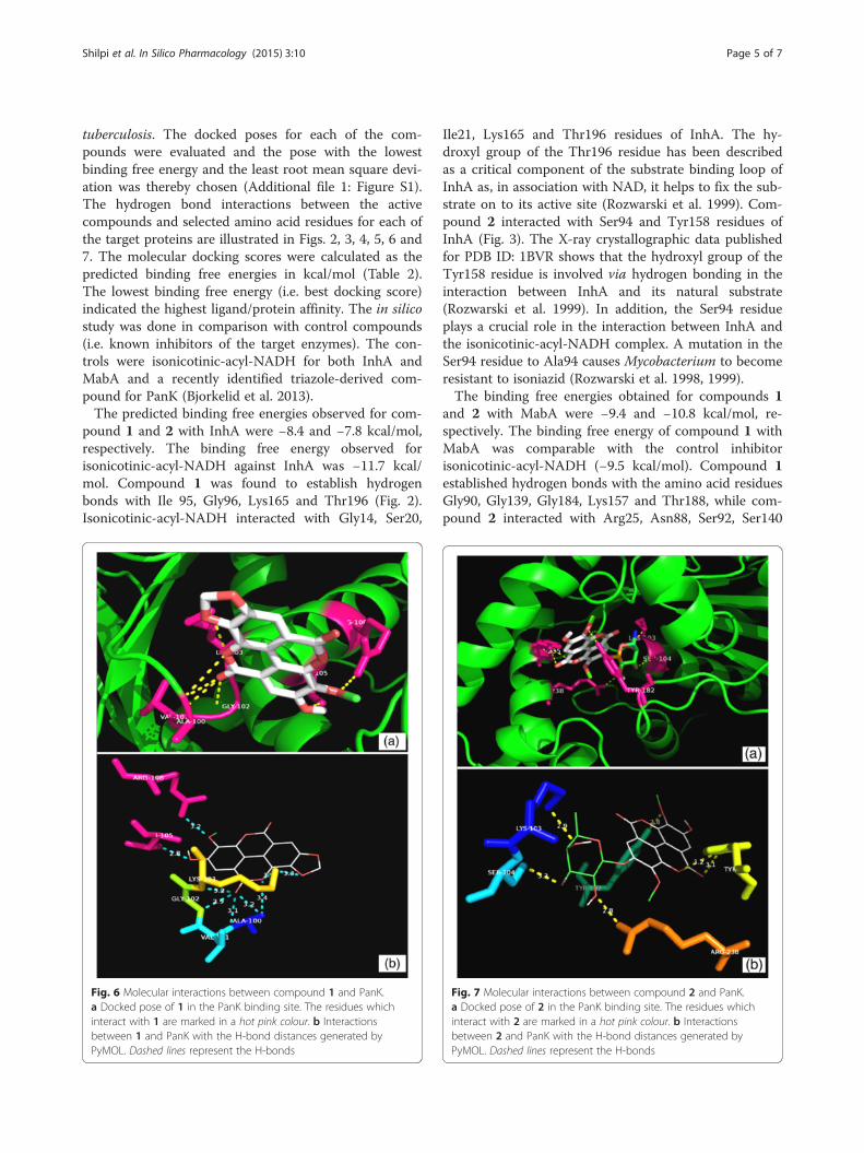
Fig. 7 Molecular interactions between compound 2 and PanK.a Docked pose of 2 in the PanK binding site. The residues whichinteract with 2 are marked in a hot pink colour. b Interactionsbetween 2 and PanK with the H-bond distances generated byPyMOL. Dashed lines represent the H-bonds
Shilpi et al. In Silico Pharmacology (2015) 3:10 Page 5 of 7

and Tyr153 (Figs. 4 and 5). Isonicotinic-acyl-NADHinteracted with Gly22, Asn24, Arg25, Ile27, Asp61,Asn88, Tyr153, Lys157, Thr188 and Thr191 residues ofMabA. Among these amino acids, the Ser140, Tyr153and Lys157 residues are those associated with the cata-lytic triad of MabA. The side chain of Tyr153 has a cen-tral role in the acid–base catalysis performed by thisenzyme (Kavanagh et al. 2008). Any mutation in theSer140 residue causes a loss of enzyme activity (Rosadoet al. 2012). The amino acid Gly90 has been shown to beinvolved in the complexation of MabA with its naturalNADPH cofactor while any mutation of Gly139 intoAla139 causes complete protein inactivation by freezingthe catalytic triad into a closed form (Poncet-Montangeet al. 2007; Rosado et al. 2012).The predicted binding energies for compounds 1 and
2 with PanK (type I) were −9.7 and −11.3 kcal/mol, re-spectively. The triazole-derived control inhibitor showeda binding energy of −10.6 kcal/mol towards PanK. Com-pound 1 established hydrogen bonds with the aminoacid residues Ala100, Val101, Gly102, Lys103, Thr105and Arg108 whereas compound 2 interacted with Lys103,Ser104, Tyr123, Tyr182 and Arg238 (Figs. 6 and 7). Thetriazole-derived control inhibitor displayed an interactiononly with the Tyr135 residue of PanK. The Ala100 toSer104 residues are known to be part of the PanK P-loopwhich is responsible for the holding of ATP duringcatalysis (Cheek et al. 2002; Bjorkelid et al. 2013). TheArg238 acts as a connecting residue between the phos-phorylated pantothenate and ATP, thereby aiding catalysis(Chetnani et al. 2010).
ConclusionsKnowledge on the molecular interactions of naturalproducts with essential M. tuberculosis targets is a po-tentially useful tool for the design and development ofnew anti-TB drugs. This in silico study revealed that twoplant-derived compounds had the potential to interact
with selected enzymes that were essential to M. tubercu-losis. One of them, identified as pteleoellagic acid (1)had a docking score to MabA comparable to the controlinhibitory substrate for this enzyme. Further work is re-quired to gain a better insight into structure-active siterelationships using a wider variety of structurally-relatedderivatives as well as to correlate the results of the dock-ing study with in vitro enzymatic experiments in thesearch for new anti-TB drugs.
Additional file
Additional file 1: Figure S1. Docked complexes of compound 1 andcompound 2 at the protein binding sites. Docking of 1 (a) and 2 (b) withInhA. Docking of 1 (c) and 2 (d) with MabA. Docking of 1 (e) and 2 (f)with PanK. Surfaces represent the protein while sticks represent the activecompounds. Different surface colours were chosen to represent differentproteins. (JPG 306 kb)
AbbreviationsInhA: 2-trans-enoyl-ACP reductase; MabA: β-ketoacyl-ACP reductase;MDR: Multi-drug resistant; NADH: Nicotinamide adenine dinucleotide(reduced form); PanK: Pantothenate kinase; PDB: Protein data bank;TB: Tuberculosis; XDR: Extensively-drug resistant.
Competing interestsAll authors declared that they have no competing interest.
Authors’ contributionsJAS, VS and AIG carried out the experiments and analysis of the dataassociated with the isolation and characterisation work. JAS, SS, MTA andSH carried out the experiments and analysis of the data associated with themolecular docking work. JAS, MTA, AIG and VS wrote the manuscript. VS, SS,JAS and SH co-worked on associated data collection and interpretation ofresults. All authors have read and approved the final manuscript.
AcknowledgementsThe authors would like to thank S.J. Uddin, M.M. Rahman, A. Khatun and ProfD.M. Ali (University of Rajshahi, Bangladesh) for collecting the plant material.They acknowledge H. Noltie (Royal Botanic Gardens, Edinburgh), B. Khan(Bangladesh National Herbarium, Dhaka) for helping with the botanicalidentification of Trewia nudiflora and Dr. T. Zhang (University of Strathclyde)and J. Tweedie (University of Glasgow) for running MS experiments. J.A.Shilpi would like to thank the University of Strathclyde for financial support.
Table 2 Predicted binding free energies (docking scores) and detailed interactions observed between compounds 1, 2 and thetarget enzymes
Test compound Protein name Predicted binding energy (kcal/mol) Interaction with amino acid residues
1 InhA −8.4 Ile95, Gly96, Lys165, Thr196.
MabA −9.4 Gly90, Gly139, Lys157, Gly184, Thr188.
PanK −9.7 Ala100, Val101, Gly102, Lys103, Thr105, Arg108.
2 InhA −7.8 Ser94, Tyr158.
MabA −10.8 Arg25, Ser92, Asn88, Ser140, Tyr153.
PanK −11.3 Lys103, Ser104, Tyr182, Tyr123, Arg238.
Isonicotinic-acyl-NADH (control) InhA −11.7 Gly14, Ser20, Ile21, Lys165, Thr196.
MabA −9.5 Gly22, Asn24, Arg25, Ile27, Asp61, Asn88, Tyr153,Lys157, Thr188, Thr191.
Triazole-derived compound (control) PanK −10.6 Tyr235.
Shilpi et al. In Silico Pharmacology (2015) 3:10 Page 6 of 7

Author details1Natural Products Research Laboratories, Strathclyde Institute of Pharmacyand Biomedical Sciences, University of Strathclyde, Glasgow, UK. 2PharmacyDiscipline, Life Science School, Khulna University, Khulna, Bangladesh.3Department of Biochemistry and Molecular Biology, University of Dhaka,Dhaka, Bangladesh. 4Bioinformatics Laboratory, QIMR Berghofer MedicalResearch Institute, Brisbane, Australia. 5School of Medicine, University ofQueensland, Brisbane, Australia.
Received: 1 September 2015 Accepted: 25 November 2015
ReferencesAllinger NL. Conformational analysis. 130. MM2. A hydrocarbon force field
utilizing V1 and V2 torsional terms. J Am Chem Soc. 1977;99(25):8127–34.Bjorkelid C, Bergfors T, Raichurkar AK, Mukherjee K, Malolanarasimhan K,
Bandodkar B, et al. Structural and biochemical characterization ofcompounds inhibiting Mycobacterium tuberculosis pantothenate kinase.J Biol Chem. 2013;288(25):18260–70.
Cheek S, Zhang H, Grishin NV. Sequence and structure classification of kinases.J Mol Biol. 2002;320(4):855–81.
Chetnani B, Kumar P, Surolia A, Vijayan M. M. tuberculosis pantothenate kinase:dual substrate specificity and unusual changes in ligand locations. J Mol Biol.2010;400(2):171–85.
Dashti Y, Grkovic T, Quinn RJ. Predicting natural product value, an exploration ofanti-TB drug space. Nat Prod Rep. 2014;31(8):990–8.
Ducasse-Cabanot S, Cohen-Gonsaud M, Marrakchi H, Nguyen M, Zerbib D,Bernadou J, et al. In vitro inhibition of the Mycobacterium tuberculosis β-ketoacyl-acyl carrier protein reductase MabA by isoniazid. Antimicrob AgentsCh. 2004;48(1):242–9.
Gasteiger J, Marsili M. Iterative partial equalization of orbital electronegativity—a rapid access to atomic charges. Tetrahedron. 1980;36(22):3219–28.
Gupta A, Bhakta S, Kundu S, Gupta M, Srivastava BS, Srivastava R. Fast growing,non-infectious and intracellularly surviving drug-resistant Mycobacteriumaurum: a model for high-throughput antituberculosis drug screening.J Antimicrob Chemother. 2009;64(4):774–81.
Guzman JD, Gupta A, Bucar F, Gibbons S, Bhakta S. Antimycobacterials fromnatural sources: ancient times, antibiotic era and novel scaffolds. Front Biosci.2012;17(5):1861–81.
Jackson M, McNeil MR, Brennan PJ. Progress in targeting cell envelopebiogenesis in Mycobacterium tuberculosis. Future Microbiol. 2013;8(7):855–75.
Johnson MM, Odell JA. Nontuberculous mycobacterial pulmonary infections.J Thorac Dis. 2014;6(3):210–20.
Kang QJ, Yang XW, Wu SH, Ma YL, Li L, Shen YM. Chemical constituents from thestem bark of Trewia nudiflora L. and their antioxidant activities. Planta Med.2008;74(4):445–8.
Kavanagh KL, Jornvall H, Persson B, Oppermann U. Medium- and short-chaindehydrogenase/reductase gene and protein families : the SDR superfamily:functional and structural diversity within a family of metabolic and regulatoryenzymes. Cel Mol Life Sci. 2008;65(24):3895–906.
Kondo Y, Toida T, Kusano G, Imai J. Specific inhibition of formation of acid-fastness in mycobacteria by 3,3′-di-O-methylellagic acid. Experientia.1979;35(5):599–600.
Marrakchi H, Lanéelle G, Quémard A. InhA, a target of the antituberculous drugisoniazid, is involved in a mycobacterial fatty acid elongation system, FAS-II.Microbiology. 2000;146(2):289–96.
Marrakchi H, Ducasse S, Labesse G, Montrozier H, Margeat E, Emorine L, et al.MabA (FabG1), a Mycobacterium tuberculosis protein involved in the long-chain fatty acid elongation system FAS-II. Microbiology. 2002;148(4):951–60.
Morris GM, Huey R, Lindstrom W, Sanner MF, Belew RK, Goodsell DS, et al.AutoDock4 and AutoDockTools4: automated docking with selective receptorflexibility. J Comput Chem. 2009;30(16):2785–91.
O’Boyle NM, Banck M, James CA, Morley C, Vandermeersch T, Hutchison GR.Open Babel: an open chemical toolbox. J Cheminform. 2011;3:33.
Poncet-Montange G, Ducasse-Cabanot S, Quemard A, Labesse G, Cohen-GonsaudM. Lack of dynamics in the MabA active site kills the enzyme activity:practical consequences for drug-design studies. Acta Crystallogr D.2007;63(8):923–5.
Quemard A, Sacchettini JC, Dessen A, Vilcheze C, Bittman R, Jacobs WR, et al.Enzymic characterization of the target for isoniazid in Mycobacteriumtuberculosis. Biochemistry. 1995;34(26):8235–41.
Rosado LA, Caceres RA, de Azevedo Jr WF, Basso LA, Santos DS. Role of serine140in the mode of action of Mycobacterium tuberculosis beta-ketoacyl-ACPreductase (MabA). BMC Res Notes. 2012;5:526.
Rozwarski DA, Grant GA, Barton DH, Jacobs Jr WR, Sacchettini JC. Modification ofthe NADH of the isoniazid target (InhA) from Mycobacterium tuberculosis.Science. 1998;279(5347):98–102.
Rozwarski DA, Vilcheze C, Sugantino M, Bittman R, Sacchettini JC. Crystalstructure of the Mycobacterium tuberculosis enoyl-ACP reductase, InhA, incomplex with NAD+ and a C16 fatty acyl substrate. J Biol Chem.1999;274(22):15582–9.
Santhosh RS, Suriyanarayanan B. Plants: a source for new antimycobacterialdrugs. Planta Med. 2014;80(1):9–21.
Shilpi JA (2009) Phytochemical and antimicrobial studies on Ludwigia ascendens,Trewia nudiflora and Hygrophila auriculata from Bangladesh. Ph. DDissertation, University of Strathclyde. http://ethos.bl.uk/OrderDetails.do?uin=uk.bl.ethos.510832. Accessed 12 Nov 2015.
Shilpi JA, Gray AI, Seidel V. Chemical constituents from Ludwigia adscendens.Biochem Syst Ecol. 2010;38(1):106–9.
Takayama K, Wang C, Besra GS. Pathway to synthesis and processing of mycolicacids in Mycobacterium tuberculosis. Clin Microbiol Rev. 2005;18(1):81–101.
Trott O, Olson AJ. AutoDock Vina: improving the speed and accuracy of dockingwith a new scoring function, efficient optimization, and multithreading.J Comput Chem. 2010;31(2):455–61.
WHO (World Health Organization) 2014, Global Tuberculosis Control WHO report,Geneva, Switzerland. http://www.who.int/tb/publications/global_report/en/.Accessed 02 Jun 2015.
Zumla A, Nahid P, Cole ST. Advances in the development of new tuberculosisdrugs and treatment regimens. Nat Rev Drug Discov. 2013;12(5):388–404.
Submit your manuscript to a journal and benefi t from:
7 Convenient online submission
7 Rigorous peer review
7 Immediate publication on acceptance
7 Open access: articles freely available online
7 High visibility within the fi eld
7 Retaining the copyright to your article
Submit your next manuscript at 7 springeropen.com
Shilpi et al. In Silico Pharmacology (2015) 3:10 Page 7 of 7