Embed Size (px)
Citation preview

i
Molecular Cytogenetic Methods for
Studying Curcuma Plants from Thailand
Tidarat Puangpairote
Faculty of Life and Environmental Sciences
University of Iceland
2013


Molecular Cytogenetic Methods for Studying Curcuma Plants from Thailand
Tidarat Puangpairote
30 ECTS LÍF 014M Research Project in Biology for Foreign Students
Advisor Kesara Anamthawat-Jónsson, Professor
Faculty of Life and Environmental Sciences School of Engineering and Natural Sciences
University of Iceland Reykjavik, January 2013

Molecular Cytogenetic Methods for Studying Curcuma Plants from Thailand
30 ECTS LÍF 014M Research Project in Biology for Foreign Students
Copyright © 2013 Tidarat Puangpairote
All rights reserved
Faculty of Life and Environmental Sciences
School of Engineering and Natural Sciences
University of Iceland
Askja
101, Reykjavik
Iceland
Telephone: 525 4000
Bibliographic information:
Tidarat Puangpairote, 2013, Molecular Cytogenetic Methods for Studying Curcuma Plants
from Thailand, Faculty of Life and Environmental Sciences, University of Iceland, pp. 63.
Reykjavik, Iceland, January 2013

Abstract
Species in the genus Curcuma L. (Zingiberacace) that are cultivated widely in Thailand for
phytoestrogen-producing rhizomes are called Wan chak motluk. Enzymatic-based methods
were modified in order to obtain metaphase spreads with high quality of chromosomes
from root tips of these Curcuma species. The results revealed that protoplast dropping with
modification in the hypotonic and the post drying steps was an efficient method for
chromosome preparation of C. comosa (2n=42, SB5403) using the combination of 8%
Cellulase and 3% Pectinase (K8) enzyme mixture. In addition, a new enzymatic drop-
squash method was developed in the present study produced well-spread metaphases with
very little cytoplasmic background in Curcuma sp.2 (NK5501). Moreover, the enzymatic
squash method using the combination of 10% Cellulase and 12% Pectinase (K12) enzyme
mixture gave satisfactory results for C. latifolia (KB5451), C. elata (CB5316) and
Curcuma sp.1 (PR5319), but moderate quality with C. comosa (2n=42, NP5411),
C. comosa (2n=63, SB5403) and Curcuma sp.3 (TK5502).
Conditions of the chromosome pretreatment and fluorescent in situ hybridization (FISH)
were optimized in order to obtain suitable protocols for rDNA-FISH mapping of Curcuma
species. The results showed that Proteinase K treatment could be omitted for chromosomes
prepared from protoplast dropping and enzymatic drop-squash methods, whereas 3-10
µg/ml of Proteinase K was needed to treat chromosomes prepared from the conventional
enzymatic squash method. Then, combined denaturation at 89°C for 10 min for re-probing
was successful to obtain strong fluorescent signals of 18S-25S rDNA in most Curcuma
species. However, the optimal denaturation temperature for C. comosa (2n=63, SB5403)
was 92°C for 10 min followed by 89°C for 10 min in the second denaturation. The separate
denaturation protocol whereby chromosomes were denatured in a Formamide and 1xSSC
solution at temperature up to 80°C was also experimented. This work will be continued
until the most efficient protocol for mapping of the ribosomal genes on chromosomes can
be obtained for all Wan chak motluk species and cultivars from different regions in
Thailand and then it will be possible to establish the genomic and genetic relationships
among these taxa.

vi
Table of Contents
List of Figures ................................................................................................................... viii
List of Tables ....................................................................................................................... ix
Acknowledgements ...............................................................................................................x
1 Introduction .....................................................................................................................1
1.1 Rationales .................................................................................................................1
1.2 Objectives .................................................................................................................3
2 Literature reviews ............................................................................................................4
2.1 Taxonomc studies .....................................................................................................4
2.2 Medicinal properties .................................................................................................5
2.3 Cytogenetic studies...................................................................................................6
2.3.1 Chromosome preparation ............................................................................... 6
2.3.2 Investigation of somatic chromosome number .............................................. 7
2.4 Molecular cytogenetic studies ................................................................................11
3 Materials and methods ..................................................................................................13
3.1 Plant materials ........................................................................................................13
3.2 Procedure of chromosome preparation and fluorescent in situ hybridization
(FISH) for Curcuma species ..................................................................................15
3.2.1 Chemicals and reagents ............................................................................... 15
3.2.2 Laboratory supplies ...................................................................................... 17
3.2.3 Equipments .................................................................................................. 17
4 Results .............................................................................................................................18
4.1 Procedures of chromosome isolation .....................................................................18
4.1.1 Chromosome pretreatment ........................................................................... 18
4.1.2 Chromosome preparation ............................................................................. 18
4.2 Pretreatment of chromosome preparation ..............................................................23
4.2.1 Removal of the coverslip ............................................................................. 23
4.2.2 Refixation .................................................................................................... 23
4.2.3 RNase treatment ........................................................................................... 23
4.2.4 ProteinaseK treatment .................................................................................. 24
4.2.5 Paraformaldehyde treatment ........................................................................ 24
4.2.6 Dehydration.................................................................................................. 24

vii
4.3 Chromosome hybridization and washing .............................................................. 24
4.3.1 Preparation of probe mixture ....................................................................... 24
4.3.2 Denaturation and hybridization .................................................................... 25
4.3.3 Washing and mounting ................................................................................ 26
5 Discussion ...................................................................................................................... 44
5.1 Techniques of chromosome preparation ............................................................... 44
5.2 Fluorescent in situ hybridization (FISH) ............................................................... 45
6 Conclusions .................................................................................................................... 46
References ........................................................................................................................... 47

viii
List of Figures
Figure 2.1: Geographical distribution of genus Curcuma L. .................................................... 5
Figure 2.2: Outline of the in situ hybridization procedure ...................................................... 11
Figure 3.1: Inflorescences and leaves of Wan chak motluk from seven provinces in
Thailand ................................................................................................................. 14
Figure 4.1: Modified steps of the drop enzyme method ........................................................... 22
Figure 4.2: Chromosome preparations of phytoestrogen-producing Curcuma species .......... 22
Figure 4.3: Reference micropipette tip .................................................................................... 26
Figure 4.4: Fluorescent in situ hybridization (FISH) of the 18S-25S rDNA genes of
phytoestrogen-producing Curcuma (Wan chak motluk) in NP5411,
TK5502 and KB5451. ............................................................................................ 41
Figure 4.5: Fluorescent in situ hybridization (FISH) of the 18S-25S rDNA genes of
phytoestrogen-producing Curcuma (Wan chak motluk) in CB5316 and
PR5319 ................................................................................................................. 42
Figure 4.6: Fluorescent in situ hybridization (FISH) of the 18S-25S rDNA genes of
phytoestrogen-producing Curcuma (Wan chak motluk) in NK5501 and
SB5403. .................................................................................................................. 43

ix
List of Tables
Table 2.1: Chromosome numbers of Curcuma species . ............................................................ 8
Table 3.1: Plant materials examined in this study, their origin and identification .................. 13
Table 3.2: Methods of chromosome isolation for Curcuma species ........................................ 16
Table 4.1: FISH conditions of NP5411 (C.comosa, 2n=42) using combined
denaturation ........................................................................................................... 28
Table 4.2: FISH condition of CB5316 (C.elata, 2n=63) using combined denaturation .......... 30
Table 4.3: FISH conditions of PR5319 (Curcuma sp.1, 2n=63) using combined
denaturation ........................................................................................................... 31
Table 4.4: FISH conditions of NK5501 (Curcuma sp.2, 2n=63) using combined
denaturation ........................................................................................................... 32
Table 4.5: FISH conditions of TK5502 (Curcuma sp.3, 2n=63) using combined
denaturation ........................................................................................................... 33
Table 4.6: FISH conditions of KB5451 (C.latifolia, 2n=63) using combined
denaturation ........................................................................................................... 34
Table 4.7: FISH conditions of SB5403 (C.comosa, 2n=63) using combined
denaturation ........................................................................................................... 35
Table 4.8: FISH conditions of SB5403 (C.comosa, 2n=63) using separated and
separated together with combined denaturation .................................................. 38
Table 4.9: Optimal condition for FISH in phytoestrogen-producing Curcuma species .......... 40

x
Acknowledgements
I would like to express my deepest and sincere gratitude to my advisor, Professor Kesara
Anamthawat-Jónsson, for her kindness, constructive guidance and suggestions as well as
thoughtful and meaningful discussions throughout my study. I would also like to express
my greatest appreciation to my PhD advisor, Associate Professor Puangpaka
Soontornchainaksaeng, for her guidance and valuable knowledge about the plants and the
research project.
My special thanks go to all members of the plant genetic laboratory (Room 383, Askja) for
all their help and wonderful friendship.
During the course of my study, I was fully supported by a scholarship from the Institute for
the Promotion of Teaching Science and Technology under the Development and Promotion
of Science and Technology Talents Project (DPST) of the Ministry of Higher Education,
Thailand.
Finally, my deepest appreciation is expressed to my parents for their love and
encouragement.

1
1 INTRODUCTION
1.1 Rationales
Wan chak motluk is one of the Thai medicinal plants, which has been popularily used for
treatment of gynecological disorders. This plant belongs to the genus Curcuma, tribe
Zingibereae of the subfamily Zingiberoideae, family Zingiberaceae (Kress et al., 2002;
Maknoi, 2006). As Thai traditional medicine, its rhizome or underground stem has been
used in the form of fresh, single-herbal, polyherbal capsules or hydro-alcoholic extracts as
remedy of abnormality of the uterus and ovarian hormone deficit. Products from Wan chak
motluk, which are extensively used for this purpose, come from the species Curcuma
comosa Roxb.
Predominant active compound extracted from rhizomes of C. comosa is diarylheptonoid
(Suksamrarn et al., 2007). This compound provides estrogenic-like actions that can be
supplemented for estrogenic hormone deficit in women. Moreover, the diarylheptanoid and
rhizome extracts of this species have been used as anti-inflammatory (Sodsai et al., 2007;
Thampithak et al., 2009) and nematocidal agents (Jurgens et al., 1994). In hamsters the
compound has been shown to reduce plasma cholesterol (Piyachaturawat et al., 1999).
Taxonomic assignment of genus Curcuma is ambiguous, most probably due to homoploid
hybridization, polyploidy speciation and also in cultivation peculiar morphology has been
selected. This has apparently led to the different levels of genetic and morphological
variation among taxa with obscure species boundaries (Škorničková et al., 2007; Záveská
et al., 2012).
In the case of Wan chak motluk, the product of these plants is commercialized based on
high medicinal values. For this reason Wan chak motluk has become popular in the
traditional herbal industries in Thailand and therefore is widely cultivated especially in the
north, northeastern, and central regions. However, Wan chak motluk is usually grown in the
same area with other Curcuma species which are quite similar in the morphological
appearances especially in the aboveground vegetative and the reproductive parts.
Moreover, related Curcuma species are called by the same local name Wan chak motluk
and to make the matter more confusing the typical Wan chak motluk is also called by
different local names when cultivated in different locations. The large morphological
variation, in both intra- and inter-population levels, has also caused a big confusion in plant
identification and medicinal uses. In general it is believed that Curcuma species with
similar morphology are the source of phytoestrogen and hence used together as polyherbal
products. However, the different plant varieties cultivated in different regions usually give
different chemical and active compounds. Precise identification of the raw materials should

2
therefore be seriously concerned, to avoid misuse which can make the medicinal end
products toxic and harmful.
The identification of Curcuma has traditionally been achieved by using taxonomic key
based on morphological data. Inflorescence is the best part that can be used to identify plant
species, but it is not always available in a complete form as Curcuma inflorescence is
perishable and short-lived and the flowering is highly seasonal. In addition to the
morphological variation even within species, this limited source of inflorescent data makes
identification of Wan chak motluk extremely difficult. To overcome this problem, a genetic
based approach which relies on characters that are independent of phenotypic variation,
must be developed. But prior to that we must understand how the species and cultivars of
phytoestrogen-producing Curcuma are genetically and genomically related.
Cytogenetics of Curcuma L., especially of Indian species, has been studied extensively in
terms of chromosome number and genome size (e.g. Leong-Skornickova et al., 2007). It is
known however that species with the same chromosome number may be distinctive in their
karyotypes. Chromosome size, position of centromere, number and position of secondary
constriction have been shown to vary significantly between species (Fukui and Nakayama,
1996). Characteristics about chromosome complements are therefore useful in
cytotaxonomy and can reveal phylogenetic relationships among closely related taxa at the
species and sub-species levels. However, there is very limited information about
chromosome morphology and structure in Curcuma because members of this genus possess
very small and numerous chromosomes and very high cytoplasmic contents resulting in
poor chromosome preparations using classical cytogenetic techniques. By using appropriate
modification of chromosome preparation techniques Soontornchainaksaeng and Jenjitikul
(2010) have successfully identified chromosomal variations in Wan chak motluk. Together
with the morphological data they have discovered that Wan chak motluk in Thailand
comprises five cultivars, possessing different cytotypes, but belonging to three Curcuma
species, i.e. C. comosa, C. elata and C. latifolia. In order to characterize these species and
cytotypes with better resolution a molecular cytogenetic approach must be adopted.
Fluorescence in situ hybridization (FISH), molecular cytogenetic technique, has proven to
be a power tool in plant cytotaxonomy: for identification of chromosome complements and
its variation due to hybridization and polyploidy, for studying genome and species
relationships, and for identifying ancestral origin of natural hybrids and allopolyploids
(Kato et al., 2005; Anamthawat-Jónsson, 2001). Physical mapping of highly conserved
sequences such 5S and 18S-25S ribosomal genes has been performed extensively and
successfully in numerous plant species. For Curcuma the first successful attempt has
already been made (Soontornchainaksaeng and Anamthawat-Jónsson, 2011). However,
different Wan chak modluk species and cultivars are invariably recalcitrant and so a much
more effective method of chromosome preparation must be developed in order to obtain
the quality suitable for further in situ hybridization analysis.

3
1.2 Objectives
1) To develop efficient methods of preparation of metaphase spreads in
phytoestrogen-producing Curcuma species (Wan chak motluk) for molecular cytogenetic
studies.
2) To develop suitable protocols for mapping of the ribosomal genes on
chromosome of Wan chak motluk species and cultivars from different growing regions in
Thailand.

4
2 Literature reviews 2.1 Taxonomic studies Zingiberaceae, one of the largest monocotyledon families, is found worldwide especially in
the tropics. It comprises of 53 genera and more than 1,500 species (Augsonkitt et al., 2004;
Záveská et al., 2012), with its center of diversity in South and Southeast Asia. Thailand is
one of the richest ginger plant diversity in the world. Twenty-six genera and nearly 300
species have been identified (re. Chen, 1989 in Larsen and Larsen, 2005). The family
Zingiberaceae was previously divided into four tribes: Alpinieae, Globbeae, Hedychieae
and Zingibereae, based on morphological characters (Larsen and Larsen, 2005). However,
Kress and colleagues (2002) proposed a new system based on DNA sequences of the
nuclear internal transcribed spacer (ITS) and plastid matK regions. This new proposed
system divides the family into four subfamilies and six tribes: subfamily Siphonochiloideae
(tribe Siphonochileae), subfamily Tamijioideae (tribe Tamijieae), subfamily Alpinioideae
(tribes Alpinieae and Ridelieae) and subfamily Zingiberoideae (tribes Globeae and
Zingibereae).
The genus Curcuma L. is rhizomatous perennial herbs with at least 120 species distributed
in tropical and subtropical Asia especially in South and Southeast Asia (Joseph et al., 1999;
Záveská et al., 2012) with a few species distributing to China, Australia and the South
Pacific (Škorničková et al., 2007). The distribution of Curcuma L. is shown in Figure 2.1.
Thirty-six species of Curcuma were preliminary reported in Thailand (Larsen, 1996) but
increased to 38 species recently (Maknoi, 2006). New species are expectedly to be
continuously discovered which would lead to a further increase in the number of species in
Curcuma. This genus was previously classified to the tribe Hedychieae, which is newly
assigned to the tribe Zingibereae of subfamily Zingiberoideae (Maknoi, 2006). Most
species have been utilized for economic, medicinal, ornamental and cultural purposes.
Wan chak motluk is phytoestrogen-producing Curcuma that has been popularly used for
treatment of gynecological diseases. It is now identified to five cultivars belonging to three
species: C. comosa, C. elata and C. latifolia based on the differentiation of inflorescent,
floral and leaf morphology (Soontornchainaksaeng and Jenkittikul, 2010).

5
Figure 2.1: Geographical distribution of genus Curcuma L. (from Maknoi, 2006)
2.2 Medicinal properties
Zingiberaceae is one of the largest plant families that composed of many medicinally
important species especially members of genus Curcuma. In addition to medicinal
purposes, several species can be used for food, spices, condiments, dyes, perfumes,
aesthetics, and as ornamental plants (Sirirugsa, 1999; Chareerach et al., 2007). More than
50 species of this genus has been found throughout Thailand (Chaveerach et al., 2007, see
Sirirugsa, 1996). Nearly all of Curcuma species have been used for medicinal purposed
since ancient time with anti-inflammatory, hypocholestraemic, choleratic, antimicrobial,
insect repellent, antirheumatic, antifibrotic, antivenomous, antiviral, antidiabetic,
antihepatotoxic and anticancerous (Sasikumar, 2005).
Wan chak motluk is one of Thai medicinal plants that have been popularly used for
treatment of gynecological diseases. As Thai traditional medicine, its rhizome,
underground stem, has been used in the form of fresh, single-herbal, poly-herbal capsules
or hydro-alcoholic extracts to remedy several symptoms. It is used for women suffered
from uterus and ovarian hormone deficit (re. Soontornchainaksaeng and Jenjittikul, 2010)
and as remedy of uterine inflammation and pain (Piyachaturawat et al., 1999). Moreover, it
can be used for treatment of postpartum uterine bleeding (Piyachaturawat et al., 1999) and
general tonic after childbirth (re. Soontornchainaksaeng and Jenjittikul, 2010). In addition,
women suffered from menstrual pain, cramp, and hot flashes during menopause can be
relieved by using this plant (Suksanrarn et al., 2008; Winuthayanon et al., 2009). It is also
used as emmenagogue, abortificient, cholagogue and carminative (Phipitphibunsuk, 2007).
Products from Wan chak motluk used for these purposes come from the species Curcuma
comosa Roxb.

6
2.3 Cytogenetic studies
2.3.1 Chromosome preparation
High quality of chromosome preparation is the principal requirement for further
chromosome analysis. The cytological techniques, therefore, have been developed from
time to time to obtain the precise information of the chromosome numbers and
chromosome structures in plant species. The basic principles for handling the mitotic and
meiotic chromosomes of all plant species are similar and consist of collection of plant
materials, pretreatment (skipped for meiotic studies), fixation, storage, maceration and
staining. However, these procedures are modified depending on objectives of the
experiments and personal preference of the cytologists. Nevertheless, there are valid
reasons for testing different steps of maceration which is the major and critical stage in
obtaining well-spread chromosomes and high quality of preparation.
Softening the plant tissues with hydrochloric acid (HCl) has been widely used in plant
cytogenetic techniques because of several advantages. It is easy to process, not so time-
consuming but give satisfactory results in many plant species (Fukui and Nakayama, 1996).
Hydrolysis in 1N HCl at 60°C was extensively used in cytological studies of family
Zingiberaceae; for instance, some species of Kaempferia and Zingiber (Saensouk and
Saensouk, 2004), many species of Curcuma in Bangladesh (Islam, 2004), some species of
Curcuma (Thepsen, 2000) and phytoestrogenic-Curcuma species in Thailand
(Soontornchainaksaeng and Jenjittikul, 2010). However, a few modifications were applied
for some studies, for example, Škorničková and co-workers (2007) used the combination of
hydrochloric acid and ethanol (1:1) for maceration step to study the chromosome numbers
of Indian Curcuma species. However, with this treatment it was difficult to obtain a
monolayer of separated cells on slide. The observation was supported by several researches
on alfalfa chromosome preparations using similar procedures. The presence of cell walls is
a major obstacle in making a good chromosome preparation. The bulk tissues with thick
cell walls resist the pressure of squashing and tapping steps and, thus, make it difficult to
spread the chromosomes.
In 1944, Emsweller and Stuart first suggested the possibility of using enzymatic maceration
of plant tissues to prepare good chromosome spreads (Fukui and Nakayama, 1996). Since
then, enzyme treatment with mainly combination of pectinase and cellulase has been
developed continually in both cytological and molecular cytogenetic studies. Song and co-
workers (1988) developed an efficient procedure for alfalfa chromosome preparation using
enzyme treatment. They found that a combination of 5% Pectinase and 2.5% Cellulase and
incubation for 60 – 90 minutes had several advantages; for example, good cell distribution
and well spread chromosomes on a single plane. Since then the enzyme treatment has been
widely applied in molecular cytogenetic studies especially in the in situ hybridization
(ISH). This is mainly because of the advantages of sample preparation using this enzymatic
method, especially that chromosomes are spreaded evenly in a single layer and also free of
cytoplasmic debris. Besides the enzymatic squash method, protoplast dropping, an
alternative preparation technique, has been found suitable for shoot tips and small

7
chromosomes (Anamthawat-Jónsson, 2003). This preparation has been carried out with
several plant species; for example, Betula (Thórsson et al., 2007), Castanopsis,
Lithocarpus and Quercus in the family Fagaceae (Chokchaichamnankit et al., 2007). The
maceration of shoot tips from these plants was successful in the combination of 2.5%
Pectinase and 2.5% Cellulase. In the case of Curcuma, the combination of 2% Pectinase
and 2% Cellulase was used to study chromosome variations in Curcuma zedoaria from
Bangladesh (Islam et al., 2007).
2.3.2 Investigation of somatic chromosome number
Chromosome numbers of Curcuma have been investigated extensively. Somatic
chromosome numbers of this genus are known to be highly variable, 2n=20-105
(Škorničková et al., 2007). Moreover, high variation in the 2n number has been found
within species; for instance, Curcuma angustifolia (2n=46, 81 and 92) and C. parviflora
(2n=24, 28, 30, 32, 34, 36 and 56) (Maknoi, 2006). The somatic chromosome numbers of
Curcuma are summarized in Table 2.1.
Curcuma species in Thailand can be classified into five groups based on the information of
chromosome numbers complemented with morphological characters (Maknoi, 2006, see
Sirisawad et al., 2003). Group 1: 2n=2x=42 comprises Curcuma species with long,
cylindrical inflorescences, large leaves and psuedostem. This group includes C. aurantiaca,
C. petiolata and C. roscoeana. Curcuma species in group 2, 2n=3x=63, are early-flowering
with normally spreading of coma bracts and with big and branching of rhizomes. This
group comprises of C. aeruginosa, C. elata, C. rubescens, C. xanthorhiza and C. zedoaria.
Group 3, 2n=2x=48, is also early-flowering Curcuma with very short peduncles and non-
branching rhizome. Curcuma angustifolia is the only species in this group. Curcuma in
group 4, 2n=24 or 32, has long and erect peduncles, short spike and narrow or linear leaves.
Curcuma alismatifolia and C.parviflora are in this group. Group 5 with various somatic
numbers, 2n=20, 24, 28, 34, 36 and 56, has under-developed rhizomes, white staminode
and anther without spur. This group is composed of C. harmandii, C. parviflora and C.
rhabdota.
Moreover, a recent study has shown that Wan chak motluk (phytoestrogenic-Curcuma)
cultivated in Thailand can be classified into three species or five cultivars based on
chromosome numbers together with morphological characters. These are C. comosa with
2n = 42 and 63, C. elata with 2n = 63 and C. latifolia with 2n = 63 and 84
(Soontornchainaksaeng and Jenjittikul, 2010).

8
Table 2.1: Chromosome number of Curcuma species
Species Chromosome numbers Origin of plant
material
material
n 2n
C. aeruginosa Roxb 63 Thailand
63 Thailand
28-35 63 Thailand
63, 84 Bangladesh
C. alismatifolia Gagnep. 16 32 Thailand
32 Thailand
32 Thailand
32 Thailand
16 32 Thailand
C. amada Roxb 42 Bangladesh
C. angustifolia Roxb. 42 Bangladesh
(C. attenuate Wall. Ex Baker) 42 84 Thailand
42 84 Thailand
(C. sessile Gage) 46, 92 Thailand
C. aromatica Roxb. 63 Thailand
63 Bangladesh
C. aurantiaca van Zijp 21 42 Thailand
C. bicolor J. Mood & K. Larsen Thailand
C. cochinchinensis Gagnep. Thailand
C. comosa Roxb. 42 India
C. cf. comosa Roxb. 63 Thailand
C. ecomata Craib Thailand
C. elata Roxb. 28-35 63 Thailand
C. flaviflora S.Q. Tong Thailand
C. glans K. Larsen & J. Mood Thailand
C. gracillima Gagnep. 24 Thailand
24 Thailand
16 32 Thailand
C. cf. gracillima Gagnep. 40 Thailand
C. harmandii Gagnep. 10 20 Thailand
20 Thailand
10 20 Thailand
C. latifolia Roxb. 63 Bangladesh
C. leucorhiza Roxb. Thailand

9
Table 2.1 (cont.)
Species Chromosome numbers Origin of plant
material
material
n 2n
C. longa L. 63 Thailand
63 Bangladesh
C. mangga Val. 42 Indonesia
63 Indonesia
C. maehongson C. Maknoi Thailand
C. nakornsawan C. Maknoi Thailand
C. parviflora Wall. 14, 17, 18, 28 28, 34, 36 Thailand
32 Thailand
30 Thailand
42 Thailand
32 Thailand
30 Thailand
16 32 Thailand
12, 14, 17, 18,
28
24, 28, 34,
36, 56
Thailand
C. petiolata Roxb. 42 Thailand
21 42 Thailand
42 Bangladesh
C. cf. petiolata Roxb. 42 Thailand
C. pierreana Gagnep. Thailand
C. pitsanulok C. Maknoi Thailand
C. raktakanta Mangaly & M. Sabu 21 42 Thailand
C. ranong C. Maknoi Thailand
C. rhabdota Sirirugsa & M.
Newman
24 Thailand
12 24 Thailand
C. roscoeana Wall. 21 42 Thailand
C. rubescens Roxb. 28-35 63 Thailand
63 Bangladesh
C. rubrobrateata Skornickova,
Sabu & Prasanthk.
Thailand
C. singularis Gagnep.
(C. aff. oligantha Trimen)
42 Thailand
40 Thailand
C. sparganiifolia Gagnep. Thailand
C. stenochila Gagnep. Thailand

10
Table 2.1 (cont.)
Species Chromosome numbers Origin of plant
material
material
n 2n
C. thorelii Gagnep. 36 Thailand
17 34 Thailand
36 Thailand
38? Thailand
34 Thailand
17 34 Thailand
C. viridiflora Roxb. Thailand
C. xanthorhiza Roxb. 63 Thailand
28-35 63 Thailand
63 Bangladesh
C. zedoaria (Christ.) Rosc. 63 Thailand
42 Thailand
28-35 63 Thailand
63 Bangladesh
Species in bracket is the previous name before re-identification.
However, species with the same chromosome number may be distinctive in its karyotype.
Chromosome complements contain such characters as chromosome size, type (position of
centromere) and presence of secondary constriction have been found to be varied
significantly between species (Fukui and Nakayama, 1996). Chromosome numbers and
karyomorphology data, therefore, are valuable tools in the studies of taxonomic
relationships and evolutionary patterns. In Curcuma species, earlier studies were
concentrated only on the investigation of chromosome number but no information on
chromosome morphology and structure, due to very small sizes of chromosome (Joseph et
al., 1999). Moreover, very high cytoplasmic contents commonly found in Curcuma species
make an analysis difficult and time-consuming. Nevertheless, Joseph and his colleagues
(1999) carried out the karyotyping of six species of the genus Curcuma for the first time by
using computational karyomorphology indices. The results showed that three diploid
species with 2n=2x=42, C. comosa, C. haritha and C. malabarica, and three triploid
species with 2n=3x=63, C. aerugenosa, C. caesia and C. raktacanta, had symmetrical
karyotypes. However, some variations were also found in chromosome morphology and
structure among species.

11
2.4 Molecular cytogenetic studies
In situ hybridization (ISH), a molecular cytogenetic technique, was described for the first
time in the late sixties (Pardue and Gall, 1969). About ten years later a non-isotopic version
of ISH was developed for use with plant and mammal chromosomes (e.g. Gerlach and
Peacock, 1980; Gerhard et al., 1981), after which a fluorescent based technique,
Fluorescent in situ hybridization (FISH), became possible in human cytogenetics
(Manuelidis et al., 1982). FISH quickly obtained a world-wide popularity due to its great
sensitivity and specificity, and was proven applicable for use with practically any species.
This technique is a powerful method for localization of nucleic acid sequences (either DNA
or RNA) in the cytoplasm, organelles, chromosomes or nuclei of biological material
(Leitch et al., 1994). ), It has proven to be a useful tool in plant cytotaxonomy: for
identification of variation due to hybridization and polyploidy; for studying genome and
species relationships; and for identifying parental origin of hybrids and allopolyploids
(Anamthawat-Jónsson et al, 1990; Anamthawat-Jónsson, 2001; Kato et al., 2005). Even
though there are several modifications of ISH depending on the objectives, the procedures
are very similar in the main steps that are outlined in Figure 2.2.
Figure 2.2: Outline of the in situ hybridization procedure (modified from Schwarzacher
and Heslop-Harrison, 2000)
Preparation of target materials Labeling of probes
Pretreatment of chromosome targets
Denaturation of probe and target materials
Detection of hybridization sites
Hybridization
Microscopy and imaging data analysis

12
Several sources of DNA or RNA can be used as probe for FISH depending on the
objectives of the experiment. These are, for example, repetitive sequences, both highly
conserved sequences (18S-25S and 5S ribosomal genes and telomeres) and fast evolving
species-specific sequences, e.g. pericentromeric sequences, mobile elements,
heterochromatic and satellite sequences (see review in Anamthawat-Jónsson, 2010). Of all
the DNA sequences, ribosomal DNA (rDNA) is extensively applied in plant molecular
cytogenetics, whereas the use of whole genomic DNA as probe (GISH, Anamthawat-
Jónsson et al., 1990) is equally popular but for different biological investigations.
Many researches involve the mapping of 5S and 18S-25S ribosomal genes on plant
chromosomes and the main objective is to differentiate individual ribosomal gene bearing
chromosomes and in association with the secondary constrictions or NORs (Nucleolar
Organizing regions). Both similarity and variation of the 5S and 18S-25S rDNA can be
used to clarify the karyotype differentiation and to infer phylogenetic and evolutionary
relationships among closely related taxa. The rDNA-FISH mapping has been used
successfully to study numerous plant species in a wide range of genera and families of both
gymnosperms and angiosperms. However, physical mapping of rDNA genes is still very
limited regarding the family Zingiberaceae, due to minute chromosome size and high
cytoplasmic content. Only one study of chromosome mapping of the 18S-25S ribosomal
genes on phytoestrogen-producing Curcuma species (Wan chak motluk) has been reported
(Soontornchainaksaeng and Anamthawat-Jónsson, 2011). The results revealed for the first
time hybrid features in these Curcuma taxa. The next questions are how these different
Wan chak motluk cultivars in Thailand have evolved, by natural means or in cultivation,
and by what molecular and cellular mechanisms exactly. This study should be further
carried out using more intensive molecular cytogenetic techniques and a complete sample
size and then it will be possible to reveal the genetic and genomic relationships in all of
cultivars and species of Wan chak motluk cultivated in Thailand.

13
3 Materials and Methods
3.1 Plant materials
Rhizomes of phytoestrogen-producing Curcuma were collected from the cultivated sites in
seven provinces throughout Thailand (Table 3.1), including one northern province (Prae),
one north-eastern province (Nongkai), one eastern province (Chanthaburi), two western
provinces (Kanchanaburi and Tak) and two in central Thailand (NakhonPathom and
Saraburi). The plant samples were grown in pots at Mahidol University, Salaya Campus,
during this study. Actively growing root tips were collected from the germinating rhizomes
grown in pots for chromosome isolation.
Inflorescences and leaves of selected plant materials were shown in Figure 3.1.
Table 3.1: Plant materials examined in this study, their origin and identification
Plant
identity
Province Location Species Chromosome
number (2n)
NP 5411 Nakhon
Pathom
Central Thailand C. comosa 42
SB 5403 Saraburi Central Thailand C. comosa 63
CB 5316 Chanthaburi Eastern Thailand C. elata 63
PR 5319 Prae Northern Thailand Curcuma sp.1 63
NK 5501 Nongkai North-eastern Thailand Curcuma sp.2 63
TK 5502 Tak Western Thailand Curcuma sp.3 63
KB 5451 Kanchanaburi Western Thailand C. latifolia 63
The phytoestrogen-producing Curcuma in Prae, Nongkai and Tak are still under
identification. However, these were primarily identified as C. elata - C. latifolia complex
due to some ambiguous morphological characters.

14
Figure 3.1 Inflorescences and leaves of Wan chak motluk from seven provinces in
Thailand. A-B Curcuma comosa (2n=42) from Nakhonpathom. C-D Curcuma comosa
(2n=63) from Saraburi. E-F Curcuma elata (2n=63) from Chantaburi. G-H Curcuma sp.1
(2n=63) from Prae. I-J Curcuma sp.2 (2n=63) from Nongkai. K-L Curcuma sp.3 (2n=63)
from Tak. M-N Curcuma latifolia (2n=63) from Kanchanaburi.
A B C D
E F G H
I J K L
M N

15
3.2 Procedures of chromosome preparation and fluorescent in situ hybridization (FISH) for Curcuma species
Chromosomes of selected plant materials were prepared using three different methods:
enzymatic squash, drop-enzyme and protoplast dropping as described in Table 3.2.
Table 3.2: Methods of chromosome isolation for Curcuma species
Plant
identity Species
Method of chromosome preparation
Enzymatic
squash
Drop-enzymatic
squash
Protoplast
dropping
NP 5411 C. comosa, 2n=42 /
SB 5403 C. comosa, 2n =63 / /
CB 5316 C. elata /
PR 5319 Curcuma sp.1 /
NK 5501 Curcuma sp.2 /
TK 5502 Curcuma sp.3 / /
KB 5451 C. latifolia /
Chemicals, reagents, equipment and laboratory supplies used in chromosome preparation
and in situ hybridization in this study are listed as following.
3.2.1 Chemicals and reagents
1) Paradichlorobenzene solution: Dissolve 16 g of Paradichlorobenzene crystal in
1000 ml of distilled water and incubate the solution in an oven at 60°C overnight.
2) Enzyme mixture for enzymatic squash method (K12), 5 ml: Cellulase Onozuka
R10 0.5 g, Pectinase (Sigma, P4716) 0.6 ml in enzyme buffer 5 ml. It is convenient to
make 500 µl aliquots in 1.5 ml microtubes and store at -20°C.
3) Enzyme buffer for the squash method, 500 ml: Mix 20 ml of stock A and 30 ml
of stock B solution in 450 ml of distilled water. To prepare stock A (Citric acid 0.1 M),
dissolve Citric acid monohydrate (Sigma, C7129) 10.5 g in 500 ml of distilled water; Stock
B (Trisodium citrate 0.1 M), dissolve trisodium citrate (Sigma, S4641) 14.7 g in 500 ml of
distilled water.
4) Enzyme mixture for protoplast dropping method (K8), 10 ml: Cellulase Onozuka
R10 0.8 g, Pectinase (Merck) 0.3 g in 10 ml of enzyme buffer (containing 75 mM KCl and
7.5 mM EDTA, pH 4). It is convenient to make 100-150 µl aliquot in 0.5 ml microtubes
and store at -20°C.
5) 75 mM KCl solution: Stock 1 M KCl solution, dissolve KCl (Sigma, P9541) 7.5
g in 100 ml of distilled water. The stock solution should be filter-sterilized. To prepare

16
working solution, mix 7.5 ml of stock solution with 92.5 ml of distilled water and keep ice-
cold.
6) Acetic acid, 45%: Make 45% (v/v) from glacial acetic acid (Sigma, 27225) and
keep in small dropper bottles.
7) DAPI, 1 µg/ml: Prepare 100 µg/ml stock solution of DAPI (4,6-diamidino-2-
phenylindole) in distilled water. Make 1 µg/ml working solution by diluting the stock
solution at 1:100 in McIlvaine’s citrate buffer, pH 7 (mixing 18 ml of 0.1 M citric acid
monohydrate and 82 ml of 0.2 M Na2HPO4 dihydrate). Store in aliquots at -20°C.
8) Fixative: A mixture of 1 part glacial acetic acid and 3 parts 96% ethanol. Prepare
fresh daily and keep ice-cold.
9) Ethanol, 96%: Keep ice-cold for dehydrating slides after protoplast dropping.
10) Liquid nitrogen or dry ice
11) Dehydration solutions (70%, 85%, 90% and 96% ethanol): 96% ethanol is
diluted to make 70%, 85% and 90% (v/v) ethanol solutions.
12) RNaseA solution: Prepare 10 mg/ml stock solution by dissolving 10 mg of
RNaseA (Sigma, R6513) in 10 mM Tris pH 8, and 15 mM NaCl. Store in aliquots at
-20°C. For RNase treatment, add 20 µl of stock solution in 40 ml of 2xSSC (for a small
Coplin jar) or 30 µl of stock solution in 60 ml of 2xSSC (for a big Coplin jar) to give a
final concentration of 5 µg/ml.
13) Proteinase-K solution: Prepare 0.5 mg/ml stock solution by dissolving 0.025 g
of Proteinase-K in 50 ml buffer (20 mM Tris pH 8.0 and 2 mM CaCl2). Store the aliquots
at -20°C. Dilute the stock solution in Proteinase-K buffer to 2-10 µg/ml of working
solutions.
14) 50 mM MgCl2 (Proteinase-K stop buffer): Dissolve 4.7605 g of MgCl2in 1000
ml of distilled water.
15) Paraformaldehyde, 4%, 50 ml: Add 2 g of Paraformaldehyde in 40 ml distilled
water. Heat the solution up to 60-70°C and swirl periodically. Add 1-2 drops of 4 M NaOH
to clear the solution. Measure pH and if it is 7.0-7.5, the solution is ready to use. Make up
the volume to 50 ml and let cool to room temperature. It is convenient to prepare a larger
quantity and make aliquots of 50 ml that can be stored at -20°C. The solution can be reused
up to 5 times.
16) SSC buffers: To prepare the stock solution 20xSSC, dissolve 175.4 g of NaCl
(3 M) and 88.2 g of Trisodium citrate (0.3 M) in 800 ml of distilled water. Adjust pH to
7.0-7.5 with 1 N HCl and autoclave. The stock 20xSSC is diluted to make 2xSSC and other
SSC working solutions, which can be stored at room temperature.
17) 4xSSC with Tween 20, 500 ml: Mix 100 ml of 20xSSC with 400 ml of distilled
water. Add 1 ml of Tween 20 and mix well.
18) Dextran sulfate, 50%: Dissolve 2.5 g of dextran sulfate in 5 ml of distilled
water. Heat up to 70°C to help dissolving. Aliquot in 1.5 ml microtubes and store at room
temperature. Heat up to 40°C before use.
19) Formamide: Make 50 ml aliquots of 100% formamide and store in -20°C.
20) SDS, 10%: Dissolve sodium dodecyl sulfate 1 g in 10 ml distilled water. Keep
aliquots at room temperature.
21) Probes: Two ribosomal DNA probes were used in this study. (1) Clone pTa71
which contains a part of 18S and the entire 5.8S and 25S coding region together with non-
transcribed spacers from wheat (Gerlach and Bedbrook, 1979) were used as 18S-25S rDNA
probe. (2) Clone pTa794 which contains a complete fragment of the 5S rDNA gene and

17
spacer regions from wheat (Gerlach and Dyer, 1980) were used as 5S rDNA. Probe
labelling followed Anamthawat-Jónsson (2010).
22) Double distilled water (ddH2O): Autoclave distilled water and keep aliquot at
room temperature.
23) Antifade mountant: Citifluor antifade is used for this study
3.2.2 Laboratory supplies
1) Plastic Petri-dishes
2) Plastic transfer pipettes
3) Acid-cleaned microscopic slides: Clean new slides in chromosulfuric acid 2%
(Merck, 102499) at least overnight, washed in running tap water for 15-20 minutes and
rinsed briefly in distilled water. Dry the slides in an incubator at 37°C and keep the cleaned
slides in 96% ethanol (for the squash method) or in distilled water (for the protolast
droping method) until use.
4) Plastic forceps, fine-forceps, dissecting needles and razor blades
5) Filter paper (Whatman 1001-110)
6) Coverslips (18x18 mm, 22x22 mm and 22x30 mm)
7) Dropper bottles
8) Lint-free wipes
9) Diamond-tip pen
10) Test-tube racks
11) Slide storage box
12) Micropipette (P10, P100, P200 and P1000) and disposable micropipette tips
13) Microtube rack
14) Microtubes 1.5 ml
15) Nylon mesh, fine pores
16) Plastic centrifuge tubes 50 ml with cap
17) Coplin jar (5-slots and 8-slots)
18) Scissors
19) Nail vanish
20) Air-tight plastic box and test tubes to make a humid chamber
21) Slide tray
22) Black paper
23) Thermometer
3.2.3 Equipments
1) Incubator 37°C (GALLENKAMP)
2) Stereo microscope (Leica L2)
3) Phase-contrast microscope (Nikon, ALPHAPHOT-2, YS2)
4) Epifluorescent microscope with the filter for visualization of DAPI (Nikon
ECLIPSE E800)
5) Microcentrifuge (MIKRO 200, Hettich)

18
4 Results
4.1 Procedures of chromosome isolation
4.1.1 Chromosome pretreatment
Root tips of phytoestrogen-producing Curcuma species (Wan chak motluk) were collected
in the afternoon and pretreated in saturated paradichlorobenzene (PDB) solution for 6 hours
in a refrigerator. Then, the materials were fixed in a 3:1 mixture of alcohol and glacial
acetic acid until use. This produced satisfactory results in term of high metaphase index.
4.1.2 Chromosome preparation
Chromosome preparations in this study were based on enzymatic digestion to remove cell
wall and cytoplasm. Two main methods of preparing chromosome spreads in Curcuma
species were squashing and protoplast dropping.
Enzymatic squash method
1) Briefly blot dry fixed root tips (2-3 roots each time) and transfer into the enzyme
buffer. Submerge the root tips and change the buffer twice in 20 minutes at room
temperature.
2) Place the root tips in the 37°C pre-incubated enzyme mixture.
3) Incubate root-tips in the enzyme mixture at 37°C for 20-40 minutes depending
on root sizes and time in fixative.
- Fixed roots that have been stored in fixative for a long time require longer
digestion time.
- Roots from soil such as Curcuma spp. also require longer time than those from
seedlings and germinated seeds.
4) Transfer the digested root tips to the enzyme buffer at room temperature. If not
used within 1 hour, the root tips in buffer should be kept at 4°C until squashing. However,
these should be kept longer than one day.
5) Place one root tip on a wipe-dried microscope slide and add one small drop of
45% acetic acid.
6) Cut the tip, 1-2 mm long under a stereo microscope. Keep the meristematic tip
on the slide and discard the rest. Remove the surrounding acid with a filter paper and
replace with another drop of 45% acetic acid. Let the meristem stay in the acetic acid for 3-
5 minutes.
7) Tease the meristem into the acetic acid with dissecting needles, under the stereo
microscope. Remove the debris as much as possible with a forcep and place a coverslip
(18x18 mm). If too big drop of 45% acetic acid, let the solution evaporate for a while.

19
8) Tap vertically and very gently with a tip of needle over the coverslip to spread
cells under the coverslip. Place the slide in between two sheets of filter papers and press
firmly with a thumb to make cells flat.
9) Label the slide clearly with a pencil on the matted end and mark the coverslip
edges underneath using diamond-tip pen.
10) Examine briefly in the phase-contrast microscope to check mitotic index and
the quality of cells and chromosome spreads.
11) Dip the slide into liquid nitrogen using a thong and let the slide stay submerged
until bubbles have disappeared, pull it up and flick the coverslip off with a razor blade.
(While dipping, hold the slide not too tightly to prevent the slide broken-up or cracked).
12) Air-dry the slide on the test-tube rack. The slides can be stored in a storage box
at 4°C for a few months.
13) Add 8-10 µl of DAPI (1 µg/ml) and place a coverslip (22x22 mm). Examine the
DAPI-stained slide in an epifluorescent microscope using UV filter block with 340-380
excitation and 430-450 emission wavelengths. Write down coordinates of the best mitotic
spreads and observe the quality of cells in terms of the cytoplasm remaining and the cell
flatness. Capture images of well spread metaphase chromosomes at 100x objective lens
with Nikon Digital Camera DXM1200F for chromosome analysis.
Protoplast dropping method
1) Transfer the fixed root tips to a Petri-dish. Rinse the root tips with distilled water
and change the water twice in 30 minutes at room temperature.
2) Blot dry fixed root tips briefly and place the samples in 100 µl of enzyme
mixture (two roots medium-size or one root large-size). Incubate overnight (18-24 hours) at
room temperature, however, the incubation time depending on the root size and how long
they have been kept in fixative.
3) Break the root tissue in its enzyme mixture into cell suspension using
micropipette tip (200 µl). Filter the suspension by transferring them with the same pipette
tip onto nylon mesh, placed over an opened 1.5 ml microtube. Let the suspension run down
the inside wall of microtube. Remove the nylon mesh with cell debris.
4) Add 1.5 ml of cold 75 mM KCl solution into the cell/protoplast suspension.
Invert gently to mix and let stand at room temperature for 15-20 minutes.
5) Spin down the protoplast suspension at 7000 rpm for 5 minutes and discard the
supernatant.
6) Add 1 ml of fresh and cold fixative to the pellet. Re-suspend the protoplasts very
gently using the same pipette tip as before. Let stand at room temperature for 15 minutes.
7) Spin down the suspension at 7000 rpm for 5 minutes and discard the supernatant.
8) Repeat the step of fixative treatment (step 6 and 7) at least two more times to
clean cytoplasm. It is best if one of the fixative treatments stays cold over night.
9) Add 50-150 µl of fresh and cold fixative into the protoplast pellet and gently mix
again. The volume of fixative is depended on the amount of pellet.
10) Protoplast suspension should be checked for the overall quality (e.g. cloudy is
good, but flaky is not) and density of cell (thin or thick) by mixing the cell suspension and
let stand for a few minutes. Pipette only the upper part of cell suspension onto ice-cold and
wet slide from a distance over 20 cm in height. This method was modified slightly to
obtain cell flatness as follows. When the drop is beginning to dry up, add a drop of 45%

20
acetic acid and then place 22x22 mm of coverslip. Place the slide in between two sheets of
filter papers and press firmly with a thumb to make cells flat.
11) Dip the slide into liquid nitrogen for a moment and leave it there until bubbles
have disappeared or place the slide on dry ice for a while. Then, flick the coverslip off with
a razor blade.
12) Label the slide clearly with a pencil on the matted end and mark the coverslip
edges underneath with the diamond-tip pen.
13) Add 8-10 µl of DAPI (1 µg/ml) and place a coverslip (22x30 mm). Examine the
DAPI-stained slide in an epifluorescent microscope using UV filter block. If cells are too
dense, repeat the step of fixative treatment (step 6 and 7). Add more volume of fixative to
dilute the cell suspension. The remaining protoplast suspension can be kept in a refrigerator
for several days.
Drop enzyme method
This method was slightly modified from the enzymatic squash to avoid a loss of very small
size of meristematic root tips in enzyme mixture. The protocol is described as following.
1) Briefly blot dry fixed root tips and transfer into enzyme buffer. Submerge the
root tips and change the buffer twice in 20 minutes at room temperature.
2) Cut root tips for 1-2 mm long, and place on the slide. Remove the excess buffer
as much as possible with a filter paper.
3) Drop 10 µl of enzyme mixture to the root tip area (Figure 4.1A). Place the slide
into the Petri-dish and cover with the lid (Figure 4.1B). Then, incubate at 37°C for 20-30
minutes (Figure 4.1C).
4) Pipette the rest of enzyme mixture back and add a drop of enzyme buffer to the
root tip (Figure 4.1D). Let stand for a while and remove the surrounding buffer with filter
paper. Replace another drop of 45% acetic acid. Leave the meristem in the acetic acid for at
least 10 minutes. Before squashing, remove the old acetic acid and add a new drop of 45%
acetic acid.
5) Tease the meristem into the acetic acid with dissecting needles, under the stereo
microscope. Remove the debris as much as possible and place a coverslip (18x18 mm). If
too big drop of 45% acetic acid, let the solution evaporate for a while.
6) Tap vertically and very gently with a tip of needle over the coverslip to spread
cells under the coverslip. Place the slide in between two sheets of filter papers and press
firmly with a thumb to make cells flat.
7) Label the slide clearly with a pencil on the matted area and mark the coverslip
edges underneath with diamond-tip pen.
8) Examine briefly in the phase-contrast microscope to check for mitotic index and
the quality of cells and chromosome spreads.
9) Dip the slide into liquid nitrogen for a moment, leave it there until bubbles
disappear and flick the coverslip off with a razor blade. (While dipping, hold the slide not
too tightly to prevent the slide broken-up or cracked).
10) Air-dry the slide on the test-tube rack. The slides can be stored in a storage box
at 4°C for a few months.
11) Add 8-10 µl of DAPI (1 µg/ml) and place a coverslip (22x22 mm). Examine the
DAPI-stained slide in an epifluorescent microscope using UV filter block with 340-380

21
excitation and 430-450 emission wavelengths. Write down the coordinates of mitotic cells
as many as possible and notice the quality of cells in terms of the cytoplasm remaining and
the cell flatness. Capture images of well spread metaphase chromosomes at 100x objective
lens with Nikon Digital Camera DXM1200F for chromosome analysis later.
Figure 4.1: Modified steps of the drop enzyme method
To obtain well spread chromosome, in a single layer, and free or very little of
cytoplasm, different procedures of chromosome preparation were carried out in several
species of phytoestrogen-producing Curcuma (Wan chak motluk) as shown in Table 3.2.
The results shown in Figure 4.3 indicated that the enzymatic squash method (enzyme
mixture, K12) gave moderate spread of chromosomes and thick cytoplasm in NP5411 (A),
SB5403 (B) and TK5502 (C). However, this method produced desirable results in KB5451
(D), CB5316 (E) and PR5319 (F), i.e. better spread chromosomes and less cytoplasm left.
The protoplast dropping (enzyme mixture, K8) and the drop enzyme squash (enzyme
mixture, K12) turned out to be the best methods in SB5403 (G and H) and NK5501 (K and
L), respectively, but this was not a good preparation for TK5502 (I and J).
A B A
C D

22
Figure 4.2: Chromosome preparations of phytoestrogen-producing Curcuma species. . A-F
Enzymatic squash method: NP5411 (A); SB5403 (B); TK5502 (C); KB5451 (D); CB5316
(E); and PR5319 (F). G-J Protoplast dropping method: SB5403 (G-H) and TK5502 (I-J).
K-L Drop enzyme method with NK5501.
A B C
B
D E F
G
L
H
L
I
L
J
L
K
L
L
L

23
4.2 Pretreatment of chromosome preparation
4.2.1 Removal of the coverslip
DAPI scanning is necessary for Curcuma species due to the very small size of
chromosomes. Furthermore, DAPI gives unequivocal identification of chromosomes as it
binds specifically to DNA, whereas the presence of cytoplasm makes it very difficult to
differentiate chromosomes from the cytoplasmic content if examined with phase-contrast
microscope. After the DAPI screening, the coverslip must be removed before proceeding to
the chromosome pretreatment.
Removal of coverslip after DAPI scanning
1) Drop 2xSSC or 4xSSC around the edges of coverslip. Let stand for a few
minutes and pull the coverslip out gently.
2) Treat the slide in fresh fixative for 5 minutes.
3) Dehydrate in the ethanol series, 70%, 85% and 96%, 2 minutes each and air dry.
4) Keep slides in a storage box in the refrigerator until use.
Removal of coverslip for re-probing
Chromosomes can be re-probed if the fluorescent signal is too weak and the chromosomes
still have good structure and bright DAPI signal.
1) Drop 2xSSC around the edges of coverslip. Let stand for a moment and pull the
coverslip very gently.
2) Treat slides in 2xSSC for 2 hours at room temperature and then re-fixation as
4.2.2
4.2.2 Re-fixation
1) Treat slides in fresh fixative for 10 minutes at room temperature.
2) Wash twice in 96% ethanol, 10 minute each and air dry.
4.2.3 RNase treatment
1) Treat slide with 5 µg/ml of RNaseA at 37°C for 1 hour.
2) Wash twice with 2xSSC at 37°C, 5 minutes each.

24
4.2.4 Proteinase-K treatment (Optional)
This treatment is depending on the quality of chromosome preparation.
1) Incubate slides in Proteinase-K buffer at 37°C for 5 min.
2) Treat slides in Proteinase-K 2-10 µg/ml at 37°C for 20 minutes. (Use high
concentration of Proteinase-K if there is a lot of cytoplasm.)
3) Stop the reaction with Proteinase-K stop buffer for 1 minute at room
temperature.
4) Wash twice with 2xSSC at room temperature, 5 minutes each.
4.2.5 Paraformaldehyde treatment
1) Treat the slides with 4% Paraformaldehyde for 15-20 minutes at room
temperature.
2) Wash twice with 2xSSC, 5 minutes each at room temperature.
4.2.6 Dehydration
Dehydrate the slides in the ethanol series, 70%, 90% and 96%, 2 minutes each and
air dry.
Notes:
- In condition of pretreatment at 37°C, Coplin jars and solutions such as 2xSSC
should be pre-incubated at least 2 hours to make sure that the temperature is up to 37°C.
4.3 Chromosome hybridization and washing
4.3.1 Preparation of probe mixture
Reagents Quantity (µl) per slide
1. Dextran sulfate (20% final) 4
2. Formamide (50% final) 10
3. 20xSSC (2x final) 2
4. 10% SDS (0.5% final) 0.5
5. Green probe x
6. Red probe y
7. ddH20 z
Total volume 20 µl per slide

25
1) Preparation of master mix for solution 1-4 is recommended and over-prepare it
by one reaction, for example, master mix of 5 reactions should be prepared for 4 slides to
avoid insufficient solution. Mix very well, spin down and distribute 16.5 µl of master mix
to each reaction in microtubes.
2) Before adding probes to each microtube, quick-spin to bring down the probe to
the bottom and mix well before use. Probe quantity is depending on its concentration.
3) Mix all of components well and centrifuge briefly.
4) Boil the probe mixture for 6 min and keep on ice for at least 5 minutes or keep
on ice until use. Quick spin before applying probe mixture to slide.
5) Apply the probe mixture to the slide and place 22x22 mm (for enzymatic squash
preparation) or 22x30 mm (for protoplast dropping preparation) coverslip over. Avoid air
bubbles. Seal all edges of coverslip with nail vanish and wait a few minute to dry.
4.3.2 Denaturation and hybridization
Two kinds of denaturation condition, combined denaturation and separated together with
combined denaturation, were carried out with phytoestrogen-producing Curcuma species.
Combined denaturation
After applying the probe mixture to the slide, denature together in an in situ thermocycle
chamber at 89-92°C, for 10-15 minutes. Denaturation condition was depended on the plant
species and the cell quality. Appropriate temperature regimes must be set up for each type
of denaturation equipment, for example if the denaturation is based on hot and dry air the
temperatures should be lower than with moist air or steamed heat. After the denaturation,
incubate the slides in humid chamber at 37°C overnight, for hybridization.
Separated together with combined denaturation
Chromosomes and probes were separately denatured and further denaturation together in
the thermal control machine.
1) Pretreatment of chromosome was followed as section 4.2 except for dehydration
step. Slides can be soaked in 2xSSC at room temperature until use.
2) For separated denaturation, slides were treated in the solution for separated
denaturation (usually 70% formamide and 1xSSC) at 72-80°C for 3-5 minutes. Then,
dehydrate slides in ice-cold ethanol series, 70%, 90% and 96%, twice and 3 minutes each
and air dry briefly. During the slide treatment, boil probe mixtures for 7 minutes and keep
on ice for at least 5 minutes.
3) Apply probe mixture to the slide and place 22x30 mm coverslip. Denature
together in an in situ thermocycle chamber at 92°C for 10 minutes.
4) Incubate slides in humid chamber at 37°C overnight.

26
4.3.3 Washing and mounting
1) Remove nail vanish carefully and soak the slides in 2xSSC at 37°C briefly to let
the coverslip down and remove out of probe mixture and wash in the second 2xSSC at
37°C again (optional).
2) Wash the slides in 0.1xSSC at 55°C 3 times, 5 minutes each, to remove unbound
and loosely bound probes.
3) Treat slides with warm 2xSSC, 37°C and let stand at room temperature (at least
30 minutes for a small jar and 45 minutes for a bigger jar)
4) Wash slides in 4xSSC with Tween at room temperature for 5 minutes.
5) Apply 10 µl of DAPI to wet slide, rinse briefly with distilled water in either front
or back side of slide after 1.5 – 2 minutes and air-dry briefly.
6) Apply 19-20 µl of Citifluor antifade and place 22x30 mm coverslip. Gently press
out the air bubbles.
7) Keep slides in a dark and cool place until ready for examination.
Notes:
- To pipette up the viscous solution such dextran sulfate, Tween 20 and Citifluor,
the micropipette tip should be cut up to 1-2 mm. Draw the solution up slowly and wait for
15-20 seconds.
- Pipetting water up to the exact amount of solution and marking its level with a
permanent marker are recommended to be a reference as shown in Figure 4.3. Compare the
pipetted amount of viscous solution to a reference every time to obtain the exact quantity.
Figure 4.3: Reference micropipette tip

27
To obtain the best resolution of rDNA genes signals, condition of chromosome
pretreatment and FISH was varied in phytoestrogen-producing Curcuma species (Wan chak
motluk) as shown in Table 4.1-4-8. The modification was depending on the quality of
chromosome preparation; for example,
1) ProteinaseK treatment was applied in species with high content of cytoplasm.
2) Chromosomes and probes were normally denatured by combined denaturation at
89°C for 10 min in most selected Curcuma species, C. comosa (2n=42, NP5411), C.
comosa (2n=63, SB5403), C. elata (2n=63, CB5316), Curcuma sp.1 (PR5319), Curcuma
sp.2 (NK5501), Curcuma sp.3 (TK5502) and C. latifolia (KB5451). However, this
separation at 89°C was not successful for SB5403, therefore, higher temperature and also
separation in Formamide solution were tried out. The optimal condition for FISH and the
signals of rDNA were shown in Table 4.9 and Figure 4.4-4.6.

28
Table 4.1: FISH condition of NP5411 (C. comosa, 2n=42) using combined denaturation
P12-1 P12-12
NP5411 S01 K10-13 NP5411 S06 K12-1
Enzymatic squash Enzymatic squash
FISH run FISH 12-1 (new) FISH 12-10 (reprobed) FISH 12-15 (new) FISH 12-21 (reprobed)
reprobe reprobe
Pretreatment condition no refixative refixative refixative refixative
Standard pretreatment re-pretreatment full pretreatment re-pretreatment
without ProK with ProK 2 µg/ml, 20 min with ProK 4 µg/ml, 20 min without ProK
with Paraformaldehyde 20 min
with Paraformaldehyde 20 min with Paraformaldehyde 20 min with Paraformaldehyde 20 min
Total ProK = 2 µg/ml Total ProK = 4 µg/ml
Total Paraformaldehyde = 40 min Total Paraformaldehyde = 40 min
Temperature and time
for denaturation
89°C, 15 min
89°C, 10min
89°C, 10 min
89°C, 10 min
probe Green = pTa71 Green = pTa71 Green = pTa71 Green = pTa794
Red = pTa794 Red = pTa71 Red = pTa71 Red = pTa71
DAPI good weak to good good good
Results green = negative, both = negative both = negative to positive green = negative
(1 cell positive, may be not real
signal) (very weak to weak signal) red = most are negative
red = negative (questionable)

29
Table 4.1 (cont.)
P12-13
NP5411 S04 K12-1
Enzymatic squash
FISH run FISH 12-15 (new) FISH 12-21 (reprobe)
reprobe
Pretreatment condition 2x SSC, 1 hr refixative
refixative
full pretreatment re-pretreatment
with ProK 4 µg/ml, 20 min without ProK
with Paraformaldehyde 20 min with Paraformaldehyde 20 min
Total ProK = 4 µg/ml
Total Paraformaldehyde = 40 min
Temperature and time for 89°C, 10 min 89°C, 10 min
denaturation
probe Green = pTa71 Green = pTa794
Red = pTa71 Red = pTa71
DAPI good weak to good
results both = negative to positive green = questionable
(very weak to weak) red = positive (weak to OK)

30
Table 4.2: FISH condition of CB5316 (C. elata, 2n=63) using combined denaturation
P12-3
CB5316 S01
Enzymatic squash
FISH run FISH 12-2 (new) FISH 12-15 (reprobe)
reprobe
Pretreatment condition no refixative refixative
standard pretreatment re-pretreatment
without ProK with ProK 4 µg/ml, 20 min
with Paraformaldehyde 20 min with Paraformaldehyde 20 min
Total ProK 4 µg/ml
Total Paraformaldehyde = 40 min
Temperature and time for 89°C, 10 min 89°C, 10 min
denaturation
probe Green = pTa71 Green = pTa71
Red = pTa794 red = pTa71
DAPI good to very good very weak to OK
cell lifted
results questionable positive (bright signals)

31
Table 4.3: FISH condition of PR5319 (Curcuma sp.1, 2n=63) using combined denaturation
P12-5 P12-11
PR5306 S01 K12-2 PR5319 S04 K12-1
Enzymatic squash Enzymatic squash
FISH run FISH 12-4 (new) FISH 12-6 (reprobe) FISH 12-12 (new) FISH 12-23 (reprobe)
reprobe reprobe
Pretreatment condition refixative refixative refixative refixative
standard pretreatment re-pretreatment full pretreatment full pretreatment
without ProK without ProK with ProK 4 µg/ml, 20 min with ProK 6 µg/ml, 20 min
with Paraformaldehyde 20 min with Paraformaldehyde 20 min with Paraformaldehyde 20 min with Paraformaldehyde 24 min
No ProK Total ProK = 10 µg/ml
Total Paraformaldehyde = 40 min Total Paraformaldehyde = 44 min
Temperature and time
for denaturation 89°C, 15 min 89°C, 15 min 89°C, 10 min 89°C, 10 min
Probe Green = pTa71 Green = pTa71 Green = pTa71 no green
Red = pTa794 Red = pTa794 Red = pTa71 Red = pTa71
DAPI OK Weak to OK OK Weak to OK
Results both = negative both = negative both = positive Red = positive (OK-good)
(in cell of no or very few
cytoplasm) (better signal)

32
Table 4.4: FISH condition of NK5501 (Curcuma sp.2, 2n=63) using combined denaturation
P12-17 P12-18
NK5501 S01 K12-6 NK5501 S04 K12-6
Drop enzymatic squash Drop enzymatic squash
FISH run FISH 12-18 (new) FISH 12-19 (reprobe) FISH 12-20 (new) FISH 12-21 (reprobe)
Pretreatment condition refixative refixative refixative refixative
standard pretreatment re-pretreatment standard pretreatment re-pretreatment
without ProK without ProK without ProK with ProK 4 µg/ml, 20 min
with Paraformaldehyde 20 min with Paraformaldehyde 20 min with Paraformaldehyde 15 min with Paraformaldehyde 20 min
No ProK Total ProK = 4 µg/ml
Total Paraformaldehyde = 40 min Total Paraformaldehyde = 35 min
Temperature and time 89°C, 10 min 89°C, 10 min 89°C, 15 min 89°C, 10 min
for denaturation
Probe no green Green = pTa794 Green = pTa794 Green = pTa794
Red = pTa71 Red = pTa71 Red = pTa71 Red = pTa71
DAPI OK- good weak - OK weak - OK very weak - bad
results negative to positive (OK) green = negative green = negative green = negative
red = positive (OK) red = negative to positive red = negative to positive
(very weak) (???)

33
Table 4.5: FISH condition of TK5502 (Curcuma sp. 3, 2n=63) using combined denaturation
P12-6
TK5502 S03
Enzymatic squash
FISH run FISH 12-7 (new) FISH 12-10 (reprobe)
reprobe
Pretreatment condition no refixative refixative
standard pretreatment re-pretreatment
without ProK with ProK 2 µg/ml, 20 min
with Paraformaldehyde 20 min with Paraformaldehyde 20 min
further pretreatment
with ProK 1 µg/ml, 20 min
without Paraformaldehyde
Total ProK = 3 µg/ml
Total Paraformaldehyde = 40 min
Temperature and time 89°C, 10 min 89°C, 10 min
for denaturation
Probe Green = pTa71 Green = pTa71
Red = pTa794 Red = pTa71
DAPI good weak to good
Results both = negative both = positive
(in cell of very few to no cytoplasm)

34
Table 4.6: FISH condition of KB5451 (C. latifolia, 2n=63) using combined denaturation
P12-16
KB5451 S04
Enzymatic squash
FISH run FISH 12-17 (new) FISH 12-21 (reprobe)
reprobe
Pretreatment condition 2xSSC, 1 hr and refixative refixative
full pretreatment re-pretreatment
with ProK 10 µg/ml, 20 min without ProK
with Paraformaldehyde 20 min with Paraformaldehyde 20 min
Total ProK = 10 µg/ml
Total Paraformaldehyde = 40 min
Temperature and time 89°C, 10 min 89°C, 10 min
for denaturation
Probe Green = pTa71 Green = pTa794
Red = pTa71 Red = pTa71
DAPI OK- good weak - OK
results both = negative to positive green = questionable
(very weak to weak) red = positive (OK)

35
Table 4.7: FISH condition of SB5403 (C. comosa, 2n=63) using combined denaturation
P12-2 P12-20
SB5403 S02/2 SB5403 S02 K8
Enzymatic squash protoplast dropping
FISH run FISH 12-2 (new) FISH 12-5 (reprobe) FISH 12-21 (new) FISH 12-22 (reprobe)
reprobe wihout
re-pretreatment reprobe
Pretreatment condition no refixative refixative refixative
standard pretreatment standard pretreatment re-pretreatment
without ProK without ProK with ProK 2 µg/ml, 20 min
with Paraformaldehyde 20 min with Paraformaldehyde 20 min with Paraformaldehyde 20 min
NO ProK Total ProK = 2 µg/ml
Total Paraformaldehyde 20 min Total Paraformaldehyde = 40 min
Temperature and time 89°C, 10 min 89°C, 10 min 90°C, 10 min 90°C, 10 min
for denaturation
Probe Green = pTa71 Green = pTa71 Green = pTa794 Green = pTa71
Red = pTa794 Red = pTa794 Red = pTa71 Red = pTa71
DAPI good weak to OK good OK
Results negative negative negative negative
(probes are outside the
cell)

36
Table 4.7 (cont.)
P12-21 P12-22
SB5403 S03 K8 SB5403 S01 K8
protoplast dropping protoplast dropping
FISH run FISH 12-22 (new) FISH 12-23 (reprobe) FISH 12-23 (new) FISH 12-24 (reprobe)
reprobe reprobe
Pretreatment condition refixative refixative refixative refixative
standard pretreatment re-pretreatment standard pretreatment re-pretreatment
without ProK without ProK without ProK without ProK
with Paraformaldehyde 20 min with Paraformaldehyde 24 min with Paraformaldehyde 24 min with Paraformaldehyde 20 min
NO ProK No ProK
Total Paraformaldehyde = 44 min Total Paraformaldehyde = 44 min
Temperature and time 92°C, 10 min 89°C, 10 min 89°C, 25 min 92°C, 10 min
for denaturation
Probe Green = pTa71 no green no green no green
Red = pTa71 Red = pTa71 Red = pTa71 Red = pTa71
DAPI OK weak to OK OK - good weak to OK
Results negative positive (OK-very good) negative negative
(but cells are lifted and not
flat)

37
Table 4.7 (cont.)
P12-23 P12-25
SB5403 S04 K8 SB5403 S04 K12
protoplast dropping Enzymatic squash
FISH 12-23 (new) FISH 12-24 (reprobe) FISH 12-26 (new)
reprobe
Pretreatment condition refixative refixative refixative
full pretreatment re-pretreatment Standard pretreatment
with ProK 6 µg/ml, 20 min without ProK without ProK
with Paraformaldehyde 24 min with Paraformaldehyde 20 min with Paraformaldehyde 20 min
Total ProK = 6 µg/ml No ProK
Total Paraformaldehyde = 44 min
Total Paraformaldehyde 20
min
Temperature and time 89°C, 25 min 92°C, 10 min 93°C, 10 min
for denaturation
Probe no green no green no green
Red = pTa71 Red = pTa71 Red = pTa71
DAPI OK-good OK very weak to weak
Cells are damaged
Results negative most are negative negative
some are positive (weak)
38
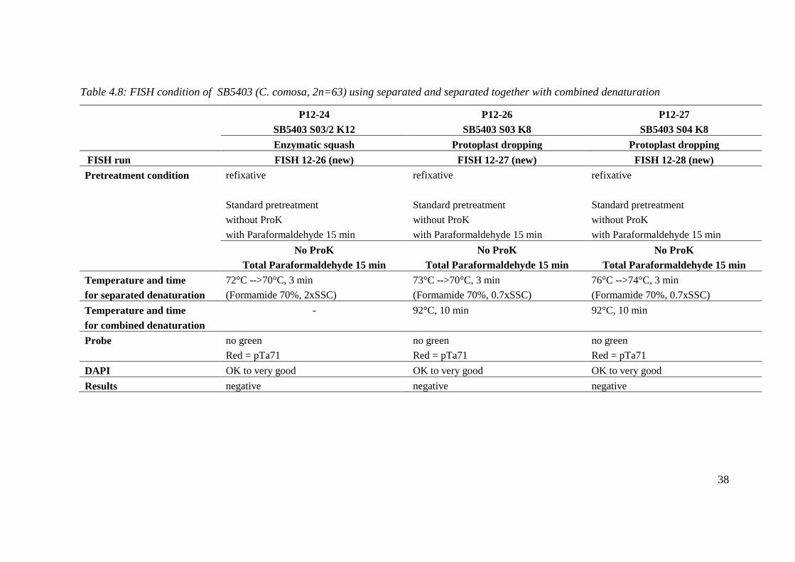
Table 4.8: FISH condition of SB5403 (C. comosa, 2n=63) using separated and separated together with combined denaturation
P12-24 P12-26 P12-27
SB5403 S03/2 K12 SB5403 S03 K8 SB5403 S04 K8
Enzymatic squash Protoplast dropping Protoplast dropping
FISH run FISH 12-26 (new) FISH 12-27 (new) FISH 12-28 (new)
Pretreatment condition refixative refixative refixative
Standard pretreatment Standard pretreatment Standard pretreatment
without ProK without ProK without ProK
with Paraformaldehyde 15 min with Paraformaldehyde 15 min with Paraformaldehyde 15 min
No ProK No ProK No ProK
Total Paraformaldehyde 15 min Total Paraformaldehyde 15 min Total Paraformaldehyde 15 min
Temperature and time 72°C -->70°C, 3 min 73°C -->70°C, 3 min 76°C -->74°C, 3 min
for separated denaturation (Formamide 70%, 2xSSC) (Formamide 70%, 0.7xSSC) (Formamide 70%, 0.7xSSC)
Temperature and time - 92°C, 10 min 92°C, 10 min
for combined denaturation
Probe no green no green no green
Red = pTa71 Red = pTa71 Red = pTa71
DAPI OK to very good OK to very good OK to very good
Results negative negative negative

39
Table 4.8: (cont.)
P12-28 P12-29
SB5403 S05 K8 SB5403 S06 K8
Protoplast dropping Protoplast dropping
FISH run FISH 12-28 (new) FISH 12-29 (new)
Pretreatment condition refixative refixative
re-pretreatment re-pretreatment
without ProK without ProK
with Paraformaldehyde 15 min with Paraformaldehyde 15 min
No ProK No ProK
Total Paraformaldehyde 15 min Total Paraformaldehyde 15 min
Temperature and time 73°C -->70°C, 6 min 80°C -->77°C, 5 min
for separated denaturation (Formamide 70%, 0.7xSSC) (Formamide 70%, 0.7xSSC)
Temperature and time 92°C, 10 min 92°C, 10 min
for combined denaturation
Probe no green no green
Red = pTa71 Red = pTa71
DAPI OK to very good OK to very good
Results negative negative

40
Table 4.9: Optimal condition for FISH in phytoestrogen-producing Curcuma species
Plant
identity
Method of
preparation
Slide that
obtain
signals
Chromosome pretreatment Combined denaturation
Refixative RNase
(for new slide)
Proteinase K Paraformaldehyde Dehydration Probe Temperature
and Time New slide Reprobe New
slide
Reprobe
NP5411 Enzymatic
squash
New / / 4 µg/ml,
20 min
- 20 min - / pTa71
(red and green)
89°C, 10 min
TK5502 Enzymatic
squash
Reprobed / / 1 µg/ml,
20 min
2 µg/ml,
20 min
20 min 20 min / pTa71
(red and green)
89°C, 10 min
KB5451 Enzymatic
squash
New / / 10 µg/ml,
20 min
- 20 min - / pTa71
(red and reen)
89°C, 10 min
CB5316 Enzymatic
squash
Reprobed / / - 4 µg/ml,
20 min
20 min 20 min / pTa71
(red and green)
89°C, 10 min
PR5319 Enzymatic
squash
New / / 4 µg/ml,
20 min
- 20 min - / pTa71
(red and green)
89°C, 10 min
NK5501
Drop
enzyme
Reprobed / / - - 20 min 20 min / Red= pTa71
Green = pTa794
89°C, 10 min
SB5403
Protoplast
dropping
Reprobed / / - - 20 min 20 min / pTa71
(red and green)
92°C, 10 min -->
89°C, 10 min

41
Figure 4.4: Fluorescent in situ hybridization (FISH) of the 18S-25S rDNA genes of
phytoestrogen-producing Curcuma (Wan chak motluk). Figs. A-C NP5411 (C. comosa,
2n=42). Figs. D-F TK5502 (Curcuma sp.3). Figs. G-L KB5451 (C. latifolia, 2n=63); Figs.
G-I metaphase cell and Figs. J-L interphase cell
A B C
D E F
G H I
J K L

42
Figure 4.5: Fluorescent in situ hybridization (FISH) of the 18S-25S rDNA genes of
phytoestrogen-producing Curcuma (Wan chak motluk). Figs. A-FCB5316 (C. elata,
2n=63); Figs. A-C metaphase cell; and Figs. D-F interphase cell. Figs. G-L PR5319
(Curcuma sp.1, 2n=63); Figs. G-I interphase cell and Figs. J-L prometaphase and
interphase cell.
A B C
D E F
G H I
J K L

43
Figure 4.6: Fluorescent in situ hybridization (FISH) of the 18S-25S rDNA genes of
phytoestrogen-producing Curcuma (Wan chak motluk). Figs. A-D NK5501 (Curcuma sp.2,
2n=63). Signals were presented in the incomplete metaphase cells. Figs. E-H SB5304 (C.
comosa, 2n=63); Figs. E-F interphase cell; and Figs. G-H incomplete metaphase cell.
A B
C D
E F
G H

44
5 Discussion
5.1 Techniques of chromosome preparation
Enzymatic-based methods were modified to prepare the chromosome spreads for
phytoestrogenic-Curcuma species (Wan chak motluk). In addition to the quality of plant
materials, the procedures and enzyme compositions are the main point in obtaining good
quality of chromosome preparations with well-spread metaphases and clean background
without cytoplasm (Anamthawat-Jónsson, 2010). In this study, enzyme mixtures K8 and
K12 were proven very good enzyme compositions for protoplast dropping and drop
enzyme squash methods, respectively, if based on the very good chromosome preparation
in C. comosa (2n=63, SB5403) and Curcuma sp.2 (NK5501). However, most of metaphase
cells were incomplete when using drop enzyme squash, the newly developed-method. This
may be due to high concentration of enzymes in the mixture if considering the proportion
between the size of root and the amount of enzyme mixture. Tapping step, therefore,
should be done carefully and very gently, in order not to disperse the chromosomes too far
away. Further experiments are required. For example, the TG1 enzyme mixture for shoot
tips used for the preparation of chromosomes from some species of phytoestrogenic-
Curcuma (Soontornchainakseang and Anamthawat-Jónsson, 2011) should be tried out for
the preparation of root tips by protoplast dropping method with slight modification to
obtain better cell quality and flatness. The enzymatic squash method using K12 enzyme
mixture produced good chromosome spreads and little cytoplasm in C. latifolia (KB5451),
C. elata (CB5316) and Curcuma sp.1 (PR5319), but this was not good enough for C.
comosa (2n=42, NP5411), C. comosa (2n=63, SB5403) or Curcuma sp.3 (TK5502) due to
too much cytoplasm. Therefore, higher concentration of Cellulase in the enzyme mixture
may be increased in order to get rid of cell wall as much as possible.
The effectiveness of enzyme digestion methods is depending on the following factors. (1)
Freshness of plant materials. New root tips and short storage time in fixative are preferable.
(2) Source and size of plant materials. For Curcuma species under study, root tips with
small sizes were sensitive to enzyme digestion probably because the cell wall may not be
so thick and the amount of differentiated cells not so much, in contrast to those with larger
sizes which normally gave many debris of differentiated cells in the preparation. Although
root tips of Curcuma species are often available during the growing season, metaphase
chromosomes tend to be highly condensed, whereas shoot tips tend to produce more
elongated metaphase chromosomes. This could, however, be due to the chemicals and time
of treatment – root and shoot meristems may require different regimes for the metaphase
arrest (3) Pretreatment solution. Saturated paradichlorobenzene (PCB) solution gave high
metaphase index in Curcuma species. Even though this was popularily used to accumulate
metaphase cells in ginger plants and to get rid of oil contents, this chemical might disturb
some processes in the in situ hybridization and consequently decrease the signals of

45
ribosomal genes. Therefore, ice-water treatment, to arrest metaphases, should be tried out
in both root tips and shoot tips for Curcuma species.
5.2 Fluorescent in situ hybridization (FISH)
Several conditions in the pretreatment of chromosome preparations and fluorescent in situ
hybridization (FISH) were adjusted to obtain the best resolution and intensity of fluorescent
signal in Curcuma species. The critical steps to obtain brilliant signals are (1) the quality of
preparation as mentioned previously and (2) the denaturation system. Bright signals of 18S-
25S rDNA have been successfully achieved in some of Curcuma species, i.e. C. comosa
(2n=42 and 63) from Kanchanaburi and C. elata (2n=63) from Phetchaboon, Prae, Sakon
Nakhon and Prachuap Khirikhan (Soontornchainakseang and Anamthawat-Jónsson, 2011).
The optimal condition there was combined denaturation at 89°C for 10 min and without re-
denaturation. This was quite different from this study (Table 4.9). Moderate to bright
signals were mostly accomplished from re-probed slides (two denaturation cycles at 89°C
for 10 min) such as in C. elata (CB5316), Curcuma sp.2 (NK5501) and Curcuma sp.3
(TK5502). The denaturation temperature used with SB5403 was aggressively higher, 92°C
for 10 min in the first round followed by 89°C for 10 min in the second denaturation
(reprobing). In contrast, C. comosa (2n=42, NP5411), C. latifolia (KB5451) and Curcuma
sp.1 (PR5319) showed poor or moderate fluorescent signals after the denaturation at 89°C
for 10 min. Such huge variation indicates that the quality of chromosome preparation as
mentioned previously and different plant species require different denaturation systems
(Anamthawat-Jónsson, 2010). Moreover, different types of thermal control machine clearly
affect the temperature of denaturation. Higher tempertures were required for the machine
with dry in situ chamber (MJ Research PTC-100), as used in the present study, when
compared to the machine with humidity i.e. steamed heat chamber (Thermo-Hybaid PCR
machine) used for the same Curcuma species in Soontornchainakseang and Anamthawat-
Jónsson (2011). On the contrary, the MJ Research thermocycler is perfect for plant species
including cereals and grasses (see for example FISH results in Anamthawat-Jónsson et al.,
2009). This is likely to be due to some differences in chromosome packaging or the amount
of repetitive DNA versus chromosomal proteins.
Although strong fluorescent signals were obtained from re-probing in most species of
Curcuma in this study, there were disadvantages. Many cells, especially the thin
metaphase cells, were lost from repeated treatments. The chromosomes might have been
damaged as well, and consequently the DAPI signal became weak. Therefore, the bright
fluorescent signals should be accomplished in the first time of denaturation. Higher
temperature of denaturation may be increased from 89°C to 90-91°C for 10 min for other
species thanSB5403. In this case, chromosomes of SB5403 should be denatured separately
in a denaturation solution of 70% formamide and low salt (0.7x – 1xSSC) at 80°C for 10-
15 min, followed by a combined denaturation with probe at 92°C for 10-15 min.
Formamide is known to preserve the chromosomes and as a denaturation agent itself it
circumvents the use of extremely high heat.

46
6. Conclusions
In this study, pretreatment of root tips of phytoestrogen-producing Curcuma species in
saturated paradichlorobenzene solution for 6 hours was the optimal condition to
accumulate high metaphase index. Protoplast dropping is an efficient method of
preparation of metaphase spreads in Curcuma species if using the combination of 8%
Cellulase and 3% Pectinase (K8). This method was slightly modified to obtain cell flatness
by pressing the preparation in a drop of 45% acetic acid under a coverslip as done in
enzymatic squash method. In addition, the newly developed drop enzyme squash method
was proven very promising for Curcuma if using the combination of 10% Cellulase and
12% Pectinase (K12). These procedures were highly applicable for molecular cytogenetic
studies in general.
Proteinase K treatment was not necessary for chromosomes prepared from protoplast
dropping and drop enzyme squash methods, whereas 3-10 µg/ml of Proteinase K was
needed to treat chromosomes prepared from enzymatic squash method. Then, combined
denaturation at 89°C for 10 min for re-probing was successful to obtain strong fluorescent
signals of 18S-25S rDNA in most Curcuma species. However, the optimal denaturation
temperature for C. comosa (2n=63, SB5403) was 92°C for 10 min followed by using 89°C
for 10 min in the second denaturation. Different species and cultivars clearly require
different denaturation regimes and this must be further investigated before an extensive
project on species and genomic relationships in Wan chak motluk may begin.

47
References
Anamthawat-Jónsson, K., Schwarzacher, T., Leitch, A.R., Bennett, M.D. & Heslop-
Harrison, J.S. 1990. Discrimination between closely related Triticeae species using
genomic DNA as a probe. Theoretical and Applied Genetics, 79, 721-728.
Anamthawat-Jónsson, K. 2001. Molecular cytogenetics of introgressive hybridization in
plants. Methods in Cell Science, 23, 139-148.
Anamthawat-Jónsson, K. 2003. Preparation of chromosomes from plant leaf meristems for
karyotype analysis and in situ hybridization. Methods in Cell Science, 25, 91-95.
Anamthawat-Jónsson, K., Wenke, T., Thórsson, Æ. Th., Sveinsson, S., Zakrzewski, F. &
Schmidt, T. 2009 Evolutionary diversification of satellite DNA sequences
from Leymus (Poaceae: Triticeae). Genome, 54 (4), 381-390.
Anamthawat-Jónsson, K. 2010. Selected Methods in Plant Cytogenetics. Reykjavík,
Iceland. Háskólaprent edition.
Augsonkitt, A., Eksomtramage, L. & Sirirugsa, P. 2004. Chromosome numbers of some
Zingiberaceae in Thailand. Songklanakarin Journal of Science and Technology,
26(4), 549-557.
Chaveerach, A., Sudmoon, R., Tanee, T., Sattayasai, N. & Sattayasai, J. 2007. A New
Species of the Genus Curcuma L., Zingiberaceae. The Japanese Society for Plant
Systematics, 58(2/3), 78-82.
Chokchaichamnankit, P., Chulalaksananukul, W., Phengklai, C. & Anamthawat-Jónsson,
K. 2007. Karyotypes of some species of Castanopsis, Lithocarpus and Quercus
(Fagaceae) from Khun Mae Kuong Forest in Chiang Mai province, northern
Thailand. Thai Forest Bulletin, 35, 38-44.
Fukui, K. & Nakayama, S. (Ed). 1996. Plant Chromosomes: Laboratory Methods. CRC
Press, Inc.
Gerhard, D.S., Kawasaki, E.S., Bancroft, F.C. & Szabo, P. 1981. Localization of a unique
gene by direct hybridization in situ. Proceedings of the National Academy of
Sciences of the United States of America, 78, 3755-3759.
Gerlash, W. L. & Bedbrook, J. R. 1979. Cloning and characterization of ribosomal genes
from wheat and barley. Nucleic Acids Research, 7, 1869-1885.

48
Gerlach, W.L. & Peacock, W.J. 1980. Chromosomal locations of highly repeated DNA
sequences in wheat. Heredity,44, 269-276.
Gerlach, W. L. & Dyer Ta, T. A. 1980. Sequence organization of the repeating units in the
nucleus of wheat which contain 5S rRNA genes. Nucleic Acids Research, 8, 4851-
4865.
Islam, M.A. 2004. Genetic diversity of the genus Curcuma in Bangladesh and
further biotechnological approaches for in vitro regeneration and long-term
conservation of C. longa germplasm. Ph.D. thesis, University of Hannover,
Germany.
Islam1, M.A., Meister, A., Schubert, V., Kloppstech, K. & Esch E. 2007. Genetic diversity
and cytogenetic analyses in Curcuma zedoaria (Christm.) Roscoe from Bangladesh.
Genetic Resources and Crop Evolution, 54, 149–156.
Joseph, R., Joseph, T. & Jose, J. 1999. Karyomorphological Studies in the Genus Curcuma
Linn.. The Japan Mendel Society, 64, 313-317.
Jurgens, T.M., Frazier, E.G., Schaeffer, J.M., Jones, T.E., Zink, D.L., Borris, R.P.,
Nanakorn, W., Beck, H.T. & Balick, M.J. 1994. Novel nematocidal agents from
Curcuma comosa. Journal of Natural Products, 57, 230–235.
Kato, A., Vega, J. M., Han, F., Lamb, J. C. & Birchler, J. A. 2005. Advances in plant
chromosome identification and cytogenetic techniques. Current Opinion in Plant
Biology, 8, 148-154.
Kress, W.J., Prince, L.M & Williams, K.J. 2002. The phylogeny and a new classification of
the gingers (Zingiberaceae): evidence from molecular data. American Journal of
Botany, 89, 1682–1696.
Larsen, K. 1996. Preliminary checklist of the Zingiberaceae of Thailand. Thai Forest
Bulletin, 22, 35-49.
Larsen, K. & Larsen, S. S. 2006. Gingers of Thailand. Queen Sirikit Botanical Garden
Organization, Ministry of Natural Resources and Environment
Maknoi, C. 2006. Taxonomy and Phylogeny of the Genus Curcuma L. (Zingiberaceae)
with Particular Reference to Its Occurrence in Thailand. Ph.D. Thesis, Prince of
Songkla University.
Manuelidis, L., Langer-Safer, P.R. & Ward, D. 1982. High-resolution mapping of satellite
DNA using biotin-labelled DNA probes. Journal of Cell Biology, 95, 619-625.
Pardue, M.L. & Gall, J.G. 1969. Molecular hybridization of radioactive DNA to the DNA
of cytological preparations. Proceedings of the National Academy of Sciences of the
United States of America, 64, 600-604.

49
Phiphitphibunsuk, W. 2007. Identification and specification of Wan Chak Motluk
(Curcuma spp.) in Thailand. M.Sc. Thesis, Mahidol University.
Piyachaturawat, P., Charoenpiboonsin, J., Toskulkao, C. & Suksamran, A. 1999. Reduction
of plasma cholesterol by Curcuma comosa extract in hypercholesterolaemic
hamsters. Journal of Ethnopharmacology, 66, 199-204.
Saensouk, S. & Saensouk,P. 2004. Chromosome numbers of some Zingiberaceae in
Thailand. Khonkhan University Research Journal, 9 (1), 3-9.
Sasikumar, B. 2005. Genetic Resources of Curcuma: diversity, characterization and
utilization. Plant Genetic Resources, 3(2), 230-251.
Schwarzacher, T. & Heslop-Harrison, P. 2000. Practical in situ Hybridization. Bios
Scientific Publishers, Oxford.
Sirirugsa, P. 1999. Thai Zingiberaceae: SpeciesDiversity and Their Uses. Retrieved from
http:// www.iupac.org/symposia/proceeding/phuket97/ sirirugsa.html.
Škorničková, J. L., Šída, O., Jarolímová, V., Sabu, M., Fér, T., Trávníček, P., et al. 2007.
Chromosome Numbers and Genome Size Variation in Indian Species of Curcuma
(Zingiberaceae). Annals of Botany, 100, 505-526.
Sodsai, A., Piyachaturawat, P., Sophasan, S., Suksamrarn, A., Vongsakul, M. 2007.
Suppression by Curcuma comosa Roxb. of proinflammatory cytokine secretion in
phorbol-12-myristate-13-acetate stimulated human mononuclear cells. International
Journal of Immunopharmacology, 7, 524–531.
Soontornchainaksaeng, P. & Jenjittikul, T. 2010. Chromosome number variation of
phytoestrogen-producing Curcuma (Zingiberaceae) from Thailand. Journal of
Natural Medicine, 64, 370-377.
Soontornchainaksaeng, P. & Anamthawat-Jónsson. 2011. Ribosomal FISH mapping
reveals hybridity in phytoestrogen-producing Curcuma species from Thailand.
Plant Systematics and Evolution, 292, 41-49.
Song, J. S., Sorensen, E. L. & Liang, G. H. 1988. A new method to prepare root tip
chromosome in alfalfa. Cytologia, 53 (4), 641-645.
Suksamrarn, A., Ponglikitmongkol, M., Wongkrajang, K., Chindaduang, A., Kittidanairak,
S., Jankan, A., et al. 2008. Diarylheptanoid, new phytoestrogens from the rhizomes
of Curcuma comosa: Isolation, chemical modification and estrogenic activity
evaluation. Bioorganic and Medicinal Chemistry, 16, 6891-6902.
Thampithak, A., Jaisin, Y., Meesarapee, B., Chongthammakun, S., Piyachaturawat, P.,
Govitrapong, P., Supavilai, P. & Sanvarinda, Y. 2009. Transcriptional regulation of
iNOS and COX-2 by a novel compound from Curcuma comosa in
lipopolysaccharide induced microglial activation. Neuroscience Letters, 462, 171–
175.

50
Thepsen, A. 2000. Identification and classification of the genus Curcuma using
morphological, palynological and cytological evidences. M.Sc. Thesis. Kasetsart
University, Thailand.
Thórsson, Æ.Th., Pálsson, S., Sigurgeirsson, A., Anamthawat-ónsson,
K. 2007. Morphological variation among Betula nana (diploid), B.
pubescens (tetraploid) and their triploid hybrids in Iceland. Annals of Botany, 99,
1183-1193.
Winuthayanon, W., Piyachaturawat, P., Suksamrarn, A., Ponglikitmongkul, M., Arao, Y.,
Hewitt, S. C. et al. 2009. Diarylheptanoid Phytoestrogens Isolated from the Medicinal
Plant Curcuma comosa: Biologic Actions in Vitro and in Vivo Indicate Estrogen
Receptor-Dependent Mechanisms. Environmental Health Prospect, 117, 1155-1161.
Záveská, E., Fér, T., Šída, O., Krak, K., Marhold, K. &; Leong-Škorničková, J. 2012.
Curcuma (Zingiberaceae) based on plastid and nuclear sequences: Proposal of the
new subgenus Ecomata. Taxon, 61 (4), 747-763.