Embed Size (px)
Citation preview

Vol. 61, No. 12INFECTION AND IMMUNITY, Dec. 1993, p. 5097-51050019-9567/93/125097-09$02.00/0Copyright © 1993, American Society for Microbiology
Molecular Characterization and Expression of p23 (OspC)from a North American Strain of Borrelia burgdorffen
STEVEN J. PADULA,* ALICIA SAMPIERI,' FELICIANO DIAS,2 ANDREW SZCZEPANSKI,3AND RAYMOND W. RYAN2
Department ofMedicine, 1 Department of Laboratory Medicine,2 and Central Electron Microscope Facility,3University of Connecticut Health Center, 263 Farmington Avenue, Farmington, Connecticut 06030-1310
Received 24 June 1993/Returned for modification 23 August 1993/Accepted 26 September 1993
We have found that sera from patients with early stages of Lyme disease contain predominant immuno-globulin M reactivity to a major 23-kDa protein (p23) from Borrelia burgdorfieri 2591 isolated in Connecticut.To characterize this immunodominant antigen, we cloned and sequenced p23 and found it to be 83% identicalby nucleotide sequence and 75% identical by amino acid sequence to pC (recently renamed OspC), anabundantly expressed protein on the outer surface of PKo, a European strain of B. burgdorfieri (B. Wilske, V.Preac-Mursic, S. Jauris, A. Hofmann, I. Pradel, E. Soutschek, E. Schwab, G. Will, and G. Wanner, Infect.Immun. 61:2182-2191, 1993). In addition, immunoelectron microscopy localized p23 to the outer membrane,confirming that p23 is the strain 2591 homolog of OspC. The North American strain B31, commonly used inserologic assays for Lyme disease, does not express OspC. Northern (RNA) blot analysis detected low levels ofospC mRNA in B31, and DNA sequencing of the ospC gene from B31 revealed a 54-bp deletion in the upstreamregulatory region, possibly accounting for the low transcriptional activity ofospC. The ospC coding region fromB31 was cloned and antibody-reactive OspC was expressed in Escherichia coli. An immunoglobulin Menzyme-linked immunosorbent assay using recombinant OspC as the target antigen shows promise for theserodiagnosis of early stages of Lyme disease.
Lyme disease is a multisystem infection caused by thetick-borne spirochete, Borrelia burgdorferi (25). Because ofthe low yield by both culture and direct visualization tech-niques for identification of this organism, the diagnosis ofLyme disease has relied on serologic confirmation in patientswith characteristic clinical findings. Accurate serodiagnosis,however, has been complicated by a delayed humoral re-sponse to the spirochete and by cross-reactions with pro-teins from other bacteria (13, 16). Arriving at a timely andaccurate diagnosis of Lyme disease is clinically important asprompt and appropriate antibiotic treatment can prevent thepotentially serious sequelae that affect the central nervousand musculoskeletal systems (7, 26).
In North America, immunoblot studies of sera from pa-tients with early stages of the disease suggested that the firstdetectable humoral response to B. burgdorferi is an immu-noglobulin M (IgM) antibody restricted primarily to the41-kDa flagellar antigen (3, 6). Similar studies in Europe,however, have reported antibodies in sera during earlystages of the disease to be predominantly directed to anapproximately 20-kDa protein, which was named pC (veryrecently pC has been renamed outer surface protein C[OspC] to denote its expression on the outer membrane ofthe spirochete [30]).At our institution we have found predominant IgM reac-
tivity to a protein with a size of approximately 23 kDa, whichwe named p23, in sera which tested positive by enzyme-linked immunosorbent assay (ELISA) for IgM reactivity toB. burgdorferi (9). For both the immunoblot and ELISA, weused B. burgdorferi 2591, an isolate which expresses a majorprotein at approximately 23 kDa, as the source of antigen.We now report the molecular characterization of p23 and
show that it is the strain 2591 homolog of pC (OspC). We
* Corresponding author.
also describe a molecular defect in B. burgdorferi B31, acommonly distributed isolate used in North America forserodiagnosis, which may explain its lack of OspC expres-sion and the generally unappreciated reactivity of sera frompatients with early stages of Lyme disease with OspC inNorth America. We also report the use of recombinant OspCfor the serodiagnosis of Lyme disease.
MATERIALS AND METHODS
B. burgdorferi strains and antigen preparation. B. burgdor-feri 2591 was obtained from L. Magnarelli, Department ofEntomology, The Connecticut Agricultural Experiment Sta-tion, New Haven, Conn. It was initially isolated from awhite-footed mouse caught in East Haddam, Conn.; B.burgdorferi B31 (type strain) was obtained from the Ameri-can Type Culture Collection (Rockville, Md.) (ATCC35210). The spirochetes were grown in BSK II medium in aclosed flask at 33°C as previously described (2). After 10 to14 days of growth, the organisms were washed three times inDulbecco's phosphate-buffered saline (DPBS) (GIBCO,Grand Island, N.Y.) and sonicated on ice by a cell disruptor(model 185; Branson, Danbury, Conn.) with 10 15-s blasts at60% of maximum power. The sonicate was cleared bycentrifugation at 10,000 x g and 4°C for 20 min. The proteinconcentration in the supernatant was determined by theBradford method (5).Genomic DNA isolation. Washed spirochetes were sus-
pended in SET buffer (25% sucrose, 50 mM Tris-HCl [pH7.5], 5 mM Na2EDTA) and lysed by adding sodium dodecylsulfate (SDS, final concentration of 0.5%)-RNase A (0.1mg/ml)-proteinase K (0.1 mg/ml) for 45 min at 37°C withgentle agitation. The DNA was extracted two times withbuffered phenol and one time with phenol-chloroform-isoamyl alcohol (25:24:1) and ethanol precipitated.PAGE and electroelution. The B. burgdorferi sonicate (40
5097
on June 5, 2018 by guesthttp://iai.asm
.org/D
ownloaded from

5098 PADULA ET AL.
to 80 p,g per well (0.8-mm thickness and 80-mm width) wasmixed with an equal volume of sample buffer (0.125 MTris-HCl [pH 6.8], 4% SDS, 20% glycerol, 2% 2-mercapto-ethanol, 0.001% bromophenol blue), boiled for 5 min, andsubjected to polyacrylamide gel electrophoresis (PAGE) in adiscontinuous 0.1% SDS-12% polyacrylamide slab gel withbuffers described by Laemmli (15). Molecular mass stan-dards included myosin (200,000 Da), Escherichia coli ,B-ga-lactosidase (116,250 Da), rabbit muscle phosphorylase b(97,400 Da), bovine serum albumin (BSA; 66,200 Da), henegg white ovalbumin (45,000 Da), bovine carbonic anhydrase(29,000 Da), soybean trypsin inhibitor (21,500 Da), and henegg white lysozyme (14,400 Da). The gels were stained andfixed with 0.25% Coomassie brilliant blue R in 50% metha-nol-10% acetic acid and destained with 40% methanol-10%acetic acid. For the preparative gel, 250 to 825 p,g of proteinwas added to a well (1.5 mm by 140 mm). The bandcorresponding to p23 was visualized by precipitation withcold 0.1 M KCl and was cut from the remainder of the gel.The protein was isolated by electroelution and dialyzedsuccessively against 0.02 M ammonium bicarbonate-0.1%SDS for 12 h and 0.1 M ammonium bicarbonate-0.02% SDSfor 12 h. The protein concentration was determined by thebicinchoninic acid protein assay (Pierce Chemical Co.,Rockford, Ill.).Immunoblot analysis. Proteins separated by SDS-PAGE
were transferred to nitrocellulose and incubated with sera orsupernatants containing monoclonal antibody (MAb) by amodification of the method described by Towbin et al. (28).Transfer of the proteins to nitrocellulose (Bio-Rad Labora-tories, Hercules, Calif.) was done in a Trans-Blot cell(Bio-Rad Laboratories) containing 192 mM glycine, 25 mMTris base, and 20% methanol at 0.5 A for 1 h with cooling.The transferred proteins were visualized by staining thenitrocellulose membrane with 0.5% Ponceau S in 1% glacialacetic acid. Nonspecific binding to the blots was blocked byincubation for 1 h at 20°C in TBS (20 mM Tris-HCI [pH 7.5],150 mM NaCI) with 1% BSA. The blots were washed threetimes with TBST (TBS with 0.05% Tween-20) and thenincubated with patient's sera (1:100 in TBS-1% BSA) orhybridoma supernatant (1:5 in TBS-1% BSA) for 1 h at 20°C.After the blots were washed four times with TBST, theywere incubated with goat anti-mouse IgM and IgG (heavyand light chains) conjugated to alkaline phosphatase (Kirke-gaard and Perry Laboratories, Gaithersburg, Md.) or goatanti-human -y chain and anti-human ,u chain conjugated toperoxidase (Sigma, St. Louis, Mo.). The blots were thenwashed four times with TBST, and substrate (nitrobluetetrazolium-BCIP [5-bromo-4-chloro-3-indolylphosphatetoluidinium] for the alkaline phosphatase conjugate or 3,3'-diaminobenzidine-hydrogen peroxide for the peroxidaseconjugate) was added.
Partial amino acid sequence determination of p23. Trypsindigestion was performed as previously described (22).Briefly, the electroeluted protein was lyophilized, resus-pended in 20% trichloroacetic acid, and precipitated at 4°Cfor 12 h. After centrifugation, the pellet was resuspended in0.5 ml of cold 0.2% HCI in acetone and incubated at -20°Cfor several days. After two washes with cold acetone, theprotein (approximately 25 ,g) was dried at 20°C and digestedwith 0.25 nmol of trypsin (Worthington, Freehold, N.J.) in2.3 M urea-0.1 M Tris-HCl [pH 8.06]-0.2 M ammoniumbicarbonate for a maximum of 24 h at 20°C. The peptidemixture was resolved by reverse-phase high-performanceliquid chromatography (HPLC) (23). The sequence analysisof the peptides was carried out on an Applied Biosystems
model 470A gas-phase sequencer equipped with a model120A PTH analyzer according to instructions from themanufacturer.
Isolation of the gene encoding p23. Degenerate oligonucle-otide primers were synthesized on the basis of the aminoacid sequence of two trypsin-digested peptide fragments.The polymerase chain reaction (PCR) was used to amplifythe intervening segment of DNA between the two primers:upstream primer, 5'-GT(AT) AAG GAG GT(AT) GA(AG)AC-3', and downstream primer, 5'-CC GTT (TC)TG (AG)TT(GATC)GC (GATC)CC-3'. Amplification was performed in avolume of 100 RI in a thermal controller (MJ Research,Watertown, Mass.) under the following conditions: 94°C for5 min, 40°C for 1 min, 72°C for 1 min, 94°C for 1 min, 39°Cfor 1 min, 72°C for 1 min; 94°C for 1 min, 38°C for 1 min, 72°Cfor 1 min for 30 cycles; and 72°C for 5 min for extension.Each primer was used at a final concentration of 0.5 ,uM, and50 ng of genomic DNA was used as the template. Theamplification buffer included 50 mM KCl, 20 mM Tris-HCl(pH 8.4) (at 25°C), 2mM MgCl2, 0.1 mg of BSA per ml, 0.125nM (each) deoxynucleoside triphosphate, and 2.5 U of TaqDNA polymerase. The amplified DNA was radiolabeled bythe random primer technique and used to probe a Southernblot of genomic B. burgdorfen DNA separately restrictedwith eight different restriction enzymes. Genomic DNA wascut with the appropriate restriction enzyme, and fragmentswith corresponding sizes were isolated from low-melt agar-ose and cloned into pBS (Stratagene, La Jolla, Calif.) andtransformed into DHSa (Bethesda Research Laboratories,Gaithersburg, Md.). The radiolabeled PCR-amplified frag-ment was used to probe the selected library by colonyhybridization, and positive colonies were grown. The clonedinsert was sequenced in both orientations by dideoxy chaintermination with Sequenase version 2.0 (U.S. Biochemicals,Cleveland, Ohio).
Northern (RNA) blot analysis and transcriptional start site.Total cellular RNA was obtained from the spirochetes in thepresence of diethylpyrocarbonate as previously described(27). RNA (15 ,ug per lane) was electrophoresed in a 0.66 Mformaldehyde-MOPS (morpholinepropanesulfonic acid)-1%agarose denaturing gel, transferred to a nylon membrane(Nytran; Schleicher & Schuell, Keene, N.H.), and hybrid-ized to a synthetic 17-mer oligonucleotide (5'-C1TI7lCCCTGAATTATTA-3'), complementary to a sequence which isidentical in strains 2591 and B31. The oligonucleotide was 3'labeled with digoxigenin, and prehybridization and hybrid-ization were performed at 42°C as recommended by themanufacturer (Genius System; Boehringer Mannheim Corp.,Indianapolis, Ind.). The membrane was washed in 6x SSC(lx SSC is 0.15 M NaCl and 0.015 M sodium citrate)-0.05%PP, twice at 20°C for 5 min and twice at 42°C for 15 min.Immunodetection of the oligonucleotide with an alkalinephosphatase-conjugated antidigoxigenin antibody and visu-alization by chemiluminescence were performed accordingto the manufacturer's recommendations.The transcriptional start site for the p23 gene was deter-
mined by primer extension analysis as previously described(14, 21). The 17-mer primer used in the Northern blotanalysis was 5' labeled with [-y-32P]ATP (Amersham, Arling-ton Heights, Ill.) and T4 polynucleotide kinase (BethesdaResearch Laboratories) and separated from unincorporatedlabel with a G25 spin column (Select-D; SPrime-*3Prime,Boulder, Colo.). Three pmoles of the labeled primer wasmixed with 15 ,g of RNA in 3 VI of hybridization buffer (100mM KCl, 50 mM Tris-HCl [pH 8.3]), heated to 90°C for 5min, annealed at 42°C for 10 min, and placed on ice for 15
INFECT. IMMUN.
on June 5, 2018 by guesthttp://iai.asm
.org/D
ownloaded from

OspC FROM A NORTH AMERICAN STRAIN OF B. BURGDORFERI 5099
min. One microliter of 5 x reverse transcriptase buffer (250mM Tris-HCl [pH 8.3], 200 mM KCl, 15mM MgCl2, 10mMdithiothreitol, 1 mM (each) deoxynucleoside triphosphate)and 1 ,ul of RNase H- reverse transcriptase (Superscript;Bethesda Research Laboratories) were added to the an-nealed reaction and incubated at 42°C for 1 h. Five microli-ters of stop solution (95% formamide, 20 mM EDTA, 0.05%bromophenol blue, 0.05% xylene cyanol FF) was added, andhalf of the volume was loaded onto a 6% polyacrylamidesequencing gel. The sizes of the extended products weredetermined by comparison with a DNA sequencing ladderobtained with the 17-mer oligonucleotide primer and aplasmid containing the p23 gene.
Expression of p23 as a fusion protein. Genomic DNA wasused as template for PCR amplification of p23 with primersbased on the sequenced DNA. The product was cloned intothe SmaI site of the expression vector pGEX-2T (Pharmacia-LKB, Piscataway, N.J.) for expression as a fusion proteinwith glutathione S-transferase at the amino terminus tofacilitate affinity purification. The cloned gene was se-quenced to confirm that it had been inserted in the appropri-ate reading frame. Colonies were grown overnight in 2 ml ofsuperbroth (32 g of tryptone, 20 g of yeast extract, 5 g ofNaCl, 5 ml of NaOH per liter), on the next day isopropyl-P-D-thiogalactopyranoside (IPTG; Sigma) was added to 0.1mM, and the culture was grown for an additional 2 h. Thecells were pelleted, resuspended in cold DPBS, and soni-cated. The supernatant was cleared by centrifugation, and 50pl of 50% (wt/vol) glutathione-agarose beads (Sigma) wasadded to the supernatant and mixed gently at 20°C for 10min. The beads were washed three times with DPBS,resuspended in SDS-PAGE sample loading buffer, and run inan SDS-12% PAGE.Use of recombinant p23 in an ELISA. Large-scale prepa-
ration of the p23 fusion protein was performed as describedabove, with the additional step of elution of the protein fromthe beads with 5 mM reduced glutathione (Sigma) in 50 mMTris-HCl (pH 8.0). Sixty microliters of the fusion protein (5,ug/ml) in DPBS was added to alternate wells of a flat-bottommicrodilution plate (Nunc-Immunoplate; Marsh BiomedicalProducts, Rochester, N.Y.) for 12 h at 4°C. An equimolaramount of the carrier protein in DPBS was added as acontrol antigen to the remaining wells. The plates wereblocked for 1 h at 37°C with 200 ,ul of DPBS containing0.05% horse serum and 0.01% dextran sulfate. The plateswere washed six times with DPBS with 0.05% Tween-20(DPBST). Patients' sera were serially diluted twofold from1:20 to 1:1,280 in DPBST. Positive and negative control serawere included on each plate. After the addition of sera, theplates were incubated for 1 h at 37°C and then washed sixtimes in DPBST. The secondary antibody used was goatanti-human IgM (I chain specific) conjugated to peroxidase(Sigma) diluted in DPBST. For screening the hybridomasupernatants for MAbs the secondary antibody used wasgoat F(ab')2 anti-mouse IgG and IgM conjugated to pero-xidase (Tago, Burlingame, Calif.). Sixty microliters ofchromogen substrate [equal volumes of 2,2'-anzino-di-(3-ethylbenzthiazoline sulfonate) and hydrogen peroxide; Kirke-gaard and Perry] was added to each well. The plates werechecked spectrophotometrically at 414 nm until the opticaldensity reading of the 1:160 dilution of the positive controlon the fusion protein-containing wells minus the backgroundon the carrier protein-containing wells was equal to 0.5. Theplates were then read immediately. A serum dilution wasconsidered positive if the net absorbance (fusion protein wellminus carrier protein well) was 3 standard deviations or
more above the mean absorbance of the negative serumwells.MAb to p23. A MAb was produced by fusion of splenic
cells from an immunized female BALB/c mouse (4 to 8weeks old) to NSO/1. The mouse was initially immunizedwith 200 pg of the fusion protein in DPBS emulsified in anequal volume of complete Freund's adjuvant (Difco Labora-tories, Detroit, Mich.) supplemented with 5 mg of desiccatedMycobacterinum tuberculosis (Difco) per ml. The mousereceived five booster injections of 100 pg of fusion protein inDPBS intraperitoneally every 2 weeks. Three days after thefinal injection, the spleen was harvested, and a single-cellsuspension of splenocytes was obtained over a fine-meshstainless-steel screen. Hybridomas were obtained essentiallyas previously described (8). The fusion was performed with50% (wt/vol) polyethylene glycol 1500 (Boehringer Mann-heim) in 75 mM HEPES (N-hydroxylethylpiperazine-N'-2-ethanesulfonic acid [pH 8.011) at a spleen-myeloma cell ratioof 5:1. The cells were initially plated out at 5 x 10⁴ myelomacells per well of a 96-well flat-bottom cluster tray (Costar,Cambridge, Mass.). Hybridomas were selected by growthfor 14 days in complete medium (Dulbecco's modified Eaglemedium with 4,500 mg of D-glucose per liter [GIBCO] perliter, 2mM L-glutamine, 100 U of penicillin G per ml, 100 ,ugof streptomycin per ml, 10% NCTC 109, 5 x 10⁻⁵ M2-mercaptoethanol, and 10 mM HEPES) supplemented with20% (vol/vol) of fetal calf serum (GIBCO) and HAT (10-4 Mhypoxanthine, 4 x 10⁻⁷ M aminopterin, 1.6 x 10⁻⁵ Mthymidine; Sigma). Subsequently, the cells were grown incomplete medium with 20% fetal calf serum and HT (HATmedium without aminopterin; Sigma) for 7 days and then incomplete medium with 20% fetal calf serum. Supernatantsfrom wells containing growing hybridomas were screenedfor selective reactivity with the fusion protein and not withthe carrier protein in an ELISA. Hybridomas from antibody-positive wells were cloned twice by limiting dilution in96-well trays with BALB/c thymus cells (one thymus per 60wells) as a feeder layer.
Immunoelectron microscopy. Spirochetes were removedfrom BSK II medium by centrifugation at 7,000 x g and 20°Cfor 20 min and washed two times by centrifugation at 7,000x g and 10°C for 20 min in Hank's balanced salt solution(Bio-Whitaker, Walkersville, Md.). Following the secondwash, spirochete pellets were fixed for 30 min in 4% form-aldehyde (Elecron Microscopy Sciences, Ft. Washington,Pa.) in 0.1 M sodium cacodylate buffer (pH 7.4) to stabilizetheir outer membranes. Fixed pellets were washed fourtimes in Ca2`-Mg2e-free PBS and sequentially incubated in0.1 M glycine (pH 7.4)-PBS containing 1% BSA (PBS-BSA)for 30 min each. Spirochete pellets were incubated withsupernatant containing MAb 4D7F5, a control isotype-matched mouse MAb, or PBS-BSA alone for 2 h at 20°C.Following three 5-min washes with PBS, spirochete pelletswere incubated with goat anti-mouse IgG-10-nm-diametergold conjugate (Amersham Life Sciences), diluted 1:20 inPBS-BSA, for 60 min at 20°C. Pellets were washed threetimes for 5 min with PBS and postfixed with 2.5% glutaral-dehyde (Electron Microscopy Sciences) in 0.1 M sodiumcacodylate buffer (pH 7.4). The pellets were osmicated,stained en bloc with uranyl acetate, dehydrated, and embed-ded in Spurr's resin. Thin sections were cut, stained withuranyl acetate-lead citrate, and viewed with a Philips CM 10transmission electron microscope.
Nucleotide sequence accession numbers. The ospC se-quences from B. burgdorferi 2591 and B31 have been as-
VOL. 61, 1993
on June 5, 2018 by guesthttp://iai.asm
.org/D
ownloaded from
5100 PADULA ET AL.
A BA B
A B
- 66.2
- 45.0
- 29.0
~-:21.5 23 kDaa_--
14.4
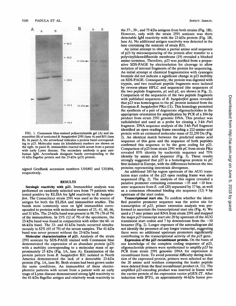
FIG. 1. Coomassie blue-stained polyacrylamide gel (A) and im-munoblot (B) of sonicated B. burgdorfen 2591 (lane A) and B31 (laneB). In panel A, the arrowhead indicates a protein band correspond-ing to p23. Molecular mass (in kilodaltons) markers are shown on
the right. In panel B, immunoblot reacted with serum from a patientwith early Lyme disease. The secondary antibody is goat anti-human IgM. Arrowheads designate bands corresponding to the41-kDa flagellar protein and the 23-kDa (p23) protein.
signed GenBank accession numbers U01892 and U01894,respectively.
RESULTSSerologic reactivity with p23. Immunoblot analysis was
performed on randomly selected sera from 79 patients whotested positive by ELISA for IgM reactivity to B. burgdor-feri. The Connecticut strain 2591 was used as the source ofantigen for both the ELISA and immunoblot studies. Thebands most commonly seen on IgM immunoblot corre-
sponded to proteins with molecular masses of 23, 41, 60, 66,and 31 kDa. The 23-kDa band was present in 98.7% (78 of 79)of the immunoblots. In 15% (12 of 79) of the specimens, the23-kDa band was found in conjunction with either the 60- or
66-kDa band. The 23- and 41-kDa bands occurred simulta-neously in 82% (65 of 79) of the serum samples. The 41-kDaband was never present without the 23-kDa band.
Molecular characterization of p23. Analysis of the strain2591 sonicate by SDS-PAGE stained with Coomassie bluedemonstrated the expression of an abundant protein (p23)with a mobility corresponding to a molecular mass of ap-proximately 23 kDa (Fig. 1A, lane A). Examination of theprotein pattern from B. burgdorfen B31 isolated in NorthAmerica demonstrated the lack of a detectable 23-kDaprotein (Fig. 1A, lane B). Both strains had been grown underthe same conditions. An immunoblot of these electro-phoretic patterns with serum from a patient with an earlystage of Lyme disease demonstrated strong IgM reactivity tothe 41-kDa flagellar antigen and relatively weak reactivity to
the 37-, 39-, and 75-kDa antigens from both strains (Fig. 1B).However, only with the strain 2591 sonicate was theredetectable IgM reactivity with the 23-kDa protein (Fig. 1B,lane A). No additional antigen reactivity was detected in thelane containing the sonicate of strain B31.An initial attempt to obtain a partial amino acid sequence
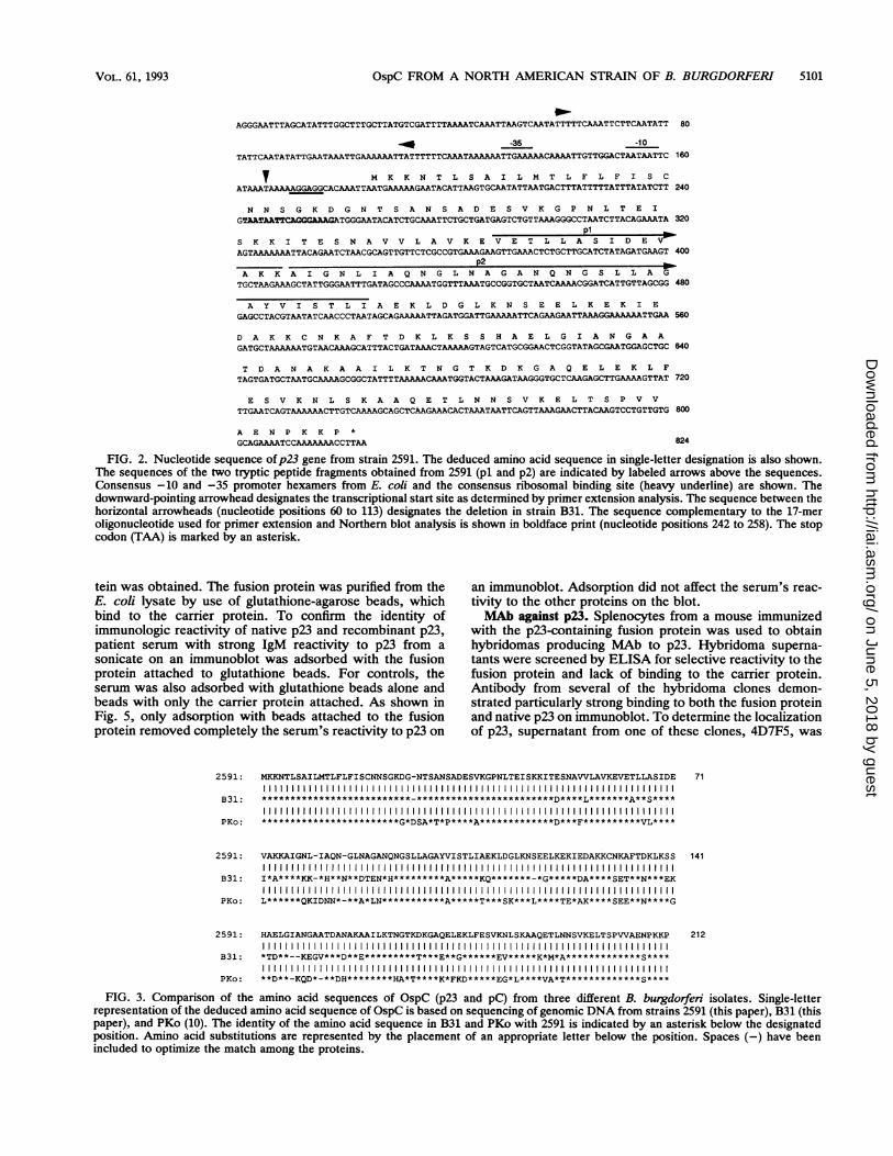
of p23 by microsequencing of the protein after transfer to apolyvinylidenedifluoride membrane (19) revealed a blockedamino terminus. Therefore, p23 was purified from a prepar-ative SDS-PAGE by electroelution for cleavage to allowisolation of internal fragments of the protein for sequencing.An initial attempt at chemical fragmentation with cyanogenbromide did not indicate a significant change in p23 mobilityon SDS-PAGE. Consequently, the protein was digested withtrypsin, and two resultant peptide fragments were isolatedby reverse-phase HPLC and sequenced (the sequences ofthe two peptide fragments, pl and p2, are shown in Fig. 2).Comparison of the sequences of the two peptide fragmentswith published sequences of B. burgdorferi genes revealedthat p23 was homologous to the pC protein isolated from theEuropean B. burgdorfen PKo (11). This homology permittedthe synthesis of a pair of degenerate oligonucleotides in theappropriate orientation for amplification by PCR of a 104-bpproduct from strain 2591 genomic DNA. This product wasradiolabeled and used as a probe for cloning a 3-kb PstIfragment. DNA sequence analysis of the 3-kb PstI fragmentidentified an open reading frame encoding a 212-amino-acidprotein with an estimated molecular mass of 22,250 Da (Fig.2). An identical match between the predicted amino acidsequence of this gene and the sequence of both peptidesconfirmed this sequence to be the gene coding for p23.Comparison of p23 from strain 2591 with pC from strain PKorevealed 83% identity by nucleotide sequence and 75%identity by amino acid sequence (Fig. 3). These resultsstrongly suggested that p23 is a homologous protein to pCfirst isolated in Europe, with the differences between the twoproteins representing interstrain divergence.An additional 185-bp region upstream of the AUG trans-
lation start codon of the p23 open reading frame was alsosequenced (Fig. 2). The analysis of this region revealed aputative promoter containing consensus -35 and -10 hex-amer sequences from E. coli (20) separated by 17 bp, as wellas a consensus ribosomal binding site sequence (12) 9 bpupstream of the start codon.
Transcriptional start site. To confirm whether the identi-fied putative promoter sequence was the active site fortranscription of p23, primer extension analysis was per-formed to ascertain the transcriptional start site (Fig. 4). Weused a 17-mer primer and RNA from strain 2591 and mappedthe majorp23 transcript start site 20 bp upstream of the AUGtranslation start codon and 7 bp downstream from the -10hexamer (Fig. 2). Longer exposure of the autoradiogram didnot identify the presence of any longer transcript, suggestingthere were no additional upstream promoters significantlycontributing to the transcriptional activity of the p23 gene.
Expression of the p23 recombinant protein. On the basis ofour knowledge of the complete coding sequence of p23,oligonucleotide primers were synthesized to amplifyp23 byPCR from strain 2591 genomic DNA for expression inrecombinant form. To avoid potential difficulty during isola-tion of the expressed protein, primers were selected so thatthe 20 amino acid residues composing the leader peptidewere deleted from the final recombinant product (4, 11). Theamplified p23-encoding product was inserted in frame withthe carrier protein of the expression vector pGEX-2T. Afterinduction with IPTG, an approximately 46-kDa fusion pro-
INFECT. IMMUN.
41 kDa ip- Ow
.............
on June 5, 2018 by guesthttp://iai.asm
.org/D
ownloaded from
OspC FROM A NORTH AMERICAN STRAIN OF B. BURGDORFERI
AGGGAATTTAGCATATTTGGCTTTGCTTATGTCGATTTTAAAATCAAATTAAGTCAATATTTTTCAAATTCTTCAATATT 80
_40 -35 -10
TATTCAATATATTGAATAAATTGAAAAAATTATTTTTTCAAATAAAAAATTGAAAAACAAAATTGTTGGACTAATAATTC 160
v M K K N T L S A I L M T L F L F I S CATAAATAAAAAGGAGGCACAAATTAATGAAAAAGAATACATTAAGTGCAATATTAATGACTTTATTTTTATTTATATCTT 240
N N S G K D G N T S A N S A D E S V K G P N L T E IGTAATAATTCAGGGAAAGATGGGAATACATCTGCAAATTCTGCTGATGAGTCTGTTAAAGGGCCTAATCTTACAGAAATA 320
p1S K K I T E S N A V V L A V K E V E T L L A S IDEVDAGTAAAAAAATTACAGAATCTAACGCAGTTGTTCTCGCCGTGAAAGAAGTTGAAACTCTGCTTGCATCTATAGATGAAGT 400
p2A K K A I G N L I A Q N G L N A G A N Q N G S L L AGTGCTAAGAAAGCTATTGGGAATTTGATAGCCCAAAATGGTTTAAATGCCGGTGCTAATCAAAACGGATCATTGTTAGCGG 480
A Y V I S T L I A E K L D G L K N S E E L K E K I EGAGCCTACGTAATATCAACCCTAATAGCAGAAAAATTAGATGGATTGAAAAATTCAGAAGAATTAAAGGAAAAAATTGAA 560
D A K K C N K A F T D K L K S S H A E L G I A N G A AGATGCTAAAAAATGTAACAAAGCATTTACTGATAAACTAAAAAGTAGTCATGCGGAACTCGGTATAGCGAATGGAGCTGC 640
T D A N A K A A I L K T N G T K D K G A Q E L E K L FTAGTGATGCTAATGCAAAAGCGGCTATTTTAAAAACAAATGGTACTAAAGATAAGGGTGCTCAAGAGCTTGAAAAGTTAT 720
E S V K N L S K A A Q E T L N N S V K E L T S P V VTTGAATCAGTAAAAAACTTGTCAAAAGCAGCTCAAGAAACACTAAATAATTCAGTTAAAGAACTTACAAGTCCTGTTGTG 800
A E N P K K P *GCAGAAAATCCAAAAAAACCTTAA 824
FIG. 2. Nucleotide sequence ofp23 gene from strain 2591. The deduced amino acid sequence in single-letter designation is also shown.The sequences of the two tryptic peptide fragments obtained from 2591 (pl and p2) are indicated by labeled arrows above the sequences.Consensus -10 and -35 promoter hexamers from E. coli and the consensus ribosomal binding site (heavy underline) are shown. Thedownward-pointing arrowhead designates the transcriptional start site as determined by primner extension analysis. The sequence between thehorizontal arrowheads (nucleotide positions 60 to 113) designates the deletion in strain B31. The sequence complementary to the 17-meroligonucleotide used for primer extension and Northern blot analysis is shown in boldface print (nucleotide positions 242 to 258). The stopcodon (TAA) is marked by an asterisk.
tein was obtained. The fusion protein was purified from theE. coli lysate by use of glutathione-agarose beads, whichbind to the carrier protein. To confirm the identity ofimmunologic reactivity of native p23 and recombinant p23,patient serum with strong IgM reactivity to p23 from asonicate on an immunoblot was adsorbed with the fusionprotein attached to glutathione beads. For controls, theserum was also adsorbed with glutathione beads alone andbeads with only the carrier protein attached. As shown inFig. 5, only adsorption with beads attached to the fusionprotein removed completely the serum's reactivity to p23 on
2591:
B31:
PKo:
2591:
B31:
PKo:
2591:
B31:
PKo:
an immunoblot. Adsorption did not affect the serum's reac-tivity to the other proteins on the blot.MAb against p23. Splenocytes from a mouse immunized
with the p23-containing fusion protein was used to obtainhybridomas producing MAb to p23. Hybridoma superna-tants were screened by ELISA for selective reactivity to thefusion protein and lack of binding to the carrier protein.Antibody from several of the hybridoma clones demon-strated particularly strong binding to both the fusion proteinand native p23 on immunoblot. To determine the localizationof p23, supernatant from one of these clones, 4D7F5, was
MKKNTLSAILMTLFLFISCNNSGKDG-NTSANSADESVKGPNLTEI SKKITESNAVVLAVKEVETLLASIDE 711111111111111111111111111111111111111111111111111111111111
111111111111111111111111111111111111111111111111111111111111111111111111
VAKKAIGNL-IAQN-GLNAGANQNGSLLAGAYVISTLIAEKLDGLKNSEELKEKIEDAKKCNKAFTDKLKSS 1411111111111111111111111111111111111111111111111111111111111
I*A****KK-*H**N**DTEN*H*********A*****KQ*******-*G*****DA****SET**N***EK
L******QKIDNN*-**A*LN***********A*****T***SK***L****TE*AK****SEE**N****G
11111111111111111111111111111111111111111111111111111111111111111*TD**--KEGV***D**E*********T***E**G******EV*****K*M*A*************S****
**D**-KQD*-**DH********HA*T****K*FKD*****EG*L****VA*T*************S****
212
FIG. 3. Comparison of the amino acid sequences of OspC (p23 and pC) from three different B. burgdorferi isolates. Single-letterrepresentation of the deduced amino acid sequence of OspC is based on sequencing of genomic DNA from strains 2591 (this paper), B31 (thispaper), and PKo (10). The identity of the amino acid sequence in B31 and PKo with 2591 is indicated by an asterisk below the designatedposition. Amino acid substitutions are represented by the placement of an appropriate letter below the position. Spaces (-) have beenincluded to optimize the match among the proteins.
VOL. 61, 1993 5101
on June 5, 2018 by guesthttp://iai.asm
.org/D
ownloaded from

5102 PADULA ET AL.
C T A G 1~~~~~~~~~~~CT
ATAATTCA
TAA
A
AA
: ~~~~~~~~~~~~A A B C
41 kDa_
AAGGAGGcAcAA
FIG. 4. Determination of the 5' end of p23 mRNA from B.burgdorferi 2591 by primer extension analysis. The size of thereverse transcriptase primer extension product ofp23 mRNA wascompared to a DNA sequencing ladder ofp23 from strain 2591. Theanalysis was performed as described in Materials and Methods.Lanes C, T, A, and G represent DNA sequencing reactions with theappropriate dideoxynucleotide triphosphate by using the same 17-mer oligonucleotide primer used for primer extension of mRNA.Lane 1 shows the primer extension product. The sequence to theright of lane 1 is the sequence in the region of the 5' end of p23mRNA, and the arrow indicates the residue at the 5' end of theextended product.
selected for use in immunoelectron microscopy of strain2591. As shown in Fig. 6, p23 was found to be expressed onthe outer surface of the spirochete. A control isotype-matched antibody did not bind (data not shown). In addition,MAb 4D7F5 did not label strain B31, confirming the lack ofexpression of p23 by this strain (data not shown).
Molecular defect in p23 gene from strain B31. The lack ofreactivity of the sera and the MAbs to both the B31 sonicateand whole organism led us to investigate the molecularreason for the lack of expression of p23 in this strain.Northern blot analysis for p23 mRNA revealed the strongexpression of an approximately 700-bp transcript from strain2591 and a very weak signal of the same size from strain B31(Fig. 7). Examination of the RNA stained with ethidiumbromide had revealed that an equivalent amount of RNAfrom each strain was loaded in the gel and that there was noappreciable degradation of the RNA. In addition, primerextension analysis also suggested a significantly reducedlevel ofp23 mRNA in strain B31 (data not shown). Also, thesize of the p23 primer extension termination product fromstrain B31 was found to be 1 base longer than the transcriptfrom strain 2591 (data not shown). To gain a better under-standing of the reason for the relative lack of p23 expression
23kDapm.-d ig
FIG. 5. Removal of IgM anti-p23 reactivity from serum by ad-sorption with recombinant p23. The IgM immunoblot of sonicate ofB. burgdorferi 2591 with patient serum after adsorption on glutathi-one beads alone (lane A) revealed strong reactivity with native p23.Lane B shows reactivity of serum after adsorption on carrier proteinattached to glutathione beads. Lane C shows complete removal ofserum's reactivity after adsorption with p23 fusion protein attachedto glutathione beads. Reactivity with other proteins including the41-kDa flagellar protein was not affected.
by strain B31, we proceeded to clone and sequence the p23gene from strain B31 in the same fashion as we had donepreviously for strain 2591. Comparison of the deduced aminoacid sequences of p23 from strains 2591 and B31 as well aspC from PKo is shown in Fig. 3. p23 from strain B31 wasfound to contain 210 amino acids, 2 less than the other twohomologs, and shares 80% amino acid sequence identity and85% nucleotide sequence identity with p23 from strain 2591.Comparison of the proteins from the three strains showedconservation of sequence at the 5' (mostly leader peptidesequence) and 3' ends of the protein, with increased vari-ability in the central region.
Analysis of the B31 DNA sequence 5' to the coding regionof p23 revealed a 54-bp deletion upstream to the consensus-10 and -35 promoter sequences found to be the activepromoter region in strains 2591 and B31. The finding of thisdeletion in close proximity to the functional promoter regionsuggested the loss of an enhancing element which results inlow transcriptional activity ofp23 and lack of expression ofthe p23 protein in strain B31.To confirm that the p23 gene from B31 could encode
antigenic protein, the coding region ofp23 was cloned intoan expression vector and expressed in E. coli as a fusionprotein. The recombinant p23 protein derived from strainB31 was as strongly reactive with sera and the MAbs as thenative and recombinant forms of p23 from strain 2591 (datanot shown).Use of recombinant p23 for testing of sera. On the basis of
INFECT. IMMUN.
on June 5, 2018 by guesthttp://iai.asm
.org/D
ownloaded from

OspC FROM A NORTH AMERICAN STRAIN OF B. BURGDORFERI
0
A - ~.M
0 S
.t
FIG. 6. Immunoelectron micrograph of thin-sectioned B. burgdorferi 2591 labeled with 4D7F5, an MAb with reactivity to p23.Localization of the MAb was detected with goat anti-mouse IgG conjugated to 10-nm-diameter gold beads. The immunogold is seen along theouter surface (see insert of cross-section view). Bar, 0.1 pm.
our observation that IgM reactivity to p23 may be a signifi-cant serologic marker for early stages of Lyme disease, weproceeded to test the feasibility of using recombinant p23 inan ELISA. We initially tested sera from 15 patients withclinically suspected Lyme disease and positive IgM immu-noblots with a minimum of three positive bands includingreactivity to p23. All 15 of these serum samples were
A B
23S-
16S-
FIG. 7. Northern blot analysis of p23 transcript. Total cellularRNA (15 ,ug per lane) was isolated from B. burgdorfen B31 (lane A)and 2591 (lane B), fractionated in a formaldehyde denaturing gel,transferred to a nylon membrane, and hybridized to a 17-meroligonucleotide based on the sequence of p23. The arrowheaddesignates the p23 transcript. The positions of 23S and 16S rRNAare indicated.
strongly positive by the recombinant p23 ELISA. Sera fromfive patients with syphilis, 10 patients with high-titeredrheumatoid factor, 5 patients with Epstein-Barr virus infec-tion, and 10 patients with high-titer antinuclear antibodies alltested negative with this assay. All of these sera also testednegative when examined by immunoblot with a sonicatefrom strain 2591.
DISCUSSION
Establishing the diagnosis of Lyme disease continues torely on serologic confirmation of exposure to the causativeagent, B. burgdorfen, in the setting of characteristic clinicalfindings. Recognizing Lyme disease in the early stages canbe difficult, however, because these patients may not mani-fest the characteristic rash or may have only nonspecificflu-like symptoms (25). This difficulty of diagnosis is com-pounded by the delayed emergence of a humoral response tothe spirochete as detected by available serologic tests (25).These tests, which currently lack standardization, also donot readily distinguish between reactivity to B. burgdorferiproteins and cross-reactive proteins from commensual orother pathogenic organisms (13, 16). Delay in establishingthe diagnosis of Lyme disease in its early stages is clinicallyimportant because timely institution of appropriate antibiotictreatment can prevent the serious sequelae from this poten-tially chronic infection (7, 26).
Initial studies with immunoblot analysis of patients fromNorth America with the early manifestations of Lyme dis-ease found IgM reactivity predominantly against the 41-kDaflagellar protein of B. burgdorferi (3, 6). Detection of anti-
VOL. 61, 1993 5103
on June 5, 2018 by guesthttp://iai.asm
.org/D
ownloaded from

5104 PADULA ET AL.
body to this protein, however, is not specific for Lymedisease, as flagellar protein sequences are conserved amongspirochetes, including the oral pathogens found commonly inperiodontal disease (17).
In similar studies of patients in Europe, utilizing differentstrains of B. burgdorfen as the source of antigen, IgM wasmost frequently detected against an abundant 21- to 22-kDaprotein, which was designated pC (32). A major protein withthis size was detected in approximately 45% of B. burgdor-feri strains from Europe but only rarely observed in NorthAmerican isolates (31, 32).
Recently, Dressler et al. reported the most prominent IgMresponse in American patients with early stages of thedisease was to a 21-kDa protein (10). This protein wasreported to be reactive with an MAb specific for pC. Thisfinding contrasts with this same group's previous finding of apredominant early response to the 41-kDa flagellar antigen(6). The discrepancy between these two findings was attrib-uted to different antigen preparations despite the use of thesame isolate, highlighting the potential confusion introducedby the current lack of test standardization.
In our laboratory we have found IgM reactivity predomi-nantly to a protein with a molecular mass of 23 kDa (p23) insera from patients with early stages of Lyme disease. In ourserologic tests we have used a Connecticut isolate, B.burgdorfeni 2591, which produces an abundant protein witha size of 23 Da. We therefore sought to identify andcharacterize p23, compare it to the European pC, andexpress it in recombinant form for testing in an easilystandardized assay for the detection of a serologic responsein early stages of Lyme disease.We cloned and sequenced the p23 gene from strain 2591
and predicted the protein to contain 212 amino acids with amolecular mass of 22,250 kDa. Comparison of the nucleicacid and predicted amino acid sequences of p23 with those ofpC from the European isolate PKo revealed a high degree ofhomology (11). Furthermore, as recently reported for pC, wehave localized p23 to the outer surface of 2591 by immuno-electron microscopy (30). Thus, p23 and pC are homologs ofthe same outer surface protein, OspC.
During the course of these studies, we confirmed the lackof expression of OspC by strain B31, an isolate which hasbeen commonly used as the source of antigen for commer-cially available serologic assays in North America. StrainB31, as well as strain 2591, belongs to genospecies I, B.burgdorfen sensu stricto, whereas strain PKo belongs togenospecies III, group VS461 (1, 29, and unpublished re-sults). We could not detect expression of OspC in a B31sonicate by SDS-PAGE and immunoblot and on the wholeorganism by immunoelectron microscopy. We did confirmhowever that the ospC gene is present in B31 and identifieda deletion of 54 bp in the 5' noncoding region of the genelocated just upstream of -35 and -10 consensus E. colipromoter sequences. Northern blot analysis of B31 revealeda low level of ospC mRNA, and primer extension studiesshowed a transcription start site for ospC 1 base upstream ofthat found for strain 2591. Recently, Marconi et al. havereported that ospC transcription in some strains may beunder the control of one or more promoters in addition to themajor functional promoter that we have described in thispaper (18). We could not detect in strains 2591 and B31,however, any evidence of longer primer extension termina-tion products by overexposure of the autoradiographs.We propose that the deleted 54-bp segment contains an
enhancer element(s) required for increased ospC transcrip-tional activity and detectable levels of expression of OspC.
At this time, the evidence supporting this contention remainscircumstantial. Additional supportive documentation of aregulatory element of transcription in the deleted sequencewould require gene transfer technology which is currentlyunavailable for Borrelia species.
Recently, Marconi et al. (18) and Sadziene et al. (24)localized the ospC gene to a 26- to 27-kDa circular plasmid,the first gene mapped to a circular plasmid in B. burgdorferi.Marconi et al. mapped the gene by using a variety ofelectrophoretic separation techniques and Southern blotting.Sadziene et al. used a group of B31-derived isolates whichcontained their linear chromosome and circular plasmids buthad been antibody selected for the loss of a variable numberof their linear plasmids. Sadziene et al. also found a corre-lation of an isolate's ability to express OspC and the loss ofa linear plasmid of 16 kb (lp 16). They hypothesized that aprotein or RNA encoded by lp 16 may function as arepressor of ospC expression. Loss of this plasmid couldthereby lead to the loss of repression and new expression ofthe protein. In addition, these investigators noted that theloss of lp 16 also led to a failure of the mutant to grow onsolid medium. Interestingly, in a recent report, Wilske et al.described the new expression of OspC by nonexpressingisolates after their growth on solid medium (30). The molec-ular reason(s) for these changes in growth requirements andhow they may affect the regulation of the expression ofproteins including OspC is as yet unexplained. How thesefindings and ours may relate to each other in the regulation ofospC transcription remains to be elucidated.To determine whether the coding region of ospC from B31
is capable of encoding an immunologically detectable pro-tein, we expressed recombinant OspC from B31 in E. coli.We found that patients' sera and MAbs reactive with p23(OspC) from strain 2591 were strongly reactive with recom-binant OspC from strain B31. Because of the observationthat p23 from strain 2591 is an early immunodominant targetof the humoral response in Lyme disease, we produced arecombinant form of the protein for testing in diagnosticassays. Preliminary studies with recombinant p23 suggestthat it may be a sensitive and specific antigen for serologicdetection early in disease. In addition, immunoassays whichutilize recombinant forms of antigens have the added advan-tage of being easily standardized. Currently, we are extend-ing these studies and are optimizing the assay to assess thelimits of early detection.
ACKNOWLEDGMENTSThis work was supported by Public Health Service grant 5R29-
AR39361 (S.J.P.).We thank Juris Ozols for assistance with the protein sequencing
and Naomi Rothfield and Peter Setlow for advice during the courseof these studies and for critical review of the manuscript.
REFERENCES1. Baranton, G., D. Postic, I. Saint Girons, P. Boerlin, J.-C.
Piffaretti, M. Assous and P. A. D. Grimont. 1992. Delineation ofBorrelia burgdorferi sensu stricto, Borrelia garinii sp. nov., andGroup VS461 associated with Lyme borreliosis. Int. J. Syst.Bacteriol. 42:378-383.
2. Barbour, A. G. 1984. Isolation and cultivation of Lyme diseasespirochetes. Yale J. Biol. Med. 57:521-525.
3. Barbour, A. G., S. F. Hayes, R. A. Heiland, M. E. Schrumpf,and S. L. Tessier. 1986. A Borrelia-specific monoclonal antibodybinds to a flagellar epitope. Infect. Immun. 52:549-554.
4. Bassford, P. J., Jr., T. J. Silhavy, and J. R. Beckwith. 1979. Useof gene fusion to study secretion of maltose-binding protein intoEscherichia coli periplasm. J. Bacteriol. 139:19-31.
INFECT. IMMUN.
on June 5, 2018 by guesthttp://iai.asm
.org/D
ownloaded from

OspC FROM A NORTH AMERICAN STRAIN OF B. BURGDORFERI 5105
5. Bradford, M. M. 1976. A rapid and sensitive method for thequantitation of microgram quantities of protein utilizing theprinciple of protein-dye binding. Anal. Biochem. 72:248-254.
6. Craft, J. E., D. K. Fischer, G. T. Shimamoto, and A. C. Steere.1986. Antigens of Borrelia burgdorferi recognized during Lymedisease. Appearance of a new immunoglobulin M response andexpansion of the immunoglobulin G response late in the illness.J. Clin. Invest. 78:934-939.
7. Dattwyler, R. J., D. J. Volkman, S. M. Conaty, S. P. Platkin, andB. J. Luft. 1990. Amoxycillin plus probenecid versus doxycy-cline for treatment of erythema migrans borreliosis. Lancet336:1404-1406.
8. de St. Groth, S. F., and D. Scheidegger. 1980. Production ofmonoclonal antibodies: strategy and tactics. J. Immunol. Meth-ods 35:1-21.
9. Dias, F., R. W. Ryan, and H. M. Feder. 1992. Interpretation ofIgM western immunoblot banding patterns in patients suspectedof having early Lyme disease, abstr. A100, p. 19. ProgramAbstr. V Int. Conf. on Lyme Borreliosis, Arlington, Va.
10. Dressler, F., J. A. Whalen, B. N. Reinhardt, and A. C. Steere.1993. Western blotting in the serodiagnosis of Lyme disease. J.Infect. Dis. 167:392-400.
11. Fuchs, R., S. Jauris, F. Lottspeich, V. Preac-Mursic, B. Wilske,and E. Soutschek. 1992. Molecular analysis and expression of aBorrelia burgdorfen gene encoding a 22 kDa protein (pC) inEscherichia coli. Mol. Microbiol. 6:503-509.
12. Gold, L., D. Pribnow, T. Schneider, S. Shinedling, B. S. Singer,and G. Stormo. 1981. Translation initiation in prokaryotes.Annu. Rev. Microbiol. 35:365-403.
13. Hansen, K., J. M. Bangsborg, H. Fjordvang, N. S. Pedersen, andP. Hindersson. 1988. Immunochemical characterization of andisolation of the gene for a Borrelia burgdorferi immunodominant60-kilodalton antigen common to a wide range of bacteria.Infect. Immun. 56:2047-2053.
14. Jones, K A., R. Yamamoto, and R. Tian. 1985. Two distincttranscription factors bind to the HSV thymidine kinase pro-moter in vitro. Cell 42:559-572.
15. Laemmli, U. K 1970. Cleavage of structural proteins during theassembly of the head of bacteriophage T4. Nature (London)227:680-685.
16. Magnarelli, L. A., J. A. Anderson, and R. C. Johnson. 1987.Cross-reactivity in serological tests for Lyme disease and otherspirochetal infections. J. Infect. Dis. 156:183-188.
17. Magnarelli, L. A., J. N. Miller, J. F. Anderson, and G. R.Riviere. 1990. Cross-reactivity of nonspecific treponemal anti-body in serologic tests for Lyme disease. J. Clin. Microbiol.28:1276-1279.
18. Marconi, R. T., D. S. Samuels, and C. F. Garon. 1993. Tran-scriptional analyses and mapping of the ospC gene in Lyme
disease spirochetes. J. Bacteriol. 175:926-932.19. Matsudaira, P. 1987. Sequence from picomole quantities of
protein electroblotted onto polyvinylidene difluoride mem-branes. J. Biol. Chem. 262:10035-10038.
20. McClure, W. R. 1985. Mechanism and control of transcriptioninitiation in prokaryotes. Annu. Rev. Biochem. 54:171-204.
21. McKnight, S. L., and R. Kingsbury. 1982. Transcription controlsignals of a eukaryotic protein-coding gene. Science 217:316-324.
22. Ozols, J. 1990. Covalent structure of liver microsomal flavin-containing monooxygenase form 1. J. Biol. Chem. 265:10289-10299.
23. Ozols, J., F. S. Heinemann, and C. Gerard. 1980. High pressureliquid chromatography of polar and hydrophobic peptides ofcytochromes from microsomal membranes, p. 417-429. In C.Birr (ed.), Methods in peptide and protein sequence analysis.Elsevier/North Holland Biomedical Press, Amsterdam.
24. Sadziene, A., B. Wilske, M. S. Ferdows, and A. G. Barbour.1993. The cryptic ospC gene of Borrelia burgdorferi B31 islocated on a circular plasmid. Infect. Immun. 61:2192-2195.
25. Steere, A. C. 1989. Lyme disease. N. Engl. J. Med. 321:586-596.26. Steere, A. C., G. J. Hutchinson, D. W. Rahn, L. H. Sigal, J. E.
Craft, E. T. DeSanna, and S. E. Malawista. 1983. Treatment ofthe early manifestations of Lyme disease. Ann. Intern. Med.99:22-26.
27. Summers, W. C. 1970. A simple method for extraction of RNAfrom E. coli utilizing diethyl pyrocarbonate. Anal. Biochem.33:459-463.
28. Towbin, H., T. Staehelin, and J. Gordon. 1979. Electrophoretictransfer of proteins from polyacrylamide gels to nitrocellulosesheets: procedure and some applications. Proc. Natl. Acad. Sci.USA 76:4350-4354.
29. Wilske, B., V. Preac-Mursic, U. B. Gobel, B. Graf, S. Jauris, E.Soutschek, E. Schwab, and G. Zumstein. 1993. An OspA sero-typing system for Borrelia burgdorferi based on reactivity withmonoclonal antibodies and OspA sequence analysis. J. Clin.Microbiol. 31:340-350.
30. Wilske, B., V. Preac-Mursic, S. Jauris, A. Hofmann, I. Pradel,E. Soutschek, E. Schwab, G. Will, and G. Wanner. 1993.Immunological and molecular polymorphisms of OspC, an im-munodominant major outer surface protein of Borrelia burgdor-fen. Infect. Immun. 61:2182-2191.
31. Wilske, B., V. Preac-Mursic, G. Schierz, and V. Busch. 1986.Immunochemical and immunological analysis of European Bor-relia burgdorferi strains. Zentralbl. Bakteriol. Mikrobiol. Hyg.Ser. A 263:92-102.
32. Wilske, B., V. Preac-Mursic, G. Schierz, R. Kuhbeck, A. G.Barbour, and M. Kramer. 1988. Antigenic variability of Borreliaburgdorferi. Ann. N.Y. Acad. Sci. 539:126-143.
VOL. 61, 1993
on June 5, 2018 by guesthttp://iai.asm
.org/D
ownloaded from