Embed Size (px)
Citation preview
1521-0103/366/2/251–264$35.00 https://doi.org/10.1124/jpet.118.248831THE JOURNAL OF PHARMACOLOGY AND EXPERIMENTAL THERAPEUTICS J Pharmacol Exp Ther 366:251–264, August 2018Copyright ª 2018 by The American Society for Pharmacology and Experimental Therapeutics
Minireviews
Molecular Basis of the Brain Renin Angiotensin System inCardiovascular and Neurologic Disorders: Uncovering a Key Rolefor the Astroglial Angiotensin Type 1 Receptor AT1R
Dhanush Haspula and Michelle A. ClarkDepartment of Biomedical Engineering, Medical College of Wisconsin, Milwaukee, Wisconsin (D.H.); and College of Pharmacy,Department of Pharmaceutical Sciences, Nova Southeastern University, Ft. Lauderdale, Florida (M.A.C.)
Received February 28, 2018; accepted May 8, 2018
ABSTRACTThe central renin angiotensin system (RAS) is one of the mostwidely investigated cardiovascular systems in the brain. It isimplicated in a myriad of cardiovascular diseases. However,studies from the last decade have identified its involvement inseveral neurologic abnormalities. Understanding the molecularfunctionality of the various RAS components can thus provideconsiderable insight into the phenotypic differences and mech-anistic drivers of not just cardiovascular but also neurologicdisorders. Since activation of one of its primary receptors, theangiotensin type 1 receptor (AT1R), results in an augmentationof oxidative stress and inflammatory cytokines, it becomes
essential to investigate not just neuronal RAS but glial RAS aswell. Glial cells are key homeostatic regulators in the brain andare critical players in the resolution of overt oxidative stress andneuroinflammation. Designing better and effective therapeuticstrategies that target the brain RAS could well hinge on un-derstanding the molecular basis of both neuronal and glial RAS.This review provides a comprehensive overview of the majorstudies that have investigated the mechanisms and regulation ofthe brain RAS, and it also provides insight into the potential roleof glial AT1Rs in the pathophysiology of cardiovascular andneurologic disorders.
IntroductionThe renin angiotensin system (RAS) is a cardioregulatory,
peptidergic, hormonal system that is involved primarily in theregulation of blood pressure. Its pivotal function is to elevateblood pressure in hypotensive states. In response to a drop inblood pressure, low salt concentration, or low blood volume,the juxtaglomerular apparatus in the kidneys triggers theRAS cascade, which ultimately culminates in a significantantinatriuretic, antidiuretic, and vasoconstrictive effect (Hall,1986; Montani and Van Vliet, 2004). Components of the RAS,as will be discussed in the succeeding sections, are found invirtually every organ/tissue of the body. Hence, their functionsgo beyond regulation of blood pressure. Of significant interest
is the action of the RAS in the brain. An overactive brain RAShas been demonstrated to be a characteristic feature ofmultiple cardiovascular diseases. However, evidence showsthat the underlying pathologic mechanisms of the RAS can beextrapolated to several neurologic disorders as well. Anincrease in oxidative stress, proinflammatory mediators, anda decrease in anti-inflammatory cytokines are tightly inter-twined with the progression of both cardiovascular andneurologic disorders. The RAS plays a pivotal role in control-ling these drivers of cardiovascular and neurologic disorders.
Classic and Nonclassic RAS and TherapeuticInterventions
The RAS is composed of several different components thatencompass precursor and active peptides, enzymes, andreceptors. Renin, secreted from the kidneys, converts angio-tensinogen (AGT), secreted from the liver, to angiotensin
This work was supported by Nova Southeastern University [President’sFaculty Research and Development Grant 335309 and Health ProfessionsDivision Grant 335585].
https://doi.org/10.1124/jpet.118.248831.
ABBREVIATIONS: ACE, angiotensin-converting enzyme; ACEI, angiotensin-converting enzyme inhibitor; ADHD, attention deficit hyperactivedisorder; AGT, angiotensinogen; Ang, angiotensin; ARB, angiotensin receptor blocker; AT1R, angiotensin type 1 receptor; AT2R, angiotensin type2 receptor; AT4R, angiotensin type 4 receptor; C21, compound 21; ERK, extracellular signal–regulated kinase; GPCR, G protein–coupled receptor;IL, interleukin; MAPK, mitogen-activated protein kinase; MS, multiple sclerosis; NF-kB, nuclear factor kB; NO, nitric oxide; NOS, nitric oxidesynthase; NTS, nucleus of the solitary tract; PI3K, phosphoinositide 3-kinase; PVN, paraventricular nucleus; RAS, renin angiotensin system; ROS,reactive oxygen synthase; RVLM, rostral ventrolateral medulla; SFO, subfornical organ; SHR, spontaneously hypertensive rat; SON, supraopticnucleus; WKY, Wistar Kyoto.
251
at ASPE
T Journals on A
pril 25, 2020jpet.aspetjournals.org
Dow
nloaded from

(Ang) I. Ang I is converted into Ang II by the action of theangiotensin-converting enzyme (ACE), an enzyme produced inthe lungs and blood vessels (Montani and Van Vliet, 2004). Itis now widely accepted that Ang II is secreted by a diversegroup of cell types, and its source is not restricted to only liverAGT (Ganong, 1994; Paul et al., 2006). The former is part ofthe local RAS, whereas the latter constitutes the systemicRAS. Optimal functioning of both the systemic and local RASis critical for overall cardiovascular homeostasis (Lavoie andSigmund, 2003). Ang II, the major effector peptide of the RAS,is a circulating hormone that has a major physiologic andpathophysiological bearing on cardiovascular functions. Al-though most of their functions converge to have one singularoutcome (i.e., an elevation in blood pressure), the RAS also hasa role in digestion, reproduction, and prenatal development(Paul et al., 2006).Thewidely studied and documented actions of Ang II, such as
aldosterone secretion and its vasoconstrictive and ionotropiceffects, are due to its ability to interact with the angiotensintype 1 receptor (AT1R) (Fyhrquist et al., 1995). Ang II coupleswith AT1R, a pertussis toxin–insensitive G protein–coupledreceptor (GPCR), to produce a spike in calcium levels. Eleva-tions in calcium activate kinases, signaling pathways, andtranscription factors and consequently cause several physio-logic actions such as smooth muscle contraction and aldoste-rone synthesis (de Gasparo et al., 2000). By interacting withAT1R on renal, cardiac, and vascular cells, Ang II is able toincrease aldosterone levels, elevate salt intake, cause sympa-thetic nervous system hyperactivation, have a positive iono-tropic effect, and elicit potent vasoconstriction (Fyhrquist et al.,1995). Ang II also interactswith the angiotensin type 2 receptor(AT2R), which is known to elicit functions that are antagonisticto AT1R, such as a reduction of oxidative stress and neutral-izing proinflammatory states (Stoll et al., 1995; Namsollecket al., 2014). At a physiologic level, AT2R activation results invasodilatory and cardioprotective effects (Li et al., 2012).Several other RAS peptides that are both functionally
similar and dissimilar to Ang II have also been identified,characterized, and studied (Paul et al., 2006). Ang III, Ang IV,and Ang (1-7) are physiologically active degradation productsof Ang II. Ang III interacts with AT1R, whereas Ang IV andAng (1-7) interact with their own cognate receptors, the Angtype 4 receptor (AT4R) and the Mas receptor, respectively(Varagic et al., 2008). Ang II is degraded by aminopeptidasesto Ang III and Ang IV and by ACE2 to Ang (1-7) (Paul et al.,2006). Alternatively, Ang (1-7) can also be synthesized fromAng I by the action of neprilysin (Paul et al., 2006). Ang IIIexhibits functional similarity to Ang II, whereas Ang (1-7)counteracts the deleterious effects of Ang II (Ferrario et al.,1991). Owing to the anti-inflammatory and antioxidant prop-erties of the Mas receptor, Ang (1-7) has been demonstratedto have potent cardioprotective and neuroprotective proper-ties (Bennion et al., 2015). Ang IV also has cardioprotectiveand neuroprotective effects. Ang IV is not only known to elicitpotent vasodilatory effects (Kramár et al., 1997; Hamiltonet al., 2001), but it has also been demonstrated to diminishthe production of several proinflammatory cytokines (Konget al., 2015). Ang IV receptor activation is linked to animprovement in cognitive abilities such as learning andmemory, and it has been suggested as a potential therapeutictarget for Alzheimer disease (Wright and Harding, 2008;Royea et al., 2017).
Although a dysregulated RAS is one of the hallmarks ofcardiovascular diseases (Veerasingham and Raizada, 2003), itsfunctions are fundamental for survival under physiologicconditions. Through multiple mechanisms, Ang II controlsand maintains blood pressure and blood volume within setboundaries. Although vasoconstriction is often viewed as itsmajor mechanism, its ability to increase water retention andelevate sympathetic activity makes it a powerful and uniquesystem in understanding the pathophysiology of cardiovasculardiseases. Ang II–mediated hypertrophy and hyperplasia arecharacteristic features of multiple risk factors of cardiovasculardiseases, such as hypertension and diabetes (Fyhrquist et al.,1995). Owing to the ubiquitous nature of AT1R expression inthe body, overactivity of AT1R is linked to multiple organ andtissue dysfunctions. Ang II is a well established mitogen that isknown to trigger hypertrophy and cell migration via AT1Ractivation (Ushio-Fukai et al., 1996; Takeuchi et al., 2006; Leeet al., 2007; Campos et al., 2012). Cardiac hypertrophy, renaldisease, and endothelial dysfunction have all been demon-strated to have an augmented RAS component. Since both AngII synthesis and AT1R activity are fundamental to RAS-mediated elevation in blood pressure, drugs that impedesynthesis of Ang II, or those that antagonize the deleteriouseffects of AT1Rs, are the mainstays in the pharmacologicalmanagement of numerous cardiovascular diseases, their riskfactors, and their complications (Burnier and Zanchi, 2006;Atlas, 2007). In addition to its augmented ability to elevateblood pressure by multiple mechanisms in pathologic condi-tions, Ang II can also cause extensive damage to the heart,kidneys, and vasculature (Long et al., 2004; Montezano et al.,2014; Wang et al., 2014). For example, AT1R angiotensinreceptor blockers (ARBs) and angiotensin-converting enzymeinhibitors (ACEIs) are routinely employed in the managementof heart failure (Aranda and Conti, 2003; Atlas, 2007).An alternative strategy is to counteract the effects of AT1R by
activating receptors and systems that exhibit functional antag-onism to the receptor. In this regard, several groups haveexplored the therapeutic potential of AT2R agonists, whichexhibit vasodilatory and cardioprotective effects such as areduction in inflammation and fibrosis as well as cause pro-oxidant states (Danyel et al., 2013;Matavelli and Siragy, 2015).Although AT2R activation has been shown to result in signif-icant vasodilation, the AT2R agonist compound 21 (C21) did notalter blood pressure when administered alone in hypertensiveand nonhypertensive rats (Carey et al., 2001; Bosnyak et al.,2010). C21 caused a significant reduction only in the presence ofAT1Rblockers. Owing to the relatively low expression of AT2Rscompared with AT1Rs, it could be that the AT1R tone is fargreater than AT2R-mediated vasorelaxation under baselineconditions. In addition, the Ang (1-7)–ACE2–Mas axis has alsobeen viewed as a potential therapeutic target to negate thedeleterious effects of AT1R activation. Several in vitro andin vivo studies have demonstrated a significant protective rolefor this pathway in cardiac tissue under pathologic conditions(Zhong et al., 2010; Patel et al., 2012). The ability of Ang (1-7) toimprove not only endothelial and vascular function (Sampaioet al., 2007; McKinney et al., 2014) but also the metabolic paneland to serve as a renoprotectant in diabetic states (Mori et al.,2014a,b) makes the Mas receptor an attractive druggabletarget. The schematic representation of the RAS pathway andthe role of its receptors in regulation of cardiovascular functionsis shown in Fig. 1.
252 Haspula and Clark
at ASPE
T Journals on A
pril 25, 2020jpet.aspetjournals.org
Dow
nloaded from

Crosstalk between different components of the RAS has alsobeen reported. For instance, Ang II has been shown toupregulate ACE and downregulate ACE2 in kidney cells andneuronal cells (Koka et al., 2008; Xiao et al., 2013). Receptorsbelonging to RAS and also other molecular systems are knownto negatively or positively regulate AT1R activity by severaldifferent modes of crosstalk. For instance, AT2R has beendemonstrated to cause heterologous desensitization of AT1Rthrough a protein kinase C–dependent mechanism and thusdiminish its activity in transfected cells (Inuzuka et al., 2016).Interestingly, AT1R activity was reduced in the amygdalaof mice brains that lacked the MAS receptor, further high-lighting the crosstalk among the various components of theRAS (Von Bohlen und Halbach et al., 2000). Interestingly, asubpressor dose of Ang II has a similar angiogenic responseto a low dose of Ang (1-7), further highlighting the complexinterplay between various components of the RAS (Hoffmannet al., 2017). Although the cannabinoid type 1 receptor hasbeen demonstrated to potentiate AT1R profibrinogenic activ-ity by way of heterodimerization (Rozenfeld et al., 2011), it hasalso has been demonstrated to diminish AT1R’s ability toactivate key signaling pathways in astrocytes (Haspula andClark, 2017b). In addition, receptor interactions with adren-ergic receptors also enable drugs such as b-blockers to influenceAT1R activity (Barki-Harrington et al., 2003). Heterodimersbetween AT2R and MAS have been shown to be function-ally relevant in astrocytes, whereby knockout of either ofthe receptors led to a diminished functionality of the other(Leonhardt et al., 2017).
Although AT1R is considered to be a prototypical plasmamembrane receptor, evidence of Ang II binding sites that arelocalized intracellularly and bear similarity with AT1R hasbeen reported in various tissues such as the liver (Booz et al.,1992; Tang et al., 1992). Evidence of an intracellular RAS inthe brain comes from studies on a nonsecretory variant ofrenin that retains its enzymatic activity (Lee-Kirsch et al.,1999) and was also shown to modulate thirst and bloodpressure in transgenic animals (Lavoie et al., 2006). Inneurons, Ang receptors were recently identified on nuclei aswell as on other organelles such as mitochondria, where theyhave a role in neuroprotection and respiration (Valenzuelaet al., 2016; Villar-Cheda et al., 2017). It becomes important tounderstand the functional significance of intracellular bindingsites for Ang II in brain cells such as astrocytes, since it wasshown in cardiomyocytes that Ang II performed functionsintracellularly that are different from those of extracellularAng II (De Mello, 2008).
Brain RAS and Blood Pressure ControlStudies on borderline hypertensive humans and hyperten-
sive animal models have confirmed the importance of anaugmented central sympathetic activity in the pathogenesis ofhypertension (Anderson et al., 1989; Korner et al., 1993;Fisher et al., 2009; Fisher and Paton, 2012). This has alsoled to the inception of the neuroadrenergic hypothesis forhypertension put forth by Grassi et al. (2010), which under-scored the role of sympathetic hyperactivity in triggering and
Fig. 1. The RAS pathway and the role of its receptors in the regulation of cardiovascular functions. AGT is the precursor protein for the synthesis of themajor Ang peptides. The actions of these peptides are mediated by the four receptors listed. APA, aminopeptidase A; APN, aminopeptidase N.
Key Role of Astroglial AT1Rs in Cardiovascular and Neurologic Disorders 253
at ASPE
T Journals on A
pril 25, 2020jpet.aspetjournals.org
Dow
nloaded from
in perpetuating hypertensive conditions. Cardiovascular cen-ters in the brainstem, rostral ventrolateral medulla (RVLM),and nucleus of the solitary tract (NTS) and in the hypothal-amus and paraventricular nucleus (PVN) serve as bothimportant convergence and divergence points for the regula-tion of blood pressure. Studies in hypertensive rat modelscharacterized by an augmented sympathetic tone haverevealed a dysfunction in cardiovascular centers (Allen,2002; Ito et al., 2002). In addition, several other nuclei suchas the subfornical organ (SFO), parabrachial nucleus, orga-num vasculosum of the lamina terminalis, and supraopticnucleus (SON) also play a vital role in regulation of cardio-vascular function.Understanding the significance of the brain RAS is vital,
since its augmentation has been reported and impli-cated in several cardiovascular diseases and conditions(Veerasingham and Raizada, 2003; Huang and Leenen,2009; Campos et al., 2012). Although functional AT1Rsin the brain were identified in the 1960s (Bickerton andBuckley, 1961), the notion that brain cells could pro-duce Ang II was suggested much later. Evidence of AngII–synthesizing enzymes and Ang II precursors in brain cellsprovided the early foundation for the conception of anindependently functioning brain RAS (Brooks and Malvin,1979; Phillips, 1983; Campbell et al., 1984). In the centralnervous system, AT1R levels are particularly greater in thecardiovascular centers of the hypothalamus and brainstem,further highlighting the importance of the RAS in theregulation of cardiovascular parameters (Phillips et al.,1993; Lenkei et al., 1995; Hu et al., 2002). Ang II, a potentdipsogen, can increase water intake by activating AT1Rs inthe SFO, SON, and organum vasculosum of the laminaterminalis (Simpson et al., 1978; Qadri et al., 1993; Morriset al., 2002; Coble et al., 2014).In addition, brain Ang II also has critical neuromodulatory
functions, whereby it activates neuronal AT1Rs to altersynaptic strength and activity by modulating impulses gen-erated by several neurotransmitters such as glutamate,GABA, and norepinephrine (Tsuda, 2012). The PVN andRVLM cardiovascular centers are especially critical for AngII–mediated sympathoexcitation. By disinhibiting GABAergicneurons in the PVN, Ang II is able to stimulate neuronsprojecting from the PVN to the RVLM, thereby facilitating anincrease in sympathetic activity (Cato and Toney, 2005; Li andPan, 2005). AT1Rs present in the cardioregulatory centers ofthe brainstem are known to not only attenuate baroreflexsensitivity but also increase firing rates of sympatheticneurons (Matsumura et al., 1998; Matsuura et al., 2002).Viral transfection of a constitutively active form of the AT1Rinto the RVLM caused a spike in blood pressure (Allen et al.,2006), whereas inhibiting it resulted in a decrease in bloodpressure in several animal models of hypertension (Ito et al.,2002; de Oliveira-Sales et al., 2010). These findings providefurther evidence of its central role in hypertension. Ang IIeffects in the NTS, however, suggest a more complex mecha-nism of action. Low and high doses of Ang II elicited differentresponses, or even a lack of significant response, on bloodpressure and heart rate regulation (Rettig et al., 1986;Mosqueda-Garcia et al., 1990; Paton and Kasparov, 1999).Ang II in the NTS was also shown to play a vital role indiminishing baroreflex sensitivity (Campagnole-Santos et al.,1988; Polson et al., 2007).
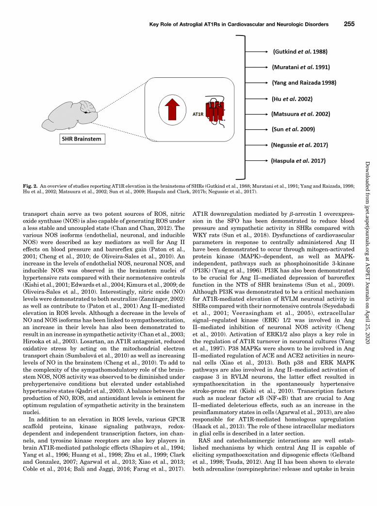
Further evidence for a role of brain RAS in cardiovasculardisorders came from studies in spontaneously hypertensiverats (SHRs), widely regarded as the best model to studyessential hypertension. The brain RAS was demonstrated tobe overactive in SHRs compared with their normotensivecontrols (Veerasingham and Raizada, 2003; Haspula andClark, 2018). AT1R levels were observed to be higher in thebrainstems of SHRs compared with their normotensive con-trols, Wistar Kyoto (WKY) rats (Gutkind et al., 1988; Hu et al.,2002). RAS activity was demonstrated to be markedly poten-tiated in the cardioregulatory regions of the brainstem andhypothalamus of SHRs compared with their normotensivecontrols (Casto and Phillips, 1985; Matsuda et al., 1987;Gutkind et al., 1988; Muratani et al., 1991; Stadler et al.,1992; Zhu et al., 1998; Hu et al., 2002; Ito et al., 2002; Sunet al., 2009; Haspula and Clark, 2017b; Negussie et al., 2017).Both synthesis and turnover of Ang II were also enhanced inSHRs compared with their normotensive controls (Gantenet al., 1983; Hermann et al., 1984). An overview of studies thathave reported an elevation in AT1R activity in the brainstemsof SHRs is shown in Fig. 2.Apart from AT1R, other components of the RAS were also
observed to be altered in rodentmodels of hypertension. ACE2levels were reported to be markedly lower in the hypothala-mus of SHRs compared with WKY rats, resulting in anaugmented ACE–Ang II–AT1R axis (Wang et al., 2017).AT2R activation was observed to have a greater antihyper-tensive effect in hypertensive models compared with normo-tensive models (de Kloet et al., 2017; Steckelings et al., 2017).Ang (1-7) was demonstrated to restore normal baroreflexfunctioning and autonomic activity in transgenic (mRen2)27hypertensive rats (Kangussu et al., 2015). In response tohypoxia, often accompanied by an elevation in sympatheticactivity, ACE levels were shown to be elevated in the medianpreoptic nucleus (Faulk et al., 2017). Brain AT2R activationcan negate the sympathoexcitatory response that is a conse-quence of AT1R-mediated sympathoexcitation and also renalvolume expansion (Stegbauer et al., 2005; Gao and Zucker,2011; Abdulla and Johns, 2017). Increased blood pressureand attenuated baroreflex sensitivity in the offspring ofbetamethasone-exposed sheep was shown to be a consequenceof central RAS impairment, which was improved by Ang (1-7)(Hendricks et al., 2017). Ang (1-7) also negated the AngII–mediated chronotropic response in the hypothalamic neu-rons of prehypertensive SHRs (Modgil et al., 2012).At a molecular level, several mechanisms such as endoplas-
mic reticulum stress, mitochondrial dysfunction, and redox-sensitive transcriptional factors have all been attributed tobrain-derived Ang II promoting neurogenic hypertension andbaroreflex impairment (Nautiyal et al., 2013; Coble et al.,2015; Young and Davisson, 2015; Case et al., 2017). Anincrease in reactive oxygen species (ROS) levels in thebrainstem cardioregulatory centers is a crucial step by whichAng II is able to augment multiple cardiovascular parameterssuch as sympathetic tone, heart rate, and water intake(Zimmerman et al., 2002, 2004; Nozoe et al., 2008; Chan andChan, 2012). In a model of renovascular hypertension, in-creased levels of both oxidative stress markers and AT1Rlevels in the PVN and RVLM were demonstrated to besignificant contributors to sympathoexcitation and hyperten-sion (Oliveira-Sales et al., 2009; de Oliveira-Sales et al., 2010).Although NADPH oxidase and the mitochondrial electron
254 Haspula and Clark
at ASPE
T Journals on A
pril 25, 2020jpet.aspetjournals.org
Dow
nloaded from
transport chain serve as two potent sources of ROS, nitricoxide synthase (NOS) is also capable of generating ROS undera less stable and uncoupled state (Chan and Chan, 2012). Thevarious NOS isoforms (endothelial, neuronal, and inducibleNOS) were described as key mediators as well for Ang IIeffects on blood pressure and baroreflex gain (Paton et al.,2001; Cheng et al., 2010; de Oliveira-Sales et al., 2010). Anincrease in the levels of endothelial NOS, neuronal NOS, andinducible NOS was observed in the brainstem nuclei ofhypertensive rats compared with their normotensive controls(Kishi et al., 2001; Edwards et al., 2004; Kimura et al., 2009; deOliveira-Sales et al., 2010). Interestingly, nitric oxide (NO)levels were demonstrated to both neutralize (Zanzinger, 2002)as well as contribute to (Paton et al., 2001) Ang II–mediatedelevation in ROS levels. Although a decrease in the levels ofNO and NOS isoforms has been linked to sympathoexcitation,an increase in their levels has also been demonstrated toresult in an increase in sympathetic activity (Chan et al., 2003;Hirooka et al., 2003). Losartan, an AT1R antagonist, reducedoxidative stress by acting on the mitochondrial electrontransport chain (Sumbalová et al., 2010) as well as increasinglevels of NO in the brainstem (Cheng et al., 2010). To add tothe complexity of the sympathomodulatory role of the brain-stem NOS, NOS activity was observed to be diminished underprehypertensive conditions but elevated under establishedhypertensive states (Qadri et al., 2003). A balance between theproduction of NO, ROS, and antioxidant levels is eminent foroptimum regulation of sympathetic activity in the brainstemnuclei.In addition to an elevation in ROS levels, various GPCR
scaffold proteins, kinase signaling pathways, redox-dependent and independent transcription factors, ion chan-nels, and tyrosine kinase receptors are also key players inbrain AT1R-mediated pathologic effects (Shapiro et al., 1994;Yang et al., 1996; Huang et al., 1998; Zhu et al., 1999; Clarkand Gonzalez, 2007; Agarwal et al., 2013; Xiao et al., 2013;Coble et al., 2014; Bali and Jaggi, 2016; Farag et al., 2017).
AT1R downregulation mediated by b-arrestin 1 overexpres-sion in the SFO has been demonstrated to reduce bloodpressure and sympathetic activity in SHRs compared withWKY rats (Sun et al., 2018). Dysfunctions of cardiovascularparameters in response to centrally administered Ang IIhave been demonstrated to occur through mitogen-activatedprotein kinase (MAPK)–dependent, as well as MAPK-independent, pathways such as phosphoinositide 3-kinase(PI3K) (Yang et al., 1996). PI3K has also been demonstratedto be crucial for Ang II–mediated depression of baroreflexfunction in the NTS of SHR brainstems (Sun et al., 2009).Although PI3K was demonstrated to be a critical mechanismfor AT1R-mediated elevation of RVLM neuronal activity inSHRs comparedwith their normotensive controls (Seyedabadiet al., 2001; Veerasingham et al., 2005), extracellularsignal–regulated kinase (ERK) 1/2 was involved in AngII–mediated inhibition of neuronal NOS activity (Chenget al., 2010). Activation of ERK1/2 also plays a key role inthe regulation of AT1R turnover in neuronal cultures (Yanget al., 1997). P38 MAPKs were shown to be involved in AngII–mediated regulation of ACE and ACE2 activities in neuro-nal cells (Xiao et al., 2013). Both p38 and ERK MAPKpathways are also involved in Ang II–mediated activation ofcaspase 3 in RVLM neurons, the latter effect resulted insympathoexcitation in the spontaneously hypertensivestroke-prone rat (Kishi et al., 2010). Transcription factorssuch as nuclear factor kB (NF-kB) that are crucial to AngII–mediated deleterious effects, such as an increase in theproinflammatory states in cells (Agarwal et al., 2013), are alsoresponsible for AT1R-mediated homologous upregulation(Haack et al., 2013). The role of these intracellular mediatorsin glial cells is described in a later section.RAS and catecholaminergic interactions are well estab-
lished mechanisms by which central Ang II is capable ofeliciting sympathoexcitation and dipsogenic effects (Gelbandet al., 1998; Tsuda, 2012). Ang II has been shown to elevateboth adrenaline (norepinephrine) release and uptake in brain
Fig. 2. An overview of studies reporting AT1R elevation in the brainstems of SHRs (Gutkind et al., 1988; Muratani et al., 1991; Yang and Raizada, 1998;Hu et al., 2002; Matsuura et al., 2002; Sun et al., 2009; Haspula and Clark, 2017b; Negussie et al., 2017).
Key Role of Astroglial AT1Rs in Cardiovascular and Neurologic Disorders 255
at ASPE
T Journals on A
pril 25, 2020jpet.aspetjournals.org
Dow
nloaded from
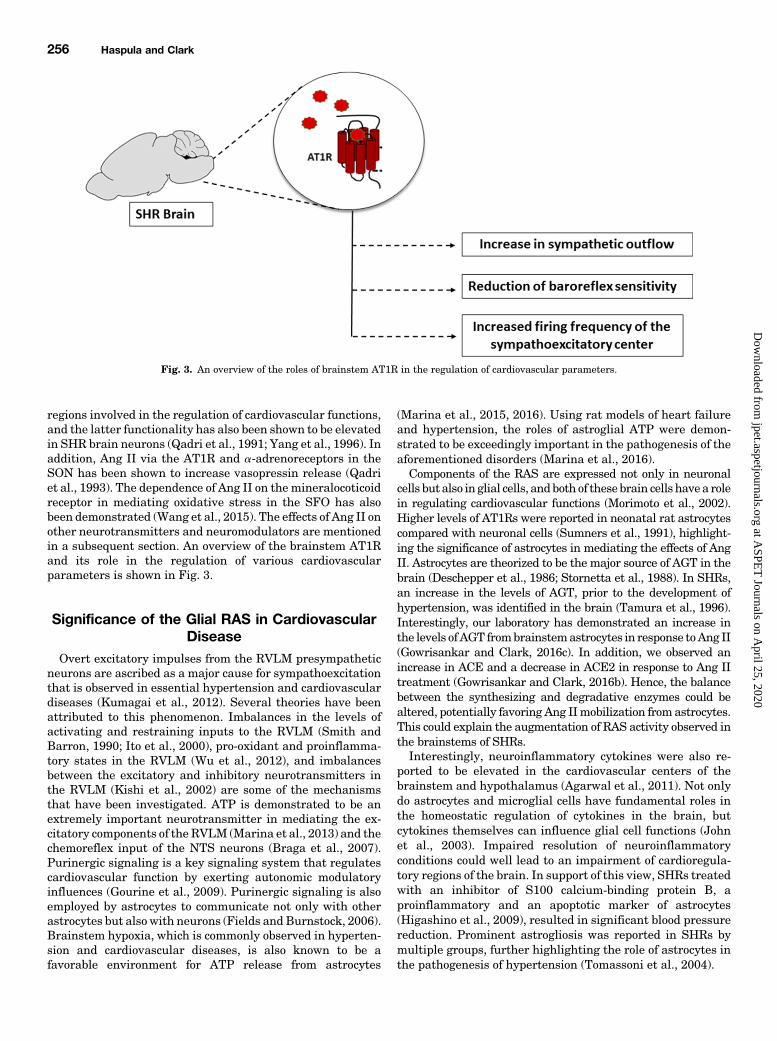
regions involved in the regulation of cardiovascular functions,and the latter functionality has also been shown to be elevatedin SHR brain neurons (Qadri et al., 1991; Yang et al., 1996). Inaddition, Ang II via the AT1R and a-adrenoreceptors in theSON has been shown to increase vasopressin release (Qadriet al., 1993). The dependence of Ang II on the mineralocoticoidreceptor in mediating oxidative stress in the SFO has alsobeen demonstrated (Wang et al., 2015). The effects of Ang II onother neurotransmitters and neuromodulators are mentionedin a subsequent section. An overview of the brainstem AT1Rand its role in the regulation of various cardiovascularparameters is shown in Fig. 3.
Significance of the Glial RAS in CardiovascularDisease
Overt excitatory impulses from the RVLM presympatheticneurons are ascribed as a major cause for sympathoexcitationthat is observed in essential hypertension and cardiovasculardiseases (Kumagai et al., 2012). Several theories have beenattributed to this phenomenon. Imbalances in the levels ofactivating and restraining inputs to the RVLM (Smith andBarron, 1990; Ito et al., 2000), pro-oxidant and proinflamma-tory states in the RVLM (Wu et al., 2012), and imbalancesbetween the excitatory and inhibitory neurotransmitters inthe RVLM (Kishi et al., 2002) are some of the mechanismsthat have been investigated. ATP is demonstrated to be anextremely important neurotransmitter in mediating the ex-citatory components of the RVLM (Marina et al., 2013) and thechemoreflex input of the NTS neurons (Braga et al., 2007).Purinergic signaling is a key signaling system that regulatescardiovascular function by exerting autonomic modulatoryinfluences (Gourine et al., 2009). Purinergic signaling is alsoemployed by astrocytes to communicate not only with otherastrocytes but also with neurons (Fields and Burnstock, 2006).Brainstem hypoxia, which is commonly observed in hyperten-sion and cardiovascular diseases, is also known to be afavorable environment for ATP release from astrocytes
(Marina et al., 2015, 2016). Using rat models of heart failureand hypertension, the roles of astroglial ATP were demon-strated to be exceedingly important in the pathogenesis of theaforementioned disorders (Marina et al., 2016).Components of the RAS are expressed not only in neuronal
cells but also in glial cells, andboth of these brain cells havea rolein regulating cardiovascular functions (Morimoto et al., 2002).Higher levels of AT1Rs were reported in neonatal rat astrocytescompared with neuronal cells (Sumners et al., 1991), highlight-ing the significance of astrocytes in mediating the effects of AngII. Astrocytes are theorized to be the major source of AGT in thebrain (Deschepper et al., 1986; Stornetta et al., 1988). In SHRs,an increase in the levels of AGT, prior to the development ofhypertension, was identified in the brain (Tamura et al., 1996).Interestingly, our laboratory has demonstrated an increase inthe levels ofAGT frombrainstemastrocytes in response toAng II(Gowrisankar and Clark, 2016c). In addition, we observed anincrease in ACE and a decrease in ACE2 in response to Ang IItreatment (Gowrisankar and Clark, 2016b). Hence, the balancebetween the synthesizing and degradative enzymes could bealtered, potentially favoringAng IImobilization fromastrocytes.This could explain the augmentation of RAS activity observed inthe brainstems of SHRs.Interestingly, neuroinflammatory cytokines were also re-
ported to be elevated in the cardiovascular centers of thebrainstem and hypothalamus (Agarwal et al., 2011). Not onlydo astrocytes and microglial cells have fundamental roles inthe homeostatic regulation of cytokines in the brain, butcytokines themselves can influence glial cell functions (Johnet al., 2003). Impaired resolution of neuroinflammatoryconditions could well lead to an impairment of cardioregula-tory regions of the brain. In support of this view, SHRs treatedwith an inhibitor of S100 calcium-binding protein B, aproinflammatory and an apoptotic marker of astrocytes(Higashino et al., 2009), resulted in significant blood pressurereduction. Prominent astrogliosis was reported in SHRs bymultiple groups, further highlighting the role of astrocytes inthe pathogenesis of hypertension (Tomassoni et al., 2004).
Fig. 3. An overview of the roles of brainstem AT1R in the regulation of cardiovascular parameters.
256 Haspula and Clark
at ASPE
T Journals on A
pril 25, 2020jpet.aspetjournals.org
Dow
nloaded from

Although the importance of the astroglial RAS has beenunderscored by several studies, the molecular mechanismsunderlying these effects have not yet been well investi-gated. Several groups have demonstrated the ability ofproinflammatory cytokines to regulate sympathetic activity(Winklewski et al., 2015). The idea that a dysregulatedneuroinflammatory state, in the cardiovascular centers ofthe brainstem, could contribute to sympathoexcitation wassuggested by Paton and colleagues less than a decade ago(Waki et al., 2008). This was based on gene expression studiesin the NTS of SHRs, where they observed altered inflamma-tory states, specifically in the levels of junctional adhesionmolecule 1, monocyte chemotactic protein 1, and interleukin(IL)-6 (Waki et al., 2008; Paton and Waki, 2009). Prominentneuroinflammatory states were observed at very early stagesof hypertension in SHRs, indicating a causative factor in thepathogenesis of essential hypertension (Waki et al., 2008).Whether an increase in proinflammatory cytokines, in thecardiovascular centers of the brain, was an important medi-ator of Ang II–mediated sympathoexcitation was investigatedbyKang et al. (2009). They demonstrated that chronic infusionof Ang II in Sprague-Dawley rats caused sympathoexcitation,which was characterized by a proinflammatory and a pro-oxidant state in the PVN. Blockade of AT1Rs and NF-kB wasalso demonstrated to normalize proinflammatory cytokines aswell as sympathetic activity, further authenticating the roleof neuroinflammation and the RAS in sympathoexcitation(Kang et al., 2009). Chronic infusion of Ang II resulting in aprominent inflammatory status in the brain vasculature wasshown to occur via an increase in ROS levels (Zhang et al.,2010). Since glial cells play a critical role in the regulation ofneuroinflammatory and oxidative states in the brain, glialAT1Rs could well be important factors in the augmentation ofsympathetic activity. In support of this, ablation of astroglialAT1Rs in the brainstem has been demonstrated to result in animprovement in the symptoms of heart failure by normaliza-tion of sympathetic activity (Isegawa et al., 2014).The link between the glial RAS, neuroinflammatory states,
and sympathoexcitation was discussed in a review by Shi et al.(2010b). They conceptualized that glial AT1R activation resultsin an increase in the levels of proinflammatory cytokines, whichcan then act as neuromodulators of synaptic activity (Shi et al.,2010b). It is plausible that mobilization of cytokines from glialcells can alter neuronal activity, since low levels of cytokinescan alter neuronal activity (Waki and Gouraud, 2014). De-finitive evidence of the role of inflammatory cytokines and AngII–mediated elevation in sympathetic nervous system activitycame from studies in the hypothalamus by the same group (Shiet al., 2010a). Chronic Ang II infusion in the PVN resulted in anincrease in proinflammatory cytokines and a decrease in anti-inflammatory cytokines, which then caused an elevation inblood pressure (Shi et al., 2010a). This effect could be blocked byminocycline, indicating that this effect was mediated by micro-glial AT1Rs (Shi et al., 2010a). Circulating Ang II has also beenshown to gain access to the cardiovascular centers in the brainby promoting blood-brain barrier breakdown (Vital et al., 2010;Biancardi and Stern, 2016). Both microglial AT1R-mediatedelevation in oxidative stress and proinflammatory cytokines aswell as molecular alterations in the brain microvasculature areviewed as the major mechanisms (Vital et al., 2010; Biancardiand Stern, 2016). Microglial cells were transformed predomi-nantly into the M1 phenotype when exposed to Ang II, which
represents a proinflammatory and a cytotoxic version of micro-glia, highlighting the role of microglial AT1Rs in promotingneuroinflammation and neurotoxicity (Rodriguez-Perez et al.,2016; Labandeira-Garcia et al., 2017). Further support camefrom another study investigating the crosstalk between theAT1R and Toll-like receptor 4 in the PVN. Ang II–mediatedincreases in ROS productionwere demonstrated to be due to aninteraction between AT1R and Toll-like receptor 4 inmicroglialcells in the PVN (Biancardi et al., 2016). A synergistic effect onsympathetic activity has also been reported between proin-flammatory cytokines and Ang II in the PVN (Shi et al., 2011).In addition to Ang II, other components of the RAS (e.g.,prorenin) have also been highlighted as a potential inducer ofneuroinflammation by microglial activation (Shi et al., 2014;Zhu et al., 2015). Interestingly, Ang (1-7) has not only beendemonstrated to counteract the effects of prorenin, but it alsoinduces microglial cells to assume an anti-inflammatory phe-notype under basal conditions (Liu et al., 2016).Microglial cell activation is often followed by astroglial
activation in several neurologic disorders (Liu et al., 2011b).Hence, microglia may initiate the inflammatory response, andastrocytes may aid in perpetuating the proinflammatorystates that have been reported in cardiovascular nuclei ofthe SHR brainstem and hypothalamus (Agarwal et al., 2011).Not just Ang II but other RAS peptides can also elicitproinflammatory states by interacting with the astroglialAT1R. PVN astrocytes isolated from SHRs, when treated withprorenin, resulted in an augmented increase in proinflamma-tory cytokines (Rodríguez et al., 2015). Ang III was alsoobserved to have potent proinflammatory effects in normo-tensive brainstem astrocytes (Kandalam et al., 2015).Our group observed an elevation of pro-oxidant and proin-
flammatory cytokines IL-1 and IL-6 in brainstem astrocytes,isolated from SHRs and Wistar rats, in response to Ang IItreatment (Gowrisankar and Clark, 2016a; Haspula andClark, 2017a). Baseline levels of the neuroinflammatorycytokines were also elevated in SHR brainstem astrocytes(Waki et al., 2008; Gowrisankar and Clark, 2016a; Haspulaand Clark, 2017a). Interestingly, we observed an elevation inthe levels of both pro- and anti-inflammatory cytokines inthese cells (Haspula and Clark, 2017a). Since astrocytes areknown to play a role in neutralizing oxidative stress andproinflammatory states, an increase in anti-inflammatorystatus could well be a compensatory/protective mechanismthat needs to be further investigated. Studies from ourlaboratory have focused on uncovering the molecular path-ways triggered by astroglial AT1R under hypertensive andnormotensive conditions. Ang II via AT1R elevates ROS aswell as activates key signal transduction pathways such as theERK, p38, and Janus kinase/signal transducer and activatorof transcription pathways. These are important systems thatare critical to astroglial functions such as cell proliferation aswell as to cardiovascular functions such as mobilization ofinflammatory cytokines from astrocytes (Kandalam andClark, 2010; Clark et al., 2013; Alanazi et al., 2014;Negussie et al., 2016; Haspula and Clark, 2017b).
Brain RAS and Neurologic DisordersHigher prevalence rates of hypertension have been reported
in neurologic impairments characterized by prominent neuro-inflammation, such as cognitive decline (Skoog et al., 1996;
Key Role of Astroglial AT1Rs in Cardiovascular and Neurologic Disorders 257
at ASPE
T Journals on A
pril 25, 2020jpet.aspetjournals.org
Dow
nloaded from

Tzourio, 2007; Nelson et al., 2014; Muela et al., 2017). Someconflicting data exist relating to the correlation betweenhypertension and cognitive decline. Although some studieshave shown a strong correlation between the two, others haveshown no correlation or even a negative correlation (Morriset al., 2000; Qiu et al., 2003; Reitz et al., 2007; Warchol-Celinska et al., 2015). Since activation of brain AT1Rs leads toan increase in proinflammatory and pro-oxidant states, itcomes as little surprise that blockade of brain AT1Rs wasinvestigated as a therapeutic strategy for several neurologicdisorders (Wright et al., 2013; Mascolo et al., 2017). Evidencepoints to neutralization of the brain RAS as a treatment optionin disorders pertaining to cognitive decline and memory loss(Mogi et al., 2012; Bodiga and Bodiga, 2013). Centrally actingARBs were observed to have a greater efficacy than otherantihypertensives in memory preservation in older hyperten-sive individuals (Ho andNation, 2017). An observational case-control study assessing the efficacy of ACEIs on cognitivefunctions in elderly individuals also had favorable results(Gao et al., 2013). Furthermore, in a recently published study,an improvement in cognitive functions was observed withcentrally acting ARBs in individuals diagnosed with Alz-heimer disease (Fazal et al., 2017). ARBs also showed favor-able neuropathological outcomes in hypertensive individualswith cognitive impairments (Hajjar et al., 2012). Consideringthat a strong correlation exists between cognitive decline andhypertension (Skoog et al., 1996; Tzourio, 2007; Nelson et al.,2014; Muela et al., 2017), hyperactivity of brain RAS, oftenassociated with hypertension, could well be a hallmark of notonly cardiovascular but also neurologic disorders as well.An overactive brain RAS has also been linked to other
neurodegenerative diseases such as Parkinson disease(Wright et al., 2013; Laudisio et al., 2017). In a prospectivecase-controlled study conducted on hypertensive individualson ACEIs and ARBs, a reduced incidence of Parkinson diseasewas reported (Lee et al., 2014). In a recently published study,the ARB azilsartan was demonstrated to counteract apoptosisof dopaminergic neurons in a rat model of Parkinson disease(Gao et al., 2017). Telmisartan, an ARB that has peroxisomeproliferator–activated receptorg activating ability, has beenshown to reduce neuronal loss and inflammation in a mousemodel of Alzheimer disease (Saavedra, 2012). Several pre-clinical studies have highlighted the role exogenous andendogenous Ang II in worsening Alzheimer disease patho-physiology, highlighting the potential deleterious role of AT1Rin neurologic indices (Ongali et al., 2014; Takane et al., 2017).In addition to AT1R, other components of the RAS have alsobeen shown to be altered in neurologic disorders. ACE2activity was observed to be markedly reduced in the post-mortem brain tissue of individuals diagnosed with Alzheimerdisease (Kehoe et al., 2016). Prospective cohort analysis inmales diagnosed with Alzheimer disease revealed a signifi-cant improvement in the progression of the disease inindividuals taking ARBs compared with other cardiovasculardrugs (Li et al., 2010). By employing animal models, the RAShas also been demonstrated to worsen autoimmune disorderssuch as multiple sclerosis (MS) by activating transforminggrowth factor b (Lanz et al., 2010). Imbalances in cerebrospi-nal fluid Ang II levels, along with an impairment in perivas-cular astrocytes, were observed in patients with MS(Matsushita et al., 2010). Additionally, immunohistochemis-try analysis revealed that plaques from the brains of patients
withMS showed a strong upregulation inAT1R levels (Plattenet al., 2009). Blocking of Ang II production was demonstratedto suppress the neuroinflammatory phenotype induced byactivation of NF-kB via AT1R (Platten et al., 2009). Owing tothe potential neuroprotective effect of ARBs, blocking of AT1Rwas shown to have promise in traumatic brain injury (Villapolet al., 2015). Since strong evidence exists linking centrally andnoncentrally acting ACEIs or AT1R blockers with improve-ment of cognitive function, dementia, and neurodegeneration(Saxby et al., 2008; Mogi and Horiuchi, 2009; Davies et al.,2011), it is imperative that we understand and fully elucidatethe mechanisms by which Ang II contributes to neuronaldamage. Since astrocytes have a central role in brain homeo-stasis by regulating levels of cytokines and ROS, hyperfuc-tional astroglial AT1Rsmay be a prominent feature of not onlycardiovascular but also neurologic disorders.Astrocytes from brain regions other than the brainstem,
such as the cerebellum, are also responsive to Ang IItreatment (Clark et al., 2013). Ang II caused a significantincrease in the proinflammatory cytokine, IL-6, and ROSlevels in astrocytes isolated from the cerebellum from bothWistar rats and SHRs (Gowrisankar and Clark, 2016a). AngII–mediated ROS elevation and proinflammatory conditionsare associated with neurodegeneration and also astrocytesenescence (Lanz et al., 2010; Liu et al., 2011a; Min et al.,2011). In addition, the SHR, which is characterized by ahyperactive brain RAS, is routinely employed as a model forattention deficit hyperactivity disorder (ADHD) (Adrianiet al., 2003). Traits that are often observed in individualswithADHD, such as shorter attention spans, inability to focus,and hyperexcitability, are also observed in the SHR, making itan ideal model to investigate the etiology of ADHD (Adrianiet al., 2003). Similar to individuals with ADHD, SHRs are alsocharacterized by cerebellar atrophy and cerebellar impair-ment (Yun et al., 2014). Evidence of a greater incidence oflearning disabilities has also been reported in children withADHD diagnosed with primary hypertension than in thosewithout (Adams et al., 2010). These studies further highlightthe importance of neuroinflammation in the pathogenesis ofnot only cardiovascular disorders but neurologic impairmentsas well. Considering that the RAS is a premium hormonalsystem that is augmented in the brains of SHRs, it issurprising to observe a paucity of studies investigating theeffects of the RAS in ADHD. Nevertheless, the ability of Ang IIto promote a proinflammatory state in different regions of thebrain may lead to significant alteration in brain functions,eventually leading to neurologic disorders or an exacerbationof several neurologic conditions. A schematic of the potentialrole of glial AT1Rs in cardiovascular and neurologic diseases isshown in Fig. 4.Exogenous administration of Ang II in the rat striatum
was demonstrated to cause an increase in dopamine andserotonin metabolites, 3,4-dihydroxyphenylacetic acid and5-hydroxyindoleacetic acid, respectively (Mendelsohn et al.,1993). A decrease in acetylcholine levels in the rat entorhinalcortex was also observed (Barnes et al., 1989), furtherhighlighting the importance of the RAS in not only cardio-vascular disorders but also neurologic disorders. AlthoughAT1R is known to be associated with an increase in oxidativeand inflammatory status, the role of AT2R in neuronalfunctions cannot be overlooked. The administration of bothan AT2R agonist and an AT1R antagonist was shown to
258 Haspula and Clark
at ASPE
T Journals on A
pril 25, 2020jpet.aspetjournals.org
Dow
nloaded from

lower 3,4-dihydroxyphenylacetic acid levels (Mertens et al.,2010). Activation of AT2R has been reported to improvememory performance (Matavelli and Siragy, 2015) and toalso confer neuroprotection in middle cerebral arteryocclusion–induced stroke (Joseph et al., 2014; Min et al.,2014). The mechanisms could be due to its role in neuronaldifferentiation, excitability, regeneration, apoptosis, andreduction in oxidative stress and inflammation (Côté et al.,1999; Gendron et al., 2003; Min et al., 2014). AT2R is shownto be mostly expressed on neuronal cells and not glial cells(Bennion et al., 2017). It is mostly the neuronal AT2R that isresponsible for the cerebroprotective effects conferred by AT2Ragonism after middle cerebral artery occlusion–induced stroke(Bennion et al., 2017).In addition to AT1Rs and AT2Rs, AT4R has also been
identified to have a significant impact on cognition. Ang IVand other AT4R ligands have been shown to significantlyimprove several different facets of memory and learningperformance in rats (Wright et al., 1993; Lee et al., 2004;Wright and Harding, 2008). An elevation in acetylcholinerelease and facilitation of long-termpotentiation, independentof glutamergic signaling, were reported to be a few of the keymechanisms of AT4R’s ability to improve cognitive ability(Wright and Harding, 2008).
Conclusion and Future PerspectivesAt a molecular level, elevations in ROS and inflammatory
cytokines are identified as an overarching paradigm for AT1R-mediated effects (Mehta and Griendling, 2007). Convincing
evidence in the last decade has underscored the role ofcentrally expressed proinflammatory and pro-oxidant media-tors in the progression of cardiovascular diseases and ashallmarks/risk factors for these diseases (Kishi et al., 2004;Wu et al., 2012; Haspula and Clark, 2018). By employinganimal models, it was shown that elevations in centralsympathetic outflow could be corrected by counteractingproinflammatory and pro-oxidant states (Shi et al., 2010a;Winklewski et al., 2015). The past decade has seen asignificant increase in the number of studies highlightingthe importance of the brain RAS in neurologic disorders suchas dementia (Mogi et al., 2012) and MS (Lanz et al., 2010) andin neurodegenerative disorders such as Alzheimer andParkinson diseases (Wright et al., 2013). Neuroinflammationand oxidative stress appear to be the common threads thatconnect the brain RAS to both cardiovascular and neurologicdisorders (Zhang et al., 2010; Saavedra, 2012). AT1R-mediated elevation in ROS and multiple proinflammatorymediators are key factors in Ang II driving the progression ofseveral pathologic conditions. Similar to many GPCRs, AT1Rsare known to crosstalk with other receptors, including thoseinvolved in mediating neuroprotection such as the Mas re-ceptor (Von Bohlen und Halbach et al., 2000), AT2R (Inuzukaet al., 2016), and cannabinoid type-1 receptor (Rozenfeld et al.,2011; Szekeres et al., 2012; Haspula and Clark, 2016, 2017b).Hence, the pathologic significance of AT1R crosstalk withother receptors and systems present in different regions of thebrain needs to be further explored. However, evidence of brainAT1R-mediated changes in receptors and systems that play acrucial role in neuroprotection has not been extensively
Fig. 4. A schematic of potential roles of astroglial AT1Rs in cardiovascular and neurologic diseases. Ang II activates AT1Rs leading to the stimulation ofa host of intracellular mediators. These mediators lead to an increase in proinflammatory cytokines that can trigger a diverse array of events in thecentral nervous system. AP-1, activator protein-1; BBB, blood-brain barrier; DAG, diacylglycerol; IP3, inositol trisphosphate; JNK, Jun N-terminalkinase; PIP2, phosphatidylinositol biphosphate; PKC, protein kinase C; PLC, phospholipase C.
Key Role of Astroglial AT1Rs in Cardiovascular and Neurologic Disorders 259
at ASPE
T Journals on A
pril 25, 2020jpet.aspetjournals.org
Dow
nloaded from

researched. Understanding AT1R-mediated interactions withGPCRs that are known to be both functionally similar, as wellas those that are deemed to be functionally antagonistic toAT1Rs, could aid in identifying the important players in AngII–mediated neuroinflammation and oxidative stress, leadingto the identification of therapeutic strategies that could begiven in combination with central RAS antagonists. Inaddition, ARBs that are known to exhibit neuroprotectiveproperties, independent of AT1R blockade, could well havepotential for the treatment of neurologic disorders that arecharacterized by excessive inflammation. ARBs such astelmisartan are known to have pleiotropic effects apart fromnegating AT1R-induced oxidative stress and inflammation inthe brain. Telmisartan has already been identified to haveneuroprotective roles in preclinical studies conducted inmouse models of Alzheimer disease and stroke (Thoene-Reineke et al., 2011; Saavedra, 2012; Torika et al., 2017).However, clinical studies have confirmed a lack of effective-ness of telmisartan in conferring neuroprotection in subacutestroke (Sare et al., 2013). Since neuroprotection has beendemonstrated for ARBs such as candesartan and telmisartan,it could be worthwhile to investigate these drugs not only forcardiovascular disorders but also for neurologic outcomes(Benicky et al., 2009; Noda et al., 2012; Villapol and Saavedra,2015). Whether ARBs such as telmisartan that target neuro-inflammation through multiple modes of mechanisms havebeneficial roles in neurologic disorders such as Alzheimer andParkinson diseases remains to be seen.
Authorship Contributions
Wrote or contributed to the writing of the manuscript: Haspula,Clark.
References
Abdulla MH and Johns EJ (2017) The role of brain angiotensin II (type 2) receptorsand nitric oxide in the renal sympathoinhibitory response to acute volume ex-pansion in conscious rats. J Hypertens 35:338–347.
Adams HR, Szilagyi PG, Gebhardt L, and Lande MB (2010) Learning and attentionproblems among children with pediatric primary hypertension. Pediatrics 126:e1425–e1429.
Adriani W, Caprioli A, Granstrem O, Carli M, and Laviola G (2003) The spontane-ously hypertensive-rat as an animal model of ADHD: evidence for impulsive andnon-impulsive subpopulations. Neurosci Biobehav Rev 27:639–651.
Agarwal D, Dange RB, Raizada MK, and Francis J (2013) Angiotensin II causesimbalance between pro- and anti-inflammatory cytokines by modulating GSK-3bin neuronal culture. Br J Pharmacol 169:860–874.
Agarwal D, Welsch MA, Keller JN, and Francis J (2011) Chronic exercise modulatesRAS components and improves balance between pro- and anti-inflammatory cy-tokines in the brain of SHR. Basic Res Cardiol 106:1069–1085.
Alanazi AZ, Patel P, and Clark MA (2014) p38 Mitogen-activated protein kinase isstimulated by both angiotensin II and angiotensin III in cultured rat astrocytes. JRecept Signal Transduct Res 34:205–211.
Allen AM (2002) Inhibition of the hypothalamic paraventricular nucleus in sponta-neously hypertensive rats dramatically reduces sympathetic vasomotor tone. Hy-pertension 39:275–280.
Allen AM, Dosanjh JK, Erac M, Dassanayake S, Hannan RD, and Thomas WG (2006)Expression of constitutively active angiotensin receptors in the rostral ventrolat-eral medulla increases blood pressure. Hypertension 47:1054–1061.
Anderson EA, Sinkey CA, Lawton WJ, and Mark AL (1989) Elevated sympatheticnerve activity in borderline hypertensive humans. Evidence from direct intra-neural recordings. Hypertension 14:177–183.
Aranda JM, Jr and Conti CR (2003) Angiotensin II blockade: a therapeutic strategywith wide applications. Clin Cardiol 26:500–502.
Atlas SA (2007) The renin-angiotensin aldosterone system: pathophysiological roleand pharmacologic inhibition. J Manag Care Pharm 13 (Suppl B):9–20.
Bali A and Jaggi AS (2016) Angiotensin II-triggered kinase signaling cascade in thecentral nervous system. Rev Neurosci 27:301–315.
Barki-Harrington L, Luttrell LM, and Rockman HA (2003) Dual inhibition of beta-adrenergic and angiotensin II receptors by a single antagonist: a functional role forreceptor-receptor interaction in vivo. Circulation 108:1611–1618.
Barnes JM, Barnes NM, Costall B, Horovitz ZP, and Naylor RJ (1989) Angiotensin IIinhibits the release of [3H]acetylcholine from rat entorhinal cortex in vitro. BrainRes 491:136–143.
Benicky J, Sánchez-Lemus E, Pavel J, and Saavedra JM (2009) Anti-inflammatoryeffects of angiotensin receptor blockers in the brain and the periphery. Cell MolNeurobiol 29:781–792.
Bennion DM, Haltigan E, Regenhardt RW, Steckelings UM, and Sumners C (2015)Neuroprotective mechanisms of the ACE2-angiotensin-(1-7)-Mas axis in stroke.Curr Hypertens Rep 17:3.
Bennion DM, Isenberg JD, Harmel AT, DeMars K, Dang AN, Jones CH, PignataroME, Graham JT, Steckelings UM, Alexander JC, et al. (2017) Post-stroke angio-tensin II type 2 receptor activation provides long-term neuroprotection in agedrats. PLoS One 12:e0180738.
Biancardi VC and Stern JE (2016) Compromised blood-brain barrier permeability:novel mechanism by which circulating angiotensin II signals to sym-pathoexcitatory centres during hypertension. J Physiol 594:1591–1600.
Biancardi VC, Stranahan AM, Krause EG, de Kloet AD, and Stern JE (2016) Crosstalk between AT1 receptors and Toll-like receptor 4 in microglia contributes toangiotensin II-derived ROS production in the hypothalamic paraventricular nu-cleus. Am J Physiol Heart Circ Physiol 310:H404–H415.
Bickerton RK and Buckley JP (1961) Evidence for a central mechanism in angio-tensin induced hypertension. Exp Biol Med 106:834–836.
Bodiga VL and Bodiga S (2013) Renin angiotensin system in cognitive function anddementia. Asian J Neurosci 2013:102602.
Booz GW, Conrad KM, Hess AL, Singer HA, and Baker KM (1992) Angiotensin-II-binding sites on hepatocyte nuclei. Endocrinology 130:3641–3649.
Bosnyak S, Welungoda IK, Hallberg A, Alterman M, Widdop RE, and Jones ES(2010) Stimulation of angiotensin AT2 receptors by the non-peptide agonist, com-pound 21, evokes vasodepressor effects in conscious spontaneously hypertensiverats. Br J Pharmacol 159:709–716.
Braga VA, Soriano RN, Braccialli AL, de Paula PM, Bonagamba LGH, Paton JFR,and Machado BH (2007) Involvement of L-glutamate and ATP in the neurotrans-mission of the sympathoexcitatory component of the chemoreflex in the commis-sural nucleus tractus solitarii of awake rats and in the working heart-brainstempreparation. J Physiol 581:1129–1145.
Brooks VL and Malvin RL (1979) An intracerebral, physiological role for angiotensin:effects of central blockade. Fed Proc 38:2272–2275.
Burnier M and Zanchi A (2006) Blockade of the renin-angiotensin-aldosterone sys-tem: a key therapeutic strategy to reduce renal and cardiovascular events in pa-tients with diabetes. J Hypertens 24:11–25.
Campagnole-Santos MJ, Diz DI, and Ferrario CM (1988) Baroreceptor reflex modu-lation by angiotensin II at the nucleus tractus solitarii.Hypertension 11:I167–I171.
Campbell DJ, Bouhnik J, Ménard J, and Corvol P (1984) Identity of angiotensinogenprecursors of rat brain and liver. Nature 308:206–208.
Campos LA, Bader M, and Baltatu OC (2012) Brain renin-angiotensin system inhypertension, cardiac hypertrophy, and heart failure. Front Physiol 2:115.
Carey RM, Howell NL, Jin XH, and Siragy HM (2001) Angiotensin type 2 receptor-mediated hypotension in angiotensin type-1 receptor-blocked rats. Hypertension38:1272–1277.
Case AJ, Tian J, and Zimmerman MC (2017) Increased mitochondrial superoxide inthe brain, but not periphery, sensitizes mice to angiotensin II-mediated hyper-tension. Redox Biol 11:82–90.
Casto R and Phillips MI (1985) Neuropeptide action in nucleus tractus solitarius:angiotensin specificity and hypertensive rats. Am J Physiol 249:R341–R347.
Cato MJ and Toney GM (2005) Angiotensin II excites paraventricular nucleus neu-rons that innervate the rostral ventrolateral medulla: an in vitro patch-clampstudy in brain slices. J Neurophysiol 93:403–413.
Chan JYH, Wang L-L, Chao Y-M, and Chan SHH (2003) Downregulation of basal iNOSat the rostral ventrolateral medulla is innate in SHR. Hypertension 41:563–570.
Chan SHH and Chan JYH (2012) Brain stem oxidative stress and its associatedsignaling in the regulation of sympathetic vasomotor tone. J Appl Physiol (1985)113:1921–1928.
Cheng W-H, Lu P-J, Ho W-Y, Tung C-S, Cheng P-W, Hsiao M, and Tseng C-J (2010)Angiotensin II inhibits neuronal nitric oxide synthase activation through theERK1/2-RSK signaling pathway to modulate central control of blood pressure. CircRes 106:788–795.
Clark MA and Gonzalez N (2007) Angiotensin II stimulates rat astrocyte mitogen-activated protein kinase activity and growth through EGF and PDGF receptortransactivation. Regul Pept 144:115–122.
Clark MA, Nguyen C, and Tran H (2013) Distinct molecular effects of angiotensin IIand angiotensin III in rat astrocytes. Int J Hypertens 2013:782861.
Coble JP, Grobe JL, Johnson AK, and Sigmund CD (2015) Mechanisms of brain reninangiotensin system-induced drinking and blood pressure: importance of the sub-fornical organ. Am J Physiol Regul Integr Comp Physiol 308:R238–R249.
Coble JP, Johnson RF, Cassell MD, Johnson AK, Grobe JL, and Sigmund CD (2014)Activity of protein kinase C-a within the subfornical organ is necessary for fluidintake in response to brain angiotensin. Hypertension 64:141–148.
Côté F, Do TH, Laflamme L, Gallo JM, and Gallo-Payet N (1999) Activation of the AT(2) receptor of angiotensin II induces neurite outgrowth and cell migration inmicroexplant cultures of the cerebellum. J Biol Chem 274:31686–31692.
Danyel LA, Schmerler P, Paulis L, Unger T, and Steckelings UM (2013) Impact ofAT2-receptor stimulation on vascular biology, kidney function, and blood pressure.Integr Blood Press Control 6:153–161.
Davies NM, Kehoe PG, Ben-Shlomo Y, and Martin RM (2011) Associations of anti-hypertensive treatments with Alzheimer’s disease, vascular dementia, and otherdementias. J Alzheimers Dis 26:699–708.
de Gasparo M, Catt KJ, Inagami T, Wright JW, and Unger T (2000) InternationalUnion of Pharmacology. XXIII. The angiotensin II receptors. Pharmacol Rev 52:415–472.
de Kloet AD, Steckelings UM, and Sumners C (2017) Protective angiotensin type2 receptors in the brain and hypertension. Curr Hypertens Rep 19:46.
De Mello WC (2008) Intracellular and extracellular renin have opposite effects on theregulation of heart cell volume. Implications for myocardial ischaemia. J ReninAngiotensin Aldosterone Syst 9:112–118.
de Oliveira-Sales EB, Nishi EE, Boim MA, Dolnikoff MS, Bergamaschi CT,and Campos RR (2010) Upregulation of AT1R and iNOS in the rostral
260 Haspula and Clark
at ASPE
T Journals on A
pril 25, 2020jpet.aspetjournals.org
Dow
nloaded from

ventrolateral medulla (RVLM) is essential for the sympathetic hyperactivity andhypertension in the 2K-1C Wistar rat model. Am J Hypertens 23:708–715.
Deschepper CF, Bouhnik J, and Ganong WF (1986) Colocalization of angiotensinogenand glial fibrillary acidic protein in astrocytes in rat brain. Brain Res 374:195–198.
Edwards MA, Loxley RA, Powers-Martin K, Lipski J, McKitrick DJ, Arnolda LF,and Phillips JK (2004) Unique levels of expression of N-methyl-D-aspartate receptorsubunits and neuronal nitric oxide synthase in the rostral ventrolateral medulla ofthe spontaneously hypertensive rat. Brain Res Mol Brain Res 129:33–43.
Farag E, Sessler DI, Ebrahim Z, Kurz A, Morgan J, Ahuja S, Maheshwari K,and John Doyle D (2017) The renin angiotensin system and the brain: new de-velopments. J Clin Neurosci 46:1–8.
Faulk K, Shell B, Nedungadi TP, and Cunningham JT (2017) Role of angiotensin-converting enzyme 1 within the median preoptic nucleus following chronic in-termittent hypoxia. Am J Physiol Regul Integr Comp Physiol 312:R245–R252.
Fazal K, Perera G, Khondoker M, Howard R, and Stewart R (2017) Associations ofcentrally acting ACE inhibitors with cognitive decline and survival in Alzheimer’sdisease. BJPsych Open 3:158–164.
Ferrario CM, Brosnihan KB, Diz DI, Jaiswal N, Khosla MC, Milsted A, and TallantEA (1991) Angiotensin-(1-7): a new hormone of the angiotensin system. Hyper-tension 18:III126–III133.
Fields RD and Burnstock G (2006) Purinergic signalling in neuron-glia interactions.Nat Rev Neurosci 7:423–436.
Fisher JP and Paton JFR (2012) The sympathetic nervous system and blood pressurein humans: implications for hypertension. J Hum Hypertens 26:463–475.
Fisher JP, Young CN, and Fadel PJ (2009) Central sympathetic overactivity: mal-adies and mechanisms. Auton Neurosci 148:5–15.
Fyhrquist F, Metsärinne K, and Tikkanen I (1995) Role of angiotensin II in bloodpressure regulation and in the pathophysiology of cardiovascular disorders. J HumHypertens 9 (Suppl 5):S19–S24.
Ganong WF (1994) Origin of the angiotensin II secreted by cells. Proc Soc Exp BiolMed 205:213–239.
Ganten D, Hermann K, Bayer C, Unger T, and Lang RE (1983) Angiotensin synthesisin the brain and increased turnover in hypertensive rats. Science 221:869–871.
Gao L and Zucker IH (2011) AT2 receptor signaling and sympathetic regulation. CurrOpin Pharmacol 11:124–130.
Gao Q, Ou Z, Jiang T, Tian Y-Y, Zhou J-S, Wu L, Shi J-Q, and Zhang Y-D (2017)Azilsartan ameliorates apoptosis of dopaminergic neurons and rescues charac-teristic parkinsonian behaviors in a rat model of Parkinson’s disease. Oncotarget 8:24099–24109.
Gao Y, O’Caoimh R, Healy L, Kerins DM, Eustace J, Guyatt G, Sammon D,and Molloy DW (2013) Effects of centrally acting ACE inhibitors on the rate ofcognitive decline in dementia. BMJ Open 3:e002881.
Gelband CH, Sumners C, Lu D, and Raizada MK (1998) Angiotensin receptors andnorepinephrine neuromodulation: implications of functional coupling. Regul Pept73:141–147.
Gendron L, Payet MD, and Gallo-Payet N (2003) The angiotensin type 2 receptor ofangiotensin II and neuronal differentiation: from observations to mechanisms. JMol Endocrinol 31:359–372.
Gourine AV, Wood JD, and Burnstock G (2009) Purinergic signalling in autonomiccontrol. Trends Neurosci 32:241–248.
Gowrisankar YV and Clark MA (2016a) Angiotensin II induces interleukin-6 ex-pression in astrocytes: role of reactive oxygen species and NF-kB. Mol Cell Endo-crinol 437:130–141.
Gowrisankar YV and Clark MA (2016b) Angiotensin II regulation of angiotensin-converting enzymes in spontaneously hypertensive rat primary astrocyte cultures.J Neurochem 138:74–85.
Gowrisankar YV and Clark MA (2016c) Regulation of angiotensinogen expression byangiotensin II in spontaneously hypertensive rat primary astrocyte cultures. BrainRes 1643:51–58.
Grassi G, Seravalle G, and Quarti-Trevano F (2010) The ‘neuroadrenergic hypothesis’in hypertension: current evidence. Exp Physiol 95:581–586.
Gutkind JS, Kurihara M, Castren E, and Saavedra JM (1988) Increased concentra-tion of angiotensin II binding sites in selected brain areas of spontaneously hy-pertensive rats. J Hypertens 6:79–84.
Haack KKV, Mitra AK, and Zucker IH (2013) NF-kB and CREB are required forangiotensin II type 1 receptor upregulation in neurons. PLoS One 8:e78695.
Hajjar I, Brown L, Mack WJ, and Chui H (2012) Impact of angiotensin receptorblockers on Alzheimer disease neuropathology in a large brain autopsy series. ArchNeurol 69:1632–1638.
Hall JE (1986) Control of sodium excretion by angiotensin II: intrarenal mechanismsand blood pressure regulation. Am J Physiol 250:R960–R972.
Hamilton TA, Handa RK, Harding JW, and Wright JW (2001) A role for the angio-tensin IV/AT4 system in mediating natriuresis in the rat. Peptides 22:935–944.
Haspula D and Clark M (2017a) Regulation of neuroinflammatory cytokines by an-giotensin and cannabinoid systems in SHR astrocytes. FASEB J 31:lb554–lb554.
Haspula D and Clark MA (2016) Heterologous regulation of the cannabinoid type1 receptor by angiotensin II in astrocytes of spontaneously hypertensive rats. JNeurochem 139:523–536.
Haspula D and Clark MA (2017b) MAPK activation patterns of AT1R and CB1R inSHR versus Wistar astrocytes: evidence of CB1R hypofunction and crosstalk be-tween AT1R and CB1R. Cell Signal 40:81–90.
Haspula D and Clark MA (2018) Neuroinflammation and sympathetic overactivity:mechanisms and implications in hypertension. Auton Neurosci 210:10–17.
Hendricks AS, Shaltout HA, Chappell MC, and Diz DI (2017) Central angiotensin-(1-7) treatment attenuates ERK 1/2 expression and oxidative stress in the dorsalmedulla of betamethasone-exposed sheep that associates with improved bloodpressure and baroreflex sensitivity (Abstract). Hypertension 70 (Suppl 1):A145.
Hermann K, McDonald W, Unger T, Lang RE, and Ganten D (1984) Angiotensinbiosynthesis and concentrations in brain of normotensive and hypertensive rats. JPhysiol (Paris) 79:471–480.
Higashino H, Niwa A, Satou T, Ohta Y, Hashimoto S, Tabuchi M, and Ooshima K(2009) Immunohistochemical analysis of brain lesions using S100B and glialfibrillary acidic protein antibodies in arundic acid- (ONO-2506) treated stroke-prone spontaneously hypertensive rats. J Neural Transm (Vienna) 116:1209–1219.
Hirooka Y, Shigematsu H, Kishi T, Kimura Y, Ueta Y, and Takeshita A (2003) Re-duced nitric oxide synthase in the brainstem contributes to enhanced sympatheticdrive in rats with heart failure. J Cardiovasc Pharmacol 42 (Suppl 1):S111–S115.
Ho JK and Nation DA; Alzheimer’s Disease Neuroimaging Initiative (2017) Memoryis preserved in older adults taking AT1 receptor blockers. Alzheimers Res Ther 9:33.
Hoffmann BR, Stodola TJ, Wagner JR, Didier DN, Exner EC, Lombard JH,and Greene AS (2017) Mechanisms of Mas1 receptor-mediated signaling in thevascular endothelium. Arterioscler Thromb Vasc Biol 37:433–445.
Hu L, Zhu D-N, Yu Z, Wang JQ, Sun Z-J, and Yao T (2002) Expression of angiotensinII type 1 (AT(1)) receptor in the rostral ventrolateral medulla in rats. J ApplPhysiol (1985) 92:2153–2161.
Huang BS and Leenen FHH (2009) The brain renin-angiotensin-aldosterone system:a major mechanism for sympathetic hyperactivity and left ventricular remodelingand dysfunction after myocardial infarction. Curr Heart Fail Rep 6:81–88.
Huang X-C, Deng T, and Sumners C (1998) Angiotensin II stimulates activation ofFos-regulating kinase and c-Jun NH2-terminal kinase in neuronal cultures fromrat brain. Endocrinology 139:245–251.
Inuzuka T, Fujioka Y, Tsuda M, Fujioka M, Satoh AO, Horiuchi K, Nishide S, NanboA, Tanaka S, and Ohba Y (2016) Attenuation of ligand-induced activation of an-giotensin II type 1 receptor signaling by the type 2 receptor via protein kinase C.Sci Rep 6:21613.
Isegawa K, Hirooka Y, Katsuki M, Kishi T, and Sunagawa K (2014) Angiotensin IItype 1 receptor expression in astrocytes is upregulated leading to increased mor-tality in mice with myocardial infarction-induced heart failure. Am J Physiol HeartCirc Physiol 307:H1448–H1455.
Ito S, Komatsu K, Tsukamoto K, Kanmatsuse K, and Sved AF (2002) Ventrolateralmedulla AT1 receptors support blood pressure in hypertensive rats. Hypertension40:552–559.
Ito S, Komatsu K, Tsukamoto K, and Sved AF (2000) Excitatory amino acids in therostral ventrolateral medulla support blood pressure in spontaneously hyperten-sive rats. Hypertension 35:413–417.
John GR, Lee SC, and Brosnan CF (2003) Cytokines: powerful regulators of glial cellactivation. Neuroscientist 9:10–22.
Joseph JP, Mecca AP, Regenhardt RW, Bennion DM, Rodríguez V, Desland F, PatelNA, Pioquinto DJ, Unger T, Katovich MJ, et al. (2014) The angiotensin type 2 re-ceptor agonist Compound 21 elicits cerebroprotection in endothelin-1 induced is-chemic stroke. Neuropharmacology 81:134–141.
Kandalam U and Clark MA (2010) Angiotensin II activates JAK2/STAT3 pathwayand induces interleukin-6 production in cultured rat brainstem astrocytes. RegulPept 159:110–116.
Kandalam U, Sarmiento N, Haspula D, and Clark MA (2015) Angiotensin III inducessignal transducer and activator of transcription 3 and interleukin-6 mRNA levelsin cultured rat astrocytes. J Renin Angiotensin Aldosterone Syst 16:758–767.
Kang Y-M, Ma Y, Zheng J-P, Elks C, Sriramula S, Yang Z-M, and Francis J (2009)Brain nuclear factor-kappa B activation contributes to neurohumoral excitation inangiotensin II-induced hypertension. Cardiovasc Res 82:503–512.
Kangussu LM, Guimaraes PS, Nadu AP, Melo MB, Santos RAS, and Campagnole-Santos MJ (2015) Activation of angiotensin-(1-7)/Mas axis in the brain lowers bloodpressure and attenuates cardiac remodeling in hypertensive transgenic (mRen2)27rats. Neuropharmacology 97:58–66.
Kehoe PG, Wong S, Al Mulhim N, Palmer LE, and Miners JS (2016) Angiotensin-converting enzyme 2 is reduced in Alzheimer’s disease in association with in-creasing amyloid-b and tau pathology. Alzheimers Res Ther 8:50.
Kimura Y, Hirooka Y, Kishi T, Ito K, Sagara Y, and Sunagawa K (2009) Role ofinducible nitric oxide synthase in rostral ventrolateral medulla in blood pressureregulation in spontaneously hypertensive rats. Clin Exp Hypertens 31:281–286.
Kishi T, Hirooka Y, Ito K, Sakai K, Shimokawa H, and Takeshita A (2002) Cardio-vascular effects of overexpression of endothelial nitric oxide synthase in the rostralventrolateral medulla in stroke-prone spontaneously hypertensive rats. Hyper-tension 39:264–268.
Kishi T, Hirooka Y, Kimura Y, Ito K, Shimokawa H, and Takeshita A (2004) In-creased reactive oxygen species in rostral ventrolateral medulla contribute toneural mechanisms of hypertension in stroke-prone spontaneously hypertensiverats. Circulation 109:2357–2362.
Kishi T, Hirooka Y, Konno S, Ogawa K, and Sunagawa K (2010) Angiotensin IItype 1 receptor-activated caspase-3 through ras/mitogen-activated proteinkinase/extracellular signal-regulated kinase in the rostral ventrolateral medulla isinvolved in sympathoexcitation in stroke-prone spontaneously hypertensive rats.Hypertension 55:291–297.
Kishi T, Hirooka Y, Sakai K, Shigematsu H, Shimokawa H, and Takeshita A (2001)Overexpression of eNOS in the RVLM causes hypotension and bradycardia viaGABA release. Hypertension 38:896–901.
Koka V, Huang XR, Chung ACK, Wang W, Truong LD, and Lan HY (2008) Angio-tensin II up-regulates angiotensin I-converting enzyme (ACE), but down-regulatesACE2 via the AT1-ERK/p38 MAP kinase pathway. Am J Pathol 172:1174–1183.
Kong J, Zhang K, Meng X, Zhang Y, and Zhang C (2015) Dose-dependent bi-directional effect of angiotensin IV on abdominal aortic aneurysm via variableangiotensin receptor stimulation. Hypertension 66:617–626.
Korner P, Bobik A, Oddie C, and Friberg P (1993) Sympathoadrenal system is criticalfor structural changes in genetic hypertension. Hypertension 22:243–252.
Kramár EA, Harding JW, and Wright JW (1997) Angiotensin II- and IV-inducedchanges in cerebral blood flow. Roles of AT1, AT2, and AT4 receptor subtypes.Regul Pept 68:131–138.
Kumagai H, Oshima N, Matsuura T, Iigaya K, Imai M, Onimaru H, Sakata K, OsakaM, Onami T, Takimoto C, et al. (2012) Importance of rostral ventrolateral medulla
Key Role of Astroglial AT1Rs in Cardiovascular and Neurologic Disorders 261
at ASPE
T Journals on A
pril 25, 2020jpet.aspetjournals.org
Dow
nloaded from
neurons in determining efferent sympathetic nerve activity and blood pressure.Hypertens Res 35:132–141.
Labandeira-Garcia JL, Rodríguez-Perez AI, Garrido-Gil P, Rodriguez-Pallares J,Lanciego JL, and Guerra MJ (2017) Brain renin-angiotensin system and microglialpolarization: implications for aging and neurodegeneration. Front Aging Neurosci9:129.
Lanz TV, Ding Z, Ho PP, Luo J, Agrawal AN, Srinagesh H, Axtell R, Zhang H,Platten M, Wyss-Coray T, et al. (2010) Angiotensin II sustains brain inflammationin mice via TGF-b. J Clin Invest 120:2782–2794.
Laudisio A, Lo Monaco MR, Silveri MC, Bentivoglio AR, Vetrano DL, Pisciotta MS,Brandi V, Bernabei R, and Zuccalà G (2017) Use of ACE-inhibitors and falls inpatients with Parkinson’s disease. Gait Posture 54:39–44.
Lavoie JL, Liu X, Bianco RA, Beltz TG, Johnson AK, and Sigmund CD (2006) Evi-dence supporting a functional role for intracellular renin in the brain.Hypertension47:461–466.
Lavoie JL and Sigmund CD (2003) Minireview: overview of the renin-angiotensinsystem–an endocrine and paracrine system. Endocrinology 144:2179–2183.
Lee HM, Lee C-K, Lee SH, Roh HY, Bae YM, Lee K-Y, Lim J, Park P-J, Park T-K, LeeYL, et al. (2007) p38 mitogen-activated protein kinase contributes to angiotensinII-stimulated migration of rat aortic smoothmuscle cells. J Pharmacol Sci 105:74–81.
Lee J, Albiston AL, Allen AM, Mendelsohn FA, Ping SE, Barrett GL, Murphy M,Morris MJ, McDowall SG, and Chai SY (2004) Effect of I.C.V. injection of AT4receptor ligands, NLE1-angiotensin IV and LVV-hemorphin 7, on spatial learningin rats. Neuroscience 124:341–349.
Lee Y-C, Lin C-H, Wu R-M, Lin J-W, Chang C-H, and Lai M-S (2014) Antihyper-tensive agents and risk of Parkinson’s disease: a nationwide cohort study. PLoSOne 9:e98961.
Lee-Kirsch MA, Gaudet F, Cardoso MC, and Lindpaintner K (1999) Distinct reninisoforms generated by tissue-specific transcription initiation and alternativesplicing. Circ Res 84:240–246.
Lenkei Z, Corvol P, and Llorens-Cortes C (1995) The angiotensin receptor subtypeAT1A predominates in rat forebrain areas involved in blood pressure, body fluidhomeostasis and neuroendocrine control. Brain Res Mol Brain Res 30:53–60.
Leonhardt J, Villela DC, Teichmann A, Münter L-M, Mayer MC, Mardahl M, KirschS, Namsolleck P, Lucht K, Benz V, et al. (2017) Evidence for heterodimerizationand functional interaction of the angiotensin Type 2 receptor and the receptorMAS. Hypertension 69:1128–1135.
Li D-P and Pan H-L (2005) Angiotensin II attenuates synaptic GABA release andexcites paraventricular-rostral ventrolateral medulla output neurons. J PharmacolExp Ther 313:1035–1045.
Li N-C, Lee A, Whitmer RA, Kivipelto M, Lawler E, Kazis LE, and Wolozin B (2010)Use of angiotensin receptor blockers and risk of dementia in a predominantly malepopulation: prospective cohort analysis. BMJ 340:b5465.
Li Y, Li X-H, and Yuan H (2012) Angiotensin II type-2 receptor-specific effects on thecardiovascular system. Cardiovasc Diagn Ther 2:56–62.
Liu G, Hosomi N, Hitomi H, Pelisch N, Fu H, Masugata H, Murao K, Ueno M,Matsumoto M, and Nishiyama A (2011a) Angiotensin II induces human astrocytesenescence through reactive oxygen species production. Hypertens Res 34:479–483.
Liu M, Shi P, and Sumners C (2016) Direct anti-inflammatory effects of angiotensin-(1-7) on microglia. J Neurochem 136:163–171.
Liu W, Tang Y, and Feng J (2011b) Cross talk between activation of microglia andastrocytes in pathological conditions in the central nervous system. Life Sci 89:141–146.
Long DA, Price KL, Herrera-Acosta J, and Johnson RJ (2004) How does angiotensinII cause renal injury? Hypertension 43:722–723.
Marina N, Ang R, Machhada A, Kasymov V, Karagiannis A, Hosford PS, Mosienko V,Teschemacher AG, Vihko P, Paton JFR, et al. (2015) Brainstem hypoxia contrib-utes to the development of hypertension in the spontaneously hypertensive rat.Hypertension 65:775–783.
Marina N, Tang F, Figueiredo M, Mastitskaya S, Kasimov V, Mohamed-Ali V, RoloffE, Teschemacher AG, Gourine AV, and Kasparov S (2013) Purinergic signalling inthe rostral ventro-lateral medulla controls sympathetic drive and contributes tothe progression of heart failure following myocardial infarction in rats. Basic ResCardiol 108:317.
Marina N, Teschemacher AG, Kasparov S, and Gourine AV (2016) Glia, sympatheticactivity and cardiovascular disease. Exp Physiol 101:565–576.
Mascolo A, Sessa M, Scavone C, De Angelis A, Vitale C, Berrino L, Rossi F, Rosano G,and Capuano A (2017) New and old roles of the peripheral and brain renin-angiotensin-aldosterone system (RAAS): focus on cardiovascular and neurologicaldiseases. Int J Cardiol 227:734–742.
Matavelli LC and Siragy HM (2015) AT2 receptor activities and pathophysiologicalimplications. J Cardiovasc Pharmacol 65:226–232.
Matsuda T, Shibata K, Abe M, Tomonaga M, and Furukawa T (1987) Potentiation ofpressor response to angiotensin II at the preoptic area in spontaneously hyper-tensive rat. Life Sci 41:749–754.
Matsumura K, Averill DB, and Ferrario CM (1998) Angiotensin II acts at AT1 re-ceptors in the nucleus of the solitary tract to attenuate the baroreceptor reflex. AmJ Physiol 275:R1611–R1619.
Matsushita T, Isobe N, Kawajiri M, Mogi M, Tsukuda K, Horiuchi M, Ohyagi Y,and Kira J (2010) CSF angiotensin II and angiotensin-converting enzyme levels inanti-aquaporin-4 autoimmunity. J Neurol Sci 295:41–45.
Matsuura T, Kumagai H, Kawai A, Onimaru H, Imai M, Oshima N, Sakata K,and Saruta T (2002) Rostral ventrolateral medulla neurons of neonatal Wistar-Kyoto and spontaneously hypertensive rats. Hypertension 40:560–565.
McKinney CA, Fattah C, Loughrey CM, Milligan G, and Nicklin SA (2014) Angio-tensin-(1-7) and angiotensin-(1-9): function in cardiac and vascular remodelling.Clin Sci (Lond) 126:815–827.
Mehta PK and Griendling KK (2007) Angiotensin II cell signaling: physiological andpathological effects in the cardiovascular system. Am J Physiol Cell Physiol 292:C82–C97.
Mendelsohn FA, Jenkins TA, and Berkovic SF (1993) Effects of angiotensin II ondopamine and serotonin turnover in the striatum of conscious rats. Brain Res 613:221–229.
Mertens B, Vanderheyden P, Michotte Y, and Sarre S (2010) Direct angiotensin IItype 2 receptor stimulation decreases dopamine synthesis in the rat striatum.Neuropharmacology 58:1038–1044.
Min L-J, Mogi M, Iwanami J, Sakata A, Jing F, Tsukuda K, Ohshima K, and HoriuchiM (2011) Angiotensin II and aldosterone-induced neuronal damage in neuronsthrough an astrocyte-dependent mechanism. Hypertens Res 34:773–778.
Min L-J, Mogi M, Tsukuda K, Jing F, Ohshima K, Nakaoka H, Kan-No H, Wang X-L,Chisaka T, Bai H-Y, et al. (2014) Direct stimulation of angiotensin II type 2 re-ceptor initiated after stroke ameliorates ischemic brain damage. Am J Hypertens27:1036–1044.
Modgil A, Zhang Q, Pingili A, Singh N, Yao F, Ge J, Guo L, Xuan C, O’Rourke ST,and Sun C (2012) Angiotensin-(1-7) attenuates the chronotropic response to an-giotensin II via stimulation of PTEN in the spontaneously hypertensive rat neu-rons. Am J Physiol Heart Circ Physiol 302:H1116–H1122.
Mogi M and Horiuchi M (2009) Effects of angiotensin II receptor blockers on de-mentia. Hypertens Res 32:738–740.
Mogi M, Iwanami J, and Horiuchi M (2012) Roles of brain angiotensin II in cognitivefunction and dementia. Int J Hypertens 2012:169649.
Montani J-P and Van Vliet BN (2004) General physiology and pathophysiology of therenin-angiotensin system, in Angiotensin (Unger T and Schölkens BA eds) vol 1, pp3–29, Springer, Berlin.
Montezano AC, Nguyen Dinh Cat A, Rios FJ, and Touyz RM (2014) Angiotensin IIand vascular injury. Curr Hypertens Rep 16:431.
Mori J, Patel VB, Abo Alrob O, Basu R, Altamimi T, Desaulniers J, Wagg CS, KassiriZ, Lopaschuk GD, and Oudit GY (2014a) Angiotensin 1-7 ameliorates diabeticcardiomyopathy and diastolic dysfunction in db/db mice by reducing lipotoxicityand inflammation. Circ Heart Fail 7:327–339.
Mori J, Patel VB, Ramprasath T, Alrob OA, DesAulniers J, Scholey JW, LopaschukGD, and Oudit GY (2014b) Angiotensin 1-7 mediates renoprotection against di-abetic nephropathy by reducing oxidative stress, inflammation, and lipotoxicity.Am J Physiol Renal Physiol 306:F812–F821.
Morimoto S, Cassell MD, and Sigmund CD (2002) Glia- and neuron-specific expres-sion of the renin-angiotensin system in brain alters blood pressure, water intake,and salt preference. J Biol Chem 277:33235–33241.
Morris MC, Scherr PA, Hebert LE, Bennett DA, Wilson RS, Glynn RJ, and Evans DA(2000) The cross-sectional association between blood pressure and Alzheimer’sdisease in a biracial community population of older persons. J Gerontol A Biol SciMed Sci 55:M130–M136.
Morris MJ, Wilson WL, Starbuck EM, and Fitts DA (2002) Forebrain circum-ventricular organs mediate salt appetite induced by intravenous angiotensin II inrats. Brain Res 949:42–50.
Mosqueda-Garcia R, Tseng CJ, Appalsamy M, and Robertson D (1990) Cardiovas-cular effects of microinjection of angiotensin II in the brainstem of renal hyper-tensive rats. J Pharmacol Exp Ther 255:374–381.
Muela HC, Costa-Hong VA, Yassuda MS, Moraes NC, Memória CM, Machado MF,Macedo TA, Shu EBS, Massaro AR, Nitrini R, et al. (2017) Hypertension severity isassociated with impaired cognitive performance. J Am Heart Assoc 6:e004579.
Muratani H, Averill DB, and Ferrario CM (1991) Effect of angiotensin II in ventro-lateral medulla of spontaneously hypertensive rats. Am J Physiol 260:R977–R984.
Namsolleck P, Recarti C, Foulquier S, Steckelings UM, and Unger T (2014) AT(2)receptor and tissue injury: therapeutic implications. Curr Hypertens Rep 16:416.
Nautiyal M, Arnold AC, Chappell MC, and Diz DI (2013) The brain renin-angiotensinsystem and mitochondrial function: influence on blood pressure and baroreflex intransgenic rat strains. Int J Hypertens 2013:136028.
Negussie S, Lymperopoulos A, and Clark MA (2017) Differential barrestin1-mediatedsignaling in spontaneously hypertensive versus normotensive rat-derived astro-cytes (Abstract). FASEB J 31 (Suppl):827.3.
Negussie S, Lymperopoulos A, and Clark MA (2016) Unexpected negative role of theAT1R adaptor protein barrestin1 in the ERK1/2 activation of SHR-derived primaryastrocytes (Abstract). FASEB J 30 (Suppl):719.6.
Nelson L, Gard P, and Tabet N (2014) Hypertension and inflammation in Alzheimer’sdisease: close partners in disease development and progression! J Alzheimers Dis41:331–343.
Noda A, Fushiki H, Murakami Y, Sasaki H, Miyoshi S, Kakuta H, and Nishimura S(2012) Brain penetration of telmisartan, a unique centrally acting angiotensin IItype 1 receptor blocker, studied by PET in conscious rhesus macaques. Nucl MedBiol 39:1232–1235.
Nozoe M, Hirooka Y, Koga Y, Araki S, Konno S, Kishi T, Ide T, and Sunagawa K(2008) Mitochondria-derived reactive oxygen species mediate sympathoexcitationinduced by angiotensin II in the rostral ventrolateral medulla. J Hypertens 26:2176–2184.
Oliveira-Sales EB, Nishi EE, Carillo BA, Boim MA, Dolnikoff MS, Bergamaschi CT,and Campos RR (2009) Oxidative stress in the sympathetic premotor neuronscontributes to sympathetic activation in renovascular hypertension. Am J Hyper-tens 22:484–492.
Ongali B, Nicolakakis N, Tong X-K, Aboulkassim T, Papadopoulos P, Rosa-Neto P,Lecrux C, Imboden H, and Hamel E (2014) Angiotensin II type 1 receptor blockerlosartan prevents and rescues cerebrovascular, neuropathological and cognitivedeficits in an Alzheimer’s disease model. Neurobiol Dis 68:126–136.
Patel VB, Bodiga S, Fan D, Das SK, Wang Z, Wang W, Basu R, Zhong J, Kassiri Z,and Oudit GY (2012) Cardioprotective effects mediated by angiotensin II type1 receptor blockade and enhancing angiotensin 1-7 in experimental heart failure inangiotensin-converting enzyme 2-null mice. Hypertension 59:1195–1203.
Paton JF, Deuchars J, Ahmad Z, Wong LF, Murphy D, and Kasparov S (2001) Ad-enoviral vector demonstrates that angiotensin II-induced depression of the cardiacbaroreflex is mediated by endothelial nitric oxide synthase in the nucleus tractussolitarii of the rat. J Physiol 531:445–458.
262 Haspula and Clark
at ASPE
T Journals on A
pril 25, 2020jpet.aspetjournals.org
Dow
nloaded from

Paton JF and Kasparov S (1999) Differential effects of angiotensin II on cardiore-spiratory reflexes mediated by nucleus tractus solitarii - a microinjection study inthe rat. J Physiol 521:213–225.
Paton JFR and Waki H (2009) Is neurogenic hypertension related to vascular in-flammation of the brainstem? Neurosci Biobehav Rev 33:89–94.
Paul M, Poyan Mehr A, and Kreutz R (2006) Physiology of local renin-angiotensinsystems. Physiol Rev 86:747–803.
Phillips MI (1983) New evidence for brain angiotensin and for its role in hyperten-sion. Fed Proc 42:2667–2672.
Phillips MI, Shen L, Richards EM, and Raizada MK (1993) Immunohistochemicalmapping of angiotensin AT1 receptors in the brain. Regul Pept 44:95–107.
Platten M, Youssef S, Hur EM, Ho PP, Han MH, Lanz TV, Phillips LK, Goldstein MJ,Bhat R, Raine CS, et al. (2009) Blocking angiotensin-converting enzyme inducespotent regulatory T cells and modulates TH1- and TH17-mediated autoimmunity.Proc Natl Acad Sci USA 106:14948–14953.
Polson JW, Dampney RA, Boscan P, Pickering AE, and Paton JF (2007) Differentialbaroreflex control of sympathetic drive by angiotensin II in the nucleus tractussolitarii. Am J Physiol Regul Integr Comp Physiol 293:R1954–R1960.
Qadri F, Arens T, Schwarz E-C, Häuser W, Dendorfer A, and Dominiak P (2003)Brain nitric oxide synthase activity in spontaneously hypertensive rats during thedevelopment of hypertension. J Hypertens 21:1687–1694.
Qadri F, Badoer E, Stadler T, and Unger T (1991) Angiotensin II-induced nor-adrenaline release from anterior hypothalamus in conscious rats: a brain micro-dialysis study. Brain Res 563:137–141.
Qadri F, Culman J, Veltmar A, Maas K, Rascher W, and Unger T (1993) AngiotensinII-induced vasopressin release is mediated through alpha-1 adrenoceptors and angio-tensin II AT1 receptors in the supraoptic nucleus. J Pharmacol Exp Ther 267:567–574.
Qiu C, von Strauss E, Fastbom J, Winblad B, and Fratiglioni L (2003) Low bloodpressure and risk of dementia in the Kungsholmen project: a 6-year follow-upstudy. Arch Neurol 60:223–228.
Reitz C, Tang MX, Manly J, Mayeux R, and Luchsinger JA (2007) Hypertension andthe risk of mild cognitive impairment. Arch Neurol 64:1734–1740.
Rettig R, Healy DP, and Printz MP (1986) Cardiovascular effects of microinjections ofangiotensin II into the nucleus tractus solitarii. Brain Res 364:233–240.
Rodríguez V, de Kloet A, Llerena V, Kitchen-Pareja MC, and Sumners C (2015)Prorenin-induced pro-inflammatory effect in hypothalamic astrocytes from spon-taneously hypertensive rats (Abstract). Hypertension 62 (Suppl 1):163.
Rodriguez-Perez AI, Borrajo A, Diaz-Ruiz C, Garrido-Gil P, and Labandeira-GarciaJL (2016) Crosstalk between insulin-like growth factor-1 and angiotensin-II indopaminergic neurons and glial cells: role in neuroinflammation and aging.Oncotarget 7:30049–30067.
Royea J, Zhang L, Tong X-K, and Hamel E (2017) Angiotensin IV receptors mediatethe cognitive and cerebrovascular benefits of losartan in a mouse model of Alz-heimer’s disease. J Neurosci 37:5562–5573.
Rozenfeld R, Gupta A, Gagnidze K, Lim MP, Gomes I, Lee-Ramos D, Nieto N,and Devi LA (2011) AT1R-CB1R heteromerization reveals a new mechanism for thepathogenic properties of angiotensin II. EMBO J 30:2350–2363.
Saavedra JM (2012) Angiotensin II AT(1) receptor blockers as treatments for in-flammatory brain disorders. Clin Sci (Lond) 123:567–590.
Sampaio WO, Henrique de Castro C, Santos RAS, Schiffrin EL, and Touyz RM (2007)Angiotensin-(1-7) counterregulates angiotensin II signaling in human endothelialcells. Hypertension 50:1093–1098.
Sare GM, Ghadami A, Ankolekar S, England T, Hammonds F, Adrian M, Clarke J,Stokes L, Auer D, and Bath PMW (2013) Effect of telmisartan on cerebral andsystemic haemodynamics in patients with recent ischaemic stroke: a randomisedcontrolled trial. ISRN Stroke 2013:1–9.
Saxby BK, Harrington F, Wesnes KA, McKeith IG, and Ford GA (2008) Candesartanand cognitive decline in older patients with hypertension: a substudy of the SCOPEtrial. Neurology 70:1858–1866.
Seyedabadi M, Goodchild AK, and Pilowsky PM (2001) Differential role of kinases inbrain stem of hypertensive and normotensive rats. Hypertension 38:1087–1092.
Shapiro MS, Wollmuth LP, and Hille B (1994) Angiotensin II inhibits calcium and Mcurrent channels in rat sympathetic neurons via G proteins.Neuron 12:1319–1329.
Shi P, Diez-Freire C, Jun JY, Qi Y, Katovich MJ, Li Q, Sriramula S, Francis J,Sumners C, and Raizada MK (2010a) Brain microglial cytokines in neurogenichypertension. Hypertension 56:297–303.
Shi P, Grobe JL, Desland FA, Zhou G, Shen XZ, Shan Z, Liu M, Raizada MK,and Sumners C (2014) Direct pro-inflammatory effects of prorenin on microglia.PLoS One 9:e92937.
Shi P, Raizada MK, and Sumners C (2010b) Brain cytokines as neuromodulators incardiovascular control. Clin Exp Pharmacol Physiol 37:e52–e57.
Shi Z, Gan X-B, Fan Z-D, Zhang F, Zhou Y-B, Gao X-Y, De W, and Zhu G-Q (2011)Inflammatory cytokines in paraventricular nucleus modulate sympathetic activityand cardiac sympathetic afferent reflex in rats. Acta Physiol (Oxf) 203:289–297.
Simpson JB, Epstein AN, and Camardo JS, Jr (1978) Localization of receptors for thedipsogenic action of angiotensin II in the subfornical organ of rat. J Comp PhysiolPsychol 92:581–601.
Skoog I, Lernfelt B, Landahl S, Palmertz B, Andreasson LA, Nilsson L, Persson G,Odén A, and Svanborg A (1996) 15-year longitudinal study of blood pressure anddementia. Lancet 347:1141–1145.
Smith JK and Barron KW (1990) GABAergic responses in ventrolateral medulla inspontaneously hypertensive rats. Am J Physiol 258:R450–R456.
Stadler T, Veltmar A, Qadri F, and Unger T (1992) Angiotensin II evokes nor-adrenaline release from the paraventricular nucleus in conscious rats. Brain Res569:117–122.
Steckelings UM, Kloet A, and Sumners C (2017) Centrally mediated cardiovascularactions of the angiotensin II Type 2 receptor. Trends Endocrinol Metab 28:684–693.
Stegbauer J, Vonend O, Habbel S, Quack I, Sellin L, Gross V, and Rump LC (2005)Angiotensin II modulates renal sympathetic neurotransmission through nitricoxide in AT2 receptor knockout mice. J Hypertens 23:1691–1698.
Stoll M, Steckelings UM, Paul M, Bottari SP, Metzger R, and Unger T (1995) Theangiotensin AT2-receptor mediates inhibition of cell proliferation in coronary en-dothelial cells. J Clin Invest 95:651–657.
Stornetta RL, Hawelu-Johnson CL, Guyenet PG, and Lynch KR (1988) Astrocytessynthesize angiotensinogen in brain. Science 242:1444–1446.
Sumbalová Z, Kucharská J, and Kristek F (2010) Losartan improved respiratoryfunction and coenzyme Q content in brain mitochondria of young spontaneouslyhypertensive rats. Cell Mol Neurobiol 30:751–758.
Sumners C, Tang W, Zelezna B, and Raizada MK (1991) Angiotensin II receptorsubtypes are coupled with distinct signal-transduction mechanisms in neurons andastrocytes from rat brain. Proc Natl Acad Sci USA 88:7567–7571.
Sun C, Zubcevic J, Polson JW, Potts JT, Diez-Freire C, Zhang Q, Paton JFR,and Raizada MK (2009) Shift to an involvement of phosphatidylinositol 3-kinase inangiotensin II actions on nucleus tractus solitarii neurons of the spontaneouslyhypertensive rat. Circ Res 105:1248–1255.
Sun J-C, Liu B, Zhang R-W, Jiao P-L, Tan X, Wang Y-K, and Wang W-Z (2018)Overexpression of ß-arrestin1 in the rostral ventrolateral medulla downregulatesangiotensin receptor and lowers blood pressure in hypertension. Front Physiol 9:297.
Szekeres M, Nádasy GL, Turu G, Soltész-Katona E, Tóth ZE, Balla A, Catt KJ,and Hunyady L (2012) Angiotensin II induces vascular endocannabinoid release,which attenuates its vasoconstrictor effect via CB1 cannabinoid receptors. J BiolChem 287:31540–31550.
Takane K, Hasegawa Y, Lin B, Koibuchi N, Cao C, Yokoo T, and Kim-Mitsuyama S(2017) Detrimental effects of centrally administered angiotensin II are enhanced ina mouse model of Alzheimer disease independently of blood pressure. J Am HeartAssoc 6:e004897.
Takeuchi Y, Yamauchi K, Nakamura J, Shigematsu S, and Hashizume K (2006)Angiotensin II regulates migration in mouse cultured mesangial cells: evidence forthe presence of receptor subtype-specific regulation. J Endocrinol 191:361–367.
Tamura K, Umemura S, Nyui N, Yamakawa T, Yamaguchi S, Ishigami T, Tanaka S,Tanimoto K, Takagi N, Sekihara H, et al. (1996) Tissue-specific regulation ofangiotensinogen gene expression in spontaneously hypertensive rats. Hypertension27:1216–1223.
Tang SS, Rogg H, Schumacher R, and Dzau VJ (1992) Characterization of nuclearangiotensin-II-binding sites in rat liver and comparison with plasma membranereceptors. Endocrinology 131:374–380.
Thoene-Reineke C, Rumschüssel K, Schmerbach K, Krikov M, Wengenmayer C,Godes M, Mueller S, Villringer A, Steckelings U, Namsolleck P, et al. (2011) Pre-vention and intervention studies with telmisartan, ramipril and their combinationin different rat stroke models. PLoS One 6:e23646.
Tomassoni D, Avola R, Di Tullio MA, Sabbatini M, Vitaioli L, and Amenta F (2004)Increased expression of glial fibrillary acidic protein in the brain of spontaneouslyhypertensive rats. Clin Exp Hypertens 26:335–350.
Torika N, Asraf K, Cohen H, and Fleisher-Berkovich S (2017) Intranasal telmisartanameliorates brain pathology in five familial Alzheimer’s disease mice. Brain BehavImmun 64:80–90.
Tsuda K (2012) Renin-Angiotensin system and sympathetic neurotransmitter releasein the central nervous system of hypertension. Int J Hypertens 2012:474870.
Tzourio C (2007) Hypertension, cognitive decline, and dementia: an epidemiologicalperspective. Dialogues Clin Neurosci 9:61–70.
Ushio-Fukai M, Zafari AM, Fukui T, Ishizaka N, and Griendling KK (1996) p22phoxis a critical component of the superoxide-generating NADH/NADPH oxidase sys-tem and regulates angiotensin II-induced hypertrophy in vascular smooth musclecells. J Biol Chem 271:23317–23321.
Valenzuela R, Costa-Besada MA, Iglesias-Gonzalez J, Perez-Costas E, Villar-ChedaB, Garrido-Gil P, Melendez-Ferro M, Soto-Otero R, Lanciego JL, Henrion D, et al.(2016) Mitochondrial angiotensin receptors in dopaminergic neurons. Role in cellprotection and aging-related vulnerability to neurodegeneration. Cell Death Dis 7:e2427.
Varagic J, Trask AJ, Jessup JA, Chappell MC, and Ferrario CM (2008) New angio-tensins. J Mol Med (Berl) 86:663–671.
Veerasingham SJ and Raizada MK (2003) Brain renin-angiotensin system dysfunc-tion in hypertension: recent advances and perspectives. Br J Pharmacol 139:191–202.
Veerasingham SJ, Yamazato M, Berecek KH, Wyss JM, and Raizada MK (2005)Increased PI3-kinase in presympathetic brain areas of the spontaneously hyper-tensive rat. Circ Res 96:277–279.
Villapol S, Balarezo MG, Affram K, Saavedra JM, and Symes AJ (2015) Neuro-restoration after traumatic brain injury through angiotensin II receptor blockage.Brain 138:3299–3315.
Villapol S and Saavedra JM (2015) Neuroprotective effects of angiotensin receptorblockers. Am J Hypertens 28:289–299.
Villar-Cheda B, Costa-Besada MA, Valenzuela R, Perez-Costas E, Melendez-Ferro M,and Labandeira-Garcia JL (2017) The intracellular angiotensin system buffersdeleterious effects of the extracellular paracrine system. Cell Death Dis 8:e3044.
Vital SA, Terao S, Nagai M, and Granger DN (2010) Mechanisms underlying thecerebral microvascular responses to angiotensin II-induced hypertension. Micro-circulation 17:641–649.
Von Bohlen und Halbach O, Walther T, Bader M, and Albrecht D (2000) Interactionbetween Mas and the angiotensin AT1 receptor in the amygdala. J Neurophysiol83:2012–2021.
Waki H and Gouraud SS (2014) Brain inflammation in neurogenic hypertension.World J Hypertens 4:1–6.
Waki H, Gouraud SS, Maeda M, and Paton JF (2008) Specific inflammatory conditionin nucleus tractus solitarii of the SHR: novel insight for neurogenic hypertension?Auton Neurosci 142:25–31.
Wang H-W, White R, Huang BS, Chen A, Ahmad M, and Leenen FH (2015) Bothmineralocorticoid receptor and angiotensin II type 1 receptors in the subfornicalorgan mediate angiotensin II induced reactive oxygen species (ROS) in brainangiotensinergic pathways (Abstract). Hypertension 66 (Suppl 1):P192.
Key Role of Astroglial AT1Rs in Cardiovascular and Neurologic Disorders 263
at ASPE
T Journals on A
pril 25, 2020jpet.aspetjournals.org
Dow
nloaded from
Wang K, Xu Y, Yang W, and Zhang Y (2017) Insufficient hypothalamic angiotensin-converting enzyme 2 is associated with hypertension in SHR rats. Oncotarget 8:20244–20251.
Wang Y, Li Y, Wu Y, Jia L, Wang J, Xie B, Hui M, and Du J (2014) 5TNF-a and IL-1bneutralization ameliorates angiotensin II-induced cardiac damage in male mice.Endocrinology 155:2677–2687.
Warchol-Celinska E, Styczynska M, Prejbisz A, Przybylowska K, Chodakowska-Zebrowska M, Kurjata P, Piotrowski W, Polakowska M, Kabat M, Zdrojewski T,et al. (2015) Hypertension in patients with Alzheimer’s disease–prevalence, char-acteristics, and impact on clinical outcome. Experience of one neurology center inPoland. J Am Soc Hypertens 9:711–724.
Winklewski PJ, Radkowski M, Wszedybyl-Winklewska M, and Demkow U (2015)Brain inflammation and hypertension: the chicken or the egg? J Neuro-inflammation 12:85.
Wright JW and Harding JW (2008) The angiotensin AT4 receptor subtype as a targetfor the treatment of memory dysfunction associated with Alzheimer’s disease. JRenin Angiotensin Aldosterone Syst 9:226–237.
Wright JW, Kawas LH, and Harding JW (2013) A role for the brain RAS in Alz-heimer’s and Parkinson’s diseases. Front Endocrinol (Lausanne) 4:158.
Wright JW, Miller-Wing AV, Shaffer MJ, Higginson C, Wright DE, Hanesworth JM,and Harding JW (1993) Angiotensin II(3-8) (ANG IV) hippocampal binding: po-tential role in the facilitation of memory. Brain Res Bull 32:497–502.
Wu KL, Chan SH, and Chan JY (2012) Neuroinflammation and oxidative stress inrostral ventrolateral medulla contribute to neurogenic hypertension induced bysystemic inflammation. J Neuroinflammation 9:212.
Xiao L, Haack KKV, and Zucker IH (2013) Angiotensin II regulates ACE and ACE2in neurons through p38 mitogen-activated protein kinase and extracellular signal-regulated kinase 1/2 signaling. Am J Physiol Cell Physiol 304:C1073–C1079.
Yang H, Lu D, Vinson GP, and Raizada MK (1997) Involvement of MAP kinase inangiotensin II-induced phosphorylation and intracellular targeting of neuronalAT1 receptors. J Neurosci 17:1660–1669.
Yang H, Lu D, Yu K, and Raizada MK (1996) Regulation of neuromodulatory actionsof angiotensin II in the brain neurons by the Ras-dependent mitogen-activatedprotein kinase pathway. J Neurosci 16:4047–4058.
Yang H and Raizada MK (1998) MAP kinase-independent signaling in angiotensin IIregulation of neuromodulation in SHR neurons. Hypertension 32:473–481.
Young CN and Davisson RL (2015) Angiotensin-II, the brain, and hypertension: anupdate. Hypertension 66:920–926.
Yun H-S, Park M-S, Ji E-S, Kim T-W, Ko I-G, Kim H-B, and Kim H (2014) Treadmillexercise ameliorates symptoms of attention deficit/hyperactivity disorder throughreducing Purkinje cell loss and astrocytic reaction in spontaneous hypertensiverats. J Exerc Rehabil 10:22–30.
Zanzinger J (2002) Mechanisms of action of nitric oxide in the brain stem: role ofoxidative stress. Auton Neurosci 98:24–27.
Zhang M, Mao Y, Ramirez SH, Tuma RF, and Chabrashvili T (2010) Angio-tensin II induced cerebral microvascular inflammation and increasedblood-brain barrier permeability via oxidative stress. Neuroscience 171:852–858.
Zhong J, Basu R, Guo D, Chow FL, Byrns S, Schuster M, Loibner H, Wang XH,Penninger JM, Kassiri Z, et al. (2010) Angiotensin-converting enzyme 2 suppressespathological hypertrophy, myocardial fibrosis, and cardiac dysfunction. Circulation122:717–728, 18, 728.
Zhu DN, Moriguchi A, Mikami H, Higaki J, and Ogihara T (1998) Central aminoacids mediate cardiovascular response to angiotensin II in the rat. Brain Res Bull45:189–197.
Zhu M, Gelband CH, Posner P, and Sumners C (1999) Angiotensin II decreasesneuronal delayed rectifier potassium current: role of calcium/calmodulin-dependent protein kinase II. J Neurophysiol 82:1560–1568.
Zhu T, Miller AG, Deliyanti D, Berka DR, Agrotis A, Campbell DJ, and Wilkinson-Berka JL (2015) Prorenin stimulates a pro-angiogenic and pro-inflammatory re-sponse in retinal endothelial cells and an M1 phenotype in retinal microglia. ClinExp Pharmacol Physiol 42:537–548.
Zimmerman MC, Lazartigues E, Lang JA, Sinnayah P, Ahmad IM, Spitz DR,and Davisson RL (2002) Superoxide mediates the actions of angiotensin II in thecentral nervous system. Circ Res 91:1038–1045.
Zimmerman MC, Lazartigues E, Sharma RV, and Davisson RL (2004) Hypertensioncaused by angiotensin II infusion involves increased superoxide production in thecentral nervous system. Circ Res 95:210–216.
Address correspondence to: Dr. Michelle A. Clark, College of Pharmacy,Department of Pharmaceutical Sciences, Nova Southeastern University,3200 South University Dr., Fort Lauderdale, FL 33328. E-mail: [email protected]
264 Haspula and Clark
at ASPE
T Journals on A
pril 25, 2020jpet.aspetjournals.org
Dow
nloaded from


![Within the Brain: The Renin Angiotensin System€¦ · Renin angiotensin system (RAS) research has a long and rich history dating back to the discovery of renin in 1898 [1]. In the](https://img.dokumen.tips/doc/110x75/6060b8ac7a8e2361e061e02e/within-the-brain-the-renin-angiotensin-system-renin-angiotensin-system-ras-research.jpg)