Embed Size (px)
Citation preview

ctagw
ica
Molecular Basis of Hereditary Colorectal CancerMatthew R. Hughes, MD, and Emina H. Huang, MD
Advances in molecular biology have defined the molecular basis for colorectal cancer(CRC). Although only a fraction of CRC has been determined to have a hereditary compo-nent, the discovery of genetic alterations in these clinical syndromes has permitteddefinition of similar discoveries in sporadic CRC. Here we delineate the molecular basis forthe most common of these defined syndromes, including familial adenomatous polyposis,hereditary nonpolyposis colon cancer, MUTYH-associated polyposis, juvenile polyposis,Peutz–Jeghers syndrome, and Cowden’s syndrome. The newest paradigm with implica-tions for the pathogenesis of sporadic CRC is called the cancer stem cell hypothesis. Asthis paradigm also implicates aberrations in molecular pathways, a brief discussion of thishypothesis is included.
Semin Colon Rectal Surg 22:65-70 © 2011 Elsevier Inc. All rights reserved.gcbgcimasstco
As the third most common cause of cancer and secondmost common cause of cancer-related death in the USA,
olorectal cancer (CRC) is a serious health problem. This yearhere will be an estimated 50,000 deaths in the US alonelong with over 140,000 new cases.1 The World Health Or-anization estimates that almost 700,000 people died of CRCorldwide in 2008.2
Over the last 3 decades, research has been able to shedlight on the genetics of this disease. Although only about 5%of CRC cases are attributable to the hereditary syndromes,those patients and their tumors have allowed many questionsto be answered in regard to the molecular basis of CRC. Mostsporadic CRCs share some of the genetic mutations seen inthe hereditary syndromes.
Along with discussing some of the genetics seen in sporadicCRC, this review covers the well-known hereditary syndromes:hereditary nonpolyposis colorectal cancer (HNPCC), familialadenomatous polyposis (FAP), Peutz-Jegher’s syndrome,MUTYH-associated polyposis (MAP), familial juvenile polyp-osis, and Cowden’s syndrome. A new paradigm that impli-cates alterations in molecular genetics/pathways is known asthe “cancer stem cell” hypothesis.3 As these alterations arenvolved in the maintenance of the protean characteristic ofolon cancer stem cells, that is, “self-renewal,” this new par-digm is also included.
Department of Surgery, University of Florida, Gainesville, FL.Supported in part by NIH R01 CA142808 (to EHH).Address reprint requests to: Emina H. Huang, MD, Department of Surgery,
1600 SW Archer Rd., PO Box 100109, University of Florida, Gainesville,
sFL 32610. E-mail: [email protected]1043-1489/$-see front matter © 2011 Elsevier Inc. All rights reserved.doi:10.1053/j.scrs.2010.12.002
Hallmarks of CancerMost mutations along the pathway to tumorigenesis eitherproduce oncogenes with gain of function or block tumorsuppressor genes, resulting in loss of function. Hanahan andWeinberg noted that tumorigenesis is a multistep processand hypothesized that through these many steps tumorsgained their abilities to grow and prosper. They posited that6 essential alterations in cell physiology dictated malignantcell growth, including evading apoptosis, self-sufficiency ingrowth signals, insensitivity to antigrowth signals, sustainedangiogenesis, limitless replicative potential, and tissue inva-sion and metastasis.4 In CRC, genetic mutations result inneoplasms with the capacity for this pathophysiology.
Adenoma-Carcinoma PathwayThe adenoma-carcinoma pathway for sporadic CRC was firsthypothesized by Fearon and Vogelstein (Fig. 1).5 Their ele-ant model described a process by which normal colonic cellsould transform to tumor cells. Along with describing a com-ination of mutated proto-oncogenes and tumor-suppressorenes, they noted that 4 to 5 mutations were required toreate a malignant cancer. APC, ras, DCC, and p53 were allmplicated. They also noted that the total accumulation of
utations was more responsible than the order of appear-nce for phenotype. Moreover, some mutant tumor suppres-or genes have a biological effect even in the heterozygoustate.5 Further, this accumulation of mutations is likely toake 15-17 years.6 The time needed for this accumulation,oupled with an average age at the discovery of CRC in the USf 65 years, has resulted in the recommendation to start
poradic CRC screening at the age of 50 years. The effect of65
n
cm
acgaFa
r
ntT
Ea
slcmVccTAad(
aabty
66 M.R. Hughes and E.H. Huang
APC mutations and the Wnt signaling pathway are delineatedbelow in regards to FAP. Although the contribution of DCC is
ow thought to most likely reflect alterations in the TGF-�pathway, alterations in ras and p53 contribute to the patho-genesis of sporadic CRC.
KRASKRAS is a proto-oncogene that encodes a guanine nucleotide-binding protein. Upwards of 25% of all human cancers alongwith 35-50% of all CRCs have a mutation in this gene.7 KRASis a member of the RAS family of proteins and is involved inactivating the RAF/MEK/ERK signaling cascade. This cascademediates cell growth and entry into the cell cycle. Mutationsin KRAS cause the protein to have reduced GTPase activity,thereby resulting in a protein with a persistently active con-formation. CRCs with KRAS mutations are resistant to anti-epidermal growth factor receptor agents, such as cetuximab.8
DCCDCC is a gene located on 18q21 that is felt to act as a tumorsuppressor gene in CRC. The DCC protein is a netrin-depen-dent receptor that helps regulate apoptosis and may play arole in cell motility. DCC can induce apoptosis throughaspase-mediated cleavage of the addiction dependence do-ain, a proapoptotic domain. DCC mutations are only found
in 10-15% of sporadic colorectal carcinomas and are felt tooccur late in the tumorigenesis pathway.9
p53Although there exists a plethora of literature and indeed en-tire careers are devoted to the study of this gatekeeper mole-cule, this protein is well-known for its role as a tumor sup-pressor gene. Located on 17p12, it is the most commonlymutated gene in multiple different kinds of malignant tu-mors.10 p53 produces a protein that normally induces cellcycle arrest to allow for DNA repair. Conversely, if there is anexcess amount of DNA damage, it will induce apoptosis.About 80% of p53 mutations are due to missense mutationsbetween exons 5 and 8. Mutations in this gene are seen in40-50% of sporadic colon cancers and are also felt to occur
Figure 1 The adenoma to carcinoma sequence. Adapted from Fearonand Vogelstein’s landmark discoveries,5 this schema has beenmended to include contemporary modifications in which geneticnd epigenetic alterations parallel the neoplastic progression fromenign colonic mucosa to the invasive phenotype. APC, adenoma-ous polyposis coli; MSI, microsatellite instability; Cox-2, cycloox-genase; k-ras, kirsten ras; DCC, deleted in colon cancer.
late in the tumorigenesis pathway.10-12
Familial Adenomatous PolyposisFAP is an autosomal-dominantly inherited syndrome causedby a genetic mutation in the adenomatous polyposis coligene. FAP patients typically develop hundreds to thousandsof adenomatous polyps usually during the second to thirddecades of life. These polyps inexorably progress to CRC ifnot removed. APC gene mutation is also seen in 60-80% ofsporadic CRC.13 In FAP, a germ line allelic mutation is usu-lly followed by loss of heterozygosity. There is a mutationluster region between codons 1286 and 1585 of the APCene.12 Mutations outside of this mutation cluster region aressociated with a milder phenotype, referred to as attenuatedAP. This subset of patients develops fewer polyps, at a laterge, and with a lower risk of CRC (69%).14
Positional cloning identified the APC gene as the causativeelement for FAP.15,16 The APC gene is found on chromosome5q21 and its gene product is a 310-kDa protein. The genecontains 15 exons, all of which can be affected by germline mutations; however, most of the mutations are present inexon 15. This protein binds to the axin/�-catenin/GSK3Bcomplex, which leads to phosphorylation of �-catenin. Thistriggers it to bind to slim (�-TrCP), which leads to the deg-adation of the proteasome that contains the phosphorylated
�-catenin, thus preventing �-catenin from migrating to theucleus. Mutations in the APC gene interfere with this func-ion and lead to an increased cytoplasmic pool of �-catenin.his allows �-catenin to migrate into the nucleus, where it
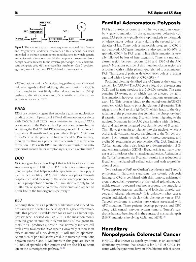
activates downstream targets via binding to the Tcf-Lef pro-moter. Such targets include c-myc, a proto-oncogene, andcyclin D1, resulting in cell proliferation (Fig. 2A). Binding ofTcf-Lef among others also leads to a downregulation of E-cadherin transcription (CDH1). E-cadherin is normally pres-ent at cell interfaces where it mediates adhesion. Activation ofthe Tcf-Lef promoter via �-catenin results in a reduction of
-cadherin-mediated cell-cell adhesion and leads to prolifer-tion of cells.
Two variants of FAP are Gardner’s syndrome and Turcot’syndrome. In Gardner’s syndrome, the colonic polyposiseading to CRC is combined with skin tumors, epidermoidysts, congenital hypertrophy of the retinal epithelium, des-oids tumors, duodenal carcinoma around the ampulla ofater, hepatoblastoma, papillary and follicular thyroid can-er, and adrenal adenomas.17 It is unknown what causesertain individuals to display this phenotype versus FAP.urcot’s syndrome is another rare variant associated withPC mutation. These patients develop polyposis and CRClong with central nervous system tumors. Turcot’s syn-rome has also been found in the context of mismatch repairMMR) mutations involving MLH1 and MSH2.18
HereditaryNonpolyposis Colorectal CancerHNPCC, also known as Lynch syndrome, is an autosomal-dominant syndrome that accounts for 3-4% of CRCs. Pa-
tients with the syndrome have an 80% lifetime risk of cancer.
nfegitrmcM
g. (Col
Molecular basis of hereditary colorectal cancer 67
Originally described in the 1900s by the pathologist, War-thin, and elaborated on by Henry Lynch and coworkers in1974,19 the clinical features seen in HNPCC are due to ge-
etic changes in MMR genes. The defects resulting from de-ective MMR genes were originally defined in yeast by Fishelt al and correlated to human pathogenesis that year.20 MMRene products help preserve genomic integrity by recogniz-ng and correcting base-pair mismatches and small nucleo-ide insertion/deletion mutations that occur during DNAeplication (Fig. 2B).21 Cells with MMR deficiency have autation rate that is 100- to 1000-fold greater than normal
ells.22 At least 4 MMR genes have been linked to HNPCC:LH1, MSH2, MSH6, and PMS2. Germ line mutations of
these genes have been identified in up to 80% of affectedfamilies. MLH1 and MSH2 account for 90% of the mutations.Study of the non-encoding microsatellite regions of HNPCCtumor DNA show genetic instability secondary to multiple
Figure 2 Molecular mechanisms in hereditary cancer.�-catenin, is sequestered by APC in a complex and tadenomatous polyposis, mutations in the APC complexnucleus and interact with the promoter, TCF-Lef. This(B) Mismatch repair. In hereditary nonpolyposis colonThus, errors in replication are not recognized. (C) PTENsyndrome. PTEN normally inhibits the PI3-Kinase/Akt cprogress unchecked. (D) TGF-�/SMAD. Germ line mufamilial juvenile polyposis. These proteins are the dominabsence of inhibition, the SMAD molecules translocateWnt, Wingless; APC, adenomatous polyposis coli; GSK-3enhancing factor; PTEN, phosphatase and tensin homserine/threonine protein kinase; mTOR, mammalian tarof p53; Smad, mothers against decapentaplegic homolo
mutations specifically targeting repetitive sequences. This is
referred to as microsatellite instability (MSI) and delineatedvia alterations in microsatellite sequences, including BAT25,BAT26, D5S346, D2S123, and D17S250. Tumors are classi-fied as MSI-high (�2 markers), MSI-low (�2 markers), orMSI-stable based on the frequency of mutations detected.22
Most HNPCC tumors are MSI-high and CpG island methy-lator phenotype negative. Fifteen percent of sporadic CRCtumors show MSI, although most of these tumors are CpGisland methylator phenotype positive and have BRAF muta-tions, and the MSI occurs late in the tumorigenesis.
Phenotypically, patients with HNPCC have a predilectionfor right-sided CRC tumors. They are also at risk of develop-ing tumors in the endometrium, stomach, ovary, hepatobili-ary tract, upper urinary tract, pancreas, small bowel, andcentral nervous system. A subset of this syndrome is referredto as Muir-Torre syndrome and includes those with skintumors consisting of sebaceous adenomas, basal cell cancers,
e WNT signaling pathway. The transcription factor,arked for degradation by the proteasome. In familialn excessive free �-catenin, which can translocate to theg facilitates cell proliferation via c-myc and cyclin D1., proteins that recognize and repair DNA are mutated.efects in PTEN, a tumor suppressor, result in Cowden’s. If PTEN does not inhibit this complex, growth signalsin the SMAD transcription factors are responsible fordiators of the transforming growth factor family. In thenucleus where they promote growth-enhancing genes.ogen synthase kinase; Tcf/Lef, T-cell factor/lymphocytedeleted on chromosome 10; Akt, protein kinase B, aapamycin; mdm2, murine double minute, an inhibitoror version of figure is available online.)
(A) Thhus mresult ibindincancer/Akt. Domplextationsant meto the�, glyc
ologueget of r
and keratoacanthomas.

sstpcimp
id
aBpt
sis
fgfoPtpshP
68 M.R. Hughes and E.H. Huang
There are 2 sets of criteria to support the diagnosis ofHNPCC: the family history based Amsterdam II criteria andthe Revised Bethesda criteria.23 The revised Bethesda criteriaerve as guidance for initiating genetic testing in absence of atriking family history. They combine patient and tumor fea-ures and take the MSI status of the tumor into account. Inarticular, features that should alert for possible HNPCC in-lude CRC with MSI-high histology, the presence of tumornfiltrating lymphocytes, Crohn’s-like lymphocytic reaction,
ucinous/signet ring differentiation, or medullary growthattern, and patient age of �50 years.24
MUTYH Associated PolyposisMUTYH associated polyposis (MAP, also known as MYH-associated polyposis) is an inherited polyposis syndromewith a phenotype similar to FAP but with an autosomal-recessive mode of inheritance. The MUTYH gene is located onchromosome 1p34. Its gene product, MUTYH glycosylase, isa 535-amino-acid base excision repair protein involved in therepair of guanine oxidation, 1 of the most common forms ofoxidative DNA damage.25,26 Guanine oxidation results in theappearance of G:C to T:A transversions. MUTYH is requiredunder oxidative stress for normal cell-cycle progression andnuclear division. This suggests a role in the maintenance ofgenome stability and tumor prevention. Mutations in MU-TYH can cause severely hampered or completely absent DNAbinding and repair activity. Molecular profiles of tumorsfrom MAP patients show G:C to T:A transversions in the APCgene and K-ras, a proto-oncogene. MAP tumors are usuallyMSI-low.
Because of the autosomal-recessive inheritance pattern,patients with MAP have biallelic mutations in MUYTH. Theytypically develop 10-100 adenomas, often with a more prox-imal distribution pattern. On average, MUTYH patients arediagnosed with CRC in their late 40s. MUTYH heterozygotesare at marginally increased risk of developing CRC.25
Juvenile Polyposis SyndromeJuvenile polyposis syndrome27 is a syndrome with a predis-position to the development of hamartomatous polyps in thestomach, small intestine, colon, and rectum. Most polyps arebenign, but some polyps have the potential for malignanttransformation. The incidence of CRC in juvenile polyposissyndrome patients is 68% by the age of 60, and the averageage when CRC is diagnosed in this syndrome is 42.27
The polyps develop because of a germ line mutation inSMAD4 (homologs of the Caenorhabditis elegans proteinsmooth muscle actin and the drosophila protein mothersagainst decapentaplegic) or BMPR1A (bone morphogenicprotein) (Fig. 2D).
SMAD4 is a tumor-suppressor gene that produces a 552-amino-acid protein encoded by 1656 nucleotides. It is a crit-ical cytoplasmic mediator of the TGF-� superfamily regulat-ng pathway. The MH1 domain of the SMAD4 proteinirectly binds DNA in response to TGF-� signaling. Patho-
logic allelic variants involving that N-terminal binding site
significantly reduce DNA binding activity. Mutations in theMH2 domain appear to cause problems with nuclear local-ization and interaction with MAD proteins and transcrip-tional activation.
The BMPR1A gene product is a 533-amino-acid proteinthat is a type 1 receptor of the TGF-� superfamily that medi-tes BMP intracellular signaling through SMAD4. AbnormalMPR1A proteins result from pathologic DNA variants in therotein kinase domain, occasionally by variants in the cys-eine-rich region of the extracellular domain.
Cowden’s SyndromeCowden’s syndrome is an autosomal-dominant conditionassociated with multiple hamartomas in multiple areas ofthe body. The lifetime risk of CRC approaches 10%.28
Germ line mutations of PTEN (phosphatase and tensinhomologue deleted on chromosome 10) are found in 80%of Cowden’s patients. PTEN is a tumor suppressor genethat encodes a 403-amino-acid protein that acts as a lipidphosphatase to negatively regulate the PI3K/AKT pathway(Fig. 2C).28 Mutations in PTEN have been shown to causeincreased nuclear �-catenin leading to increased expres-ion of c-Myc and cyclin D1 (see FAP above). Interest-ngly, although both PTEN loss and aberrations in the Wntignaling pathway lead to increased nuclear �-catenin,
and both alterations result in a polyposis phenotype, thetypes of polyps present are different. In the former, thepolyps are hamartomatous, representing both epithelialand stromal proliferation, while in FAP, the polyps areadenomas, with a dominantly epithelial histology. Al-though both pathways engage �-catenin as a transcriptionactor at the minimum, the differences in phenotype sug-est the potential for overlap in signaling pathways, dif-erential levels of expression, or influences of the stroman the ultimate phenotype. Regarding CRC, specifically,TEN mutations have been shown to lead to an increase inhe invasion and migration of CRC cells. Low PTEN ex-ression correlates with CRC tumor size, depth of inva-ion, lymphatic invasion, lymph node metastasis, andigher TNM staging.28,29 Patients with CRCs withoutTEN expression have shorter survival PTEN suppression
or loss is believed to contribute to tumor invasion andmetastasis in advanced CRC disease.29 One of the func-tions of PTEN is to inhibit the PI3K/AKT pathway. Activa-tion of the PI3K/AKT pathway is common in several can-cers. AKT has been shown to help regulate apoptosis andPI3K activation may play a role in chemotherapy resis-tance.
Cowden’s syndrome typically involves mucocutaneouslesions of the face and mouth, along with tumors of thebreast, thyroid gland, genitourinary tract, gastrointestinaltract, nervous system, and skeletal system. Breast canceris the most common malignancy. Gastrointestinal involve-ment with polyps is seen in up to 85% of patients, andthese almost always involve the sigmoid colon and therectum. Hamartomas tend to be the predominant his-
tologic type, but lipomatous, fibromatous, hyperplastic,
lwbGrrr
3t
na
“tuiivfca
mci
Molecular basis of hereditary colorectal cancer 69
inflammatory, and adenomatous lesions may also bepresent.28
Peutz–Jeghers SyndromePeutz–Jeghers syndrome (PJS) is an autosomal-dominantsyndrome in which affected patients develop gastrointes-tinal hamartomas and mucocutaneous hyperpigmenta-tion. Most patients with PJS have a germ line mutation ofthe gene STK11 (LKB1), a tumor suppressor gene locatedon chromosome 19p13. STK11 encodes a serine–threo-nine kinase that modulates cell polarity and cell prolifer-ation.27 It also helps in responding to low cellular energyevels. STK11 inhibits the AMP-activated protein kinase,hich in turn regulates the gene product of tuberin. Tu-erin facilitates the generation of rheb-GDP from rheb-TP, which activates mTOR. Germ line STK11 mutations
esult in the downregulation of tuberin, upregulationheb-GTP, and induction of mTOR. This inhibition is dys-egulated in PJS patients.30
Gastrointestinal cancers are frequent in PJS. The overallincidence of carcinomas ranges from 20% to 50%. Specif-ically, colon polyps are present in 53%, while rectal polypsare seen in 32% of affected subjects.30 PJS patients have a
9% chance of developing colon cancer over their life-ime.27
Stem Cells in CRCStem cells are defined by the 2 following properties: self-renewal and multipotency. Self-renewal is the cell’s ability
Figure 3 Isolating and perpetuation of colon cancer steoperative specimens are implanted into the subcutaneoutumors have grown to 1-1.5 cm, they are harvested andfor tumor initiating activity (green). (D) As few as 10 ce
the parent tumor (H&E, �400). Note resemblance to the H&to perpetuate itself for an extended amount of time. Mul-tipotency is the ability to generate all the differentiatedcells of the tissue of origin. Normal colon stem cells can befound at the bottom of the epithelial crypts.31 The termi-
ally differentiated cells at the luminal surface of the cryptre derived from these stem cells.
A new paradigm regarding tumorigenesis is termed thecancer stem cell hypothesis.” This paradigm suggests thatumors are generated and maintained by a small subset ofndifferentiated cells able to self-renew and differentiate
nto the bulk tumor population.3 This paradigm was orig-nally substantiated in leukemia, but now appears to bealidated in CRC. Three recent studies appear to haveound that colon cancer stem cells can be identified usingertain cell markers: CD44, CD133, EpCAM, CD166, andldehyde dehydrogenase (Fig. 3).32-35
Both normal stem cell self-renewal and cancer stem cellself-renewal are regulated by many pathways. However,for both leukemias and colon cancer, the WNT pathway,for example, is likely to play a role.36,37 Other pathwayswith implications for CRC include the Notch, Hedgehog,PTEN/AKT, Bmi, and p53 molecular signaling pathways.The cancer stem cell theory has important implications forprevention and therapy. The fact that most cytotoxicagents used to treat colon cancer are designed to kill ac-tively proliferating cells means that the cancer-causingstem cells are left behind and contribute to selecting resis-tance.38 In the future, the use of cancer stem cell enrich-
ent and evaluation will be invaluable to seek these rareells as biomarkers for diagnosis and prognosis, and todentify targets for therapy.
s. (A) Tumor fragments (1-2 mm) isolated from freshof immunocompromised mice (H&E, �400). (B) Onceated. (C) Flow cytometry identifies those cells enriched
this enriched population are capable of recapitulating
m cells spacedissocills from
E in (A). (Color version of figure is available online.)

1
1
1
1
1
1
2
2
2
2
2
2
2
2
2
2
3
3
3
3
3
3
3
3
3
70 M.R. Hughes and E.H. Huang
ConclusionsAlthough the hereditary syndromes account for only 10-15%of cases, the study of their mutations has shed a great amountof light on the genetic mutations and molecular basis of CRC.As research continues in these areas, we may be able to targetspecific mutations with new therapies. Identification of cer-tain mutations in tumors may assist us in better predictingprognosis and defining treatment strategies. The new cancerstem cell hypothesis for CRC lends an explanation for thosepatients who develop late recurrences or who develop asyn-chronous metastases, Further, additional research will resultin focused treatment and eradication of these tumor initiatingand propagating cells.
References1. Jemal A, Siegel R, Xu J, et al: Cancer statistics, 2010. CA Cancer J Clin
60:277-300, 20102. Saif MW, Chu E: Biology of colorectal cancer. Cancer J 16:196-201,
20103. Wicha MS, Liu S, Dontu G: Cancer stem cells: An old idea—A paradigm
shift. Cancer Res 66:1883-1890 [discussion 1895-1886], 20064. Hanahan D, Weinberg RA: The hallmarks of cancer. Cell 100:57-70,
20005. Fearon ER, Vogelstein B: A genetic model for colorectal tumorigenesis.
Cell 61:759-767, 19906. Wood LD, Parsons DW, Jones S, et al: The genomic landscapes of
human breast and colorectal cancers. Science 318:1108-1113, 20077. Bos JL, Fearon ER, Hamilton SR, et al: Prevalence of ras gene mutations
in human colorectal cancers. Nature 327:293-297, 19878. Kranenburg O: The KRAS oncogene: Past, present, and future. Biochim
Biophys Acta 1756:81-82, 20059. Grady WM: Making the case for DCC and UNC5C as tumor-suppressor
genes in the colon. Gastroenterology 133:2045-2049, 200710. Baker SJ, Fearon ER, Nigro JM, et al: Chromosome 17 deletions and
p53 gene mutations in colorectal carcinomas. Science 244:217-221,1989
11. Takayama T, Miyanishi K, Hayashi T, et al: Colorectal cancer: Geneticsof development and metastasis. J Gastroenterol 41:185-192, 2006
12. Baker SJ, Preisinger AC, Jessup JM, et al: p53 gene mutations occur incombination with 17p allelic deletions as late events in colorectal tu-morigenesis. Cancer Res 50:7717-7722, 1990
13. Powell SM, Zilz N, Beazer-Barclay Y, et al: APC mutations occur earlyduring colorectal tumorigenesis. Nature 359:235-237, 1992
4. Knudsen AL, Bulow S, Tomlinson I, et al: Attenuated Familial adeno-matous polyposis (AFAP) results from an international collaborativestudy. Colorectal Dis 2010 [epub ahead of print]
5. Groden J, Thliveris A, Samowitz W, et al: Identification and character-ization of the familial adenomatous polyposis coli gene. Cell 66:589-600, 1991
6. Kinzler KW, Nilbert MC, Su LK, et al: Identification of FAP locus genesfrom chromosome 5q21. Science 253:661-665, 1991
7. Juhn E, Khachemoune A: Gardner syndrome: Skin manifestations, dif-ferential diagnosis and management. Am J Clin Dermatol 11:117-122,
20108. Lebrun C, Olschwang S, Jeannin S, et al: Turcot syndrome confirmedwith molecular analysis. Eur J Neurol 14:470-472, 2007
9. Lynch HT, Smyrk T, Lynch JF: Molecular genetics and clinical-pathol-ogy features of hereditary nonpolyposis colorectal carcinoma (Lynchsyndrome): Historical journey from pedigree anecdote to moleculargenetic confirmation. Oncology 55:103-108, 1998
0. Fishel R, Lescoe MK, Rao MR, et al: The human mutator gene homologMSH2 and its association with hereditary nonpolyposis colon cancer.Cell 75:1027-1038, 1993
1. Fishel R, Wilson T: MutS: Homologs in mammalian cells. Curr OpinGenet Dev 7:105-113, 1997
2. Narayan S, Roy D: Role of APC and DNA mismatch repair genes in thedevelopment of colorectal cancers. Mol Cancer 2:41, 2003
3. NCCN. Clinical practice guidelines in oncology: Colorectal cancerscreening. NCCN V.1, 2010
4. Gruber SB: New developments in Lynch syndrome (hereditary nonpol-yposis colorectal cancer) and mismatch repair gene testing. Gastroen-terology 130:577-587, 2006
5. Nielsen M, Morreau H, Vasen HF, et al: MUTYH-associated polyposis(MAP). Crit Rev Oncol/Hematol2010 [epub ahead of print]
6. Poulsen ML, Bisgaard ML: MUTYH associated polyposis (MAP). CurrGenomics 9:420-435, 2008
7. Giardiello FM, Brensinger JD, Tersmette AC, et al: Very high risk ofcancer in familial Peutz–Jeghers syndrome. Gastroenterology 119:1447-1453, 2000
8. Farooq A, Walker LJ, Bowling J, et al: Cowden syndrome. Cancer TreatRev 36:577-583, 2010
9. Bowen KA, Doan HQ, Zhou BP, et al: PTEN loss induces epithelial--mesenchymal transition in human colon cancer cells. Anticancer Res29:4439-4449, 2009
0. Chen HM, Fang JY: Genetics of the hamartomatous polyposis syn-dromes: A molecular review. Int J Colorectal Dis 24:865-874, 2009
1. Barker N, Clevers H: Leucine-rich repeat-containing G-protein-cou-pled receptors as markers of adult stem cells. Gastroenterology 138:1681-1696, 2010
2. Dalerba P, Dylla SJ, Park IK, et al: Phenotypic characterization of hu-man colorectal cancer stem cells. Proc Natl Acad Sci USA 104:10158-10163, 2007
3. Huang EH, Hynes MJ, Zhang T, et al: Aldehyde dehydrogenase 1 is amarker for normal and malignant human colonic stem cells (SC) andtracks SC overpopulation during colon tumorigenesis. Cancer Res 69:3382-3389, 2009
4. O’Brien CA, Pollett A, Gallinger S, et al: A human colon cancer cellcapable of initiating tumour growth in immunodeficient mice. Nature445:106-110, 2007
5. Wang Q, Chen ZG, Du CZ, et al: Cancer stem cell marker CD133�
tumour cells and clinical outcome in rectal cancer. Histopathology55:284-293, 2009
6. Vermeulen L, De Sousa EMF, van der Heijden M, et al: Wnt activitydefines colon cancer stem cells and is regulated by the microenviron-ment. Nat Cell Biol 12:468-476, 2010
7. Wang Y, Krivtsov AV, Sinha AU, et al: The Wnt/beta-catenin pathway isrequired for the development of leukemia stem cells in AML. Science327:1650-1653, 2010
8. Dylla SJ, Beviglia L, Park IK, et al: Colorectal cancer stem cells areenriched in xenogeneic tumors following chemotherapy. PLoS ONE
3:e2428, 2008