Embed Size (px)
Citation preview

Molecular aptamers for drug deliveryWeihong Tan1,2, Hui Wang2, Yan Chen1,2,3, Xiaobing Zhang1, Haizhen Zhu1,Chaoyong Yang3, Ronghua Yang1 and Chen Liu2
1 State Key Laboratory of Chemo/Biosensing and Chemometrics, College of Biology, College of Chemistry and Chemical
Engineering, Hunan University, Changsha, 410082, China2 Center for Research at Bio/Nano Interface, Department of Chemistry, Department of Physiology and Functional Genomics,
Department of Pathology and Laboratory Medicine, Shands Cancer Center, UF Genetics Institute and McKnight Brain Institute,
University of Florida, Gainesville, FL 32611, USA3 State Key Laboratory for Physical Chemistry of Solid Surfaces, The Key Laboratory for Chemical Biology of Fujian Province and
Department of Chemical Biology, College of Chemistry and Chemical Engineering, Xiamen University, Xiamen Fujian, 361005,
China
Review
The active targeting of drugs in a cell-, tissue- or disease-specific manner represents a potentially powerful tech-nology with widespread applications in medicine, in-cluding the treatment of cancers. Aptamers haveproperties such as high affinity and specificity for tar-gets, easy chemical synthesis and modification, andrapid tissue penetration. They have become attractivemolecules in diagnostics and therapeutics rivaling and,in some cases, surpassing other molecular probes, suchas antibodies. In this review, we highlight the recentprogress in aptamer-mediated delivery for therapeuticsand disease-targeting based on aptamer integrationwith a variety of nanomaterials, such as gold nanorods,DNA micelles, DNA hydrogels and carbon nanotubes.
IntroductionAptamers are single-stranded (ss) oligonucleotides thatcan bind to their target molecules with high affinity andselectivity by folding into distinct secondary and tertiarystructures. They are identified from an initial library con-taining 1013–1016 random ssDNA or ssRNA sequencesthrough an in vitro selection process termed SELEX (sys-tematic evolution of ligands by exponential enrichment)(recently reviewed in [1,2]). Aptamers are different fromantibodies, yet they mimic properties of antibodies in avariety of diagnostic formats. Antibodies have made sub-stantial contributions towards the advancement of diag-nostic assays and have become indispensable in mostdiagnostic tests that are routinely used in clinics today.As an emerging class of molecules that rivals antibodies inboth therapeutic and diagnostic application, aptamershave become the focus of attention for their novel proper-ties, such as highly selective and specific target recognitionand binding. Synthetic aptamers possess several advan-tages over natural antibodies [3–6], including economicaland reproducible synthesis, as well as excellent molecularrecognition ability, with Kd values in the nanomolar range.Examples include the DNA aptamers to thrombin(Kd = 25–200 nM [7]), Mucin-1 (MUC1) (Kd = 47.3 nM[8]), apple stem pitting virus coat proteins (PSA-H) protein(Kd = 8.0 nM [9]) and leukemic lymphoid (CEM) cells(Kd = 0.8 nM [10]). These values are all superior to those
Corresponding author: Tan, W. ([email protected]).
634 0167-7799/$ – see front matter � 2011 Elsevier Ltd. All rights reserved. do
of antibodies, for which Kd values are in the micromolar tomillimolar range, depending on concentration. Comparedwith antibodies, aptamers have additional advantages,such as biocompatibility and flexible modification, easyand controllable modification to fulfill different diagnosticand therapeutic purposes, long-term stability as dry pow-ders or in solution, ability to sustain reversible denatur-ation, lack of toxicity and immunogenicity, and rapid tissuepenetration. These physical and chemical properties makeaptamers ideal candidates as probes for use in molecularmedicine to elucidate the molecular foundations of dis-eases, particularly cancer and infectious diseases.
After more than a decade of development, aptamershave attracted interest for use as therapeutic agents anddiagnostic tools by, for example, helping to improve thesensitivity and specificity of diagnostic assays throughmolecular imaging [10,11], inhibiting disease processes[12,13] or targeting the delivery of drugs to diseased tis-sues [14–16]. This review focuses on aptamer-mediateddelivery for therapeutics and disease-targeting based onaptamer integration with a variety of nanomaterials, suchas gold nanorods, DNA micelles, DNA hydrogels and car-bon nanotubes.
Aptamers as therapeutic agentsIn current research, aptamers have been effectively usedfor therapeutic applications, such as cancer cell detectionand diagnostics [17–19] and targeted therapy [20–22], aswell as sorting and enrichment [23,24]. The generation of apool of DNA aptamers for various types of cancer cells hasbeen reported, including small-cell lung, non-small-celllung (NSCLC), acute myelogenous leukemia (AML), liverand colon cancer, as well as virus-infected cells [25–28].Using a similar strategy, DNA aptamers for mesenchymalstem cells [29], porcine endothelial precursor cells [30] andlive bacterial cells [31] have also been developed by otherresearch groups during the past two years. With theirsuperior targeting performance, incorporation of aptamerswith a defined therapeutic function and recognition capa-bility for cancer therapy has raised considerable interest.Thus far, aptamers and aptamer assemblies have beenvalidated as essential molecular tools in the areas of anti-infectives [32], anticoagulation [33], anti-inflammation
i:10.1016/j.tibtech.2011.06.009 Trends in Biotechnology, December 2011, Vol. 29, No. 12

Review Trends in Biotechnology December 2011, Vol. 29, No. 12
[34], anti-angiogenesis [35], antiproliferation [36] and im-mune therapy [37].
In addition to their ability to recognize a target moleculewith high specificity, certain aptamers can also modulatethe activities of proteins implicated in pathological condi-tions, making aptamers potentially useful as pharmaceu-tical agents. For instance, Pfizer’s Macugen1 (pegaptanib),an aptamer-based anti-VEGF treatment for age-relatedmacular degeneration, was approved by the United StatesFDA in 2004 based on findings from two clinical trialsinvolving 1200 patients and all subtypes of neovascularAMD (Age-Related Macular Degeneration). Another exam-ple is AS1411, an aptamer which specifically targetsnucleolin, a bcl-2 mRNA binding protein involved in cellproliferation, which is found on the surface of many cancercells. Once bound, the AS1411 aptamer is taken into thecancer cell, where it causes death by apoptosis (pro-grammed cell death). AS1411 has also undergone clinicaltrials [38,39].
Aptamers as delivery agentsAptamers can be designed as targeting ligands, particu-larly when generated by cell-based SELEX, and can dif-ferentiate diseased cells from healthy cells, thus enablingthe selective delivery of therapeutic compounds to targetcells (Table 1).
The emerging integration of aptamers with nanotech-nology and chemical biology is envisioned to produce moreversatile target-specific molecules, stimulate further newdiagnostic and therapeutic nanotechnologies, and providesignificant potential for many research and clinical appli-cations in the near future. Compared with applications ofantibodies, aptamer research is still in its infancy but isprogressing at a rapid pace.
SELEX: aptamer selection processThe technologies involved in the SELEX process can varyfrom traditional capillary electrophoresis or flow cytometryto, most recently, microfluidic systems [40]. SELEX hasundergone several refinements and modifications. Mostadvances in aptamer isolation have aimed at improvingselection efficiency, aptamer biostability and bioavailabili-ty. However, the overall goal of the various selectionapproaches has remained the same – to increase selectionspeed and efficiency, while identifying ligands that bind totargets with high specificity and affinity – and progress inthis area has been recently reviewed [1,2,41]. Althoughmany complex forms of SELEX exist, in this section wefocus on the fundamental ‘cell-SELEX’ strategy, which is ofparticular interest for targeted drug delivery where apta-
Table 1. Aptamers for drug delivery
Target name Aptamer Selection techni
Epidermal growth factor receptor (EGFR) RNA Purified extracel
Immunoglobin heavy mu chain (IGHM) DNA Cell-SELEX
Mucin-1 (MUC1) DNA Recombinant pe
Prostate-specific membrane antigen (PSMA) RNA Purified extracel
Protein tyrosine kinase-7 (PTK7) DNA Cell-SELEX
mers are integrated with nanomaterials, such as liposomes[42] or DNA micelles [14].
Cell-SELEX: a promising tool to generate clinically
useful aptamers
Cell-SELEX is the process whereby live cells are used toselect aptamers for target recognition (Figure 1). By yield-ing ligands that preferentially bind to diseased cells, com-pared with normal cells, cell-SELEX is a particularlypromising selection strategy for the development of apta-mers able to transport a payload of nanomaterials todiseased cells (Table 1). Live cells of different cancers havebeen used in this process and, as a result, cell-SELEX wassuccessfully used to develop an aptamer against hepato-cellular carcinoma (HCC), one of the most common andhighly malignant cancers in the world, found in a human Tcell acute lymphoblastic leukemia cell line CEM (used cell-SELEX target) [43]. More recently, cell-SELEX was ap-plied to isolate aptamers that recognized AML cells withdissociation constants (Kd) in the nanomolar range [25].
The cell-SELEX process is easily managed, rapid andreproducible. Generation of aptamers using this technologyhas become an effective tool for molecular medicine andbiomarker discovery. Specifically, when bound with cellmembrane receptors, aptamers provide an effective ap-proach for the discovery of biomarkers as disease signals.Because aptamer probes recognize molecular signatures onthe cancer cell surface with high specificity, tumor cellprofiles can be defined, perhaps leading to more ‘personal-ized’ cancer treatment. In addition, with their ability todistinguish normal from cancer cells, aptamers allow acomparative strategy to identify differences at the molecularlevel and promote the discovery of molecular features ofcancer cells.
A recent promising approach using cell-SELEX was de-veloped for the generation of aptamer molecular probes thatspecifically recognize Burkitt’s lymphoma cells, an acuteblood cell cancer [44]. Early diagnosis and targeted therapyare crucial for patient treatment. However, the lack ofmolecular probes able to recognize cell-surface biomarkersprevents early diagnosis of cancers, such as Burkitt’s lym-phoma, makes the study of their developing mechanism(s)difficult, if not impossible. The authors were able to identifya panel of cell-specific aptamers, called TDO5, againstRamos cells, a B cell lymphoma cell line, leading to increasedinsight into the molecular activity of Ramos cells. As aneffective molecular tool for identification of target cells inreal biological samples for clinically meaningful biomedicalstudies and biomarker discovery, TD05 is advancing thepotential for improved cancer diagnostics and treatment.
que Delivery application Refs
lular domain of EGFR Nanoparticle delivery [63]
Micelle nanoparticles for drug delivery [14]
ptides Photodynamic therapy (PDT)
Radionuclide delivery
[64]
[65]
lular domain of PSMA siRNA delivery
Cytotoxin delivery
Chemotherapeutic drug delivery
[66]
[67]
[68]
Chemotherapeutic drug delivery [56]
635

Target cell
Positiveselection
Wash to removeunbound sequences
Control cell
Extracted boundsequences
Remove boundsequences
Negativeselection
Sequencing andscreeningpotential DNAaptamer
ssDNA library
TRENDS in Biotechnology
Retain unboundsequences
PCR amplification
Cloning evolvedDNA pool
Cell-SELEX
Figure 1. Schematic representation of DNA aptamer selection using the cell-SELEX (systematic evolution of ligands by exponential enrichment) strategy. DNA sequences
that have specific recognition to target cells are evolved to enrich the selection pools. The enriched pools are cloned, and the positive clones are sequenced to identify
individual aptamers. The single-stranded DNA (ssDNA) pool is first incubated with target cells. After washing, the bound DNAs are eluted by thermal denaturation. The
eluted DNAs are amplified by PCR. The double-stranded PCR products are then separated into ssDNAs and the sense strand DNAs collected for the next round of selection
or tested by flow cytometry to monitor the SELEX progression. When the selected pool is sufficiently enriched, the PCR product of the evolved pool is cloned and
sequenced for aptamer identification. Adapted from [69].
Review Trends in Biotechnology December 2011, Vol. 29, No. 12
Aptamers in nanotechnologyThe ability of DNA or RNA aptamers to act as targetingagents enables these molecules to be conjugated withtherapeutic agents for use in targeted drug delivery. Earlydiagnosis of disease relies not only on the specificity of themolecular probes but also on the detection sensitivity.Because of their predictable structures and functionalgroups for chemical modification, aptamers can readilybe linked to advanced signaling mechanisms in the devel-opment of diagnostics or disease treatments. Some cancercells, especially those in the early stages of disease devel-opment, could have a very low density of target on the cellsurface available for detection. With their relatively smallsize, aptamers have shown promise in specifically target-ing tumor cells and transporting small molecules, such asproteins, drugs or siRNA, through the microvasculature orthe tumor interstitium [45]. In addition, by taking advan-tage of straightforward synthesis and chemical modifica-tion, aptamers can be conjugated to functional groups withrelative ease, enabling their use as effective nanomater-ials. Because each type of nanomaterial has differentoptical, electrochemical and mechanical properties, medi-cal diagnostic and drug delivery agents with diverse char-acteristics can be used for different applications, movingaptamer-based nanomedicine closer to reality. In this sec-tion, we mainly focus on the applications of aptamers innanotechnology for diagnostics and therapeutics.
Aptamers meet nanomaterials: nanoparticle conjugates
Applications for aptamers at the interface of nanotechnol-ogy and medicine in the form of aptamer-nanoparticle
636
conjugates are actively being investigated. The large sur-face areas of nanoparticles offer excellent platforms forconjugating multiple aptamers. In addition, the interiorvolumes of nanoparticles can be used to store large quan-tities of drug molecules, thereby enhancing loading capaci-ty.
Huang et al. synthesized Au-Ag nanorods (NRs) to serveas a platform for binding several aptamer molecules(Figure 2) [46]. In their work, an NR–scg8 aptamer conju-gate combined the high absorption efficiency of Au–Ag NRswith the target specificity of molecular aptamers. Wheninvestigated with cell mixtures, the aptamer–NRs weredemonstrated to have excellent hyperthermia efficiency onexposure to near-infrared laser radiation, as well as selec-tivity for the target CEM cells. This technique can be usedin clinical detection to enhance both binding affinity andsignal strength when the concentration of target cells isrelatively low. By functionalizing the surface of Au nano-particles (NPs) with an RNA aptamer that binds to pros-tate-specific membrane antigen (PSMA), NP–aptamerconjugates were used for targeted molecular computedtomography imaging and treatment of prostate cancer[47]. Other than metallic NPs, aptamer-conjugated poly-mer particles are also attractive agents for drug encapsu-lation and controlled release in a cell- or tissue-specificmanner. A bioconjugate composed of controlled releasepolymeric aptamer–NPs has been used for targeted deliv-ery to a prostate cancer cell line, LNCaP, which expressesPSMA [48]. These bioconjugates efficiently targeted andwere taken up by LNCaPs. Compared with solid, metallicNPs, polymeric nanomaterials offer a more promising

TRENDS in Biotechnology
Sgc8Aptamer
Target cells Fluorescent signalsAu-Ag
nanorod
Aptamer NanorodConjugate
Excitation
Figure 2. Schematic representation of aptamer–nanorod signal enhancement.
Review Trends in Biotechnology December 2011, Vol. 29, No. 12
solution for encapsulation of chemotherapeutics and havebeen shown to reduce toxicity by providing a protectivehousing for the drug that limits its interaction with healthycells [49].
Advanced aptamer–nanomaterial conjugates
Hydrogels are networks of polymer chains that are water-insoluble and superabsorbent, and they possess a degree offlexibility very similar to natural tissue. Target-responsivehydrogels that crosslink DNA aptamers with linear poly-acrylamide chains have been fabricated (Figure 3) [50].Competitive binding of the target to the aptamer causes adecrease in crosslinking density and, hence, dissolution of
Aptamer
2 hours 12 hours
Lipid
Se
(a)
(b)
(f) 0 ` 2 `
50µm 50 µm
10µm
(c)
Figure 3. (a) Design scheme of aptamer–micelles containing dye. Fluorescent images of
2 h. (e) Enlarged fluorescent image after post-labeling the biotinylated TDO5 aptamer w
cell. (f) Real-time monitoring of doped special dyes released from the core of the mice
the hydrogel. Therapeutic applications can therefore bedevised using small molecules and proteins as the targets.To this end, an in situ injectable hydrogel has been function-alized with nucleic acid aptamers to control the release ofproteins for human disease treatment [51]. The resultsshowed that the protein release rate can be controlled byadjusting the affinity of the aptamers. Both of these studies[48,49] demonstrate that aptamer-based hydrogels providea highly selective and controllable system, whereby efficientrelease of therapeutic agents can occur in the specific envi-ronment where the target biomarker is found.
In another report, a diacyl lipid tail was incorporated atthe 50 end of oligonucleotides by solid-phase DNA synthe-
2 hours Post-labeled with QD-streptavidin
lf-assembly
Dye
50µm 5µm
4 ` 8 `
(d) (e)
TRENDS in Biotechnology
Ramos cells for (b) 2 h and (c) 12 h, or (d) incubation with biotin–TDO5–micelle for
ith QD705 streptavidin. The inset in image (e) is the fluorescent image of the dead
lles and activated by intracellular enzymes.
637
Review Trends in Biotechnology December 2011, Vol. 29, No. 12
sis. When dispersed in an aqueous solution, these amphi-philic DNA molecules spontaneously self-assembled intomonodispersed micelle structures [52]. In one applicationof this technology, a self-assembling aptamer–micellenanostructure was formed of hydrophilic aptamers linkedto hydrophobic lipids by poly(ethylene glycol) (PEG) [14].In aqueous solution, these conjugates self-assembled into3D spherical micelle structures with a hydrophilic aptamertargeting Ramos cells (from human Burkitt’s lymphomacell line) on the outside, and the lipid core on the inside(Figure 3a). The presence of more than one aptamer on themicelle surface provides an approximately 750-fold in-crease in target binding affinity. The aptamer–micelleassembly is also able to be internalized [37], indicatingit is a promising strategy for clinical applications by in-creasing therapeutic effectiveness. Furthermore, after twodays of incubation with the aptamer–micelle assembly,normal cells maintained over 80% viability.
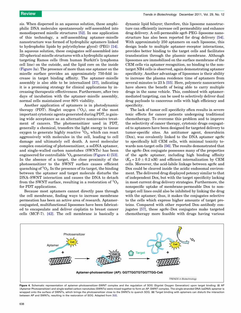
Another application of aptamers is in photodynamictherapy (PDT). Singlet oxygen (1O2), one of the mostimportant cytotoxic agents generated during PDT, is gain-ing wide acceptance as an alternative noninvasive treat-ment of cancers. The photosensitizer used in PDT,generally a chemical, transfers the light energy to tissueoxygen to generate highly reactive 1O2, which can reactaggressively with molecules in the cell, leading to celldamage and ultimately cell death. A novel molecularcomplex consisting of photosensitizer, a ssDNA aptamer,and single-walled carbon nanotubes (SWNTs) has beenengineered for controllable 1O2 generation (Figure 4) [53].In the absence of a target, the close proximity of thephotosensitizer to the SWNT surface causes efficientquenching of 1O2. In the presence of its target, the bindingbetween the aptamer and target molecule disturbs theDNA–SWNT interaction and causes the DNA to detachfrom the SWNT surface, resulting in a restoration of 1O2
for PDT applications.Because most aptamers cannot directly pass through
the cell membrane, finding ways to increase membranepermeation has been an active area of research. Aptamer-conjugated, multifunctional liposomes have been fabricat-ed to encapsulate and deliver cisplatin to breast cancercells (MCF-7). [42]. The cell membrane is basically a
Aptamer
Aptamer-photosensitizer (AP): G
+ Ce6
Ce6
Photosensitizer
Figure 4. Schematic representation of aptamer–photosensitizer–SWNT complex and
(Aptamer Photosensitizer) and single-walled carbon nanotubes (SWNTs) were mixed tog
wrapped onto the surface of SWNTs, which brings the photosensitizer close to the SW
between AP and SWNTs, resulting in the restoration of SOG. Adapted from [53].
638
dynamic lipid bilayer; therefore, this liposome nanostruc-ture can efficiently increase cell permeability and enhancedrug delivery. A cell-permeable sgc8–PEG–liposome nano-structure has also been reported for drug delivery [54].With approximately 250 aptamers on each liposome, thisdesign leads to multiple aptamer–receptor interactions,provides better binding to the target cells and facilitatestranslocation through the plasmic membrane. Althoughliposomes are immobilized on the surface membrane of theCEM cells via aptamer recognition, no binding to the non-target NB4 cells is observed, again demonstrating aptamerspecificity. Another advantage of liposomes is their abilityto increase the plasma residence time of aptamers fromseveral minutes to 23 h [55]. Here, polymeric nanocarriershave shown the benefit of being able to carry multipledrugs in the same vehicle. This, combined with aptamer-mediated targeting, can be used to selectively deliver dualdrug payloads to cancerous cells with high efficiency andspecificity.
The lack of tumor cell specificity often results in severetoxic effects for cancer patients undergoing traditionalchemotherapy. To overcome this problem and to improvethe selectivity of cancer therapy, cytotoxic drugs conjugat-ed to aptamers have been designed for targeted delivery totumor-specific sites. An antitumor agent, doxorubicin(Dox), was covalently linked to the DNA aptamer sgc8cto specifically kill CEM cells, with minimal toxicity to-wards non-target cells [56]. The results demonstrated thatthe sgc8c–Dox conjugate possesses many of the propertiesof the sgc8c aptamer, including high binding affinity(Kd = 2.0 � 0.2 nM) and efficient internalization by CEMcells. Moreover, the acid-labile linkage between sgc8c andDox could be cleaved inside the acidic endosomal environ-ment. The delivered drug displayed potency similar to thatof independent Dox, but with the target specificity lackingin most current drug delivery strategies. Furthermore, thenonspecific uptake of membrane-permeable Dox to non-target cell lines could also be inhibited by linking the drugwith the aptamer; thus, it makes the conjugates selectiveto the cells which express higher amounts of target pro-teins. Compared with other reported Dox–antibody con-jugates [57], these sgc8c–Dox conjugates make targetedchemotherapy more feasible with drugs having various
Target
GTTGGTGTGGTTGG-Ce6
+
1O2
TRENDS in Biotechnology
Ce6
the regulation of SOG (Signlet Oxygen Generation) upon target binding: (i) AP
ether to form an AP–SWNT complex. The single-stranded DNA (ssDNA) aptamer is
NTs to quench SOG. (ii) Target binding with aptamers can disturb the interaction

Review Trends in Biotechnology December 2011, Vol. 29, No. 12
potencies. When combined with the large number of re-cently created DNA aptamers that specifically target awide variety of cancer cells, this drug–aptamer conjugationmethod has broad implications for targeted drug delivery.
Aptamer-mediated cell type-specific small interferingRNA (siRNA) deliveryAlso known as chemical antibodies, aptamers are poised tosurpass natural antibodies in therapeutics, diagnosticsand drug development. The pharmacologic properties ofaptamers include wide therapeutic margins, stability,adjustable pharmacokinetics and very low immunogenic-ity and toxicity – advantages that are drawing attentionfrom major pharmaceutical companies. It has also beendemonstrated that exogenous siRNAs can silence geneexpression via the RNAi pathway in mammalian cells[58]. The challenge is learning how to direct those RNAmolecules to the target cells and then deliver themthrough the membrane. With their specific recognitionability, aptamers are ideal candidates for this purpose.For the first time, cell type-specific delivery of anti-humanimmunodeficiency virus (anti-HIV) siRNAs through fu-sion to an anti-gp120 aptamer has been demonstrated[59]. The envelope glycoprotein is expressed on the surfaceof HIV-1-infected cells, allowing binding and internaliza-tion of the aptamer–siRNA chimeric molecules. The chi-mera is specifically taken up by cells expressing HIV-1gp120, and the appended siRNA is processed by Dicer; thisreleases an anti-tat/rev siRNA, which, in turn, inhibitsHIV replication.
In a further study of this method [60], an anti-PSMARNA aptamer (A10) was appended to a 21-mer siRNAportion, resulting in a chimera that targets polo-like kinase1 (PLK1) and BCL2, two survival genes overexpressed inmost human tumors [61] The aptamer portion of thechimeras selectively binds to PSMA, whereas the thera-peutic siRNA portion interferes and knocks down geneexpression and inhibits the xenograft growth activity ofthe cancer cells both in cell culture and in vivo. Becausethis delivery system consists only of RNA components, itoffers several potential advantages as a therapeutic agent,including lack of immunogenicity, the possibility for chem-ical synthesis and stabilizing modifications for in vivoapplication.
Concluding remarks and future perspectivesAptamers are rapidly maturing into therapeutic tools withcommercial potential. Given that aptamers mimic andextend many features of monoclonal antibodies (mAbs),they have the potential to make an impact in molecularmedicine. For instance, aptamers that are highly specific tocancer cells can be used as drug targeting agents, therebyreducing toxicity (mice body weight loss only around 7%)while improving upon the efficacy of current therapeuticdrugs (tumor size reduction from fifth day of injection andall the mice survived in 109 days treatment) [62]. Com-pared with antibodies in the 1990s, aptamers are on aneven more accelerated trajectory for commercialization.Using cell-SELEX, aptamer probes capable of recognizingvaccinia virus-infected lung cancer A549 cells havebeen created [28]. It is therefore possible that a range of
antiviral aptamers can be generated easily and that thesemight show synergistic activity, opening new prospects forantiviral prophylaxis or therapy.
With the improvements in SELEX technology and apta-mer functionalities, as illustrated above, we believe thataptamers are poised to successfully compete with mAbs intherapeutics and drug development within the next fewdecades.
AcknowledgementsWe acknowledge the Interdisciplinary Center for Biotechnology Research(ICBR) at the University of Florida for technical support. This work issupported by grants awarded by the National Key Scientific Program ofChina (2011CB911000) and China National Grand Program(2009ZX10004-312) and by the National Institutes of Health(GM066137, GM079359 and CA133086).
References1 Cerchia, L. and de Franciscis, V. (2010) Targeting cancer cells with
nucleic acid aptamers. Trends Biotechnol. 28, 517–5252 Keefe, A.D. et al. (2010) Aptamers as therapeutics. Nat. Rev. Drug
Discov. 9, 537–5503 Osborne, S.E. et al. (1997) Aptamers as therapeutic and diagnostic
reagents: problems and prospects. Curr. Opin. Chem. Biol. 1, 5–94 Famulok, M. et al. (2007) Functional aptamers and aptazymes in
biotechnology, diagnostics, and therapy. Chem. Rev. 107, 3715–37435 Navani, N.K. and Li, Y.F. (2006) Nucleic acid aptamers and enzymes as
sensors. Curr. Opin. Chem. Biol. 10, 272–2816 Jiang, Y.X. et al. (2003) Specific aptamer-protein interaction studied by
atomic force microscopy. Anal. Chem. 75, 2112–21167 Griffin, L.C. et al. (1993) The discovery and characterization of a novel
nucleotide-based thrombin inhibitor. Gene 137, 25–318 Ferreira, C.S. et al. (2008) DNA aptamers against the MUC1 tumour
marker: design of aptamer-antibody sandwich ELISA for the earlydiagnosis of epithelial tumours. Anal. Bioanal. Chem. 390, 1039–1050
9 Balogh, Z. et al. (2010) Selection and versatile application of virus-specific aptamers. FASEB J. 24, 4187–4195
10 Shangguan, D. et al. (2006) Aptamers evolved from live cells as effectivemolecular probes for cancer study. Proc. Natl. Acad. Sci. U.S.A. 103,11838–11843
11 Li, J.W.J. et al. (2002) Molecular aptamer beacons for real-time proteinrecognition. Biochem. Biophys. Res. Commun. 292, 31–40
12 Gutsaeva, D.R. et al. (2011) Inhibition of cell adhesion by anti-P-selectin aptamer: a new potential therapeutic agent for sickle celldisease. Blood 117, 727–735
13 Lu, C. et al. (2010) Targeting pericytes with a PDGF-B aptamer inhuman ovarian carcinoma models. Cancer Biol. Ther. 9, 176–182
14 Wu, Y.R. et al. (2010) DNA aptamer-micelle as an efficient detection/delivery vehicle toward cancer cells. Proc. Natl. Acad. Sci. U.S.A. 107,5–10
15 Huang, Y.F. et al. (2008) Selective photothermal therapy for mixedcancer cells using aptamer-conjugated nanorods. Langmuir 24, 11860–
1186516 Chu, T.C. et al. (2006) Aptamer mediated siRNA delivery. Nucleic Acids
Res. 34, e7317 Davis, K.A. et al. (1998) Staining of cell surface human CD4 with 20-F-
pyrimidine-containing RNA aptamers for flow cytometry. Nucleic AcidsRes. 26, 3915–3924
18 Conrad, R. and Ellington, A.D. (1996) Detecting immobilizedprotein kinase C isozymes with RNA aptamers. Anal. Biochem.242, 261–265
19 Murphy, M.B. et al. (2003) An improved method for the in vitroevolution of aptamers and applications in protein detection andpurification. Nucleic Acids Res. 31, e110
20 Floege, J. et al. (1999) Novel approach to specific growth factorinhibition in vivo-antagonism of platelet-derived growth factor inglomerulonephritis by aptamers. Am. J. Pathol. 154, 169–179
21 Ostendorf, T. et al. (2002) The effects of platelet-derived growth factorantagonism in experimental glomerulonephritis are independent ofthe transforming growth factor-beta system. J. Am. Soc. Nephrol. 13,658–667
639

Review Trends in Biotechnology December 2011, Vol. 29, No. 12
22 White, R.R. et al. (2003) Inhibition of rat corneal angiogenesis by anuclease-resistant RNA aptamer specific for angiopoietin-2. Proc. Natl.Acad. Sci. U.S.A. 100, 5028–5033
23 Mayer, G. et al. (2010) Fluorescence-activated cell sorting for aptamerSELEX with cell mixtures. Nat. Protoc. 5, 1993–2004
24 Raddatz, M.S. et al. (2008) Enrichment of cell-targeting andpopulation-specific aptamers by fluorescence-activated cell sorting.Angew. Chem. Int. Ed. Engl. 47, 5190–5193
25 Sefah, K. et al. (2009) Molecular recognition of acute myeloid leukemiausing aptamers. Leukemia 23, 235–244
26 Zhao, Z.L. et al. (2009) Recognition of subtype non-small celllung cancer by DNA aptamers selected from living cells. Analyst134, 1808–1814
27 Chen, H.W. et al. (2008) Molecular recognition of small-cell lung cancercells using aptamers. Chem. Med. Chem. 3, 991–1001
28 Tang, Z.W. et al. (2009) Generating aptamers for recognition of virus-infected cells. Clin. Chem. 55, 813–822
29 Guo, K.T. et al. (2006) A new technique for the isolation and surfaceimmobilization of mesenchymal stem cells from whole bone marrowusing high-specific DNA aptamers. Stem Cells 24, 2220–2231
30 Hoffmann, J. et al. (2008) Immobilized DNA aptamers used as potentattractors for porcine endothelial precursor cells. J. Biomed. Mater.Res. A 84A, 614–621
31 Hamula, C.L.A. et al. (2008) Selection of aptamers against livebacterial cells. Anal. Chem. 80, 7812–7819
32 Ulrich, H. et al. (2002) In vitro selection of RNA aptamers that bind tocell adhesion receptors of Trypanosoma cruzi and inhibit cell invasion.J. Biol. Chem. 277, 20756–20762
33 Kuliczkowski, W. et al. (2010) Aptamers: the emerging class of futureanticoagulation for vascular disease. Expert Rev. Cardiovasc. Ther. 8,503–507
34 Joeng, C.B. et al. (2009) ssDNA aptamers that recognize diclofenac and2-anilinophenylacetic acid. Bioorg. Med. Chem. 17, 5380–5387
35 Nonaka, Y. et al. (2010) Screening and improvement of an anti-VEGFDNA aptamer. Molecules 15, 215–225
36 Choi, J.H. et al. (2009) DNA aptamer-passivated nanocrystal synthesis:a facile approach for nanoparticle-based cancer cell growth inhibition.Small 5, 672–675
37 Dollins, C.M. et al. (2008) Aptamers in immunotherapy. Hum. GeneTher. 19, 443–450
38 Bates, P.J. et al. (2009) Discovery and development of the G-richoligonucleotide AS1411 as a novel treatment for cancer. Exp. Mol.Path. 86, 151–164
39 Ireson, C.R and Kelland, L.R. (2006) Discovery and development ofanticancer aptamers. Mol. Cancer Ther. 5, 2957–2962
40 Lou, X.H. et al. (2009) Micromagnetic selection of aptamers inmicrofluidic channels. Proc. Natl. Acad. Sci. U.S.A. 106, 2989–2994
41 Sefah, K. et al. (2010) Development of DNA aptamers using Cell-SELEX. Nat. Protoc. 5, 1169–1185
42 Cao, Z.H. et al. (2009) Reversible cell-specific drug deliverywith aptamer-functionalized liposomes. Angew. Chem. Int. Ed. 48,6494–6498
43 Shangguan, D.H. et al. (2008) Identification of liver cancer-specificaptamers using whole live cells. Anal. Chem. 80, 721–728
44 Tang, Z.W. et al. (2007) Selection of aptamers for molecular recognitionand characterization of cancer cells. Anal. Chem. 79, 4900–4907
45 Dyke, C.K. et al. (2006) First-in-human experience of an antidote-controlled anticoagulant using RNA aptamer technology-a phase 1apharmacodynamic evaluation of a drug-antidote pair for the controlledregulation of factor IXa activity. Circulation 114, 2490–2497
640
46 Huang, Y.F. et al. (2008) Cancer cell targeting using multiple aptamersconjugated on nanorods. Anal. Chem. 80, 567–572
47 Kim, D. et al. (2010) A drug-loaded aptamer-gold nanoparticlebioconjugate for combined CT imaging and therapy of prostatecancer. ACS Nano 4, 3689–3696
48 Farokhzad, O.C. et al. (2004) Nanopartide-aptamer bioconjugates:a new approach for targeting prostate cancer cells. Cancer Res. 64,7668–7672
49 Li, L. et al. (2010) Triggered content release from optimized stealththermosensitive liposomes using mild hyperthermia. J. Control.Release 143, 274–279
50 Yang, H. et al. (1994) Comparative-studies of in-vitro and in-vivofunction of 3 different shaped bioartificial pancreases made ofagarose hydrogel. Biomaterials 15, 113–120
51 Soontornworajit, B. et al. (2010) Aptamer-functionalized in situinjectable hydrogel for controlled protein release. Biomacromolecules11, 2724–2730
52 Liu, H. et al. (2010) DNA-based micelles: synthesis, micellar propertiesand size-dependent cell permeability. Chemistry 16, 3791–3797
53 Zhu, Z. et al. (2008) Regulation of singlet oxygen generation usingsingle-walled carbon nanotubes. J. Am. Chem. Soc. 130, 10856–10857
54 Kang, H.Z. et al. (2010) A liposome-based nanostructure for aptamerdirected delivery. Chem. Commun. 46, 249–251
55 Willis, M.C. et al. (1998) Liposome anchored vascular endothelialgrowth factor aptamers. Bioconjug. Chem. 9, 573–582
56 Huang, Y.F. et al. (2009) Molecular assembly of an aptamer-drugconjugate for targeted drug delivery to tumor cells. Chem. Biochem.10, 862–868
57 Inoh, K. et al. (2006) Doxorubicin-conjugated anti-midkine monoclonalantibody as a potential anti-tumor drug. Jpn. J. Clin. Oncol. 36,207–211
58 Elbashir, S.M. et al. (2001) Duplexes of 21-nucleotide RNAs mediateRNA interference in cultured mammalian cells. Nature 411, 494–498
59 Zhou, J. et al. (2008) Novel dual inhibitory function aptamer-siRNAdelivery system for HIV-1 therapy. Mol. Ther. 16, 1481–1489
60 Dassie, J.P. et al. (2009) Systemic administration of optimizedaptamer-siRNA chimeras promotes regression of PSMA-expressingtumors. Nat. Biotechnol. 27, 839–849
61 McNamara, J.O., 2nd et al. (2006) Cell type-specific delivery of siRNAswith aptamer-siRNA chimeras. Nat. Biotechnol. 24, 1005–1015
62 Farokhzad, O.C. et al. (2006) Targeted nanoparticle-aptamerbioconjugates for cancer chemotherapy in vivo. Proc. Natl. Acad. Sci.U.S.A. 103, 6315–6320
63 Li, N. et al. (2010) Directed evolution of gold nanoparticle delivery tocells. Chem. Commun. (Camb.) 46, 392–394
64 Ferreira, C.S.M. et al. (2009) Phototoxic aptamers selectively enter andkill epithelial cancer cells. Nucleic Acids Res. 37, 866–876
65 Da Pieve, C. et al. (2009) Anti-MUC1 aptamers: radiolabelling with Tc-99m and biodistribution in MCF-7 tumour-bearing mice. Nucl. Med.Biol. 36, 703–710
66 McNamara, J.O. et al. (2006) Cell type-specific delivery of siRNAs withaptamer-siRNA chimeras. Nat. Biotechnol. 24, 1005–1015
67 Chu, T.C. et al. (2006) Aptamer: toxin conjugates that specificallytarget prostate tumor cells. Cancer Res. 66, 5989–5992
68 Bagalkot, V. et al. (2007) Quantum dot-aptamer conjugates forsynchronous cancer imaging, therapy, and sensing of drug deliverybased on Bi-fluorescence resonance energy transfer. Nano Lett. 7,3065–3070
69 Sefah, K. et al. (2010) Development of DNA aptamers using cell-SELEX. Nat. Protoc. 5, 1169–1185


![Molecular Selection, Modification and Development of ...€¦ · 1990 [3,4,6]. Compared to monoclonal antibodies, aptamers possess similar affinity and specificity, but have minimal](https://img.dokumen.tips/doc/110x75/5f89c78334544f44117e5b45/molecular-selection-modification-and-development-of-1990-346-compared.jpg)