Embed Size (px)
Citation preview
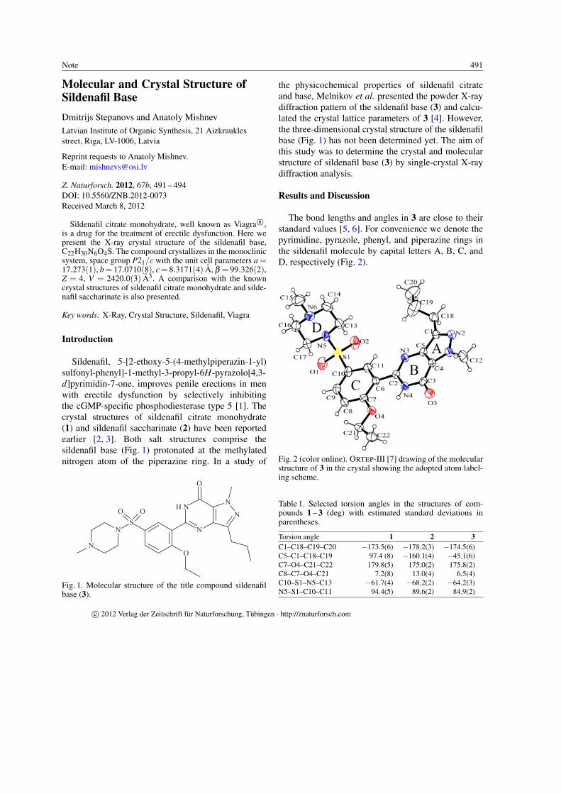
491Note
Molecular and Crystal Structure ofSildenafil Base
Dmitrijs Stepanovs and Anatoly Mishnev
Latvian Institute of Organic Synthesis, 21 Aizkrauklesstreet, Riga, LV-1006, Latvia
Reprint requests to Anatoly Mishnev.E-mail: [email protected]
Z. Naturforsch. 2012, 67b, 491 – 494DOI: 10.5560/ZNB.2012-0073Received March 8, 2012
Sildenafil citrate monohydrate, well known as Viagra R©,is a drug for the treatment of erectile dysfunction. Here wepresent the X-ray crystal structure of the sildenafil base,C22H30N6O4S. The compound crystallizes in the monoclinicsystem, space group P21/c with the unit cell parameters a =17.273(1), b = 17.0710(8), c = 8.3171(4) A, β = 99.326(2),Z = 4, V = 2420.0(3) A3. A comparison with the knowncrystal structures of sildenafil citrate monohydrate and silde-nafil saccharinate is also presented.
Key words: X-Ray, Crystal Structure, Sildenafil, Viagra
Introduction
Sildenafil, 5-[2-ethoxy-5-(4-methylpiperazin-1-yl)sulfonyl-phenyl]-1-methyl-3-propyl-6H-pyrazolo[4,3-d]pyrimidin-7-one, improves penile erections in menwith erectile dysfunction by selectively inhibitingthe cGMP-specific phosphodiesterase type 5 [1]. Thecrystal structures of sildenafil citrate monohydrate(1) and sildenafil saccharinate (2) have been reportedearlier [2, 3]. Both salt structures comprise thesildenafil base (Fig. 1) protonated at the methylatednitrogen atom of the piperazine ring. In a study of
O
O
SOO
N
N N
NHN
N
Fig. 1. Molecular structure of the title compound sildenafilbase (3).
the physicochemical properties of sildenafil citrateand base, Melnikov et al. presented the powder X-raydiffraction pattern of the sildenafil base (3) and calcu-lated the crystal lattice parameters of 3 [4]. However,the three-dimensional crystal structure of the sildenafilbase (Fig. 1) has not been determined yet. The aim ofthis study was to determine the crystal and molecularstructure of sildenafil base (3) by single-crystal X-raydiffraction analysis.
Results and Discussion
The bond lengths and angles in 3 are close to theirstandard values [5, 6]. For convenience we denote thepyrimidine, pyrazole, phenyl, and piperazine rings inthe sildenafil molecule by capital letters A, B, C, andD, respectively (Fig. 2).
Fig. 2 (color online). ORTEP-III [7] drawing of the molecularstructure of 3 in the crystal showing the adopted atom label-ing scheme.
Table 1. Selected torsion angles in the structures of com-pounds 1 – 3 (deg) with estimated standard deviations inparentheses.
Torsion angle 1 2 3C1–C18–C19–C20 −173.5(6) −178.2(3) −174.5(6)C5–C1–C18–C19 97.4 (8) −160.1(4) −45.1(6)C7–O4–C21–C22 179.8(5) 175.0(2) 175.8(2)C8–C7–O4–C21 7.2(8) 13.0(4) 6.5(4)C10–S1–N5–C13 −61.7(4) −68.2(2) −64.2(3)N5–S1–C10–C11 94.4(5) 89.6(2) 84.9(2)
c© 2012 Verlag der Zeitschrift fur Naturforschung, Tubingen · http://znaturforsch.com

492 Note
Fig. 3 (color online). Superimposed structures of the molecu-lar structure of 3 with 1 (sildenafil citrate) (a) and 2 (sildenafilsaccharinate) (b).
The pyrazolopyrimidine bicyclic system (A + B)and the phenyl ring (C) in 1 – 3 are almost copla-nar. The dihedral angle between least-squares planesof the pyrazolopyrimidine system and the phenyl ring
Table 2. Intramolecular hydrogen bonding geometry (A,deg).
Compound D–H· · ·A D–H H· · ·A D· · ·A ∠D–H· · ·A1 N4–H· · ·O4 0.88 1.94 2.622(6) 1342 N4–H· · ·O4 0.88 1.94 2.653(3) 1373 N4–H· · ·O4 0.86 1.95 2.645(3) 137
(A + B/C) is 11.6◦ in 1, 2.4◦ in 2 and 5.6◦ in themolecular structure of 3. The ethoxy groups in 1 – 3lie also almost in the A + B + C plane. In sildenafilsaccharinate (2) the propyl group lies close to theA + B + C average plane. In sildenafil citrate 1 thepropyl group is situated out of the A + B + C plane andat the side opposite to the methylpiperazine fragment.In the sildenafil base (3) the propyl group is also situ-ated out of the A + B + C plane but on the same sideas the methylpiperazine fragment. In all three crystalstructures the piperazine ring is in a chair conformationwith methyl and sulfonyl groups attached equatorially.The values of selected torsion angles for 1, 2 and 3 aregiven in Table 1.
A graphical comparison of the molecular structuresof 1, 2 and 3 by means of the superimposed pyra-zolopyrimidone ring systems is shown in Fig. 3.
The molecule of sildenafil in the structures of 1, 2and 3 has one intramolecular hydrogen bond with thegeometric parameters given in Table 2. There are someshort intermolecular C–H· · ·O contacts in the crystalstructure of 3, which can be characterized as weak hy-drogen bonds with electrostatic or mostly electrostaticnature [5].
The crystal structure determination of the sildenafilbase (3) allows the inspection of the correctness ofthe powder diffraction pattern indexing of 3 as pub-lished by Melnikov et al. [4]. The theoretical diffrac-tion pattern calculated from the atomic coordinates of3 is consistent with the experimental powder diffrac-tion pattern published in ref. [4]. Unfortunately, the lat-tice parameters of 3 as calculated in ref. [4] (a = 8.66,b = 34.27, c = 8.93 A, β = 96.63◦, V = 2632.5 A3)are far from the correct values, giving a wrong calcu-lated crystal density of 1.18 g cm−3 instead of the cor-rect value of 1.30 g cm−3
Conclusion
The molecular and crystal structure of sildenafilbase have been determinated by single-crystal X-raystructure analysis. The sildenafil molecule is builtfrom a rigid central core fragment consisting of a π-conjugated bicyclic pyrazolopyrimidone and a phenylring. The bulky methylpiperazine-sulfonyl fragmentseams to be insensitive to its rotation with respect tothe central core fragment. The only flexible part of themolecule is the propyl group which adopts three dif-ferent positions in the three known derivatives of silde-
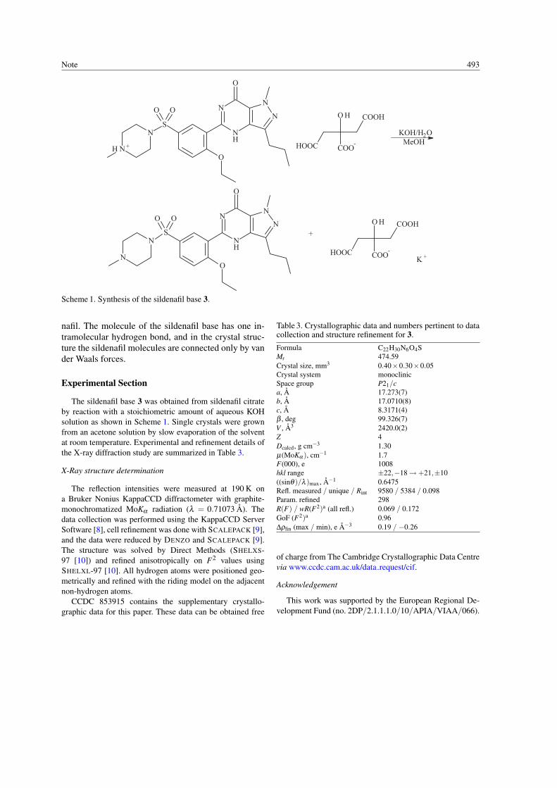
Note 493
K +
O HOO
O
O
NH
N
NN
SN
N
OO
O
O
NH
N
NN
S
N +H
O H
+
MeOHKOH/H2O
COOH
COO- HOOC
N
COOH
HOOC COO-
Scheme 1. Synthesis of the sildenafil base 3.
nafil. The molecule of the sildenafil base has one in-tramolecular hydrogen bond, and in the crystal struc-ture the sildenafil molecules are connected only by vander Waals forces.
Experimental Section
The sildenafil base 3 was obtained from sildenafil citrateby reaction with a stoichiometric amount of aqueous KOHsolution as shown in Scheme 1. Single crystals were grownfrom an acetone solution by slow evaporation of the solventat room temperature. Experimental and refinement details ofthe X-ray diffraction study are summarized in Table 3.
X-Ray structure determination
The reflection intensities were measured at 190 K ona Bruker Nonius KappaCCD diffractometer with graphite-monochromatized MoKα radiation (λ = 0.71073 A). Thedata collection was performed using the KappaCCD ServerSoftware [8], cell refinement was done with SCALEPACK [9],and the data were reduced by DENZO and SCALEPACK [9].The structure was solved by Direct Methods (SHELXS-97 [10]) and refined anisotropically on F2 values usingSHELXL-97 [10]. All hydrogen atoms were positioned geo-metrically and refined with the riding model on the adjacentnon-hydrogen atoms.
CCDC 853915 contains the supplementary crystallo-graphic data for this paper. These data can be obtained free
Table 3. Crystallographic data and numbers pertinent to datacollection and structure refinement for 3.
Formula C22H30N6O4SMr 474.59Crystal size, mm3 0.40×0.30×0.05Crystal system monoclinicSpace group P21/ca, A 17.273(7)b, A 17.0710(8)c, A 8.3171(4)β , deg 99.326(7)V , A3 2420.0(2)Z 4Dcalcd, g cm−3 1.30µ(MoKα ), cm−1 1.7F(000), e 1008hkl range ±22,−18→+21,±10((sinθ)/λ )max, A−1 0.6475Refl. measured / unique / Rint 9580 / 5384 / 0.098Param. refined 298R(F) / wR(F2)a (all refl.) 0.069 / 0.172GoF (F2)a 0.96∆ρfin (max / min), e A−3 0.19 / −0.26
of charge from The Cambridge Crystallographic Data Centrevia www.ccdc.cam.ac.uk/data request/cif.
Acknowledgement
This work was supported by the European Regional De-velopment Fund (no. 2DP/2.1.1.1.0/10/APIA/VIAA/066).

494 Note
[1] A. Laties, E. Zrenner, Prog. Retin. Eye Res. 2002, 21,485 – 506.
[2] H. S. Yathirajan, B. Nagaraj, P. Nagaraja, M. Bolte,Acta Crystallogr. 2005, E61, o489 – o491.
[3] R. Banerjee, P. M. Bhat, G. R. Desiraju, Cryst. GrowthDes. 2006, 6, 1468 – 1478.
[4] P. Melnikov, P. P. Corbi, A. Cuin, M. Cavicchioli, W. R.Guimares, J. Pharm. Sci. 2003, 92, 2140 – 2143.
[5] G. Gilli in Fundamentals of Crystallography, (Ed.:C. Giacovazzo), Oxford University Press, Oxford 2002,pp. 585 – 666.
[6] F. H. Allen, O. Kennard, D. G. Watson, L. Brammer,A. G. Orpen, R. Taylor, J. Chem. Soc., Perkin Trans. 21987, S1 – S19.
[7] C. K. Johnson, M. N. Burnett, ORTEP-III (version1.0.2), Rep. ORNL-6895, Oak Ridge National Labora-
tory, Oak Ridge, TN (USA) 1996. Windows version:L. J. Farrugia, University of Glasgow, Glasgow, Scot-land (UK) 1999. See also: L. J. Farrugia, J. Appl.Crystallogr. 1997, 30, 565.
[8] KappaCCD Server Software. Nonius BV, Delft (TheNetherlands) 1997.
[9] Z. Otwinowski, W. Minor in Methods in Enzymol-ogy, Vol. 276, Macromolecular Crystallography, PartA (Eds.: C. W. Carter Jr, R. M. Sweet), AcademicPress, New York, 1997, pp. 307 – 326.
[10] G. M. Sheldrick, SHELXS/L-97, Programs for CrystalStructure Determination, University of Gottingen,Gottingen (Germany) 1997. See also: G. M. Sheldrick,Acta Crystallogr. 1990, A46, 467 – 473; ibid. 2008,A64, 112 – 122.