Embed Size (px)
Citation preview

Molecular analysis of early onset Indonesian breast cancer
Dewajani Purnomosari

ISBN-10: 90-393-4390-X ISBN-13: 978-90-393-4390-6

Molecular analysis of early onset Indonesian breast cancer Moleculaire analyse van borstkanker bij jonge Indonesische vrouwen (met een samenvatting in het Nederlands) Analisis molekuler penderita kanker payudara usia muda di Indonesia (dengan ringkasan dalam bahasa Indonesia) Proefschrift ter verkrijging van de graad van doctor aan de Universiteit Utrecht op gezag van de rector magnificus, prof.dr.W.H. Gispen, ingevolge van het besluit van het college voor promoties in het openbaar te verdedigen op woensdag 29 november 2006 des ochtends te 10.30 uur door Dewajani Purnomosari geboren op 15 december 1969 te Surabaya, Indonesië

Promotor: Prof.dr. P.J. van Diest Co-promotor: Dr. G.Pals Dit onderzoek werd mede mogelijk gemaakt door grant NKB IN 2001-08 van KWF-Kankerbestrijding.

I dedicate this thesis to my late Beloved Father, the living inspiration ever in my journey, Poernomo Sidhi


Contents Chapter 1: General Introduction
9
Chapter 2: A novel BRCA2 mutation in an Indonesian family found with a new, rapid, and sensitive mutation detection method based on pooled denaturing gradient gel electrophoresis and targeted sequencing. J Clin Pathol 2005;58:493-499.
41
Chapter 3: BRCA1 and BRCA2 germline mutation analysis in the Indonesian population. Submitted for publication
55
Chapter 4: Comparison of multiplex ligation dependent probe amplification to immunohistochemistry for assessing HER-2/neu amplification in invasive breast cancer. Biotech Histochem 2006;81:79-85.
69
Chapter 5: Histopathological features of early onset Indonesian breast cancer pointing to BRCA1/2 germline mutations. Manuscript
79
Chapter 6: High throughput analysis of promoter hypermethylation status of 22 tumor suppressor genes in invasive breast cancer. Manuscript
91
Chapter 7: High throughput analysis of gene amplification of 27 genes in invasive breast cancer by Multiplex Ligation dependent Probe Amplification. Manuscript
105
Chapter 8: General Discussion
121
Chapter 9: Summaries
133
Samenvatting in het Nederlands 134 Summary in English 135 Ringkasan dalam bahasa Indonesia 136 A word of gratitude 138 Curriculum Vitae 140 List of publications 141


Chapter 1:
General Introduction
chapter 1 General Introduction
Contents Chapter 1: General Introduction 1. Clinical Aspects of breast cancer 11 1.1. Epidemiology of breast cancer 11 1.2. Etiology of breast cancer 1.3. The multi-step progression model of breast cancer 12 1.4. Treatment of breast cancer based on prognostic and predictive
factors 13
2 Genetic Aspects of breast cancer 14 2.1. BRCA1 and BRCA2 17 2.1.1. Structure of BRCA1 and BRCA2 17 2.1.2. Function of BRCA1 and BRCA2 19 2.1.2.1. DNA repair 19 2.1.2.2. Transcriptional response to DNA damage 20 2.1.2.3. DNA damage-responsive cell cycle checkpoints 20 2.1.3. Germline mutation in BRCA1 and BRCA2 22 2.1.4. Ethnic differences in frequency of BRCA1/2 mutations 23 2.1.5. Penetrance of BRCA1 and BRCA2 mutations 23 2.1.6. Pathology of BRCA1 and BRCA2 related breast cancer 24 2.1.7. Clinical presentation of BRCA1 and BRCA2 related breast
cancer 25
2.2. Mutation detection methods 26 2.3. Epigenetics of sporadic and hereditary breast cancer 27 2.3.1. DNA methylation 27 2.3.2. CpG island methylator phenotype 28 2.3.3. Hypermethylation of genes in breast cancer 28 3. Summary and scope of thesis 29 30 4. References
10

chapter 1 General Introduction
I. CLINICAL ASPECTS OF BREAST CANCER 1.1. Epidemiology of breast cancer Breast cancer is the second leading cause of cancer deaths in women today (after lung cancer) and is the most common cancer among women, excluding non-melanoma skin cancers. According to the World Health Organization, more than 1.2 million people will be diagnosed with breast cancer this year worldwide (http://www.who.int/whosis/whostat2006.pdf.). The American Cancer Society estimates that in 2005, approximately 211,240 women in the United States will have been diagnosed with invasive breast cancer (http://www.cancer.org/downloads/STT/CAFF2005BrF.pdf). The chance of developing invasive breast cancer during a woman's lifetime is approximately 1 in 8 (13.4%). Another 58,490 women will be diagnosed with in situ breast cancer, an early form of the disease. Though much less common, breast cancer also occurs in men. An estimated 1,690 cases will have been diagnosed in men in 2005. There are no nationwide data on the incidence of breast cancer in Indonesia, but locally published data [1] on a pathology based cancer registry in Jogjakarta in 1982 showed that breast cancer incidence ranked second (18.9%) after cervical cancer (20%). Ghozali [2] showed in 1995 that breast cancer was even the most common cancer among women in Jogjakarta (24.58%), followed by cervical cancer (17.28%). Unpublished data on early onset (< 40 years) breast cancer patients between 1998 to 2004 in Sardjito Hospital Jogjakarta, showed that the proportion of these patients was 26% of total breast cancer cases. This number is higher compared to data from The National Cancer Institute’s Surveillance, Epidemiology and End Results (SEER) program that reveals that 75% of breast tumors occur in women aged >50 years and only 6.5% in women aged <40 years. This indicates that breast cancer is also a major health care problem in Indonesian women, especially among young women. 1.2. Etiology of breast cancer The etiology of breast cancer is still poorly understood. Several known risk factors can only explain a small proportion of breast cancer cases [3]. At first, age is associated with increasing breast cancer risk. However, remarkably, most risk increase occurs during the reproductive years as breast cancer incidence is very low before age 25, and increases up to 100 fold by age 45 [4]. This pattern suggests the involvement of reproductive hormones in breast cancer etiology. Other known risk factors involve life style factors, environmental factors, a history of benign proliferative breast lesions, and genetic factors. Reproductive factors such as nulliparity, early menarche and older age at first pregnancy have been associated with an increased breast cancer risk [4, 5]. In addition, exogenous hormonal influences like using oral contraceptives may increase breast cancer risk. However, the opposite was demonstrated in many epidemiologic studies in which no association between the use of oral contraceptive and the risk of breast cancer was shown. Recently, however, a large meta-analysis calculated a small but significant increase in relative risk of breast cancer (RR = 1.24) in current oral contraceptive users [6]. The use of hormone replacement therapy (HRT) by postmenopausal women was also shown to be associated with enhanced breast cancer risks, predominantly affecting the chance of development of a hormone receptor-positive breast cancer [7].
11

chapter 1 General Introduction
Life style factors, like increased height and weight have been associated with a higher breast cancer risk in a number of studies [8, 9]. Dietary factors such as high fat intake, low vegetables/fruit and low fibre intake may also increase risk [10]. Furthermore, alcohol consumption was significantly associated with a higher risk of breast cancer [11, 12]. Some studies related socio-economic status to breast cancer risk, but these findings can probably be explained by differential life styles such as alcohol, diet and reproductive patterns [13]. Besides these life style factors, other medical history factors are involved with increased breast cancer risk. Mammography density >75% is another well-established risk factor for breast cancer in both pre-and postmenopausal women [14]. Increased mammography density is seen predominantly in nulliparous women and thin women. Furthermore, a history of a benign proliferative breast conditions, especially atypical ductal hyperplasia (ADH) and fibroadenoma, is associated with an increased risk for breast cancer [15-17]. Other exogenous risk factors include exposure of the mammary gland to high dose ionizing radiation during childhood [4]. This latter relation is dose dependent and decreases gradually over time. Finally, a positive family history of breast cancer is most significantly associated with increased risk of breast cancer. This is most strongly seen in families with a germline mutation in a breast cancer susceptibility gene such as BRCA1 and BRCA2 which were identified in 1990 and 1994, respectively [18, 19]. Such germline mutations are associated with a 50-80% life time breast cancer risk. In addition to the BRCA genes, several other high susceptibility genes have been identified, such as p53, ATM, CHEK2 and PTEN [3]. However, because of their low allele frequencies in the general population, it is believed that only 5 to 10 percent of all breast cancers are associated with the presence a specific germline mutation. Besides the above mentioned well known genetic alterations, epigenetic alterations are among the most common molecular alterations in human neoplasia [20-22]. Epigenetic changes differ from genetic changes mainly in that they occur at a higher frequency than genetic changes, are reversible upon treatment with pharmacological agents and occur at defined regions in a gene [21]. 1.3. The multistep progression model of breast cancer Breast cancer tumorigenesis can be described as a multi-step process [23] in which each step is thought to correlate with one or more distinct mutations in major or minor regulatory genes. Breast development begins in the embryonic period. Ductal morphogenesis starts from a bud-like structure with branching, elongation and then canalization. Basal cells, expressing both smooth muscle actin, as well as high molecular weight cytokeratins appear at the end of the second trimester. In the adult breast two major cell types can be distinguished: the myoepithelial cell and the luminal secretory cell, that derive from a pluripotent CK5/6 positive stem cell that shows no compartimentalization. Clinically and histopathologically, various morphologically definable steps can be identified during progression to malignancy [24]. Ductal hyperplasia, characterized by proliferation of unevenly distributed polyclonal epithelial cells with overlapping nuclei of varying shape and chromatin pattern and haphazard lumina, is often a first sign of tendency towards malignancy. The cells have relatively little cytoplasm and no clear cell borders. Cytologically the cells are benign. The transition from hyperplasia to (clonal) atypical hyperplasia
12

chapter 1 General Introduction
with cells with more distinct cytoplasm, more regular nuclei with less overlap and more regular lumina, is clinically associated with an increased risk of breast cancer. The next step is development of carcinoma in situ, either ductal or lobular, which is defined as a proliferation of cells with cytological characteristics of malignancy, but without stromal invasion across the basement membrane. Lobular carcinoma in situ derives from the lobuli, and is usually diffusely spread throughout the breast (and even often bilateral), and is typically not palpable and unvisible by imaging. In contrast, ductal carcinoma in situ is a segmental ductal lesion that may reveal itself through microcalcifications and may be palpable. As cells detach from the basement membrane and invade the stroma, the tumour becomes invasive. Through dissemination via blood and lymph vessels, invasive cells can give rise to metastases, either to loco regional lymph nodes or to distant organs. In this process, tumor cells need to escape the immune system to prevent clearance. At each of these steps, genetic events occur that give the cell new properties with a resulting clonal selective advantage for that cell. These genetic events range from small point mutations, via chromosomal deletions, translocations and amplifications to large-scale changes as whole chromosome losses or duplications. The result of these alterations could be modification of gene expression or functional alteration of gene products that are relevant for tumour progression. The accumulation of these genetic events is best believed to follow the “bingo principle” (in random order) than a fixed stepwise order, although some events seem to occur more often earlier than others. It has become clear that the genetic events that play a role in carcinogenesis of sporadic and hereditary breast cancer show overlap as well as differences. It is beyond the scope to discuss of this thesis and introduction to review the panoply of different genetic events that have been described in sporadic breast cancer. Rather, we will focus on some important genetic aspects of hereditary breast carcinogenesis. 1.4. Treatment of breast cancer based on prognostic and predictive factors Because adjuvant systemic therapy has associated risks, it should only be given to high risk patients. Therefore, it is essential to be able to estimate an individual patient’s risk to develop clinically manifest metastatic disease using prognostic factors [25-27]. With predictive factors, patients that most likely to benefit from a certain therapy can be identified. The most significant prognostic indicator for breast cancer is the presence or absence of lymph node involvement as mentioned earlier. There is also a relationship between the number of involved axillary nodes and the risk of distance recurrence [28]. Therefore, axillary node status is the most important prognostic factor used in adjuvant therapy decision making. Secondly, tumor size is also an independent prognostic factor, with distant recurrence rates increasing with larger tumor size [29, 30]. Histologically, tumor grade scored according to the widely accepted Bloom-Richardson classification [31] has also prognostic significance. Of the three features encompassing grade, mitotic index is known to have overriding prognostic value. Furthermore, lymphovascular invasion is associated with recurrence and overall survival [32].
13

chapter 1 General Introduction
The estrogen (ER) and progesterone receptors (PgR) are useful for predicting the clinical response to endocrine therapy [33]. HER-2/neu (c-erbB-2) has recently been added as a routine immunohistochemical assessment, as protein overexpression of HER-2/neu predicts both higher chance to respond to trastuzumab treatment as well as taxane chemotherapy [34]. Further, such patients respond better to aromatase inhibitors than to tamoxifen [35-39]. Trastuzumab is a recombinant DNA-derived humanized monoclonal antibody that selectively binds to the extracellular domain of HER2. This specific biological drug has been approved in the USA since 1998 for HER2-positive patients with metastatic breast cancer. It is most effective in combination with chemotherapy. Recent clinical studies have shown that trastuzumab may also be useful as adjuvant therapy in breast cancer patients who overexpress HER2. The drug is generally well tolerated but cardiotoxicity has been reported in up to 27% of cases when used in combination with anthracycline and cyclophosphamide [40, 41]. This, and the significant costs of the therapy underline the need to optimally select patients for treatment with this drug. Immunohistochemical (IHC) staining has been approved as a screening method for HER2 protein activity, accepting a 3+ score as “positive” for treatment. However, there are some problems with IHC interpretation due to its subjective nature, so it is generally accepted that a 2+ score requires gene status confirmation by an amplification test. Further, there are indications that gene amplification status predicts response better than IHC [42]. The most widely used gene amplification test is fluorescence in situ hybridization (FISH). However, FISH is difficult, expensive, probes have a limited half life, and interpretation is not without its problems, so FISH is not very suitable for routine use. Chromagen ISH (CISH) overcomes some but definitely not all these drawbacks. Other more easy to perform tests are therefore urgently required. Recently, a test based on multiplex ligation dependent probe amplification [43] has become available that deserved to be validated. II. GENETIC ASPECTS OF HEREDITARY BREAST CANCER Approximately 5% of breast cancers show a familial pattern of occurrence [44]. This is often related to germline mutations in different (tumor suppressor) genes of which the proteins have a crucial function in the breast. Like in the classical tumor suppressor, in patients with a germline mutation, loss of expression of the other allele by point mutations or deletions will lead to a significant or complete loss of protein function. In familial breast cancer patients, germline mutations have been described in BRCA1, BRCA2, PTEN, p53, ATM and CHEK2. Together, these account for most but certainly not all hereditary cases, so the search for other hereditary breast cancer genes continuous. BRCA1 and BRCA2 will be dealt with extensively further on as they form the focus of this thesis, so here we mainly discuss the other proteins. The BRCA1 gene, located on chromosome 17q12-21, was cloned in 1994 [18]. BRCA1 is involved in many transcriptional processes. It has been associated with more than 15 different proteins involved in transcription, either in transcriptional activation or transcriptional repression [45]. It also plays a role in apoptosis. As a tumour suppressor, BRCA1 is a factor in maintaining genomic stability. It interacts with various proteins, and the complexes formed are involved in DNA recognition
14

chapter 1 General Introduction
and repair [46, 47]. The BRCA2 gene is located on chromosome 13q12-13. The gene codes for proteins involved in DNA repair, cell cycle control and transcription [48], and may have a function in terminal differentiation of breast epithelial cells [49]. In sporadic breast cancer, mutational inactivation of BRCA2 is rare as inactivation requires both gene copies to be mutated or totally lost [47, 48, 50]. Surprisingly, despite the inherited predisposition to cancer associated with BRCA1 and BRCA2 (see below) somatic disease-causing mutations in either of these genes are extremely rare in sporadic breast cancers [51, 52].
Figure 1. — Breast cancer susceptibility genes. Hereditary breast cancer (right) constitutes only approximately 5% to 10% of all breast cancer cases (left). Germline mutations in the two major susceptibility genes BRCA1 and BRCA2 account for less than 5% of all breast cancer cases, while mutations in genes such as ATM, CHEK2, PTEN, and TP53 account for only about 1% of all breast cancer cases (adopted from Dapic et al, 2005 [53]). TP53 is a tumor suppressor gene located on 17p13.1 encoding a nuclear phosphoprotein that acts as a transcription factor involved in the control of cell cycle progression, repair of DNA damage, genomic stability, and apoptosis [54]. In response to DNA damage, the p53 protein arrests cells in the G1 phase of the cell cycle, allowing the DNA repair mechanism to proceed prior to DNA synthesis. Loss of p53 function abolishes this growth arrest response to DNA damage. TP53 is one of the most frequently mutated genes in sporadic human cancer [50]. Most mutations are point mutations leading to proteins defective for sequence-specific DNA binding and activation of p53-responsive genes [55-57]. In sporadic breast carcinomas the occurrence of TP53 mutations is usually a late event. Interestingly, TP53 mutations are frequently found in BRCA1-linked tumors and several studies have suggested that the status of BRCA1/BRCA2 influences the type and distribution of TP53 mutations in breast cancer [57-59]. Germline mutations in TP53 (Li-Fraumeni syndrome) are very rare. Analysis of 475 tumors in 91 families with p53 germline mutations showed that breast carcinomas are most frequent (24.0%), followed by bone sarcomas (12.6%), brain tumors (12.0%), and soft tissue sarcomas (11.6%) [60]
15

chapter 1 General Introduction
PTEN (also known as MMAC1) on chromosome 10q23.3 was originally identified as a tumor suppressor gene defective in a variety of human cancers [61, 62]. Germline mutations in PTEN, causing Cowden disease, a rare autosomal dominant inherited cancer syndrome characterized by a high risk of breast, thyroid, and endometrial carcinomas and hamartomas (a common benign tumor as a result from an abnormal formation of basically normal tissue components). Hamartomas, while generally benign, can cause problems due to their location. They are particularly likely to cause major health issues when located in the hypothalamus, spleen or kidneys. [63-65]. Most cancer associated PTEN mutations are truncations that cause a 25% to 50% lifetime breast cancer risk among women affected with Cowden disease [66, 67]. PTEN mutations are rare in sporadic breast cancer and have been found in only 5% of the sporadic cases [68, 69]. However, 29% to 48% of sporadic breast cancer cases show loss of heterozygosity at the PTEN locus, while no alterations have been found in the remaining allele [70]. In addition, approximately 40% of breast cancers show a decrease or absence of PTEN protein levels. The cell cycle checkpoint kinase CHEK2 gene on chromosome 22q12.1 is a key mediator in DNA damage-response [71, 72]. In mammalian cells, CHEK2 is phosphorylated by ATM in response to double strand breaks (DSB) [73]. Activated CHEK2 phosphorylates a number of target proteins that in turn prevent cellular entry into mitosis and activate DNA repair pathways. In addition, CHEK2 acts in the G1-S checkpoint by phosphorylating p53 and mediating activation and stabilization of p53 by ATM [74, 75]. In another important connection, CHEK2 phosphorylates Cdc25C and BRCA1 [76, 77]. Mutation screening of the CHEK2 gene among Li-Fraumeni cases revealed the CHEK2 1100delC mutation which inactivates the kinase activity of the protein [78]. This allele has also been proposed to be a low-penetrance breast cancer susceptibility allele [79, 80]. Additional screening of CHEK2 did not identify any other variant that occurs at significantly elevated frequency, indicating that 1100delC may be the only CHEK2 allele with a significant contribution to breast cancer susceptibility [81]. Interestingly, CHEK2 1100delC is associated with breast cancer only in non-carriers of BRCA1 and BRCA2 [79]. A recent search for new breast cancer susceptibility genes among families with no BRCA1 and BRCA2 mutation suggested a model in which CHEK2 1100delC interacts with an as yet unknown gene to increase breast cancer risk [82]. The ATM (ataxia telangiectasia-mutated) protein was identified as the product of the gene mutated in the rare human autosomal recessive disorder ataxia telangiectasia (AT) [83]. ATM plays a key role in monitoring genomic integrity and triggering appropriate cell-cycle checkpoints, DNA repair, or apoptotic pathways in response to DNA double-strand breaks. In response to ionizing radiation, a potent inducer of DNA double strand breaks, ATM associates with and phosphorylates a number of different substrates, including p53, MDM2, Nibrin, CtIP and BRCA1 [84-87]. Phosphorylation of p53 and MDM2 results in p53 stabilization and accumulation that activates the G1/S cell cycle checkpoint. ATM phosphorylation of CtIP in response to radiation exposure modulates the ability of BRCA1 to induce expression of DNA-damage response-molecules such as GADD45 [87]. ATM also directly phosphorylates BRCA1, and this phosphorylation is required for normal cellular survival after exposure to ionizing radiation [86]. Therefore, there are direct functional links between ATM and BRCA1, both of which have been implicated in
16

chapter 1 General Introduction
breast carcinoma susceptibility. ATM heterozygotes have an approximately ninefold-increased risk of developing a type of breast cancer characterized by frequent bilateral occurrence, early age at onset and long-term survival [88]. 2.1. BRCA1 and BRCA2 In 1990, genetic studies provided initial evidence that the risk of breast cancer in some families is linked to chromosome 17q21 [89]. This 17q-associated syndrome was characterized by autosomal dominant inheritance with incomplete penetrance. In fact, loss of heterozygosity (LOH) at 17q was found in most familial breast and ovarian tumors, suggesting the involvement of tumor suppressor gene(s) [90, 91]. In 1994, the breast-cancer susceptibility gene, BRCA1, was identified by positional cloning; subsequently, this gene has been the subject of intensive research [18]. BRCA1 is composed of 22 coding exons distributed over 100 kb of genomic DNA. This gene encodes 1863 amino acids, and more than 200 different germline mutations associated with cancer susceptibility have so far been identified (http://www.research.nhgri.nih.gov/projects/bic). Many disease-predisposing alleles of BRCA1 concern insertions, deletions, frameshifts, base substitutions and inferred regulatory mutations [92], the majority resulting in premature truncation of the protein leading to loss of protein function. Germline mutations in BRCA1 confer susceptibility to breast, ovarian and Fallopian tube cancer, as well as cancers of the corpus uteri, the cervix and the peritoneum. Because only 45% of familial breast cancers showed evidence of linkage to BRCA1, the search for a second breast cancer susceptibility gene continued. In 1995, the BRCA2 gene was identified at chromosome 13q12.3 [19, 93]. So far, around 3000 different germline mutations associated with cancer susceptibility have been identified (http://www.research.nhgri.nih.gov/projects/bic). Germline mutation carriers also have increased susceptibility to ovarian, Fallopian tube, pancreatic, prostate, and male breast cancers. 2.1.1. Structure of BRCA1 and BRCA2 Although there is no sequence similarity between the two genes, many structural and functional features of BRCA1 and BRCA2 are similar. Both genes have complex genomic structures (BRCA1 is composed of 24 exons and BRCA2 of 27 exons), and they both encode very large proteins (BRCA1 1863 amino acids and BRCA2 3418 amino acids) [18, 19, 94]. In both, exon 1 is noncoding and exon 11 is unusually large, 3.4 kb in BRCA1 and 5 kb in BRCA2. BRCA1 and BRCA2 are expressed in a wide range of tissues and show remarkably similar temporal and spatial patterns of expression. Both BRCA1 and BRCA2 exhibit approximately 60% amino acid identity with their murine counterparts, although several short domains of both proteins are relatively well conserved compared to the remainder [95-97]. This level of evolutionary conservation is lower than that of other cancer-susceptibility genes, most of which exhibit more than 90% human/mouse sequence identity. Three regions of BRCA1 show sequence similarity to previously described proteins. A highly conserved zinc-binding RING finger domain is located close to the amino-terminus (residues 20–68). Although some classes of zinc finger act as transcriptional regulators by binding to specific DNA sequences in promoter/enhancer regions, there is little current evidence in favor of this function
17

chapter 1 General Introduction
for the RING class of zinc finger. In contrast, exist data indicating that RING fingers are involved in protein-protein interactions [98]. Indeed, a search for interacting proteins, using the BRCA1 RING finger domain as bait, uncovered a further RING-domain protein designated BARD1 (BRCA1-associated RING domain), which binds to BRCA1. The RING domains of both BARD1 and BRCA1 are necessary, but not sufficient, to mediate this interaction [99]. Toward the carboxyl terminus of BRCA1 are two tandem copies of a motif (designated the BRCT domain), which are located at residues 1699–1736 and 1818–1855 [100]. Similar BRCT motifs have been found in 53BPI, a protein capable of binding p53, and several proteins involved in cell cycle regulation or DNA repair such as RAD9, XRCC1, RAD4, RAP1, Ect2, terminal deoxynucleotidyltransferases, and three DNA ligases [101]. The BRCT domains show strong sequence conservation in the murine protein and are found in a similar position in BARD1. Their function is unknown, but they are located within the region of BRCA1 that is reported to activate transcription when fused to a DNA-binding domain [102].
Figure 2. The BRCA1 and BRCA2 genes, showing some functional domains and founder mutations (adopted from Thompson and Easton, 2004 [103]) RING finger or BRCT domains are not present in BRCA2, nor does it bear substantial similarity to any other sequence presently registered in the databases. However, eight copies of a 20–30 amino acid repeat (termed BRC repeats) are located between residues 1000–2030. Currently, the only other protein known to contain a similar sequence is a predicted C. elegans protein of unknown function. Most of the BRC repeats in BRCA2 show >80% amino acid sequence identity between human, mouse, and chicken, and are therefore better conserved than the protein overall [95].
18

chapter 1 General Introduction
2.1.2. Function of BRCA1 and BRCA2 2.1.2.1. DNA repair Subsequent studies demonstrated the involvement of BRCA1 and BRCA2 in complexes that activate the repair of doublestrand breaks (DSBs) and initiate homologous recombination (HR), linking the maintenance of genomic integrity to tumor suppression. BRCA1 and BRCA2 co-localize with Rad51 to form complexes [104, 105]. Eukaryotic Rad51 proteins are homologues of bacterial RecA and are required for recombination during mitosis and meiosis, as well as for HR repair of DSBs [106]. Rad51 coats single-stranded DNA to form a nucleoprotein filament that invades and pairs with a homologous region in duplex DNA, and then activates strand exchange to generate a crossover between the juxtaposed DNA [107, 108]. Co-localization of BRCAs with Rad51 at sites of recombination and DNA damage-induced foci strongly suggests that BRCAs have a role in both the detection and the repair of DSBs [104]. In this regard, focus formation of Rad51 is reduced after treatment with DNA-damaging agents and is deficient during repair of DSBs by HR in BRCA1-deficient cells [109, 110]. However, accumulating evidence suggests that BRCA1 might not directly regulate Rad51, since interactions between BRCA1 and Rad51 are indirect and stoichiometrically negligible [111]. The roles played by BRCA1 and BRCA2 in the repair of DSBs by HR appear to differ. Available evidence indicates a more direct role of BRCA2. BRCA2-deficient cells exhibit increased sensitivity to ionizing radiation, indicative of a defect in DSB repair, whereas the cell cycle checkpoint and apoptotic responses to DNA damage remain intact [112, 113]. In addition, BRCA2-deficient cells accumulate chromosomal breaks and aberrant mitotic exchanges during culture. Rad51-deficient cells show similar phenotypes, providing genetic evidence that interactions of BRCA2 with Rad51 are fundamental for the maintenance of cell division and chromosome structure. Physiologically, interactions between BRCA2 and Rad51 are mediated by the BRC repeat and an unrelated domain located at the C-terminus. Recent studies have shown that BRCA2 regulates the intracellular localization and function of Rad51 [114]. In BRCA2-deficient cells, nuclear transport of Rad51 is impaired, suggesting that BRCA2 moves Rad51 from the site of synthesis to the site of DNA damage processing [114]. Some unexpected and potentially informative insight into the role of BRCA genes in human DNA repair has come from recent studies of Fanconi anemia (FA) [115-117]. To date, twelve genetic FA subgroups have been identified based on the complementation analysis, and eleven of these have been cloned (FancA/B/C/D1/D2/E/F/G/J/L/M) [118, 119]. The proteins encoded by FA genes are intimately related to each other in molecular pathways involved in DNA repair, and FANCA, FANCC, FANCE, FANCF, and FANCG interact directly to form a multisubunit nuclear complex [120]. In response to DNA damage this complex is translocated to DNA repair foci containing BRCA1 and BRCA2 [121]. Howlett et al. [115] have provided evidence that FANCD1 is identical with BRCA2. The cellular consequences of homozygosity for mutated BRCA2, including spontaneous chromosome instability and hypersensitivity to DNA crosslinking agents, are similar to those observed in cells derived from FA patients. Another FA-complementation-group protein, FANCD2, can interact and colocalize with BRCA1 [121]. Moreover, both FANCD2 and BRCA1 can be phosphorylated by ATM [122], which has also recently been implicated in susceptibility to breast cancer [123]. Hence, it appears
19
chapter 1 General Introduction
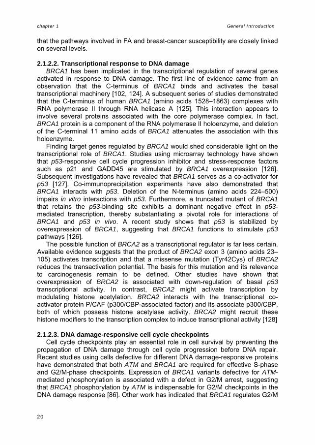
that the pathways involved in FA and breast-cancer susceptibility are closely linked on several levels. 2.1.2.2. Transcriptional response to DNA damage BRCA1 has been implicated in the transcriptional regulation of several genes activated in response to DNA damage. The first line of evidence came from an observation that the C-terminus of BRCA1 binds and activates the basal transcriptional machinery [102, 124]. A subsequent series of studies demonstrated that the C-terminus of human BRCA1 (amino acids 1528–1863) complexes with RNA polymerase II through RNA helicase A [125]. This interaction appears to involve several proteins associated with the core polymerase complex. In fact, BRCA1 protein is a component of the RNA polymerase II holoenzyme, and deletion of the C-terminal 11 amino acids of BRCA1 attenuates the association with this holoenzyme. Finding target genes regulated by BRCA1 would shed considerable light on the transcriptional role of BRCA1. Studies using microarray technology have shown that p53-responsive cell cycle progression inhibitor and stress-response factors such as p21 and GADD45 are stimulated by BRCA1 overexpression [126]. Subsequent investigations have revealed that BRCA1 serves as a co-activator for p53 [127]. Co-immunoprecipitation experiments have also demonstrated that BRCA1 interacts with p53. Deletion of the N-terminus (amino acids 224–500) impairs in vitro interactions with p53. Furthermore, a truncated mutant of BRCA1 that retains the p53-binding site exhibits a dominant negative effect in p53-mediated transcription, thereby substantiating a pivotal role for interactions of BRCA1 and p53 in vivo. A recent study shows that p53 is stabilized by overexpression of BRCA1, suggesting that BRCA1 functions to stimulate p53 pathways [126]. The possible function of BRCA2 as a transcriptional regulator is far less certain. Available evidence suggests that the product of BRCA2 exon 3 (amino acids 23–105) activates transcription and that a missense mutation (Tyr42Cys) of BRCA2 reduces the transactivation potential. The basis for this mutation and its relevance to carcinogenesis remain to be defined. Other studies have shown that overexpression of BRCA2 is associated with down-regulation of basal p53 transcriptional activity. In contrast, BRCA2 might activate transcription by modulating histone acetylation. BRCA2 interacts with the transcriptional co-activator protein P/CAF (p300/CBP-associated factor) and its associate p300/CBP, both of which possess histone acetylase activity. BRCA2 might recruit these histone modifiers to the transcription complex to induce transcriptional activity [128] 2.1.2.3. DNA damage-responsive cell cycle checkpoints Cell cycle checkpoints play an essential role in cell survival by preventing the propagation of DNA damage through cell cycle progression before DNA repair. Recent studies using cells defective for different DNA damage-responsive proteins have demonstrated that both ATM and BRCA1 are required for effective S-phase and G2/M-phase checkpoints. Expression of BRCA1 variants defective for ATM-mediated phosphorylation is associated with a defect in G2/M arrest, suggesting that BRCA1 phosphorylation by ATM is indispensable for G2/M checkpoints in the DNA damage response [86]. Other work has indicated that BRCA1 regulates G2/M
20

chapter 1 General Introduction
DNA damage induced checkpoints through its ability to activate Chk1 kinase and thereby induce signaling cascades downstream of Chk1 [129]. In this context, the finding that BRCA1-deficient cells exhibit defective G2/M arrest in response to ionizing radiation further supports a role of BRCA1 in the regulation of G2/M checkpoints. As mentioned above, BRCA1 functions as a co-activator of p53-mediated gene transcription. In BRCA1-deficient cells, the expression of 14-3-3σ, which is regulated by p53, is significantly diminished [130]. Since 14-3-3σ is a major G2/M checkpoint control gene, 14-3-3σ induction by BRCA1 may also be involved in BRCA1-mediated G2/M checkpoints. Other studies have shown that overexpression of BRCA1 results in the transcriptional activation of GADD45 in a p53-dependent manner [131, 132]. As GADD45 has been implicated in G2/M checkpoints, BRCA1 may in part activate G2/M checkpoints by induction of GADD45 protein. Interestingly, another p53 target gene, the G1 cyclin-dependent kinase inhibitor p21, is also transactivated by exogenous expression of BRCA1 to block S phase entry in a p53-independent manner [133]. Importantly, cancer-associated mutant BRCA1 failed to activate the p21 promoter. BRCA1 has also been found to transactivate the cyclin-dependent kinase inhibitor p27KIP1 [134]. The induction of G1 arrest by exogenous BRCA1 expression is likely to be associated with activation of p27KIP1. There is evidence that FANCD2, like BRCA1, participates in the events that are triggered by DNA damage during S and G2 phases and that lead to cell-cycle arrest. As well as modifying BRCA1, the ATM protein kinase can phosphorylate FANCD2 on several residues, including serine 222, following exposure to ionizing radiation [122]. Ser222 phosphorylation is required for the intra-S-phase arrest and is, in turn, dependent on the protein kinase NBS1, which is a component of the trimeric MRE11–RAD50–NBS1 complex [135]. FANCD2 can also be phosphorylated in an ATM-independent manner by the ATR kinase [136], which signals an intra-S-phase arrest that is triggered by ultraviolet radiation or DNA-crosslinking agents [137]. Here, too, FANCD2 phosphorylation depends on NBS1, which is required to fully enforce the S-phase checkpoint. The participation of FANCD2 in ionizing radiation-activated checkpoint responses through phosphorylation on Ser222 seems to be independent [135] of its mono-ubiquitylation on Lys561, which promotes the translocation of FANCD2 to damage-induced nuclear foci that contain BRCA1 and RAD51, and that are presumed sites of DNA repair. Similarly, FANCD2 foci do not require ATR-dependent phosphorylation [136]. So, these findings distinguish functions of FANCD2 in two aspects of DNA-damage responses during S and G2 phases through apparently discrete post-translational modifications — through Ser/Thr phosphorylation in checkpoint arrest and through Lys561 mono-ubiquitintation in DNA repair. It remains unclear whether BRCA2 participates directly in cell cycle regulation or checkpoint functions. Available evidence suggests that BRCA2 mediates G2/M-phase control by interacting with a novel protein, BRCA2-associated factor 35 (BRAF35), which binds to branched DNA structures [138]. Nuclear staining has revealed a close association of BRAF35/BRCA2 complex with condensed chromatin, coincident with histone H3 phosphorylation. Importantly, antibody microinjection experiments suggest a role of BRCA2/BRAF35 complex in modulation of metaphase progression [138]. However, it is premature to conclude
21

chapter 1 General Introduction
that BRCA2 is directly involved in mitotic progression. Since BRCA2 has a major role in DNA repair, its suppression is thought to induce unrepaired DNA lesions, which cause cell cycle arrest by activating checkpoint signaling, including mitotic progression. 2.1.3. Germline mutations in BRCA1 and BRCA2 BRCA1 and BRCA2 are the most important breast cancer susceptibility genes in high-risk families, and identification of mutations in these genes forms an important component of the management of high-risk women. The Breast Cancer Information Core (BIC) database had recorded (as of June 2006) 1536 distinct germline BRCA1 mutations and 1885 BRCA2 mutations. Of these, 878 (57%) and 1140 (60%) have been reported just once. Mutations appear to be reasonably evenly distributed across the coding sequences, with no obvious “mutation hot-spots.” Most mutations found in breast and/or ovarian cancer families are predicted to truncate the protein product. The most common types of mutation are small frameshift insertions or deletions, nonsense mutations, or mutations affecting splice sites resulting in deletion of complete or partial exons or insertion of intronic sequence. The Breast Cancer Linkage Consortium (BCLC) has estimated that approximately 70% of BRCA1 mutations and 90% of BRCA2 mutations in linked families are of this type [103]. Large-scale rearrangements, including insertions, deletions, or duplications of more than 500kb of DNA, have also been identified, but as these are not identifiable by exonic sequencing or other conventional screening techniques they are likely to be underreported. To date there have been reports of at least 19 distinct large genomic rearrangements in BRCA1 and two in BRCA2, identified using protein truncation analyses, Southern blots.or relatively new method, multiplex ligation dependent probe amplification (MLPA) [139, 140].The majority are deletions of one or more exons [140]. The higher density of Alu repetitive sequences in the BRCA1 gene (42% vs. 20%) [141] is thought to contribute to the larger number of large deletions and duplications observed in this gene. In addition to protein truncating mutations, large numbers of amino-acid substitutions have been identified in both BRCA1 and BRCA2. A small number of these, principally involving cysteine residues in the BRCA1 RING domain, have occurred consistently in high-risk families and are regarded as disease-associated missense mutations. Some do not cause amino acid changes and are thereby harmless polymorphisms, but the status of the majority (termed “unclassified variants”) is uncertain. Given their frequency and the fact that many occur in patients with another, deleterious, mutation, it is clear that the large majority of these variants cannot be strongly associated with disease. At present no reliable functional assay exists to determine whether such a variant is likely to be deleterious, and only the epidemiological evidence on the frequency of the variant in breast cancer cases and controls, or co-segregation of the variant with disease in families, can be regarded as definitive. Also association with the typical morphology of BRCA related cancers (see below) may help in this respect. Unfortunately this evidence is lacking for most variants. Only two variants outside known functional domains of BRCA1 are classified as missense mutations by BIC, and for some of these the evidence that they are pathogenic is not totally convincing. No clearly deleterious missense BRCA2 mutations have yet been defined.
22

chapter 1 General Introduction
It has been suggested that common polymorphisms in BRCA1 and BRCA2 may be associated with moderately increased risks of breast or ovarian cancer. This hypothesis has been tested by comparing polymorphism frequencies in cases and controls, but there is no consistent evidence that any of the BRCA1 polymorphisms tested so far confers an increased risk of breast cancer [142, 143]. One common BRCA2 variant, N372H, has been shown to be associated with a moderately increased risk of breast cancer [144, 145]. Intriguingly, among female controls including newborns, the frequency of homozygotes was significantly lower than that expected under Hardy-Weinberg equilibrium, whereas among newborn males a deficit of heterozygotes was identified, suggesting that BRCA2 has different roles in the fetal development of males and females, leading to differential selection [144].
2.1.4. Ethnic differences in frequency of BRCA1/2 mutations Whilst the majority of BRCA1 and BRCA2 mutations are infrequently observed, certain mutations in BRCA1 and BRCA2 have been observed to be common in specific populations. Such founder mutations in BRCA1 and BRCA2 have been described in French Canadians [146], Swedes [147], Icelandic [148], Norwegians [149], Finns [150], Dutch [151, 152], Russians [153], Japanese [154], African Americans [155] and Ashkenazi Jews [156-158]. The best characterized examples occur in The Icelandic and the Ashkenazi Jewish Population. Three mutations are commonly found in the Ashkenazi Jewish population: 185delAG and 5382insC in BRCA1 [146] and 6174delT in BRCA2 [94]. Although the large majority of 185delAG carrier families are Ashkenazi, the mutation has also been reported in other Jewish groups, indicating an older origin [159]. The 6174delT mutation appears to be virtually restricted to the Ashkenazi Jews, and has only once been reported in anyone of proven non-Ashkenazi Jewish heritage [160]. The 5382insC mutation is, however, more widespread, being common in Poland, Russia, and other parts of Eastern Europe and occurring in most European populations. On the basis of a pooled analysis of five population studies, the frequencies of the 185delAG and 6174delT mutations in the Ashkenazi Jews have been estimated to be about 1 in 100, with the frequency of 5382insC being about 1 in 400 [161]. In this population, these mutations are present in approximately 30% of breast cancer cases diagnosed below age 40 years [158, 162-165] and in 40–60% of ovarian cancer cases [166, 167]. A single BRCA2 mutation, 999del5, has been identified in the geographically isolated population of Iceland, and is present in the majority of multiple case breast cancer families in this population [148, 168]. About 1 in 200 Icelanders are thought to carry a 999del5 mutation, a much higher frequency than that of all mutations together in larger, genetically more heterogeneous populations [169, 170]. In this population, the 999del5 mutation is estimated to account for around 8% of ovarian cancers and female breast cancers, rising to 24% of female breast cancers diagnosed before age 40 years, and 38% of male breast cancer cases [148, 169]. 2.1.5. Penetrance of BRCA1 and BRCA2 mutations Penetrance estimates from a recent meta analysis of 22 population studies [171] showed that the cumulative risks of both breast and ovarian cancer are lower in BRCA1 carriers than BRCA2 carriers, but the difference is more marked for ovarian cancer (39% vs. 11% by age 70). The difference is also more marked for
23

chapter 1 General Introduction
breast cancer at younger ages. This is a consequence of the fact that BRCA1 breast cancer incidence rates rise steeply to approximately 3–4% per annum in the 40–49 age group, and are roughly constant thereafter, whereas the BRCA2 rates show a pattern similar to that in the general population (though approximately 10-fold higher), rising steeply up to age 50 and more slowly thereafter. Ovarian cancer risk in BRCA1 carriers is very low below age 40, rising thereafter to 1–2% per annum, whereas it is very low below age 50 in BRCA2 carriers but thereafter increases sharply. 2.1.6. Pathology of BRCA1 and BRCA2 related breast cancer The morphology of breast tumours arising in BRCA1 carriers is markedly different from those occurring in non-carriers. Several studies have demonstrated that BRCA1-associated tumours tend to be high grade (usually grade 3) and, more specifically, have high mitotic count [172, 173]. The majority of BRCA1-associated tumours are infiltrating ductal, but there is a significantly higher frequency of tumours classified as medullary or atypical medullary type than in non-carriers (21% vs. 2% in the BCLC study). Conversely, BRCA1 tumours are less likely to be lobular, mucinous, cribriform or tubular, or to be associated with ductal or lobular carcinoma in situ. More detailed analysis has demonstrated that BRCA1 tumours are more likely to exhibit continuous pushing margins and marked lymphocytic infiltration. Consistent with their higher grade, BRCA1 tumours have been shown to more often be DNA aneuploid, with a higher average S-phase fraction [174, 175]. Other studies have suggested that BRCA1 tumours are larger [176-178] and more often associated with axillary lymph node involvement [179], although the evidence for these associations is less convincing than for grade. Some studies have indicated that somatic p53 mutations are more common in these tumours [180], although the evidence from immunohistochemical staining with TP53 antibodies is less clear [181]. Several studies have shown that BRCA1 tumours are likely to be estrogen (ER) and progesterone receptor (PR) negative; in the largest study, over 90% of BRCA1 tumours exhibited no staining for ER [181]. This finding suggests that breast tumours arising in BRCA1 carriers are less likely to be responsive to hormonal therapies such as tamoxifen, and moreover that tamoxifen might be unable to prevent breast cancer in BRCA1 carriers, although this has not always been seen in practice (e.g., [182]). Tumours in BRCA1 carriers are also often EGFR positive [183-186], but unlikely to be HER2 positive [175, 181]. More recently, microarray studies have suggested that BRCA1 tumours fall into a category of “basal-like” tumours, recognized by staining for high molecular weight cytokeratin types 5,6 and 14 [187, 188]. Although much less is know about the pathological phenotype of breast precursor lesions, it is our experience that DCIS in carriers is also usually of high grade and shows associated lymphoplasmocytic infiltrate. The infiltrate already presents in high frequency in the morphologically normal breast in prophylactic mastectomies as T-cell lobulitis [189]. In prophylactic mastectomies, a high frequency has been found of ductal hyperplasia of usual type, atypical ductal/lobular hyperplasia, LCIS, DCIS, and invasive cancer [190], also when compared to age matched mammoplasties in non-carriers [189, 191], although this was not conformed in one study [192]. A patient with a family history of
24

chapter 1 General Introduction
breast/ovarian cancer has been described that presented with multiple fibroadenomas harbouring DCIS and LCIS [193]. Recently, gene expression profiling studies have included some hereditary breast cancers [194-198]. In one study, the molecular profiles of sporadic breast cancers were compared with those in BRCA1 and BRCA2 germline mutation carriers [199]. Almost 200 genes were found to be significantly differently expressed between tumours associated with BRCA1 and BRCA2 germline mutations [199]. Interestingly, one patient with a sporadic breast cancer was demonstrated to have a gene expression profile that was highly similar to that of BRCA1 mutants, suggesting a mechanism of the inactivation of this gene. They found that the down-regulation of the expression of BRCA1 in this tumor was associated with hypermethylation of the promoter region. Wessels et al. [200] described a molecular classification of breast cancer based on somatic genetic profiles using comparative genomic hybridization (CGH). They developed a profile and classification rule with which tumors with a BRCA1 mutation can be distinguished from control tumors with accuracy of 84% in regions on chromosomes 3p, 3q and 5q. Whereas van Beers et al., [201] report the chromosomal gains and losses as measured by CGH in 25 BRCA2-associated breast tumors and compared them with existing 36 BRCA1 and 30 control profiles. All chromosomal regions were compared and the regions of differential gain or loss were determined between tumor classes and controls. BRCA2 and control tumors have very similar genomic profiles. As a consequence, and in contrast to BRCA1-associated tumors, CGH profiles from BRCA2-associated tumors could not be distinguished from control tumors using the classification methodology developed previously by Wessels et al,. The largest number of significant differences existed between BRCA1 and controls, followed by BRCA1 compared with BRCA2, suggesting different tumor development pathways for BRCA1 and BRCA2 [201] In summary, these features establish the following pathological phenotype of BRCA1 germline mutation related breast cancer: morphologically ductal or medullary type, pushing margins, marked lymphoplasmocytic infiltration, high grade, high mitotic index, and immunophenotypically ER/PR/HER2 negative, and CK5/CK6/EGFR positive. This phenotype may provide a powerful basis for identifying likely BRCA1 carriers amongst early onset breast cancer patients, and help to establish the deleterious nature of “unclassified variant” BRCA mutations. The pathological characteristics of tumours in BRCA2 carriers are less clear than for BRCA1, and overall their pathological phenotype appears to be between carriers and non carriers [184, 202]. The distribution of ER and PR is similar to that in non-carriers [203]. 2.1.7. Clinical presentation of BRCA1 and BRCA2 related breast cancer Patients with BRCA1/2 germline mutation related breast cancer present at younger age, have more often bilateral breast cancer and also more often develop multiple tumors in other organs than patients with sporadic breast cancer [ref]. The above described pathological characteristics of breast cancers in BRCA1 germline mutation carriers would suggest that the prognosis in these patients is likely to be quite poor. Direct evidence of bad prognosis in BRCA1 carriers is, however, still conflicting (e.g., [173, 174, 177, 204]). Although overall BRCA1/2 carriers may
25
chapter 1 General Introduction
perhaps have a slightly worse prognosis compared to sporadic cases, it is clear prognosis is not as poor as would be expected from the pathological phenotype. 2.2. Mutation detection methods The increasing demand for mutation detection in disease genes, either known or presumed, can be solved by automated sequencing using fluorescent dyes [205]. However, only a few laboratories are equipped for the broad application of this costly and labour intensive strategy. As alternatives to sequencing methods, which determine the exact nature and location of each base along a DNA fragment, various mutation scanning procedures have been developed. These methods, which rely on the recognition of a sequence variation between mutant and wild-type DNA on the basis of an altered electrophoretic migration pattern, provide a simple means for determining whether a given DNA sample harbours a mutation in a particular gene. The most well established scanning procedures are single strand conformational polymorphism (SCCP) analysis, DGGE, chemical cleavage of mismatch, RNase cleavage, the protein truncation test (PTT), and heteroduplex analysis. Among these methods, SCCP, DGGE, PTT, and heteroduplex analysis are the most widely used because of their accuracy, simplicity, lack of toxicity, and/or relative affordability. DGGE is believed to have the highest mutation detection rate (close to 100%) [206] compared with SCCP and heteroduplex analysis. Additional advantages of this methodology are the possibility of optimising the analysis by computer simulation and the non-radioactive approach. Double gradient DGGE is based on the combination of two linear gradients, a primary denaturing gradient (urea and formamide) and a collinear secondary porosity gradient (polyacrylamide) [207]. This secondary gradient suppresses band broadening during electrophoresis and thus improves the resolution of the DGGE banding pattern. Hayes et al compared double gradient DGGE gels with various porosity gradients to a standard 9% polyacrylamide gel, and showed that mutations with different melting profiles cannot be appropriately detected using a single DGGE condition [208]. Even though DGGE seems to be the most attractive technique to screen naïve populations such as the Indonesian population for BRCA1/2 mutations, standard DGGE techniques are not high throughput and mutation detection based on standard DGGE would be very laborious since BRCA1 and BRCA2 are very large genes. Therefore, DGGE needs to be high-throughputized first. PTT is a widely applied screening technique [209] but is especially helpful for known mutations in particular populations, and less suitable as a primary screening approach for new mutations in naive populations. Furthermore, PTT only detects mutations that result in stop codons and lead to premature termination of translation, thereby producing truncated proteins. A possible advantage of PTT is that it conveniently misses harmless polymorphisms. PTT is usually only applied for detecting mutations in exon 11 of the BRCA1 gene and exons 10 and 11 of the BRCA2 gene, which account for more than 60% of the coding sequence. However, mutations are distributed throughout the entire coding sequence, with no apparent clustering or hot spots [207]. In spite of these methods being well established for mutation detection of BRCA1 and BRCA2, they do not detect all mutations. With sequencing, especially
26

chapter 1 General Introduction
large genomic deletions can be missed. Recently, an MLPA technique has been developed that may especially detect such large genomic deletions [139]. In summary, to screen Indonesian breast cancer patients for BRCA1/2 mutations, a combination of high-throughput DGGE and MLPA seems to be the best combination of techniques. 2.3. Epigenetics of sporadic and hereditary breast cancer Decades of research have led to a substantial understanding of the factors involved in the development of breast cancer. All these factors cause or are at least associated with development of breast cancer and lead to a `new type of tissue' (neoplasm) characterized by a variety of genetic events including gene amplifications, gene deletions, point mutations, chromosomal rearrangements, and chromosomal aneuploidy. Besides the above mentioned well known genetic alterations, epigenetic alterations are among the most common molecular alterations in human neoplasia [20-22]. Epigenetic changes differ from genetic changes mainly in that they occur at a higher frequency than genetic changes, are reversible upon treatment with pharmacological agents and occur at defined regions in a gene, usually the promoters. Epigenetics can be understood as the mechanisms that initiate and maintain heritable patterns of gene expression and gene function in an inheritable manner without changing the sequence of the genome. Therefore, epigenetics provides the best explanation about how the same genotype can be translated to different phenotypes. The epigenetics network has many layers of complexity that could be summarized in four: DNA methylation, histone modifications, chromatin remodeling and microRNAs. The latter 3 are beyond the scope of this thesis and will not be discussed here. DNA methylation is the most well known epigenetic-mechanism and it has become clear in recent years that there is a synergy between genetic and epigenetic changes and that Knudson's two-hit hypothesis has to be revised: instead of two possibilities (loss of heterozygosity or homozygous deletion), a third possibility - transcriptional silencing by DNA methylation of promoters - can disable tumor-suppressor genes [21]. 2.3.1. DNA methylation Cytosines are methylated in the human genome mostly when located 5' to a guanosine. These CpG nucleotides have been severely depleted in the vertebrate genome to about 20% of the predicted frequency and most CpG dinucleotides (over 70%) are methylated. However, in small stretches of DNA termed CpG islands, which are about 500 to 2000 bp in length [210, 211], the CpG dinucleotide occurs at near the expected frequency and these areas are frequently located in and around the transcription start sites of approximately half of human genes. It has been increasingly recognized over the past 4 to 5 years that the CpG islands of a large number of genes, which are mostly unmethylated in normal tissues, are methylated to varying degrees in human cancers, including breast cancer [212]. In human cancer, the observed DNA methylation aberrations can be considered as falling into one of two categories: transcriptional silencing of tumor suppressor genes by CpG island promoter hypermethylation [21, 213, 214], or genomic hypomethylation that takes place predominantly in DNA repetitive sequences and
27

chapter 1 General Introduction
has been linked to the generation of chromosomal instability [213, 215]. CpG islands become hypermethylated with the result that the expression of the contiguous gene is shut down. If this aberration affects a tumor suppressor gene, it confers a selective advantage on that cell and is selected generation after generation. A long list of hypermethylated genes in human neoplasias has been identified, and this epigenetic alteration is now considered to be a common hallmark of all human cancers affecting all cellular pathways [21, 213, 214]. Extremely important genes in cancer biology, such as the cell cycle inhibitor p16INK4a, the p53-regulator p14ARF, the DNA-repair genes hMLH1, BRCA1 and MGMT the cell-adherence gene E-cadherin, or the estrogen and retinoid receptors undergo methylation-associated silencing in cancer cells [21, 213, 214]. The profiles of CpG island hypermethylation are known to depend on the tumor type [216, 217]. Each tumor subtype can now be assigned a DNA hypermethylome that almost completely defines that particular malignancy in a similar fashion as do genetic and cytogenetic markers. Establishing a DNA hypermethylome can be very useful for classifying these malignancies according to their aggressiveness or sensitivity to chemotherapy. Single-gene approaches can also be extremely useful, such as it was firstly demonstrated with the DNA repair gene MGMT [218]. 2.3.2. CpG island methylator phenotype Two important issues in the study of DNA hypermethylation pattern are the following: (a) whether gene-specific patterns of methylation can distinguish breast cancer phenotypes; and (b) whether there is a CpG island methylator phenotype for breast cancer. Both of these possibilities might be expected based on methylation patterns that have been observed in other types of human cancers. For example, unique profiles of methylation for 12 different genes have been found to distinguish 15 different types of human cancer [217], leading us to question whether subsets of breast cancer, which is biologically heterogeneous, could be distinguished in a similar manner. In addition, a distinctively high frequency of methylation has been described for a subset of colorectal cancers [219, 220], leading us to consider the possibility of a similar CpG island methylator phenotype for breast cancer. There is some evidence in breast cancer that gene methylation might identify phenotypes with different histology or clinical properties. For example, a recent study using an array-based method found that poorly differentiated tumours exhibit more hypermethylated CpG islands than their moderately- or well-differentiated counterparts [221]. 2.3.3. Hypermethylation of genes in breast cancer A significant amount of data has established a list of genes hypermethylated in cancer and recently whole genome approaches have identified methylation signatures of breast cancer cells [222-224]. These methylation signatures, which are the unique combination of methylated CpG islands in a cancer cell, were correlated with breast cancer stage and have been proposed to be a diagnostic marker of breast cancer cells. In addition to their diagnostic value in breast cancer it is clear from the repertoire of methylated genes that silencing of these genes by DNA methylation plays a role in the transformation process. Amongst the methylated genes are tumor suppressor genes such as p16 and CCND2 whose
28

chapter 1 General Introduction
methylation is proposed to silence this gene and override cell growth regulatory signals [225, 226]. p16 methylation in DNA prepared from plasma of breast cancer patients was associated with nodal metastasis [227]. Another group of methylated genes in breast cancer is composed of damage response genes such as BRCA1 and GSTP1 [228], which is also mutated in familial breast cancer. Disruption of repair genes might increase sporadic mutations frequency, a hallmark of cancer cells. Steroid receptor genes family members such as the estrogen receptor [229] and retinoic acid beta 2 (RARb2) receptor are methylated and silenced in a fraction of breast cancers [230]. Interaction of RARb2 receptor with retinoic acid might have an antiproliferative effect and its silencing confers a selective advantage on advanced breast cancer cells. Cell adhesion and cell surface molecules such as E-cadherin, CDH13 [231, 232] and inhibitors of proteases such as TIMP-3 [233] whose silencing might promote metastases are also found to be methylated in breast cancer as well as two alternative forms of tumor suppressor in the Ras mediated signal transduction pathway: RASSF1 [234], PTEN [235] and APC [236]. All this information has been gained on sporadic breast cancer (cells), and as yet no studies haven been performed on hereditary cases. III. SUMMARY AND SCOPE OF THESIS Little is known about the role of BRCA1/2 germline mutations in Indonesian breast cancer. Since breast cancer shows a high and increasing incidence in Indonesia, and many females develop breast cancer at young age, it is likely that such mutations do play an important role in the Indonesian population. We therefore set out to screen a series of Indonesian breast cancer patients and their family member for germline mutations in BRCA1 and BRCA2. To this end, a fast and cheap method to screen for these mutations was needed. In chapter 2, we describe a rapid and sensitive method to screen for BRCA1/2 mutations based on pooled DGGE and targeted sequencing. Using this method, and adding MLPA to detect genomic deletions, we analyzed a group of 116 early onset breast cancer patients and some of their family members for BRCA1/2 mutations in chapter 3. The phenotype and genotype of a group of early onset Indonesian breast cancer patients is described in chapter 4 and chapter 5, as such phenotype can give clues for the “BRCA-ness” of a breast cancer. Absence of HER-2/neu amplification and overexpression is a feature of BRCA1/2 germline mutated breast cancer. In view of the fact that HER-2 status determines the type of adjuvant chemotherapy, we studied in chapter 4 we evaluated for the first time the potential value of a new MLPA based PCR based technique to assess HER-2/neu amplification. In chapter 5, we further investigated the histopathological and immuno-histochemical characteristics of early onset (< 40 years) Indonesian breast cancer patients, as such features can be used as to distinguish between BRCA and non-BRCA carriers among these young women. This could help to limit expensive mutation screening to those patients at highest risk to harbour a germline BRCA mutation. Breast cancer derives through accumulation of a wide variety of genetic and epigenetic events, also in patients predisposed to breast cancer due to a germline BRCA1/2 mutation. As promoter methylation of tumor suppressor genes and amplification of oncogenes are well known phenomena in sporadic breast cancer but have hardly been studied yet in hereditary breast cancer, we analyzed a group
29

chapter 1 General Introduction
of early onset Indonesian breast cancer for promoter methylation of tumor suppressor genes in chapter 6 and for gene copy number in chapter 7. In chapter 8, the different papers are discussed together to underline their coherence within the framework of early onset and hereditary breast cancer. IV. REFERENCES 1. Soeripto: Penelitian Registrasi Kanker "population based" di Daerah Istimewa Yogyakarta.
In: Fakultas Kedokteran Universitas Gadjah Mada. Yogyakarta: Fakultas Kedokteran Universitas Gadjah Mada; 1988.
2. Ghozali AaS: Pathological Based cancer Registery in the Department of Pathology. In. Yogyakarta: Faculty of Medicine, Gadjah Mada University; 1999.
3. Dumitrescu RG, Cotarla I: Understanding breast cancer risk -- where do we stand in 2005? J Cell Mol Med 2005, 9(1):208-221.
4. Hulka BS, Moorman PG: Breast cancer: hormones and other risk factors. Maturitas 2001, 38(1):103-113; discussion 113-106.
5. Feigelson HS, Henderson BE: Estrogens and breast cancer. Carcinogenesis 1996, 17(11):2279-2284.
6. Breast cancer and hormonal contraceptives: collaborative reanalysis of individual data on 53 297 women with breast cancer and 100 239 women without breast cancer from 54 epidemiological studies. Collaborative Group on Hormonal Factors in Breast Cancer. Lancet 1996, 347(9017):1713-1727.
7. Chen WY, Hankinson SE, Schnitt SJ, Rosner BA, Holmes MD, Colditz GA: Association of hormone replacement therapy to estrogen and progesterone receptor status in invasive breast carcinoma. Cancer 2004, 101(7):1490-1500.
8. Ahlgren M, Melbye M, Wohlfahrt J, Sorensen TI: Growth patterns and the risk of breast cancer in women. N Engl J Med 2004, 351(16):1619-1626.
9. Lahmann PH, Gullberg B, Olsson H, Boeing H, Berglund G, Lissner L: Birth weight is associated with postmenopausal breast cancer risk in Swedish women. Br J Cancer 2004, 91(9):1666-1668.
10. McTiernan A: Behavioral risk factors in breast cancer: can risk be modified? Oncologist 2003, 8(4):326-334.
11. Schatzkin A, Longnecker MP: Alcohol and breast cancer. Where are we now and where do we go from here? Cancer 1994, 74(3 Suppl):1101-1110.
12. Smith-Warner SA, Spiegelman D, Yaun SS, van den Brandt PA, Folsom AR, Goldbohm RA, Graham S, Holmberg L, Howe GR, Marshall JR et al: Alcohol and breast cancer in women: a pooled analysis of cohort studies. Jama 1998, 279(7):535-540.
13. Kelsey JL: Breast cancer epidemiology: summary and future directions. Epidemiol Rev 1993, 15(1):256-263.
14. Boyd NF, Lockwood GA, Martin LJ, Knight JA, Byng JW, Yaffe MJ, Tritchler DL: Mammographic densities and breast cancer risk. Breast Dis 1998, 10(3-4):113-126.
15. London SJ, Connolly JL, Schnitt SJ, Colditz GA: A prospective study of benign breast disease and the risk of breast cancer. Jama 1992, 267(7):941-944.
16. Dupont WD, Page DL: Risk factors for breast cancer in women with proliferative breast disease. N Engl J Med 1985, 312(3):146-151.
17. Rizou H, Bardi G, Arnaourti M, Apostolikas N, Sfikas K, Charlaftis A, Polichronis A, Agnantis NJ, Pandis N: Metaphase and interphase cytogenetics in fibroadenomas of the breast. In Vivo 2004, 18(6):703-711.
18. Miki Y, Swensen J, Shattuck-Eidens D, Futreal PA, Harshman K, Tavtigian S, Liu Q, Cochran C, Bennett LM, Ding W et al: A strong candidate for the breast and ovarian cancer susceptibility gene BRCA1. Science 1994, 266(5182):66-71.
19. Wooster R, Bignell G, Lancaster J, Swift S, Seal S, Mangion J, Collins N, Gregory S, Gumbs C, Micklem G: Identification of the breast cancer susceptibility gene BRCA2. Nature 1995, 378(6559):789-792.
20. Baylin SB, Herman JG: DNA hypermethylation in tumorigenesis: epigenetics joins genetics. Trends Genet 2000, 16(4):168-174.
21. Jones PA, Laird PW: Cancer epigenetics comes of age. Nat Genet 1999, 21(2):163-167. 22. Jones PA: DNA methylation errors and cancer. Cancer Res 1996, 56(11):2463-2467.
30

chapter 1 General Introduction
23. Beckmann MW, Niederacher D, Schnurch HG, Gusterson BA, Bender HG: Multistep carcinogenesis of breast cancer and tumour heterogeneity. J Mol Med 1997, 75(6):429-439.
24. Osin PP, Anbazhagan R, Bartkova J, Nathan B, Gusterson BA: Breast development gives insights into breast disease. Histopathology 1998, 33(3):275-283.
25. Jansen RL, Hillen HF, Schouten HC: Prognostic and predictive factors in breast cancer. Neth J Med 1997, 51(2):65-77.
26. Cianfrocca M, Goldstein LJ: Prognostic and predictive factors in early-stage breast cancer. Oncologist 2004, 9(6):606-616.
27. Gradishar WJ: The future of breast cancer: the role of prognostic factors. Breast Cancer Res Treat 2005, 89 Suppl 1:S17-26.
28. Saez RA, McGuire WL, Clark GM: Prognostic factors in breast cancer. Semin Surg Oncol 1989, 5(2):102-110.
29. Carter CL, Allen C, Henson DE: Relation of tumor size, lymph node status, and survival in 24,740 breast cancer cases. Cancer 1989, 63(1):181-187.
30. Rosen PP, Groshen S, Kinne DW, Norton L: Factors influencing prognosis in node-negative breast carcinoma: analysis of 767 T1N0M0/T2N0M0 patients with long-term follow-up. J Clin Oncol 1993, 11(11):2090-2100.
31. Bloom HJ, Richardson WW: Histological grading and prognosis in breast cancer; a study of 1409 cases of which 359 have been followed for 15 years. Br J Cancer 1957, 11(3):359-377.
32. Schoppmann SF, Bayer G, Aumayr K, Taucher S, Geleff S, Rudas M, Kubista E, Hausmaninger H, Samonigg H, Gnant M et al: Prognostic value of lymphangiogenesis and lymphovascular invasion in invasive breast cancer. Ann Surg 2004, 240(2):306-312.
33. Harvey JM, Clark GM, Osborne CK, Allred DC: Estrogen receptor status by immunohistochemistry is superior to the ligand-binding assay for predicting response to adjuvant endocrine therapy in breast cancer. J Clin Oncol 1999, 17(5):1474-1481.
34. Konecny GE, Thomssen C, Luck HJ, Untch M, Wang HJ, Kuhn W, Eidtmann H, du Bois A, Olbricht S, Steinfeld D et al: Her-2/neu gene amplification and response to paclitaxel in patients with metastatic breast cancer. J Natl Cancer Inst 2004, 96(15):1141-1151.
35. Horton J: Trastuzumab use in breast cancer: clinical issues. Cancer Control 2002, 9(6):499-507.
36. Slamon DJ, Clark GM, Wong SG, Levin WJ, Ullrich A, McGuire WL: Human breast cancer: correlation of relapse and survival with amplification of the HER-2/neu oncogene. Science 1987, 235(4785):177-182.
37. Tetu B, Fradet Y, Allard P, Veilleux C, Roberge N, Bernard P: Prevalence and clinical significance of HER/2neu, p53 and Rb expression in primary superficial bladder cancer. J Urol 1996, 155(5):1784-1788.
38. Thurlimann B, Hess D, Koberle D, Senn I, Ballabeni P, Pagani O, Perey L, Aebi S, Rochlitz C, Goldhirsch A: Anastrozole ('Arimidex') versus tamoxifen as first-line therapy in postmenopausal women with advanced breast cancer: results of the double-blind cross-over SAKK trial 21/95--a sub-study of the TARGET (Tamoxifen or 'Arimidex' Randomized Group Efficacy and Tolerability) trial. Breast Cancer Res Treat 2004, 85(3):247-254.
39. Abrial C, Mouret-Reynier MA, Cure H, Feillel V, Leheurteur M, Lemery S, Le Bouedec G, Durando X, Dauplat J, Chollet P: Neoadjuvant endocrine therapy in breast cancer. Breast 2006, 15(1):9-19.
40. Slamon DJ, Leyland-Jones B, Shak S, Fuchs H, Paton V, Bajamonde A, Fleming T, Eiermann W, Wolter J, Pegram M et al: Use of chemotherapy plus a monoclonal antibody against HER2 for metastatic breast cancer that overexpresses HER2. N Engl J Med 2001, 344(11):783-792.
41. Ross JS, Fletcher JA, Linette GP, Stec J, Clark E, Ayers M, Symmans WF, Pusztai L, Bloom KJ: The Her-2/neu gene and protein in breast cancer 2003: biomarker and target of therapy. Oncologist 2003, 8(4):307-325.
42. Mass RD, Press MF, Anderson S, Murphy M, Slamon D: Improved survival benefit from Herceptin (transtuzumab) in patients selected by fluorescence in situ hybridization (FISH). Proc Am Soc Clin Oncol 2001, 20:85.
43. Schouten JP, McElgunn CJ, Waaijer R, Zwijnenburg D, Diepvens F, Pals G: Relative quantification of 40 nucleic acid sequences by multiplex ligation-dependent probe amplification. Nucleic Acids Res 2002, 30(12):e57.
44. Rosen EM, Fan S, Pestell RG, Goldberg ID: BRCA1 gene in breast cancer. J Cell Physiol 2003, 196(1):19-41.
31

chapter 1 General Introduction
45. Cable PL, Wilson CA, Calzone FJ, Rauscher FJ, 3rd, Scully R, Livingston DM, Li L, Blackwell CB, Futreal PA, Afshari CA: Novel consensus DNA-binding sequence for BRCA1 protein complexes. Mol Carcinog 2003, 38(2):85-96.
46. Jhanwar-Uniyal M: BRCA1 in cancer, cell cycle and genomic stability. Front Biosci 2003, 8:s1107-1117.
47. Venkitaraman AR: Cancer susceptibility and the functions of BRCA1 and BRCA2. Cell 2002, 108(2):171-182.
48. Kerr P, Ashworth A: New complexities for BRCA1 and BRCA2. Curr Biol 2001, 11(16):R668-676.
49. Vidarsson H, Mikaelsdottir EK, Rafnar T, Bertwistle D, Ashworth A, Eyfjord JE, Valgeirsdottir S: BRCA1 and BRCA2 bind Stat5a and suppress its transcriptional activity. FEBS Lett 2002, 532(1-2):247-252.
50. Lerebours F, Lidereau R: Molecular alterations in sporadic breast cancer. Crit Rev Oncol Hematol 2002, 44(2):121-141.
51. Futreal PA, Liu Q, Shattuck-Eidens D, Cochran C, Harshman K, Tavtigian S, Bennett LM, Haugen-Strano A, Swensen J, Miki Y et al: BRCA1 mutations in primary breast and ovarian carcinomas. Science 1994, 266(5182):120-122.
52. Lancaster JM, Wooster R, Mangion J, Phelan CM, Cochran C, Gumbs C, Seal S, Barfoot R, Collins N, Bignell G et al: BRCA2 mutations in primary breast and ovarian cancers. Nat Genet 1996, 13(2):238-240.
53. Dapic V, Carvalho MA, Monteiro AN: Breast cancer susceptibility and the DNA damage response. Cancer Control 2005, 12(2):127-136.
54. Vogelstein B, Lane D, Levine AJ: Surfing the p53 network. Nature 2000, 408(6810):307-310. 55. Ko LJ, Prives C: p53: puzzle and paradigm. Genes Dev 1996, 10(9):1054-1072. 56. Sigal A, Rotter V: Oncogenic mutations of the p53 tumor suppressor: the demons of the
guardian of the genome. Cancer Res 2000, 60(24):6788-6793. 57. Gasco M, Yulug IG, Crook T: TP53 mutations in familial breast cancer: functional aspects.
Hum Mutat 2003, 21(3):301-306. 58. Crook T, Crossland S, Crompton MR, Osin P, Gusterson BA: p53 mutations in BRCA1-
associated familial breast cancer. Lancet 1997, 350(9078):638-639. 59. Smith PD, Crossland S, Parker G, Osin P, Brooks L, Waller J, Philp E, Crompton MR, Gusterson
BA, Allday MJ et al: Novel p53 mutants selected in BRCA-associated tumours which dissociate transformation suppression from other wild-type p53 functions. Oncogene 1999, 18(15):2451-2459.
60. Kleihues P, Schauble B, zur Hausen A, Esteve J, Ohgaki H: Tumors associated with p53 germline mutations: a synopsis of 91 families. Am J Pathol 1997, 150(1):1-13.
61. Li J, Yen C, Liaw D, Podsypanina K, Bose S, Wang SI, Puc J, Miliaresis C, Rodgers L, McCombie R et al: PTEN, a putative protein tyrosine phosphatase gene mutated in human brain, breast, and prostate cancer. Science 1997, 275(5308):1943-1947.
62. Steck PA, Pershouse MA, Jasser SA, Yung WK, Lin H, Ligon AH, Langford LA, Baumgard ML, Hattier T, Davis T et al: Identification of a candidate tumour suppressor gene, MMAC1, at chromosome 10q23.3 that is mutated in multiple advanced cancers. Nat Genet 1997, 15(4):356-362.
63. Liaw D, Marsh DJ, Li J, Dahia PL, Wang SI, Zheng Z, Bose S, Call KM, Tsou HC, Peacocke M et al: Germline mutations of the PTEN gene in Cowden disease, an inherited breast and thyroid cancer syndrome. Nat Genet 1997, 16(1):64-67.
64. Nelen MR, van Staveren WC, Peeters EA, Hassel MB, Gorlin RJ, Hamm H, Lindboe CF, Fryns JP, Sijmons RH, Woods DG et al: Germline mutations in the PTEN/MMAC1 gene in patients with Cowden disease. Hum Mol Genet 1997, 6(8):1383-1387.
65. Tsou HC, Teng DH, Ping XL, Brancolini V, Davis T, Hu R, Xie XX, Gruener AC, Schrager CA, Christiano AM et al: The role of MMAC1 mutations in early-onset breast cancer: causative in association with Cowden syndrome and excluded in BRCA1-negative cases. Am J Hum Genet 1997, 61(5):1036-1043.
66. Marsh DJ, Kum JB, Lunetta KL, Bennett MJ, Gorlin RJ, Ahmed SF, Bodurtha J, Crowe C, Curtis MA, Dasouki M et al: PTEN mutation spectrum and genotype-phenotype correlations in Bannayan-Riley-Ruvalcaba syndrome suggest a single entity with Cowden syndrome. Hum Mol Genet 1999, 8(8):1461-1472.
67. Eng C, Peacocke M: PTEN and inherited hamartoma-cancer syndromes. Nat Genet 1998, 19(3):223.
32

chapter 1 General Introduction
68. Feilotter HE, Coulon V, McVeigh JL, Boag AH, Dorion-Bonnet F, Duboue B, Latham WC, Eng C, Mulligan LM, Longy M: Analysis of the 10q23 chromosomal region and the PTEN gene in human sporadic breast carcinoma. Br J Cancer 1999, 79(5-6):718-723.
69. Teng DH, Hu R, Lin H, Davis T, Iliev D, Frye C, Swedlund B, Hansen KL, Vinson VL, Gumpper KL et al: MMAC1/PTEN mutations in primary tumor specimens and tumor cell lines. Cancer Res 1997, 57(23):5221-5225.
70. Singh B, Ittmann MM, Krolewski JJ: Sporadic breast cancers exhibit loss of heterozygosity on chromosome segment 10q23 close to the Cowden disease locus. Genes Chromosomes Cancer 1998, 21(2):166-171.
71. Rouse J, Jackson SP: Interfaces between the detection, signaling, and repair of DNA damage. Science 2002, 297(5581):547-551.
72. Myung K, Datta A, Kolodner RD: Suppression of spontaneous chromosomal rearrangements by S phase checkpoint functions in Saccharomyces cerevisiae. Cell 2001, 104(3):397-408.
73. Matsuoka S, Rotman G, Ogawa A, Shiloh Y, Tamai K, Elledge SJ: Ataxia telangiectasia-mutated phosphorylates Chk2 in vivo and in vitro. Proc Natl Acad Sci U S A 2000, 97(19):10389-10394.
74. Chehab NH, Malikzay A, Appel M, Halazonetis TD: Chk2/hCds1 functions as a DNA damage checkpoint in G(1) by stabilizing p53. Genes Dev 2000, 14(3):278-288.
75. Shieh SY, Ahn J, Tamai K, Taya Y, Prives C: The human homologs of checkpoint kinases Chk1 and Cds1 (Chk2) phosphorylate p53 at multiple DNA damage-inducible sites. Genes Dev 2000, 14(3):289-300.
76. Matsuoka S, Huang M, Elledge SJ: Linkage of ATM to cell cycle regulation by the Chk2 protein kinase. Science 1998, 282(5395):1893-1897.
77. Lee JS, Collins KM, Brown AL, Lee CH, Chung JH: hCds1-mediated phosphorylation of BRCA1 regulates the DNA damage response. Nature 2000, 404(6774):201-204.
78. Bell DW, Varley JM, Szydlo TE, Kang DH, Wahrer DC, Shannon KE, Lubratovich M, Verselis SJ, Isselbacher KJ, Fraumeni JF et al: Heterozygous germ line hCHK2 mutations in Li-Fraumeni syndrome. Science 1999, 286(5449):2528-2531.
79. Meijers-Heijboer H, van den Ouweland A, Klijn J, Wasielewski M, de Snoo A, Oldenburg R, Hollestelle A, Houben M, Crepin E, van Veghel-Plandsoen M et al: Low-penetrance susceptibility to breast cancer due to CHEK2(*)1100delC in noncarriers of BRCA1 or BRCA2 mutations. Nat Genet 2002, 31(1):55-59.
80. Vahteristo P, Bartkova J, Eerola H, Syrjakoski K, Ojala S, Kilpivaara O, Tamminen A, Kononen J, Aittomaki K, Heikkila P et al: A CHEK2 genetic variant contributing to a substantial fraction of familial breast cancer. Am J Hum Genet 2002, 71(2):432-438.
81. Schutte M, Seal S, Barfoot R, Meijers-Heijboer H, Wasielewski M, Evans DG, Eccles D, Meijers C, Lohman F, Klijn J et al: Variants in CHEK2 other than 1100delC do not make a major contribution to breast cancer susceptibility. Am J Hum Genet 2003, 72(4):1023-1028.
82. Oldenburg RA, Kroeze-Jansema K, Kraan J, Morreau H, Klijn JG, Hoogerbrugge N, Ligtenberg MJ, van Asperen CJ, Vasen HF, Meijers C et al: The CHEK2*1100delC variant acts as a breast cancer risk modifier in non-BRCA1/BRCA2 multiple-case families. Cancer Res 2003, 63(23):8153-8157.
83. Savitsky K, Sfez S, Tagle DA, Ziv Y, Sartiel A, Collins FS, Shiloh Y, Rotman G: The complete sequence of the coding region of the ATM gene reveals similarity to cell cycle regulators in different species. Hum Mol Genet 1995, 4(11):2025-2032.
84. Shafman T, Khanna KK, Kedar P, Spring K, Kozlov S, Yen T, Hobson K, Gatei M, Zhang N, Watters D et al: Interaction between ATM protein and c-Abl in response to DNA damage. Nature 1997, 387(6632):520-523.
85. Khosravi R, Maya R, Gottlieb T, Oren M, Shiloh Y, Shkedy D: Rapid ATM-dependent phosphorylation of MDM2 precedes p53 accumulation in response to DNA damage. Proc Natl Acad Sci U S A 1999, 96(26):14973-14977.
86. Cortez D, Wang Y, Qin J, Elledge SJ: Requirement of ATM-dependent phosphorylation of brca1 in the DNA damage response to double-strand breaks. Science 1999, 286(5442):1162-1166.
87. Li S, Ting NS, Zheng L, Chen PL, Ziv Y, Shiloh Y, Lee EY, Lee WH: Functional link of BRCA1 and ataxia telangiectasia gene product in DNA damage response. Nature 2000, 406(6792):210-215.
88. Broeks A, Urbanus JH, Floore AN, Dahler EC, Klijn JG, Rutgers EJ, Devilee P, Russell NS, van Leeuwen FE, van 't Veer LJ: ATM-heterozygous germline mutations contribute to breast cancer-susceptibility. Am J Hum Genet 2000, 66(2):494-500.
33
chapter 1 General Introduction
89. Hall JM, Lee MK, Newman B, Morrow JE, Anderson LA, Huey B, King MC: Linkage of early-onset familial breast cancer to chromosome 17q21. Science 1990, 250(4988):1684-1689.
90. Smith SA, Easton DF, Evans DG, Ponder BA: Allele losses in the region 17q12-21 in familial breast and ovarian cancer involve the wild-type chromosome. Nat Genet 1992, 2(2):128-131.
91. Neuhausen SL, Marshall CJ: Loss of heterozygosity in familial tumors from three BRCA1-linked kindreds. Cancer Res 1994, 54(23):6069-6072.
92. Wooster R, Weber BL: Breast and ovarian cancer. N Engl J Med 2003, 348(23):2339-2347. 93. Wooster R, Neuhausen SL, Mangion J, Quirk Y, Ford D, Collins N, Nguyen K, Seal S, Tran T,
Averill D et al: Localization of a breast cancer susceptibility gene, BRCA2, to chromosome 13q12-13. Science 1994, 265(5181):2088-2090.
94. Tavtigian SV, Simard J, Rommens J, Couch F, Shattuck-Eidens D, Neuhausen S, Merajver S, Thorlacius S, Offit K, Stoppa-Lyonnet D et al: The complete BRCA2 gene and mutations in chromosome 13q-linked kindreds. Nat Genet 1996, 12(3):333-337.
95. Bignell G, Micklem G, Stratton MR, Ashworth A, Wooster R: The BRC repeats are conserved in mammalian BRCA2 proteins. Hum Mol Genet 1997, 6(1):53-58.
96. Connor F, Smith A, Wooster R, Stratton M, Dixon A, Campbell E, Tait TM, Freeman T, Ashworth A: Cloning, chromosomal mapping and expression pattern of the mouse Brca2 gene. Hum Mol Genet 1997, 6(2):291-300.
97. Sharan SK, Wims M, Bradley A: Murine Brca1: sequence and significance for human missense mutations. Hum Mol Genet 1995, 4(12):2275-2278.
98. Saurin AJ, Borden KL, Boddy MN, Freemont PS: Does this have a familiar RING? Trends Biochem Sci 1996, 21(6):208-214.
99. Wu LC, Wang ZW, Tsan JT, Spillman MA, Phung A, Xu XL, Yang MC, Hwang LY, Bowcock AM, Baer R: Identification of a RING protein that can interact in vivo with the BRCA1 gene product. Nat Genet 1996, 14(4):430-440.
100. Koonin EV, Altschul SF, Bork P: BRCA1 protein products. Functional motifs. Nat Genet 1996, 13(3):266-268.
101. Callebaut I, Mornon JP: From BRCA1 to RAP1: a widespread BRCT module closely associated with DNA repair. FEBS Lett 1997, 400(1):25-30.
102. Chapman MS, Verma IM: Transcriptional activation by BRCA1. Nature 1996, 382(6593):678-679.
103. Thompson D, Easton D: The genetic epidemiology of breast cancer genes. J Mammary Gland Biol Neoplasia 2004, 9(3):221-236.
104. Scully R, Chen J, Plug A, Xiao Y, Weaver D, Feunteun J, Ashley T, Livingston DM: Association of BRCA1 with Rad51 in mitotic and meiotic cells. Cell 1997, 88(2):265-275.
105. Chen J, Silver DP, Walpita D, Cantor SB, Gazdar AF, Tomlinson G, Couch FJ, Weber BL, Ashley T, Livingston DM et al: Stable interaction between the products of the BRCA1 and BRCA2 tumor suppressor genes in mitotic and meiotic cells. Mol Cell 1998, 2(3):317-328.
106. Shinohara A, Ogawa H, Ogawa T: Rad51 protein involved in repair and recombination in S. cerevisiae is a RecA-like protein. Cell 1992, 69(3):457-470.
107. Sung P: Catalysis of ATP-dependent homologous DNA pairing and strand exchange by yeast RAD51 protein. Science 1994, 265(5176):1241-1243.
108. Baumann P, Benson FE, West SC: Human Rad51 protein promotes ATP-dependent homologous pairing and strand transfer reactions in vitro. Cell 1996, 87(4):757-766.
109. Scully R, Ganesan S, Vlasakova K, Chen J, Socolovsky M, Livingston DM: Genetic analysis of BRCA1 function in a defined tumor cell line. Mol Cell 1999, 4(6):1093-1099.
110. Moynahan ME, Cui TY, Jasin M: Homology-directed dna repair, mitomycin-c resistance, and chromosome stability is restored with correction of a Brca1 mutation. Cancer Res 2001, 61(12):4842-4850.
111. Venkitaraman AR: Functions of BRCA1 and BRCA2 in the biological response to DNA damage. J Cell Sci 2001, 114(Pt 20):3591-3598.
112. Yu VP, Koehler M, Steinlein C, Schmid M, Hanakahi LA, van Gool AJ, West SC, Venkitaraman AR: Gross chromosomal rearrangements and genetic exchange between nonhomologous chromosomes following BRCA2 inactivation. Genes Dev 2000, 14(11):1400-1406.
113. Moynahan ME, Pierce AJ, Jasin M: BRCA2 is required for homology-directed repair of chromosomal breaks. Mol Cell 2001, 7(2):263-272.
114. Davies AA, Masson JY, McIlwraith MJ, Stasiak AZ, Stasiak A, Venkitaraman AR, West SC: Role of BRCA2 in control of the RAD51 recombination and DNA repair protein. Mol Cell 2001, 7(2):273-282.
34

chapter 1 General Introduction
115. Howlett NG, Taniguchi T, Olson S, Cox B, Waisfisz Q, De Die-Smulders C, Persky N, Grompe M, Joenje H, Pals G et al: Biallelic inactivation of BRCA2 in Fanconi anemia. Science 2002, 297(5581):606-609.
116. Witt E, Ashworth A: Biomedicine. D-Day for BRCA2. Science 2002, 297(5581):534. 117. Zdzienicka MZ, Arwert F: Breast cancer and Fanconi anemia: what are the connections?
Trends Mol Med 2002, 8(10):458-460. 118. Thompson LH, Hinz JM, Yamada NA, Jones NJ: How Fanconi anemia proteins promote the
four Rs: replication, recombination, repair, and recovery. Environ Mol Mutagen 2005, 45(2-3):128-142.
119. Thompson LH: Unraveling the Fanconi anemia-DNA repair connection. Nat Genet 2005, 37(9):921-922.
120. Medhurst AL, Huber PA, Waisfisz Q, de Winter JP, Mathew CG: Direct interactions of the five known Fanconi anaemia proteins suggest a common functional pathway. Hum Mol Genet 2001, 10(4):423-429.
121. Garcia-Higuera I, Taniguchi T, Ganesan S, Meyn MS, Timmers C, Hejna J, Grompe M, D'Andrea AD: Interaction of the Fanconi anemia proteins and BRCA1 in a common pathway. Mol Cell 2001, 7(2):249-262.
122. Taniguchi T, Garcia-Higuera I, Xu B, Andreassen PR, Gregory RC, Kim ST, Lane WS, Kastan MB, D'Andrea AD: Convergence of the fanconi anemia and ataxia telangiectasia signaling pathways. Cell 2002, 109(4):459-472.
123. Chenevix-Trench G, Spurdle AB, Gatei M, Kelly H, Marsh A, Chen X, Donn K, Cummings M, Nyholt D, Jenkins MA et al: Dominant negative ATM mutations in breast cancer families. J Natl Cancer Inst 2002, 94(3):205-215.
124. Monteiro AN, August A, Hanafusa H: Evidence for a transcriptional activation function of BRCA1 C-terminal region. Proc Natl Acad Sci U S A 1996, 93(24):13595-13599.
125. Scully R, Anderson SF, Chao DM, Wei W, Ye L, Young RA, Livingston DM, Parvin JD: BRCA1 is a component of the RNA polymerase II holoenzyme. Proc Natl Acad Sci U S A 1997, 94(11):5605-5610.
126. MacLachlan TK, Takimoto R, El-Deiry WS: BRCA1 directs a selective p53-dependent transcriptional response towards growth arrest and DNA repair targets. Mol Cell Biol 2002, 22(12):4280-4292.
127. Zhang H, Somasundaram K, Peng Y, Tian H, Zhang H, Bi D, Weber BL, El-Deiry WS: BRCA1 physically associates with p53 and stimulates its transcriptional activity. Oncogene 1998, 16(13):1713-1721.
128. Siddique H, Zou JP, Rao VN, Reddy ES: The BRCA2 is a histone acetyltransferase. Oncogene 1998, 16(17):2283-2285.
129. Yarden RI, Pardo-Reoyo S, Sgagias M, Cowan KH, Brody LC: BRCA1 regulates the G2/M checkpoint by activating Chk1 kinase upon DNA damage. Nat Genet 2002, 30(3):285-289.
130. Aprelikova O, Pace AJ, Fang B, Koller BH, Liu ET: BRCA1 is a selective co-activator of 14-3-3 sigma gene transcription in mouse embryonic stem cells. J Biol Chem 2001, 276(28):25647-25650.
131. Harkin DP, Bean JM, Miklos D, Song YH, Truong VB, Englert C, Christians FC, Ellisen LW, Maheswaran S, Oliner JD et al: Induction of GADD45 and JNK/SAPK-dependent apoptosis following inducible expression of BRCA1. Cell 1999, 97(5):575-586.
132. MacLachlan TK, Somasundaram K, Sgagias M, Shifman Y, Muschel RJ, Cowan KH, El-Deiry WS: BRCA1 effects on the cell cycle and the DNA damage response are linked to altered gene expression. J Biol Chem 2000, 275(4):2777-2785.
133. Somasundaram K, Zhang H, Zeng YX, Houvras Y, Peng Y, Zhang H, Wu GS, Licht JD, Weber BL, El-Deiry WS: Arrest of the cell cycle by the tumour-suppressor BRCA1 requires the CDK-inhibitor p21WAF1/CiP1. Nature 1997, 389(6647):187-190.
134. Williamson EA, Dadmanesh F, Koeffler HP: BRCA1 transactivates the cyclin-dependent kinase inhibitor p27(Kip1). Oncogene 2002, 21(20):3199-3206.
135. Nakanishi K, Taniguchi T, Ranganathan V, New HV, Moreau LA, Stotsky M, Mathew CG, Kastan MB, Weaver DT, D'Andrea AD: Interaction of FANCD2 and NBS1 in the DNA damage response. Nat Cell Biol 2002, 4(12):913-920.
136. Pichierri P, Rosselli F: The DNA crosslink-induced S-phase checkpoint depends on ATR-CHK1 and ATR-NBS1-FANCD2 pathways. Embo J 2004, 23(5):1178-1187.
137. Shiloh Y: ATM and related protein kinases: safeguarding genome integrity. Nat Rev Cancer 2003, 3(3):155-168.
35

chapter 1 General Introduction
138. Marmorstein LY, Kinev AV, Chan GK, Bochar DA, Beniya H, Epstein JA, Yen TJ, Shiekhattar R: A human BRCA2 complex containing a structural DNA binding component influences cell cycle progression. Cell 2001, 104(2):247-257.
139. Hogervorst FB, Nederlof PM, Gille JJ, McElgunn CJ, Grippeling M, Pruntel R, Regnerus R, van Welsem T, van Spaendonk R, Menko FH et al: Large genomic deletions and duplications in the BRCA1 gene identified by a novel quantitative method. Cancer Res 2003, 63(7):1449-1453.
140. Montagna M, Dalla Palma M, Menin C, Agata S, De Nicolo A, Chieco-Bianchi L, D'Andrea E: Genomic rearrangements account for more than one-third of the BRCA1 mutations in northern Italian breast/ovarian cancer families. Hum Mol Genet 2003, 12(9):1055-1061.
141. Smith TM, Lee MK, Szabo CI, Jerome N, McEuen M, Taylor M, Hood L, King MC: Complete genomic sequence and analysis of 117 kb of human DNA containing the gene BRCA1. Genome Res 1996, 6(11):1029-1049.
142. Durocher F, Shattuck-Eidens D, McClure M, Labrie F, Skolnick MH, Goldgar DE, Simard J: Comparison of BRCA1 polymorphisms, rare sequence variants and/or missense mutations in unaffected and breast/ovarian cancer populations. Hum Mol Genet 1996, 5(6):835-842.
143. Dunning AM, Chiano M, Smith NR, Dearden J, Gore M, Oakes S, Wilson C, Stratton M, Peto J, Easton D et al: Common BRCA1 variants and susceptibility to breast and ovarian cancer in the general population. Hum Mol Genet 1997, 6(2):285-289.
144. Healey CS, Dunning AM, Teare MD, Chase D, Parker L, Burn J, Chang-Claude J, Mannermaa A, Kataja V, Huntsman DG et al: A common variant in BRCA2 is associated with both breast cancer risk and prenatal viability. Nat Genet 2000, 26(3):362-364.
145. Spurdle AB, Hopper JL, Chen X, Dite GS, Cui J, McCredie MR, Giles GG, Ellis-Steinborner S, Venter DJ, Newman B et al: The BRCA2 372 HH genotype is associated with risk of breast cancer in Australian women under age 60 years. Cancer Epidemiol Biomarkers Prev 2002, 11(4):413-416.
146. Simard J, Tonin P, Durocher F, Morgan K, Rommens J, Gingras S, Samson C, Leblanc JF, Belanger C, Dion F et al: Common origins of BRCA1 mutations in Canadian breast and ovarian cancer families. Nat Genet 1994, 8(4):392-398.
147. Johannsson O, Ostermeyer EA, Hakansson S, Friedman LS, Johansson U, Sellberg G, Brondum-Nielsen K, Sele V, Olsson H, King MC et al: Founding BRCA1 mutations in hereditary breast and ovarian cancer in southern Sweden. Am J Hum Genet 1996, 58(3):441-450.
148. Thorlacius S, Olafsdottir G, Tryggvadottir L, Neuhausen S, Jonasson JG, Tavtigian SV, Tulinius H, Ogmundsdottir HM, Eyfjord JE: A single BRCA2 mutation in male and female breast cancer families from Iceland with varied cancer phenotypes. Nat Genet 1996, 13(1):117-119.
149. Andersen TI, Borresen AL, Moller P: A common BRCA1 mutation in Norwegian breast and ovarian cancer families? Am J Hum Genet 1996, 59(2):486-487.
150. Huusko P, Paakkonen K, Launonen V, Poyhonen M, Blanco G, Kauppila A, Puistola U, Kiviniemi H, Kujala M, Leisti J et al: Evidence of founder mutations in Finnish BRCA1 and BRCA2 families. Am J Hum Genet 1998, 62(6):1544-1548.
151. Peelen T, van Vliet M, Petrij-Bosch A, Mieremet R, Szabo C, van den Ouweland AM, Hogervorst F, Brohet R, Ligtenberg MJ, Teugels E et al: A high proportion of novel mutations in BRCA1 with strong founder effects among Dutch and Belgian hereditary breast and ovarian cancer families. Am J Hum Genet 1997, 60(5):1041-1049.
152. Petrij-Bosch A, Peelen T, van Vliet M, van Eijk R, Olmer R, Drusedau M, Hogervorst FB, Hageman S, Arts PJ, Ligtenberg MJ et al: BRCA1 genomic deletions are major founder mutations in Dutch breast cancer patients. Nat Genet 1997, 17(3):341-345.
153. Gayther SA, Harrington P, Russell P, Kharkevich G, Garkavtseva RF, Ponder BA: Frequently occurring germ-line mutations of the BRCA1 gene in ovarian cancer families from Russia. Am J Hum Genet 1997, 60(5):1239-1242.
154. Inoue R, Fukutomi T, Ushijima T, Matsumoto Y, Sugimura T, Nagao M: Germline mutation of BRCA1 in Japanese breast cancer families. Cancer Res 1995, 55(16):3521-3524.
155. Gao Q, Neuhausen S, Cummings S, Luce M, Olopade OI: Recurrent germ-line BRCA1 mutations in extended African American families with early-onset breast cancer. Am J Hum Genet 1997, 60(5):1233-1236.
156. Struewing JP, Abeliovich D, Peretz T, Avishai N, Kaback MM, Collins FS, Brody LC: The carrier frequency of the BRCA1 185delAG mutation is approximately 1 percent in Ashkenazi Jewish individuals. Nat Genet 1995, 11(2):198-200.
157. Tonin P, Serova O, Lenoir G, Lynch H, Durocher F, Simard J, Morgan K, Narod S: BRCA1 mutations in Ashkenazi Jewish women. Am J Hum Genet 1995, 57(1):189.
36
chapter 1 General Introduction
158. Neuhausen S, Gilewski T, Norton L, Tran T, McGuire P, Swensen J, Hampel H, Borgen P, Brown K, Skolnick M et al: Recurrent BRCA2 6174delT mutations in Ashkenazi Jewish women affected by breast cancer. Nat Genet 1996, 13(1):126-128.
159. Bar-Sade RB, Kruglikova A, Modan B, Gak E, Hirsh-Yechezkel G, Theodor L, Novikov I, Gershoni-Baruch R, Risel S, Papa MZ et al: The 185delAG BRCA1 mutation originated before the dispersion of Jews in the diaspora and is not limited to Ashkenazim. Hum Mol Genet 1998, 7(5):801-805.
160. Berman DB, Costalas J, Schultz DC, Grana G, Daly M, Godwin AK: A common mutation in BRCA2 that predisposes to a variety of cancers is found in both Jewish Ashkenazi and non-Jewish individuals. Cancer Res 1996, 56(15):3409-3414.
161. Fodor FH, Weston A, Bleiweiss IJ, McCurdy LD, Walsh MM, Tartter PI, Brower ST, Eng CM: Frequency and carrier risk associated with common BRCA1 and BRCA2 mutations in Ashkenazi Jewish breast cancer patients. Am J Hum Genet 1998, 63(1):45-51.
162. FitzGerald MG, MacDonald DJ, Krainer M, Hoover I, O'Neil E, Unsal H, Silva-Arrieto S, Finkelstein DM, Beer-Romero P, Englert C et al: Germ-line BRCA1 mutations in Jewish and non-Jewish women with early-onset breast cancer. N Engl J Med 1996, 334(3):143-149.
163. Warner E, Foulkes W, Goodwin P, Meschino W, Blondal J, Paterson C, Ozcelik H, Goss P, Allingham-Hawkins D, Hamel N et al: Prevalence and penetrance of BRCA1 and BRCA2 gene mutations in unselected Ashkenazi Jewish women with breast cancer. J Natl Cancer Inst 1999, 91(14):1241-1247.
164. Satagopan JM, Offit K, Foulkes W, Robson ME, Wacholder S, Eng CM, Karp SE, Begg CB: The lifetime risks of breast cancer in Ashkenazi Jewish carriers of BRCA1 and BRCA2 mutations. Cancer Epidemiol Biomarkers Prev 2001, 10(5):467-473.
165. Offit K, Gilewski T, McGuire P, Schluger A, Hampel H, Brown K, Swensen J, Neuhausen S, Skolnick M, Norton L et al: Germline BRCA1 185delAG mutations in Jewish women with breast cancer. Lancet 1996, 347(9016):1643-1645.
166. Moslehi R, Chu W, Karlan B, Fishman D, Risch H, Fields A, Smotkin D, Ben-David Y, Rosenblatt J, Russo D et al: BRCA1 and BRCA2 mutation analysis of 208 Ashkenazi Jewish women with ovarian cancer. Am J Hum Genet 2000, 66(4):1259-1272.
167. Abeliovich D, Kaduri L, Lerer I, Weinberg N, Amir G, Sagi M, Zlotogora J, Heching N, Peretz T: The founder mutations 185delAG and 5382insC in BRCA1 and 6174delT in BRCA2 appear in 60% of ovarian cancer and 30% of early-onset breast cancer patients among Ashkenazi women. Am J Hum Genet 1997, 60(3):505-514.
168. Gudmundsson J, Johannesdottir G, Arason A, Bergthorsson JT, Ingvarsson S, Egilsson V, Barkardottir RB: Frequent occurrence of BRCA2 linkage in Icelandic breast cancer families and segregation of a common BRCA2 haplotype. Am J Hum Genet 1996, 58(4):749-756.
169. Johannesdottir G, Gudmundsson J, Bergthorsson JT, Arason A, Agnarsson BA, Eiriksdottir G, Johannsson OT, Borg A, Ingvarsson S, Easton DF et al: High prevalence of the 999del5 mutation in icelandic breast and ovarian cancer patients. Cancer Res 1996, 56(16):3663-3665.
170. Thorlacius S, Sigurdsson S, Bjarnadottir H, Olafsdottir G, Jonasson JG, Tryggvadottir L, Tulinius H, Eyfjord JE: Study of a single BRCA2 mutation with high carrier frequency in a small population. Am J Hum Genet 1997, 60(5):1079-1084.
171. Antoniou A, Pharoah PD, Narod S, Risch HA, Eyfjord JE, Hopper JL, Loman N, Olsson H, Johannsson O, Borg A et al: Average risks of breast and ovarian cancer associated with BRCA1 or BRCA2 mutations detected in case Series unselected for family history: a combined analysis of 22 studies. Am J Hum Genet 2003, 72(5):1117-1130.
172. Lakhani SR, Jacquemier J, Sloane JP, Gusterson BA, Anderson TJ, van de Vijver MJ, Farid LM, Venter D, Antoniou A, Storfer-Isser A et al: Multifactorial analysis of differences between sporadic breast cancers and cancers involving BRCA1 and BRCA2 mutations. J Natl Cancer Inst 1998, 90(15):1138-1145.
173. Pathology of familial breast cancer: differences between breast cancers in carriers of BRCA1 or BRCA2 mutations and sporadic cases. Breast Cancer Linkage Consortium. Lancet 1997, 349(9064):1505-1510.
174. Marcus JN, Watson P, Page DL, Narod SA, Lenoir GM, Tonin P, Linder-Stephenson L, Salerno G, Conway TA, Lynch HT: Hereditary breast cancer: pathobiology, prognosis, and BRCA1 and BRCA2 gene linkage. Cancer 1996, 77(4):697-709.
175. Johannsson OT, Idvall I, Anderson C, Borg A, Barkardottir RB, Egilsson V, Olsson H: Tumour biological features of BRCA1-induced breast and ovarian cancer. Eur J Cancer 1997, 33(3):362-371.
37

chapter 1 General Introduction
176. Hamann U, Sinn HP: Survival and tumor characteristics of German hereditary breast cancer patients. Breast Cancer Res Treat 2000, 59(2):185-192.
177. Stoppa-Lyonnet D, Ansquer Y, Dreyfus H, Gautier C, Gauthier-Villars M, Bourstyn E, Clough KB, Magdelenat H, Pouillart P, Vincent-Salomon A et al: Familial invasive breast cancers: worse outcome related to BRCA1 mutations. J Clin Oncol 2000, 18(24):4053-4059.
178. Turchetti D, Cortesi L, Federico M, Bertoni C, Mangone L, Ferrari S, Silingardi V: BRCA1 mutations and clinicopathological features in a sample of Italian women with early-onset breast cancer. Eur J Cancer 2000, 36(16):2083-2089.
179. Robson M, Gilewski T, Haas B, Levin D, Borgen P, Rajan P, Hirschaut Y, Pressman P, Rosen PP, Lesser ML et al: BRCA-associated breast cancer in young women. J Clin Oncol 1998, 16(5):1642-1649.
180. Phillips KA, Nichol K, Ozcelik H, Knight J, Done SJ, Goodwin PJ, Andrulis IL: Frequency of p53 mutations in breast carcinomas from Ashkenazi Jewish carriers of BRCA1 mutations. J Natl Cancer Inst 1999, 91(5):469-473.
181. Lakhani SR, Van De Vijver MJ, Jacquemier J, Anderson TJ, Osin PP, McGuffog L, Easton DF: The pathology of familial breast cancer: predictive value of immunohistochemical markers estrogen receptor, progesterone receptor, HER-2, and p53 in patients with mutations in BRCA1 and BRCA2. J Clin Oncol 2002, 20(9):2310-2318.
182. Narod SA, Brunet JS, Ghadirian P, Robson M, Heimdal K, Neuhausen SL, Stoppa-Lyonnet D, Lerman C, Pasini B, de los Rios P et al: Tamoxifen and risk of contralateral breast cancer in BRCA1 and BRCA2 mutation carriers: a case-control study. Hereditary Breast Cancer Clinical Study Group. Lancet 2000, 356(9245):1876-1881.
183. van der Groep P, Bouter A, van der Zanden R, Menko FH, Buerger H, Verheijen RH, van der Wall E, van Diest PJ: Re: Germline BRCA1 mutations and a basal epithelial phenotype in breast cancer. J Natl Cancer Inst 2004, 96(9):712-713; author reply 714.
184. van der Groep P, Bouter A, van der Zanden R, Siccama I, Menko F, Gille J, van Kalken C, van der Wall E, Verheijen RH, Van Diest PJ: Classifying hereditary and sporadic breast cancers based on clinicopathological data. J Clin Pathol 2006.
185. van Diest PJ, van der Groep P, van der Wall E: EGFR expression predicts BRCA1 status in patients with breast cancer. Clin Cancer Res 2006, 12(2):670; author reply 671.
186. Lakhani SR, Reis-Filho JS, Fulford L, Penault-Llorca F, van der Vijver M, Parry S, Bishop T, Benitez J, Rivas C, Bignon YJ et al: Prediction of BRCA1 status in patients with breast cancer using estrogen receptor and basal phenotype. Clin Cancer Res 2005, 11(14):5175-5180.
187. Rodriguez-Pinilla SM, Sarrio D, Honrado E, Hardisson D, Calero F, Benitez J, Palacios J: Prognostic significance of basal-like phenotype and fascin expression in node-negative invasive breast carcinomas. Clin Cancer Res 2006, 12(5):1533-1539.
188. Foulkes WD, Stefansson IM, Chappuis PO, Begin LR, Goffin JR, Wong N, Trudel M, Akslen LA: Germline BRCA1 mutations and a basal epithelial phenotype in breast cancer. J Natl Cancer Inst 2003, 95(19):1482-1485.
189. Hermsen BB, von Mensdorff-Pouilly S, Fabry HF, Winters HA, Kenemans P, Verheijen RH, van Diest PJ: Lobulitis is a frequent finding in prophylactically removed breast tissue from women at hereditary high risk of breast cancer. J Pathol 2005, 206(2):220-223.
190. Hoogerbrugge N, Bult P, de Widt-Levert LM, Beex LV, Kiemeney LA, Ligtenberg MJ, Massuger LF, Boetes C, Manders P, Brunner HG: High prevalence of premalignant lesions in prophylactically removed breasts from women at hereditary risk for breast cancer. J Clin Oncol 2003, 21(1):41-45.
191. Kauff ND, Brogi E, Scheuer L, Pathak DR, Borgen PI, Hudis CA, Offit K, Robson ME: Epithelial lesions in prophylactic mastectomy specimens from women with BRCA mutations. Cancer 2003, 97(7):1601-1608.
192. Adem C, Reynolds C, Soderberg CL, Slezak JM, McDonnell SK, Sebo TJ, Schaid DJ, Myers JL, Sellers TA, Hartmann LC et al: Pathologic characteristics of breast parenchyma in patients with hereditary breast carcinoma, including BRCA1 and BRCA2 mutation carriers. Cancer 2003, 97(1):1-11.
193. Kuijper A, Preisler-Adams SS, Rahusen FD, Gille JJ, van der Wall E, van Diest PJ: Multiple fibroadenomas harbouring carcinoma in situ in a woman with a family history of breast/ovarian cancer. J Clin Pathol 2002, 55(10):795-797.
194. Wang Y, Klijn JG, Zhang Y, Sieuwerts AM, Look MP, Yang F, Talantov D, Timmermans M, Meijer-van Gelder ME, Yu J et al: Gene-expression profiles to predict distant metastasis of lymph-node-negative primary breast cancer. Lancet 2005, 365(9460):671-679.
38
chapter 1 General Introduction
195. van 't Veer LJ, Dai H, van de Vijver MJ, He YD, Hart AA, Mao M, Peterse HL, van der Kooy K, Marton MJ, Witteveen AT et al: Gene expression profiling predicts clinical outcome of breast cancer. Nature 2002, 415(6871):530-536.
196. van de Vijver MJ, He YD, van't Veer LJ, Dai H, Hart AA, Voskuil DW, Schreiber GJ, Peterse JL, Roberts C, Marton MJ et al: A gene-expression signature as a predictor of survival in breast cancer. N Engl J Med 2002, 347(25):1999-2009.
197. Ahr A, Karn T, Solbach C, Seiter T, Strebhardt K, Holtrich U, Kaufmann M: Identification of high risk breast-cancer patients by gene expression profiling. Lancet 2002, 359(9301):131-132.
198. Huang E, Cheng SH, Dressman H, Pittman J, Tsou MH, Horng CF, Bild A, Iversen ES, Liao M, Chen CM et al: Gene expression predictors of breast cancer outcomes. Lancet 2003, 361(9369):1590-1596.
199. Hedenfalk I, Duggan D, Chen Y, Radmacher M, Bittner M, Simon R, Meltzer P, Gusterson B, Esteller M, Kallioniemi OP et al: Gene-expression profiles in hereditary breast cancer. N Engl J Med 2001, 344(8):539-548.
200. Wessels LF, van Welsem T, Hart AA, van't Veer LJ, Reinders MJ, Nederlof PM: Molecular classification of breast carcinomas by comparative genomic hybridization: a specific somatic genetic profile for BRCA1 tumors. Cancer Res 2002, 62(23):7110-7117.
201. van Beers EH, van Welsem T, Wessels LF, Li Y, Oldenburg RA, Devilee P, Cornelisse CJ, Verhoef S, Hogervorst FB, van't Veer LJ et al: Comparative genomic hybridization profiles in human BRCA1 and BRCA2 breast tumors highlight differential sets of genomic aberrations. Cancer Res 2005, 65(3):822-827.
202. Palacios J, Honrado E, Osorio A, Cazorla A, Sarrio D, Barroso A, Rodriguez S, Cigudosa JC, Diez O, Alonso C et al: Immunohistochemical characteristics defined by tissue microarray of hereditary breast cancer not attributable to BRCA1 or BRCA2 mutations: differences from breast carcinomas arising in BRCA1 and BRCA2 mutation carriers. Clin Cancer Res 2003, 9(10 Pt 1):3606-3614.
203. Jazaeri AA, Yee CJ, Sotiriou C, Brantley KR, Boyd J, Liu ET: Gene expression profiles of BRCA1-linked, BRCA2-linked, and sporadic ovarian cancers. J Natl Cancer Inst 2002, 94(13):990-1000.
204. Eerola H, Vahteristo P, Sarantaus L, Kyyronen P, Pyrhonen S, Blomqvist C, Pukkala E, Nevanlinna H, Sankila R: Survival of breast cancer patients in BRCA1, BRCA2, and non-BRCA1/2 breast cancer families: a relative survival analysis from Finland. Int J Cancer 2001, 93(3):368-372.
205. Rosenthal A, Charnock-Jones DS: New protocols for DNA sequencing with dye terminators. DNA Seq 1992, 3(1):61-64.
206. Macek M, Jr., Mercier B, Mackova A, Miller PW, Hamosh A, Ferec C, Cutting GR: Sensitivity of the denaturing gradient gel electrophoresis technique in detection of known mutations and novel Asian mutations in the CFTR gene. Hum Mutat 1997, 9(2):136-147.
207. Shattuck-Eidens D, McClure M, Simard J, Labrie F, Narod S, Couch F, Hoskins K, Weber B, Castilla L, Erdos M et al: A collaborative survey of 80 mutations in the BRCA1 breast and ovarian cancer susceptibility gene. Implications for presymptomatic testing and screening. Jama 1995, 273(7):535-541.
208. Hayes VM, Wu Y, Osinga J, Mulder IM, van der Vlies P, Elfferich P, Buys CH, Hofstra RM: Improvements in gel composition and electrophoretic conditions for broad-range mutation analysis by denaturing gradient gel electrophoresis. Nucleic Acids Res 1999, 27(20):e29.
209. Zweemer RP, Verheijen RH, Gille JJ, van Diest PJ, Pals G, Menko FH: Clinical and genetic evaluation of thirty ovarian cancer families. Am J Obstet Gynecol 1998, 178(1 Pt 1):85-90.
210. Jones PA, Takai D: The role of DNA methylation in mammalian epigenetics. Science 2001, 293(5532):1068-1070.
211. Takai D, Jones PA: Comprehensive analysis of CpG islands in human chromosomes 21 and 22. Proc Natl Acad Sci U S A 2002, 99(6):3740-3745.
212. Yang X, Yan L, Davidson NE: DNA methylation in breast cancer. Endocr Relat Cancer 2001, 8(2):115-127.
213. Esteller M: Aberrant DNA methylation as a cancer-inducing mechanism. Annu Rev Pharmacol Toxicol 2005, 45:629-656.
214. Herman JG, Baylin SB: Gene silencing in cancer in association with promoter hypermethylation. N Engl J Med 2003, 349(21):2042-2054.
215. Feinberg AP, Tycko B: The history of cancer epigenetics. Nat Rev Cancer 2004, 4(2):143-153.
39

chapter 1 General Introduction
216. Costello JF, Fruhwald MC, Smiraglia DJ, Rush LJ, Robertson GP, Gao X, Wright FA, Feramisco JD, Peltomaki P, Lang JC et al: Aberrant CpG-island methylation has non-random and tumour-type-specific patterns. Nat Genet 2000, 24(2):132-138.
217. Esteller M, Corn PG, Baylin SB, Herman JG: A gene hypermethylation profile of human cancer. Cancer Res 2001, 61(8):3225-3229.
218. Esteller M, Garcia-Foncillas J, Andion E, Goodman SN, Hidalgo OF, Vanaclocha V, Baylin SB, Herman JG: Inactivation of the DNA-repair gene MGMT and the clinical response of gliomas to alkylating agents. N Engl J Med 2000, 343(19):1350-1354.
219. Toyota M, Ahuja N, Ohe-Toyota M, Herman JG, Baylin SB, Issa JP: CpG island methylator phenotype in colorectal cancer. Proc Natl Acad Sci U S A 1999, 96(15):8681-8686.
220. Toyota M, Ohe-Toyota M, Ahuja N, Issa JP: Distinct genetic profiles in colorectal tumors with or without the CpG island methylator phenotype. Proc Natl Acad Sci U S A 2000, 97(2):710-715.
221. Yan PS, Perry MR, Laux DE, Asare AL, Caldwell CW, Huang TH: CpG island arrays: an application toward deciphering epigenetic signatures of breast cancer. Clin Cancer Res 2000, 6(4):1432-1438.
222. Huang TH, Laux DE, Hamlin BC, Tran P, Tran H, Lubahn DB: Identification of DNA methylation markers for human breast carcinomas using the methylation-sensitive restriction fingerprinting technique. Cancer Res 1997, 57(6):1030-1034.
223. Huang TH, Perry MR, Laux DE: Methylation profiling of CpG islands in human breast cancer cells. Hum Mol Genet 1999, 8(3):459-470.
224. Chen CM, Chen HL, Hsiau TH, Hsiau AH, Shi H, Brock GJ, Wei SH, Caldwell CW, Yan PS, Huang TH: Methylation target array for rapid analysis of CpG island hypermethylation in multiple tissue genomes. Am J Pathol 2003, 163(1):37-45.
225. Herman JG, Merlo A, Mao L, Lapidus RG, Issa JP, Davidson NE, Sidransky D, Baylin SB: Inactivation of the CDKN2/p16/MTS1 gene is frequently associated with aberrant DNA methylation in all common human cancers. Cancer Res 1995, 55(20):4525-4530.
226. Evron E, Umbricht CB, Korz D, Raman V, Loeb DM, Niranjan B, Buluwela L, Weitzman SA, Marks J, Sukumar S: Loss of cyclin D2 expression in the majority of breast cancers is associated with promoter hypermethylation. Cancer Res 2001, 61(6):2782-2787.
227. Hu XC, Wong IH, Chow LW: Tumor-derived aberrant methylation in plasma of invasive ductal breast cancer patients: clinical implications. Oncol Rep 2003, 10(6):1811-1815.
228. Esteller M, Corn PG, Urena JM, Gabrielson E, Baylin SB, Herman JG: Inactivation of glutathione S-transferase P1 gene by promoter hypermethylation in human neoplasia. Cancer Res 1998, 58(20):4515-4518.
229. Piva R, Rimondi AP, Hanau S, Maestri I, Alvisi A, Kumar VL, del Senno L: Different methylation of oestrogen receptor DNA in human breast carcinomas with and without oestrogen receptor. Br J Cancer 1990, 61(2):270-275.
230. Widschwendter M, Berger J, Hermann M, Muller HM, Amberger A, Zeschnigk M, Widschwendter A, Abendstein B, Zeimet AG, Daxenbichler G et al: Methylation and silencing of the retinoic acid receptor-beta2 gene in breast cancer. J Natl Cancer Inst 2000, 92(10):826-832.
231. Graff JR, Herman JG, Lapidus RG, Chopra H, Xu R, Jarrard DF, Isaacs WB, Pitha PM, Davidson NE, Baylin SB: E-cadherin expression is silenced by DNA hypermethylation in human breast and prostate carcinomas. Cancer Res 1995, 55(22):5195-5199.
232. Toyooka S, Toyooka KO, Harada K, Miyajima K, Makarla P, Sathyanarayana UG, Yin J, Sato F, Shivapurkar N, Meltzer SJ et al: Aberrant methylation of the CDH13 (H-cadherin) promoter region in colorectal cancers and adenomas. Cancer Res 2002, 62(12):3382-3386.
233. Bachman KE, Herman JG, Corn PG, Merlo A, Costello JF, Cavenee WK, Baylin SB, Graff JR: Methylation-associated silencing of the tissue inhibitor of metalloproteinase-3 gene suggest a suppressor role in kidney, brain, and other human cancers. Cancer Res 1999, 59(4):798-802.
234. Agathanggelou A, Honorio S, Macartney DP, Martinez A, Dallol A, Rader J, Fullwood P, Chauhan A, Walker R, Shaw JA et al: Methylation associated inactivation of RASSF1A from region 3p21.3 in lung, breast and ovarian tumours. Oncogene 2001, 20(12):1509-1518.
235. Khan S, Kumagai T, Vora J, Bose N, Sehgal I, Koeffler PH, Bose S: PTEN promoter is methylated in a proportion of invasive breast cancers. Int J Cancer 2004, 112(3):407-410.
236. Jin Z, Tamura G, Tsuchiya T, Sakata K, Kashiwaba M, Osakabe M, Motoyama T: Adenomatous polyposis coli (APC) gene promoter hypermethylation in primary breast cancers. Br J Cancer 2001, 85(1):69-73.
40

Chapter 2: A novel BRCA2 mutation in an Indonesian family found with
a new, rapid, and sensitive mutation detection method based on pooled denaturing gradient gel electrophoresis
and targeted sequencing
J Clin Pathol 2005;58:493–499.
D Purnomosari, D K Paramita, T Aryandono, G Pals, P J van Diest
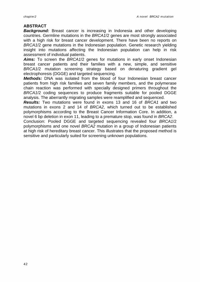
chapter2 A novel BRCA2 mutation
ABSTRACT Background: Breast cancer is increasing in Indonesia and other developing countries. Germline mutations in the BRCA1/2 genes are most strongly associated with a high risk for breast cancer development. There have been no reports on BRCA1/2 gene mutations in the Indonesian population. Genetic research yielding insight into mutations affecting the Indonesian population can help in risk assessment of individual patients. Aims: To screen the BRCA1/2 genes for mutations in early onset Indonesian breast cancer patients and their families with a new, simple, and sensitive BRCA1/2 mutation screening strategy based on denaturing gradient gel electrophoresis (DGGE) and targeted sequencing. Methods: DNA was isolated from the blood of four Indonesian breast cancer patients from high risk families and seven family members, and the polymerase chain reaction was performed with specially designed primers throughout the BRCA1/2 coding sequences to produce fragments suitable for pooled DGGE analysis. The aberrantly migrating samples were reamplified and sequenced. Results: Two mutations were found in exons 13 and 16 of BRCA1 and two mutations in exons 2 and 14 of BRCA2, which turned out to be established polymorphisms according to the Breast Cancer Information Core. In addition, a novel 6 bp deletion in exon 11, leading to a premature stop, was found in BRCA2. Conclusion: Pooled DGGE and targeted sequencing revealed four BRCA1/2 polymorphisms and one novel BRCA2 mutation in a group of Indonesian patients at high risk of hereditary breast cancer. This illustrates that the proposed method is sensitive and particularly suited for screening unknown populations.
42

chapter2 A novel BRCA2 mutation
INTRODUCTION Breast cancer is the most common malignancy in women in the Western world, accounting for 32% of all female cancers, and is responsible for 18% of cancer deaths in women. The hereditary form of breast cancer constitutes about 5% of breast cancer cases overall [1]. The first major gene responsible for hereditary breast cancer susceptibility, BRCA1, was mapped to 17q21 in 1990 [2] and was cloned in 1994 [3]. A second gene involved in the hereditary breast cancer syndrome, BRCA2, was identified several months later.4 Both genes have long coding sequences and complex genomic structures: BRCA1 comprises 5592 bp, composed of 22 coding exons that encode 1863 amino acids [3], and BRCA2 comprises 10443 bp, with 26 coding exons that encode 3418 amino acids [4]. In addition to breast cancer, ovarian cancer [5] fallopian tube cancer [6] and primary peritoneal cancer are part of the BRCA1/2 cancer spectrum in women. ‘‘By identifying the endemic Indonesian mutations, we hope to offer better risk assessment for women in the Indonesian population who are susceptible to the BRCA1/ 2 related hereditary cancers’’ Over 80% of families with two or more cases of premenopausal breast cancer and two or more cases of ovarian cancer are believed to carry a germline BRCA1 or BRCA2 mutation [7, 8]. Comprehensive evaluation of breast cancer susceptibility genes can be used to provide women with information concerning their risk of developing cancer, and guide the decision on the necessity of preventive surgical measures. It has been shown that ethnically different populations exhibit different germline mutation spectra in the BRCA1 and BRCA2 genes [9-11]. There are no previous publications on BRCA1/2 mutation detection in the Indonesian population. By identifying the endemic Indonesian mutations, we hope to offer better risk assessment for women in the Indonesian population who are susceptible to the BRCA1/2 related hereditary cancers. There are no exact data on breast cancer incidence in Indonesia, but a study by Soeripto et al in 1982 (unpublished results) showed that the age standardized incidence rate in the Jogjakarta region was 6.17, ranking second after cervical cancer (7.69). One of the central themes in cancer risk assessment is the efficiency and accuracy of genetic screening methods. Currently, the ability to conduct large scale, population based studies is constrained by the lack of an accurate and inexpensive method for mutation detection. The large size of the BRCA1 and BRCA2 genes, and the scattered distribution of mutations throughout the genes, complicate the task of mutation detection and make rapid screening for mutations a major technical challenge. A technique that scans stretches of DNA for unknown mutations should be optimised to detect as close to 100% of the sequence alteration as possible. Here, we describe the results of the application of a recently developed rapid and sensitive method for the detection of BRCA1/2 mutations [12] based on denaturing gradient gel electrophoresis (DGGE) [13] and targeted sequencing on an Indonesian group of high risk patients. This method appears to be particularly suited for the analysis of unknown populations.
43

chapter2 A novel BRCA2 mutation
MATERIALS AND METHODS Patients From 1996 to 1998, four Indonesian families were selected from patient pools at three hospitals in Jogjakarta, Indonesia; three with hereditary breast cancer based on Moller’s criteria [14], and one with one early onset patient without a family history. Figure 1 shows the pedigrees of these four families. Information regarding potential risk factors for breast cancer was obtained through a structured face to face interview. Information on family history (up to the date of diagnosis of the patient’s breast cancer) was elicited by asking each patient to identify all first and
44

chapter2 A novel BRCA2 mutation
second degree female blood relatives. For each identified relative, the interviewer then asked the year of birth, vital status, year of death (if applicable), history, and type of cancer (if any). The patients were interviewed for family history using standardised questionnaires and blood was taken after informed consent. DNA amplification DNA was isolated from peripheral blood using simple and rapid genomic DNA extraction. Two types of buffer—cell membrane lysis buffer, containing sucrose and triton X-100, and nuclear membrane lysis buffer, containing guanidine thiocyanate, sodium N-lauroyl sarcocinate, and b mercaptoethanol—were used. By using this method, only a small amount of peripheral blood (0.5 ml) was needed. The DNA was stored at 4˚C. Polymerase chain reaction (PCR) fragments suitable for DGGE analysis were designed based on the theoretical melting profile of each exon, as described previously.12 A 40 mer GC clamp was attached to the short oligonucleotide to obtain optimal melting profiles. Tables 1 and 2 provide the PCR primer sequences for BRCA1 and BRCA2, respectively. Genomic DNA was amplified using 100–200 ng of template DNA, 10 pmol of the mixture of 60 mer primers, 1mM MgCl, and 1 U Platinum Taq in 50 ml PCR reactions. The amplification conditions in a Robocyler (Stratagene, Canada) were five minutes at 94˚C (hot start), followed by five cycles of one minute at 94˚C, one minute at 52˚C, and one minute at 72˚C; five cycles of one minute at 94˚C, one minute at 50˚C, and one minute at 72˚C; and 30 cycles of one minute at 94˚C, one minute at 48˚C, and one minute at 72˚C; with five minutes extra at 72˚C. The final incubation was followed by five minutes at 94˚C and 15 minutes at 50˚C to enhance the formation of heteroduplex molecules. Denaturing gradient gel electrophoresis Primers for DGGE were obtained from Ingeny (Goes, the Netherlands). Aliquots (4–6 ml) of the PCR products were mixed with 2 ml of standard dye loading buffer and electrophoresed through a 20 cm 9% polyacrylamide gel (acrylamide/bisacrylamide, 37.5/1) containing a linear 20– 65% denaturing gradient (100% UF: 7M urea/40% deionised formamide). A 12% polyacrylamide stacking gel was poured to create solid slots for efficient loading of the PCR products, which prevented difficulties caused by the high urea concentration. Electrophoresis was performed in 0.5 Tris acetate EDTA buffer at 58˚C for 16 hours [15]. Gels were stained with ethidium bromide and photographed under an ultraviolet transilluminator. To increase the throughput of the DGGE procedure, three to four different amplicons with relatively large melting temperature differences were pooled. The fragment pool was designed using a computer program that was based around melting profiles and sequences (tables 3 and 4). For each different exon, seven samples from four different families were loaded side by side to compare their band patterns. The aberrantly migrating samples were reamplified using sequencing primers and sequencing was performed using the Big Dye cycle sequencing kit according to the manufacturer’s instructions. The reaction products were analysed using an ABI DNA sequencer 310 and sequence files were edited using the Sequence Navigator program.
45

chapter2 A novel BRCA2 mutation
46

chapter2 A novel BRCA2 mutation
47

chapter2 A novel BRCA2 mutation
RESULTS In the 11 patients, aberrantly migrating bands (fig 2) were found in exons 13 and 16 of the BRCA1 gene, and in exons 2, 11, and 14 of the BRCA2 gene (table 5). We then sequenced the exons with aberrantly migrating bands (fig 3). The aberrantly migrating bands found in exons 13 and 16 of BRCA1 and exons 2 and 14 of BRCA2 appeared to be caused by single nucleotide substitutions producing the same amino acid. Consultation with the Breast Cancer Information Core (last accessed 1 February 2005; http://www.research.nhgri.nih.gov/projects/bic) revealed that these substitutions were indeed established polymorphisms. We detected one deleterious BRCA2 mutation in patient BIII-5 (fig 4). This mutation was a deletion of six nucleotides in exon 11 (c.2472delTAAATG) at codon 824. The mutation did not cause a shift of the reading frame, but affected three codons and resulted in a premature stop codon. There is no such mutation registered in the Breast Cancer Information Core (http://www.research.nhgri.nih.gov/projects/bic), so this is a novel mutation. This patient had early onset breast cancer and there was no family history of cancer. Table 5 summarises the genetic aberrations found, and table 6 shows the clinicopathological data of the patients with breast cancer.
Figure 2. Pooling of five samples of 24 exons in BRCA1 gene for DGGE. Each pool consist of three to four amplicons selected based on their melting behaviour. The fragment for each pool can be seen in Table 3. The aberrant bands appeared in fragment 9 (in Pool B), fragment 16B (in Pool C), fragment 2A and 16A (in Pool E), fragment 13 (in Pool F) and fragment 3 (in Pool G), and fragment 11-15 (in Pool H) were further analysed by sequencing.
48

chapter2 A novel BRCA2 mutation
Figure 3. Results of DGGE analysis of BRCA1 DGGE of Indonesian patients at high risk for hereditary breast cancer. Exon 16, Lanes 1, 4 and 6 display a darker single band as opposed to samples in lanes 2, 3, 5 and 7 which showed lighter double bands. The dark band sat in the same position as one of the double bands. Exon 13 was similar: lanes 8, 11 and 13 displayed a dark single band, whereas lanes 9, 10, 12 and 14 showed light double bands. These aberrant bands appeared to represent single nucleotide substitutions with no consequences for the amino acid sequence of the protein (polymorphisms). DISCUSSION The increasing demand for mutation detection in disease genes, either known or presumed, can be solved by automated sequencing using fluorescent dyes [16]. However, only a few laboratories are equipped for the broad application of this costly and labour intensive strategy. As alternatives to sequencing methods, which determine the exact nature and location of each base along a DNA fragment, various mutation scanning procedures have been developed. These methods, which rely on the recognition of a sequence variation between mutant and wild-type DNA on the basis of an altered electrophoretic migration pattern, provide a simple means for determining whether a given DNA sample harbours a mutation in a particular gene.
igure 4. Sequencing graphs of exon 11 of BRCA2 of a 30 years old early onset breast cancer patient
The most well established scanning procedures are single strand
Fshowing the 2699delTAAATG mutation on the left with the normal control on the right. conformational polymorphism (SCCP) analysis, DGGE, chemical cleavage of mismatch, RNase cleavage, the protein truncation test (PTT), and heteroduplex analysis. Among these methods, SCCP, DGGE, PTT, and heteroduplex analysis are the most widely used because of their accuracy, simplicity, lack of toxicity,
49

chapter2 A novel BRCA2 mutation
and/or relative affordability. We focused on DGGE because this method, when optimised, has the highest mutation detection rate (close to 100% [17]) compared with SCCP and heteroduplex analysis. Additional advantages of this methodology are the possibility of optimising the analysis by computer simulation and the non-radioactive approach. In DGGE, during electrophoresis, double stranded DNA amplified by PCR migrates through a gel containing an increasing concentration of denaturant (urea and formamide). As double stranded DNA is electrophoresed through the denaturing gradient, it will melt and change its conformation in such a way that the mobility of the molecule is dramatically reduced. To prevent complete strand dissociation and to facilitate the detection of mutations in the higher melting domains, a GC rich fragment (GC clamp) is introduced during fragment amplification. The GC clamp increases the percentage of single base changes detectable by DGGE, theoretically to 100% [18, 19]. In principle, four bands are detectable in a heterozygous state after denaturation and renaturation, corresponding to the two homodimers and two heterodimers. Although DGGE has been applied to BRCA mutation screening before [20-22] we used a new technique based on DGGE after exon by exon PCR amplification of the complete BRCA1 and BRCA2 sequences [12], and sequenced aberrantly moving bands. This yielded four polymorphisms consisting of single nucleotide substitutions, underlining the sensitivity of the method. Intelligent pooling of the amplicons before electrophoresis greatly improved the throughput of the method.
Double gradient DGGE is based on the combination of two linear gradients, a
primary denaturing gradient (urea and formamide) and a collinear secondary porosity gradient (polyacrylamide) [23]. This secondary gradient suppresses band broadening during electrophoresis and thus improves the resolution of the DGGE banding pattern. Hayes et al compared double gradient DGGE gels with various porosity gradients to a standard 9% polyacrylamide gel, and showed that mutations with different melting profiles cannot be appropriately detected using a single DGGE condition [15]. PTT is a widely applied screening technique [5], but is especially helpful for known mutations in particular populations, and less suitable as a primary screening approach for new mutations in unknown populations. Furthermore, PTT only detects mutations that result in stop codons and lead to premature termination of translation, thereby producing truncated proteins. A possible advantage of PTT is that it conveniently misses harmless polymorphisms. PTT is usually only applied for detecting mutations in exon 11 of the BRCA1 gene and exons 10 and 11 of the BRCA2 gene, which account for more than 60% of the
50

chapter2 A novel BRCA2 mutation
coding sequence. However, mutations are distributed throughout the entire coding sequence, with no apparent clustering or hot spots [24]. ‘‘Intelligent pooling of the amplicons before electrophoresis greatly improved the throughput of the method’’
More than 1893 distinct germline BRCA2 mutations have been found to date
(http://www.research.nhgri.nih.gov/projects/bic), and the number is expected to increase further. The pattern of mutations is similar to that seen in BRCA1 and in many other tumour suppressor genes. At present, all mutations that clearly cause disease result in premature termination of translation or absence of a transcript. Approximately 75% of truncating mutations are small deletions, 15% are small insertions, and 10% are base substitutions leading directly to termination codons (http://www.research.nhgri.nih.gov/projects/bic) [25-27]. In addition, the novel 2699delTAAATG mutation described in our present paper leads to a premature stop codon. The patient with this mutation had early onset breast cancer and no family history of cancer. It has been suggested that the probability of harbouring a BRCA2 mutation among patients with early onset breast cancer is relatively independent of a positive family history for the disease [25]. Mutations in the BRCA2 gene have been found mainly in families with a high
, we describe a novel BRCA2 mutation and four polymorphisms in
CKNOWLEDGEMENTS 01-008 of the Dutch Cancer Society. We thank Dr J
Hilgers who was instrumental in setting up the Familial Cancer Clinic initiative in Jogjakarta.
incidence of female and male breast cancer [4, 26, 28, 29], whereas the risk of ovarian cancer is lower than in BRCA1 families [4, 26, 30, 31]. Loss of heterozygosity has been found in 30–45% of sporadic breast tumours [32, 33], indicating a role for BRCA2 also in sporadic breast tumours, although very few somatic mutations have been reported [24, 34, 35]. Other tumour types seen repeatedly in BRCA2 families include those of the prostate, larynx, pancreas, and colon [36, 37]. In conclusionthe BRCA1/2 genes found in the Indonesian population using a new high throughput and highly sensitive method to screen BRCA1 and BRCA2 for mutations based on exon by exon PCR amplification followed by pooled DGGE and sequencing of aberrant bands. This is an ideal approach for screening populations for unknown mutations, and has the ability to detect single base differences using non-toxic and relatively simple and inexpensive methods. ASupported by grant IN-20
51

chapter2 A novel BRCA2 mutation
REFERENCES 1. Jardines L HB, Doroshow JH, Fisher P, Weitzel J, Theriault RL: Breast cancer overview, in
ment: A Multidiciplinary approach; 2001. , Newman B, Morrow JE, Anderson LA, Huey B, King MC: Linkage of early-onset
C,
6. H,
er
10.
ner
ncer
,
Cancer Manage. Hall JM, Lee MK2
familial breast cancer to chromosome 17q21. Science 1990, 250(4988):1684-1689. 3. Miki Y, Swensen J, Shattuck-Eidens D, Futreal PA, Harshman K, Tavtigian S, Liu Q, Cochran
Bennett LM, Ding W et al: A strong candidate for the breast and ovarian cancer susceptibility gene BRCA1. Science 1994, 266(5182):66-71. Wooster R, Bignell G, Lancaster J, Swift S, Seal S, Mangion J, Collins N, Gregory S, Gu4. mbs C, Micklem G: Identification of the breast cancer susceptibility gene BRCA2. Nature 1995, 378(6559):789-792. Zweemer RP, Verheijen RH, Gille JJ, van Diest P5. J, Pals G, Menko FH: Clinical and genetic evaluation of thirty ovarian cancer families. Am J Obstet Gynecol 1998, 178(1 Pt 1):85-90. Zweemer RP, van Diest PJ, Verheijen RH, Ryan A, Gille JJ, Sijmons RH, Jacobs IJ, Menko FKenemans P: Molecular evidence linking primary cancer of the fallopian tube to BRCA1germline mutations. Gynecol Oncol 2000, 76(1):45-50. Narod SA, Ford D, Devilee P, Barkardottir RB, Lynch HT, Smith SA, Ponder BA, Weber BL, Ga7. rbJE, Birch JM et al: An evaluation of genetic heterogeneity in 145 breast-ovarian cancer families. Breast Cancer Linkage Consortium. Am J Hum Genet 1995, 56(1):254-264. Pathology of familial breast cancer: differences between breast canc8. ers in carriers of BRCA1or BRCA2 mutations and sporadic cases. Breast Cancer Linkage Consortium. Lancet 1997, 349(9064):1505-1510. Liede A, Narod SA: Hereditary breast and ovarian cancer in Asia: genetic epidemiolo9. gy of BRCA1 and BRCA2. Hum Mutat 2002, 20(6):413-424.
Neuhausen SL: Ethnic differences in cancer risk resulting from genetic variation. Cancer 1999, 86(11 Suppl):2575-2582.
11. Verhoog LC, van den Ouweland AM, Berns E, van Veghel-Plandsoen MM, van Staveren IL, WagA, Bartels CC, Tilanus-Linthorst MM, Devilee P, Seynaeve C et al: Large regional differences in the frequency of distinct BRCA1/BRCA2 mutations in 517 Dutch breast and/or ovarian cafamilies. Eur J Cancer 2001, 37(16):2082-2090.
12. van der Hout AH MI, Wu Y, van der Vlies P, Huisman M, Oosterwijk JC, Buys CHCM, Hoftra RMW: A DGGE system for comprehensive mutation screening of the complete coding regions of BRCA1 and BRCA2 outside exon 11. Am J Hum Genet 1999, 65(suppl S Oct):2322. Fischer SG, Lerman LS: DNA fragments differin13. g by single base-pair substitutions are separated in denaturing gradient gels: correspondence with melting theory. Proc Natl Acad Sci U S A 1983, 80(6):1579-1583. Moller P, Evans G, Haites N, Vasen H, Reis MM, Anderson E, Apold J, Hodgson S, Ecc14. les DOlsson H et al: Guidelines for follow-up of women at high risk for inherited breast cancer: consensus statement from the Biomed 2 Demonstration Programme on Inherited Breast Cancer. Dis Markers 1999, 15(1-3):207-211.
15. Hayes VM, Wu Y, Osinga J, Mulder IM, van der Vlies P, Elfferich P, Buys CH, Hofstra RM: Improvements in gel composition and electrophoretic conditions for broad-range mutationanalysis by denaturing gradient gel electrophoresis. Nucleic Acids Res 1999, 27(20):e29. Rosenthal A, Sproat B, Voss H, Stegemann J,16. Schwager C, Erfle H, Zimmermann J, Coutelle C, Ansorge W: Automated sequencing of fluorescently labelled DNA by chemical degradation. DNA Seq 1990, 1(1):63-71. Macek M, Jr., Mercier B, Mackova A, Miller PW, Hamosh A, Ferec C, Cutting GR: Sensitivity o17. f the denaturing gradient gel electrophoresis technique in detection of known mutations and novel Asian mutations in the CFTR gene. Hum Mutat 1997, 9(2):136-147. Abrams ES, Murdaugh SE, L18. erman LS: Comprehensive detection of single base changes in human genomic DNA using denaturing gradient gel electrophoresis and a GC clamp. Genomics 1990, 7(4):463-475. Sheffield VC, Cox DR, Lerman LS, Myers RM: Attachment of a 40-base-pair19. G + C-rich sequence (GC-clamp) to genomic DNA fragments by the polymerase chain reaction results in improved detection of single-base changes. Proc Natl Acad Sci U S A 1989, 86(1):232-236.
52

chapter2 A novel BRCA2 mutation
20. Muller D, Bonaiti-Pellie C, Abecassis J, Stoppa-Lyonnet D, Fricker JP: BRCA1 testing in band/or ovarian cancer families from northeastern France identifies two common mutations with a founder effect. Fam Cancer 2004, 3(1):15-20.
reast
sh and non-Jewish carriers. Fam Cancer
22. ast
23. , Zunino F, Della Torre G, Pierotti MA, Righetti PG: Detection of p53 point ,
tilla
ity gene. Implications for presymptomatic testing and screening. Jama
25. ed
n
net 1996, 13(1):123-125.
, Moslehi
enet 1996, 12(3):333-337. s of 308.
31. odels for familial ovarian
32.
ins ,
36. BRCA2 mutation carriers.The Breast Cancer Linkage Consortium. J Natl
37. BA, Easton DF, Pharoah PD: BRCA1 es
21. Gal I, Gershoni Baruch R, Haber D, Dagan E, Eisenberg-Barzilai S, Zidan J, Friedman E: The 1100delAT BRCA1 and the 8765delAG BRCA2 mutations: occurrence in high-risk non-Ashkenazi Jews and haplotype comparison of Jewi2004, 3(1):11-14. Atoum MF, Al-Kayed SA: Mutation analysis of the breast cancer gene BRCA1 among brecancer Jordanian females. Saudi Med J 2004, 25(1):60-63. Gelfi C, Righetti SCmutations by double-gradient, denaturing gradient gel electrophoresis. Electrophoresis 199718(15):2921-2927.
24. Shattuck-Eidens D, McClure M, Simard J, Labrie F, Narod S, Couch F, Hoskins K, Weber B, CasL, Erdos M et al: A collaborative survey of 80 mutations in the BRCA1 breast and ovarian cancer susceptibil1995, 273(7):535-541. Plaschke J, Commer T, Jacobi C, Schackert HK, Chang-Claude J: BRCA2 germline mutationsamong early onset breast cancer patients unselected for family history of the disease. J MGenet 2000, 37(9):E17.
26. Couch FJ, Farid LM, DeShano ML, Tavtigian SV, Calzone K, Campeau L, Peng Y, Bogden B, CheQ, Neuhausen S et al: BRCA2 germline mutations in male breast cancer cases and breast cancer families. Nat Ge
27. Teng DH, Bogden R, Mitchell J, Baumgard M, Bell R, Berry S, Davis T, Ha PC, Kehrer R, Jammulapati S et al: Low incidence of BRCA2 mutations in breast carcinoma and other cancers. Nat Genet 1996, 13(2):241-244.
28. Phelan CM, Lancaster JM, Tonin P, Gumbs C, Cochran C, Carter R, Ghadirian P, Perret CR, Dion F et al: Mutation analysis of the BRCA2 gene in 49 site-specific breast cancer families. Nat Genet 1996, 13(1):120-122.
29. Tavtigian SV, Simard J, Rommens J, Couch F, Shattuck-Eidens D, Neuhausen S, Merajver S, Thorlacius S, Offit K, Stoppa-Lyonnet D et al: The complete BRCA2 gene and mutations in chromosome 13q-linked kindreds. Nat G
30. Prevalence and penetrance of BRCA1 and BRCA2 mutations in a population-based seriebreast cancer cases. Anglian Breast Cancer Study Group. Br J Cancer 2000, 83(10):1301-1Antoniou AC, Gayther SA, Stratton JF, Ponder BA, Easton DF: Risk mand breast cancer. Genet Epidemiol 2000, 18(2):173-190. Cleton-Jansen AM, Collins N, Lakhani SR, Weissenbach J, Devilee P, Cornelisse CJ, Stratton MR: Loss of heterozygosity in sporadic breast tumours at the BRCA2 locus on chromosome 13q12-q13. Br J Cancer 1995, 72(5):1241-1244.
33. van den Berg J, Johannsson O, Hakansson S, Olsson H, Borg A: Allelic loss at chromosome 13q12-q13 is associated with poor prognosis in familial and sporadic breast cancer. Br J Cancer 1996, 74(10):1615-1619.
34. Lancaster JM, Wooster R, Mangion J, Phelan CM, Cochran C, Gumbs C, Seal S, Barfoot R, CollN, Bignell G et al: BRCA2 mutations in primary breast and ovarian cancers. Nat Genet 199613(2):238-240.
35. Miki Y, Katagiri T, Kasumi F, Yoshimoto T, Nakamura Y: Mutation analysis in the BRCA2 gene in primary breast cancers. Nat Genet 1996, 13(2):245-247. Cancer risks inCancer Inst 1999, 91(15):1310-1316. Basham VM, Lipscombe JM, Ward JM, Gayther SA, Ponderand BRCA2 mutations in a population-based study of male breast cancer. Breast Cancer R2002, 4(1):R2.
53

chapter2 A novel BRCA2 mutation
54

Chapter 3 BRCA1 and BRCA2 germline mutation analysis in the
Indonesian population
Submitted for publication
Dewajani Purnomosari 1, Gerard Pals 5, Artanto Wahyono 2 , Teguh Aryandono 2, Tjakra W Manuaba 3, Samuel J Haryono 4, Paul J van Diest 6

chapter 3 BRCA1 and BRCA2 germline mutation analysis
ABSTRACT Specific mutations in BRCA1 and BRCA2 genes have been identified in specific populations and ethnic groups. However, little is known about the contribution of BRCA1 and BRCA2 mutations to breast cancers in the Indonesian population. One hundred-twenty moderate to high risk breast cancer patients were tested using PCR-DGGE, and any aberrant band was sequenced. Multiplex ligation-dependent probe amplification (MLPA) was performed on all samples to detect large deletions in the two genes. Twenty-three different mutations were detected in 30 individuals, ten were deleterious mutations and 20 were “unclassified variants” with uncertain clinical consequences. Three of seven (c.2784_2875insT, p.Leu1415X and del exon 13-15) and two of four (p.Glu2183X and p.Gln2894X) deleterious mutations that were found in BRCA1 and BRCA2 respectively, are novel. Several novel, pathogenic BRCA1 and BRCA2 germline mutations are found in early onset Indonesian breast cancer patients, these may therefore be specific for the Indonesian population.
56

chapter 3 BRCA1 and BRCA2 germline mutation analysis
INTRODUCTION Breast cancer is the most common cancer in women. In 5% to 10% of breast cancer cases, the disease results from a hereditary predisposition [1, 2], which can to a large extent be attributed to mutations in either of two tumour suppressor genes, BRCA1 (MIM# 113705) and BRCA2 (MIM# 600185) [3-5]. These genes are involved in DNA repair as well as transcriptional regulation [6, 7]. Women carrying pathogenic germline mutations in either of these genes tend to develop breast cancer at an early age [8, 9]. The BRCA1 and BRCA2 genes encode large proteins of 1863 and 3418 amino acids, respectively. Over 300 distinct mutations in BRCA1 and BRCA2 have been described [10, 11]. These mutations are widely scattered across both genes and most affect the structure and function of the gene. Nevertheless, a significant proportion (34% of BRCA1 and 38% of BRCA2 mutations) (http://www.nhgri.nih.gov/Intramural_research/Lab_transfer/Bic) are missense mutations that alter one amino acid, but do not truncate the protein and are rare sequence variants of unknown functional consequence. Moreover, a number of base substitutions do not alter the amino acid sequence or result in amino acid changes not associated with disease (polymorphisms) [12]. Hence the biggest challenge in interpreting the mutation analysis of BRCA1 and BRCA2 genes is to distinguish between harmless polymorphisms and deleterious mutations associated with increased cancer risk. In addition, mutations specific for certain populations and ethnic groups have been identified in both genes. For example, specific BRCA1 and BRCA2 mutations were reported for Ashkenazi Jews [13]. Other common BRCA1 mutations were especially found in Italian, Canadian, Belgian or Dutch breast cancer families [14-16]. In Indonesia, the contribution of the BRCA1/BRCA2 mutations to the population incidence of early-onset breast cancer is largely unknown. In one pilot study, however, a new BRCA2 mutation was identified [17] indicating that it was worthwhile to more extensively study the Indonesian population, which was the aim of this study. The accumulating knowledge about the prevalence and nature of BRCA1 and BRCA2 mutations in specific populations may facilitate the interpretation of genetic analysis with regard to breast cancer risk of individual patients. MATERIALS AND METHODS Patients A total of 120 unrelated breast cancer patients and 16 of their family members from three Indonesian cities (Jakarta and Jogjakarta on the Java island, Denpasar on the Bali island) were analyzed. Breast cancer patients at moderate to high-risk of a hereditary predisposition were selected according to the following criteria: A. Breast cancer before the age of 41 (n=102); B. Two cases of breast cancer in the same family before the age of 60 (n=9); C. Three or more cases of breast cancer in the same family (n=2); D. Bilateral breast cancer (n=7). Subjects were asked to fill out questionnaires to evaluate their personal and family histories, and blood specimens were collected for determination of BRCA mutations. Informed consent was obtained from all the subjects in this study.
57

chapter 3 BRCA1 and BRCA2 germline mutation analysis
DNA extraction and PCR amplification Genomic DNA was isolated by the saturated salt extraction procedure as described in [18]. All 22 coding exons of BRCA1 and 26 coding exons of BRCA2 were amplified using primer sequences developed by the University of Groningen, The Netherlands [19]. Primers for DGGE were obtained from Ingeny (Goes, The Netherlands). Genomic DNA was amplified using 100 – 200 ng of template DNA, 10 pmol of the mixture of 40-mer primers, 30 mM of MgCl, 3 mM dNTPs (Invitrogen) and 0.7 unit of Platinum Taq (Invitrogen) in 9 µl PCR reactions. In order to speed up the test, the PCR reaction was placed in 384 well plates using a pipetting robot (TECAN Miniprep 75). PCR conditions were performed as previously described [17]. Denaturing Gradient Gel Electrophoresis and DNA sequencing A 4 – 6 µl aliquot of each PCR product with relatively large melting temperature differences were pooled as previously described [17] with some modifications for optimal results. The fragment pool was designed based on melting profiles and sequence. Electrophoresis was performed in 0.5 TAE buffer at 58°C, 120 V for 16 hours for BRCA1 gene, and 55°C, 100V for 18 hours for BRCA2. Gels were stained with ethidium bromide and photographed under a UV transilluminator. The aberrantly migrating samples were re-amplified using sequencing primers and sequencing was performed using Big Dye Cycle-sequencing kit according to the manufacturer’s instructions. The reaction products were analyzed using an ABI 3100 DNA Sequencer (Applied Biosystems, Torrence, CA, USA) and sequence files were edited using the Bio Edit program. The classification of gene alterations was performed in accordance with the entries in the Breast Cancer Information Core (BIC, Bethesda, MD). Multiplex ligation-dependent probe amplification (MLPA) The principle of the MLPA technique has been described elsewhere [20]. The MLPA test for BRCA1 (P02) and BRCA2 (P45) mutations were obtained from MRC-Holland, Amsterdam, The Netherlands. The fragments were analyzed on an ABI model 310 capillary sequencer (Applied Biosystems, Torrence, CA, USA) using Genescan-TAMRA 500 size standards (Applied Biosystems). Fragment analysis was performed with Genescan software. RESULTS AND DISCUSSIONS We identified 120 incident Indonesian breast cancer cases diagnosed before the age of 41 years, or having family history of breast cancer, or harboring bilateral breast cancer during September 1999 – April 2005 (Jogjakarta) and during July 2004 – April 2005 (Jakarta and Denpasar). In addition, 16 of their family members were analyzed. The entire coding regions and exon-intron junctions of BRCA1 and BRCA2 were screened in these 136 persons of breast cancer patients and their families using PCR-DGGE (Figure 1) followed by sequencing (Figure 2) for samples with aberrant migrating bands. To optimize the screening, MLPA, a relatively new technique, was also performed in all samples (Figure 3). Here, we report on 116/120 women (96.7%) for whom BRCA1/2 analysis were completed. The
58

chapter 3 BRCA1 and BRCA2 germline mutation analysis
remaining 4 patients (all from group A) had to be excluded due to the small amount of extracted DNA that did not allow complete screening of the BRCA1 and BRCA2 genes. BRCA1 and BRCA2 pathogenic mutations The analysis of 116 unrelated breast cancer patients with breast cancer revealed that 9 patients (7.8%) carried pathogenic germline mutations especially the early onset patients: 3 within BRCA1 (2.6%) and 6 within BRCA2 (5.2%) which is comparable to previous studies [21]. We only found BRCA1 and BRCA2 mutations in groups A (“early onset”, n=7 out of 98, 7.1%) and B (two cases of breast cancer in the same family before the age of 60, n=2 out of 9 (22.2%)) (Table 1). There were twice as many BRCA2 mutations as BRCA1 mutations. Although the absolute numbers are low and no firm conclusions can therefore be drawn, this is comparable to other Asian regions [22-24] but seems to discern the Indonesian population from non-Asian ethnic groups where the reverse trend is seen.
Figure 1. DGGE analysis of fragments 11.15g, 11.4 and 11.10 of the BRCA2 gene in ten unrelated breast cancer patients. The arrows show altered band mobility compare to other patients. Seven pathogenic mutations were found in nine probands: three in BRCA1 (c.2784_2785insT, pL1415X (c.4361_4362insT), del exon 13-15) and four in BRCA2 (c.3040_3043delGCAA, p.Glu2183X (c.6775G>T), p.Leu824X (c.2699_2704delTAAATG), p.Gln2894X (c.9008C>T)). All these mutations were classified as pathogenic as they are predicted to result in protein truncation. The three pathogenic mutations found in BRCA1 were not previously reported in the BIC database as well as two novel nonsense mutations (p.Glu2183X and p.Gln2894X) identified in BRCA2. The p.Glu2183X mutation was found in 2 related patients that had breast cancer above the age of 60. One of seven pathogenic mutations found in BRCA1 and BRCA2 showed a significant clinical impact on the patient (Table 2). Patient AE with a one nucleotide insertion (Thymine) between nucleotide 2784 and 2785 (c.2784_2785insT) in exon 11 of BRCA1 suffered from bilateral breast cancer at a relatively early age (25 years). The insertion leads to frameshift and creates a premature stop codon in
59

chapter 3 BRCA1 and BRCA2 germline mutation analysis
exon 11. The mutation takes place in the sequence within BRCA1 encoding for aminoacids 758-1064 which interact with RAD51 protein that is required for homologous recombination (HR) repair of double strand breaks (DSBs) [25], which is one of the most important functions of the BRCA1 protein. This patient presented in a late stage (stage III for both breasts) and only survived for 9 weeks after treatment. Her mother did not carry this mutation. Although her father may be carrier, the mutation is probably de novo as there was no family history of breast or other cancers.
A
B
Figure 2. Sequence electropherogram of a normal individual showing (A) wild-type BRCA2 exon 11 sequence and (B) of breast cancer patient (B-3-5) showing c.2699_2704delTAAATG mutation.
Figure 3. MLPA analysis of BRCA1 gene of patient sample (blue) compare to the normal control (red). X and Y axis represent peak size and peak height respectively. There are reduced peaks in the patient sample compared to the normal control in exons 13, 14 and 15 (arrows) indicating deletions.
60

chapter 3 BRCA1 and BRCA2 germline mutation analysis
Table 1. BRCA1 or BRCA2 germline mutations in Indonesian women with early onset breast cancer.
Patient Age1 gene Exon Mutation2 mutation type Pathogenic
mutation BIC3
AE 25 BRCA1 11 c.2784_2785insT frameshift + no
B10 31 BRCA1 13 p.Leu1415X nonsense + no
AA 40 BRCA1 13-15 - 4large
rearrangement + no
AB 34 BRCA2 11 c.3040_3043del4 frameshift + 1
B5 66 BRCA2 11 p.Glu2183X nonsense + no
B6 65 BRCA2 11 p.Glu2183X nonsense + no
B-III-5 30 BRCA2 11 p.Leu824X nonsense + no
AZ 40 BRCA2 11 p.Leu824X nonsense + no
W-II 37 BRCA2 21 p.Gln2894X nonsense + no
Q-II 40 BRCA1 2 c.101-10T>C IVS + 6
P-III-19 19 BRCA1 9 p.Val191Ile Missense + 6
J22 32 BRCA1 11 p.Leu1209Val Missense ? No
AZ 40 BRCA1 16 p.Met1652Ile Missense + 35
B1 24 BRCA1 20 c.5313-31A>G IVS ? No
B7 31 BRCA1 24 p.Arg1835Gln Missense ? No
216 33 BRCA1 24 p.Thr1852Ile Missense ? No
P-III-19 19 BRCA2 5 p.Gln147Arg Missense + 6
B3 24 BRCA2 10 p.Gln609Glu Missense ? no
C-II-7 39 BRCA2 11 p.Met1149Val Missense + 5
AO 28 BRCA2 11 p.Met1149Val Missense + 5
AQ 44 BRCA2 11 p.Met1149Val Missense + 5
BH 38 BRCA2 11 p.Met1149Val Missense + 5
172 36 BRCA2 11 p.Gln699Leu Missense ? no
J32 29 BRCA2 11 p.Arg2108Cys Missense + 16
J6 33 BRCA2 11 p.Val950Ile Missense ? No
206 37 BRCA2 25 c.9485-16T>C IVS + 4
BC 35 BRCA2 27 p.Ile3412Val Missense + 109
166 33 BRCA2 27 p.Ile3412Val Missense + 109
J24 35 BRCA2 27 p.Ile3412Val Missense + 109
206 37 BRCA2 27 p.Lys3326X nonsense + 289
1 Age at time of diagnosis 2 Gen Bank Accession number, BRCA1: U14680, BRCA2: U43746 3 number of times reported in BIC 4 not determined, detected by MLPA
61
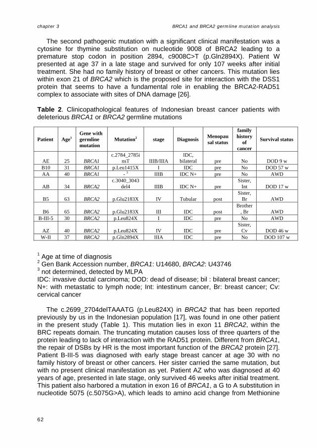
chapter 3 BRCA1 and BRCA2 germline mutation analysis
The second pathogenic mutation with a significant clinical manifestation was a cytosine for thymine substitution on nucleotide 9008 of BRCA2 leading to a premature stop codon in position 2894, c9008C>T (p.Gln2894X). Patient W presented at age 37 in a late stage and survived for only 107 weeks after initial treatment. She had no family history of breast or other cancers. This mutation lies within exon 21 of BRCA2 which is the proposed site for interaction with the DSS1 protein that seems to have a fundamental role in enabling the BRCA2-RAD51 complex to associate with sites of DNA damage [26]. Table 2. Clinicopathological features of Indonesian breast cancer patients with deleterious BRCA1 or BRCA2 germline mutations
Patient Age1Gene with germline mutation
Mutation2 stage Diagnosis Menopausal status
family history
of cancer
Survival status
AE 25 BRCA1 c.2784_2785i
nsT IIIB/IIIA IDC,
bilateral pre No DOD 9 w B10 31 BRCA1 p.Leu1415X I IDC pre No DOD 57 w AA 40 BRCA1 - 3 IIIB IDC N+ pre No AWD
AB 34 BRCA2 c.3040_3043
del4 IIIB IDC N+ pre Sister,
Int DOD 17 w
B5 63 BRCA2 p.Glu2183X IV Tubular post Sister,
Br AWD
B6 65 BRCA2 p.Glu2183X III IDC post Brother
, Br AWD B-III-5 30 BRCA2 p.Leu824X I IDC pre No AWD
AZ 40 BRCA2 p.Leu824X IV IDC pre Sister,
Cv DOD 46 w W-II 37 BRCA2 p.Gln2894X IIIA IDC pre No DOD 107 w
1 Age at time of diagnosis 2 Gen Bank Accession number, BRCA1: U14680, BRCA2: U43746 3 not determined, detected by MLPA IDC: invasive ductal carcinoma; DOD: dead of disease; bil : bilateral breast cancer; N+: with metastatic to lymph node; Int: intestinum cancer, Br: breast cancer; Cv: cervical cancer The c.2699_2704delTAAATG (p.Leu824X) in BRCA2 that has been reported previously by us in the Indonesian population [17], was found in one other patient in the present study (Table 1). This mutation lies in exon 11 BRCA2, within the BRC repeats domain. The truncating mutation causes loss of three quarters of the protein leading to lack of interaction with the RAD51 protein. Different from BRCA1, the repair of DSBs by HR is the most important function of the BRCA2 protein [27]. Patient B-III-5 was diagnosed with early stage breast cancer at age 30 with no family history of breast or other cancers. Her sister carried the same mutation, but with no present clinical manifestation as yet. Patient AZ who was diagnosed at 40 years of age, presented in late stage, only survived 46 weeks after initial treatment. This patient also harbored a mutation in exon 16 of BRCA1, a G to A substitution in nucleotide 5075 (c.5075G>A), which leads to amino acid change from Methionine
62

chapter 3 BRCA1 and BRCA2 germline mutation analysis
to Isoleucine, (p.Met16521Ile) which has to date been reported 35 times in BIC as a UV mutation. As the c.2699_2704delTAAATG mutation was found in two unrelated patients, this mutation could be a good candidate as a founder mutation. None of the families with more than 3 cases of breast cancer and families with bilateral breast cancer showed pathogenic mutations in the BRCA1 and BRCA2 genes. Family U had four first-degree relatives that were affected by breast cancer. Two of four members had bilateral breast cancer. In spite of this high familial breast cancer incidence, no BRCA1/2 mutations were found. BRCA 1 and BRCA2 unclassified variants Sixteen (7 BRCA1 and 9 BRCA2) rare mutations of so far unknown significance (“unclassified variants”, UVs) were detected in 18 patients: 13 missense changes and 3 intronic variants. Of these 16 UVs, 7 were novel, whereas the other UVs have been previously reported in the BIC database (Table 1). From the 18 patients which carried UV mutations, two patients were detected in families from group D; one patient in a group B family and the other fifteen patients in families from group A. Seven UV were found in the BRCA1 gene, two mutations occurring in the intronic region between exons 1 and 2 (c.101-10T>C) and between exons 19 and 20 (c.5313-31A>G), and five missense mutations identified: p.Val191Ile (c.690G>A), p.Leu1209Val (c.3744T>G), p.Met1652Ile (c.5075G>A), p.Arg1835Gln (c.5623G>A) and p.Thr1852Ile (c.5674C>T). Four out of seven BRCA1 missense mutations; p.Leu1209Val (c.3744T>G), c.5313-31A>G, p.Arg1835Gln (c.5623G>A) and p.Thr1852Ile (c.5674C>T) were have not been described previously in the BIC. The p.Leu1209Val may not be a significant change as both Leucine and Valine belong to the same group of non polar amino acids. However, p.Arg1835Gln is possibly an important alteration since a positively charged Arginine is replaced by an uncharged Glutamine, which may have an effect on the structure and/or function of the protein. Another potentially important alteration concerns p.Thr1852Ile, where the hydrophilic amino acid Threonine is replaced by a hydrophobic Isoleucine. The sites of mutation of both p.Arg1835Gln and p.Thr1852Ile also have to be considered as they lie within the site for the activation domain of the BRCA1 protein [28]. The intronic UV c.5313-31A>G also deserves further investigation as it may theoretically have an effect on splicing. However, according to splice site finder (http://www. genet.sickkids.on.ca/~ali/splicesitefinder.html), the splicing sites in the wild type and mutant alleles are similar, so therefore we can suggest that the c.5313-31A>G has no effect on splicing. Nine different UVs of the BRCA2 gene were found in fourteen patients (Table1), and three of them were novel; p.Gln609Glu 9c.2053C>G), p.Gln699Leu (2324A>T) and p.Val950Ile (3076G>A). One truncating mutation near the C-terminal end of BRCA2, p.Lys3326X (c.10204A>T) is probably not pathogenic. Since the truncating mutation is at the very end of the protein, it is possible that protein functions are not affected. Most of the few entries in databanks describing nonsense mutations near the C terminus of BRCA2 between codon 3308 and 3408 are described as UVs. Thus, the effect of this truncating mutation on cancer predisposition remains unclear.
63

chapter 3 BRCA1 and BRCA2 germline mutation analysis
The p.Val950Ile may not be a significant change as both Valine and Isoleucine belong to the same group of non polar, hydrophobic amino acids. However, pGln609Glu and p.Gln699Leu are potentially important alterations as for p.Gln609Glu, a non acidic, polar, hydrophilic Glutamine is replaced by a negatively charged Glutamic acid, whereas for p.Gln699Leu, an uncharged hydrophilic Glutamine is replaced by a hydrophobic Leucine. As it takes place within the BRC repeats of the BRCA2 protein, the pGln699Leu alteration might affect protein structure and function. To know more about the importance of amino acid substitutions for protein function, we compared the amino acid sequence of interest in seven other species, i.e. Mus musculus, Rattus rattus, Bos taurus, Gallus gallus, Canis familiaris, Macaca mullata and Monodelphis domestica. The missense mutation p.Leu1209Val lies in the conserved region of exon 11 of the BRCA1 gene as the sequence is maintained in seven other species, whereas p.Arg1835Gln and p.Thr1852Ile are only conserved in four and three other species (comparison of p.Arg1835Gln and p.Thr1852Ile with Bos taurus sequence is not possible because the BRCA1 gene is shorter). Therefore, even tough the Leucine to Valine changes may not give any effect on amino acid charge, its conservation in evolution is suggestive of a functional role. Interestingly, p.Gln609Glu and p.Gln699Leu of BRCA2 that result in a quite dramatic amino acid subtitution that might lead to protein structure changes, are only conserved in four and five species respectively. As for the p.Val950Ile, the conservation in evolution is quite low. Although p.Gln609Glu is less conserved, we still believe that Glutamine to Glutamic acid substitution may have an effect on protein conformation as two adjacent acidic amino acids will be formed as the result of the substitution. Table 3. The amino acid properties of novel unclassified mutations in BRCA1 and BRCA2 within an Indonesian breast cancer population
Gene Amino acid
Change Change of
Charge
Change of amino acid
group
Similarity score*
# species with conserved sequence
BRCA1 Leu to Val None No 32 7a,b,c,d,e,f,g
BRCA1 Arg to Gln Pos to no charge Yes 43 4a,c,f,g
BRCA1 Thr to Ile polar to non polar Yes 89 3a,c,g
BRCA2 Gln to Glu No charge to neg Yes 29 4a,b,c,g
BRCA2 Gln to Leu Polar to non polar Yes 113 5a,b,d,e,f
BRCA2 Val to Ile None No 29 2f,g
* based on Grantham table [Grantham et al. 1974], a score above 100 indicates significance changes a = Macaca mullata, b = Bos taurus, c = Canis familiaris, d = Rattus rattus, e = Mus musculus, f = Gallus gallus, h = Monodelphis domestica
64

chapter 3 BRCA1 and BRCA2 germline mutation analysis
Glycosylation moiety of an amino acid also plays a role in protein function. Amino acid substitutions involving Serine, Threonine and Asparagine, should also be checked for their O-GlcNac potential and threshold. Here we have a Threonine to Isoleucine substitution (p.Thr1852Ile) that after checking with YinOYang (http://www.cbs.dtu.dk/services/YinOYang) showed no significant threshold changes between the wildtype and the mutant allele. The possible effect of amino acid changes in proteins can also be assessed using similarity scores (based on Grantham table [29]), in which a value above 100 for an amino acid substitution indicates a higher chance of impact on protein function. Among seven novel UVs in the BRCA1 and BRCA2 genes found in the present study, only p.Gln699Leu in BRCA2 has a similarity score above 100, whereas p.Gln609Glu and p.Val905Ile in BRCA2 have the lowest score (Table 3). Overall, we propose that among the seven novel UVs, there are three mutations that are possibly pathogenic: p.Leu1209Val for its location in a conserved region, and p.Gln609Glu and the p.Gln699Leu because of two adjacent acidic amino acid being formed and a high similarity score, respectively. When comparing the three different Indonesian regions, the percentages of breast cancer patients with pathogenic BRCA1/2 mutations was significantly higher in Denpasar (Bali island) than in Jogjakarta and Jakarta (Java island) ((25% (3/12), 7.2% (6/83) and 0% (0/25) respectively (p=0.0255, chi-square test)). The percentages of breast cancer patients with UV mutations in Jakarta, Jogjakarta, and Denpasar were 16% (4/25), 12% (10/83), and 25% (3/12), respectively (n.s.). Although the number of patients is too small to draw firm conclusions, these data may point to geographic differences within Indonesia. It was initially suggested that the BRCA1 and BRCA2 genes would be responsible for most cases of inherited breast cancer, but more recent studies suggest that they would account for a far smaller proportion, with considerable variation among different populations [30]. We found that the incidence of mutations in these genes varies, depending on the diagnostic group. In this sense, mutations were present in (22/102) 21.6% of early onset patients (group A), 28.7% (2/7) in patients with bilateral breast cancer (group D) and (2/9) 22.2% of patients with two cases of breast cancer before the age of 60 (group B). The proportion of families affected by BRCA1/2 mutations depends on the population analyzed and on the criteria used to select the patients. Family history of breast cancer was, however, absent or not suggestive of a hereditary predisposition in three-fourth of the deleterious mutations carriers and in more than 90% of UV carriers. This suggests that BRCA screening policies based on family history only would miss a considerable proportion of mutation carriers. In conclusion, a relatively high percentage of early onset Indonesian breast cancer patients carry a germline mutation in either BRCA1 or BRCA2. Several novel, pathogenic BRCA1 and BRCA2 germline mutations have been found, as well as a variety of novel “unclassified variant” mutations that may therefore be specific for the Indonesian population. It is likely that some of the “unclassified variant” mutations may have a functional role in breast cancer development, which deserves to be explored further.
65

chapter 3 BRCA1 and BRCA2 germline mutation analysis
ACKNOWLEDGEMENTS: Supported by grant IN-2001-008 of the Dutch Cancer Society.We thank dr. Jo Hilgers who has been instrumental in setting up the Familial Cancer Clinic initiative in Jogjakarta. The MLPA test for BRCA1 (P02) and BRCA2 (P45) mutations were kindly provided by Jan Schouthen, MRC-Holland, Amsterdam, The Netherlands. REFERENCES 1. Easton D, Peto J: The contribution of inherited predisposition to cancer incidence. Cancer
Surv 1990, 9(3):395-416. 2. Claus EB, Schildkraut JM, Thompson WD, Risch NJ: The genetic attributable risk of breast and
ovarian cancer. Cancer 1996, 77(11):2318-2324. 3. Hall JM, Lee MK, Newman B, Morrow JE, Anderson LA, Huey B, King MC: Linkage of early-
onset familial breast cancer to chromosome 17q21. Science 1990, 250(4988):1684-1689. 4. Miki Y, Swensen J, Shattuck-Eidens D, Futreal PA, Harshman K, Tavtigian S, Liu Q, Cochran C,
Bennett LM, Ding W et al: A strong candidate for the breast and ovarian cancer susceptibility gene BRCA1. Science 1994, 266(5182):66-71.
5. Wooster R, Bignell G, Lancaster J, Swift S, Seal S, Mangion J, Collins N, Gregory S, Gumbs C, Micklem G: Identification of the breast cancer susceptibility gene BRCA2. Nature 1995, 378(6559):789-792.
6. Scully R, Livingston DM: In search of the tumour-suppressor functions of BRCA1 and BRCA2. Nature 2000, 408(6811):429-432.
7. Zheng L, Li S, Boyer TG, Lee WH: Lessons learned from BRCA1 and BRCA2. Oncogene 2000, 19(53):6159-6175.
8. Rahman N, Stratton MR: The genetics of breast cancer susceptibility. Annu Rev Genet 1998, 32:95-121.
9. Stratton MR, Wooster R: Hereditary predisposition to breast cancer. Curr Opin Genet Dev 1996, 6(1):93-97.
10. Shattuck-Eidens D, McClure M, Simard J, Labrie F, Narod S, Couch F, Hoskins K, Weber B, Castilla L, Erdos M et al: A collaborative survey of 80 mutations in the BRCA1 breast and ovarian cancer susceptibility gene. Implications for presymptomatic testing and screening. Jama 1995, 273(7):535-541.
11. Shattuck-Eidens D, Oliphant A, McClure M, McBride C, Gupte J, Rubano T, Pruss D, Tavtigian SV, Teng DH, Adey N et al: BRCA1 sequence analysis in women at high risk for susceptibility mutations. Risk factor analysis and implications for genetic testing. Jama 1997, 278(15):1242-1250.
12. Monteiro AN, August A, Hanafusa H: Common BRCA1 variants and transcriptional activation. Am J Hum Genet 1997, 61(3):761-762.
13. Roa BB, Boyd AA, Volcik K, Richards CS: Ashkenazi Jewish population frequencies for common mutations in BRCA1 and BRCA2. Nat Genet 1996, 14(2):185-187.
14. Simard J, Tonin P, Durocher F, Morgan K, Rommens J, Gingras S, Samson C, Leblanc JF, Belanger C, Dion F et al: Common origins of BRCA1 mutations in Canadian breast and ovarian cancer families. Nat Genet 1994, 8(4):392-398.
15. Peelen T, van Vliet M, Petrij-Bosch A, Mieremet R, Szabo C, van den Ouweland AM, Hogervorst F, Brohet R, Ligtenberg MJ, Teugels E et al: A high proportion of novel mutations in BRCA1 with strong founder effects among Dutch and Belgian hereditary breast and ovarian cancer families. Am J Hum Genet 1997, 60(5):1041-1049.
16. Petrij-Bosch A, Peelen T, van Vliet M, van Eijk R, Olmer R, Drusedau M, Hogervorst FB, Hageman S, Arts PJ, Ligtenberg MJ et al: BRCA1 genomic deletions are major founder mutations in Dutch breast cancer patients. Nat Genet 1997, 17(3):341-345.
17. Purnomosari D, Paramita DK, Aryandono T, Pals G, van Diest PJ: A novel BRCA2 mutation in an Indonesian family found with a new, rapid, and sensitive mutation detection method based on pooled denaturing gradient gel electrophoresis and targeted sequencing. J Clin Pathol 2005, 58(5):493-499.
18. Miller SA, Dykes DD, Polesky HF: A simple salting out procedure for extracting DNA from human nucleated cells. Nucleic Acids Res 1988, 16(3):1215.
19. Hout AH, Ouweland AM, Luijt RB, Gille HJ, Bodmer D, Bruggenwirth H, Mulder IM, Vlies PV, Elfferich P, Huisman MT et al: A DGGE system for comprehensive mutation screening of BRCA1 and BRCA2: application in a Dutch cancer clinic setting. Hum Mutat 2006.
66

chapter 3 BRCA1 and BRCA2 germline mutation analysis
20. Schouten JP, McElgunn CJ, Waaijer R, Zwijnenburg D, Diepvens F, Pals G: Relative quantification of 40 nucleic acid sequences by multiplex ligation-dependent probe amplification. Nucleic Acids Res 2002, 30(12):e57.
21. de Sanjose S, Leone M, Berez V, Izquierdo A, Font R, Brunet JM, Louat T, Vilardell L, Borras J, Viladiu P et al: Prevalence of BRCA1 and BRCA2 germline mutations in young breast cancer patients: a population-based study. Int J Cancer 2003, 106(4):588-593.
22. Liede A, Narod SA: Hereditary breast and ovarian cancer in Asia: genetic epidemiology of BRCA1 and BRCA2. Hum Mutat 2002, 20(6):413-424.
23. Ho GH, Phang BH, Ng IS, Law HY, Soo KC, Ng EH: Novel germline BRCA1 mutations detected in women in singapore who developed breast carcinoma before the age of 36 years. Cancer 2000, 89(4):811-816.
24. Takeda M, Ishida T, Ohnuki K, Suzuki A, Sakayori M, Ishioka C, Nomizu T, Noguchi S, Matsubara Y, Ohuchi N: Collaboration of breast cancer clinic and genetic counseling division for BRCA1 and BRCA2 mutation family in Japan. Breast Cancer 2004, 11(1):30-32.
25. Shinohara A, Ogawa H, Ogawa T: Rad51 protein involved in repair and recombination in S. cerevisiae is a RecA-like protein. Cell 1992, 69(3):457-470.
26. Gudmundsdottir K, Lord CJ, Witt E, Tutt AN, Ashworth A: DSS1 is required for RAD51 focus formation and genomic stability in mammalian cells. EMBO Rep 2004, 5(10):989-993.
27. Rudkin TM, Foulkes WD: BRCA2: breaks, mistakes and failed separations. Trends Mol Med 2005, 11(4):145-148.
28. Somasundaram K: Breast cancer gene 1 (BRCA1): role in cell cycle regulation and DNA repair--perhaps through transcription. J Cell Biochem 2003, 88(6):1084-1091.
29. Grantham R: Amino acid difference formula to help explain protein evolution. Science 1974, 185(4154):862-864.
30. Szabo CI, King MC: Population genetics of BRCA1 and BRCA2. Am J Hum Genet 1997, 60(5):1013-1020.
67

chapter 3 BRCA1 and BRCA2 germline mutation analysis
68

Chapter 4 Comparison of multiplex ligation dependent probe
amplification to immunohistochemistry for assessing HER-2/neu amplification in invasive breast cancer
Biotechnic & Histochemistry 2006, 81(2-3): 79-85
D Purnomosari, T Aryandono, K Setiaji, SB Nugraha, G Pals, PJ van Diest

chapter 4 HER-2/neu amplification in invasive breast cancer
ABSTRACT The HER-2/neu transmembrane tyrosine kinase receptor is both a prognostic marker and a therapeutic target for breast cancer. Accurate determination of HER-2/neu status is a prerequisite for selecting breast tumors for HER-2/neu immunotherapy or for taxan based chemotherapy. Unfortunately, there is no consensus concerning how this determination should be reached. We compared assessment of HER-2/neu status using Multiplex ligation-dependent probe amplification (MLPA) and immunohistochemistry (IHC). The patient group comprised 60 Indonesian breast cancers patients. IHC was performed on paraffin sections using the CB11 antibody from Novocastra. Results were scored according to the Hercept test. For MLPA, DNA was extracted from frozen samples, PCR amplified with a probe set containing three hemi-primer sets for the HER-2 locus and another nine control probes spread over chromosome 17 and other chromosomes, and analyzed on a gene scanner. A ratio above two for at least two HER-2 locus probes compared to the control probes was regarded as amplification. IHC for HER-2/neu was negative in 36 cases, and 24 cases (40%) showed expression. Seven, eight and nine of the latter cases were 1+, 2+ and 3+ positive, respectively. Forty-seven cases showed no amplification by MLPA, and 13 cases (22%) were amplified. Comparison of IHC and MPLA showed that none of the 36 IHCnegative or seven IHC 1+ cases was amplified. Five of the eight (63%) 2+ cases were amplified, and eight of nine (89%) of the IHC 3+ tumors showed gene amplification by MLPA assay. For HER-2/neu, there is a good correlation between gene amplification detected by MLPA and overexpression by IHC in invasive breast cancer. It appears that MLPA can detect the HER-2 amplified cases in the IHC 2+ class. Because MLPA is quick and inexpensive, it is an attractive method for detecting HER-2/neu amplification in daily laboratory practice.
Key words: amplification, breast cancer, HER-2/neu, MLPA, polymerase chain reaction
70

chapter 4 HER-2/neu amplification in invasive breast cancer
INTRODUCTION The HER-2/neu proto-oncogene located on human chromosome 17 encodes a 185 kD transmembrane tyrosine kinase growth factor receptor of the epidermal growth factor receptor family (Bargmann et al. 1986) that is involved in cell growth and development (Popescu et al. 1989). HER-2 is amplified in 20-30% of breast carcinomas. Amplification of HER-2/neu is now known to confer a poor prognosis (Slamon et al. 1987; Baak et al. 1991) and may also predict a poor response to hormonal therapy (Wright et al. 1992) and standard chemotherapy regimens (Konecny et al. 2004). HER-2/neu proto-oncogene amplification usually is accompanied by overexpression of its protein (Slamon et al. 1989) as determined by immunohistochemistry (IHC). The recent development of trastuzumab (Herceptin), a humanized monoclonal antibody to the extracellular domain of HER-2/neu, offers a new therapeutic approach for women with HER-2/neu positive breast cancer. Further, HER-2/neu positive breast cancer patients respond better to taxane chemotherapy. The significant cost and toxicity of trastuzumab (Sparano 2001) and taxanes, however, have focused attention on accurate determination of HER-2/neu status. Currently, HER-2/neu status is determined using two methods: one that reveals gene amplification and one aimed at detecting the overexpressed HER-2/neu protein (Hanna 2001; Tubbs et al. 2001; Di Leo et al. 2002; Press et al. 2002). Some studies claim that gene amplification status predicts response to therapy better than protein overexpression (Mass et al. 2001). IHC is the most commonly used method for assessing protein overexpression. It is a rather easy morphological method that has many advantages, but it may be hampered by technical problems and it requires strict quality control and standardization (Ginestier et al. 2004). Moreover, the different IHC technical steps are highly dependent on fixation conditions that significantly modify membrane staining (Penault-Llorca et al. 1994; Press et al. 2002; Rhodes et al. 2002; Rhodes et al. 2002). Consequently, significant variability of IHC results has been demonstrated in inter-laboratory quality control studies. For scoring IHC staining, the 0 to 3+ visual system developed for the Herceptest (Dako) is widely used. While there is little difficulty assigning the 0 and 3+ scores, interpretation is more problematic for the two intermediate levels. For cases scoring 2+ (10-15% of all breast cancers), the concordance with gene amplification by FISH is barely 25%, yet a proportion of these 2+ cases are true HER-2/neu amplified tumors. These cases, therefore, require a second amplification test. Fluorescence hybridization (FISH) currently is the most popular method for HER-2/neu gene amplification testing. FISH is expensive, technically challenging and sensitive to differences in digestion methods, however, and the commercially available kits have a limited half-life. Therefore, FISH is not a practical primary screening tool (Jacobs et al. 1999), although it has been recognized for this purpose by the United States Food and Drug Administration (Birner et al. 2001). Its use, therefore, usually is limited to equivocal cases. We evaluated the potential value of a new PCR based technique to assess HER-2/neu amplification called multiplex ligation-dependent probe amplification (MLPA). MLPA permits detection of a gain in copy number in samples containing only 20-100 ng of human DNA extracted from paraffin blocks. MLPA reactions are inexpensive, simple to perform, and the results are easy to interpret.
71

chapter 4 HER-2/neu amplification in invasive breast cancer
MATERIALS AND METHODS Breast tumor samples Sixty frozen and accompanying formaldehyde fixed, paraffin embedded breast cancer samples were obtained from women treated at Sardjito Hospital, Yogyakarta, Indonesia. The collection and storage of samples conformed to specific local guidelines and local ethical guidelines were followed for this study. Immunohistochemistry Immunohistochemical staining was performed using a two-step immunoperoxidase technique on 4 mm paraffin sections. Sections were mounted on silan coated glass slides and stored overnight at 600 C. Endogenous peroxidase was blocked prior to immunohistochemical staining. The primary antibody c-ErbB2 (CB11, Novocastra, Newcastle upon Tyne, UK) was used at a dilution of 1:100 in phosphate buffered saline (PBS) containing 1% bovine serum albumin and 0.2% sodium azide. After incubation for 1 h, the slides were incubated with a horse anti-mouse biotine-conjugated antibody (Vector laboratories, Burlingame, CA) followed by a streptavidin-peroxidase conjugate (Immunotech, Margency, France), both for 30 min. Visualization of the peroxidase label was accomplished with 3,3’-diaminobenzidine tetrahydrochloride (Sigma, St. Louis, MO). The sections were counterstained with Mayer’s hematoxylin. Scoring was performed as proposed for the Hercept test by one experienced pathologist (PvD) as negative, 1+, 2+ or 3+ (Fig. 1). DNA extraction DNA was extracted from the frozen samples using the QIAamp DNA mini kit (Qiagen GmbH, Hilden, Germany) according to the manufacturer’s protocol. MLPA analysis The principle of the MLPA technique has been described elsewhere (Schouten et al. 2002) and is summarized in Fig. 2. The MLPA test for HER-2/neu amplification was obtained from MRC-Holland (Amsterdam, The Netherlands). The P012 probe mix contains three sets of hemi-probes that recognize different sequences of the human erbB2/HER-2 gene. In addition to these HER-2/neu specific probes, nine control hemi-probe sets are included: one probe for TOP2A, a gene located on chromosome 17 a short distance from the HER-2 gene, and eight probes for single copy sequences on regions on other chromosomes that harbor infrequent copy number changes in breast cancers (Fig. 3) according to Comparative Genomic Hybridization analysis (Kallioniemi et al. 1992) experiments. Briefly, 50-500 ng of target DNA per 5 ml of 10 mM pH 8 Tris 0.1 mM EDTA was denatured for 5 min at 980 C after which 3 ml of the probe mix was added. The mixture was heated at 950 C for 1 min and incubated at 600 C overnight (16 h). Ligation was performed with the temperature stable Ligase-65 enzyme (MRC-Holland) for 15 min at 540 C. Next, the ligase was heat inactivated in the thermocycler for 5 min at 980 C. Ten microliters of the ligation mixture was premixed with 30 ml of PCR buffer and put in a PCR machine at 600 C. Subsequently, a 10 ml mix was added that contained deoxynucleoside triphosphate, Taq polymerase, and one unlabeled and one carboxyfluorescein labeled PCR primer that are complementary to the universal primer sequences.
72

chapter 4 HER-2/neu amplification in invasive breast cancer
PCR was carried out for 33 cycles (30 sec at 950 C, 30 sec at 600 C, and 1 min at 720 C). The fragments were analyzed with an ABI model 310 capillary sequencer (Applied Biosystems, Torrence, CA, USA) using Genescan-TAMRA 500 size standards (Applied Biosystems). Fragment analysis was performed using Genescan software. DNA from Centre d’Etude Polymorphisme duHumain (CEPH) was used as a control sample and analyzed simultaneously with breast cancer samples in each run. To objectify interpretation of the fragment analysis, the relative quantity of the amplified probes in each sample was determined using an Excel template. For this purpose, the relative peak areas for each probe were calculated as fractions of the sum of peak areas in a given sample. Subsequently, the fraction of each peak was divided by the average peak fraction of the corresponding probe in control samples. Finally, the values were normalized using the values obtained for the autosomal control probes that served as a reference for the copy number of 2.0. Cases that showed a copy number greater than 2 for at least two of the probes on the HER-2/neu locus could be considered amplified.
73

chapter 4 HER-2/neu amplification in invasive breast cancer
Fig. 2. MLPA uses a mixture of hemi-probe sets that consist of two oligonucleotides, both having PCR primer sequences (x/y) on the outer ends, while both on the inner ends have a sequence complementary to a part of the target sequence (A or B). One of the primers has a spacer (green) of variable length between the PCR primer sequence and the complementary target sequence. When the complementary target sequences of both hemi-probes hybridize adjacent to each other on the target sequence (top figure), they can be ligated to each other (bottom figure), and subsequently amplified using the PCR primer sequences. Because the PCR primers are the same for all hemi-probe sets, they can be amplified in a single PCR, which will provide amplicons of unique and defined lengths due to the specific spacer length within each probe set. RESULTS Figure 3 shows an example of a HER-2/neu MLPA test on a HER-2 amplified invasive breast cancer compared to a normal sample. Table 1 shows the correlation of IHC staining intensity with MLPA assay. Of the 60 cases, 36 were HER-2/neu-negative and seven, eight and nine cases showed 1+, 2+ and 3+ HER-2/neu overexpression, respectively. A total of 13/60 cases (22%) showed gene amplification by MLPA assay. Of these amplified cases, eight (62%) showed 3+ IHC, five (38%) 2+ IHC and no cases were 1+ or IHC negative. None of the 36 IHC negative and none of the seven 1+ cases were amplified. Five of the eight (63%) 2+ cases were amplified, and as many as eight of nine (89%) IHC 3+ tumors showed gene amplification by MLPA assay. Table 1. Comparison of HER-2/neu IHC and MLPA to assess protein overexpression for detecting HER-2/neu gene amplification.
MLPA IHC score normal amplified
0 36 36 0 1 + 7 7 0 2 + 8 3 5 3 + 9 1 8
Total 60 47 13
74

chapter 4 HER-2/neu amplification in invasive breast cancer
Ratio Sample vs AVR CTRLS, A ratio of 1 corresponds to a copynumber of 2
AVERAGE CONTROL AREA 31N
Fc(ocasnfe D spitoc pSioaico
0
1
2
3
4
5
6
MOX2 ERBB2-D02
NP220 EMS1 IL1A NFKB1 ERBB2-D04
APP IL2 ERBB2-D01
ING1 TOP2A
probe gene
copy
num
ber
Ratio Sample vs AVR CTRLS, A ratio of 1 corresponds to a copynumber of 2
AVERAGE CONTROL AREA 124
0
1
2
3
4
5
6
MOX2 ERBB2-D02
NP220 EMS1 IL1A NFKB1 ERBB2-D04
APP IL2 ERBB2-D01
ING1 TOP2A
probe gene
copy
num
ber
ig 3. Example of a HER-2/neu MLPA test in two invasive breast cancers (31N and 124) and copy number alculations. Amplification products were separated by capillary electrophoresis and compared to normal control DNA blue peaks). (A) Breast cancer without HER-2 amplification showing peaks comparable peaks for the all probes (left ne of the peak pairs) and the control peaks (right one of the peak pairs), further demonstrated by copy number alculations (B) yielding a ratio of approximately 1 for all probes. (C) Invasive breast cancer with HER-2 amplification s demonstrated by three tumor HER-2 peaks that are clearly higher than the red autosomal control peaks for this ample and the normal control peaks indicating increased HER-2 gene copy number, further demonstrated by copy umber calculations (D) yielding a ratio between 3 and 5 for the HER-2 probes compared to ratios of approximately 1 or the control probes. Note that the chromosome 17 located TOP2A gene (far right) shows a normal copy number, xcluding chromosome 17 polysomy as an explanation for the HER-2 gene amplification.
ISCUSSION Since the first reports by Slamon et al. (Slamon et al. 1987; Slamon et al. 1989) howing that HER-2/neu amplification in breast carcinoma correlates with poor rognosis, accurate detection of HER-2/neu gene alteration status has become
ncreasingly important. Moreover, the selection of patients for trastuzumab and axane chemotherapy relies on the presence of gene amplification or protein verexpression. Given the toxicity of trastuzumab (Sparano 2001) and taxane hemotherapy, accurate determination of HER-2/neu status is crucial. Several methods are in use for detecting HER-2/neu gene amplification or rotein overexpression including immunostaining of the protein, FISH, quantitative outhern blotting, and real time PCR. The most widely applied test for HER-2/neu
s IHC. Depending on the antibody and scoring system used, however, HER-2/neu verexpression rates in the literature vary between 14 (van de Vijver et al. 1988) nd 60% (Roche and Ingle 1999). The subjectivity of IHC tends to decrease with
ncreasing positivity, so that inter-observer correlation is higher for strongly positive ases. Chromosome 17 polysomy has been postulated to have played a role in ther studies showing discrepancies between protein expression and gene
75

chapter 4 HER-2/neu amplification in invasive breast cancer
amplification. Pauletti et al. (Pauletti et al. 2000) attributed 3+ positive, FISH-negative cases to chromosome 17 polysomy and also found this subset of patients to have clinical outcomes similar to those for patients without the HER-2/neu gene alteration. In the literature, concordance rates between IHC and FISH range from 79 to 100% for 3+ cases (Lebeau et al. 2001; Tubbs et al. 2001) and between 12% and 36% for 2+ cases (Ridolfi et al. 2000; Perez et al. 2002), which demonstrates the importance of a gene amplification test. Recently, two new methods have been described for measuring gene copy number: multiplex amplifiable probe hybridization (Armour et al. 2000) and MLPA (Schouten et al. 2002). Both techniques rely on comparative quantitation of specifically bound probes that are amplified by PCR with universal primers. The introduction of universal primers has advantages because multiplexing numerous targets becomes much easier, and when fluorescence detection of products is used, only one fluorescent primer is required, thus reducing the cost compared to buying fluorescent probes for each target. Technically, FISH has disadvantages compared to MLPA for determining partial gene deletions and remains a relatively low throughput method compared to other molecular genetic techniques. The latter limitation also applies to Southern blotting, where only a few samples can be run per gel, a limited number of loci can be queried per blot, and the tests may take several days (Sellner and Taylor 2004). Real time PCR also can be used as a semiquantitative technique when an internal amplification control is incorporated, and it has the advantage of not requiring post-PCR analysis; however, the number of targets that can be interrogated in a reaction is limited by the number of fluorophores available and the detection capabilities of the instrument. In general, PCR-based techniques for gene dosage determination offer a less labor intensive alternative with higher throughput. In the study reported here, we evaluated for the first time MLPA as a simple and rapid PCR based test for HER-2/neu gene amplification. In our group of 60 Indonesian breast cancer patients, HER-2/neu overexpression was found in 24 cases (40%). Forty-seven cases showed no amplification by MLPA, and 13 cases (22%) were amplified. Comparison between IHC and MPLA showed that none of the 36 IHC negative cases were amplified and as many as eight of nine (89%) IHC 3+ tumors showed gene amplification by MLPA assay. Therefore, there was complete concordance between IHC and MLPA in (36+8)/60 (73%) of cases. None of the IHC 1+ cases was MLPA amplified. FISH has been shown to detect amplification in 6- 7% of negatively staining tumors (Pauletti et al. 2000); MLPA also may prove to have some additional value in these cases in larger future studies. As many as five of eight (63%) 2+ cases, however, were amplified by MLPA, indicating that the additional value of MLPA lies especially in the IHC 2+ cases. Therefore, we propose MLPA as a quick and inexpensive method for detecting HER-2/neu amplification in daily laboratory practice. Although IHC still is useful as an initial screening tool, MLPA may be an attractive alternative to FISH for amplification testing for IHC 2+ cases. Because MLPA can be used readily on paraffin embedded tissue, the method is widely applicable. The percentage of patients with tumors that overexpressed HER-2/neu in our study was similar to the 10-30% that has been described for Western women, as well as other studies of white, African- American and Hispanic women (Elledge et al. 1994). Another study of HER-2/neu expression in Asian women also reported a
76
chapter 4 HER-2/neu amplification in invasive breast cancer
positivity rate similar to that found in Western women (Horiguchi et al. 2000). A higher incidence of HER-2/neu positivity (approximately 50%) has been noted, however, in Japanese (Osaki et al. 1992; Yokota et al. 1999) and Korean women (Choi et al. 2003) who are ethnically related. Merchant et al. (Merchant et al. 1999) compared expression levels of HER-2/neu in an effort to explain the differences in outcome between British and Japanese women. Those authors found similar rates of expression in both populations. Overall, there are no strong indications that there are significant differences in HER-2/neu overexpression or amplification between Western and Asian women. ACKNOWLEDGMENTS Supported by grant IN-2001-008 of the Dutch Cancer Society. We thank Dr. Jo Hilgers, who has been instrumental in establishing the Familial Cancer Clinic initiative in Jogjakarta, Mrs. Sabrina Elshof for help with the immunohistochemistry, and Mr. Anders Nygren for help with MLPA data analysis. REFERENCES Armour JA, Sismani C, Patsalis PC, Cross G (2000) Measurement of locus copy number by hybridisation with amplifiable probes. Nucleic Acids Res 28:605-609 Baak JP, Chin D, van Diest PJ, Ortiz R, Matze-Cok P, Bacus SS (1991) Comparative long-term prognostic value of quantitative HER-2/neu protein expression, DNA ploidy, and morphometric and clinical features in paraffin-embedded invasive breast cancer. Lab Invest 64:215-223 Bargmann CI, Hung MC, Weinberg RA (1986) The neu oncogene encodes an epidermal growth factor receptor-related protein. Nature 319:226-230 Birner P, Oberhuber G, Stani J, Reithofer C, Samonigg H, Hausmaninger H, Kubista E, Kwasny W, Kandioler-Eckersberger D, Gnant M, Jakesz R (2001) Evaluation of the United States Food and Drug Administration-approved scoring and test system of HER-2 protein expression in breast cancer. Clin Cancer Res 7:1669-1675 Choi DH, Shin DB, Lee MH, Lee DW, Dhandapani D, Carter D, King BL, Haffty BG (2003) A comparison of five immunohistochemical biomarkers and HER-2/neu gene amplification by fluorescence in situ hybridization in white and Korean patients with early-onset breast carcinoma. Cancer 98:1587-1595 Di Leo A, Dowsett M, Horten B, Penault-Llorca F (2002) Current status of HER2 testing. Oncology 63 Suppl 1:25-32 Elledge RM, Clark GM, Chamness GC, Osborne CK (1994) Tumor biologic factors and breast cancer prognosis among white, Hispanic, and black women in the United States. J Natl Cancer Inst 86:705-712 Ginestier C, Charafe-Jauffret E, Penault-Llorca F, Geneix J, Adelaide J, Chaffanet M, Mozziconacci MJ, Hassoun J, Viens P, Birnbaum D, Jacquemier J (2004) Comparative multi-methodological measurement of ERBB2 status in breast cancer. J Pathol 202:286-298 Hanna W (2001) Testing for HER2 status. Oncology 61 Suppl 2:22-30 Horiguchi J, Iino Y, Takei H, Maemura M, Koibuchi Y, Takeyoshi I, Ohwada S, Yokoe T, Nakajima T, Oyama T, Morishita Y (2000) Immunohistochemical study on primary and recurrent tumors in patients with local recurrence in the conserved breast. Oncol Rep 7:295-298 Jacobs TW, Gown AM, Yaziji H, Barnes MJ, Schnitt SJ (1999) Specificity of HercepTest in determining HER-2/neu status of breast cancers using the United States Food and Drug Administration-approved scoring system. J Clin Oncol 17:1983-1987 Kallioniemi A, Kallioniemi OP, Sudar D, Rutovitz D, Gray JW, Waldman F, Pinkel D (1992) Comparative genomic hybridization for molecular cytogenetic analysis of solid tumors. Science 258:818-821 Konecny GE, Thomssen C, Luck HJ, Untch M, Wang HJ, Kuhn W, Eidtmann H, du Bois A, Olbricht S, Steinfeld D, Mobus V, von Minckwitz G, Dandekar S, Ramos L, Pauletti G, Pegram MD, Janicke F, Slamon DJ (2004) Her-2/neu gene amplification and response to paclitaxel in patients with metastatic breast cancer. J Natl Cancer Inst 96:1141-1151
77

chapter 4 HER-2/neu amplification in invasive breast cancer
Lebeau A, Deimling D, Kaltz C, Sendelhofert A, Iff A, Luthardt B, Untch M, Lohrs U (2001) Her-2/neu analysis in archival tissue samples of human breast cancer: comparison of immunohistochemistry and fluorescence in situ hybridization. J Clin Oncol 19:354-363 Mass RD, Press MF, Anderson S, Murphy M, Slamon D (2001) Improved survival benefit from Herceptin (transtuzumab) in patients selected by fluorescence in situ hybridization (FISH). Proc Am Soc Clin Oncol 20:22a Merchant WJ, Millis RR, Smith P, Chaudary MA, Barnes DM (1999) Expression of c-erbB2 and p53 protein is similar in breast cancer from British and Japanese women. Int J Cancer 84:278-283 Osaki A, Toi M, Yamada H, Kawami H, Kuroi K, Toge T (1992) Prognostic significance of co-expression of c-erbB-2 oncoprotein and epidermal growth factor receptor in breast cancer patients. Am J Surg 164:323-326 Pauletti G, Dandekar S, Rong H, Ramos L, Peng H, Seshadri R, Slamon DJ (2000) Assessment of methods for tissue-based detection of the HER-2/neu alteration in human breast cancer: a direct comparison of fluorescence in situ hybridization and immunohistochemistry. J Clin Oncol 18:3651-3664 Penault-Llorca F, Adelaide J, Houvenaeghel G, Hassoun J, Birnbaum D, Jacquemier J (1994) Optimization of immunohistochemical detection of ERBB2 in human breast cancer: impact of fixation. J Pathol 173:65-75 Perez EA, Roche PC, Jenkins RB, Reynolds CA, Halling KC, Ingle JN, Wold LE (2002) HER2 testing in patients with breast cancer: poor correlation between weak positivity by immunohistochemistry and gene amplification by fluorescence in situ hybridization. Mayo Clin Proc 77:148-154 Popescu NC, King CR, Kraus MH (1989) Localization of the human erbB-2 gene on normal and rearranged chromosomes 17 to bands q12-21.32. Genomics 4:362-366 Press MF, Slamon DJ, Flom KJ, Park J, Zhou JY, Bernstein L (2002) Evaluation of HER-2/neu gene amplification and overexpression: comparison of frequently used assay methods in a molecularly characterized cohort of breast cancer specimens. J Clin Oncol 20:3095-3105 Rhodes A, Jasani B, Anderson E, Dodson AR, Balaton AJ (2002) Evaluation of HER-2/neu immunohistochemical assay sensitivity and scoring on formalin-fixed and paraffin-processed cell lines and breast tumors: a comparative study involving results from laboratories in 21 countries. Am J Clin Pathol 118:408-417 Rhodes A, Jasani B, Couturier J, McKinley MJ, Morgan JM, Dodson AR, Navabi H, Miller KD, Balaton AJ (2002) A formalin-fixed, paraffin-processed cell line standard for quality control of immunohistochemical assay of HER-2/neu expression in breast cancer. Am J Clin Pathol 117:81-89 Ridolfi RL, Jamehdor MR, Arber JM (2000) HER-2/neu testing in breast carcinoma: a combined immunohistochemical and fluorescence in situ hybridization approach. Mod Pathol 13:866-873 Roche PC, Ingle JN (1999) Increased HER2 with U.S. Food and Drug Administration-approved antibody. J Clin Oncol 17:434 Schouten JP, McElgunn CJ, Waaijer R, Zwijnenburg D, Diepvens F, Pals G (2002) Relative quantification of 40 nucleic acid sequences by multiplex ligation-dependent probe amplification. Nucleic Acids Res 30:e57 Sellner LN, Taylor GR (2004) MLPA and MAPH: new techniques for detection of gene deletions. Hum Mutat 23:413-419 Slamon DJ, Clark GM, Wong SG, Levin WJ, Ullrich A, McGuire WL (1987) Human breast cancer: correlation of relapse and survival with amplification of the HER-2/neu oncogene. Science 235:177-182 Slamon DJ, Godolphin W, Jones LA, Holt JA, Wong SG, Keith DE, Levin WJ, Stuart SG, Udove J, Ullrich A, et al. (1989) Studies of the HER-2/neu proto-oncogene in human breast and ovarian cancer. Science 244:707-712 Sparano JA (2001) Cardiac toxicity of trastuzumab (Herceptin): implications for the design of adjuvant trials. Semin Oncol 28:20-27 Tubbs RR, Pettay JD, Roche PC, Stoler MH, Jenkins RB, Grogan TM (2001) Discrepancies in clinical laboratory testing of eligibility for trastuzumab therapy: apparent immunohistochemical false-positives do not get the message. J Clin Oncol 19:2714-2721 van de Vijver MJ, Peterse JL, Mooi WJ, Wisman P, Lomans J, Dalesio O, Nusse R (1988) Neu-protein overexpression in breast cancer. Association with comedo-type ductal carcinoma in situ and limited prognostic value in stage II breast cancer. N Engl J Med 319:1239-1245 Wright C, Nicholson S, Angus B, Sainsbury JR, Farndon J, Cairns J, Harris AL, Horne CH (1992) Relationship between c-erbB-2 protein product expression and response to endocrine therapy in advanced breast cancer. Br J Cancer 65:118-121 Yokota T, Imamura M, Teshima S, Suzuki H, Tezuka F, Kikuchi S, Kunii Y, Yamauchi H (1999) c-erbB-2, p53 protein expression and steroid hormone receptors in breast carcinomas: an immunohistochemical study. Anticancer Res 19:4007-4012
78

Chapter 5 Histopathological features of early onset Indonesian breast
cancer pointing to BRCA1/2 germline mutations
Manuscript
Purnomosari D, van der Groep P, Wahyono A, Aryandono T, Pals G, van Diest PJ

Chapter 5 Histopathology of early-onset breast cancer
ABSTRACT Breast cancer under 40 years concerns a relatively small subgroup of cases that tend to display a more aggressive phenotype. Compatible with this, early age of onset has been known as one of clinical characteristic of hereditary breast cancers associated with germline BRCA1 or BRCA1 mutations. As early onset breast cancer is frequent in Indonesia, we investigated the histopathological and immunohistochemical characteristics of early onset (< 40 years) Indonesian breast cancer patients, as such features can be used to distinguish between BRCA and non-BRCA germline mutation carriers among these young women. Thirty-five formalin-fixed and paraffin-embedded tissue sections of young women (mean 36 years, range 22-40 years) who underwent surgical resection at the Department of Surgery of the Sardjito Hospital, Jogjakarta were examined for pathological features, estrogen and progesterone receptor status, proliferation as determined by Ki67 labeling, EGFR and CK5/6 and the presence of HER-2/neu and p53 protein. Additionally, mutation analysis for BRCA1 and BRCA2 was performed in 30 young women. The control group consisted of carcinomas from women above 50 years (mean 59.02, range 50-80 years). Carcinomas occurring in women aged below 40 years were more often advanced stage and higher proliferating (p=0.006). Among the early onset breast cancer patients, ductal type, grade 3, ER and HER-2/neu negativity, high Ki67 index and CK5/6 and EGFR positivity were typical for BRCA1 patients. Unfortunately, there were no typical phenotypical features for BRCA2 carriers. However, grade I and lobular cases were never BRCA1/2 germline mutated. In conclusion, early onset Indonesian breast cancer shows increased proliferation compared to late onset patients. Within the early onset group, the strongest features pointing to a sporadic cancer seem to be grade I and lobular differentiation. Features increasing the chance of a germline BRCA1/2 mutation are CK5/6 and EGFR expression, p53 accumulation and high proliferation as measured by Ki67 labeling. This is potentially useful to optimize selection of early onset breast cancer patients for BRCA1/2 mutation testing.
80
Chapter 5 Histopathology of early-onset breast cancer
INTRODUCTION Breast cancer in patient under 40 years old is uncommon. The National Cancer Institute’s Surveillance, Epidemiology and End Results (SEER) program reveals that 75% of breast tumors occur in women age >50 years, only 6.5% in women age <40 years, and a mere 0.6% in women age <30 years [1, 2]. Carcinomas from younger patients tend to be more aggressive with a higher incidence of poorly differentiated (grade III) carcinomas and more hormone insensitive (ER/PR negative) tumors. Additionally, the tumors are often rapidly proliferating and express abnormal p53 [2-4]. Of the this relatively small fraction of young patients, a disproportionally large fraction has hereditary breast cancer. It has been known that over 80% of families with two or more cases of premenopausal breast cancer and two or more cases of ovarian cancer carry a germline BRCA1 or BRCA2 mutation [5, 6]. Therefore, a diagnosis of breast carcinoma at young age greatly impacts these patients and their families. Clinically, a hereditary basis of breast cancer is, apart from early onset and family history of (early onset) breast cancer, recognized by contralateral breast cancer, male breast cancer, and ovarian/Fallopian tube cancer [7, 8]. Sometimes, however, family history is incomplete, even in developed countries. In Indonesia, incompleteness of family history is common as a significant number of patients do not seek medical treatment, families tend to be bigger, and patients are thereby often poorly informed on the exact medical problems in their families. So, young age at presentation is in developing countries even more important to indicate a potential hereditary condition. Genetic testing for BRCA1 and BRCA2 is complex and time-consuming due to the large size of both genes, and the presence of mutations throughout their entire coding regions. Therefore, it is important to find additional clinical or pathological factors that could suggest the presence of BRCA1 or BRCA2 mutations in a patient. Several studies have compared the characteristics of breast cancers in BRCA1 carriers and sporadic controls. Distinct features of BRCA1-associated tumor have been proposed, such as high tumor grade, estrogen (ER) and progesterone receptor (PgR) negativity [9, 10], accumulation of p53 [9, 10], expression of the epidermal growth factor receptor (EGFR) [11-13], and absence of amplification and overexpression of HER-2/neu [14, 15]. Additionally, hereditary breast cancers are preferentially of the ductal and medullary carcinoma types [16, 17]. cDNA expression analyses and expression of cytokeratins 5/6 have suggested a basal epithelial phenotype for BRCA1 related cancers [18]. However, the phenotype of BRCA2 related cancers is much less outspoken, and seems to be in between that of BRCA1 related and sporadic cancers [19]. Whereas in Western countries most hereditary breast cancers are BRCA1 related, the reverse trend is seen in Indonesia where most hereditary breast cancers seem to be BRCA2 related [20]. In the current study, we therefore investigated the histopathological and immunohistochemical characteristics of early onset (< 40 years) Indonesian breast cancer patients, as such features can be used as to identify the BRCA1/2 germline mutation carriers among these young women. This could help to limit expensive
81

Chapter 5 Histopathology of early-onset breast cancer
mutation screening to those patients at highest risk to harbor a germline BRCA mutation. MATERIALS AND METHODS Tumor specimens Formalin-fixed and paraffin-embedded tissue sections were obtained from 35 young women (mean age 36.11 years, range 22–40 years) who underwent surgical resection of primary breast cancer between 2002-2004 at the Department of Surgery, Sardjito Hospital, Yogyakarta, Indonesia. Tumors were staged according to the 5th edition of the American Joint Committee on Cancer TNM classification system [21]. Fourteen tumors were early stage (I/II) and nineteen tumors presented at late stage (III/IV). Specimens included 33 invasive ductal carcinomas (IDCs), and 2 invasive lobular carcinomas (ILCs) according to the WHO [22]. Three tumors were classified as Grade I, 15 as Grade II and 17 as Grade III according to the modified Elston and Ellis grading [23]. Concerning family history, 3/35 patients had a history of breast or ovarian cancers in first-degree female relatives and 3/35 patients had a family history positive for other cancer types. Germline mutation analyses of the entire coding region of BRCA1/BRCA2 was done in 30 of these patients by pooled denaturing gradient gel electrophoresis with direct sequencing of aberrantly moving bands as described before [20], complemented by multiplex ligation dependent probe amplification to detect genomic deletions [24]. There were two mutations in BRCA1 (deletion exon 13-15 and exon 16 p.Met165Ile) and five mutations in BRCA2 (c.3040_3043del 4, p.Met1149Ile, p.Glu699Leu, p.Leu824X all in exon 11 and c.9485-16T>C in exon 25). One of these latter patients (AZ) had in addition to a pathogenic c.2699_2704 delTAAATG mutation in exon 11 of BRCA2 an unclassified variant mutation in exon 16 of BRCA1 (p.Met165Ile). Fifty-four late onset breast cancer (>50 years) from the same hospital were included as a control group (mean 59.02, range 50 – 80 years). On this series, data concerning family history, BRCA mutation status, lymph node status, and EGFR and CK5/6 status were lacking. Immunohistochemistry Immunohistochemistry was performed on 4 µm thick sections. After deparaffination and rehydration, endogenous peroxidase activity was blocked for 30 minutes in a methanol solution containing 0.3 % hydrogen peroxide. After antigen retrieval in citrate buffer (autoclave except for ER where the microwave was used, a cooling off period of 30 minutes preceded the incubation (overnight at 4°C) with the primary antibodies (p53: DO7, Dako, 1:500; EGFR: Novocastra, 1:10; ER: Dako, 1:50; PR: Novocastra, 1:50; HER-2/neu: Novocastra 1:100; Ki67: Dako, 1:40). The primary antibodies were detected using a biotinylated rabbit anti-mouse antibody (DAKO). The signal was amplified by avidin-biotin complex formation and developed with diaminobenzidine followed by counterstaining with haematoxylin, dehydrated in alcohol and xylene and mounted. For Ki67, p53, ER, and PR, only nuclear staining was considered and diffuse cytoplasmic staining was ignored, leading to an estimated percentage of positively stained nuclei. HER-2/neu was scored according to the HERCEPT system as negative, 1+, 2+ or 3+, and EGFR staining was scored positive when a clear membrane staining pattern was seen.
82

Chapter 5 Histopathology of early-onset breast cancer
Scoring was done blinded to BRCA1/2 mutation status by a single experienced pathologist (PvD). HER-2/neu amplification testing In addition to immunohistochemistry, HER-2/neu was tested for amplification by multiplex ligation dependent probe amplification (MLPA) as described before [25]. In short, 50- 500 ng of target DNA was denatured and the probe mix was added afterward. The mixture was heated and incubated overnight (16 h). Ligation was performed with the temperature stable Ligase-65 enzyme (MRC-Holland) then was inactivated in the thermocycler. Ligated mixture was amplified with one unlabeled and one carboxyfluorescein labeled PCR primers that are complementary to the universal primer sequences. PCR was carried out for 33 cycles. The fragments were analyzed with an ABI model 310 capillary sequencer (Applied Biosystems, Torrence, CA, USA) using Genescan-TAMRA 500 size standards (Applied Biosystems). Fragment analysis was performed using Genescan software. DNA from Centre d’Etude Polymorphisme duHumain (CEPH) was used as a control sample and analyzed simultaneously with breast cancer samples in each run. To objectify interpretation of the fragment analysis, the relative quantity of the amplified probes in each sample was determined using an Excel template. For this purpose, the relative peak areas for each probe were calculated as fractions of the sum of peak areas in a given sample. Subsequently, the fraction of each peak was divided by the average peak fraction of the corresponding probe in control samples. Finally, the values were normalized using the values obtained for the autosomal control probes that served as a reference for the copy number of 2.0. Cases that showed a copy number greater than 2 for at least two of the probes on the HER-2/neu locus could be considered amplified. HER-2/neu was considered to be “positive” when it was either 3+ overexpressed or amplified by MLPA. Statistics Statistical analysis was carried out by SPSS software version 11 for Windows. Continuous variables were tested for differences between the non-mutated and mutated cases using the Mann-Whitney test, and discrete variables with the chi-square test using logical classes. P-values <0.05 were considered significant. RESULTS Table 1 shows the main clinicopathological features of our early onset Indonesian breast cancer study group compared to the control group and data from other relevant published studies. There was high prevalence of advanced stage, tumors with poor differentiation, and tumors with lymph node metastases. Most of the cancers were of the ductal type, and special type cancers were lacking. With regard to the immunophenotype, about half the cases were ER and PgR positive, 30% were EGFR positive, 23% were HER-2/neu positive, and 23% were CK5/6 positive. The mean percentages of p53 and MIB-1 positive cells were both 30%.
83

Chapter 5 Histopathology of early-onset breast cancer
Table 1. Clinicopathological features of a group of early onset (< 40 years) Indonesian breast cancers compare to control group (>50 years) and relevant studies. Present study Sidoni et al Rodrigues et al early late early late early late Feature N (%) N (%) N (%) N (%) N (%) N (%) Stage I/II 13 (41%) 32 (60%) nd Nd Nd Nd III/IV 19 (59%) 21 (40%) nd nd Nd Nd p=0.105 Type Lobular 2 (6%) 2 (4%) 2 (4%) 1 (2%) Nd Nd Ductal 33 (94%) 45 (83%) 44(88%) 45 (90%) Nd Nd p=0.165 Grade I 3 (9%) 2 (4%) 2 (4%) 4 (8%) 3 (15%) 7 (21%) II 15 (43%) 19 (36%) 27 (58%) 36 (75%) 9 (45%) 14 (41%) III 17 (48%) 32 (60%) 19 (38%) 8 (17%) 8 (40%) 13 (38%) p=0.438 P=0.04 P=0.88 nodal status Negative 11 (37%)
nd 22(47%) 29 (58%) Nd Nd
Positive 19 (63%) nd 25(53%) 21 (42%) Nd Nd family history
Negative 25 (86%)
nd 26 (76%) Nd 11 (55%) 22 (65%)
Breast/ovarian cancer 1 (3%)
nd 8/34 (24%)
5/30 (17%)
8 (40%) 9 (26%)
Other cancers 3 (10%) nd nd Nd 1 (5%) Nd ER Negative 19 (58%) 23 (43%) 23 (46%) 11 (20%) 2 (10%) 7 (21%) Positive 14 (42%) 31 (57%) 27 (54%) 39 (80%) 18 (90%) 27 (79%) p=0.175 P=0.01 P=0.31 PgR Negative 19 (58%) 25 (46%) 25 (50%) 16 (30%) 4 (20%) 10 (30%) Positive 14 (42%) 29 (54%) 25 (50%) 34 (70%) 16 (80%) 23 (70%) p=0.307 P=0.067 P=0.41 HER-2/neu not amplified 27 (77%) 43 (80%) 26 (52%) 37 (74%) 7 (35%) 21 (62%) Amplified 8 (23%) 11 (20%) 24 (48%) 13 (26%) 13 (65%) 13 (38%) p=0.78 P=0.03 P=0.06 EGFR Negative 23 (70%) nd nd Nd Nd Nd Membranous 10 (30%) nd nd Nd Nd Nd CK5/6 Negative 27 (77%) nd nd Nd Nd Nd Positive 8 (23%) nd nd Nd Nd Nd p53 Negative 21 (62%) 39 (72%) 35 (70%) 46 (92%) 16 (84%) 28 (82%) Positive 13 (38%) 15 (28%) 15 (30%) 4 (8%) 3 (16%) 6 (18%) p=0.305 P=0.01 P=0.86 Ki67 low 9 (26%) 30 (56%) nd Nd 12 (60%) 25 (74%) high 26 (74%) 24 (44%) nd nd 8 (40%) 9 (26%) p= 0.006 P=0.30
Comparison with control group of late onset (>50 years) breast cancer for several clinicopathologic features (Table 1) showed that high Ki67 (>10%) was more frequent in early onset patients (74% to 44%, p= 0.006). Tumor stage, tumor type and ER staining only showed tendency for significance (p=0.105, p=0.165 and p=0.175 respectively) while the other features showed no significance at all. Comparison of continuous features between non-BRCA and BRCA mutation carriers did not reveal statistically significant differences (Table 2). Further comparison between non-BRCA and BRCA carriers among early onset breast cancer with regard to the discrete histopathological features did also not show significant differences although BRCA germline mutation carriers had more advanced stage and had never lobular or grade I carcinomas (Table 3), except for
84

Chapter 5 Histopathology of early-onset breast cancer
family history (p=0.025) where twenty-two non-BRCA cases all had negative family history for breast/ovarian cancer or other tumors. Table 2. Means of continuous clinicopathological features of early onset Indonesian breast cancers (=< 40 years) according to BRCA1/2 mutation status. None of the values were statistically significant between BRCA mutation carriers and patients without mutations.
no mutation BRCA mutation BRCA1 mutation BRCA2 mutation
N mean N Mean N mean n Mean N
Age 30 35.96 23 36.14 7 40 2 34.6 5 Tumor size 29 3.09 23 3.33 6 4 2 3 4 ER 28 29.81 21 41.43 7 0 2 58 5 PgR 28 24.95 21 24.43 7 25 2 24.2 5 Ki67 30 25.96 23 32.14 7 20 2 37 5 p53 29 24.23 22 53.14 7 90 2 38.4 5
DISCUSSION It is widely believed that breast cancer in young women is characterized by relatively unfavorable prognostic pathologic features. Published studies on this issue are however not easily compared because of differences in the age ranges, and the prognostic features considered. The majority of epidemiological studies have documented an adverse outcome of breast cancer in young patients [26, 27] independent from other factors [27]. With these considerations in mind the present study was undertaken to analyze some well-defined pathological prognostic factors in a series of 35 invasive breast cancer patients below 40 years of age and compared them with a control group of postmenopausal aged over 50 years. The results of our study show that breast carcinoma in Indonesian women below 40 years of age differ especially in proliferation as measured by Ki67 positivity. As rate of proliferation is of overriding prognostic importance and the most important component of grade [28], this is consistent with those studies showing higher prevalence of grade 3 cancers [29, 30] and worse prognosis in young women [3, 26, 31], although Rodrigues et al [30] found no significance for Ki67 in their study. Grade itself was not significant in our study, and the results on grade in the literature and conflicting [29, 30]. ER, PR, p53 and HER-2/neu were not significant in our study as well, and also for these features other studies are conflicting [29, 30]. Early onset breast cancer is often hereditary [32]. In recent years, the histopathological features of BRCA1 germline mutation related tumors have been well characterized. They are most frequently of ductal and medullary histological type [6], more frequently poorly differentiated [6, 17, 33-36], usually ER [14, 17, 34-38] , PR [14, 34-36, 39] and HER-2/neu [40, 41] negative, and show a high degree of positivity for CK5/6 [40], EGFR [11-13] and p53 accumulation [39, 42, 43]. Fully consistent with this, our few BRCA1 related cases were advanced stage, ductal type, grade 3, ER and HER-2/neu negative, and EGFR, CK5/6 and p53 positive.
85

Chapter 5 Histopathology of early-onset breast cancer
Table 3. Frequencies of discrete clinicopathological features of early onset Indonesian breast cancers (=< 40 years) according to BRCA1/2 mutation status.
no mutation
BRCA mutation
BRCA1 mutation
BRCA2 mutation Features n
freq (%) freq (%) freq (%) freq (%) stage 30
I/II 12 9 39 3 25 0 0 3 60 III/IV 18 14 61 4 22 2 100 2 40 type 30 lob 2 2 9 0 0 0 0 0 0 duc 28 21 91 7 100 2 100 5 100
grade 29 well 3 3 14 0 0 0 0 0 0 mod 11 7 32 4 57 0 0 4 80 poor 15 12 55 3 43 2 100 1 20
nodal status 29
negative 10 8 36 2 29 1 50 1 20 positive 19 14 64 5 71 1 50 4 80
fam history 29
no 25 22 100 3 43 1 50 2 40 yes 1 0 0 1 14 1 50 0 0
other 3 0 0 3 43 0 0 3 60 p= 0.025
ER 28 negative 15 11 52 4 57 2 100 2 40 positive 13 10 48 3 43 0 0 3 60
PgR 28 negative 14 11 52 3 43 1 50 2 40 positive 14 10 48 4 57 1 50 3 60
her2 30 not ampl 23 17 74 6 86 2 100 4 80 amplified 7 6 26 1 14 0 0 1 20
EGFR 28 negative 19 16 76 3 43 0 0 3 60 positive 9 5 24 4 57 2 100 2 40 CK5/6 30
negative 24 20 87 4 57 0 0 4 80 positive 6 3 13 3 43 2 100 1 20
p53 29 negative 18 15 68 3 43 0 0 3 60 positive 11 7 32 4 57 2 100 2 40
Ki67 30 negative 8 7 30 1 14 1 50 0 0 positive 22 16 70 6 86 1 50 5 100
The phenotype and genotype of breast cancers in BRCA2-mutation carriers seems to be less outspoken and lies between that of BRCA1 mutation carriers and sporadic controls [19]. Better defining the phenotype of BRCA2 related breast cancers is especially important in Asian populations like ours, as the prevalence of BRCA2 related cancers exceeds that of BRCA1 related cancers [24]. BRCA2 tumors are more frequently of ductal type [44] as in the present study where all
86
Chapter 5 Histopathology of early-onset breast cancer
BRCA2 related tumors were ductal. They are also more frequently grade 2 and 3 than sporadic controls [6, 34, 35]. In line with these previous studies, BRCA2 carriers had in the present study moderately or poorly differentiated tumors and none of them were well differentiated. The frequency of ER and PR expression in BRCA2 tumors has been reported as similar to that in sporadic breast tumors most studies [14, 35, 36, 39], in line with the present study. As to p53, some studies have found p53 accumulation in around 20-50% of BRCA2 related carcinomas [14], In the present study, 60% of the BRCA2 associated tumors showed p53 accumulation. Data on HER-2/neu expression in BRCA2-associated tumors vary from series to series, probably as a consequence of differences in the techniques employed. For example, Armes et al. [39] and Eerola et al. [36] found no differences in the expression of HER-2/neu in BRCA2 and sporadic breast tumors. However, other studies revealed low frequencies between 0 and 3% in HER-2/neu overexpression in BRCA2 tumors [14, 17, 35]. Combining immunohistochemistry with a new amplification test, we found a 20% frequency of HER-2/neu positivity in BRCA2 associated tumors. Concerning cytokeratin 5/6, 20% of our BRCA2 cases were positive, compared to 15.4% (N=48) in the literature [13]. There are few data on EGFR and Ki67 index in breast cancers of BRCA2 mutated cases. One study reported a 100% EGFR positivity [19] in BRCA2 related breast cancer (N=5), another one 8% (N=48) [13] compared to 40% in the present study. As to Ki67, one study reported a median Ki67 of 20% in BRCA2 cancers, a little lower than the 37% mean in our study. These studies, like ours, concern however just a few cases, so these results will not be significantly different. The results of the present study add to the concept that breast cancer arising in BRCA1 and BRCA2 mutation carriers of mutation in the genes differ from sporadic breast cancer of age matched controls. This is especially clear for BRCA1 related cancers. Unfortunately, also our study has not firmly established a clear phenotype for BRCA2 related breast cancers. One of the most important applications of this information would be its use as a guide for genetic testing. Although we have developed a fast and relatively cheap method for BRCA1/2 mutation detection [20], such testing is still difficult to afford for most Indonesian patients. Currently, young age and positive family history are the best predictors of a high likelihood of carrying a BRCA1/2 mutation. However, to optimize screening, it would for early onset Indonesian patients that are often oblivious to their family history be quite useful if histopathological features, in conjunction with clinical data, could be used to raise the a priori chance of a germline mutation (table 3). To this end, the strongest features pointing to a sporadic cancer seem to be grade I and lobular differentiation. Features increasing the chance of a germline BRCA1/2 mutation are CK5/6 and EGFR expression, p53 accumulation and high proliferation as measured by Ki67 labeling. In conclusion, early onset Indonesian breast cancer is characterized by increased proliferation. Within the early onset group, the strongest features pointing to a sporadic cancer seem to be the absence of family history of breast and or ovarian cancer, grade I and lobular differentiation. Features increasing the chance of a germline BRCA1/2 mutation are family history of breast and or ovarian cancer, CK5/6 and EGFR expression, p53 accumulation and high proliferation as
87

Chapter 5 Histopathology of early-onset breast cancer
measured by Ki67 labeling. This may be potentially useful to optimize selection of early onset breast cancer patients for BRCA1/2 mutation testing. Acknowledgements: Supported by grant IN-2001-008 of the Dutch Cancer Society. We thank dr. Jo Hilgers who has been instrumental in setting up the Familial Cancer Clinic initiative in Jogjakarta.
REFERENCES 1. Batori M, Ruggieri M, Chatelou E, Straniero A, Mariotta G, Palombi L, Casella G, Basile M, Casella
MC: Breast cancer in young women: case report and a review. Eur Rev Med Pharmacol Sci 2006, 10(2):51-52.
2. Kothari AS, Fentiman IS: 11. Breast cancer in young women. Int J Clin Pract 2002, 56(3):184-187.
3. Walker RA, Lees E, Webb MB, Dearing SJ: Breast carcinomas occurring in young women (< 35 years) are different. Br J Cancer 1996, 74(11):1796-1800.
4. Henderson IC, Patek AJ: Are breast cancers in young women qualitatively distinct? Lancet 1997, 349(9064):1488-1489.
5. Narod SA, Ford D, Devilee P, Barkardottir RB, Lynch HT, Smith SA, Ponder BA, Weber BL, Garber JE, Birch JM et al: An evaluation of genetic heterogeneity in 145 breast-ovarian cancer families. Breast Cancer Linkage Consortium. Am J Hum Genet 1995, 56(1):254-264.
6. Pathology of familial breast cancer: differences between breast cancers in carriers of BRCA1 or BRCA2 mutations and sporadic cases. Breast Cancer Linkage Consortium. Lancet 1997, 349(9064):1505-1510.
7. Zweemer RP, Verheijen RH, Menko FH, Gille JJ, van Diest PJ, Coebergh JW, Shaw PA, Jacobs IJ, Kenemans P: Differences between hereditary and sporadic ovarian cancer. Eur J Obstet Gynecol Reprod Biol 1999, 82(2):151-153.
8. Zweemer RP, van Diest PJ, Verheijen RH, Ryan A, Gille JJ, Sijmons RH, Jacobs IJ, Menko FH, Kenemans P: Molecular evidence linking primary cancer of the fallopian tube to BRCA1 germline mutations. Gynecol Oncol 2000, 76(1):45-50.
9. Foulkes WD, Chappuis PO, Wong N, Brunet JS, Vesprini D, Rozen F, Yuan ZQ, Pollak MN, Kuperstein G, Narod SA et al: Primary node negative breast cancer in BRCA1 mutation carriers has a poor outcome. Ann Oncol 2000, 11(3):307-313.
10. Loman N, Johannsson O, Bendahl PO, Borg A, Ferno M, Olsson H: Steroid receptors in hereditary breast carcinomas associated with BRCA1 or BRCA2 mutations or unknown susceptibility genes. Cancer 1998, 83(2):310-319.
11. van der Groep P, Bouter A, van der Zanden R, Menko FH, Buerger H, Verheijen RH, van der Wall E, van Diest PJ: Re: Germline BRCA1 mutations and a basal epithelial phenotype in breast cancer. J Natl Cancer Inst 2004, 96(9):712-713; author reply 714.
12. van Diest PJ, van der Groep P, van der Wall E: EGFR expression predicts BRCA1 status in patients with breast cancer. Clin Cancer Res 2006, 12(2):670; author reply 671.
13. Lakhani SR, Reis-Filho JS, Fulford L, Penault-Llorca F, van der Vijver M, Parry S, Bishop T, Benitez J, Rivas C, Bignon YJ et al: Prediction of BRCA1 status in patients with breast cancer using estrogen receptor and basal phenotype. Clin Cancer Res 2005, 11(14):5175-5180.
14. Lakhani SR, Van De Vijver MJ, Jacquemier J, Anderson TJ, Osin PP, McGuffog L, Easton DF: The pathology of familial breast cancer: predictive value of immunohistochemical markers estrogen receptor, progesterone receptor, HER-2, and p53 in patients with mutations in BRCA1 and BRCA2. J Clin Oncol 2002, 20(9):2310-2318.
15. Phillips KA, Andrulis IL, Goodwin PJ: Breast carcinomas arising in carriers of mutations in BRCA1 or BRCA2: are they prognostically different? J Clin Oncol 1999, 17(11):3653-3663.
16. Eisinger F, Stoppa-Lyonnet D, Longy M, Kerangueven F, Noguchi T, Bailly C, Vincent-Salomon A, Jacquemier J, Birnbaum D, Sobol H: Germ line mutation at BRCA1 affects the histoprognostic grade in hereditary breast cancer. Cancer Res 1996, 56(3):471-474.
17. Johannsson OT, Idvall I, Anderson C, Borg A, Barkardottir RB, Egilsson V, Olsson H: Tumour biological features of BRCA1-induced breast and ovarian cancer. Eur J Cancer 1997, 33(3):362-371.
88

Chapter 5 Histopathology of early-onset breast cancer
18. Sorlie T, Tibshirani R, Parker J, Hastie T, Marron JS, Nobel A, Deng S, Johnsen H, Pesich R, Geisler S et al: Repeated observation of breast tumor subtypes in independent gene expression data sets. Proc Natl Acad Sci U S A 2003, 100(14):8418-8423.
19. van der Groep P, Bouter A, van der Zanden R, Siccama I, Menko F, Gille J, van Kalken C, van der Wall E, Verheijen RH, Van Diest PJ: Classifying hereditary and sporadic breast cancers based on clinicopathological data. J Clin Pathol 2006.
20. Purnomosari D, Paramita DK, Aryandono T, Pals G, van Diest PJ: A novel BRCA2 mutation in an Indonesian family found with a new, rapid, and sensitive mutation detection method based on pooled denaturing gradient gel electrophoresis and targeted sequencing. J Clin Pathol 2005, 58(5):493-499.
21. Sobin LH, Fleming ID: TNM Classification of Malignant Tumors, fifth edition (1997). Union Internationale Contre le Cancer and the American Joint Committee on Cancer. Cancer 1997, 80(9):1803-1804.
22. The world Health Organization Histological Typing of Breast Tumors--Second Edition. The World Organization. Am J Clin Pathol 1982, 78(6):806-816.
23. Elston CW, Ellis IO: Pathological prognostic factors in breast cancer. I. The value of histological grade in breast cancer: experience from a large study with long-term follow-up. Histopathology 1991, 19(5):403-410.
24. Purnomosari D, Pals G, Wahyono A, Aryandono T, Manuaba T, Haryono S, van Diest P: BRCA1 and BRCA2 germline mutation analysis in the Indonesian population. submitted for publication.
25. Purnomosari D, Aryandono T, Setiaji K, Nugraha S, Pals G, van Diest P: HER-2/neu amplification analysis in invasive breast cancer by Multiplex Ligation dependent Probe Amplification in comparison to immunohistochemistry. Biotechnic and Histochmistry 2006, in press.
26. Chung M, Chang HR, Bland KI, Wanebo HJ: Younger women with breast carcinoma have a poorer prognosis than older women. Cancer 1996, 77(1):97-103.
27. Xiong Q, Valero V, Kau V, Kau SW, Taylor S, Smith TL, Buzdar AU, Hortobagyi GN, Theriault RL: Female patients with breast carcinoma age 30 years and younger have a poor prognosis: the M.D. Anderson Cancer Center experience. Cancer 2001, 92(10):2523-2528.
28. van Diest PJ, van der Wall E, Baak JP: Prognostic value of proliferation in invasive breast cancer: a review. J Clin Pathol 2004, 57(7):675-681.
29. Sidoni A, Cavaliere A, Bellezza G, Scheibel M, Bucciarelli E: Breast cancer in young women: clinicopathological features and biological specificity. Breast 2003, 12(4):247-250.
30. Rodrigues NA, Dillon D, Carter D, Parisot N, Haffty BG: Differences in the pathologic and molecular features of intraductal breast carcinoma between younger and older women. Cancer 2003, 97(6):1393-1403.
31. Adami HO, Malker B, Holmberg L, Persson I, Stone B: The relation between survival and age at diagnosis in breast cancer. N Engl J Med 1986, 315(9):559-563.
32. Loman N, Johannsson O, Kristoffersson U, Olsson H, Borg A: Family history of breast and ovarian cancers and BRCA1 and BRCA2 mutations in a population-based series of early-onset breast cancer. J Natl Cancer Inst 2001, 93(16):1215-1223.
33. Armes JE, Egan AJ, Southey MC, Dite GS, McCredie MR, Giles GG, Hopper JL, Venter DJ: The histologic phenotypes of breast carcinoma occurring before age 40 years in women with and without BRCA1 or BRCA2 germline mutations: a population-based study. Cancer 1998, 83(11):2335-2345.
34. Lynch BJ, Holden JA, Buys SS, Neuhausen SL, Gaffney DK: Pathobiologic characteristics of hereditary breast cancer. Hum Pathol 1998, 29(10):1140-1144.
35. Palacios J, Honrado E, Osorio A, Cazorla A, Sarrio D, Barroso A, Rodriguez S, Cigudosa JC, Diez O, Alonso C et al: Immunohistochemical characteristics defined by tissue microarray of hereditary breast cancer not attributable to BRCA1 or BRCA2 mutations: differences from breast carcinomas arising in BRCA1 and BRCA2 mutation carriers. Clin Cancer Res 2003, 9(10 Pt 1):3606-3614.
36. Eerola H, Heikkila P, Tamminen A, Aittomaki K, Blomqvist C, Nevanlinna H: Histopathological features of breast tumours in BRCA1, BRCA2 and mutation-negative breast cancer families. Breast Cancer Res 2005, 7(1):R93-100.
37. Karp SE, Tonin PN, Begin LR, Martinez JJ, Zhang JC, Pollak MN, Foulkes WD: Influence of BRCA1 mutations on nuclear grade and estrogen receptor status of breast carcinoma in Ashkenazi Jewish women. Cancer 1997, 80(3):435-441.
38. Foulkes WD, Metcalfe K, Sun P, Hanna WM, Lynch HT, Ghadirian P, Tung N, Olopade OI, Weber BL, McLennan J et al: Estrogen receptor status in BRCA1- and BRCA2-related breast cancer: the influence of age, grade, and histological type. Clin Cancer Res 2004, 10(6):2029-2034.
89

Chapter 5 Histopathology of early-onset breast cancer
39. Armes JE, Trute L, White D, Southey MC, Hammet F, Tesoriero A, Hutchins AM, Dite GS, McCredie MR, Giles GG et al: Distinct molecular pathogeneses of early-onset breast cancers in BRCA1 and BRCA2 mutation carriers: a population-based study. Cancer Res 1999, 59(8):2011-2017.
40. Palacios J, Honrado E, Osorio A, Cazorla A, Sarrio D, Barroso A, Rodriguez S, Cigudosa JC, Diez O, Alonso C et al: Phenotypic characterization of BRCA1 and BRCA2 tumors based in a tissue microarray study with 37 immunohistochemical markers. Breast Cancer Res Treat 2005, 90(1):5-14.
41. Honrado E, Benitez J, Palacios J: The molecular pathology of hereditary breast cancer: genetic testing and therapeutic implications. Mod Pathol 2005, 18(10):1305-1320.
42. Crook T, Brooks LA, Crossland S, Osin P, Barker KT, Waller J, Philp E, Smith PD, Yulug I, Peto J et al: p53 mutation with frequent novel condons but not a mutator phenotype in BRCA1- and BRCA2-associated breast tumours. Oncogene 1998, 17(13):1681-1689.
43. Phillips KA, Nichol K, Ozcelik H, Knight J, Done SJ, Goodwin PJ, Andrulis IL: Frequency of p53 mutations in breast carcinomas from Ashkenazi Jewish carriers of BRCA1 mutations. J Natl Cancer Inst 1999, 91(5):469-473.
44. Agnarsson BA, Jonasson JG, Bjornsdottir IB, Barkardottir RB, Egilsson V, Sigurdsson H: Inherited BRCA2 mutation associated with high grade breast cancer. Breast Cancer Res Treat 1998, 47(2):121-127.
90

Chapter 6 High throughput analysis of promoter hypermethylation status
of 22 tumor suppressor genes in invasive breast cancer
Manuscript
Purnomosari D, Ameziane N, Wahyono A, Aryandono T, Meijer GA, Pals G, van Diest PJ

Chapter 6 Promoter hypermethylation of 22 genes
ABSTRACT A number of different tumor suppressor genes are known to be inactivated by aberrant hypermethylation in breast cancer, but it is still unknown to what extent these epigenetic alterations differ according to specific breast cancer phenotypes. This is largely caused by the fact that most studies concern only a few genes due to the low throughput of traditional techniques. We therefore investigated hypermethylation of 22 tumor suppressor genes in 168 breast cancers by methylation-specific multiplex ligation-probe dependent amplification (MS-MLPA), a new high throughput technique, to assess whether hypermethylation identifies breast cancers with distinctive clinicopathological features. MS-MLPA showed methylation frequencies ranging from 0% for CDKN2A (p14ARK), CDKN1B (p27KIP1), ATM, PTEN, BRCA2 and VHL to 26% for CDH13, 33% for GSTP1, 40% for APC and 61% for RASSF1. Tumors with frequent methylation (4-8 genes) were more often poorly differentiated (P=0.007) and HER-2/neu amplified (P=0.041) compared to those with infrequent methylation (0-2 genes). In conclusion, this comprehensive analysis of tumor suppressor gene promoter methylation status in invasive breast cancer by high throughput MS-MLPA reveals remarkable differences in methylation frequency for various genes. Promoter methylation of multiple genes seems to be correlated to poor differentiation and HER-2/neu amplification. A more comprehensive hypermethylation profile as assessed by MS-MLPA could therefore potentially be useful for breast cancer detection and classification, and understanding the biology of this disease.
92

Chapter 6 Promoter hypermethylation of 22 genes
INTRODUCTION Breast cancer is the most common malignancy in women and represents 18% of all female cancers. The incidence of breast cancer increases with age, and in Western countries the disease is the single most common cause of death among women. The natural history of breast cancer is characterized by a long duration and marked heterogeneity between patients. Tumors with similar histopathological appearance can follow significantly different clinical courses and show different responses to therapy. The presence of hormone or growth factor receptors identify subsets of patients responsive to targeted or endocrine therapy, but even in these groups the clinical outcome can be heterogeneous [1]. Cancer is a disease in which cells suffer from multiple genomic changes resulting in the aberrant expression of many genes which in turn form tumor specific phenotypes [2-5]. These genomic changes may comprise genetic changes such as gene copy number changes or point mutations, but also epigenetic gene silencing due to promoter methylation can contribute to the tumor specific profile of gene expression. In the mammalian genome, methylation takes place only at cytosine bases that are located 5′ to a guanosine in a CpG dinucleotide which is significantly under-represented [6]. Most cytosines within CpG dinucleotides are methylated in the human genome, but some remain unmethylated in short regions of 0.5–4 kb in length that are rich in CpG content, known as CpG islands [6, 7]. Over 50% of the protein coding genes have at least one CpG island within or near their promoters, and the expression of these genes is sensitive to the methylation status of such CpG islands [5], being generally unmethylated in normal cells. In cancer, however, hypermethylation of these promoter regions within a background of global hypomethylation is now the most well-categorized epigenetic change to occur, found in virtually every type of human neoplasm [5, 8]. Hypermethylation of the CpG islands is associated with delayed replication, condensed chromatin, and inhibition of transcription initiation. Therefore, there must be a gene-specific mechanism that determines the methylation profile of the promoter CpG islands both in normal and tumor cells. Many recent studies demonstrated that methylation can contribute to inactivation of tumor suppressor genes, a key event in tumorgenesis of a wide spectrum of human tumors [5, 9-12], including breast cancer [13, 14]. However, the majority of such studies have dealt with only one or just a few genes [15, 16], applying techniques based on the conversion of unmethylated cytosine residues into uracil after sodium bisulphite treatment [17], which are converted to thymidine during subsequent PCR. Thus, after bisulphate treatment, alleles that were originally methylated have different DNA sequences compared to their corresponding unmethylated alleles. These differences can be exploited by several techniques, such as methylation specific PCR (MSP), restriction-digestion (COBRA), methyl-light, direct sequencing, denaturing high-performance liquid chromatography (DHPLC), nucleotide extension assays (MS-SnuPE), methylation-specific oligonucleotide (MSO) microarray and HeavyMethyl [17-24]. However, most of these methods are labor intensive and/or allow the study of methylation status of only one gene at the time. Here, we performed for the first time a high throughput easy method based on methylation specific Multiplex Ligation dependent Probe Amplification (MS-MLPA) [25] to assess the methylation status of 22 selected tumor suppressor genes in a large group of breast cancer patients.
93

Chapter 6 Promoter hypermethylation of 22 genes
MATERIALS AND METHODS Tissue samples Tumor tissues were obtained during surgery from 168 breast cancer patient at the Sardjito Hospital Jogjakarta, Indonesia in the years 2002-2004. All samples were freshly obtained and put into a -80º C freezer for storage. The histological type and grade of the tumors were classified according to the World Health Organization criteria. Patient age at diagnosis ranged from 19 to 80 years (mean 49). Genomic DNA was extracted from frozen tissues using Qiamp DNA mini kit (Qiagen, Germany) as described in the manufacturer’s protocol. HER-2/neu gene amplification status was assessed by MLPA as described before [26]. For comparison, 46 paraffin embedded tissue of Dutch invasive breast cancer samples from the archives of the Department of Pathology of the VU University Medical Center were used. Methylation specific – MLPA (MS-MLPA) The principle of MLPA has been described elsewhere [27]. MLPA probes to assess methylation are similar to ordinary MLPA probes except that the sequence detected by the MS-MLPA probe contains a recognition sequence for the methylation sensitive restriction enzyme HhaI. Upon digestion of the sample DNA/probe hybrids with HhaI, probes of which the recognition sequence is unmethylated in the sample analyzed, will not generate a signal. In contrast, a normal probe signal will be detected if the site is methylated. We used the ME001 MS-MLPA probe mix (MRC Holland, Amsterdam, The Netherlands) which contains 24 sequences that correspond to the promoters of a set of tumor suppressor genes that are frequently silenced by methylation in different tumors (including breast cancer), but are unmethylated in blood derived DNA of healthy individuals: hMLH1, BRCA2, VHL, RABβ, TIMP3, DAPK1, CDKN2B (p15INK4b), CDKN2A (p14ARF), APC, PTEN, CDKN1B (p27KIP1), ATM, HIC1, CASP8, CD44, ESR1, RASSF1, TP73, FHIT, IGSF4, CDH13, and GSTP1 (there are two probes for both RASSF1 and MLH1). In addition, 15 different control probes are present that are not influenced by the HhaI digestion. The initial hybridization step of the probemix to the DNA is similar to conventional MLPA [27]. However, after 16 hrs when hybridization of the probes to the sample DNA is completed, the sample reaction is divided into two parts. The first part is processed as an ordinary MLPA reaction and provides information on copy number changes. The second part of the MLPA hybridization reaction is digested with HhaI while the hybridized probes are ligated. Hybrids of (unmethylated) probe DNA and unmethylated sample DNA are digested by the HhaI enzyme and thus can not be exponentially amplified by PCR and will not generate a signal when analyzed by capillary or gel electrophoresis. However, if the sample DNA is methylated, the hemi-methylated probe-sample DNA hybrids cannot be digested by HhaI and the ligated probes will generate a MS-MLPA probe signal after PCR. Thereby, each MS-MLPA reaction will yield two samples to be analyzed; one undigested sample that is only ligated and one sample that is simultaneously digested and ligated. Quantification of the methylation status can be done by
94

Chapter 6 Promoter hypermethylation of 22 genes
comparing the relative peak signal of these two samples for the 22 different genes. Aberrant methylation was scored when the digested:undigested peak ratio was >= 0.1. Any peak ratios below this level were regarded as background. In order to validate the MS-MLPA results, we also applied MS-PCR on 32 samples for ESR1 and hMLH1 because these genes represent high and low methylation frequencies in breast cancer, respectively [14, 28]. Statistical Analysis The χ2-test was used to determine associations between the methylation status of individual genes on the one hand and phenotypic and molecular features of breast cancer on the other. The early onset patients (=< 40 years) were analyzed separately as this subgroup is relatively large in Indonesia. Fisher’s exact test was used when individual cell numbers were less than 5. All P values were derived from two-tailed statistical tests and significance was assumed at <0.05. The Mann-Whitney U test was used to compare stage, histological type and histological grade between tumors with and without methylation. All analyses were performed using the SPSS 11.0 for Windows statistical software package. RESULTS We assessed aberrant promoter methylation of CpG islands in a total of 22 gene promoter regions in a single experiment in 168 breast tumor samples applying for the first time MS-MLPA, a new high throughput method, to invasive breast cancer. Six genes displayed no promoter methylation in any of 168 tumor samples: CDKN2A (p14ARK), CDKN1B (p27 KIP1)) ATM, PTEN, BRCA2 and VHL. Methylation frequencies for the other genes ranged from 1% for TP73 and FHIT to 61% for RASSF1 (Table 1). Of the 168 tumor samples, 33 (20%) showed no methylation at any promoter site. 30 cases (18%) were methylated at the promoter site of only 1 gene, 35 (21%) at 2 sites, 32 (19%) at 3 sites, 21 (13%) at 4 sites, 11 (7%) at 5 sites, 2 (1%) at 6 sites, 3 (2%) at 7 sites and 1 (1%) at 8 sites. No tumor was methylated at all 22 sites. Figure 1 shows an example of a breast cancer showing methylation on APC (148 bp), RASSF1 (328 bp), RASSF1 (382 bp) and GSTP1(454 bp) (Figure 1c). Figure 1b and 1d show capillary electrophoresis-pattern from the same sample but without HhaI treatment, showing the undigested peak heights that were used for quantification of the methylation levels. For APC, RASSF1 (382 bp) and GSTP1, 100% methylation occurred, indicating that methylation is present in both alleles, whereas the RASSF1 (328 bp) probe showed a 50% reduction of the MS-MLPA signal, corresponding to the presence of only one methylated allele. When comparing methylation status between early onset patients (< 40 years) with the remaining patients, there were no significant associations between methylation of each of the gene with the age of patients. Table 1 also shows associations between the methylation status of each gene and various clinicopathological features. Most associations were weak, with significant associations between CDH13 methylation and poor differentiation and amplification of HER-2/neu, RASSF1 methylation with ductal histological type, poor differentiation and positive HER-2/neu, TIMP3 methylation with lobular histological
95

96
Chapter 6 Promoter hypermethylation of 22 genes
Figure 1. Detection of aberrant methylation pattern in breast cancer samples by MS-MLPA using the ME001 probe mixture. (a). Capillary electrophoresis (CE) pattern from a control sample, showing no methylation in any probe. Total absence of all MS-MLPA probes indicates 100% efficiency in the digestion reaction. (b) CE-pattern from the same sample but without HhaI treatment, showing undigested peak heights that were used for quantification of the mehylation levels. (c) CE-pattern from a breast cancer sample showing promoter methylation of APC (148 bp), RASSF1 (328 bp), RASSF1 (382 bp) and GSTP1 (454 bp) (black dots). (d) CE-pattern from the same sample [as shown in c] but without HhaI treatment type, APC methylation with HER-2/neu positivity, RARb methylation with poor differentiation, and GSTP1 methylation with positive lymph node status and HER-2/neu amplification. Most associations were thus seen with HER-2/neu status, where four genes showed a higher methylation frequency in tumors with amplification of HER-2/neu. Tumors with infrequent methylation (0–2 genes) were more often negative for HER-2/neu compared to those with frequent methylation (3-8 genes; 63% vs 37%, P=0.041). Tumors with infrequent methylation were more often well differentiated than those with frequent methylation (63% vs 38%, P=0.007). No significant associations were seen between nodal involvement or stage or menopausal status and methylation of any gene. Methylation of APC and RASSF1, APC and GSTP1, RASSF1 and GSTP1, RARb and CD44 genes showed strong positive associations, while methylation of RARb showed trends for association with methylation of DAPK1, RASSF1 and GSTP1, as well as CDKN2B and TIMP3, HIC1 and IGSF4, GSTP1 and CD44 (Table 2). In the 46 Dutch invasive breast cancer samples, two of the six genes that displayed no methylation in Indonesian samples (ATM and VHL) also did not show any methylation, whereas the other four genes ((CDKN2A (p14ARK), CDKN1B (p27 KIP1), PTEN, and BRCA2) showed low methylation frequencies of 4% (2/46), 2% (1/46), 4% (2/46) and 4% (2/46), respectively. Methylation of RASSF1 (32/46, 70%) as well as GSTP1 (26/46, 57%) and RARB (4/46, 9%) were in line with Indonesian samples, whereas methylation frequencies were higher for ESR1 (13/46, 28%, p<0.0001) and TIMP3 (9/46, 20%, p=0.002).

Chapter 6 Promoter hypermethylation of 22 genes
Table 1. Association between gene promoter methylation status assessed by MS-MLPA and clinicopathological features of 168 breast cancer cases. Six genes (CDKN2A (p14ARK), CDKN1B (p27 KIP1)) ATM, PTEN, BRCA2 and VHL) that did not show methylation in any of the cases are not listed.
Feature (n) TIMP3 APC RARβ CDKN2B HIC1 CASP8 CD44 DAPK1 ESR1 RASFF1 TP37 FHIT IGSF4 CDH13 GSTP1 MLH1
N % N % N % N % N % N % N % N % N % N % N % N % N % N % N % N % Total 168 9 5 68 40 15 9 14 8 3 2 13 8 5 3 14 8 4 2 103 61 1 1 1 1 2 1 44 26 56 33 3 2 Age 167 <=40 44 2 5 17 39 4 9 1 2 0 0 5 11 0 0 4 9 1 2 22 50 0 0 0 0 0 0 10 23 16 36 0 2 >40 123 7 6 50 41 11 9 13 11 3 2 7 6 5 4 10 8 3 2 80 65 1 1 1 1 2 2 34 28 40 33 3 2 Menopausal status 168 pre-menopause 92 3 3 33 36 7 8 5 5 0 0 6 7 2 2 7 8 3 3 51 55 0 0 0 0 0 0 24 26 31 34 1 1 post-menopause 76 6 8 35 46 8 11 9 12 3 4 7 9 3 4 7 9 1 1 52 68 1 1 1 1 2 3 20 26 25 33 2 3 Stages 164 I/II 57 2 4 21 37 4 7 6 11 0 0 4 7 3 5 2 4 2 4 32 57 1 2 0 0 0 0 13 23 17 30 2 4 III/IV 107 7 7 45 42 10 9 7 7 2 2 8 7 1 0.9 12 11 2 2 68 64 0 0 1 1 1 1 31 29 37 35 1 1 Histological type 167 Ductal 154 6 4 66 43 15 10 11 7 3 2 13 8 5 3 14 9 3 2 100 65 1 1 1 1 2 1 43 28 53 34 3 2 Lobular 5 2 40 1 20 0 0 2 40 0 0 0 0 0 0 0 0 0 0 1 20 0 0 0 0 0 0 1 20 2 40 0 0 Other 8 1 13 1 13 0 0 1 13 0 0 0 0 0 0 0 0 1 13 2 25 0 0 0 0 0 0 0 0 1 13 0 0 P=0.004 P=0.003 Histological grade 161 I 8 0 0 3 38 0 0 1 13 0 0 0 0 0 0 1 13 1 13 4 50 0 0 0 0 0 0 2 25 0 0 0 0 II 76 3 4 32 42 5 7 3 4 1 1 5 7 3 4 6 8 1 1 43 57 1 1 0 0 1 1 12 16 27 36 0 0 III 68 3 4 51 75 10 15 7 10 2 3 8 12 2 3 6 9 1 1 50 74 0 0 0 0 1 1 27 40 25 37 3 4 P=0.03 P=0.002 P=0.002 Nodal involvement 151 Negative 39 3 8 13 33 1 3 3 8 0 0 1 3 1 3 2 5 3 8 23 59 0 0 0 0 0 0 8 21 8 21 2 5 Positive 112 6 5 47 42 2 2 9 8 3 3 11 10 3 3 11 10 1 1 70 63 0 0 1 1 2 2 13 12 43 38 1 1 P=0.042 HER-2/neu 168 Not amplified 122 7 6 42 34 8 7 11 9 3 2 11 9 3 2 12 10 3 2 65 53 1 1 0 0 2 2 22 18 34 28 3 2 Amplified 46 2 4 25 54 7 15 3 7 0 0 2 4 2 4 2 4 1 2 38 83 0 0 1 2 0 0 22 48 22 48 0 0
P=0.024 P=0.001 P=0.000 P=0.014
97

Chapte
98
r 6 Promoter hypermethylation of 22 genes
APC RARβ CDKN2B HIC1 CASP8 CD44 DAPK1 ESR1 RASFF1 TP37 FHIT IGSF4 CDH13 GSTP1 MLH1
TIMP3 NS NS 0.029 NS NS NS NS NS NS NS NS NS NS 0.007 NS
APC 0.007 NS NS NS NS NS NS 0.000 NS NS NS NS 0.000 NS
RARβ NS NS NS 0.000 0.024 NS 0.035 NS NS NS NS 0.022 NS
CDKN2B NS NS NS NS NS NS NS NS NS NS NS NS
HIC1 NS NS NS NS NS NS NS 0.036 NS NS NS
CASP8 NS 0.014 NS NS NS NS NS NS NS NS
CD44 NS NS NS 0.03 NS NS NS 0.043 NS
DAPK1 NS NS NS NS NS NS 0.016 NS
ESR1 NS NS NS NS NS NS NS
RASFF1 NS NS NS 0.004 0.000 NS
TP37 NS NS NS NS NS
FHIT NS NS NS NS
IGSF4 NS NS NS
CDH13 0.018 NS
GSTP1 NS
Table 2. Associations between promoter methylation of different genes assessed by MS-MLPA in 168 breast cancer cases. Six genes (CDKN2A (p14ARK), CDKN1B (p27 KIP1)) ATM, PTEN, BRCA2 and VHL) that did not show methylation in any of the cases are not listed.

Chapter 6 Promoter hypermethylation of 22 genes
When comparing MS-MLPA with MS-PCR, methylation status of hMLH1 by MS-MLPA and MS-PCR was concordant. The six ESR1 MS-MLPA methylated samples showed also methylation by MS-PCR, as well as 19/26 of the non-methylated MS-MLPA samples. DISCUSSION In an attempt to better understand the epigenetic events involved in breast cancer development and progression, we examined the promoter methylation status of multiple tumor suppressor genes in invasive breast cancers in a single experiment using a new high throughput method: MS-MLPA. We included cell cycle regulators operating in the p16INK4a pathway (p15INK4b) or in the p53 pathway (p14ARK), as well as p27KIP1, a cyclin-dependent kinase inhibitor. The remaining genes were the p53 analogue p73, the alternative form of a tumor suppressor in the Ras mediated signal transduction pathway (RASSF1), as well as VHL, APC and PTEN. Also included were CDH13, a gene encoding a cell membrane protein active in intercellular interactions, hMLH1 and BRCA2 that act in DNA repair and a gene acting in apoptosis, DAPK1. The MS-MLPA technique described here shows to be a robust method for high throughput analysis of DNA methylation. In MS-MLPA, the ligation of the probes while hybridized to their target sequence is combined with simultaneous digestion of these complexes with methylation sensitive restriction endonucleases such as HhaI. Complete digestion was also apparent by the disappearance of all MS-MLPA probes in a MS-MLPA reaction (figure 1a), whereas incomplete digestion would result in general background peak signals of all MS-MLPA probes. The different probes have been designed to differ enough in length so that, after multiplex amplification in one reaction, the PCR products can well be separated on a gene scanner and quantitatively analyzed. In order to avoid false negative results, MS-MLPA probes were included that only harbor HhaI recognition sites within the hybridizing sequences. Sometimes (figure 1), the results for different probes for the same gene were not fully concordant. In general, a CpG island is either completely methylated or not, but in certain genes there seems to be a critical region for methylation. In addition, several genes have parts that are always methylated. Contamination by non-tumor DNA could be another reason that leads to a reduced signal in the MS-MLPA for genes that show tissue specific expression. We also compared MS-MLPA with MS-PCR for ESR1 and hMLH1 in order to validate our results, because these two genes respectively represent high and low methylation frequencies in breast cancer in the literature [14, 28], whereas in our study both genes had low methylation frequencies. For hMLH1, MS-MLPA and MS-PCR results were completely consistent. MS-PCR on ESR1, however, showed a high frequency of methylation (25/32, 78%). The six ESR1 MS-MLPA-methylated samples also showed methylation using the MS-PCR, as well as 19/26 of the MS-MLPA non-methylated samples. By checking the primer and probe sequence, we think that the different CpG recognition site between MS-MLPA and MS-PCR is the source of the difference. Not all CpGs within a promoter region are analyzed by MS-MLPA, but only those CpGs that block digestion of methylation-sensitive endonucleases [25]. In the MS-MLPA probes, only one methylation-sensitive restriction site should be present within the recognition sequence, because not all
99

Chapter 6 Promoter hypermethylation of 22 genes
CpG sites in a CpG island need to be methylated to silence the transcription of a particular gene [29, 30]. Thus, if a signal is generated from one MS-MLPA probe but not from a second probe located elsewhere in the same promoter, this indicates that the particular gene is methylated and additional tests should be performed. The MS-MLPA is more quantitative than other methods, but is restricted to the specific sites chosen in each promoter region. CDKN2A (p14ARK), CDKN1B (p27 KIP1)) ATM, PTEN, BRCA2 and VHL displayed no promoter methylation in any of our 168 Indonesian breast cancer samples. For p14ARF, VHL and BRCA2, this is in line with a previous study [31]. However, other studies using methylation-specific PCR did show methylation for PTEN (15/44 cases, 34%) [32] and ATM (18/23 cases, 78%) [33]. Hypermethylation of the Von Hippel-Lindau (VHL) gene is mostly found in renal call carcinoma [34], while the reason behind non-hypermethylation of BRCA2 remains unsolved [35]. Methylation frequencies of individual genes reported in the literature for breast cancer vary widely and are likely to depend on the sensitivity of the procedure, quality of DNA, the number of PCR cycles and the PCR conditions used, and (ethnic) differences between study populations. Frequencies of ESR1 and TIMP3 in the present study were much lower (2.4% for ESR1 and 5.3% for TIMP3) than previous studies where their methylation ranged from 27% [13, 36] to 84% [14] and 20% [37] to 27% [28], respectively. On the other hand, the frequency of methylation of APC was at the higher end of the range compared to previous studies [13, 28], whereas the frequencies of GSTP1 and DAPK methylation were consistent with the previous findings [28]. We therefore think that the sensitivity of the new method for methylation detection that we applied for the first time to breast cancer in this study, MS-MLPA, is at least as good as conventional MS-PCR. MS-MLPA has the big advantage that it enables to detect promoter methylation in a series of genes in one reaction. For comparison, 46 Dutch invasive breast cancer samples were analyzed. Two of the six genes (ATM and VHL) that displayed no methylation in Indonesian samples also showed no methylation in Dutch samples, and methylation of the other four genes (CDKN2A (p14ARK), CDKN1B (p27KIP1), PTEN, and BRCA2) was infrequent in Dutch samples without significant statistical differences. Methylation of RASSF1 as well as GSTP1 and RARB were in line with Indonesian samples, but ESR1 and TIMP3 methylation was significantly more frequent in Dutch samples, consistent with previous studies [13, 36, 37]. The result of comparison of MS-MLPA between Dutch and Indonesian breast cancers thereby strongly suggest that variation of methylation frequencies in Indonesian patients are due to ethnic differences rather than technical limitations of MS-MLPA. Epigenetic inactivation of RASSF1A is known to be widespread in carcinomas of lung, ovary, bladder, kidney and breast [38-42]. The function of RASSF1A is thought to involve regulation of Ras-like GTPases, due to interactions at the Ras-Rab association domain, a region homologous with the Ral GDS superfamily [43, 44]. In the present study, the RASSF1A gene promoter was hypermethylated with the highest frequency (61%, 103/168). Although RASSF1A hypermethylation may be highly frequent in carcinomas, Lehmann et al. [45] recently reported epigenetic inactivation of RASSF1A expression in benign diseases like usual ductal hyperplasia and papillomas, although inactive normal breast epithelium and proliferating lactating epithelium were unmethylated. Dammann et al.,[46] also
100
Chapter 6 Promoter hypermethylation of 22 genes
found RASSF1A hypermethylation in 7.5% of normal breast specimens. Hence, as Lehmann et al. have stated, low levels of RASSF1A hypermethylation may reflect non-physiologic proliferation in breast cells and/or lesions that however not morphologically qualify as carcinoma. Whether such proliferations are, in fact, of higher clinical risk than is suggested by their morphology remains to be determined. RASSF1A methylation may be correlated with bad prognosis as it showed strong association with ductal histological type, poor differentiation and amplification of HER-2/neu. RASSF1A methylation also strongly correlated with other genes that are often found methylated in breast cancer; APC (P=0.000), RARβ (P=0.035), CDH13 (P=0.004) and GSTP1. Significant correlations of RASSF1A with APC and RARβ which are both involved in signal transduction, suggest that epigenetic inactivation of signal transduction genes plays a role in breast carcinogenesis, as well as silencing of genes for cell-cell interaction and detoxification. The most consistent association observed in the present study of specific gene methylation was with amplification of HER-2/neu where 4 genes (APC, RASSF1A, CDH13 and GSTP1) reached statistical significance. The subgroup of tumors with frequent methylation (4-8 genes) were often poorly differentiated and HER-2/neu amplified compared to those with infrequent methylation (0-2 genes). This finding is important because it links DNA hypermethylation with the well established pathological prognostic and predictive features of breast cancer. Previous studies have also reported associations between CpG island methylation and poor histological differentiation of breast tumors [47]. When comparing the early onset with the remaining patients, no significant differences were found. We analyzed this subgroup as in Indonesia it relatively large with many patient haboring BRCA1/2 mutations [48]. Although previous studies have shown genetic differences between early onset and older breast cancer patients [49, 50] such differences are not apparent with regard to promoter methylation of the tumor suppressor genes analyzed. Finally, our study allowed us to compare different genes to each other for patterns of methylation across a spectrum of breast cancers. A consistent pattern of coordinated methylation of two or more genes could suggest that these genes have complementary rather than overlapping functions in breast cancer biology. Although the functional significance of concordant gene methylation remains unclear, such findings have practical significance related to the use of methylation markers for the detection of cancer in ductal lavage fluids or other biological specimens [51]. In particular, multiple genes that show parallel profiles of methylation are likely to be redundant as markers for cancer detection, whereas non-redundant combinations of genes could provide a more useful set of markers for breast cancer detection in biological specimens. In conclusion, this comprehensive analysis of tumor suppressor promoter methylation status in invasive breast cancer by high throughput MS-MLPA reveals remarkable differences in methylation frequency for various genes. Promoter methylation of multiple genes seems to be correlated to poor differentiation and HER-2/neu amplification. A comprehensive hypermethylation profile as assessed by MS-MLPA could therefore potentially be useful for breast cancer detection and classification, and understanding the biology of this disease.
101

Chapter 6 Promoter hypermethylation of 22 genes
Acknowledgements: Supported by grant IN-2001-008 of the Dutch Cancer Society. We thank dr. Jo Hilgers who has been instrumental in setting up the Familial Cancer Clinic initiative in Jogjakarta. The MS-MLPA kit (ME001) was kindly provided by Jan Schouten, MRC-Holland, Amsterdam, The Netherlands. REFERENCES 1. Bundred NJ: Prognostic and predictive factors in breast cancer. Cancer Treat Rev 2001,
27(3):137-142. 2. Feinberg AP: DNA methylation, genomic imprinting and cancer. Curr Top Microbiol Immunol
2000, 249:87-99. 3. Feinberg AP: Cancer epigenetics takes center stage. Proc Natl Acad Sci U S A 2001, 98(2):392-
394. 4. Paulsen M, Ferguson-Smith AC: DNA methylation in genomic imprinting, development, and
disease. J Pathol 2001, 195(1):97-110. 5. Baylin SB, Herman JG: DNA hypermethylation in tumorigenesis: epigenetics joins genetics.
Trends Genet 2000, 16(4):168-174. 6. Bird A: DNA methylation patterns and epigenetic memory. Genes Dev 2002, 16(1):6-21. 7. Takai D, Jones PA: Comprehensive analysis of CpG islands in human chromosomes 21 and
22. Proc Natl Acad Sci U S A 2002, 99(6):3740-3745. 8. Jones PA, Laird PW: Cancer epigenetics comes of age. Nat Genet 1999, 21(2):163-167. 9. Xu XL, Yu J, Zhang HY, Sun MH, Gu J, Du X, Shi DR, Wang P, Yang ZH, Zhu JD: Methylation
profile of the promoter CpG islands of 31 genes that may contribute to colorectal carcinogenesis. World J Gastroenterol 2004, 10(23):3441-3454.
10. Simpson DJ, Hibberts NA, McNicol AM, Clayton RN, Farrell WE: Loss of pRb expression in pituitary adenomas is associated with methylation of the RB1 CpG island. Cancer Res 2000, 60(5):1211-1216.
11. Baldwin RL, Nemeth E, Tran H, Shvartsman H, Cass I, Narod S, Karlan BY: BRCA1 promoter region hypermethylation in ovarian carcinoma: a population-based study. Cancer Res 2000, 60(19):5329-5333.
12. Wiencke JK, Zheng S, Lafuente A, Lafuente MJ, Grudzen C, Wrensch MR, Miike R, Ballesta A, Trias M: Aberrant methylation of p16INK4a in anatomic and gender-specific subtypes of sporadic colorectal cancer. Cancer Epidemiol Biomarkers Prev 1999, 8(6):501-506.
13. Parrella P, Poeta ML, Gallo AP, Prencipe M, Scintu M, Apicella A, Rossiello R, Liguoro G, Seripa D, Gravina C et al: Nonrandom distribution of aberrant promoter methylation of cancer-related genes in sporadic breast tumors. Clin Cancer Res 2004, 10(16):5349-5354.
14. Li S, Rong M, Iacopetta B: DNA hypermethylation in breast cancer and its association with clinicopathological features. Cancer Lett 2005.
15. Jin Z, Tamura G, Tsuchiya T, Sakata K, Kashiwaba M, Osakabe M, Motoyama T: Adenomatous polyposis coli (APC) gene promoter hypermethylation in primary breast cancers. Br J Cancer 2001, 85(1):69-73.
16. Parrella P, Scintu M, Prencipe M, Poeta ML, Gallo AP, Rabitti C, Rinaldi M, Tommasi S, Paradiso A, Schittulli F et al: HIC1 promoter methylation and 17p13.3 allelic loss in invasive ductal carcinoma of the breast. Cancer Lett 2005, 222(1):75-81.
17. Frommer M, McDonald LE, Millar DS, Collis CM, Watt F, Grigg GW, Molloy PL, Paul CL: A genomic sequencing protocol that yields a positive display of 5-methylcytosine residues in individual DNA strands. Proc Natl Acad Sci U S A 1992, 89(5):1827-1831.
18. Cottrell SE, Distler J, Goodman NS, Mooney SH, Kluth A, Olek A, Schwope I, Tetzner R, Ziebarth H, Berlin K: A real-time PCR assay for DNA-methylation using methylation-specific blockers. Nucleic Acids Res 2004, 32(1):e10.
19. Deng D, Deng G, Smith MF, Zhou J, Xin H, Powell SM, Lu Y: Simultaneous detection of CpG methylation and single nucleotide polymorphism by denaturing high performance liquid chromatography. Nucleic Acids Res 2002, 30(3):E13.
20. Gitan RS, Shi H, Chen CM, Yan PS, Huang TH: Methylation-specific oligonucleotide microarray: a new potential for high-throughput methylation analysis. Genome Res 2002, 12(1):158-164.
21. Eads CA, Danenberg KD, Kawakami K, Saltz LB, Blake C, Shibata D, Danenberg PV, Laird PW: MethyLight: a high-throughput assay to measure DNA methylation. Nucleic Acids Res 2000, 28(8):E32.
102

Chapter 6 Promoter hypermethylation of 22 genes
22. Gonzalgo ML, Jones PA: Rapid quantitation of methylation differences at specific sites using methylation-sensitive single nucleotide primer extension (Ms-SNuPE). Nucleic Acids Res 1997, 25(12):2529-2531.
23. Herman JG, Graff JR, Myohanen S, Nelkin BD, Baylin SB: Methylation-specific PCR: a novel PCR assay for methylation status of CpG islands. Proc Natl Acad Sci U S A 1996, 93(18):9821-9826.
24. Xiong Z, Laird PW: COBRA: a sensitive and quantitative DNA methylation assay. Nucleic Acids Res 1997, 25(12):2532-2534.
25. Nygren AO, Ameziane N, Duarte HM, Vijzelaar RN, Waisfisz Q, Hess CJ, Schouten JP, Errami A: Methylation-specific MLPA (MS-MLPA): simultaneous detection of CpG methylation and copy number changes of up to 40 sequences. Nucleic Acids Res 2005, 33(14):e128.
26. Purnomosari D, Aryandono T, Setiaji K, Nugraha S, Pals G, van Diest P: HER-2/neu amplification analysis in invasive breast cancer by Multiplex Ligation dependent Probe Amplification in comparison to immunohistochemistry. Biotechnic and Histochemistry 2006, 81(2-3):79-85.
27. Schouten JP, McElgunn CJ, Waaijer R, Zwijnenburg D, Diepvens F, Pals G: Relative quantification of 40 nucleic acid sequences by multiplex ligation-dependent probe amplification. Nucleic Acids Res 2002, 30(12):e57.
28. Esteller M, Corn PG, Baylin SB, Herman JG: A gene hypermethylation profile of human cancer. Cancer Res 2001, 61(8):3225-3229.
29. Tischkowitz M, Ameziane N, Waisfisz Q, De Winter JP, Harris R, Taniguchi T, D'Andrea A, Hodgson SV, Mathew CG, Joenje H: Bi-allelic silencing of the Fanconi anaemia gene FANCF in acute myeloid leukaemia. Br J Haematol 2003, 123(3):469-471.
30. Taniguchi T, Tischkowitz M, Ameziane N, Hodgson SV, Mathew CG, Joenje H, Mok SC, D'Andrea AD: Disruption of the Fanconi anemia-BRCA pathway in cisplatin-sensitive ovarian tumors. Nat Med 2003, 9(5):568-574.
31. Esteller M: CpG island hypermethylation and tumor suppressor genes: a booming present, a brighter future. Oncogene 2002, 21(35):5427-5440.
32. Khan S, Kumagai T, Vora J, Bose N, Sehgal I, Koeffler PH, Bose S: PTEN promoter is methylated in a proportion of invasive breast cancers. Int J Cancer 2004, 112(3):407-410.
33. Vo QN, Kim WJ, Cvitanovic L, Boudreau DA, Ginzinger DG, Brown KD: The ATM gene is a target for epigenetic silencing in locally advanced breast cancer. Oncogene 2004, 23(58):9432-9437.
34. Herman JG, Latif F, Weng Y, Lerman MI, Zbar B, Liu S, Samid D, Duan DS, Gnarra JR, Linehan WM et al: Silencing of the VHL tumor-suppressor gene by DNA methylation in renal carcinoma. Proc Natl Acad Sci U S A 1994, 91(21):9700-9704.
35. Collins N, Wooster R, Stratton MR: Absence of methylation of CpG dinucleotides within the promoter of the breast cancer susceptibility gene BRCA2 in normal tissues and in breast and ovarian cancers. Br J Cancer 1997, 76(9):1150-1156.
36. Muller HM, Widschwendter A, Fiegl H, Ivarsson L, Goebel G, Perkmann E, Marth C, Widschwendter M: DNA methylation in serum of breast cancer patients: an independent prognostic marker. Cancer Res 2003, 63(22):7641-7645.
37. Lui EL, Loo WT, Zhu L, Cheung MN, Chow LW: DNA hypermethylation of TIMP3 gene in invasive breast ductal carcinoma. Biomed Pharmacother 2005, 59 Suppl 2:S363-365.
38. Agathanggelou A, Honorio S, Macartney DP, Martinez A, Dallol A, Rader J, Fullwood P, Chauhan A, Walker R, Shaw JA et al: Methylation associated inactivation of RASSF1A from region 3p21.3 in lung, breast and ovarian tumours. Oncogene 2001, 20(12):1509-1518.
39. Burbee DG, Forgacs E, Zochbauer-Muller S, Shivakumar L, Fong K, Gao B, Randle D, Kondo M, Virmani A, Bader S et al: Epigenetic inactivation of RASSF1A in lung and breast cancers and malignant phenotype suppression. J Natl Cancer Inst 2001, 93(9):691-699.
40. Lee MG, Kim HY, Byun DS, Lee SJ, Lee CH, Kim JI, Chang SG, Chi SG: Frequent epigenetic inactivation of RASSF1A in human bladder carcinoma. Cancer Res 2001, 61(18):6688-6692.
41. Byun DS, Lee MG, Chae KS, Ryu BG, Chi SG: Frequent epigenetic inactivation of RASSF1A by aberrant promoter hypermethylation in human gastric adenocarcinoma. Cancer Res 2001, 61(19):7034-7038.
42. Yoon JH, Dammann R, Pfeifer GP: Hypermethylation of the CpG island of the RASSF1A gene in ovarian and renal cell carcinomas. Int J Cancer 2001, 94(2):212-217.
43. Ortiz-Vega S, Khokhlatchev A, Nedwidek M, Zhang XF, Dammann R, Pfeifer GP, Avruch J: The putative tumor suppressor RASSF1A homodimerizes and heterodimerizes with the Ras-GTP binding protein Nore1. Oncogene 2002, 21(9):1381-1390.
103

Chapter 6 Promoter hypermethylation of 22 genes
44. Dreijerink K, Braga E, Kuzmin I, Geil L, Duh FM, Angeloni D, Zbar B, Lerman MI, Stanbridge EJ, Minna JD et al: The candidate tumor suppressor gene, RASSF1A, from human chromosome 3p21.3 is involved in kidney tumorigenesis. Proc Natl Acad Sci U S A 2001, 98(13):7504-7509.
45. Lehmann U, Langer F, Feist H, Glockner S, Hasemeier B, Kreipe H: Quantitative assessment of promoter hypermethylation during breast cancer development. Am J Pathol 2002, 160(2):605-612.
46. Dammann R, Yang G, Pfeifer GP: Hypermethylation of the cpG island of Ras association domain family 1A (RASSF1A), a putative tumor suppressor gene from the 3p21.3 locus, occurs in a large percentage of human breast cancers. Cancer Res 2001, 61(7):3105-3109.
47. Yan PS, Perry MR, Laux DE, Asare AL, Caldwell CW, Huang TH: CpG island arrays: an application toward deciphering epigenetic signatures of breast cancer. Clin Cancer Res 2000, 6(4):1432-1438.
48. Purnomosari D, Pals G, Wahyono A, Aryandono T, Manuaba T, Haryono S, van Diest P: BRCA1 and BRCA2 germline mutation analysis in the Indonesian population. submitted for publication.
49. Malone KE, Daling JR, Doody DR, Hsu L, Bernstein L, Coates RJ, Marchbanks PA, Simon MS, McDonald JA, Norman SA et al: Prevalence and Predictors of BRCA1 and BRCA2 Mutations in a Population-Based Study of Breast Cancer in White and Black American Women Ages 35 to 64 Years. Cancer Res 2006, 66(16):8297-8308.
50. Lalloo F, Varley J, Moran A, Ellis D, O'Dair L, Pharoah P, Antoniou A, Hartley R, Shenton A, Seal S et al: BRCA1, BRCA2 and TP53 mutations in very early-onset breast cancer with associated risks to relatives. Eur J Cancer 2006, 42(8):1143-1150.
51. Fackler MJ, Malone K, Zhang Z, Schilling E, Garrett-Mayer E, Swift-Scanlan T, Lange J, Nayar R, Davidson NE, Khan SA et al: Quantitative multiplex methylation-specific PCR analysis doubles detection of tumor cells in breast ductal fluid. Clin Cancer Res 2006, 12(11 Pt 1):3306-3310.
104

Chapter 7 High throughput analysis of gene amplification of 27 genes in
invasive breast cancer by MLPA
Manuscript
Purnomosari D, Wahyono A, Aryandono T, Pals G, van Diest PJ

Chapter 7 Analysis of gene amplification by MLPA
ABSTRACT Genomic instability is a hallmark of cancer, and specific copy number changes are thought to play a driving role in the transformation of normal cells to malignant clones. Gene copy number alteration has mostly been studied using conventional or array CGH or single gene analyses. Here we performed Multiplex Ligation dependent Probe Amplification (MLPA) to detect copy number changes of 27 genes that often have an increased copy number in one or more types of human cancer on 191 Indonesian breast cancer samples. These genes span the human genome, from MYCL1 that is located on 1p34.2 to BCL2L13 on 22q11. No cases were amplified for BIRC4, PDGFRA, PDGFRB or HMGA. Amplification frequencies by MLPA ranged from 1% for MYBL2, BIRC2, hTERT, BCL2A1, BCL2L1, BCL2L13 and MYCN to 26% for HER-2/neu, consistent with previous studies on single genes and CGH. Tumors with frequent amplification (>2 genes) were more often poorly differentiated (p=0.021) and presented at advanced stage (p=0.025) compared to those with infrequent amplification (0-2 genes). In conclusion, we present a novel high throughput MLPA method that allows a reliable, quick and cheap comprehensive analysis of copy number of a large set of genes that play a role in carcinogenesis in breast and other cancers. This method thereby allows efficient analysis of gene copy number status for better understanding the biology and possibly clinical behavior of breast and other cancers.
106

Chapter 7 Analysis of gene amplification by MLPA
INTRODUCTION Breast cancer is the most prevalent cancer worldwide and is the second leading cause of cancer-related deaths in women [1, 2]. It is a complex disease in which multiple genetic factors combine to drive pathogenesis [3-5]. Genomic instability is a hallmark of cancer, and specific subchromosomal copy number changes are thought to play a driving role in the transformation of normal cells to malignant clones. These genomic copy number changes may result in deletion of one or both alleles of tumor suppressor genes, overexpression of oncogenes and rearrangements that may alter transcription of target and downstream genes [6]. Several recent studies suggest that specific genetic abnormalities in human cancers may be highly predictive of response to targeted therapeutics. For example, HER-2/neu amplification may be more predictive of response to trastuzumab than protein overexpression with normal gene copy number. Changes in copy numbers of genes such as HER-2/neu and c-MYC have been extensively documented in breast cancer [7, 8]. Because of the stability of DNA, gene amplification is easier to measure than RNA or protein overexpression. Determination of gene amplifications would therefore be optimally suited for diagnostic applications. Indeed, gene amplification analysis is now increasingly being used for diagnostic analysis of the HER2 status instead of immunohistochemistry in clinical breast cancer samples [9]. Presence of gene amplification may not only be important because of the consecutive overexpression of the respective oncogene. Gene amplification may also serve as a surrogate parameter for increased genetic instability of a cancer and, as such, represent an indicator of poor prognosis. This may especially apply to tumors that have multiple amplifications. Indeed, a trend toward a worse prognosis in tumors with multiple amplifications was described recently in a study investigating 640 breast cancers by Southern blot for amplification of eight different oncogenes [10]. Conventional karyotyping, fluorescence in situ hybridization (FISH), spectral karyotyping, multicolor FISH, chromosomal comparative genomic hybridization (CGH) and array CGH have found a wide application in studying chromosomal alterations and detection of specific genes with copy number changes in different female cancers like breast cancer [11-13], ovarian [14] and Fallopian tube [15] cancer. Although the resolution of CGH is increasing, the loci found amplified or lost still harbour multiple genes, and the other techniques targeting single genes often suffer from low throughput. Here we performed a high throughput easy method based on Multiplex Ligation-dependent Probe Amplification (MLPA) to assess the amplification of 25 genes in a large group of breast cancer patients. MATERIALS AND METHODS Tissue samples Tumor tissues were obtained during surgery from 191 breast cancer patient at the Sardjito Hospital Jogjakarta, Indonesia in the years 2002-2004. All samples were freshly obtained and put into a -80º C freezer for storage. The histological type and grade of the tumors were classified according to the World Health Organization criteria. Patient age at diagnosis ranged from 19 to 80 years (mean 49). Genomic DNA was extracted from frozen tissues using Qiamp DNA mini kit (Qiagen, Germany) as described in the manufacturer’s protocol.
107

Chapter 7 Analysis of gene amplification by MLPA
MLPA analysis The principle of the MLPA technique has been described elsewhere [9, 16]. The Salsa MLPA kit P171 (MRC-Holland, Amsterdam, The Netherlands) was used to detect copy number changes of several human genes that often have an increased copy number in different tumors including breast cancer. The P171 probemix contains 42 different probes, with amplification products between 112 and 472 bp, for the following 27 genes: BCAS1, BCL2A1, BCL2L1, BCL2L13, BIRC2, BIRC4, CENPF, EGFR, ERBB2, ERBB4, ESR1, FGFR1, GNAS, GSTP1, HMGA1, MET, MYBL1, MYBL2, MYC, MYCL1, MYCN, PDGFRA, PDGFRB, PTK2, PTP4A3, SERPINB9, and hTERT. Several genes have more than one probe; ERBB2, MYC, MET, MYCN and PDGFRB has three probes, ESR1, EGFR, PTK2, PTP4A3 and BCL2L1 has two probes. In addition, one synthetic control probe is present. Briefly, 50_ 500 ng of target DNA per 5 ml of 10 mM pH 8 Tris _0.1 mM EDTA was denatured for 5 min at 98 C after which 3 ml of the probe mix was added. The mixture was heated at 95°C for 1 min and incubated at 60ºC overnight (16 h). Ligation was performed with the temperature stable Ligase-65 enzyme (MRC-Holland) for 15 min at 54ºC. Next, the ligase was heat inactivated in the thermocycler for 5 min at 98ºC. Ten microliters of the ligation mixture was premixed with 30 microliters of PCR buffer and put in a PCR machine at 60ºC. Subsequently, a 10 microliters mix was added that contained deoxynucleoside triphosphate, Taq polymerase, and one unlabeled and one carboxyfluorescein labeled PCR primer that are complementary to the universal primer sequences. PCR was carried out for 33 cycles (30 sec at 95ºC, 30 sec at 60ºC, and 1 min at 72ºC). The fragments were analyzed with an ABI model 310 capillary sequencer (Applied Biosystems, Torrence, CA, USA) using Genescan-TAMRA 500 size standards (Applied Biosystems). Fragment analysis was performed using Genescan software. DNA from Centre d’Etude Polymorphisme duHumain (CEPH) was used as a control sample and analyzed simultaneously with breast cancer samples in each run. To objectify interpretation of the fragment analysis, the relative quantity of the amplified probes in each sample was determined using Gene Marker software version 1.2. For this purpose, the relative peak areas for each probe were calculated as fractions of the sum of peak areas in a given sample. Subsequently, the fraction of each peak was divided by the average peak fraction of the corresponding probe in control samples. Followed the default software settings, cases that showed ratios greater than 1.5 were considered amplified. For genes with multiple probes, at least two of the probes needed to fulfill this criterion to be amplified. Statistical Analysis The χ2-test was used to determine associations between the amplification status of individual genes on the one hand and phenotypic and molecular features of breast cancer on the other. The early onset patients (=< 40 years) were analyzed separately as this subgroup is relatively large in Indonesia. Fisher’s exact test was used when individual cell numbers were less than 5. All P values were derived from two-tailed statistical tests and significance was assumed at <0.05. The Mann-Whitney U test was used to compare tumor size, histological type and
108
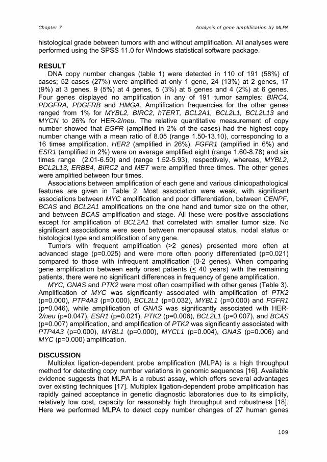
Chapter 7 Analysis of gene amplification by MLPA
histological grade between tumors with and without amplification. All analyses were performed using the SPSS 11.0 for Windows statistical software package. RESULT DNA copy number changes (table 1) were detected in 110 of 191 (58%) of cases; 52 cases (27%) were amplified at only 1 gene, 24 (13%) at 2 genes, 17 (9%) at 3 genes, 9 (5%) at 4 genes, 5 (3%) at 5 genes and 4 (2%) at 6 genes. Four genes displayed no amplification in any of 191 tumor samples: BIRC4, PDGFRA, PDGFRB and HMGA. Amplification frequencies for the other genes ranged from 1% for MYBL2, BIRC2, hTERT, BCL2A1, BCL2L1, BCL2L13 and MYCN to 26% for HER-2/neu. The relative quantitative measurement of copy number showed that EGFR (amplified in 2% of the cases) had the highest copy number change with a mean ratio of 8.05 (range 1.50-13.10), corresponding to a 16 times amplification. HER2 (amplified in 26%), FGFR1 (amplified in 6%) and ESR1 (amplified in 2%) were on average amplified eight (range 1.60-8.78) and six times range (2.01-6.50) and (range 1.52-5.93), respectively, whereas, MYBL2, BCL2L13, ERBB4, BIRC2 and MET were amplified three times. The other genes were amplified between four times. Associations between amplification of each gene and various clinicopathological features are given in Table 2. Most association were weak, with significant associations between MYC amplification and poor differentiation, between CENPF, BCAS and BCL2A1 amplifications on the one hand and tumor size on the other, and between BCAS amplification and stage. All these were positive associations except for amplification of BCL2A1 that correlated with smaller tumor size. No significant associations were seen between menopausal status, nodal status or histological type and amplification of any gene. Tumors with frequent amplification (>2 genes) presented more often at advanced stage (p=0.025) and were more often poorly differentiated (p=0.021) compared to those with infrequent amplification (0-2 genes). When comparing gene amplification between early onset patients (< 40 years) with the remaining patients, there were no significant differences in frequency of gene amplification. MYC, GNAS and PTK2 were most often coamplified with other genes (Table 3). Amplification of MYC was significantly associated with amplification of PTK2 (p=0.000), PTP4A3 (p=0.000), BCL2L1 (p=0.032), MYBL1 (p=0.000) and FGFR1 (p=0.046), while amplification of GNAS was significantly associated with HER-2/neu (p=0.047), ESR1 (p=0.021), PTK2 (p=0.006), BCL2L1 (p=0.007), and BCAS (p=0.007) amplification, and amplification of PTK2 was significantly associated with PTP4A3 (p=0.000), MYBL1 (p=0.000), MYCL1 (p=0.004), GNAS (p=0.006) and MYC (p=0.000) amplification. DISCUSSION Multiplex ligation-dependent probe amplification (MLPA) is a high throughput method for detecting copy number variations in genomic sequences [16]. Available evidence suggests that MLPA is a robust assay, which offers several advantages over existing techniques [17]. Multiplex ligation-dependent probe amplification has rapidly gained acceptance in genetic diagnostic laboratories due to its simplicity, relatively low cost, capacity for reasonably high throughput and robustness [18]. Here we performed MLPA to detect copy number changes of 27 human genes
109

Chapte
110
r 7 Analysis of gene amplification by MLPA
gene # amplified cases (%)
amplification ratio (range)
spanning the human genome that often have an increased copy number in one or more types of tumors including breast cancer. From the chromosomal region point of view, this study observed three amplified regions consistent with current breast cancer literature [19-21], by showing 38% gain of region 8q (attributed to MYC, PTK2 and PTP4A3), 26% of 17q (attributed to HER2) and 17% of 20q (attributed to BCAS1 and GNAS). Table 1. Copy number changes of 25 oncogenes as assessed by multiplex ligation dependent probe amplification of 191 breast cancer cases (n.a. = not applicable). In addition, amplification ratios (ratio=1 means 2 gene copies) are given
BCAS1 16 (8%) 1.91 (1.52-3.43) BCL2A1 1 (1%) 1.71 BCL2L1 2 (1%) 1.99 (1.93-2.05) BCL2L13 2 (1%) 1.61 (1.53-1.69) BIRC2 1 (1%) 1.62 BIRC4 0 n.a. CENPF 16 (8%) 1.75 (1.51-2.12) EGFR 3 (2%) 8.05 (1.5-13.1) ERBB2 50 (26%) 4.04 (1.60-8.78) ERBB4 4 (2%) 1.57 (1.52-1.66) ESR1 3 (2%) 3.30 (1.52-5.93) FGFR1 12 (6%) 3.30 (2.01-6.5) GNAS 17 (9%) 1.98 (1.52-3.70) GSTP1 4 (2%) 2.14 (1.59-2.57) HMGA1 0 n.a. MET 3 (2%) 1.59 (1.53-1.67) MYBL1 15 (8%) 2.08 (1.51-3.00) MYBL2 1 (1%) 1.52 MYC 35 (18%) 2.21 (1.55-4.82) MYCL1 7 (4%) 2.01 (1.59-2.52) MYCN 1 (1%) 3.13 PDGFRA 0 n.a. PDGFRB 0 n.a. PTK2 21 (11%) 2.05 (1.50-2.72) PTP4A3 18 (9%) 1.98 (1.52-2.62) SERPINB9 4 (2%) 1.76 (1.55-1.99) hTERT 1 (1%) 1.97

Chapter 7 Analysis of gene amplification by MLPA
Table 2. Associations between gene amplification status assessed by MLPA and clinicopathological features of 191 breast cancer cases. Four genes (PDGFRA, PDGFRB, BIRC4 and HMGA) that did not show amplification in any of these cases are not listed. Feature n
N % N % N % N % N % N % N % NTotal 191 50 26 35 18 3 2 1 1 3 2 3 2 21 11 18
Age 191<= 40 49 10 20 11 22 2 4 1 2 1 2 0 0 9 18 8> 40 142 40 28 24 17 2 1 0 0 2 1 3 2 13 9 10
Menopausal status 191pre-menopause 109 23 21 23 21 3 3 1 1 1 1 1 1 16 15 12post-menopause 82 27 33 12 15 1 1 0 0 2 2 2 2 6 7 6 7 0 0 6
Tumor size 185T1 8 2 25 0 0 0 0 0 0 0 0 0 0 1 13 0 0 0 0 0T2 46 9 20 8 17 0 0 0 0 1 2 2 4 4 9 3 7 0 0 3T3 36 16 44 7 19 1 3 0 0 0 0 0 0 5 14 3 8 0 0 2T4 95 21 22 17 18 3 3 1 1 1 1 1 1 10 11 11
Stage 190I/II 65 18 28 12 18 0 0 0 0 1 2 2 3 6 9 4 6 0 0 4III/IV 125 32 26 22 18 4 3 1 1 2 2 1 1 15 12 14
Histological grade 1840 10 0 0 0 0 0 0 0 0 0 0 0 0 0 0 11 6 2 33 0 0 0 0 0 0 0 0 0 0 0 0 0 0 0 0 02 90 25 28 12 13 1 1 0 0 2 2 1 1 10 11 93 78 20 26 20 26 3 4 1 1 1 1 2 3 12 15 7
Histological type 191ductal 174 46 26 32 18 4 2 1 1 3 2 3 2 21 12 16lobula
% N % N % N % N %9 2 1 15 8 1 1 16 8
16 1 2 5 10 0 0 3 67 1 1 10 7 1 1 13 9
11 2 2 9 8 0 0 5 57 1 1 11 13
0 0 0 0 07 0 0 2 46 0 0 1 3
12 2 2 8 8 1 1 13 14
6 0 0 2 311 2 2 10 8 1 1 14 11
10 0 0 0 0 0 0 1 100 0 0 0 0
10 1 1 5 6 1 1 8 99 1 1 9 12 0 0 7 9
9 2 1 13 7 1 1 15 9r 4 1 25 0 0 0 0 0 0 0 0 0 0 0 0 0 0 0 0 0
others 13 2 15 2 15 0 0 0 0 0 0 0 0 1 8 1 8 0 0 1
Nodal status 170negative 46 9 20 9 20 2 4 1 2 0 0 1 2 8 17 2 4 0 0 4positive 124 34 27 20 16 2 2 0 0 2 2 2 2 14 11 15
p=0.005
HER-2 MYC MET MYCN ESR1 EGFR PTK2 PT
0 0 0 0 08 0 0 1 8
9 1 2 5 1112 1 1 9 7 0 0 11 9
p=0.019
MYLB1 MYLB2P4A3 BCL2L1 CENPF
111
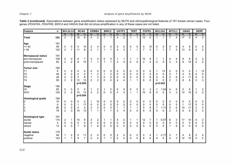
Chapter 7 Analysis of gene amplification by MLPA
Table 2 (continued). Associations between gene amplification status assessed by MLPA and clinicopathological features of 191 breast cancer cases. Four genes (PDGFRA, PDGFRB, BIRC4 and HMGA) that did not show amplification in any of these cases are not listed.
Feature nN % N % N % N % N % N % N %
Total 191 2 1 16 8 4 2 1 1 4 2 1 1 12
Age 181<= 40 49 0 0 5 10 2 4 0 0 0 0 0 0 5> 40 142 2 1 11 8 2 1 1 1 4 3 1 1 7 5
Menopausal status 191pre-menopause 109 0 0 8 7 3 3 0 0 1 1 1 1 10post-menopause 82 2 2 8 10 1 1 1 1 3 4 0 0 2 2
Tumor size 185T1 8 0 0 0 0 0 0 0 0 0 0 0 0 0T2 46 0 0 0 0 1 2 1 2 0 0 0 0 2 4T3 36 0 0 3 8 1 3 0 0 0 0 0 0 3 8T4 95 2 2 12 13 2 2 0 0 4 4 1 1 6 6
Stage 190I/II 65 0 0 0 0 1 2 1 2 0 0 0 0 2 3III/IV 125 2 2 15 12 3 2 0 0 4 3 1 1 10
Histological grade 1840 10 0 0 0 0 1 10 0 0 0 0 0 0 0 01 6 0 0 1 17 0 0 0 0 0 0 0 0 02 90 0 0 4 4 2 2 0 0 1 1 1 1 53 78 2 3 10 13 1 1 1 1 3 4 0 0 6
Histological type 191ductal 174 2 1 15 9 3 2 1 1 4 2 1 1 12lobula
N % N % N % N %6 1 1 7 4 17 9 4 2
10 0 0 0 0 4 8 0 01 1 7 5 13 9 4 3
9 1 1 4 4 9 8 2 20 0 3 4 8 10 2 2
0 1 13 0 0 0 0 1 130 0 3 7 4 9 0 00 0 2 6 4 11 0 00 0 2 2 8 8 3 3
1 1.54 4 6 5 8 1 28 0 0 3 2 12 10 3 2
0 0 0 0 0 0 0 00 0 0 0 0 1 17 0 06 1 1.11 5 6 10 11 2 28 0 0 2 3 6 8 2 3
7 1 0.57 6 3 17 10 4 2r 4 0 0 0 0 0 0 0 0 0 0 0 0 0
others 13 0 0 1 8 1 8 0 0 0 0 0 0 0
Nodal status 170negative 46 1 2 5 11 2 4 0 0 2 4 0 0 2 4positive 124 1 1 9 7 2 2 1 1 2 2 0 0 8 6
p=0.009
p=0.004
BIRC2 GSTP1 TERT FGFBCL2L13 BCAS ERBB4
0 0 0 0 0 0 0 0 00 0 0 1 8 0 0 0 0
1 2.17 3 7 4 9 2 40 0 4 3 12 10 2 2
p=0.043
SERPR1 BCL2A1 MYCL1 GNAS
112

Analysis of gene amplification by MLPA
113
Table 3. Associations between gene amplification of different genes assessed by MLPA in 192 breast cancer cases. Four genes (PDGFRA, PDGFRB, BIRC4 and HMGA) that did not show amplification in any of these cases are not listed.
MYC MET MYCN ESR1 EGFR PTK2 PTP4A3 BCL2L1 MYLB1 MYLB2 CENPF BCL13 BCAS ERBB4 BIRC2 GSTP1 TERT FGFR1 BCL2A1 MYCL1 GNAS SERPHER-2 0.014 0.038 0.047MYC 0.000 0.000 0.032 0.000 0.046METMYCNESR1 0.021EGFR 0.031PTK2 0.000 0.000 0.004 0.006PTP4A3 0.000 0.048BCL2L1 0.01 0.007MYLB1MYLB2 0.021CENPFBCL13 0.041BCAS 0.007 0.036ERBB4BIRC2GSTP1TERTFGFR1BCL2A1 0.021MYCL1GNAS 0.04
Chapter 7

Chapter 7 Analysis of gene amplification by MLPA
Consistent with previous studies on Indonesian breast cancer [9] and other studies on HER-2/neu amplification [22-24], we found 26% of the samples to show HER-2/neu amplification. A recent meta-analysis summarizing data from 81 studies with 27,171 patients, revealed that in the great majority (90%) of studies either HER-2/neu amplification or protein overexpression correlated with poor outcome of the patients [25]. Studies using CGH have also found gain of 17q12-q21.1 where HER-2/neu is located [20]. MYC amplification was found in 18% of the samples. This result is in line with previous studies that found 8 to 37% MYC amplification in breast cancer [26, 27]. A significant association between MYC amplification and poor differentiation was found in this study, consistent with a previous study that found a highly significant association between 8q24 copy number gains and high tumor grade [28]. By CGH, gains of 8q23-q24 were more often observed in grade III tumors [20]. Another gene that showed amplification frequencies above 10% was PTK2 (21/191; 11%). PTK2 (protein tyrosine kinase 2) gene is located in 8q24-ter, encodes a cytoplasmic protein tyrosine kinase which is found concentrated in the focal adhesions that form between cells growing in the presence of extracellular matrix constituents [29, 30]. The encoded protein is a member of the FAK subfamily of protein tyrosine kinases but lacks significant sequence similarity to kinases from other subfamilies. Activation of this gene may be an important early step in cell growth and intracellular signal transduction pathways triggered in response to certain neural peptides or to cell interactions with the extracellular matrix [31-33]. PTK2 was one of the amplified genes that was only recently found amplified in breast cancer, mostly together with ERBB2 (HER-2/neu), EGFR and MYC [19]. PTK2 amplification has also been found in hepatocellular carcinoma (HCC) together with amplification of MYC, MOS and EXT1 [34], and elevated expression of PTK2 was associated with a large tumor size in HCC [34]. Additionally, over expression of PTK2 is seen in a number of different tumor types, including carcinoma of colon [35], prostate [36], ovary [37] and mesenchymal tissues [38]. Several studies [11, 19, 20, 39] found 8q24 as the most frequently gained region in breast cancer samples. Additionally, gains at chromosome arm 8q were found to be correlated with the mean nuclear area (MNA), mitotic activity index (MAI) and occurred in the ‘poor prognostic features’ group [11], and Isola et al. found gain of 8q to be associated with recurrence [40]. There are three potentially important genes located in this region: MYC (8q24.12), PTK2 (8q24-ter) and PTP4A3 (8q24.3). The protein product of oncogene c-myc acts as a transcription factor involved in cell differentiation and apoptosis. Deregulation of this gene may lead to impaired differentiation and apoptosis pathways and thus to growth advantage [11]. The amplification of MYC in primary breast tumors and tumor cell lines is associated with the formation of a distinct type of chromosome changes referred as double minute chromosomes and abnormally banded or homogeneously stained regions (HSR). Both abnormalities reflect DNA amplification and like other structural alterations are associated with genomic instability and contribute to the process of carcinogenesis [41-44]. Their occurrence leads to loss of control of copy gene number that may generate subpopulations of tumor cells with increased growth
114

Chapter 7 Analysis of gene amplification by MLPA
potential, invasiveness and refractory to chemotherapeutic agents [42-44]. These alterations are also viewed as important in cancer development because they frequently involve proto-oncogenes which can alter proliferation of cells [41]. PTK2 encodes focal adhesion kinase (FAK), a non-receptor protein tyrosine kinase that is a major mediator of integrin-stimulated signal transduction pathways. There is evidence that FAK is essential for integrin stimulated cell migration [31, 45], cell spreading [45], and proliferation [31-33]. Upon activation by integrins, FAK induces cell migration through activation of small GTPases of the Rho and Ras families, as well as activation of MAPK and JNK cascades. Furthermore, it induces cell invasion through expression of motility-and invasion-associated proteins such as matrix metalloproteinases [46, 47]. Some investigators have suggested that over expression of PTK2 is a late event in carcinogenesis, contributing to invasiveness and metastasis [48, 49]. Expression of PTP4A3 or PRL3 was found clearly elevated in tumor endothelial cells relative to normal endothelial cells, bulk tissue samples, and breast tumor cell lines. Therefore it was an additional support to a preferential expression of PRL3 in breast tumor vasculature relative to epithelial cells [50]. PRL3 is a proposed tyrosine phospatase with a COOH-terminal prenylation motif that allows its association with the plasma membrane. This gene was reported recently to have role in colorectal cancer metastasis [51]. High level 20q copy number gain was shown to contribute to the cancer phenotype, especially aspects of immortalization, genome instability, apoptosis and increased proliferation [52]. Amplification at 20q13 is particularly interesting because this aberration occurs in a variety of tumor types and is associated with aggressive tumor behavior [53]. The initial CGH study showed increased copy number involving 20q13 in 40% of breast cancer cell lines and 18% of primary breast tumor [54]. Positional cloning of cancer-associated 20q13.2 amplicon identified two genes that display high mRNA levels in breast tumors. One of them was designated Novel Amplified in Breast Cancer-1 (NABC1) or BCAS1. BCAS1 was found to be highly expressed in three amplified breast cancer cell lines and in one breast tumor without amplification at 20q13.2. However, Beardsley et al [55] in their study to characterize the BCAS1 gene product, revealed that BCAS1 overexpression alone does not promote cellular transformation. Concerning that cancer is a multistep process generally involving altered function of several protein, this protein can not be excluded as a determinant in breast tumorigenesis. as overexpression of ZNF217, a gene that is very closely located to BCAS1, has been shown to lead to immortalization of breast cancer cell lines [56]. Thus, it is possible that BCAS1 may have a similar effect when overexpressed in this cell type as well, or BCAS1 expression works cooperatively or synergistically with ZNF217 to promote malignant growth. Although not consistently overexpressed in tumors [53] , BCAS1 is a candidate oncogene. In the present study, we found amplification of BCAS1 in 8% of breast cancers. Despite the low frequency of amplification, a significant association of BCAS1 amplification with advance tumor stage (p=0.004) and bigger tumor size (p=0.009) was found. This result strengthens the previous studies of association of BCAS1 as gene attributed to increased copy number of 20q with more aggressive tumor phenotype. CENPF amplification associated with bigger tumor size. CENPF, centromere protein F, 350/400ka (mitosin) is located on 1q32-q41 and encodes a protein that
115
Chapter 7 Analysis of gene amplification by MLPA
associates with the centromere-kinetochore complex. High copy number of 1q32 region has been found esophageal squamous cell carcinoma as well as glioblastoma and breast cancer [57]. BCL2A1 located on 15q24.3 was isolated from human fetal liver, is a member of the BCL-2 gene family, inhibits p-53 induced apoptosis and exhibits a potent cooperative transforming activity [58, 59]. BCL2A1 or BFL-1 was also shown to be induced by inflammatory cytokines TNF-α or IL-1β, suggesting that it may play a protective role during inflammation [60]. Yoon et al [61] observed that BCL2A1 gene was related to more advanced breast cancer and to factors that have favorable prognosis. In the present study, BCL2A1 amplification was associated with smaller tumor size which is not easy to understand. Tumors with frequent amplification (>2 genes) were more often poorly differentiated and at advanced stage compared to those with infrequent amplification (0-2 genes). This is well understandable as increasing genomic instability as also reflected in more amplified genes will accompany tumor progression. This finding is important because it links gene amplification with the well established pathological prognostic and predictive features of breast cancer. When comparing gene amplification between early-onset patients (< 40 years) with the remaining patients, there were no significant associations. Apparently, there are no clear differences between early and late onset Indonesian breast cancer patients with regard to amplification of the genes studied. In conclusion, we present a high throughput MLPA method that allows a reliable, quick and cheap comprehensive analysis of copy number of a large set of genes that play a role in carcinogenesis in breast and other cancers. This method thereby allows efficient analysis of gene copy number status for better understanding the biology and possibly clinical behavior of breast and other cancers. Acknowledgements: Supported by grant IN-2001-008 of the Dutch Cancer Society. We thank dr. Jo Hilgers who has been instrumental in setting up the Familial Cancer Clinic initiative in Jogjakarta. The MLPA kit (p171GAIN) was kindly provided by Jan Schouten, MRC-Holland, Amsterdam, The Netherlands REFERENCES 1. Parkin DM, Bray F, Ferlay J, Pisani P: Global cancer statistics, 2002. CA Cancer J Clin 2005,
55(2):74-108. 2. Bray F, McCarron P, Parkin DM: The changing global patterns of female breast cancer
incidence and mortality. Breast Cancer Res 2004, 6(6):229-239. 3. Hanahan D, Weinberg RA: The hallmarks of cancer. Cell 2000, 100(1):57-70. 4. Garnis C, Buys TP, Lam WL: Genetic alteration and gene expression modulation during
cancer progression. Mol Cancer 2004, 3:9. 5. Simpson PT, Reis-Filho JS, Gale T, Lakhani SR: Molecular evolution of breast cancer. J Pathol
2005, 205(2):248-254. 6. Weber BL: Cancer genomics. Cancer Cell 2002, 1(1):37-47. 7. Shimada M, Imura J, Kozaki T, Fujimori T, Asakawa S, Shimizu N, Kawaguchi R: Detection of
Her2/neu, c-MYC and ZNF217 gene amplification during breast cancer progression using fluorescence in situ hybridization. Oncol Rep 2005, 13(4):633-641.
8. Kallioniemi OP, Kallioniemi A, Kurisu W, Thor A, Chen LC, Smith HS, Waldman FM, Pinkel D, Gray JW: ERBB2 amplification in breast cancer analyzed by fluorescence in situ hybridization. Proc Natl Acad Sci U S A 1992, 89(12):5321-5325.
116

Chapter 7 Analysis of gene amplification by MLPA
9. Purnomosari D, Aryandono T, Setiaji K, Nugraha S, Pals G, van Diest P: HER-2/neu amplification analysis in invasive breast cancer by Multiplex Ligation dependent Probe Amplification in comparison to immunohistochemistry. Biotechnic and Histochemistry 2006, 81(2-3):79-85.
10. Cuny M, Kramar A, Courjal F, Johannsdottir V, Iacopetta B, Fontaine H, Grenier J, Culine S, Theillet C: Relating genotype and phenotype in breast cancer: an analysis of the prognostic significance of amplification at eight different genes or loci and of p53 mutations. Cancer Res 2000, 60(4):1077-1083.
11. Hermsen MA, Baak JP, Meijer GA, Weiss JM, Walboomers JW, Snijders PJ, van Diest PJ: Genetic analysis of 53 lymph node-negative breast carcinomas by CGH and relation to clinical, pathological, morphometric, and DNA cytometric prognostic factors. J Pathol 1998, 186(4):356-362.
12. Janssen EA, Baak JP, Guervos MA, van Diest PJ, Jiwa M, Hermsen MA: In lymph node-negative invasive breast carcinomas, specific chromosomal aberrations are strongly associated with high mitotic activity and predict outcome more accurately than grade, tumour diameter, and oestrogen receptor. J Pathol 2003, 201(4):555-561.
13. Farabegoli F, Hermsen MA, Ceccarelli C, Santini D, Weiss MM, Meijer GA, van Diest PJ: Simultaneous chromosome 1q gain and 16q loss is associated with steroid receptor presence and low proliferation in breast carcinoma. Mod Pathol 2004, 17(4):449-455.
14. Zweemer RP, Ryan A, Snijders AM, Hermsen MA, Meijer GA, Beller U, Menko FH, Jacobs IJ, Baak JP, Verheijen RH et al: Comparative genomic hybridization of microdissected familial ovarian carcinoma: two deleted regions on chromosome 15q not previously identified in sporadic ovarian carcinoma. Lab Invest 2001, 81(10):1363-1370.
15. Snijders AM, Nowee ME, Fridlyand J, Piek JM, Dorsman JC, Jain AN, Pinkel D, van Diest PJ, Verheijen RH, Albertson DG: Genome-wide-array-based comparative genomic hybridization reveals genetic homogeneity and frequent copy number increases encompassing CCNE1 in fallopian tube carcinoma. Oncogene 2003, 22(27):4281-4286.
16. Schouten JP, McElgunn CJ, Waaijer R, Zwijnenburg D, Diepvens F, Pals G: Relative quantification of 40 nucleic acid sequences by multiplex ligation-dependent probe amplification. Nucleic Acids Res 2002, 30(12):e57.
17. Taylor CF, Charlton RS, Burn J, Sheridan E, Taylor GR: Genomic deletions in MSH2 or MLH1 are a frequent cause of hereditary non-polyposis colorectal cancer: identification of novel and recurrent deletions by MLPA. Hum Mutat 2003, 22(6):428-433.
18. Di Fiore F, Charbonnier F, Martin C, Frerot S, Olschwang S, Wang Q, Boisson C, Buisine MP, Nilbert M, Lindblom A et al: Screening for genomic rearrangements of the MMR genes must be included in the routine diagnosis of HNPCC. J Med Genet 2004, 41(1):18-20.
19. Naylor TL, Greshock J, Wang Y, Colligon T, Yu QC, Clemmer V, Zaks TZ, Weber BL: High resolution genomic analysis of sporadic breast cancer using array-based comparative genomic hybridization. Breast Cancer Res 2005, 7(6):R1186-1198.
20. Weber-Mangal S, Sinn HP, Popp S, Klaes R, Emig R, Bentz M, Mansmann U, Bastert G, Bartram CR, Jauch A: Breast cancer in young women (< or = 35 years): Genomic aberrations detected by comparative genomic hybridization. Int J Cancer 2003, 107(4):583-592.
21. Cingoz S, Altungoz O, Canda T, Saydam S, Aksakoglu G, Sakizli M: DNA copy number changes detected by comparative genomic hybridization and their association with clinicopathologic parameters in breast tumors. Cancer Genet Cytogenet 2003, 145(2):108-114.
22. Ross JS, Fletcher JA: HER-2/neu (c-erb-B2) gene and protein in breast cancer. Am J Clin Pathol 1999, 112(1 Suppl 1):S53-67.
23. Kauraniemi P, Kallioniemi A: Activation of multiple cancer-associated genes at the ERBB2 amplicon in breast cancer. Endocr Relat Cancer 2006, 13(1):39-49.
24. Sidoni A, Ferri I, Cavaliere A, Bellezza G, Scheibel M, Bucciarelli E: Detection of HER-2/neu (c-erbB-2) overexpression and amplification in breast carcinomas with ambiguous immunohistochemical results. A further contribution to defining the role of fluorescent in situ hybridization. Anticancer Res 2006, 26(3B):2333-2337.
25. Ross JS, Fletcher JA, Linette GP, Stec J, Clark E, Ayers M, Symmans WF, Pusztai L, Bloom KJ: The Her-2/neu gene and protein in breast cancer 2003: biomarker and target of therapy. Oncologist 2003, 8(4):307-325.
26. Borg A, Baldetorp B, Ferno M, Olsson H, Sigurdsson H: c-myc amplification is an independent prognostic factor in postmenopausal breast cancer. Int J Cancer 1992, 51(5):687-691.
27. Champeme MH, Bieche I, Hacene K, Lidereau R: Int-2/FGF3 amplification is a better independent predictor of relapse than c-myc and c-erbB-2/neu amplifications in primary human breast cancer. Mod Pathol 1994, 7(9):900-905.
117

Chapter 7 Analysis of gene amplification by MLPA
28. Ioannidis P, Mahaira L, Papadopoulou A, Teixeira MR, Heim S, Andersen JA, Evangelou E, Dafni U, Pandis N, Trangas T: 8q24 Copy number gains and expression of the c-myc mRNA stabilizing protein CRD-BP in primary breast carcinomas. Int J Cancer 2003, 104(1):54-59.
29. Hanks SK, Calalb MB, Harper MC, Patel SK: Focal adhesion protein-tyrosine kinase phosphorylated in response to cell attachment to fibronectin. Proc Natl Acad Sci U S A 1992, 89(18):8487-8491.
30. Hynes RO: Cell adhesion: old and new questions. Trends Cell Biol 1999, 9(12):M33-37. 31. Gilmore AP, Romer LH: Inhibition of focal adhesion kinase (FAK) signaling in focal adhesions
decreases cell motility and proliferation. Mol Biol Cell 1996, 7(8):1209-1224. 32. Zhao JH, Reiske H, Guan JL: Regulation of the cell cycle by focal adhesion kinase. J Cell Biol
1998, 143(7):1997-2008. 33. Zhao J, Zheng C, Guan J: Pyk2 and FAK differentially regulate progression of the cell cycle. J
Cell Sci 2000, 113 (Pt 17):3063-3072. 34. Hashimoto K, Mori N, Tamesa T, Okada T, Kawauchi S, Oga A, Furuya T, Tangoku A, Oka M,
Sasaki K: Analysis of DNA copy number aberrations in hepatitis C virus-associated hepatocellular carcinomas by conventional CGH and array CGH. Mod Pathol 2004, 17(6):617-622.
35. Owens LV, Xu L, Craven RJ, Dent GA, Weiner TM, Kornberg L, Liu ET, Cance WG: Overexpression of the focal adhesion kinase (p125FAK) in invasive human tumors. Cancer Res 1995, 55(13):2752-2755.
36. Tremblay L, Hauck W, Aprikian AG, Begin LR, Chapdelaine A, Chevalier S: Focal adhesion kinase (pp125FAK) expression, activation and association with paxillin and p50CSK in human metastatic prostate carcinoma. Int J Cancer 1996, 68(2):164-171.
37. Judson PL, He X, Cance WG, Van Le L: Overexpression of focal adhesion kinase, a protein tyrosine kinase, in ovarian carcinoma. Cancer 1999, 86(8):1551-1556.
38. Weiner TM, Liu ET, Craven RJ, Cance WG: Expression of growth factor receptors, the focal adhesion kinase, and other tyrosine kinases in human soft tissue tumors. Ann Surg Oncol 1994, 1(1):18-27.
39. Buerger H, Mommers EC, Littmann R, Diallo R, Brinkschmidt C, Poremba C, Dockhorn-Dworniczak B, van Diest PJ, Bocker W: Correlation of morphologic and cytogenetic parameters of genetic instability with chromosomal alterations in in situ carcinomas of the breast. Am J Clin Pathol 2000, 114(6):854-859.
40. Isola JJ, Kallioniemi OP, Chu LW, Fuqua SA, Hilsenbeck SG, Osborne CK, Waldman FM: Genetic aberrations detected by comparative genomic hybridization predict outcome in node-negative breast cancer. Am J Pathol 1995, 147(4):905-911.
41. Bishop JM: The molecular genetics of cancer. Science 1987, 235(4786):305-311. 42. Alitalo K, Schwab M: Oncogene amplification in tumor cells. Adv Cancer Res 1986, 47:235-281. 43. Schimke RT: Gene amplification in cultured cells. J Biol Chem 1988, 263(13):5989-5992. 44. Pardue ML: Dynamic instability of chromosomes and genomes. Cell 1991, 66(3):427-431. 45. Owen JD, Ruest PJ, Fry DW, Hanks SK: Induced focal adhesion kinase (FAK) expression in
FAK-null cells enhances cell spreading and migration requiring both auto- and activation loop phosphorylation sites and inhibits adhesion-dependent tyrosine phosphorylation of Pyk2. Mol Cell Biol 1999, 19(7):4806-4818.
46. Schaller MD: Biochemical signals and biological responses elicited by the focal adhesion kinase. Biochim Biophys Acta 2001, 1540(1):1-21.
47. Hauck CR, Hsia DA, Schlaepfer DD: The focal adhesion kinase--a regulator of cell migration and invasion. IUBMB Life 2002, 53(2):115-119.
48. Agochiya M, Brunton VG, Owens DW, Parkinson EK, Paraskeva C, Keith WN, Frame MC: Increased dosage and amplification of the focal adhesion kinase gene in human cancer cells. Oncogene 1999, 18(41):5646-5653.
49. Brunton VG, Ozanne BW, Paraskeva C, Frame MC: A role for epidermal growth factor receptor, c-Src and focal adhesion kinase in an in vitro model for the progression of colon cancer. Oncogene 1997, 14(3):283-293.
50. Parker BS, Argani P, Cook BP, Liangfeng H, Chartrand SD, Zhang M, Saha S, Bardelli A, Jiang Y, St Martin TB et al: Alterations in vascular gene expression in invasive breast carcinoma. Cancer Res 2004, 64(21):7857-7866.
51. Saha S, Bardelli A, Buckhaults P, Velculescu VE, Rago C, St Croix B, Romans KE, Choti MA, Lengauer C, Kinzler KW et al: A phosphatase associated with metastasis of colorectal cancer. Science 2001, 294(5545):1343-1346.
118

Chapter 7 Analysis of gene amplification by MLPA
52. Hodgson JG, Chin K, Collins C, Gray JW: Genome amplification of chromosome 20 in breast cancer. Breast Cancer Res Treat 2003, 78(3):337-345.
53. Collins C, Rommens JM, Kowbel D, Godfrey T, Tanner M, Hwang SI, Polikoff D, Nonet G, Cochran J, Myambo K et al: Positional cloning of ZNF217 and NABC1: genes amplified at 20q13.2 and overexpressed in breast carcinoma. Proc Natl Acad Sci U S A 1998, 95(15):8703-8708.
54. Kallioniemi A, Kallioniemi OP, Piper J, Tanner M, Stokke T, Chen L, Smith HS, Pinkel D, Gray JW, Waldman FM: Detection and mapping of amplified DNA sequences in breast cancer by comparative genomic hybridization. Proc Natl Acad Sci U S A 1994, 91(6):2156-2160.
55. Beardsley DI, Kowbel D, Lataxes TA, Mannino JM, Xin H, Kim WJ, Collins C, Brown KD: Characterization of the novel amplified in breast cancer-1 (NABC1) gene product. Exp Cell Res 2003, 290(2):402-413.
56. Nonet GH, Stampfer MR, Chin K, Gray JW, Collins CC, Yaswen P: The ZNF217 gene amplified in breast cancers promotes immortalization of human mammary epithelial cells. Cancer Res 2001, 61(4):1250-1254.
57. Pimkhaokham A, Shimada Y, Fukuda Y, Kurihara N, Imoto I, Yang ZQ, Imamura M, Nakamura Y, Amagasa T, Inazawa J: Nonrandom chromosomal imbalances in esophageal squamous cell carcinoma cell lines: possible involvement of the ATF3 and CENPF genes in the 1q32 amplicon. Jpn J Cancer Res 2000, 91(11):1126-1133.
58. Choi SS, Park IC, Yun JW, Sung YC, Hong SI, Shin HS: A novel Bcl-2 related gene, Bfl-1, is overexpressed in stomach cancer and preferentially expressed in bone marrow. Oncogene 1995, 11(9):1693-1698.
59. D'Sa-Eipper C, Subramanian T, Chinnadurai G: bfl-1, a bcl-2 homologue, suppresses p53-induced apoptosis and exhibits potent cooperative transforming activity. Cancer Res 1996, 56(17):3879-3882.
60. Jung-Ha H, Kim D, Lee SB, Hong SI, Park SY, Huh J, Kim CW, Kim SS, Lee Y, Choi SS et al: Expression of Bfl-1 in normal and tumor tissues: Bfl-1 overexpression in cancer is attributable to its preferential expression in infiltrating inflammatory cells. Hum Pathol 1998, 29(7):723-728.
61. Yoon HS, Hong SH, Kang HJ, Ko BK, Ahn SH, Huh JR: Bfl-1 gene expression in breast cancer: its relationship with other prognostic factors. J Korean Med Sci 2003, 18(2):225-230.
119

Chapter 7 Analysis of gene amplification by MLPA
120

Chapter 8:
General Discussion
chapter 8 General Discussion
Breast cancer shows a high and increasing incidence in Indonesia, and many females develop breast cancer at young age. It is therefore likely that BRCA1 and BRCA2 mutations play an important role in breast carcinogenesis in the Indonesian population. However, little is known about the role of BRCA1/2 germline mutations in Indonesian breast cancer. We therefore set out to screen a series of Indonesian breast cancer patients and their family member for germline mutations in BRCA1 and BRCA2. To this end, a fast and cheap method to screen for these mutations was needed. BRCA1 and BRCA2 germline mutations Over 80% of families with two or more cases of pre-menopausal breast cancer and two or more cases of ovarian cancer are believed to carry a germline BRCA1 or BRCA2 mutation [1, 2]. It has been shown that ethnically different populations exhibit different germline mutation spectra in the BRCA1 and BRCA2 genes [3-5]. There are no previous publications on BRCA1/2 mutation detection in the Indonesian population. By identifying the endemic Indonesian mutations, we hope to offer better risk assessment for women in the Indonesian population who are susceptible to the BRCA1/2 related hereditary cancers. However, the large size of the BRCA1 and BRCA2 genes, and the scattered distribution of mutations throughout the genes, complicate the task of mutation detection and make rapid screening for mutations a major technical challenge. A technique that scans stretches of DNA for unknown mutations should be optimized to detect as close to 100% of the sequence alteration as possible. In Chapter 2, we describe the results of the application of a recently developed rapid and sensitive method for the detection of BRCA1/2 mutations [6] based on pooled denaturing gradient gel electrophoresis (DGGE) [7] and targeted sequencing to an Indonesian group of high risk patients. Theoretically, this method should be particularly suited for the analysis of naive populations. As alternatives to sequencing methods, which determine the exact nature and location of each base along a DNA fragment, various mutation scanning procedures have been developed. These methods, which rely on the recognition of a sequence variation between mutant and wild-type DNA on the basis of an altered electrophoretic migration pattern, provide a simple means for determining whether a given DNA sample harbors a mutation in a particular gene. The most well established scanning procedures are single strand conformational polymorphism (SCCP) analysis, DGGE, chemical cleavage of mismatch, RNase cleavage, the protein truncation test (PTT), and heteroduplex analysis. Among these methods, SCCP, DGGE, PTT, and heteroduplex analysis are the most widely used because of their accuracy, simplicity, lack of toxicity, and/or relative affordability. We focused on DGGE because this method, when optimized, has the highest mutation detection rate (close to 100% [8]) compared with SCCP and heteroduplex analysis. Although DGGE has been applied to BRCA mutation screening before [9-11] we used a new technique based on DGGE after exon by exon PCR amplification of the complete BRCA1 and BRCA2 sequences [12] in an Indonesian breast cancer population, and sequenced aberrantly moving bands. This yielded four polymorphisms consisting of single nucleotide substitutions, underlining the sensitivity of the method. Intelligent pooling of the amplicons before electrophoresis greatly improved the throughput of the method. In addition, a novel BRCA2
122
chapter 8 General Discussion
mutation (2699delTAAATG) was found. In conclusion, a new high throughput and highly sensitive method to screen BRCA1 and BRCA2 for mutations based on exon by exon PCR amplification followed by pooled DGGE and sequencing of aberrant bands seems to be the ideal approach for screening naïve populations for mutations, and has the ability to detect single base differences using non-toxic and relatively simple and inexpensive methods. Using this method, and adding by Multiplex ligation dependent probe amplification (MLPA) technique to detect genomic deletions in BRCA1/2, we analyzed a group of 116 early onset breast cancer patients and 16 of their family members from three Indonesian cities (Jakarta and Jogjakarta on the Java island, and Denpasar on the Bali island) for BRCA1/2 mutations in chapter 3. Mutations specific for certain populations and ethnic groups have been identified in both genes. For example, specific BRCA1 and BRCA2 mutations were reported for Ashkenazi Jews [13]. Other common BRCA1 mutations were especially found in Italian, Canadian, Belgian or Dutch breast cancer families [14-16]. In Indonesia, the contribution of the BRCA1/BRCA2 mutations to the population incidence of early-onset breast cancer was largely unknown before. The analysis of 116 unrelated breast cancer patients with breast cancer revealed that 9 patients (7.8%) carried pathogenic germline mutations especially the early onset patients: 3 within BRCA1 (2.6%) (c.2784_2785insT, pL1415X (c.4361_4362insT), del exon 13-15) and 6 within BRCA2 (5.2%) (c.3040_3043delGCAA, p.Glu2183X (c.6775G>T), p.Leu824X (c.2699_2704delTAAATG), p.Gln2894X (c.9008C>T)) which is comparable to previous studies [17]. All these mutations were classified as pathogenic as they are predicted to result in protein truncation. The three pathogenic mutations found in BRCA1 were not previously reported in the BIC database as well as two novel nonsense mutations (p.Glu2183X and p.Gln2894X) identified in BRCA2. There were twice as many BRCA2 mutations as BRCA1 mutations. Although the absolute numbers are low and no firm conclusions can therefore be drawn, this is comparable to other Asian regions [3, 18, 19] but seems to discern the Indonesian population from non-Asian ethnic groups where the reverse trend is seen. The c.2699_2704delTAAATG (p.Leu824X) in BRCA2 that has been reported previously by us in the Indonesian population [20], was found in one other patient in the present study. This mutation lies in exon 11 BRCA2, within the BRC repeats domain. The truncating mutation causes loss of three quarters of the protein leading to lack of interaction with the RAD51 protein. Different from BRCA1, the repair of DSBs by HR is the most important function of the BRCA2 protein [21]. As the c.2699_2704delTAAATG mutation was found in two unrelated patients, this mutation could be a good candidate as a founder mutation. Sixteen (7 BRCA1 and 9 BRCA2) rare mutations of so far unknown significance (“unclassified variants”, UVs) were detected in 18 patients: 13 missense changes and 3 intronic variants. Of these 16 UVs, 7 were novel, whereas the other UVs have been previously reported in the BIC database. Seven UV were found in the BRCA1 gene, two mutations occurring in the intronic region between exons 1 and 2 (c.101-10T>C) and between exons 19 and 20 (c.5313-31A>G), and five missense mutations identified: p.Val191Ile (c.690G>A), p.Leu1209Val (c.3744T>G), p.Met1652Ile (c.5075G>A), p.Arg1835Gln (c.5623G>A) and p.Thr1852Ile (c.5674C>T). Four out of seven BRCA1 missense mutations; p.Leu1209Val (c.3744T>G), c.5313-31A>G, p.Arg1835Gln (c.5623G>A) and p.Thr1852Ile
123

chapter 8 General Discussion
(c.5674C>T) were have not been described previously in the BIC. Nine different UVs of the BRCA2 gene were found in fourteen patients, and three of them were novel; p.Gln609Glu 9c.2053C>G), p.Gln699Leu (2324A>T) and p.Val950Ile (3076G>A). When comparing the three different Indonesian regions, the percentages of breast cancer patients with pathogenic BRCA1/2 mutations were significantly higher in Denpasar on the Bali island than in Jogjakarta and Jakarta on the Java island. Although the number of patients is too small to draw firm conclusions, these data may point to geographic differences within Indonesia. In conclusion, a relatively high percentage of early onset Indonesian breast cancer patients carry a germline mutation in either BRCA1 or BRCA2. Several novel, pathogenic BRCA1 and BRCA2 germline mutations have been found, as well as a variety of novel “unclassified variant” mutations that may therefore be specific for the Indonesian population. The finding of all these novel mutations illustrates the power of the optimized DGGE/MLPA technique to detect BRCA1/2 mutations in naïve populations like the Indonesian one. It is likely that some of the “unclassified variant” mutations may have a functional role in breast cancer development, which deserves to be explored further. The phenotype and genotype of a group of early onset Indonesian breast cancer patients is described in chapter 4 and chapter 5, as such phenotype can give clues for the “BRCA-ness” of a breast cancer. Distinct features of BRCA1-associated tumor have been proposed, such as high tumor grade, estrogen (ER) and progesterone receptor (PgR) negativity [22, 23], accumulation of p53 [22, 23], expression of the epidermal growth factor receptor (EGFR) [24-26], and absence of amplification and overexpression of HER-2/neu [27, 28]. Additionally, hereditary breast cancers are preferentially of the ductal and medullary carcinoma types [29, 30]. cDNA expression analyses and expression of cytokeratins 5/6 have suggested a basal epithelial phenotype for BRCA1 related cancers [31]. However, the phenotype of BRCA2 related cancers is much less outspoken, and seems to be in between that of BRCA1 related and sporadic cancers [32]. Whereas in Western countries most hereditary breast cancers are BRCA1 related, the reverse trend is seen in Indonesia where most hereditary breast cancers seem to be BRCA2 related [20]. Better defining the phenotype of BRCA2 related breast cancers is especially important in Asian populations like ours, as the prevalence of BRCA2 related cancers exceeds that of BRCA1 related cancers [3, 33]. Currently, HER-2/neu status is determined using two approaches: one that reveals gene amplification and one aimed at detecting the overexpressed HER-2/neu protein [34-37]. In chapter 4 we evaluated for the first time the potential value of a new MLPA based PCR based technique to assess HER-2/neu amplification. MLPA relies on comparative quantitation of specifically bound probes that are amplified by PCR with universal primers [38]. The introduction of universal primers has advantages because multiplexing numerous targets becomes much easier, and when fluorescence detection of products is used, only one fluorescent primer is required, thus reducing the cost compared to buying fluorescent probes for each target. Technically, FISH has disadvantages compared to MLPA for determining partial gene deletions and remains a relatively low throughput method compared to other molecular genetic techniques. Comparison between IHC and MPLA showed that none of the 36 IHC negative cases were amplified and as many
124
chapter 8 General Discussion
as eight of nine (89%) IHC 3+ tumors showed gene amplification by MLPA assay. Therefore, there was complete concordance between IHC and MLPA in (36+8)/60 (73%) of cases. None of the IHC 1+ cases was MLPA amplified. FISH has been shown to detect amplification in 6-7% of negatively staining tumors [39]; MLPA also may prove to have some additional value in these cases in larger future studies. As many as five of eight (63%) 2+ cases, however, were amplified by MLPA, indicating that the additional value of MLPA lies especially in the IHC 2+ cases. Therefore, we propose MLPA as a quick and inexpensive method for detecting HER-2/neu amplification in daily laboratory practice. Although IHC still is useful as an initial screening tool, MLPA may be an attractive alternative to FISH for amplification testing for IHC 2+ cases. Because MLPA is relatively cheap and can be used readily on paraffin embedded tissue, the method is widely applicable, also in a developing country like Indonesia. This method was further used in the following chapter. In chapter 5, we further investigated the histopathological and immuno-histochemical characteristics of early onset (< 40 years) Indonesian breast cancer patients, as such features can be used as to distinguish between BRCA and non-BRCA carriers among these young women. This could help to limit expensive mutation screening to those patients at highest risk to harbour a germline BRCA mutation. Fully consistent with previous studies, our few BRCA1 related cases were advanced stage, ductal type, grade 3, ER and HER-2/neu negative, and EGFR, CK5/6 and p53 positive. BRCA2 tumors are more frequently of ductal type [40] as in the present study where all BRCA2 related tumors were ductal. They are also more frequently grade 2 and 3 than sporadic controls [2, 41, 42]. In line with these previous studies, in the present study BRCA2 carriers had moderately or poorly differentiated tumors and none of them were well differentiated. The frequency of ER and PR expression in BRCA2 tumors has been reported as similar to that in sporadic breast tumors most studies [27, 42-44], in line with the present study. As to p53, some studies have found p53 accumulation in around 20-50% of BRCA2 related carcinomas [27], In the present study, 60% of the BRCA2 associated tumors showed p53 accumulation. Data on HER-2/neu expression in BRCA2-associated tumors vary from series to series, probably as a consequence of differences in the techniques employed. For example, Armes et al. [44] and Eerola et al. [43] found no differences in the expression of HER-2/neu in BRCA2 and sporadic breast tumors. However, other studies revealed low frequencies between 0 and 3% in HER-2/neu overexpression in BRCA2 tumors [27, 30, 42]. Combining immunohistochemistry with a new amplification test, we found a 20% frequency of HER-2/neu positivity in BRCA2 associated tumors. While immunohistochemistry result for EGFR, CK5/6 and Ki67 for BRCA2 associated tumors showed 40%, 20% and 100% positivity, respectively. The results of the present study add to the concept that breast cancer arising in BRCA1 and BRCA2 mutation carriers of mutation in the genes differ from sporadic breast cancer of age matched controls. This is especially clear for BRCA1 related cancers. Unfortunately, also our study has not firmly established a clear phenotype for BRCA2 related breast cancers. In conclusion, early onset Indonesian breast cancer is characterized by increased proliferation. Within the early onset group, the strongest features pointing to a sporadic cancer seem to be the absence of family history of breast and or ovarian cancer, grade I and lobular differentiation. Features
125

chapter 8 General Discussion
increasing the chance of a germline BRCA1/2 mutation are family history of breast and or ovarian cancer, CK5/6 and EGFR expression, p53 accumulation and high proliferation as measured by Ki67 labeling. This is potentially quite useful to optimize selection of early onset breast cancer patients for BRCA1/2 mutation testing. Breast cancer, like other cancers, derives through accumulation of a wide variety of genetic and epigenetic events [45-51], also in patients predisposed to breast cancer due to a germline BRCA1/2 mutation. As promoter methylation of tumor suppressor genes and amplification of oncogenes are well known phenomena in sporadic breast cancer but have hardly been studied yet in hereditary breast cancer, we analyzed a group of early onset Indonesian breast cancer for promoter methylation of tumor suppressor genes in chapter 6 and for gene copy number in chapter 7. The majority of previous studies on methylation have dealt with only one or just a few genes [52, 53], applying techniques based on the conversion of unmethylated cytosine residues into uracil after sodium bisulphite treatment [54], which are converted to thymidine during subsequent PCR. Here, we performed for the first time a high throughput easy method based on methylation specific Multiplex Ligation dependent Probe Amplification (MS-MLPA) [55] to assess the methylation status of 22 selected tumor suppressor genes in a large group of breast cancer patients. We used in chapter 6 the ME001 MS-MLPA probe mix (MRC Holland, Amsterdam, The Netherlands) which contains 24 sequences that correspond to the promoters of a set of tumor suppressor genes that are frequently silenced by methylation in different tumors (including breast cancer), but are unmethylated in blood derived DNA of healthy individuals. Six genes displayed no promoter methylation in any of 168 tumor samples: CDKN2A (p14ARK), CDKN1B (p27 KIP1)) ATM, PTEN, BRCA2 and VHL. Methylation frequencies for the other genes ranged from 1% for TP73 and FHIT to 61% for RASSF1. For p14ARF, VHL and BRCA2, this is in line with a previous study [56]. However, other studies using methylation-specific PCR did show methylation for PTEN (15/44 cases, 34%) [57] and ATM (18/23 cases, 78%) [58]. Hypermethylation of the Von Hippel-Lindau (VHL) gene is mostly found in renal call carcinoma [59] while a fundamental matter still remains completely unresolved regarding the reason behind non-hypermethylation of BRCA2 [60]. Of the 168 tumor samples, 33 (20%) showed no methylation at any promoter site. 30 cases (18%) were methylated at the promoter site of only 1 gene, 35 (21%) at 2 sites, 32 (19%) at 3 sites, 21 (13%) at 4 sites, 11 (7%) at 5 sites, 2 (1%) at 6 sites, 3 (2%) at 7 sites and 1 (1%) at 8 sites. No tumor was methylated at all 22 sites. The most consistent association observed in the present study of specific gene methylation was with amplification of HER-2/neu where 4 genes (APC, RASSF1A, CDH13 and GSTP1) reached statistical significance. Tumors with infrequent methylation (0–2 genes) were more often negative for HER-2/neu compared to those with frequent methylation (3-8 genes; 63% vs 37%, P=0.041). Tumors with infrequent methylation were more often well differentiated than those with frequent methylation (63% vs 38%, P=0.007). This finding is important because it links DNA hypermethylation with the well established pathological prognostic and predictive features of breast cancer. Previous studies have also reported associations between CpG island methylation and poor
126

chapter 8 General Discussion
histological differentiation of breast tumors [61]. In conclusion, this comprehensive analysis of tumor suppressor promoter methylation status in invasive breast cancer by high throughput MS-MLPA reveals remarkable differences in methylation frequency for various genes. Promoter methylation of multiple genes seems to be correlated to poor differentiation and HER-2/neu amplification. A comprehensive hypermethylation profile as assessed by MS-MLPA could therefore potentially be useful for breast cancer detection and classification, and understanding the biology of this disease. Genomic instability is a hallmark of cancer, and specific subchromosomal copy number changes are thought to play a driving role in the transformation of normal cells to malignant clones. These genomic copy number changes may result in deletion of one or both alleles of tumor suppressor genes, overexpression of oncogenes and rearrangements that may alter transcription of target and downstream genes [62]. In chapter 7 we applied a high throughput easy method based on Multiplex Ligation-dependent Probe Amplification (MLPA) to assess the amplification of 25 genes in a large group of breast cancer patients. Four genes displayed no amplification in any of 191 tumor samples: BIRC4, PDGFRA, PDGFRB and HMGA. Amplification frequencies for the other genes ranged from 1% for MYBL2, BIRC2, hTERT, BCL2A1, BCL2L1, BCL2L13 and MYCN to 26% for HER-2/neu. Of 191 tumor samples, 52 cases (27%) were amplified at only 1 gene, 24 (13%) at 2 genes, 17 (9%) at 3 genes, 9 (5%) at 4 genes, 5 (3%) at 5 genes and 4 (2%) at 6 genes. From the chromosomal region point of view, this study observed three regions that consistent with other breast cancer literatures [63-65], by showing 38% gaining in region 8q (attributed to MYC, PTK2 and PTP4A3), 26% in 17q (attributed to HER2) and 17% in 20q (attributed to BCAS1 and GNAS). MYC amplification was found in 18% of the samples. This result is in line with previous studies that found 8 to 37% MYC amplification in breast cancer [66, 67]. A significant association between MYC amplification and poor differentiation was found in this study, consistent with a previous study that found a highly significant association between 8q24 copy number gains and high tumor grade [68]. By CGH, gains of 8q23-q24 were more often observed in grade III tumors [64]. Consistent with our previous study on Indonesian breast cancer [69] and other studies on HER-2/neu amplification [70-72], we found 26% of the samples to show HER-2/neu amplification. A recent meta-analysis summarizing data from 81 studies with 27,171 patients, revealed that in the great majority (90%) of studies either HER-2/neu amplification or protein overexpression correlated with poor outcome of the patients [73]. Studies using CGH have also found gain of 17q12-q21.1 where HER-2/neu is located [64]. The subgroup of tumors with frequent amplification (>2 genes) were more often poorly differentiated and at advanced stage compared to those with infrequent amplification (0-2 genes). This is well understandable as increasing genomic instability as also reflected in more amplified genes will accompany tumor progression. This finding is important because it links gene amplification with the well established pathological prognostic and predictive features of breast cancer. In conclusion, we present a novel high throughput MLPA method that allows a reliable, quick and cheap comprehensive analysis of copy number of a large set of genes that play a role in carcinogenesis in breast and other cancers. This method thereby allows efficient analysis of gene copy
127

chapter 8 General Discussion
number status for better understanding the biology and possibly clinical behavior of breast and other cancers. When comparing methylation and gene amplification status between early onset patients (< 40 years) with the remaining patients, there were no significant differences. We analyzed this subgroup as in Indonesia it relatively large with many patient haboring BRCA1/2 mutations [33]. Although previous studies have shown genetic differences between early onset and older breast cancer patients [74, 75], such differences are not apparent with regard to promoter methylation of the tumor suppressor genes or amplification of the oncogenes analyzed. References 1. Narod SA, Ford D, Devilee P, Barkardottir RB, Lynch HT, Smith SA, Ponder BA, Weber BL, Garber
JE, Birch JM et al: An evaluation of genetic heterogeneity in 145 breast-ovarian cancer families. Breast Cancer Linkage Consortium. Am J Hum Genet 1995, 56(1):254-264.
2. Pathology of familial breast cancer: differences between breast cancers in carriers of BRCA1 or BRCA2 mutations and sporadic cases. Breast Cancer Linkage Consortium. Lancet 1997, 349(9064):1505-1510.
3. Liede A, Narod SA: Hereditary breast and ovarian cancer in Asia: genetic epidemiology of BRCA1 and BRCA2. Hum Mutat 2002, 20(6):413-424.
4. Neuhausen SL: Ethnic differences in cancer risk resulting from genetic variation. Cancer 1999, 86(11 Suppl):2575-2582.
5. Verhoog LC, van den Ouweland AM, Berns E, van Veghel-Plandsoen MM, van Staveren IL, Wagner A, Bartels CC, Tilanus-Linthorst MM, Devilee P, Seynaeve C et al: Large regional differences in the frequency of distinct BRCA1/BRCA2 mutations in 517 Dutch breast and/or ovarian cancer families. Eur J Cancer 2001, 37(16):2082-2090.
6. Hout AH, Mulder IM, Wu Y, van der Vlies P, Huisman M, Oosterwijk JC, Buys CHCM, Hoftra RMW: A DGGE system for comprehensive mutation screening of the complete coding regions of BRCA1 and BRCA2 outside exon 11. Am J Hum Genet 1999, 65(suppl S Oct):2322.
7. Fischer SG, Lerman LS: DNA fragments differing by single base-pair substitutions are separated in denaturing gradient gels: correspondence with melting theory. Proc Natl Acad Sci U S A 1983, 80(6):1579-1583.
8. Macek M, Jr., Mercier B, Mackova A, Miller PW, Hamosh A, Ferec C, Cutting GR: Sensitivity of the denaturing gradient gel electrophoresis technique in detection of known mutations and novel Asian mutations in the CFTR gene. Hum Mutat 1997, 9(2):136-147.
9. Muller D, Bonaiti-Pellie C, Abecassis J, Stoppa-Lyonnet D, Fricker JP: BRCA1 testing in breast and/or ovarian cancer families from northeastern France identifies two common mutations with a founder effect. Fam Cancer 2004, 3(1):15-20.
10. Gal I, Gershoni Baruch R, Haber D, Dagan E, Eisenberg-Barzilai S, Zidan J, Friedman E: The 1100delAT BRCA1 and the 8765delAG BRCA2 mutations: occurrence in high-risk non-Ashkenazi Jews and haplotype comparison of Jewish and non-Jewish carriers. Fam Cancer 2004, 3(1):11-14.
11. Atoum MF, Al-Kayed SA: Mutation analysis of the breast cancer gene BRCA1 among breast cancer Jordanian females. Saudi Med J 2004, 25(1):60-63.
12. van der Hout AH MI, Wu Y, van der Vlies P, Huisman M, Oosterwijk JC, Buys CHCM, Hoftra RMW: A DGGE system for comprehensive mutation screening of the complete coding regions of BRCA1 and BRCA2 outside exon 11. Am J Hum Genet 1999, 65(suppl S Oct):2322.
13. Roa BB, Boyd AA, Volcik K, Richards CS: Ashkenazi Jewish population frequencies for common mutations in BRCA1 and BRCA2. Nat Genet 1996, 14(2):185-187.
14. Simard J, Tonin P, Durocher F, Morgan K, Rommens J, Gingras S, Samson C, Leblanc JF, Belanger C, Dion F et al: Common origins of BRCA1 mutations in Canadian breast and ovarian cancer families. Nat Genet 1994, 8(4):392-398.
15. Peelen T, van Vliet M, Petrij-Bosch A, Mieremet R, Szabo C, van den Ouweland AM, Hogervorst F, Brohet R, Ligtenberg MJ, Teugels E et al: A high proportion of novel mutations in BRCA1 with strong founder effects among Dutch and Belgian hereditary breast and ovarian cancer families. Am J Hum Genet 1997, 60(5):1041-1049.
128

chapter 8 General Discussion
16. Petrij-Bosch A, Peelen T, van Vliet M, van Eijk R, Olmer R, Drusedau M, Hogervorst FB, Hageman S, Arts PJ, Ligtenberg MJ et al: BRCA1 genomic deletions are major founder mutations in Dutch breast cancer patients. Nat Genet 1997, 17(3):341-345.
17. de Sanjose S, Leone M, Berez V, Izquierdo A, Font R, Brunet JM, Louat T, Vilardell L, Borras J, Viladiu P et al: Prevalence of BRCA1 and BRCA2 germline mutations in young breast cancer patients: a population-based study. Int J Cancer 2003, 106(4):588-593.
18. Ho GH, Phang BH, Ng IS, Law HY, Soo KC, Ng EH: Novel germline BRCA1 mutations detected in women in singapore who developed breast carcinoma before the age of 36 years. Cancer 2000, 89(4):811-816.
19. Takeda M, Ishida T, Ohnuki K, Suzuki A, Sakayori M, Ishioka C, Nomizu T, Noguchi S, Matsubara Y, Ohuchi N: Collaboration of breast cancer clinic and genetic counseling division for BRCA1 and BRCA2 mutation family in Japan. Breast Cancer 2004, 11(1):30-32.
20. Purnomosari D, Paramita DK, Aryandono T, Pals G, van Diest PJ: A novel BRCA2 mutation in an Indonesian family found with a new, rapid, and sensitive mutation detection method based on pooled denaturing gradient gel electrophoresis and targeted sequencing. J Clin Pathol 2005, 58(5):493-499.
21. Rudkin TM, Foulkes WD: BRCA2: breaks, mistakes and failed separations. Trends Mol Med 2005, 11(4):145-148.
22. Foulkes WD, Chappuis PO, Wong N, Brunet JS, Vesprini D, Rozen F, Yuan ZQ, Pollak MN, Kuperstein G, Narod SA et al: Primary node negative breast cancer in BRCA1 mutation carriers has a poor outcome. Ann Oncol 2000, 11(3):307-313.
23. Loman N, Johannsson O, Bendahl PO, Borg A, Ferno M, Olsson H: Steroid receptors in hereditary breast carcinomas associated with BRCA1 or BRCA2 mutations or unknown susceptibility genes. Cancer 1998, 83(2):310-319.
24. van der Groep P, Bouter A, van der Zanden R, Menko FH, Buerger H, Verheijen RH, van der Wall E, van Diest PJ: Re: Germline BRCA1 mutations and a basal epithelial phenotype in breast cancer. J Natl Cancer Inst 2004, 96(9):712-713; author reply 714.
25. van Diest PJ, van der Groep P, van der Wall E: EGFR expression predicts BRCA1 status in patients with breast cancer. Clin Cancer Res 2006, 12(2):670; author reply 671.
26. Lakhani SR, Reis-Filho JS, Fulford L, Penault-Llorca F, van der Vijver M, Parry S, Bishop T, Benitez J, Rivas C, Bignon YJ et al: Prediction of BRCA1 status in patients with breast cancer using estrogen receptor and basal phenotype. Clin Cancer Res 2005, 11(14):5175-5180.
27. Lakhani SR, Van De Vijver MJ, Jacquemier J, Anderson TJ, Osin PP, McGuffog L, Easton DF: The pathology of familial breast cancer: predictive value of immunohistochemical markers estrogen receptor, progesterone receptor, HER-2, and p53 in patients with mutations in BRCA1 and BRCA2. J Clin Oncol 2002, 20(9):2310-2318.
28. Phillips KA, Andrulis IL, Goodwin PJ: Breast carcinomas arising in carriers of mutations in BRCA1 or BRCA2: are they prognostically different? J Clin Oncol 1999, 17(11):3653-3663.
29. Eisinger F, Stoppa-Lyonnet D, Longy M, Kerangueven F, Noguchi T, Bailly C, Vincent-Salomon A, Jacquemier J, Birnbaum D, Sobol H: Germ line mutation at BRCA1 affects the histoprognostic grade in hereditary breast cancer. Cancer Res 1996, 56(3):471-474.
30. Johannsson OT, Idvall I, Anderson C, Borg A, Barkardottir RB, Egilsson V, Olsson H: Tumour biological features of BRCA1-induced breast and ovarian cancer. Eur J Cancer 1997, 33(3):362-371.
31. Sorlie T, Tibshirani R, Parker J, Hastie T, Marron JS, Nobel A, Deng S, Johnsen H, Pesich R, Geisler S et al: Repeated observation of breast tumor subtypes in independent gene expression data sets. Proc Natl Acad Sci U S A 2003, 100(14):8418-8423.
32. van der Groep P, Bouter A, van der Zanden R, Siccama I, Menko F, Gille J, van Kalken C, van der Wall E, Verheijen RH, Van Diest PJ: Classifying hereditary and sporadic breast cancers based on clinicopathological data. J Clin Pathol 2006.
33. Purnomosari D, Pals G, Wahyono A, Aryandono T, Manuaba T, Haryono S, van Diest P: BRCA1 and BRCA2 germline mutation analysis in the Indonesian population. submitted for publication.
34. Di Leo A, Dowsett M, Horten B, Penault-Llorca F: Current status of HER2 testing. Oncology 2002, 63 Suppl 1:25-32.
35. Hanna W: Testing for HER2 status. Oncology 2001, 61 Suppl 2:22-30. 36. Tubbs RR, Pettay JD, Roche PC, Stoler MH, Jenkins RB, Grogan TM: Discrepancies in clinical
laboratory testing of eligibility for trastuzumab therapy: apparent immunohistochemical false-positives do not get the message. J Clin Oncol 2001, 19(10):2714-2721.
129

chapter 8 General Discussion
37. Press MF, Slamon DJ, Flom KJ, Park J, Zhou JY, Bernstein L: Evaluation of HER-2/neu gene amplification and overexpression: comparison of frequently used assay methods in a molecularly characterized cohort of breast cancer specimens. J Clin Oncol 2002, 20(14):3095-3105.
38. Schouten JP, McElgunn CJ, Waaijer R, Zwijnenburg D, Diepvens F, Pals G: Relative quantification of 40 nucleic acid sequences by multiplex ligation-dependent probe amplification. Nucleic Acids Res 2002, 30(12):e57.
39. Pauletti G, Dandekar S, Rong H, Ramos L, Peng H, Seshadri R, Slamon DJ: Assessment of methods for tissue-based detection of the HER-2/neu alteration in human breast cancer: a direct comparison of fluorescence in situ hybridization and immunohistochemistry. J Clin Oncol 2000, 18(21):3651-3664.
40. Agnarsson BA, Jonasson JG, Bjornsdottir IB, Barkardottir RB, Egilsson V, Sigurdsson H: Inherited BRCA2 mutation associated with high grade breast cancer. Breast Cancer Res Treat 1998, 47(2):121-127.
41. Lynch BJ, Holden JA, Buys SS, Neuhausen SL, Gaffney DK: Pathobiologic characteristics of hereditary breast cancer. Hum Pathol 1998, 29(10):1140-1144.
42. Palacios J, Honrado E, Osorio A, Cazorla A, Sarrio D, Barroso A, Rodriguez S, Cigudosa JC, Diez O, Alonso C et al: Immunohistochemical characteristics defined by tissue microarray of hereditary breast cancer not attributable to BRCA1 or BRCA2 mutations: differences from breast carcinomas arising in BRCA1 and BRCA2 mutation carriers. Clin Cancer Res 2003, 9(10 Pt 1):3606-3614.
43. Eerola H, Heikkila P, Tamminen A, Aittomaki K, Blomqvist C, Nevanlinna H: Histopathological features of breast tumours in BRCA1, BRCA2 and mutation-negative breast cancer families. Breast Cancer Res 2005, 7(1):R93-100.
44. Armes JE, Trute L, White D, Southey MC, Hammet F, Tesoriero A, Hutchins AM, Dite GS, McCredie MR, Giles GG et al: Distinct molecular pathogeneses of early-onset breast cancers in BRCA1 and BRCA2 mutation carriers: a population-based study. Cancer Res 1999, 59(8):2011-2017.
45. Baylin SB, Herman JG: DNA hypermethylation in tumorigenesis: epigenetics joins genetics. Trends Genet 2000, 16(4):168-174.
46. Xu XL, Yu J, Zhang HY, Sun MH, Gu J, Du X, Shi DR, Wang P, Yang ZH, Zhu JD: Methylation profile of the promoter CpG islands of 31 genes that may contribute to colorectal carcinogenesis. World J Gastroenterol 2004, 10(23):3441-3454.
47. Simpson DJ, Hibberts NA, McNicol AM, Clayton RN, Farrell WE: Loss of pRb expression in pituitary adenomas is associated with methylation of the RB1 CpG island. Cancer Res 2000, 60(5):1211-1216.
48. Wiencke JK, Zheng S, Lafuente A, Lafuente MJ, Grudzen C, Wrensch MR, Miike R, Ballesta A, Trias M: Aberrant methylation of p16INK4a in anatomic and gender-specific subtypes of sporadic colorectal cancer. Cancer Epidemiol Biomarkers Prev 1999, 8(6):501-506.
49. Li R, Saito T, Tanaka R, Satohisa S, Adachi K, Horie M, Akashi Y, Kudo R: Hypermethylation in promoter region of retinoic acid receptor-beta gene and immunohistochemical findings on retinoic acid receptors in carcinogenesis of endometrium. Cancer Lett 2005, 219(1):33-40.
50. Parrella P, Poeta ML, Gallo AP, Prencipe M, Scintu M, Apicella A, Rossiello R, Liguoro G, Seripa D, Gravina C et al: Nonrandom distribution of aberrant promoter methylation of cancer-related genes in sporadic breast tumors. Clin Cancer Res 2004, 10(16):5349-5354.
51. Baldwin RL, Nemeth E, Tran H, Shvartsman H, Cass I, Narod S, Karlan BY: BRCA1 promoter region hypermethylation in ovarian carcinoma: a population-based study. Cancer Res 2000, 60(19):5329-5333.
52. Jin Z, Tamura G, Tsuchiya T, Sakata K, Kashiwaba M, Osakabe M, Motoyama T: Adenomatous polyposis coli (APC) gene promoter hypermethylation in primary breast cancers. Br J Cancer 2001, 85(1):69-73.
53. Parrella P, Scintu M, Prencipe M, Poeta ML, Gallo AP, Rabitti C, Rinaldi M, Tommasi S, Paradiso A, Schittulli F et al: HIC1 promoter methylation and 17p13.3 allelic loss in invasive ductal carcinoma of the breast. Cancer Lett 2005, 222(1):75-81.
54. Frommer M, McDonald LE, Millar DS, Collis CM, Watt F, Grigg GW, Molloy PL, Paul CL: A genomic sequencing protocol that yields a positive display of 5-methylcytosine residues in individual DNA strands. Proc Natl Acad Sci U S A 1992, 89(5):1827-1831.
55. Nygren AO, Ameziane N, Duarte HM, Vijzelaar RN, Waisfisz Q, Hess CJ, Schouten JP, Errami A: Methylation-specific MLPA (MS-MLPA): simultaneous detection of CpG methylation and copy number changes of up to 40 sequences. Nucleic Acids Res 2005, 33(14):e128.
130

chapter 8 General Discussion
56. Esteller M: CpG island hypermethylation and tumor suppressor genes: a booming present, a brighter future. Oncogene 2002, 21(35):5427-5440.
57. Khan S, Kumagai T, Vora J, Bose N, Sehgal I, Koeffler PH, Bose S: PTEN promoter is methylated in a proportion of invasive breast cancers. Int J Cancer 2004, 112(3):407-410.
58. Vo QN, Kim WJ, Cvitanovic L, Boudreau DA, Ginzinger DG, Brown KD: The ATM gene is a target for epigenetic silencing in locally advanced breast cancer. Oncogene 2004, 23(58):9432-9437.
59. Herman JG, Latif F, Weng Y, Lerman MI, Zbar B, Liu S, Samid D, Duan DS, Gnarra JR, Linehan WM et al: Silencing of the VHL tumor-suppressor gene by DNA methylation in renal carcinoma. Proc Natl Acad Sci U S A 1994, 91(21):9700-9704.
60. Collins N, Wooster R, Stratton MR: Absence of methylation of CpG dinucleotides within the promoter of the breast cancer susceptibility gene BRCA2 in normal tissues and in breast and ovarian cancers. Br J Cancer 1997, 76(9):1150-1156.
61. Yan PS, Perry MR, Laux DE, Asare AL, Caldwell CW, Huang TH: CpG island arrays: an application toward deciphering epigenetic signatures of breast cancer. Clin Cancer Res 2000, 6(4):1432-1438.
62. Weber BL: Cancer genomics. Cancer Cell 2002, 1(1):37-47. 63. Naylor TL, Greshock J, Wang Y, Colligon T, Yu QC, Clemmer V, Zaks TZ, Weber BL: High
resolution genomic analysis of sporadic breast cancer using array-based comparative genomic hybridization. Breast Cancer Res 2005, 7(6):R1186-1198.
64. Weber-Mangal S, Sinn HP, Popp S, Klaes R, Emig R, Bentz M, Mansmann U, Bastert G, Bartram CR, Jauch A: Breast cancer in young women (< or = 35 years): Genomic aberrations detected by comparative genomic hybridization. Int J Cancer 2003, 107(4):583-592.
65. Cingoz S, Altungoz O, Canda T, Saydam S, Aksakoglu G, Sakizli M: DNA copy number changes detected by comparative genomic hybridization and their association with clinicopathologic parameters in breast tumors. Cancer Genet Cytogenet 2003, 145(2):108-114.
66. Borg A, Baldetorp B, Ferno M, Olsson H, Sigurdsson H: c-myc amplification is an independent prognostic factor in postmenopausal breast cancer. Int J Cancer 1992, 51(5):687-691.
67. Champeme MH, Bieche I, Hacene K, Lidereau R: Int-2/FGF3 amplification is a better independent predictor of relapse than c-myc and c-erbB-2/neu amplifications in primary human breast cancer. Mod Pathol 1994, 7(9):900-905.
68. Ioannidis P, Mahaira L, Papadopoulou A, Teixeira MR, Heim S, Andersen JA, Evangelou E, Dafni U, Pandis N, Trangas T: 8q24 Copy number gains and expression of the c-myc mRNA stabilizing protein CRD-BP in primary breast carcinomas. Int J Cancer 2003, 104(1):54-59.
69. Purnomosari D, Aryandono T, Setiaji K, Nugraha S, Pals G, van Diest P: HER-2/neu amplification analysis in invasive breast cancer by Multiplex Ligation dependent Probe Amplification in comparison to immunohistochemistry. Biotechnic and Histochemistry 2006, 81(2-3):79-85.
70. Ross JS, Fletcher JA: HER-2/neu (c-erb-B2) gene and protein in breast cancer. Am J Clin Pathol 1999, 112(1 Suppl 1):S53-67.
71. Kauraniemi P, Kallioniemi A: Activation of multiple cancer-associated genes at the ERBB2 amplicon in breast cancer. Endocr Relat Cancer 2006, 13(1):39-49.
72. Sidoni A, Ferri I, Cavaliere A, Bellezza G, Scheibel M, Bucciarelli E: Detection of HER-2/neu (c-erbB-2) overexpression and amplification in breast carcinomas with ambiguous immunohistochemical results. A further contribution to defining the role of fluorescent in situ hybridization. Anticancer Res 2006, 26(3B):2333-2337.
73. Ross JS, Fletcher JA, Linette GP, Stec J, Clark E, Ayers M, Symmans WF, Pusztai L, Bloom KJ: The Her-2/neu gene and protein in breast cancer 2003: biomarker and target of therapy. Oncologist 2003, 8(4):307-325.
74. Malone KE, Daling JR, Doody DR, Hsu L, Bernstein L, Coates RJ, Marchbanks PA, Simon MS, McDonald JA, Norman SA et al: Prevalence and Predictors of BRCA1 and BRCA2 Mutations in a Population-Based Study of Breast Cancer in White and Black American Women Ages 35 to 64 Years. Cancer Res 2006, 66(16):8297-8308.
75. Lalloo F, Varley J, Moran A, Ellis D, O'Dair L, Pharoah P, Antoniou A, Hartley R, Shenton A, Seal S et al: BRCA1, BRCA2 and TP53 mutations in very early-onset breast cancer with associated risks to relatives. Eur J Cancer 2006, 42(8):1143-1150.
131

chapter 8 General Discussion
132

Chapter 9:
Samenvatting in het Nederlands Summary in English
Ringkasan dalam bahasa Indonesia

chapter 9 Summary in Englisch, Dutch and Indonesian
SAMENVATTING Borstkanker is een belangrijk volksgezondheidsprobleem in Indonesië, met name onder jonge vrouwen. Presentatie op jonge leeftijd is een bekende factor die wijst op een mogelijke kiembaan mutatie in BRCA1/2. Gezien het feit er etnische verschillen zijn in het soort BRCA1/2 mutatie en er nog geen publicaties zijn verschenen over BRCA1/2 mutatie detectie in de Indonesische populatie, is het identificeren van Indonesië specifieke mutaties belangrijk voor betere risk assessment van Indonesische vrouwen die een predispositie hebben voor het krijgen van BRCA1/2 gerelateerde erfelijke kankers. De grootte van de BRCA1 en BRCA2 genen en de verspreide ligging van mutaties binnen de genen maken mutatie detectie echter moeilijk, en snelle mutatie screening is daarmee een grote technische uitdaging. In hoofdstuk 2 wordt een nieuwe slimme methode voor BRCA1/2 mutatie detectie beschreven die is gebaseerd op gepoolde DGGE. Deze methode lijkt ideaal voor het screenen van naïeve populaties op mutaties en bleek gevoelig genoeg te zijn voor het detecteren van veranderingen in een enkel base paar op een niet-toxische, relatief simpele en goedkope manier. Met deze methode werd een grotere groep van jonge borstkanker patiënten en enkele familie leden uit 3 Indonesische steden gescreend op BRCA1/2 mutaties in hoofdstuk 3. Een relatief hoog percentage van deze jonge Indonesische patiënten bleek een kiembaan mutatie te hebben in BRCA1 of BRCA2, waaronder verschillende nieuwe pathogene mutaties en nieuwe “unclassified variant” mutaties die specifiek zouden kunnen zijn voor de Indonesische populatie. Er leken ook wat geografische verschillen te zijn tussen de mutaties in de 3 Indonesische regio’s. In hoofdstuk 5 wordt de waarde van histopathologische en immuunhistochemische markers voor het onderscheiden van BRCA mutatie draagsters en sporadische jonge patiënten (< 40 jaar) met borstkanker onderzocht. Positieve familie anamnese, CK5/6 en EGFR expressie, p53 accumulatie en hoge proliferatie maakten een kiembaan BRCA1/2 mutatie waarschijnlijker. Deze bevindingen zijn mogelijk waardevol voor het optimaliseren van de selectie van Indonesische patiënten voor BRCA1/2 mutatie detectie. De Multiplex Ligation dependent Probe Amplification (MLPA) PCR techniek werd in hoofdstuk 4 gevalideerd als een attractief alternatief voor FISH als amplificatie test voor HER-2/neu, en voor het tegelijk testen op amplificatie van een set van 27 andere oncogenen in hoofdstuk 7. Tumoren met veel amplificaties (>2 genen) waren vaker slecht gedifferentieerd en van hoger stadium dan tumoren met weinig amplificaties (0-2 genen). Deze bevinding linkt gen amplificatie met bekende pathologische prognostische en predictieve borstkanker markers. In hoofdstuk 6 is de methylering-specifieke MS-MLPA techniek gebruikt om een uitgebreide analyse te doen van de promotor methyleringsstatus van een set van 22 tumor suppressor genen in een grote groep borstkanker patiënten. Er waren opmerkelijke verschillen in methylerings frequenties voor de geteste genen. Als meerdere genen promotor methylering toonden was er vaak sprake van slechte differentiatie graad en HER-2/neu amplificatie. Alhoewel eerdere studies genetische verschillen vonden tussen jonge en oudere borstkanker patiënten, waren er in onze Indonesische borstkanker populatie geen duidelijke verschillen in promotor methylering van tumor suppressor genen of amplificatie van oncogenen.
134
chapter 9 Summary in Englisch, Dutch and Indonesian
SUMMARY Breast cancer is a major health problem in Indonesia, especially among young women. Early onset breast cancer has been known as one of the indicators harboring germline BRCA1/2 mutation. Giving the fact that different BRCA1/2 mutations were found in different ethnic populations and no publications on BRCA1/2 mutation detection in the Indonesian population are available, identifying the Indonesian mutations is important for better risk assessment of Indonesian women who are susceptible to the BRCA1/2 related hereditary cancers. However, the large size of the BRCA1 and BRCA2, and the scattered distribution of mutations complicate the task of mutation detection and make rapid screening for mutations a major technical challenge. In chapter 2, we describe an intelligent pooled DGGE method to screen for BRCA1/2 mutations. This method seems to be the ideal approach for screening naïve populations for mutations and was able to detect single base differences using non-toxic and relatively simple and inexpensive procedures. A larger group of early onset breast cancer patients and their family members from three Indonesian cities were screened for BRCA1/2 mutations in chapter 3. A relatively high percentage of early onset Indonesian breast cancer patients were observed to carry a germline mutation in either BRCA1 or BRCA2, which comprises of several novel pathogenic and a variety of novel “unclassified variant” mutations that could be specific for the Indonesian population. Comparison of the three different Indonesian regions for BRCA1/2 mutations pointed to geographic differences. To distinguish between BRCA and non-BRCA carriers among these young women, we investigated in chapter 5 their histopathological and immuno-histochemical characteristics. Within the early onset group (< 40 years), family history of breast and or ovarian cancer, CK5/6 and EGFR expression, p53 accumulation and high proliferation features increased the chance of a germline BRCA1/2 mutation. This finding is potentially useful to optimize selection of early onset breast cancer patients for BRCA1/2 mutation testing. The Multiplex Ligation dependent Probe Amplification (MLPA) PCR technique was validated as an attractive alternative to FISH for amplification testing of HER-2/neu (chapter 4) as well as for multiplex testing of amplification of twenty-seven oncogenes (chapter 7). Tumors with frequent amplification (>2 genes) were more often poorly differentiated and at advanced stage compared to those with infrequent amplification (0-2 genes). This finding is important because it links gene amplification with well established pathological prognostic and predictive features of breast cancer. In chapter 6, we used the methylation specific MS-MLPA technique to do a comprehensive analysis of promoter methylation status of 22 tumor suppressor genes in invasive breast cancer, which revealed remarkable differences in methylation frequency for various genes. Promoter methylation of multiple genes was correlated to poor differentiation and HER-2/neu amplification. Although earlier studies have shown genetic differences between early and late onset breast cancer patients, such differences were not apparent with regard to promoter methylation of the tumor suppressor genes or amplification of the oncogenes analyzed in our Indonesian breast cancer population.
135

chapter 9 Summary in Englisch, Dutch and Indonesian
RINGKASAN Kanker payudara merupakan salah satu penyakit yang banyak ditemukan pada wanita muda di Indonesia. Kejadian kanker payudara pada usia muda telah diketahui karena adanya mutasi pada gen BRCA1 dan BRCA2. Karena terdapat perbedaan jenis mutasi BRCA1 dan BRCA2 pada berbagai etnis populasi, serta tidak ada data mengenai deteksi mutasi BRCA1 dan BRCA2 sebelumnya di Indonesia, maka identifikasi variasi mutasi di Indonesia sangat penting untuk skrining terhadap wanita yang mempunyai risiko kanker payudara turunan terkait BRCA1 dan atau BRCA2. Namun demikian mengingat besarnya ukuran gen BRCA1 dan BRCA2 serta acaknya penyebaran mutasi, maka dibutuhkan teknik skrining mutasi secara tepat dan cepat. Metode yang digunakan untuk skrining mutasi BRCA1 dan BRCA2 adalah metode pengelompokan DGGE, yang dijabarkan pada bab 2. Metode ini tidak hanya merupakan metode yang ideal untuk mendeteksi mutasi pada populasi baru namun juga aman, sederhana dan ekonomis. Pada bab 3, dijabarkan tentang deteksi mutasi BRCA1/2 pada sejumlah besar penderita kanker payudara usia muda dari tiga kota besar di Indonesia. Sebagian besar penderita tersebut diketahui mempunyai mutasi turunan pada BRCA1 atau BRCA2 yang bersifat patogenik maupun jenis mutasi yang spesifik untuk populasi di Indonesia yang belum diketahui dampak klinisnya. Sedangkan perbandingan mutasi yang ditemukan diantara 3 kota besar tersebut menunjukkan adanya perbedaan. Untuk membedakan karier BRCA dengan non-BRCA pada wanita muda, dilakukan karakterisasi berdasarkan histopatologis dan imunohistokimianya seperti dijabarkan dalam bab 5. Pada kelompok kanker payudara usia muda (< 40 tahun), riwayat kanker payudara dan atau kanker rahim pada keluarga, ekspresi CK5/6 dan EFGR, akumulasi p53 dan tingkat pertumbuhan yang tinggi meningkatkan kemungkinan mutasi BRCA1/2. Temuan ini bermanfaat untuk mengoptimalkan seleksi pada penderita kanker payudara usia muda yang akan dilakukan uji mutasi gen BRCA1/2. Teknik Multiplex Ligation dependent Probe Ampification (MLPA) terbukti sebagai metode alternatif terbaik pengganti FISH untuk mengalisis amplifikasi HER2-2/neu (bab 4), seperti juga dalam menganalisis amplifikasi dua puluh tujuh onkogen (bab 7). Tumor yang mengalami amplifikasi pada lebih dari 2 onkogen mempunyai kecenderungan berdiferensiasi buruk dan berada pada stadium lanjut. Penemuan ini berhasil menyimpulkan adanya hubungan antara amplifikasi gen dengan gambaran patologis yang digunakan untuk menentukan prognosis kanker payudara. Pada bab 6, dibicarakan mengenai analisis komprehensif status metilasi 22 promotor gen tumor supresor dengan teknik MS-MLPA pada kanker payudara invasif. Studi ini menemukan adanya perbedaan yang signifikan pada frekuensi metilasi berbagai gen. Metilasi promoter pada berbagai gen ditengarai mempunyai korelasi pada diferensiasi yang buruk serta amplifikasi HER-2/neu. Meskipun penelitian yang telah dilakukan sebelumnya menunjukkan perbedaan genetik antara penderita kanker payudara usia muda dan usia yang lebih tua, perbedaan ini tidak dijumpai pada analisis metilasi promoter gen tumor supresor dan amplifikasi onkogen yang dilakukan pada studi ini.
136

A word of gratitude Curriculum Vitae
List of Publications

A word of gratitude
A WORD OF GRATITUDE To the following people, I would like to express my deepest gratitude. Prof. dr. Paul J van Diest. I remember the first time I came to his office in the year 2000. When chaotic condition happened to my unorganized program, he saved me by giving the chance of working in the breast cancer field under his supervision. When his proposal to KWF has approved in 2001, my way to doctorate study was re-open. During my study, I was motivated by his uncompromised support, which I consider a priceless gift in my journey as a PhD researcher, especially in the last 3 months of my visit when my home town was shocked by earthquakes in May 2006. Thank you for those invaluable lessons on how to express thoughts into scientifically legible writing. I learned many good things from you. Dr. Gerard Pals. The year 2000 was a special moment for me, when the first time I met him and he accepted me to learn and do DGGE in his laboratory in very special circumstances, so that my 3 months visit to Holland at that year was worth it. His kindness has given me a chance to learn and his expertise leading me to perform my research in his laboratory for the consecutive 4 years. Among his limited times, he still has time to take care of essential things I need for living in Holland and sending stuff that made my research were possible being performed in Jogja. My study would not have been possible without the help of dr Teguh Aryandono, dr.Kunta Setiaji, dr Samuel Haryono and dr Tjakra Manuaba who continuously provided samples of their patients. My special thanks go out to my humble, diligent and faithful co-worker, dr Artanto, who always provided the patient’s medical records whenever I asked him. Mr. Jan Shouten for introducing me to his brilliant idea that makes molecular analysis easier, and his generosity to provide the kits during my four years research. Anders Nygren, that always has time to help me to transfer quantification of peak area and peak height into valuable and meaningful data. Petra van der Groep, for squishing her energy days and nights to provide good staining in a very limited time. To my paranymphs, and constant source of support, Najim Ameziane and Willy van Bragt. Thanks to Najim for insightful discussion, especially on bisulfide modification and primers design of MS-PCR. Willy for arranging my very important meetings, arranging my defence and getting my thesis printed. My room mates for the last 4 times 3 months, Thyrsa Vriesman, Gabrielle Vittoz and Nasim Faharani. Thank you for your endless warm hospitality. Our friendship makes me feel at home. Linda van Kempen for teaching me doing DGGE at the first time, also Ron Schoten, Jolanda Keek, dr. Hans Gille, Eric, dr. Resie Vervenne and Marco that were always around in time I need your help. Marianne Baert, for taking care of my lodging every year, without your help, I would have been homeless. Rommel Bacabac, my Philiphino guy. When we first met in 1999, I never thought that our friendship would be long lasting. Thank you for your time to accompany me, especially in my bad days. One valuable lesson I learn from you, never learn Dutch, yet you have survived for eight years without saying a single Dutch word!!
138

A word of gratitude
My “family” when I’m away from home, Oom Joss and Hans, tante Tinne and Oom George. Thank you for making the windy and cold Amsterdam a nice and warm place to live in. Dr. Jeike Biewenga, for not only introducing me to Immunology, but also for radiating her limitless spirit to my study. Indonesian students at “VU student flat”, for sharing experiences how so survive and enjoying Holland in “student way”. The three musketeers fellow in former Tumor Biology Laboratory, Jajah and Dewi, my ally in the laboratory battle field. The bright man, Lukman for his assistance in handling the breast cancer samples while I was away from Jogja. All Biology Molecular Laboratory “residence”, for working, sharing, shouting, eating and laughing moments that make us one. The year 2006 visit was dedicated to writing my thesis. Before I left for Amsterdam, I was ready to be alone and lonely, yet, that was not the case. I met people that “kept me warm” and never let me feel lonely. Thank you for romo Sunu, my family’s friend, who was always ready with his meals whenever I needed, Pak Hari, our tour guide, who made my weekend always relaxing and cheerful, and mas Rully, my competitor in writing the thesis. I’m so glad to have you around in my tough days. I should not forget a person that initiated my study and brought me to Amsterdam, dr Jo Hilgers. Your dream has helped my dream come true. The KWF is gratefully acknowledged for funding the “Family Cancer Clinic” project. The seed has not only been sawn, but is growing and starts to blossom. Back home, my greatest appreciation for Ervianto, my husband, who was willing to take over my responsibility when I’m away. My mother, for always being available for my children as my representative. Wisnu and Amel, my angels. Thank you for being my daily inspiration though mummy was not available for many days among your precious time. I believe my sisters, Laksmi and Nessi, and my brother, Yoyok and Seno, who probably did not understand what I’m doing for my study, always supported me in their ways. I love you all.
139

Curriculum Vitae
CURRICULUM VITAE Dewajani Purnomosari was born in Surabaya, East Java, Indonesia, on December 15, 1969. She received her degree in Biology in 1993 at Gadjah Mada University, Yogyakarta, Indonesia, and pursued her master program in Medical Biotechnology in 1994 at the same University. After graduating from her master study, she joined the department of Histology and Cell Biology at the Faculty of Medicine of the Gadjah Mada University in 1997, and was appointed to help in the Tumor Biology Laboratory in 1998 as a researcher. In 1999 she had the opportunity to do research training in the Department of Pathology of the VU University Medical Centre (VUmc) of Amsterdam, The Netherlands, and the Fräuen Klinik, Heidelberg, Germany, for 3 months. In 2000, she worked in the field of molecular genetics of breast cancer in the Department of Clinical Genetics of the VUmc for 3 months. From 2003-2006 she conducted a sandwich research program at Molecular Biology Laboratory, Medical Faculty, Gadjah Mada University and at the Molecular Diagnostics Laboratory, Department of Clinical Genetics, VUmc for 3 months each year, within the framework of a Dutch Cancer Society development project. During this period she improve her knowledge and skills as a researcher in the Laboratory Facilities of the Department of Health, Hawaii State, Hawaii, USA for 1 month in 2005 and represented Indonesia in the Novartis International Biotechnology Leadership Camp in Taiwan in the same year. Her work in The Netherlands was supervised by Prof. dr Paul J van Diest (Department of Pathology, Utrecht Medical Center (formerly at the VUmc)) and dr. Gerard Pals, (Molecular Diagnostics laboratory, Department of Clinical Genetics, VUmc), and resulted in this PhD thesis.
140

List of publications
LIST OF PUBLICATIONS Purnomosari D, Aryandono T, Setiaji K, Nugraha SB, Pals G, van Diest PJ. Comparison of multiplex ligation dependent probe amplification to immunohistochemistry for assessing HER-2/neu amplification in invasive breast cancer, Biotechnic & Histochemistry 2006, 81(2-3); 79-85 Purnomosari, D., Kartikawati, D., Aryandono, T., Pals, G., van Diest , P.J. A novel BRCA2 mutation in an Indonesian family found with a novel rapid and sensitive mutation detection method based on pooled denaturing gradient gel electrophoresis and targeted sequencing, J.Clin.Pathol 2005;58;493-499. Hery Susatyo, Dewajani Purnomosari, Teguh Aryandono, Possible Hereditary Breast Cancer Cases at dr. Sardjito Hospital 1992-2000, Jurnal Ilmu Bedah Indonesia Ropanasuri vol. 30, 2002. Dewajani Purnomosari, Moeljopawiro,S., and Sofro, A.S., 1998, Mutation Analysis of PAX3 and MITF Genes in Indonesian Waardenburg Syndrome Family, Berkala Penelitian Pascasarjana Universitas Gadjah Mada, 11 (1C).
141