Embed Size (px)
Citation preview
Volume 6 • Issue 4• 1000341J Cytol HistolISSN: 2157-7099 JCH, an open access journal
Research Article Open Access
Mokhtar et al., J Cytol Histol 2015, 6:4 DOI: 10.4172/2157-7099.1000341
Research Article Open Access
A Histological, Histochemical and Ultrastructural Study on the Fundic Region of the Stomach of Nile Catfish (Clarias gariepinus)Doaa M Mokhtar*, Enas A Abd-Elhafez and Hassan AHS
Department of Anatomy and Histology, Faculty of Veterinary Medicine, Assuit University, Egypt
*Corresponding author: Doaa M Mokhtar, Department of Anatomy andHistology, Faculty of Vet. Medicine, Assuit University, Egypt; Tel: 00201015356678; Fax: 0882080501; E-mail: doaamokhtar33@ yahoo.com
Received May 29, 2015; Accepted June 23, 2015; Published June 25, 2015.
Citation: Mokhtar DM, Abd-Elhafez EA, Hassan AHS (2015) A Histological, Histochemical and Ultrastructural Study on the Fundic Region of the Stomach of Nile Catfish (Clarias gariepinus). J Cytol Histol 6: 341. doi:10.4172/2157-7099.1000341
Copyright: © 2015 Mokhtar DM, et al. This is an open-access article distributed under the terms of the Creative Commons Attribution License, which permits unrestricted use, distribution, and reproduction in any medium, provided the original author and source are credited.
Abstract
The present work was carried out on 20 specimens of both sexes of Nile catfish in order to observe the morphological and histological as well as the fine structure of fundic gland region of the stomach. The present study demonstrated the presence of folded mucosa in the fundic region of the stomach and its surface epithelium was lined by simple columnar mucosecretory cells. The lamina propria contained simple tubular branched glands. The fundic glands were made up of oxyntico-peptic cells. The glandular cells were positive to PAS and negative to Alcian blue and showed strong positive activity for acid phosphatase. The electron microscopic examination revealed that the oxyntico-peptic cells contained a dense tubulovesicular system that may participate in hydrochloric acid production, in addition to the extensive presence of mitochondria and rough endoplasmic reticulum. The Golgi complex is involved in the formation of secretory or zymogen granules. Oval to round- shaped enteroendocrine cells were scattered among the glandular and superficial columnar cells, which stained positive to Grimelius stain. The glands were surrounded by collagenous fibers and smooth muscle fibers
Keywords: Fundic stomach; Histology; Ultrastructure; Nile catfish;Gastric glands
IntroductionThe stomach varies greatly in fishes, depending upon the diet.
In most fishes it is a simple straight or curved tube or pouch with a muscular wall and a glandular lining [1]. The histological structure of the stomach of numerous fish species is generally consisted of mucosa, submucosa, muscularis and serosa [2]. Results in previous studies have indicated that some small differences of histological structures among fish stomach are related to feeding habits, food, age, body shape and weight [3-5]. However, the information for the histophysiological features of the gastric epithelium and glands are sparse and incomplete.
The histology of stomach of fish is generally simpler than that of higher vertebrates in that the gastric glands contain only one cell type that secrete both pepsinogen and hydrochloric acid [6]. However, lacunae still remain relating to the precise digestive function of stomach of different freshwater fishes having diverse feeding habits. The microarchitecture of stomach involving electron microscopical studies has rarely been done in teleosts [7,8].
Nile catfish (Clarias gariepinus) is one of the most abundant and widely distributed fish in the River Nile. The fish had a wide geographical spread, a high growth rate, resistant to handling and stress, and well appreciated in a wide number of African countries. It is considered the third important commercial fish in Egypt after tilapia and bagrids [9]. It was recognized by its long dorsal and anal fins, which gave it a rather eel-like appearance. Its prominent barbells gave it the image of cat-like whiskers It is carnivorous fish, where tilapias were the most preferred food items especially the young ones followed by insects, crustaceans and mollusks, respectively [10].
The aim of the present study is to describe the anatomical and histological structures of the stomach of the catfish, using light and electron microscopy. This study would help to get information regarding the precise cellular structure of various cells lining the stomach of Nile catfish.
Materials and MethodsSample collection
The materials employed in this study consisted of randomly obtained 20 adult specimens of both sex of the Nile catfish claris garipinus. The materials were collected from the Nile River at Elkhazan bridge in Assuit city during the year. The specimens were 34.00 ± 1.16 cm in standard length and 444.52 ± 8.00 g in body weight.
Gross morphology: Gross morphology was done for the shape and the length of the stomach of the catfish.
Histological examination: The samples for histological examination were dissected as soon as possible from middle (fundic) region of the stomach of catfish through a middle incision in the abdominal cavity. All samples were dissected at 1 × 1 × 0.05 cm and were immediately fixed in Bouin’s fluid for 22 hours. The fixed materials were dehydrated in an ascending series of ethanol, cleared in methyl benzoate and then embedded in paraffin wax. Transverse and longitudinal paraffin sections at 5-8 µm in thickness were cut and stained with Harris haematoxylin and Eosin for general histological examination and Crossmon’s Trichrome stain for identification of collagenous and muscle fibers [11].
Histochemical analysis and enzyme histochemistry: For carbohydrate histochemistery, sections were stained with Periodic Acid Schiff (PAS) technique for demonstration of neutral mucopolysaccharides, combined alcian blue- PAS technique for
Journal of Cytology & HistologyJour
nal o
f Cytology &Histology
ISSN: 2157-7099
Page 2 of 6
Volume 6 • Issue 4 • 1000341J Cytol HistolISSN: 2157-7099 JCH, an open access journal
Citation: Mokhtar DM, Abd-Elhafez EA, Hassan AHS (2015) A Histological, Histochemical and Ultrastructural Study on the Fundic Region of the Stomach of Nile Catfish (Clarias gariepinus). J Cytol Histol 6: 341. doi:10.4172/2157-7099.1000341
acid and neutral mucins. Representative stained sections of fundic regions were stained with Grimelus silver method for identification of argyrophilic cells and Gomori lead nitrate method for acid phosphatase activity [11].
Semithin sections and transmission electron microscopic preparations: Eight fish were used for the transmission electron microscopy. Small pieces 2.0–3.0 mm long from fundic stomach were fixed in 2.5% cold glutraldehyde in phosphate buffer (PH 7.2) for 24 hours. The pieces were washed twice in 0.1 M phosphate buffer and then post-fixed in 1% osmium tetraoxide, in the same buffer. The post-fixed pieces were dehydrated in graded alcohols and embedded in araldite resin. Thin sections, obtained by a Reichert ultra- microtome, were stained with uranyl acetateand lead citrate [12] and examined with a Philips EM 400 electron microscope. Semi-thin sections (1 µm) in thickness were stained with 1% toludine blue.
Scanning electron microscopic (SEM) preparation: Tissues from fundic region were immediately washed by 0.1M Nacacodylate buffer. Then they were fixed in 2.5% paraformaldehyde and 2.5% glutaraldehyde in M Na-cacodylate buffer, pH 7.3 for 4 hours at 4°C. Thereafter, they were washed in the same buffer and post-fixed in 1% osmic acid in 0.1M Nacacodylate buffer for further 2 hours at room temperature. The samples were then dehydrated in acetone followed by isoamyl acetate and then subjected to critical point drying method with a polaron apparatus. Finally, they were coated with gold and observed with a JEOL scanning electron microscope (JSM - 5400 LV) at 15 KV.
Morphometrical measurements: Morphometric study was applied on representative stained sections of the fundic region of stomach by using Image analysis system (Leica Q500MC). Including: -Diameter of fundic regions of the stomach.
• Thickness of the wall. -Diameter of the lumen. -Thickness of the tunica mucosa.
• Number of mucosal folds/ cross section. -Height and width of the mucosal folds. -Height of the surface epithelium. -Diameter of gastric gland.
• Height of the glandular epithelium. -Thickness of the submucosa. -Thickness of the tunica mucularis.
ResultsGross morphology
The stomach appeared as a curved muscular sac, located in dorso-cranial region of the peritoneal cavity behind the liver. It extended from the oesophagus to intestine (Figure 1A). The mean length was 3.2 + 0.61 cm. The stomach of catfish was J- shaped with the ascending limb ended at the end of pyloric region. It attained its greatest diameter about the middle of its length, posterior to which it became more slender especially near the pyloric end (Figure 1B). The stomach was divided into three regions; cardiac region: an initial region that was close to the entry point of the oesophagus. Fundic region: was the large sac- shaped blind middle region, which communicated with the other two regions of the stomach. Pyloric region: was the terminal small region. The end of the pyloric region extended towards the beginning of the intestinal tube (Figure 1C).
Histological and histochemical analysisLight microscopic observations revealed that the wall of fundic
region of the stomach was composed of tunica mucosa, tunica submucosa, tunica muscularis and tunica serosa. 1-Tunica mucosa:
Figure 1: Gross morphology of the stomach of Nile catfish. 1A: Ventral view of catfish showing the relationship of the gastrointestinal tract with other organs in the abdominal cavity. Oesophagus (O) connected to pharynx (Ph) and stomach (S), which overlapped by the liver (L). Notice the few convolutions of the intestine (I). (Bar=0.92 cm). 1B: Photograph of the gastrointestinal tract of catfish, showing oesophagus (O) lead to stomach (S), which is overlapped by liver (L) and connected to intestine (I) caudally. (Bar=1.5 cm). 1C: Photograph of the gastrointestinal tract of catfish. Oesophagus (O) connected to stomach, which is divided into cardiac (C), fundic (F) and pyloric (P) regions. The intestine is divided into anterior intestine (AI), posterior intestine (PI) and rectum (R). (Bar=0.90 cm).
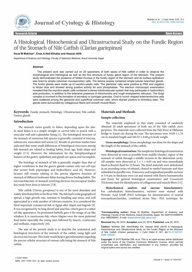
Fundic region was thrown into about 9 longitudinally oriented dome- shaped folds of different sizes. These folds were projected into a lumen, which was narrow and stellate in cross- section when empty (Figure 2A). These folds involved the lamina propria and submucosa, as well as the lining epithelium (Figures 2B and 2C).
Lamina epithelialis: Surface epithelial cells were high columnar mucosecretory in type with tapering infranuclear region. Migrating cells were seen among the epithelial cells especially in the basal part. No goblet cells were observed between the epithelial cells of fundic region.
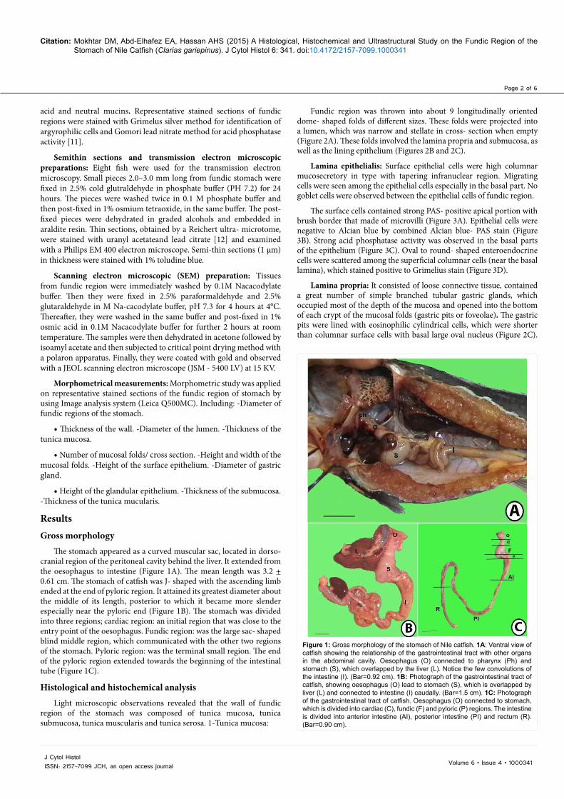
The surface cells contained strong PAS- positive apical portion with brush border that made of microvilli (Figure 3A). Epithelial cells were negative to Alcian blue by combined Alcian blue- PAS stain (Figure 3B). Strong acid phosphatase activity was observed in the basal parts of the epithelium (Figure 3C). Oval to round- shaped enteroendocrine cells were scattered among the superficial columnar cells (near the basal lamina), which stained positive to Grimelius stain (Figure 3D).
Lamina propria: It consisted of loose connective tissue, contained a great number of simple branched tubular gastric glands, which occupied most of the depth of the mucosa and opened into the bottom of each crypt of the mucosal folds (gastric pits or foveolae). The gastric pits were lined with eosinophilic cylindrical cells, which were shorter than columnar surface cells with basal large oval nucleus (Figure 2C).
Page 3 of 6
Volume 6 • Issue 4 • 1000341J Cytol HistolISSN: 2157-7099 JCH, an open access journal
Citation: Mokhtar DM, Abd-Elhafez EA, Hassan AHS (2015) A Histological, Histochemical and Ultrastructural Study on the Fundic Region of the Stomach of Nile Catfish (Clarias gariepinus). J Cytol Histol 6: 341. doi:10.4172/2157-7099.1000341
perpendicular to the gastric mucosal surface. Each gland was composed of clusters of polygonal cells called oxyntico-peptic cells, which were radially arranged around a very narrow glandular lumen. These cells had basally located spherical vesicular nuclei. The fundic glands of catfish resembled the mammalian pancreatic acini, in which they had about one fourth of the cell nearer the base was stained pale blue while the rest was stained red. The cytoplasm of these glandular cells appeared densely granular that contained acidophilic secretory granules. There were no parietal, chief or mucous neck cells as in mammals (Figure 2D).
The glandular cells were positive to PAS and negative to Alcian blue (Figure 3B). The mean height of glandular epithelium was 7.77 ± 0.36 µm. The diameter of every gastric gland was approximately the same, which their mean diameter was 17.0 ± 0.78 µm (Table 1). Oval to round- shaped enteroendocrine cells were scattered among the glandular epithelium, which stained positive to Grimelius stain (Figure 3D). The glands were surrounded by collagenous fibers and smooth muscle fibers (Figure 2B).
Semithin sections showed that the internal surface of the fundic region of the stomach was composed of folded mucosa that filled with fundic glands. The luminal surface of the mucosa was punctuated by shallow depressions of numerous gastric pits (Figure 4A). The surface epithelium was composed of tall narrow columnar cells. The apical part of the cytoplasm of epithelial cells showed muco-substances that were stained intensely with toluidine blue. The basal part of the epithelium contained polymorphic cells with many cytoplasmic processes that stained positive with toluidine blue, which may be undifferentiated basal cells (Figure 4B). The glands opened in groups of 2 or 3 into the gastric ducts. Its ducts were lined by simple squamous flat cells and opened in the gastric pits of the mucosal folds. Occasional smooth myocytes, fibroblasts and minute blood capillaries were observed between the glands (Figure 4C). The glandular cells (oxyntico-peptic cells) appeared pyramidal; their cytoplasm contained large number of vesicles of different sizes (Figure 4D).
Scanning electron microscopic observations revealed that the luminal surface of the mucosa of the fundic region had meshwork
The apices of these cells were projected into a small lumen, and their cytoplasm stained intensely with PAS (Figure 3A).
The fundic glands of fundic region were arranged in groups,
Figure 2: Histological structure of the fundic region. 2A: the mucosa (M) was thrown into many longitudinal folds (Crossmon's trichrome). 2B: the fundic region showing lamina propria (Lp), submucosa (S) and serosa (se) stained green, while, tunica muscularis (M) stained red. (Crossmon's trichrome). 2C: the folded mucosa (m) of fundic region includes epithelium (ep) and lamina propria (Lp) that contains fundic glands (fg), surrounded with submucosa (S) that contains diffuse smooth muscle bundles (arrow). (Haematoxylin and Eosin). 2D: fundic glands (fg) resemble the pancreatic acini. No parietal or chief cells. (Haematoxylin and Eosin).
Figure 3: Histochemical analysis of fundic region. 3A: PAS- positive apical part of surface simple columnar epithelium (arrow). (PAS). 3B: the fundic region showing alcian blue negative and PAS positive fundic glands (arrow). (Combined AB and PAS). 3C: the superficial lining epithelium (white arrowhead) as well as the glandular cells (black arrowheads) showed positive reactivity for acid phosphatase. (Gomori Lead Nitrate method). 3D: The mucosa of the fundic region showing positive reaction of enteroendocrine cells on the surface epithelium (arrow) and fundic glands. (Grimelius).
Measurements Fundic regionDiameter of the organ (μm) 7467.92 ± 48.39Thickness of the wall (μm) 2709.0 ± 47.42
Diameter of the lumen (μm) 2049.92 ± 71.0927.44 %
Thickness of mucosa (μm) 1518.0 ± 59.85 56.03%
Number of mucosal folds/cross section 9.50 ± 1.89
Height of mucosal folds (μm) 1279.25 ± 19.7147.22%
Width of mucosal folds (μm) 1336.13 ± 66.06
Height of the epithelium (μm) 26.6 ± 1.010.98%
Length of gastric pits(μm)
98.86 ± 3.983.64%
Diameter of gastric glands(μm) 17.0 ± 0.78
Height of glandularepithelium (μm) 7.77 ± 0.36
Thickness of submucosa (μm) 350.44 ± 14.4412.93%
Thickness of muscularis (μm) 786.93 ± 8.3129.04%
Table 1: morphometrical measurements of fundic region in relation to the thickness of the wall of the stomach of catfish.
Page 4 of 6
Volume 6 • Issue 4 • 1000341J Cytol HistolISSN: 2157-7099 JCH, an open access journal
Citation: Mokhtar DM, Abd-Elhafez EA, Hassan AHS (2015) A Histological, Histochemical and Ultrastructural Study on the Fundic Region of the Stomach of Nile Catfish (Clarias gariepinus). J Cytol Histol 6: 341. doi:10.4172/2157-7099.1000341
appearance of large number of primary longitudinal folds. The mucosal surface showed deep concavities between the mucosal folds. The most prominent feature of the fundic region was the presence of large number of gastric pits of different sizes, which punctuated the surface of the mucosa (Figure 5A). The hexagonal borders of superficial epithelial cells gave the gastric surface a honey comb- like appearance (Figure 5B). These cells were uniformly covered with few and short microvilli, which gave it a velvety appearance. Some mucous droplets were deposited on the surface (Figure 5C). In Figure 5D, the epithelial sheet of the
fundic region had been damaged and the individual columnar cells were observed. They were tall and cylindrical with narrow infranuclear region. The apical borders were covered with many short microvilli.
Transmission electron microscopic observations to the fundic glands showed that they were lined with one type of cells, which was:
Oxyntico-peptic cells: polygonal cells with basally located spherical nucleus, with prominent central electron lucent nucleolus. Spherical shaped vesicles of different sizes and density, which represented the formation of secretory or zymogen granules were distributed in cytoplasm. In many images, the granules were found in the apical portions of the cell, suggesting that their contents were about to be released via exocytosis. The luminal surface contained short microvilli, which were projected into the glandular lumen. The supranuclear portion of the cytoplasm contained a tubulovesicular network, composed of smooth membranous elements (Figure 6A). A great number of large mitochondria were located all over the cytoplasm. The rough endoplasmic reticulum was located around the nucleus. The well- developed Golgi complexes and lysosomes were arranged around the nucleus (Figures 6B-6D).
2-Tunica submucosa increased in thickness toward the pyloric region, its mean thickness was 350.44 + 14.44 µm that represented about 12.93 % of the thi ckness of the wall (Table 1). It was consisted of loose connective tissue with many collagenous, elastic fibers and numerous blood vessels with much amount of smooth muscle fibers (Figure 2B).
3-Tunica muscularis was a thick layer (Table 1). It was composed of thick inner circular and a thinner outer longitudinal layer of smooth muscle fibers (Figure 2B).
4-Tunica serosa was a thin layer of connective tissue, covered with mesothelium (Figure 2B).
All morphometrical measurements were represented in Table 1.
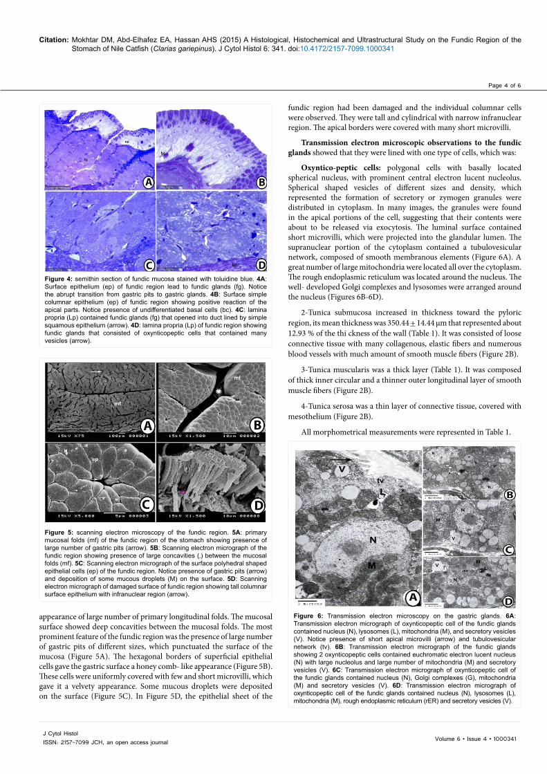
Figure 4: semithin section of fundic mucosa stained with toluidine blue. 4A: Surface epithelium (ep) of fundic region lead to fundic glands (fg). Notice the abrupt transition from gastric pits to gastric glands. 4B: Surface simple columnar epithelium (ep) of fundic region showing positive reaction of the apical parts. Notice presence of undifferentiated basal cells (bc). 4C: lamina propria (Lp) contained fundic glands (fg) that opened into duct lined by simple squamous epithelium (arrow). 4D: lamina propria (Lp) of fundic region showing fundic glands that consisted of oxynticopeptic cells that contained many vesicles (arrow).
Figure 5: scanning electron microscopy of the fundic region. 5A: primary mucosal folds (mf) of the fundic region of the stomach showing presence of large number of gastric pits (arrow). 5B: Scanning electron micrograph of the fundic region showing presence of large concavities (*) between the mucosal folds (mf). 5C: Scanning electron micrograph of the surface polyhedral shaped epithelial cells (ep) of the fundic region. Notice presence of gastric pits (arrow) and deposition of some mucous droplets (M) on the surface. 5D: Scanning electron micrograph of damaged surface of fundic region showing tall columnar surface epithelium with infranuclear region (arrow).
Figure 6: Transmission electron microscopy on the gastric glands. 6A: Transmission electron micrograph of oxynticopeptic cell of the fundic glands contained nucleus (N), lysosomes (L), mitochondria (M), and secretory vesicles (V). Notice presence of short apical microvilli (arrow) and tubulovesicular network (tv). 6B: Transmission electron micrograph of the fundic glands showing 2 oxynticopeptic cells contained euchromatic electron lucent nucleus (N) with large nucleolus and large number of mitochondria (M) and secretory vesicles (V). 6C: Transmission electron micrograph of oxynticopeptic cell of the fundic glands contained nucleus (N), Golgi complexes (G), mitochondria (M) and secretory vesicles (V). 6D: Transmission electron micrograph of oxynticopeptic cell of the fundic glands contained nucleus (N), lysosomes (L), mitochondria (M), rough endoplasmic reticulum (rER) and secretory vesicles (V).

Page 5 of 6
Volume 6 • Issue 4 • 1000341J Cytol HistolISSN: 2157-7099 JCH, an open access journal
Citation: Mokhtar DM, Abd-Elhafez EA, Hassan AHS (2015) A Histological, Histochemical and Ultrastructural Study on the Fundic Region of the Stomach of Nile Catfish (Clarias gariepinus). J Cytol Histol 6: 341. doi:10.4172/2157-7099.1000341
DiscussionThe present work was carried out on 20 specimens of both sexes of
catfish in order to observe the morphological and histological as well as fine structure of fundic gland region. The results indicated that the stomach was J-shaped in Nile catfish, while Caceci et al. [13] recorded Y-shaped stomach in O. niloticus, true cods and ocean perch, Souza and Intelizano, [14,15] found U- shaped in Salmo and Loricariidae and Chakrabarti and Ghosh [8] reported gizzard- like stomach in Liza parsia. Any of these configurations must be convenient for containing the ingested food. The stomach of fish was absent in early developmental stages, but the lack of the stomach was not restricted to larval fish. Juveniles and adults of a variety of fishes were stomachless such as cyprinids (carp, goldfish), beloniforms (needle fish) and scarids [16,17]. In the later species, the digestion was mainly done by pancreatic and intestinal mucosal enzymes [18,19].
The presence of gastric mucosal folds in fundic stomach catfish is to allow the fish to accommodate large variations in meal size, by permitting a great deal of distention when excess food is present. The distension served as a powerful stimulus for gastric digestive secretions in teleosts. It is also possible that this pattern of folding may slow down the passage of food in the stomach and divide the ingested bolus into smaller portions for more efficient mixing with the digestive fluid. These findings are in consistence with those observed by Osman and Caceci [20].
In agreement with several authors (Pedini et al., Chatchavalvanich et al., and Hernandez et al.,) [21-23], the surface epithelial cells of the stomach were PAS positive, which indicated that it contained neutral muco-substances, which play a role in digestive activity such as: facilitating the movement of the great size particles of food and then emulsifying it into chyme after mixing with several digestive enzymes. These muco-substances are also related to the absorption of easily digested molecules, such as disaccharide and short-chain fatty acids. On the other hand, the secretion of muco-substances from the gastric epithelium play a role in protecting the underlying mucosa from the acid environment and proteolysis; neutralizing the gastric PH and act as defensive mechanism against bacteria.
The gastric lamina propria observed in fundic region of the stomach of catfish may help to keep the glands in position and it was rich in collagenous fibers that might form a protective, supporting and strengthing layer in the carnivorous fish. The present study demonstrated the presence of folded mucosa in fundic region of the stomach and its surface epithelium was lined by simple columnar mucosecretory cells. The lamina propria contained simple branched tubular glands. In our results, surface cells and gastric glands of fundic region contained enteroendocrine cells, which were positive to Grimulius stain. Ghattas [24] concluded that the gastric mucosa of catfish contained numerous enteroendocrine cells that secreted gastrin, somatostatin, and serotonin. The author suggested that these hormones might stimulate the glandular cells to increase their rate of HCl secretions.
The glandular cells which made up the fundic glands were called oxyntico-peptic by most authors because they secrete both pepsin and HCl [25,26]. In the current study, the electron microscopic examination revealed that the oxyntico-peptic cells contained tubulovesicular system that may participate in hydrochloric acid production, a process involved an H+K+ ATPase. The extensive presence of mitochondria reflected the high energy requirements of the above mention ionic exchange. The rough endoplasmic reticulum observed in oxyntico-
peptic cells could be involved in the production of gastric juices. The Golgi complex is involved in the formation of secretory or zymogen granules, whose contents are eventually released into the glandular lumen. These findings are in agreement with the results obtained by Garrido et al., [27] in European eel; Gargiulo et al., [28] in Tilapia spp; Carrasson, et al., [29] in Dentex dentex and El-Habback, [30] in Tilapia nilotica.
Surface and exocrine glandular cells secrete gastric juices containing digestive enzymes; including lipolytic enzymes [27,31]. Thus, the well- developed gastric glands and the epithelial columnar cells that were rich in neutral mucins implied that the stomach of catfish had strong capabilities of digestion and absorption.
This study demonstrated smooth muscle fibers around the gastric glands of catfish that may probably play an important role by their contraction to induce release of glandular and faveaolar secretion into the gastric lumen and prevent occlusion of foveolae by ingested food. Also, these muscle fibers may aid in the movement of the folds, which blend the food bolus to facilitate mixing with gastric juice in the stomach. Similar findings were observed by Osman and Caceci [20]. The smooth muscular layer of the stomach may aid in final breakdown of the food before entering the intestine.
In conclusion, the fundic region of the stomach of Clarias gariepinus had revealed special characteristics that are undoubtedly correlated with its feeding habits. The columnar epithelial cells lining the fundic mucosa secreted neutral mucin justifying their active role to protect the underlying epithelial cells from acid, enzymes and mechanical rubbing. Positive reaction to PAS in the tubular glands may be concerned with formation of neutral mucopolysaccharides and high acid phosphatase activity was also related to metabolic activity of the cell concerned. The oxyntico-peptic cells contained tubulovesicular system, rER and Golgi apparatus which were involved in synthesis of Hcl, gastric juices and secretory granules. However, the results presented in the current study may be considered as a base line for subsequent studies on the stomach of different teleosts. Immunohistochemistery for oxntico-peptic cells and enteroendocrine cells of the fundic region of the stomach of Nile catfish should be done.
References
1. Suíçmez M, Ulus E (2005) A study of the anatomy, histology and ultrastructure of the digestive tract of Orthrias angorae Steindachner, 1897. Folia Biol (Krakow) 53: 95-100.
2. Haloi K, Kalita M, Nath P (2013) The study on the histopathological changes of stomach of Channa punctatus (Bloch) by used pesticide endosulfan. Global J Sci Front Res Bio Sci 13:1-6.
3. Fugi R, Agostinho AA, Hahn NS (2001) Trophic morphology of five benthic-feeding fish species of a tropical floodplain. Braz J Biol 61: 27-33.
4. Abdulhadi HA (2005) Some comparative histological studies on alimentary tract of tilapia fish (Tilapia spilurus) and sea bream (Mylio cuvieri). Egypt J Aquat Res 31: 387-397.
5. Khalaf Allah HMM (2013) Morphological adaptations of digestive tract according to food and feeding habits of the broomtail wrasse, Cheilinus lunulatus. Egypt Aquat Biol Fish 17: 123-141.
6. Albrecht MP, Ferreira MFN, Caramaschi EP (2001) Anatomical features and histology of the digestive tract of two related neotropical omnivorous fishes (Characiformes; Anostomidae). Fish Biol 58: 419-430.
7. Naguib SAA, EI-Shabaka HA, Ashour F (2011) Comparative Histological and Ultrastructural Studies on the Stomach of Schilbe mystus and the Intestinal Swelling of Labeo niloticus. American Sci 7: 251-263.
8. Chakrabarti P, Ghosh SK (2014) A comparative study of the histology and microanatomy of the stomach in Mystus vittatus (Bloch), Liza parsia (Hamilton) and Oreochromis mossambicus (Peters). J Microsc Ultrastr 2: 245-250.
Page 6 of 6
Volume 6 • Issue 4 • 1000341J Cytol HistolISSN: 2157-7099 JCH, an open access journal
Citation: Mokhtar DM, Abd-Elhafez EA, Hassan AHS (2015) A Histological, Histochemical and Ultrastructural Study on the Fundic Region of the Stomach of Nile Catfish (Clarias gariepinus). J Cytol Histol 6: 341. doi:10.4172/2157-7099.1000341
9. Khallaf EA, Gaber N (1991) Analysis of stomach contents and intraspecific interaction over diet of Claris lazera (Cuv. & Val.) in Bahr- Shebeen canal. JBulletin of Faculty of Science, Zagazig University 13: 481-499.
10. Amisah S, Oteng MA, Ofori JK (2009) Growth performance of the African catfish, Claris gariepinus, fed varying inclusion levels of Leucaena leucocephala leafmeal. Appl Sci Environ Manag 13: 21-26.
11. Bancroft JD, Steven A (1996) Theory and practice of histological techniques(4thedn.) Churchill Livingstone. New York. Edinburgh. London. Madrid.Melbourne. San Francisco. Tokyo.
12. REYNOLDS ES (1963) The use of lead citrate at high pH as an electron-opaque stain in electron microscopy. J Cell Biol 17: 208-212.
13. Caceci T, El-Habback HA, Smith SA, Smith BJ (1997) The stomach ofOreochromis niloticus has three regions. Fish Biol 50: 939-952.
14. Souza AM, Intelizano W (2000) Anatomy and histology of the stomach in some species of Loricariidae (Siluriformes, Ostariophysi). J Publs Avulsas do Instituto pau Brasil 10: 3-16.
15. Ghosh SK, Chakrabarti P (2015) Histological and histochemical characterization on stomach of Mystus cavasius (Hamilton), Oreochromis niloticus (Linnaeus)and Gudusia chapra (Hamilton): Comparative study. J Basic Appl Zool 70: 16-24.
16. Kent GC, Miller L (1997) Comparative anatomy of the vertebrates. (8th edn.)The McGraw- Hill Companies, Inc.
17. Manjakasy JM, Day RD, Kemp A, Tibbetts IR (2009) Functional morphology ofdigestion in the stomachless, piscivorous needlefishes Tylosurus gavialoides and Strongylura leiura ferox (Teleostei: Beloniformes). J Morphol 270: 1155-1165.
18. Kowalska A, Zakes Z, Zakes KD (2006) The impact of feeding on the results of rearing larval pikeperch, Sander lucioperca (L.), with regard to the development of the digestive tract. J Polish Agricul Univ 9: 88-96.
19. Mokhtar DM (2015) Histological, histochemical and ultrastructuralcharacterization of the pancreas of the grass carp (Ctenopharyngodon idella).Eur J Anat 19: 145-153.
20. Osman AHK, Caceci T (1991) Histology of the stomach of Tilapia nilotica(Linnaeus, 1758) from the River Nile. Fish Biol 38: 211-223.
21. Pedini V, Aglio CD, Parillo F, Scocco P (2005) Glycoconjugate distribution ingastric fundic mucosa of Umbrina cirrosa L. revealed by lectin histochemistry.Fish Biol 66: 222-229.
22. Chatchavalvanich K, Marcos R, Poonpirom J, Thongpan A, Rocha E (2006)Histology of the digestive tract of the freshwater stingray Himantura signiferCompagno and Roberts, 1982 (Elasmobranchii, Dasyatidae). Anat Embryol(Berl) 211: 507-518.
23. Hernandez DR, Gianeselli, PM, Domitrovic HA (2009) Morphology, histologyand histochemistry of the digestive system of South American Catfish (Rhamdia quelen). J Int Morphol 27: 105-111.
24. Ghattas SM (2004) A histological study on the digestive tract of eel (Anguillaanguilla L.) with special reference to ultrastructure of gastric glandular cells. JMansoura Vet Med Anostomidae) Fish Biol 58: 419-430.
25. Diaz AO, García AM, Figueroa DE, Goldemberg AL (2008) The mucosa ofthe digestive tract in Micropogonias furnieri: a light and electron microscopeapproach. Anat Histol Embryol 37: 251-256.
26. Domeneghini C, Arrighi S, Radaelli G, Bosi G, Veggetti A (2005) Histochemical analysis of glycoconjugate secretion in the alimentary canal of Anguilla anguilla L. Acta Histochem 106: 477-487.
27. Garrido MV, Oller C, Equisoain MA (1996) Effect of diet on gastric mucosa cells in the European eel (Anguilla anguilla L.). Histochemical and ultrastructuralStudy. J Micron 27 1: 25-34.
28. Gargiulo AM, Ceccarelli P, Dall’aglio C, Pedini V (1997) Ultrastructural study on the stomach of Tilapia spp (Teleostei). Anat Histol Embryol 26: 331-336.
29. Carrassón M, Grau A, Dopazo LR, Crespo S (2006) A histological, histochemical and ultrastructural study of the digestive tract of Dentex dentex (Pisces,Sparidae). Histol Histopathol 21: 579-593.
30. El-Habback HA (2007) Post- hatching development of the stomach of tilapianilotica (Oreochromis niloticus) light and electron microscopic studies. J VetMed Kafrelsheikh 5 2: 1-31.
31. Sandhu AA (2000) Comparative anatomical and histological studies of thegastrointestinal tract of some fresh water catfishes in relation to their food (Pisces: Siluriformes), Ph.D, University of the Punjab, Lahore.