Embed Size (px)
Citation preview
Module EModule E
Computed Tomography Computed Tomography Physics, Instrumentation, Physics, Instrumentation,
and Imagingand Imaging

DisclaimerDisclaimer This workforce solution was funded by a grant awarded under the This workforce solution was funded by a grant awarded under the
President’s Community-Based Job Training Grants as implemented President’s Community-Based Job Training Grants as implemented by the U.S. Department of Labor’s Employment and Training by the U.S. Department of Labor’s Employment and Training Administration. The solution was created by the grantee and does Administration. The solution was created by the grantee and does not necessarily reflect the official position of the U.S. Department of not necessarily reflect the official position of the U.S. Department of Labor. The Department of Labor makes no guarantees, warranties, Labor. The Department of Labor makes no guarantees, warranties, or assurances of any kind, express or implied, with respect to such or assurances of any kind, express or implied, with respect to such information, including any information on linked sites and including, information, including any information on linked sites and including, but not limited to, accuracy of the information or its completeness, but not limited to, accuracy of the information or its completeness, timeliness, usefulness, adequacy, continued availability, or timeliness, usefulness, adequacy, continued availability, or ownership. This solution is copyrighted by the institution that ownership. This solution is copyrighted by the institution that created it. Internal use by an organization and/or personal use by created it. Internal use by an organization and/or personal use by an individual for non-commercial purposes is permissible. All other an individual for non-commercial purposes is permissible. All other uses require the prior authorization of the copyright owner.uses require the prior authorization of the copyright owner.

AttenuationAttenuation
The reduction in the intensity of a The reduction in the intensity of a radiographic beam as it travels through radiographic beam as it travels through matter.matter.
Remnant radiationRemnant radiation

X-ray InteractionsX-ray Interactions
Coherent scatterCoherent scatter Compton EffectCompton Effect Photoelectric EffectPhotoelectric Effect Pair ProductionPair Production Triplet ProductionTriplet Production PhotodisintegrationPhotodisintegration

Diagnostic x-ray InteractionsDiagnostic x-ray Interactions
Compton – Compton – non-diagnosticnon-diagnostic
Photoelectric Effects – Photoelectric Effects – the the x-ray interaction which will result x-ray interaction which will result in a diagnostic imagein a diagnostic image
Attenuation in CTAttenuation in CT
Depends on:Depends on: the effective atomic density in the effective atomic density in
atoms/volumeatoms/volume The Z-number of the absorber (atomic The Z-number of the absorber (atomic
number)number) The energy of the x-ray photonsThe energy of the x-ray photons

Linear Attenuation CoefficientLinear Attenuation Coefficient
Although the Photoelectric effect is more predominant…. Although the Photoelectric effect is more predominant…. The Compton interaction must be considered when The Compton interaction must be considered when developing mathematical formulas to determine the developing mathematical formulas to determine the changes of x-ray attenuation through different tissue and changes of x-ray attenuation through different tissue and then reconstruct images of the anatomy scanned. then reconstruct images of the anatomy scanned.
Requires:Requires: The application of physicsThe application of physics Complex mathematics andComplex mathematics and Computer science WHY………….Computer science WHY………….
CT and x-ray attenuationCT and x-ray attenuation
The determination of x-ray attenuation in The determination of x-ray attenuation in body tissue and the use of that information body tissue and the use of that information to reconstruct images of the anatomy to reconstruct images of the anatomy scanned is the basic problem in CT.scanned is the basic problem in CT.

Homogeneous / MonochromaticHomogeneous / Monochromatic
Gamma radiation source with a pencil beam configuration Gamma radiation source with a pencil beam configuration was initially used in Hounsfield’s tests.was initially used in Hounsfield’s tests.
A radiation source with 1 frequency was needed to use the A radiation source with 1 frequency was needed to use the Lambert-Beer Law for calculations.Lambert-Beer Law for calculations.
The attenuation characteristic of the homogeneous The attenuation characteristic of the homogeneous radiation source interacting with human tissue causes radiation source interacting with human tissue causes absorption of photons but no loss in the quality or energy absorption of photons but no loss in the quality or energy of the beam. of the beam.
This allowed accurate calculations to be made determining This allowed accurate calculations to be made determining the the linear attenuation coefficientslinear attenuation coefficients for human tissue. for human tissue.

IIinin = = IIoutout ee -μx-μx
Lambert-Beer LawLambert-Beer Law
CT attenuation depends on CT attenuation depends on • Effective atomic density in atoms/volumeEffective atomic density in atoms/volume• The Z-number of the absorber (atomic The Z-number of the absorber (atomic
number)number)• The energy of the x-ray photons The energy of the x-ray photons

Monochromatic vs. PolychromaticMonochromatic vs. Polychromatic
Original experiments using monochromatic – quality of Original experiments using monochromatic – quality of the beam remained the same after interaction with the beam remained the same after interaction with tissue. Only the quantity changed allowing for the tissue. Only the quantity changed allowing for the requirements necessary to use the Lambert-Beer Law requirements necessary to use the Lambert-Beer Law for mathematic computations.for mathematic computations.
Polychromatic beam (x-ray) attenuates at various Polychromatic beam (x-ray) attenuates at various rates….. Quantity and Quality are both changed. rates….. Quantity and Quality are both changed.
In addition, the beam geometry also changed from pencil In addition, the beam geometry also changed from pencil to Fan-beam by using polychromatic x-ray radiation. to Fan-beam by using polychromatic x-ray radiation. The differences in beam energy frequencies creates the The differences in beam energy frequencies creates the fan beam configuration of a divergent x-ray source.fan beam configuration of a divergent x-ray source.

New EquationNew Equation
N = NN = Nooee-μx-μx
N = the number of transmitted x-ray photons N = the number of transmitted x-ray photons No = the number of x-ray photons entering body tissue No = the number of x-ray photons entering body tissue
(incidental photons) (incidental photons) μ = equals the linear attenuation coefficients of the tissue μ = equals the linear attenuation coefficients of the tissue (μp +μc) (μp +μc) e = the base of the natural logarithm e = the base of the natural logarithm (Euler’s Constant of 2.718)(Euler’s Constant of 2.718)
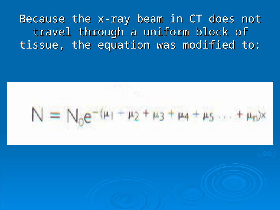
Because the x-ray beam in CT does not travel Because the x-ray beam in CT does not travel through a uniform block of tissue, the equation was through a uniform block of tissue, the equation was
modified to:modified to:

CT vs. Conventional radiographyCT vs. Conventional radiography
Cross-sectional imagingCross-sectional imaging Eliminates superimpositionEliminates superimposition Sensitive to subtle variations in x-ray Sensitive to subtle variations in x-ray
attenuationattenuation Digital imaging processing techniqueDigital imaging processing technique Images produced from topographic maps Images produced from topographic maps
of the x-ray linear attenuation coefficients.of the x-ray linear attenuation coefficients.

MaxrixMaxrix
-an array of numbers composed of -an array of numbers composed of rows and columnsrows and columns
-512 x 512 = 262,144 pixels-512 x 512 = 262,144 pixels
-1024 x 1024 = 1,042,576 pixels-1024 x 1024 = 1,042,576 pixels

MatrixMatrix
Pixels within the matrix represent various Pixels within the matrix represent various tissue types.tissue types.
Tissue differences are represented by CT Tissue differences are represented by CT numbers or Hounsfield units.numbers or Hounsfield units.
A pixel is considered a two-dimensional A pixel is considered a two-dimensional representationrepresentation

Pixel size / Voxel sizePixel size / Voxel size
P (mm2) = DFOV (mm
2) / Matrix Size
Voxel Size (mm3) = DFOV (mm2) (slice thickness in mm) / Matrix Size

Pixel and VoxelPixel and Voxel

SamplingSampling Process for determining attenuation Process for determining attenuation
valuesvalues Type of measurement techniqueType of measurement technique Enables CT to change analog Enables CT to change analog
information into digital information information into digital information without losing information.without losing information.
Two types of SamplingTwo types of Sampling Angular Angular rayray

Nyquist TheoremNyquist Theorem
““Sampling must be performed at Sampling must be performed at least twice the spatial frequency of least twice the spatial frequency of the object scanned”.the object scanned”.

Angular / Ray SamplingAngular / Ray Sampling
Angular Sampling-Angular Sampling-• Determined by the distance between each Determined by the distance between each
view obtained during the scanview obtained during the scan
Ray Sampling-Ray Sampling-• Determined by the angle between each pair Determined by the angle between each pair
of rays within a viewof rays within a view

Rays and ViewsRays and Views
44thth generation scans- generation scans- View is a set of rays that strike a single View is a set of rays that strike a single
(specific) detector (remember the tube is (specific) detector (remember the tube is rotating around the patient in 4rotating around the patient in 4 thth generation generation scanners)scanners)
The value of each ray is directly proportional The value of each ray is directly proportional to the transmitted photon measured and to the transmitted photon measured and characterized as a CT number.characterized as a CT number.
Data from each view is called “Raw Data”Data from each view is called “Raw Data”