Embed Size (px)
Citation preview

Modulation of the M2 Muscarinic Cholinergic
Receptor by Cholesterol
by
Alejandro Tienzo Colozo
A thesis submitted in conformity with the requirements for the degree of Doctor of Philosophy
Graduate Department of Pharmaceutical Sciences University of Toronto
© Copyright by Alejandro Tienzo Colozo, 2009

ii
Modulation of the M2 Muscarinic Cholinergic Receptor by Cholesterol
Doctor of Philosophy, 2009
Alejandro Tienzo Colozo
Department of Pharmaceutical Sciences, University of Toronto
ABSTRACT
M2 muscarinic receptor extracted from Sf9 cells in cholate-NaCl differs from that
extracted from porcine sarcolemmal membranes. Whereas the latter has been shown to
exhibit non-competitive effects in the binding of N-methylscopolamine (NMS) and
quinuclidinylbenzilate (QNB), which can be explained in terms of cooperativity within a
receptor that is at least tetravalent, binding to the former is essentially competitive.
Levels of cholesterol in Sf9 membranes were only 5% of those in sarcolemmal
membranes and were increased to about 100% by means of cholesterol-methyl-β-
cyclodextrin. M2 receptors extracted from CHL-treated Sf9 membranes resembled those
from heart; that is, cholesterol induced a pronounced heterogeneity detected in the
binding of both radioligands, including a shortfall in the apparent capacity for [3H]NMS,
and there were marked discrepancies in the apparent affinity of NMS as estimated
directly and via the inhibition of [3H]QNB. The data can be described quantitatively in
terms of cooperative effects among six or more interacting sites, apparently within an
oligomer. Cholesterol also was found to increase the affinity of the receptor for NMS
and QNB, and the effect was examined for its possible relationship to the known
interconversion of cardiac muscarinic receptors between an agonist-specific (R*) and an
antagonist-specific (R) state. Cholesterol and N-ethylmaleimide (NEM) were compared
for their effect on the affinity of NMS, QNB and four muscarinic agonists, and the data
were assessed in terms of an explicit mechanistic model for a receptor that interconverts
spontaneously between two states (i.e., R º R*). The data can be described equally well

iii
by an effect of cholesterol on either the distribution of receptors between R and R* or the
affinity of all ligands for both states, with an accompanying effect of NEM on either the
affinity or the distribution between states, respectively. Since NEM is known from other
data to favor R* over R, cholesterol appears to increase affinity per se. Cholesterol
therefore is a determinant of affinity and cooperativity in the binding of orthosteric
ligands to the M2 receptor. Both effects are observed in solution and therefore appear to
arise from a direct interaction between the lipid and the receptor.

iv
I never think of the future – it comes soon enough.
Albert Einstein

v
ACKNOWLEDGEMENTS
I thank my father and mother, Wilfredo and Adelina Colozo for their unwavering support in all of my endeavors and I thank them for always trying to instill in me the value of hardwork and education. I am grateful for all the help of those whom I have worked with during my time here, Chi Sum, Paul Park, Amy Ma, Luca Pisterzi, Dar’ya Redka, Rabindra Shivnaraine, David Greiss, Judy Chou and John Dong. I thank my advisory committee members, Dr. Christine Bear, Dr. Hubert Van Tol and Dr. Christopher Yip for their guidance and helpful insights throughout the completion of this work. I am grateful to my supervisor James W. Wells for always making time and to whom I am very much indebted. This work was supported by the Canadian Institutes of Health Research (CIHR), the Heart and Stroke Foundation of Ontario (HSFO), Ontario Graduate Scholarships (OGS) and an Ontario Graduate Scholarship in Science and Technology (OGSST).

vi
TABLE OF CONTENTS
ABSTRACT ....................................................................................................................................... ii ACKNOWLEDGEMENTS .................................................................................................................... v LIST OF SCHEMES .......................................................................................................................... vii LIST OF TABLES .............................................................................................................................. ix LIST OF FIGURES.............................................................................................................................. x SUMMARY OF ABBREVIATIONS ..................................................................................................... xii CHAPTER 1: GENERAL INTRODUCTION ....................................................................................... 1
1.1 The Receptor Concept.......................................................................................................... 2 1.2 The G protein-Coupled Receptor Superfamily .................................................................. 11 1.3 Mechanism of Signaling .................................................................................................... 16 1.4 Oligomers of G protein-coupled receptors......................................................................... 29 1.5 Cholesterol ......................................................................................................................... 32 1.6 An Outline of the Thesis .................................................................................................... 39
CHAPTER 2: CHOLESTEROL AS A DETERMINANT OF COOPERATIVITY IN THE M2 MUSCARINIC CHOLINERGIC RECEPTOR ...................................................................................... 43
2.1 Abstract .............................................................................................................................. 44 2.2 Introduction........................................................................................................................ 45 2.3 Materials and Methods....................................................................................................... 47 2.4 Results................................................................................................................................ 56 2.5 Discussion .......................................................................................................................... 63 2.6 Appendix............................................................................................................................ 72 2.7 Acknowledgements ............................................................................................................ 74
CHAPTER 3: EFFECT OF CHOLESTEROL ON THE BINDING OF ANTAGONISTS TO THE MUSCARINIC CHOLINERGIC RECEPTOR EXTRACTED FROM PORCINE ATRIA... 89
3.1 Abstract .............................................................................................................................. 90 3.2 Introduction........................................................................................................................ 91 3.3 Materials and Methods....................................................................................................... 92 3.4 Results................................................................................................................................ 98 3.5 Discussion ........................................................................................................................ 100 3.6 Acknowledgements .......................................................................................................... 102
CHAPTER 4: MECHANISTIC BASIS FOR THE EFFECT OF CHOLESTEROL ON THE AFFINITY OF AGONISTS AND ANTAGONISTS FOR THE M2 MUSCARINIC CHOLINERGIC RECEPTOR ......................................................................................................... 108
4.1 Abstract ............................................................................................................................ 109 4.2 Introduction...................................................................................................................... 110 4.3 Materials and Methods..................................................................................................... 113 4.4 Results.............................................................................................................................. 119 4.5 Discussion ........................................................................................................................ 125 4.6 Acknowledgements .......................................................................................................... 131
CHAPTER 5: GENERAL DISCUSSION ........................................................................................ 142 5.1 Insights from the Thesis ................................................................................................... 143 5.2 Cooperativity.................................................................................................................... 145 5.3 Functional Role of Cholesterol ........................................................................................ 149
CHAPTER 6: FUTURE DIRECTIONS........................................................................................... 156 6.1 Specificity in the Effects of Cholesterol on Cooperativity and Affinity .......................... 157 6.2 Oligomeric Status of the Receptor ................................................................................... 158
APPENDIX ................................................................................................................................. 161 REFERENCES............................................................................................................................ 179

vii
LIST OF SCHEMES
Scheme 1
Scheme 2

viii
Scheme 3

ix
LIST OF TABLES
CHAPTER 2: Cholesterol as a determinant of cooperativity in the M2 muscarinic
cholinergic receptor
Table 2-1: Empirical characterization of specific binding
Table 2-2: Affinities of NMS for M2 receptor extracted from native and cholesterol-treated Sf9 membranes, estimated empirically in terms of Scheme 1
CHAPTER 3: Effect of cholesterol on the binding of antagonists to the muscarinic
cholinergic receptor extracted from porcine atria
Table 3-1: Empirical characterization of specific binding in terms of the Hill equation
Table 3-2: Affinities of NMS for M2 receptor extracted from native and
cholesterol-depleted sarcolemmal membranes of porcine atria, estimated empirically in terms of Scheme 1
CHAPTER 4: Mechanistic basis for the effect of cholesterol on the affinity of agonists
and antagonists for the M2 muscarinic cholinergic receptor
Table 4-1: Empirical characterization of specific binding in terms of the Hill equation
Table 4-2: Affinities of agonists and antagonists, estimated in terms of
Scheme 1
Table 4-3: Affinities of agonists and antagonists, estimated in terms of Scheme 3
Table 4-4: Affinities of agonists and antagonists, estimated in terms of
Scheme 3

x
LIST OF FIGURES
CHAPTER 2: Cholesterol as a determinant of cooperativity in the M2 muscarinic
cholinergic receptor
Figure 2-1: Incorporation of cholesterol into the membranes of Sf9 cells Figure 2-2: Electrophoretic mobility of M2 receptor extracted from native and
cholesterol-treated Sf9 membranes Figure 2-3: Stability of binding to M2 receptor extracted from native Sf9
membranes Figure 2-4: Equilibration of [3H]NMS with M2 receptor extracted from
cholesterol-treated Sf9 membranes Figure 2-5: Binding to M2 receptor extracted from native Sf9 membranes,
analyzed empirically in terms of Scheme 1 Figure 2-6: Binding to M2 receptor extracted from cholesterol-treated Sf9
membranes, analyzed empirically in terms of Scheme 1. Figure 2-7: Comparison of Schemes 1 and 2 for goodness of fit to data from
cholesterol-treated Sf9 membranes Figure 2-8: Optimal number of interacting sites for the fit of Scheme 2 to data
from cholesterol-treated membranes Figure 2-9: Binding to M2 receptor extracted from cholesterol-treated Sf9
membranes, analyzed in terms of Scheme 2
CHAPTER 3: Effect of cholesterol on the binding of antagonists to the muscarinic cholinergic receptor extracted from porcine atria
Figure 3-1: Levels of cholesterol in membranes from porcine sarcolemma, Sf9
cells and CHO cells Figure 3-2: Binding to M2 receptor extracted from native and cholesterol-
depleted sarcolemmal membranes of porcine atria, analyzed empirically in terms of Scheme 1
CHAPTER 4: Mechanistic basis for the effect of cholesterol on the affinity of agonists
and antagonists for the M2 muscarinic cholinergic receptor

xi
Figure 4-1: Effect of N-ethylmaleimide on the apparent affinity of oxotremorine-M and [3H]quinuclidinylbenzilate for M2 receptor extracted from native and cholesterol-treated Sf9 membranes
Figure 4-2: Effect of cholesterol and N-ethylmaleimide on the binding of
agonists and antagonists to M2 receptor extracted from Sf9 membranes
Figure 4-3: Fit of Scheme 3 mapped with respect to KR as affected by N-
ethylmaleimide and cholesterol Figure 4-4: Domain of acceptable parametric values for an effect of N-
ethylmaleimide on KR in Scheme 3 Figure 4-5: Domain of acceptable parametric values for an effect of cholesterol
on KR in Scheme 3 APPENDIX: Assessing the oligomeric status of the M2 muscarinic receptor using
atomic force microscopy Figure A-1: Development of substrate and incorporation of the M2 receptor Figure A-2: Effect of repetitive freezing and thawing on vesicular size assessed
by electron and atomic force microscopy

xii
SUMMARY OF ABBREVIATIONS
AICc second-order Akaike’s information criterion
AFM atomic force microscopy
BRET bioluminescence resonance energy transfer
DAMGO [D-Ala2,N-MePhe4,Gly-ol5]enkephalin
BS3 bis[sulfosuccinimidyl]suberate
CHL cholesterol
CHO Chinese hamster ovary
EM Electron microscope
FRET fluorescence (Förster) resonance energy transfer
GMP-PNP guanylylimidodiphosphate
GPCR G protein-coupled receptor
GTPγS guanosine 5´-O-[3-(γ-thio)triphosphate]
HEN HEPES, EDTA and NaCl
MβCD methyl-β-cyclodextrin
p-MPPF (4-2´-methoxy)-phenyl-1-[2´-(N-2˝-pyridinyl-p-fluorobenzamido]ethyl-
piperazine
NEM N-ethylmaleimide
NMS N-methylscopolamine
Oxo-M oxotremorine-M
PC phosphatidylcholine
PMSF phenylmethylsulfonylfluoride
PS phosphatidylserine
QNB (−)-quinuclidinylbenzilate
SDS-PAGE Sodium Dodecyl Sulfate-Polyacrylamide Gel Electrophoresis
Sf9 Spodoptera frugiperda

1
CHAPTER 1
GENERAL INTRODUCTION

2
1.1 The Receptor Concept
The origins of the idea of the receptor can be traced back to the works of Paul Ehrlich and
John Newport Langley between the period of 1878 and 1908 [1]. Although the nature of
their research was quite different, the ideas that emerged from their studies eventually led
to the modern concept of the receptor. In the case of Paul Ehrlich, his early work was
concerned with the drug resistance of trypanosomes, for which he developed a ‘side chain
theory’ of immunity in order to give a theoretical platform to the chemotherapy of such
micro-organisms [2]. As for John Langley, his research interest was mainly focused on
the effects of alkaloids on muscle and nerve. In his early work as a student at Cambridge,
Langley was involved in examining the opposing actions of atropine and pilocarpine on
the secretion of saliva by the sub-maxillary gland [3].
It was in 1905 when Langley first spoke of the existence of what he referred to as
specific ‘receptive substances’ in cells [4]. He had shown that nicotine causes tonic
contraction in certain muscles of the fowl, frog and toad, that this contraction is prevented
by a sufficient quantity of curari and that the action of both ‘poisons’ in the fowl is
unaltered by degeneration of the nerve endings [5].
Paul Ehrlich initially rejected Langley’s notion of the receptor in favor of his own
‘receptive side chain’ hypothesis. In Ehrlich’s view, the protoplasm of a cell is akin to a
giant molecule which contains a central structure which possesses chemical side chains
that are responsible for the specific activity of that given cell type [1]. According to
Ehrlich, a particular ‘receptive side chain’ has an atomic group which has specific
combining properties for a particular toxin. Such groups are said to act as antibodies or
antitoxins by combining with foreign agents in the bloodstream, thus preventing the toxin
from attaching to cells. The relative ease by which drugs can be removed from tissues
with the use of solvents led Ehrlich to conclude that, unlike toxins, drugs are not bound
firmly to cells. He believed that the action of drugs did not apply to his proposed
‘receptive side chains’ because, unlike toxins which are bound to the protoplasmic
molecule of the cell by chemical union, the former in his view lacked the necessary

3
atomic groups to do so. By 1907, however, the growing body of evidence from
Langley’s work had convinced Ehrlich that the concept of the receptor as articulated by
his contemporary also applied to chemical substances in general. Thus the ‘receptive
substance’ described initially by John Newport Langley was designated a
‘chemoreceptor’ by Paul Ehrlich [6].
The receptor concept was not met with universal acceptance. During the period
between 1895 and 1930, there were several alternative hypotheses for drug action [4,7].
A few were based on homeopathy while others were based on physicochemical
considerations. Examples of the latter posed by far the stronger challenges to the receptor
concept. One of the more prominent ones was the physical theory, a notion championed
by Walther Straub who was an internationally renowned pharmacologist at the time. The
physical theory emphasized that the surface tension of the cell membrane greatly
influenced the concentration gradient between the inside and outside of the cell, and thus
controlled the effect of the drug on the receptive organ. With experiments on the isolated
heart of the snail, Walther Straub observed a decrease or a complete cessation of the heart
rate upon addition of muscarine [4,8]. He concluded that this effect depended upon the
absorption of the alkaloid into the muscle cell, rather than the direct effect of the poison
once inside the cell itself. Straub was convinced that the decisive factor that controlled
toxicity was the difference in the poison concentration between the inside and the outside
of the cell, which he referred to as the ‘concentration potential’. He reasoned that, as the
poison entered the cell through the membrane, the concentration gradient worked to
hinder the latter from excreting the chemical waste products of the former thus damaging
the cell and eventually bringing its functions to a standstill. Although it is now clear that
these alternatives were misguided, the ensuing years following Langley and Ehrlich’s
proposal would see much debate and skepticism over their concept of a receptor as the
basis for hormone and drug action. It would take several more decades before it was
shown that receptors really do exist and are not merely an abstract concept.
Although much of the early work that emerged from the notion of receptors was
qualitative in nature, some investigators began to develop the idea in more quantitative
terms. For example in 1909, Archibald Vivian Hill, who was a student of John Langley,

4
made an early attempt to provide a quantitative basis for the receptor concept [9]. In his
mathematical analysis of the size and time course of the contraction curves for nicotine
and of the relaxation curves at different concentrations of curare, Hill supported
Langley’s view in his conviction that the curves reflected a gradual combination of the
drug with some constituent of the muscle. This was in contrast to a gradual diffusion of
the drug into or out of the muscle preparation, as physical theories of drug action would
have suggested. He concluded that the action of the drug on the muscle could be
understood as a reversible chemical combination of the nicotine molecule with some
constituent of the muscle.
The idea of the receptor was further carried forward by Alfred Joseph Clark, another
student of Langley. His major contribution to the development of the concept was linked
with his research on acetylcholine, which he regarded as suitable for the study of drug
actions on cells as it produced reversible and graded effects over a wide range of
concentrations and its effects differed with different tissues. Clark worked on isolated
ventricular strips from the frog heart [10] and on the rectus abdominis muscle of the frog
[11], where he observed a decrease and an increase in the force of contraction upon
addition of acetylcholine, respectively. The resultant data resembled a sigmoid-shaped
curve when the action of acetylcholine was plotted against the logarithm of its
concentration.
The main observation that Clark gathered from his data was that the amount of
acetylcholine required to elicit an effect in fact was very small. He calculated that the
minimal amount of acetylcholine that would be sufficient to produce a demonstrable
response was roughly 2,000 molecules per cell. Such an amount was not enough to form
a continuous layer over the heart cells or to cover a large area inside them, and it
therefore was contrary to the ‘poison-potential’ hypothesis touted by Walther Straub.
Moreover, Clark observed no direct relation between the amount of acetylcholine
entering the heart muscle and the action produced. He therefore suggested that the
simplest way to explain the concentration-action relationship, which was in the form of a
rectangular hyperbola, was to assume that a reversible, monomolecular reaction occurred

5
between the drug and some receptor in the cell or on the cell surface. His conclusions
were in agreement with those of Archibald Hill.
In addition to his own work, Clark’s approach to the receptor concept also led him to
collect and collate data from a large number of diverse pharmacological studies which he
analyzed and re-analyzed quantitatively [12]. He recognized that the Law of Mass
Action which previously was used by Hill and Langmuir to describe the adsorption of a
gas onto a metal surface also can be applied to the interaction between drugs and cells,
because he noticed that, for many drugs, the relationship between drug concentration and
biological effect followed a simple hyperbolic function. Clark ultimately concluded that
the hyperbolic nature of drug action reflected an equilibrium between a drug present in
excess that reacts with a finite number of cell receptors to form an easily dissociable
complex, and that the response that is elicited is directly proportional to the number of
receptors occupied. This linear relationship between occupancy and response has been
referred to as the receptor-occupancy theory, and it allowed for thermodynamic
properties of the drug-receptor interaction to be inferred from measurements of a dose-
dependent response [13]. Clark’s scheme demonstrated the essential value of a
mechanistic model in that it allowed access to properties of a system that were not
directly measurable at the time. The accuracy of his inferences, however, would depend
on how closely this simple model represents the system. Nevertheless, his proposal
would become the commonly accepted scheme for interpreting drug dose-response
relationships up until the early 1950s.
An important contribution to the development of the receptor idea was made by John
Henry Gaddum in 1937 when he introduced the idea of competitive antagonism [14]. He
suggested that antagonistic drugs acted by competing with the agonist for the same
receptors by blocking them inertly. He demonstrated this by deriving the very simple
equations for the binding of two mutually exclusive compounds at the same population of
sites. Since the binding of drugs to their receptors could not yet be measured directly at
that time, Heinz Otto Schild further elaborated on this notion of competitive antagonism.
Schild circumvented the lack of knowledge regarding the relationship between agonist
binding and response by keeping the level of response constant; that is, he assumed only
6
that occupancy of a specified fraction of receptors by agonist would always produce the
same response regardless of whether other receptors were occupied by antagonist. His
treatment of competitive antagonism made it possible to measure the equilibrium constant
for the binding of an antagonist [15–18], as it does not include the agonist as an explicit
variable and therefore avoids the uncertainty associated with an empirical quantity such
as the inhibitory potency of an antagonist.
Although some of the early data by Clark and others were consistent with the
postulate that the fraction of receptors that is occupied by agonist correlated directly with
the fractional response that is elicited, certain data did conflict with this straightforward
relationship. For example, there were data for which the slope of the dose-response
relationship was steeper or sometimes shallower than what is predicted by the Hill-
Langmuir binding isotherm. There also were numerous examples where the application
of supramaximal concentrations of stimulatory agents did not elicit a maximal level of
response, which suggested that even at full receptor occupancy certain agonists do not
elicit maximum physiological effects [12]. Moreover, there also was the problem that
structurally similar drugs could differ considerably in their biological effects. In the
1930’s and 1940’s, researchers were still puzzled by the variety of excitatory and
inhibitory responses of different tissues to adrenaline and similar substances.
At that time, there was the notion that the mediator substance of sympathetic nerves
was what had been called sympathins. This notion was echoed by Walter B. Cannon and
Arturo Rosenblueth, two prominent physiologists who suggested that sympathins
combined with either excitatory or inhibitory receptive substances in tissues to form
either excitatory sympathin E or inhibitory sympathin I [19]. These complexes were
thought to be released into the bloodstream which led to either the stimulation or the
inhibition of other tissues. In 1948, however, Raymond P. Ahlquist proposed an
alternative view by suggesting the existence of two types of adrenoceptors which he
named alpha and beta. Ahlquist compared the potency of action of six sympathomimetic
amines in experiments on dogs, cats, rats and rabbits in terms of several physiological
functions [20]. The differences in effect of the six sympathomimetics could not be
reconciled with Cannon and Rosenblueth’s view of the receptor. According to Ahlquist,

7
epinephrine had all the chemical, physical and physiological properties necessary to be
the only adrenergic mediator. His proposal of the existence of two fundamental types of
adrenotropic receptors was directly opposed to the concept of two adrenergic mediators
(sympathin E and sympathin I), and this distinction subsequently developed into the
modern concept of nicotinic and muscarinic acetylcholine receptors.
Despite some of the inconsistencies associated with a quantitative approach to the
receptor concept, the idea as a whole was steadily gaining acceptance over those based on
physical theories of drug action. In the 1950s, the receptor concept took another step
forward when Everhardus Jacobus Ariens and Robert P. Stephenson revisited Clark’s
theory in their attempts to account for partial agonism. Ariens observed dualistic
behavior from the action of phenylethylamines in elevating blood pressure in decapitated
cats; that is, they elicited both agonistic and antagonistic effects [21]. Ariens therefore
introduced the concept of ‘intrinsic activity’ as a new factor that determined the
maximum effect of a drug. According to Ariens, the biological effect of a drug had to be
described not only as a function of its dose and its affinity for the receptor but also as a
function of the degree of effect that it is able to produce upon combining with the
receptor. Although Arien’s introduction of ‘intrinsic activity’ did not alter the
fundamental principles articulated by Alfred Clark, in that the maximal effect of a given
drug still required full occupancy of the entire receptor population, it did address some of
the anomalous observations wherein the apparent saturation of all receptor sites by some
agonists did not elicit a maximal response.
Robert Stephenson addressed the phenomenon of partial agonism with his proposed
concept of ‘efficacy’. It was a major conceptual advance in the understanding of the
quantitative relationship between receptor occupancy and receptor-elicited effects.
According to Stephenson, efficacy is a characteristic of the drug that describes its ability
to activate receptors and is distinct from its affinity for those receptors. In 1956, he
postulated three principles which he believed governed receptor-mediated effects and
could explain the anomalous observations that some agonist-response curves often were
steeper than the dose-response relationships predicted by simple law of mass action [22].
They are as follows. First, a maximum effect can be produced by an agonist when

8
occupying only a small proportion of the receptors. Second, the response is not linearly
proportional to the number of receptors occupied. Third, different drugs may have
varying capacities to initiate a response and consequently occupy different proportions of
the receptors when producing equal responses. The last point is what Stephenson
referred to as the efficacy of the drug. Critical to this idea is the notion that a maximal
tissue response does not necessarily correspond to full receptor occupancy but could
occur when only a small proportion of the receptors are occupied. Stephenson’s
approach introduced the concept of spare receptors and, in contrast to Arien’s description
of partial agonism, it moved away from Clark’s initial postulate that agonist
concentration-effect curves tracked the corresponding curves for occupancy of the
receptor.
The existence of ‘spare receptors’ was confirmed by the work of Robert F. Furchgott
with the use of haloalkylamine antagonists such as dibenamine [23]. Dibenamine is
known to block histamine and catecholamine receptors irreversibly. When used
sparingly, it yielded data on the blockade of histamine-induced contractions that
resembled the pattern expected for reversible, competitive antagonism: that is, a parallel
shift to the right of the agonist log concentration-response curves with no change in the
slope or the maximal response. At higher concentrations of dibenamine, a progressive
inactivation of the receptors was accompanied by a rightward shift in the agonist log
dose-response curves and a concomitant reduction in the maximal response. He
emphasized that a non-proportionality between occupancy and response was commonly
observed [24,25] A similar pattern was described by Nickerson when he demonstrated
that occupancy of only 1% of the histamine receptor population in guinea pig ileum was
sufficient to give rise to maximal contractile effects [26]. His observations also
suggested the existence of a large reserve for histamine receptors in this tissue. As more
dose-response studies were carried out in different tissues, it slowly became clear that the
system was rather more complicated than articulated initially by Clark in his prototypic
scheme.
It was shortly after Stephenson had introduced his concept of efficacy that the
development of several novel technologies would facilitate the merger between

9
biochemistry and pharmacology [27]. The 1960s and 1970s saw major advances in
receptor research with the emergence of radioligand binding and affinity labeling
techniques, detergent solubilization, purification via affinity chromatography and lipid
reconstitution.
It can be argued that methods based on radioligand binding were the technical
advance that opened the door to the molecular era of receptor research; through liquid
scintillation counting of compounds radiolabeled to high specific activity, it became
possible to measure directly the binding of drugs to receptors rather than to infer those
properties from activities measured at effectors downstream [27]. Sir William
Drummond Macdonald Paton and Humphrey P. Rang performed the first studies on
atropine uptake by the smooth muscle of guinea-pig ileum, where they demonstrated the
existence of saturable binding to sites with the characteristics of muscarinic receptors
[28]. Shortly thereafter, Gill and Rang also prepared and radiolabeled benzilylcholine
mustard, which is an irreversible atropine-like compound, in an attempt to purify the
receptor protein [29]. It was not very long after these pioneering studies that the nicotinic
acetylcholine receptor would be successfully labeled with α-bungarotoxin [30] and the β-
adrenoceptor with alprenolol [31]. Such methods eventually would be employed by
others and would lead to new insights into the dynamic regulation of receptor number and
behavior as well as insights into their molecular coupling properties.
In 1971, Earl Sutherland was awarded the Nobel Prize for his work on the
mechanism of action of hormones. In 1958, he isolated a previously unknown compound
called cyclic adenine monophosphate (cAMP), and by the 1970s he had shown that it
acted as a secondary messenger mediating the actions of dozens of receptors [32]. In
particular, his discovery implied that epinephrine induced the formation of cAMP in liver
cells, where the nucleotide converted the inactive phosphorylase to its active form which
ultimately led to the formation of glucose. Moreover, he also discovered the enzyme that
was responsible for the conversion of ATP to cAMP, which he then named adenylyl
cyclase.

10
In 1992, Edwin Krebs shared the Nobel Prize with Edmond Fischer for their work on
reversible phosphorylation as a mechanism of protein activation which played important
roles in regulating various cellular processes [33,34]. Their work showed that an enzyme
called protein kinase takes phosphate from adenosine triphosphate (ATP) and adds it to
inactive phosphorylase resulting in activation. Moreover, they showed that another
enzyme called protein phosphatase reverses this process and deactivates phosphorylase
by removing the phosphate. Their work had an immense impact in many areas of
biology, including signaling via G protein-coupled receptors, and it became evident over
the years that reversible protein phosphorylation is a general mechanism that cells
employ to regulate activity.
In 1994, Martin Rodbell shared the Nobel Prize with Alfred Gilman for their
discovery of GTP-binding proteins (G proteins) and for their work in uncovering the role
of those proteins in signal transduction. In particular, Rodbell studied the properties of
the glucagon-sensitive adenylyl cyclase system in liver membranes, where he noted that
ATP could reverse the binding action of glucagon to a yet undefined receptor of the cell
[35]. He later realized that his adenylyl cyclase assay was contaminated with GTP.
When he used synthetic, GTP-free, AMP-PNP as the substrate instead of “dirty” ATP, he
discovered that the hormone could not act unless GTP also was added. This observation
led him to propose, in 1971, the existence of a guanine nucleotide regulatory protein (G
protein) that serves as a transducer between hormone receptors and adenylyl cyclase.
Gilman and his colleagues eventually would demonstrate the existence of this protein,
which they successfully purified and named Gs.
Taken together, the works of Sutherland, Krebs, Fisher, Rodbell and Gilman
identified the essential components for the initial series of molecular events that are
required to communicate a signal from the outside of the cell to the inside. The ability to
tag receptors and other proteins directly, a technique that became available in the 1970s
and 1980s, allowed for the purification of each of these signaling components. One of
the earliest purifications of a receptor was achieved using affinity chromatography. The
β2-adrenoceptor [36–38] and the α2A- [39] and α1B-adrenoceptors [40] were purified
essentially to homogeneity using this procedure. It later would be demonstrated that the

11
receptor can be successfully reconstituted with purified Gs and the catalytic moiety of
adenylyl cyclase to give a fully functioning system in which catecholamines could
stimulate the enzyme [41,42]. Such experiments revealed the minimum number of
components that are necessary for the agonist-sensitive regulation of adenylyl cyclase.
The advent of receptor cloning and advances in micro-sequencing techniques by the
mid-1980s hastened the identification and characterization of a multitude of receptors as
well as the other components of the G protein-mediated signaling pathway. The earliest
example came in 1983 when the gene and cDNA encoding bovine rhodopsin was
successfully cloned [43]. Shortly thereafter, clones of other receptors including the M1
and M2 muscarinic receptors [44,45], β2-adrenoceptor [46], α2-adrenoceptor [47] and
dopamine D1 receptor [48] were obtained, as well as those of the stimulatory and
inhibitory G proteins and mammalian adenylyl cyclase. Since then, more than 800
different G protein-coupled receptor genes have been identified, and the G proteins to
which they couple to have been found to derive from at least 35 different genes [49]. The
system which Alfred Clark had attempted to describe mechanistically in the early 1930s,
when he related drug or hormone concentration to tissue response, turns out to be not
quite as simple as it first seemed.
1.2 The G protein-Coupled Receptor Superfamily
The receptor responsible for mediating the opposing actions of pilocarpine and atropine,
which John Langley first observed as a student at Cambridge, would come to be known
as a member of the superfamily of G protein-coupled receptors (GPCRs). It is the largest
group of cell surface proteins that are involved in signaling across biological membranes.
It has been estimated that there are over 1000 sequences, which constitute roughly 3–4 %
of all human genes, and of which 500 are odorant or taste receptors and 450 are receptors
for endogenous ligands [50][51]. They are involved in many physiological processes and
also are linked to a multitude of diseases such as retinitis pigmentosa [52],
hyperfunctioning thyroid adenomas [53], and acromegaly [54] to name a few. Over the

12
years, they have become an attractive avenue for therapeutic intervention in that
approximately 50% of all drugs on the market today are targeted toward G protein-
coupled receptors [55]. All members of this family of membrane proteins are likely to
operate through a similar molecular mechanism, and there accordingly is a vast potential
for drug discovery in understanding the manner by which they transduce signals across
biological membranes
G protein-coupled receptors are commonly classified into three major families based
on the homology of their amino acid sequences [56,57]. Family I is composed of the
rhodopsin-like receptors, which happens to be the largest of the three and is further
divided into three other subclasses. Family II is made up of the secretin/glucagon
receptor group, which is a small collection of peptide receptors. Family III includes the
metabotropic glutamate receptors, the calcium sensor, the γ-aminobutyric acid receptor,
and a family of ~100 pheromone receptors.
Recently, G protein-coupled receptors have been further classified into five main
families on the basis of their phylogeny [58]. This classification system has been named
GRAFS. It is an acronym for Glutamate, Rhodopsin, Adhesion, Frizzled and Secretin,
and it corresponds to the names of the five main groups. Receptors that make up the
families of Rhodopsin, Secretin and Glutamate in GRAFS are generally the same as those
receptors of Families I, II and III, respectively. In GRAFS, the newly classified
Adhesion family consists of receptors with GPCR-like transmembrane-spanning regions
fused at the N-terminus to one or several functional domains containing adhesion-like
motifs [59,60]. Such domains include EGF-like repeats, mucin-like regions and
conserved cysteine-rich motifs. The other newly classified family of G protein-coupled
receptors is that of Frizzled, and it includes the frizzled and the taste-2 (TAS2) receptors.
G protein-coupled receptors are known to undergo post-translational modifications
such as palmitoylation and phosphorylation, which have been shown to play a role in G
protein-coupling selectivity and also in the process of desensitization [57,61]. Rhodopsin

13
was the first G protein-coupled receptor for which autopalmitoylation was demonstrated
in vitro [62]; that is, palmitoylation occurs at two highly conserved cysteine residues in
the carboxyl-terminus of the protein, 14 amino acids away from the putative
membrane/cytosol border. Similarly, other G protein-coupled receptors also have been
found to contain one or more highly conserved cysteine residues within their C-terminus
located at a position analogous to that found in rhodopsin. Functional analyses of mutant
receptors lacking these conserved C-terminal cysteines have failed to reveal a common
functional role for receptor palmitoylation. It appears to depend upon the specific
receptor examined, as it has been shown to have several different effects on receptor
phosphorylation, sequestration, desensitization, cell-surface trafficking, and the efficiency
of G protein-coupling [61].
Phosphorylation by a number of kinases has been shown to initiate a cascade of
events that lead to receptor desensitization [63,64]. It has been suggested that the sites of
phosphorylation are in the vicinity of those of palmitoylation, and a model for the
concerted regulation of these two post-translational modifications has been proposed
[61]. In that context, palmitoylation has been suggested to act as a molecular switch,
regulating the accessibility of phosphorylation sites involved in the desensitization of the
receptor, rather than to exert direct control over the interaction between the receptor and
the G protein.
Despite the remarkable structural diversity of their activating ligands and the variety
of their amino acid sequences, G protein-coupled receptors share a common architecture;
that is, their tertiary structure consists of seven transmembrane α-helices which are linked
by alternating intracellular and extracellular loops [57]. The ligand-binding site of those
receptors that belong to Family I is located within a crevice formed by the cluster of
transmembrane domains; the interaction with heterotimeric G proteins occurs at the
intracellular surface and involves the C-terminal domain of the receptor [65,66].
The high-resolution crystal structures of several G protein-coupled receptors from
Family I are now available. They include rhodopsin [67], opsin [68,69], the β1- [70] and
β2-adrenergic receptors [71,72] and the A2A adenosine receptor [73]. The general

14
organization of the seven-helical bundle was initially identified by cryo-electron
microscopy, and this general topology was confirmed by the first X-ray structure of
rhodopsin [67]. The crystal was resolved at 2.8 D, and it is of the dark state (inactive) of
rhodopsin. It can be seen that the helical bundle at the intracellular side of the receptor is
tightly packed. That is in contrast to the extracellular segments, which diverge from one
another, thus creating the main ligand-binding pocket. The crevice that forms the binding
pocket is completely covered by a β-sheet structure that comprises major parts of the
second extracellular loop, forming what appears to be a lid above retinal.
For several years, the crystal structure of rhodopsin was the only one which was
available, and therefore it has been used as the structural template for many other G
protein-coupled receptors [74,75]. Recently, the crystal structure of a β2-adrenergic
receptor-T4 lysozyme fusion protein was resolved at 2.4 D [71]. The receptor
presumably was in its inactive state, as it was crystallized while bound to the partial
inverse agonist carazolol. When superimposed onto the structure of rhodopsin, it was
found that, despite an overall similarity of the transmembrane segments, the β2-adrenergic
receptor appears to have a relatively more open structure. In contrast to the buried β-
sheet structure of the second extracellular loop in rhodopsin, the corresponding region of
the β2-adrenergic receptor is more exposed to the solvent and contains an extra helical
segment. Moreover, the N-terminus of the β2-adrenergic receptor does not make
extensive contact with the second extracellular loop, which also is in contrast to that of
rhodopsin. It has been suggested that this lack of interaction between the N-terminus and
the second extracellular loop of the β2-adrenergic receptor facilitates access of a
diffusible ligand to the binding site [71].
It appears that carazolol and retinal occupy similar spaces in their respective
receptors. The main difference is that retinal extends deeper into the binding pocket of
rhodopsin and contacts residues on helices V and VI. There is a highly conserved
tryptophan residue in helix VI which has been proposed to act as a ‘toggle switch’ for
receptor activation, and the interaction between this residue and cis-retinal likely
accounts for the absence of basal activity in rhodopsin. Carazolol does not interact
directly with this proposed switch, and it has been suggested that it lowers the basal

15
activity of the β2-adrenergic receptor by interacting with two phenylalanine residues that
surround that highly conserved tryptophan in helix VI [71].
Even when the β2-adrenergic receptor is occupied by the inverse agonist carazolol,
the relatively high constitutive activity is suppressed by only 50%. It appears that the
carazolol-bound β2-adrenergic receptor is not the functional counterpart of the dark-
adapted state of rhodopsin, which is known to have no detectable basal activity. The
difference in the arrangement of the cytoplasmic ends of the transmembrane segments
between these two receptors has provided structural insights into basal receptor activity.
For example, it appears that a network of hydrogen bonds and charge interactions
referred to as the ‘ionic lock,’ which helps to maintain rhodopsin in an inactive
conformation, serves a similar role in the β2-adrenergic receptor [72]. It previously has
been shown that mutations of these amino acids in the β2-adrenergic receptor or of those
in other adrenergic receptors lead to constitutive activity [74,76].
It appears that constitutive activity is a common property of numerous wild-type G
protein-coupled receptors and many of them are members of Family 1a, which includes
the biogenic amine receptors, cannabinoid and opioid receptors [77]. It has been
suggested that the comparatively more open structure of the β2-adrenergic receptor likely
is more reflective of the general architecture of other G protein-coupled receptors,
particularly those of Family I, than is that of the known structure of rhodospin.
Unlike rhodopsin, generating high-resolution crystals of other G protein-coupled
receptors has proven difficult owing to their low natural abundance, inherent structural
flexibility, and instability in detergent solutions [72]. Successful crystallization of the β2-
adrenergic receptor skirted these issues by expression of the protein in Sf9 insect cells, by
the use of a monoclonal antibody and by incubation of the receptor with an inverse
agonist of picomolar affinity. Protein expression in Sf9 cells allowed for the production
of large quantities of the receptor, which was purified to homogeneity using antibody and
ligand-affinity chromatography. The production of a monoclonal antibody that bound to
the relatively unstructured region of the third intracellular loop of the receptor provided
sufficient conformational stability. The instability in detergent solution was avoided by

16
incubation of purified β2-adrenergic receptor with an inverse agonist of picomolar
affinity, which helped to maintain the receptor in its less labile inactive state.
It is likely that the strategies employed in the structural analysis of the β2-adrenergic
receptor are applicable to other G protein-coupled receptors, especially to those of Family
I. One therefore can expect that the crystal structures of other G protein-coupled
receptors will become available with increasing frequency, contributing in turn to a better
understanding of structure and mechanism of action within this large family of membrane
proteins.
1.3 Mechanism of Signaling
There appear to be three principal components involved in the initial stages of
transmembrane signaling: namely, the receptor, the heterotrimeric G protein to which it
couples and the effector enzyme which it regulates. The key to understanding the
mechanism of signaling via G protein-coupled receptors lies in understanding the nature
of the interaction between the receptor and its cognate G protein. Not long after the
discovery of the latter in 1971 [35], mechanistic models were conceived to rationalize
GPCR-mediated effects. Radioligand studies, which directly measure the binding of a
ligand to a receptor, have shown that there is a dispersion of affinities in the binding of
agonists [78]. At thermodynamic equilibrium, there are three possible explanations for
the observed dispersion: a heterogenous mixture of mutually independent and non-
interconverting sites (i.e., multi-site model; e.g., Reference 79), interaction of the receptor
with a limiting quantity of a third component such as a G protein (i.e., ternary complex
model; [80]), and cooperativity among interacting sites [81].
In 1972, a general approach was put forward to describe the reversible binding
reactions between multiple species of ligand and multiple species of binding molecule,
which in this case would be the receptor, in a system at thermodynamic equilibrium [82].
This general model is referred to here as the ‘multi-site’ model, and it assumes that each
species of receptor behaves as if it were mutually independent in the binding of ligand;

17
that is, there is no interconversion between different classes of sites and no interaction
between one site and another. In the absence of structural or mechanistic information on
GPCR-mediated systems, the multi-site model was widely adopted for the analysis of
data that revealed multi-phasic behavior in the binding of agonists (i.e., nH < 1). The
dispersion of affinities seen in the binding of agonists, therefore, has been attributed to
multiple classes of receptor [79]. Although the model can describe the observed
heterogeneity, it does so at the price of mechanistic consistency [83]. For example, the
effect of guanyl nucleotides suggests that the receptor can interconvert between at least
two states differing in their affinity for agonists; that is, the presence of the nucleotide
appears to convert the receptor from a state of high affinity for the agonist to a state of
low affinity [80,84,85]. This interconversion is manifested as a steepening of the binding
curve such that the value of nH approaches 1 and an overall shift of the curve to the right.
The effect has been generally referred to as the ‘GTP-shift’. Moreover, it has been shown
that the fraction of receptors in one or another class can be different for different agonists
even when characterized under the same experimental conditions [78,83,86–89]. If the
apparent heterogeneity indeed derives from different classes of receptor, such as different
receptor subtypes or the same subtype with different post-translational modifications,
then all agonists for a particular receptor are expected to exhibit the same proportion of
high- and low-affinity sites. The apparent interconversion of the receptor from high to
low affinity in the presence of guanyl nucleotides and the inconsistencies in the
proportion of high- and low-affinity sites for different agonists highlight the failure of the
multi-site model to provide a mechanistically consistent account of the data [83].
Although the multi-site model is not tenable as a mechanistic basis for G protein-
mediated signaling, it does provide a quantitative measure of the dispersion of affinities
revealed in the binding of agonists. Thus, the ratio of affinities for the states of high and
low affinity (i.e., KL/KH) correlates with pharmacological properties such as efficacy and
intrinsic activity [e.g., 86,90]. Similarly, the fraction of receptors ostensibly in the state
of high affinity for agonists and the magnitude of the shift effected by guanyl nucleotides
also correlate with efficacy [86,91]. The binding patterns thus appear to be a
manifestation of the mechanistic events that culminate in a response, and it is widely

18
accepted that sites of low and high affinity for agonists represent coexisting populations
of uncoupled and G protein-coupled receptors.
The limitations of models based on classical saturation functions such as the multi-
site model prompted De Lean et al. (1980) to propose that the low Hill coefficients arise
from an interaction among components of a ternary complex composed of agonist (A),
receptor (R), and its cognate G protein (G) [80]. Since that time, the ternary complex
model has become the most widely accepted view in rationalizing the mechanism of
signal transduction across biological membranes. Its basic premise in terms of G protein-
mediated signaling is the notion of a spontaneous equilibrium between free receptor (R)
and free G protein (G) on the one hand and a receptor-G protein complex on the other
(RG) (i.e., R + G º RG) [85]. The freely mobile components of the ternary complex are
assumed to assemble through random collisions within the membrane and, once formed,
the complex is presumed transient. Thus, the equilibrium dissociation constants for the
binding of A to R and to RG, and for the binding of G to R and to AR, can be represented
by KA, KAG, KG, and KGA, respectively [85]. Four quantities control the binding of
agonists in the ternary model: (a) the ratio of total G protein to total receptor within the
membrane ([G]t/[R]t); (b) the relative affinity of the agonist (A) for the receptor alone (R)
and for the complex (RG) between receptor and G protein (KA/KAG); (c) the relative
affinity of the radiolabeled antagonist (P) for the two forms of the receptor (KP/KPG); and
(d) the total concentration of receptor within the membrane relative to its affinity for the
G protein ([R]t/KG). Moreover, it is implicit in the model that agonists are without effect
on [G]t/[R]t.
The equilibrium between the uncoupled (R) and coupled (RG) states of the receptor
is regulated by agonists and guanyl nucleotides in accord with the principle of
microscopic reversibility; that is, compounds with higher affinity for free R or G must
promote uncoupling, and those with higher affinity for the complex must promote
coupling [85]. The model predicts that a drug may bind with higher affinity either to the
G-coupled form (KA/KAG > 1) or to the uncoupled form (KA/KAG < 1) of the receptor.
The fraction of receptors coupled to G protein thus will be either increased (KG/KGA > 1)
or decreased (KG/KGA < 1) upon addition of the drug. Data in the literature suggest that

19
agonists have higher affinity for the complex and therefore will promote the association
of the receptor with the G protein [92,93]. In contrast, guanyl nucleotides have higher
affinity for free G protein and therefore will promote uncoupling of the complex (RG),
leaving the receptor in the form for which agonists have lower affinity. Antagonists
either are indifferent to presence of the G protein (KA = KAG) or, in the case of inverse
agonists, there are data to suggest that they mimic the effect of guanyl nucleotides: that
is, they have higher affinity for the uncoupled form of the receptor and thus promote
uncoupling [94–98].
Qualitatively, the ternary model appears to describe the binding patterns of agonists
and to account for those effects that are inconsistent with the assumptions of the multi-
site model. Moreover, the model also is in apparent agreement with several biochemical
observations. For example, there was an apparent increase in the size of the α-adrenergic
[99], β-adrenergic [100] and D2-dopamine [101] receptors upon treatment of membranes
with agonists, which corresponds to the association of the receptor with its cognate G
protein. In the absence of agonist, or after treatment with either antagonist or guanyl
nucleotide, only free receptor and free G protein were detected. It also has been shown
that co-immunoprecipitation of the RG complex for muscarinic receptors from heart
requires pre-treatment of the membranes with agonists [102,103]. Moreover, the amount
of G protein that co-precipitated with the receptor exhibited a dose-dependence on the
concentration of the agonist, and the level of co-immunoprecipitation was higher with a
full agonist than with a partial agonist. Such evidence lends support to the notion of a
ligand-regulated equilibrium between free receptor and free G protein on the one hand
and the receptor-G protein complex on the other.
In order for the ternary complex model to account quantitatively for the dispersion of
affinities observed in the binding of agonists, three conditions have to be satisfied; that is,
the agonist must differ in its affinity for R and RG, the number of G proteins must be
limiting, and the total concentration of the receptor must be bracketed by its affinity for
the G protein in the absence and presence of ligand [85]. If these conditions are not
fulfilled, then the ternary complex model predicts that agonist binding curves will have
the form of a rectangular hyperbola (i.e., nH = 1).

20
It is clear that the agonist must differ in its affinity for R and RG for it to be able to
differentiate between the uncoupled and coupled forms of the receptor. Otherwise,
agonist binding curves would be monophasic. Indeed, there is evidence to show that
agonists do differ in their affinity for receptor alone and for receptor coupled to a G
protein. It has been shown that muscarinic receptor purified from porcine brain exhibits
predominantly low affinity in the binding of acetylcholine both in the absence of
nucleotide and in the presence of GMP-PNP [104]. When the receptor was reconstituted
with either Gi or Go, however, the receptor exhibited predominantly high affinity in the
binding of acetylcholine; upon treatment with GMP-PNP, the majority of sites of high
affinity were converted to sites of low affinity. Moreover, in those same preparations, the
antagonist atropine did not exhibit any difference in its affinity for purified receptor alone
or for purified receptor reconstituted with either Gi or Go. Similar patterns have been
observed in other studies of purified M2 muscarinic receptor [93], and also in studies of
purified µ-opioid receptor, [105–107], purified D2 dopamine receptor [108], and purified
A1 adenosine receptor [109].
In principle, three situations can exist with respect to the relative amounts of G
protein and receptor: [G]t greater than [R]t, [G]t equal to [R]t, and [G]t less than [R]t [85].
The nature of the dispersion of affinities in the binding of agonists that is predicted by the
ternary model derives from the fixed ratio of [G]t to [R]t and the mutual depletion of both.
The Hill coefficient is 1 when [G]t is 0 and tends to 1 as [G]t exceeds [R]t sufficiently that
relatively little change can occur in the free concentration of G. In the case when [G]t is
greater than [R]t, it has been shown that a 1.4-fold excess of the former over the latter
places a lower limit of 0.85 on the Hill coefficient, and a two-fold excess results in a
lower limit of 0.91. Experimental error in binding assays generally would render the
latter value virtually indistinguishable from 1, which highlights the fact that a
comparatively small excess of G protein over receptor would be sufficient to yield curves
that appear monophasic. It follows that the number of G proteins must be severely
limiting in order for the system to give rise to the manifestly biphasic curves observed in
the binding of agonists.
21
The number of G proteins is limiting in situations when [G]t equals [R]t or when [G]t
is less than [R]t [85]. When [G]t equals [R]t, the binding profiles for agonists can appear
biphasic with appropriate values of [R]t/KG and KA/KAG. In that case, it has been shown
that no agonist irrespective of its relative affinities for R and RG can exhibit a Hill
coefficient less than a minimum determined by the ratio of [R]t to KG. Similarly, no
change in [R]t/KG can reduce the Hill coefficient of an agonist below a minimum
determined by KA/KAG. A Hill coefficient of 0.67 is the smallest that can be obtained
when the number of G proteins equals the number of receptors. This theoretical
minimum reflects the limitation on the apparent heterogeneity possible with the ternary
model, and it shows that there is a very narrow range of values for either [R]t/KG or
KA/KAG which will give rise to binding profiles that are manifestly biphasic.
Similar restrictions on the values of [R]t/KG and KA/KAG are observed when [G]t is
less than [R]t [85]. In this case, however, the origin of a multiphasic curve is self-evident.
When there are fewer G proteins than there are receptors, some receptors will be free of
G proteins even in the presence of an agonist that has a very high affinity for RG simply
because there are not enough G proteins. Intuitively, one expects that there will be two
components to the binding profile; that is, the agonist will exhibit one affinity for the free
receptor and a different affinity for the G protein-coupled receptor. Interestingly,
however, there actually are at least three components. One component arises from the
excess of receptors over G proteins. It reflects the interaction of the agonist with free
receptor and thus exhibits a Hill coefficient near or equal to 1. The second and third
components reflect the effect of the agonist within the ternary system. The Hill
coefficient for these two components is less than 1, and the value is controlled by KA/KAG
and [R]t/KG in a manner similar to the case when [G]t equals [R]t,
Whenever [G]t approximates [R]t, the total concentration of the latter must be
bracketed by the affinity of the receptor for the G protein in the absence and presence of
the ligand in order for the ternary model to predict multi-phasic curves for the binding of
agonists (i.e., KGA < [R]t < KG or KGA > [R]t > KG) [85]. This requirement relates to the
ability of the ligand to cause maximal perturbation of the system; that is, in the coupling
or uncoupling of the receptor and G protein. This will occur when two conditions are

22
met: first, KG must be such that the system in the absence of a ligand exists
predominantly in the state not favored by that ligand; second, KGA must be such that a
sufficient concentration of the ligand can preclude measurable quantities of uncoupled R
and G (KA/KAG > 1) or the RG complex (KA/KAG < 1). For example, when KGA < [R]t <
KG, the system is such that, in the absence of ligand, the receptor exists predominantly in
the uncoupled form. Upon addition of an agonist that has a high affinity for RG, the
system is perturbed maximally in that the receptor becomes predominantly coupled to the
G protein. However, when [R]t falls outside this range (i.e., [R]t > KG or [R]t < KGA),
maximal perturbation of the system will not be achieved. When [R]t > KG, the receptor
exists predominantly in the coupled form even in the absence of ligand, and the effect of
an agonist is to shift the equilibrium further in that direction, that is, towards the coupled
state. When [R]t < KGA, the receptor exists predominantly in the uncoupled form in the
absence of ligand and remains largely uncoupled even in the presence of agonist.
Several lines of evidence are at odds both qualitatively and quantitatively with
various precepts of the ternary complex model. For example, there is evidence to suggest
that the receptor and the G protein associate to form a stable complex rather than one
which is transient as presumed in the ternary model. It previously has been shown
through immunoblotting that the M2 muscarinic receptor is accompanied by a mixture of
G proteins including Gi1, Gi2, Gi3, Go, Gq/11 and Gs when affinity-purified from the
sarcolemmal membrane of porcine atria [110,111]. G proteins which include Gi1/2 and Go
also have been found to accompany the A1 adenosine receptor when the latter is affinity-
purified from membranes of bovine cerebral cortex [112]. Similarly, µ-opioid receptor
solubilized from rat brain can be co-immunoprecipitated with the α-subunits of Go, Gi1
and Gi3 [113,114], and the vasopressin receptor was found to co-sediment with its
cognate G protein following solubilization from plasma membranes of rat liver [115].
It has been shown using fluorescence resonance energy transfer (FRET) that in the
absence of agonists in intact living cells, the α2A-adrenergic receptor, M4 muscarinic
receptor, A1 adenosine receptor and the D2 dopamine receptor are each in a complex with
Go in their resting state [116]. Similarly, the conformational rearrangements at the
interface between the α2A-adrenergic receptor and Gαi1β1γ2 heterotrimer were monitored

23
using bioluminescence resonance energy transfer (BRET), and it was demonstrated that
at least a fraction of the receptor exists in pre-associated complexes with the G protein
even in the absence of receptor activation [117]. Moreover, changes in the BRET signal
suggested that there is a conformational change between the N-terminus and the helical
domain of the γ- and α-subunits of the G protein, respectively, rather than the full
dissociation of the latter upon activation of the receptor with an agonist. Similar results
have been obtained in other studies in which resonance energy transfer techniques were
applied to G proteins of the Gi family [118,119].
The ability of the receptor-G protein complex to survive the procedures of
solubilization and purification argues against the notion that the two are loosely bound in
a transient association. Also, the observation that they can be found in a complex in the
absence of agonist-promoted receptor activation argues against a ligand-regulated
equilibrium between receptor and G protein. Moreover, the observation that the latter
may not necessarily dissociate fully from the former upon activation of the receptor by an
agonist is further evidence that the complex may not be transient. Taken together, these
data are at variance with the notion of a reversible equilibrium between two, membrane
bound proteins that are presumed to associate and dissociate in random fashion such that
all members of one pool potentially can interact with all members of the other.
There is evidence to suggest that the level of G proteins often exceeds that of the
receptor in systems where apparent heterogeneity is detected in the binding of agonists
[120 and references therein]. For example, in preparations of purified plasma membranes
from human platelets and in the postmortem brains of humans, it was found that there is a
20–100-fold excess and over a 1000-fold excess of G proteins over α2-adrenergic and :-
opioid receptors, respectively [121,122]. When studies were carried out to quantify the
components involved in signaling through the β-adrenergic receptor, it was found that the
ratio of receptor:G protein:adenylyl cyclase in S49 murine lymphoma cells was
approximately 1:100:3 [123]. It has been shown that very large quantities of the Gi and
Go subtypes of G protein can be purified from membranes of bovine brain, accounting for
about 1.5% of the total membrane protein [124]. At such high levels, those G proteins
are likely to be in great excess relative to their cognate receptors. The multi-phasic

24
binding of agonists in those systems therefore argues against the ternary model, as only a
twofold excess is required to render the model unable to predict manifestly bi-phasic
curves. In the case of the M2 muscarinic receptor, which couples to G proteins of the
Gi/Go family, saturation of the former with the latter was ensured by treatment of the
system with propylbenzilylcholine mustard, which resulted in the irreversible blockade of
about 80% of the receptors. Even in this extreme case, multi-phasic curves were obtained
for the binding of agonists [125].
Quantitative application of the ternary complex model to describe the binding of
agonists to β-adrenergic receptors [80], D2 dopaminergic receptors [126], A1 adenosine
receptors [127], α2 adrenergic receptors [128], and M2 muscarinic receptors [91,129–131]
commonly has implied that guanyl nucleotides such as GMP-PNP cause an irreversible
loss of G proteins from the system. Such an irreversible loss cannot be accounted for by
the model, and it is incompatible with the basic notion of a thermodynamic equilibrium.
In the case of the M2 muscarinic and β-adrenergic receptors, the ratio of total G protein to
total receptor ([G]t/[R]t) inferred from the model was reported to differ with different
agonists [80,82], which implied that the agonist itself determines the total number of G
proteins that is accessible to the receptor [83]. In principle, [G]t/[R]t is expected to be
independent of the agonist, and the observed varability is inconsistent with the ternary
model. The nucleotide is expected to alter KG, that is, the affinity of the receptor for the
G protein. It thereby ought to perturb the equilibrium between coupled and uncoupled
receptor, which in turn ought to yield a shift in the inhibitory potencies of agonists [85].
In the case of the M2 muscarinic receptor, those shifts are not as great as those predicted
by the model [85]. Moreover, the model also is unable to describe the high-affinity sites
recognized by agonists at saturating concentrations of GMP-PNP.
Just as heterogeneity is observed in the binding of agonists to G protein-coupled
receptors, heterogeneity also is observed in the binding of guanyl nucleotides to receptor-
coupled G proteins. It has been shown that muscarinic agonists can modulate the
interconversion of G proteins from higher to lower affinity for GDP in a manner that is
analogous to the effect of nucleotides on the binding of agonists to the M2 receptor
[132,133]. The system therefore appears symmetrical in that the effect of agonists on the

25
binding of GDP mirrors the effect of guanyl nucleotides on the binding of agonists. If
this apparent symmetry is to be described by the ternary model, then its parametric values
ought to be the same regardless of whether the system is viewed through radioligands to
the receptor or to the G protein. This is not the case with the M2 muscarinic receptor in
that estimates of KG obtained from assays with [3H]NMS imply that there is no
appreciable coupling of R and G in the absence of ligands to either protein; in contrast,
those obtained from assays with [35S]GTPγS imply that R and G are more than 95%
coupled [125]. There also is a discrepancy between the concentration of receptor as
inferred from the values of [G]t and [R]t/[G]t in assays with [35S]GTPγS and that which is
measured directly with [3H]NMS. Similarly, different guanyl nucleotides reveal different
numbers of G proteins in assays with [35S]GTPγS. Finally, there is a discrepancy in the
affinity of guanyl nucleotides for the uncoupled G protein in that the value estimated with
[35S]GTPγS differs from those values estimated with [3H]NMS.
Although there are biochemical and biophysical data to support both sides of the
argument with regard to whether or not the receptor-G protein complex is transient or
whether or not the number of G proteins is limiting, the ternary complex model fails to
provide a mechanistically consistent description of the data when it is applied in a
quantitative manner to G protein-mediated systems. Moreover, this inconsistency cannot
be resolved by expanding the model to include two pools of receptor: one that lacks G
proteins and another that contains a heterogenous population of G proteins [85,125].
The problem with using the ternary complex model and its variants to describe
signaling via G protein-coupled receptors is that it has become common practice to
analyze ligand-binding data and the accompanying effects of guanyl nucleotides in terms
of the multi-site model but then to rationalize those effects in terms of the ternary
complex model [80,83,134]. The two schemes are not equivalent, and the estimates of
high and low affinity sites from the former (KA1, KA2) are not necessarily equivalent to
the affinities of the agonist for the coupled and uncoupled forms of the receptor in the
latter (KAG, KA) [85]. The estimates of affinity from the multi-site model fall in between
those values from the ternary model (i.e., KAG < KA1 < KA2 < KA). Therefore, if the
‘GTP-shift’, which correlates well with efficacy, indeed derives from the uncoupling of

26
the RG complex as a result of the effect of guanyl nucleotides, then the resulting curve
would be markedly to the right of that obtained experimentally [85]. It therefore argues
against the validity of the ternary complex model as a mechanistic basis for signaling.
As described above, a dispersion of affinities for agonists could arise in at least three
ways if the system is at thermodynamic equilibrium. Given the inability of the multi-site
and ternary complex models to provide a mechanistically consistent account of the data,
one is left with the possibility that the apparent heterogeneity may arise from
cooperativity in the binding of successive equivalents of the ligand to interacting sites
within a receptor-oligomer.
Although now much in evidence, oligomers of G protein-coupled receptors went
almost unrecognized for many years, and a growing body of evidence also suggests that
the major components of signaling, namely, the receptor, the G protein, and the effector,
can occur in precisely packaged and stable complexes [135,136]. Such systems are
therefore in contrast to the fundamental premise underlying the ternary complex model
and related schemes [e.g., 137,138].
Non-competitive effects in ligand binding were detected early on for the β-adrenergic
receptor [139], the histamine receptor [87,140–143] and the muscarinic receptor
[110,144–146]. It has been reported that the rate of dissociation of [3H]alprenolol from
β-adrenergic receptors was faster when the process was initiated by addition of the
unlabeled analogue than when it was initiated by dilution of the reaction mixture [138].
Hill coefficients greater than 1 have been reported for the inhibitory effect of histaminic
ligands on the specific binding of [3H]histamine to membranes of guinea pig cortex
[87,140,143]. Hill coefficients which exceed 1 also have been reported for the specific
binding of [3H]QNB to cardiac membranes [144,146]. Similarly, apparently
noncompetitive behavior also has been described in the binding of antagonists to
muscarinic receptors in membranes from rat adenohypophisis [145].
Moreover, purified M2 receptors from porcine atria revealed a difference in capacity
for the antagonists [3H]NMS and [3H]QNB; that is, the former labeled only about one-

27
half the sites labeled by the latter, yet binding at saturating concentrations of [3H]QNB
was inhibited fully at comparatively low concentrations of unlabeled NMS [110].
Essentially the same pattern has been found with M2 receptor extracted from sarcolemmal
membranes of porcine atria and that purified from membranes of Sf9 cells [147,148].
Data from the former were inconsistent with the multi-site model but could be accounted
for in terms of cooperativity among at least four interacting sites, presumably within a
tetramer or larger oligomer [147]. In the case of the latter, M2 receptors were purified as
monomers but were reconstituted into phospholipid vesicles as tetramers where they gave
rise to essentially the same pattern in the binding of muscarinic antagonists as that found
with M2 receptor from porcine atria [148].
It can be seen that cooperativity in M2 muscarinic receptors is manifested in the
membrane, in solution, and in a preparation where the receptor was first purified as
monomers followed by reconstitution in phospholipid vesicles as tetramers. In those
examples, the multi-site model is unable to provide a consistent account of the data
regardless of the number of sites that are added to the model; it therefore is inadequate at
any level of complexity. In contrast, models based on cooperativity can provide a good
approximation of those same data and, although their success stems in part from their
intrinsic complexity and their comparatively large number of parameters, they do so in a
mechanistically consistent manner.
The observed differential capacity for [3H]QNB and [3H]NMS can be attributed to
pronounced negative cooperativity in the binding of the latter. Cooperativity previously
has been investigated as the mechanistic basis for effects that cannot be accounted for by
the ternary complex model with cardiac muscarinic receptors in membranes from Syrian
hamsters [81]. The dispersion of affinities observed in the binding of agonists, which the
ternary complex model attributes to uncoupled and G protein-coupled receptors, can be
rationalized in terms of cooperative interactions between sites within the oligomer; that
is, binding of one equivalent of an agonist to a site on the vacant oligomer reduces the
affinity of successive equivalents of ligand for the remaining sites. The binding of an
agonist typically is inferred from its effect on the binding of a radiolabeled antagonist,
and in such cases the binding pattern of the agonist is determined in part by the

28
magnitude of cooperative interactions between the agonist and the radioligand. The
effects of GMP-PNP on the binding of agonists can be attributed to changes in the degree
of cooperativity at various levels of receptor occupancy, with corresponding changes in
the affinity of the agonist at each level [81,148].
In addition to the non-competitive effects exhibited by G protein-coupled receptors,
they also have been found to exist in multiple interconverting states. It has been shown
previously in early studies that the sulfhydryl reagent N-ethylmaleimide (NEM) appeared
to convert the receptor from a state of low affinity to a state of high affinity for agonists
[149,150]. An explicit scheme for such an arrangement is referred to here as the two-state
model, which presupposes that the receptor interconverts spontaneously between an
inactive state (R) and an active (R*) state (Scheme 3) [e.g., 151]. The preference of a
ligand for one state over the other shifts the equilibrium accordingly; thus, ligands with
higher affinity for the R* state are agonists, and ligands that are indifferent or prefer the R
state are antagonists. Those antagonists with a preference for the R state typically are
referred to as inverse agonists [152]. This notion of a ligand-regulated equilibrium
between at least two states is consistent with the general pattern revealed in binding
studies; that is, guanyl nucleotides are found to reduce the apparent affinity of G protein-
coupled receptors for agonists. Conversely, agonists reduce the apparent affinity of
receptor-linked G proteins for GDP.
The non-competitive effects revealed in the binding of antagonists and the apparent
symmetry that is present in G protein-mediated systems—as manifested in the effect of
agonists on the binding of GDP and the reciprocal effect of guanyl nucleotides on the
binding of agonists—can be accommodated within a framework in which oligomers of
receptor and oligomers of G protein exist in a stable heteromeric complex that
interconverts spontaneously between at least two states. In this context, G proteins and
guanyl nucleotides can be regarded as allosteric effectors that shift the equilibrium of the
receptor-oligomer between R and R*. In the absence of an explicit mechanism for such
effects, the shift can be modeled empirically as a change in the corresponding equilibrium
constant (i.e., KR = [R]/[R*]). The equilibrium between R and R* also will be regulated
by agonists and antagonists to the extent that the ligand differs in its affinity for the two

29
states at each level of occupancy. It therefore appears that an oligomer of receptor that
can interconvert spontaneously between two states that differ in their cooperative
properties can account for the several phenomena that seem likely to share a common
mechanistic basis: namely, the occurrence of constitutive activity, the differential effects
of agonists and inverse agonists, the multiple states of affinity, and the effects of guanyl
nuecleotides [81].
1.4 Oligomers of G protein-coupled receptors
The oligomers of G protein-coupled receptors implied by the non-competitive effects
revealed in radioligand binding studies have now been demonstrated through various
biochemical and biophysical techniques [153]. Early biochemical evidence involved the
detection of immuno-reactive bands on western blots, which often showed the presence
of molecular masses equivalent to two, three and sometimes larger multiples of the mass
expected of a receptor monomer [e.g., 154]. For example, detection of multimeric forms
of the M2 receptor purified from porcine atrial tissue has shown that the protein can
migrate as a trimer and a tetramer during electrophoresis on SDS-polyacrylamide gels
[110]. In recent years, the most common biochemical approach used to show the
existence of oligomers has been the co-immunoprecipitation of differently tagged
receptors from extracts of cells expressing both proteins. Some of the aggregates
identified in this manner include homo-oligomers of the M2 and M3 muscarinic receptors
[155,156], the β2-adrenergic receptor [157], the D3 dopamine receptor [158], the δ-opioid
receptor [159], the somatostatin SST2A receptor [160], the mGluR5 receptor [161] and
the α-factor receptor [162].
Co-immunoprecipitation also has been used to demonstrate the existence of hetero-
oligomers of closely and distantly related G protein-coupled receptors. Examples of the
former include the R1 and R2 subunits of the GABA receptor [163–165], the δ-opioid
and κ-opioid receptors [166], the δ-opioid and µ-opioid receptors [167,168], and the SST3
and SST2a somatostatin receptors [160]. Examples of the latter include the A1 adenosine

30
and D1 dopamine receptors [169], the AT1 angiotensin and B2 bradykinin receptors [170],
and the δ-opioid and β2-adrenergic receptors [171].
Bioluminescence and fluorescence resonance energy transfer is the non-radiative
transfer of energy between the electromagnetic dipoles of a donor and an acceptor [172].
BRET and FRET recently have become valuable tools for demonstrating the existence of
receptor oligomers in living cells. Both require that an overlap exists between the
emission and excitation spectra of the donor and acceptor molecules, respectively. The
donor and acceptor molecules also must be in close proximity to one another, generally
less than 100 D apart.
One of the earliest demonstrations that G protein-coupled receptors could form
dimers or larger oligomers in living cells came from the appearance of BRET between
fusion proteins in which GFP and Renilla luciferase were linked to the carboxyl-terminus
of the human β2-adrenergic receptor [173]. Oligomers of other G protein-coupled
receptors that also have since been identified in this manner include the A1 and A2A
adenosine receptors, AT1 angiotensin receptor, δ-, κ- and µ-opioid receptors, and the
TRHR1 and TRHR2 receptors to name a few [174].
Similarly, the many different spectral mutants of the jellyfish green fluorescent
protein now available have made the application of FRET an attractive approach in the
study of GPCR oligomerization, both in solution and in living cells [174,175]. For
example, the α-mating factor receptor Ste2 has been fused to CFP and YFP, and energy
transfer was shown to occur in whole cells of yeast and in purified membrane fractions
[176]. With FRET, the prolonged fluorescence characteristics of lanthanide compounds,
such as europium and allophycocyanin, can be exploited by allowing the short-lived
autofluorescence of the cell to decay in order to achieve a greater signal-to-noise ratio.
Antibodies to G protein-coupled receptors have been labeled with these compounds and
incubated with cells expressing receptors tagged with different immunological epitopes.
Using a technique called time-resolved FRET, in which a europium-labeled anti-FLAG
antibody was used as the donor fluorophore and an allophycyanin-labeled anti-Myc
antibody was used as the acceptor fluorophore, oligomers of the δ-opioid receptor were

31
detected on the surface of HEK-293 cells [177]. Oligomers of the α1A-adrenergic
receptor also have been identified in this manner [178].
Another application of FRET stems from an intrinsic property of a fluorophore called
photobleaching, which is characterized by the fading of the fluorescent signal upon
continuous exposure to excitation light. It is useful for the detection of oligomers
because the close proximity of an acceptor fluorophore offers an alternate route for the
deactivation of the energetically excited state of the donor fluorophore thus resulting in
the slower photobleaching of the latter. Receptor oligomers identified using this
approach include the somatostatin SSTR5 receptor [179], the neuropeptide Y receptor
[180], the thyrotropin receptor [181] and hetero-oligomers of the somatostatin SSTR5
and D2 dopamine receptors [182].
High-resolution structures of membrane proteins have been obtained recently using
atomic force microscopy and from the three-dimensional reconstructions of single-
particle images obtained using cryoelectron microscopy [183]. Both biophysical
approaches are capable of monitoring the oligomeric status of G protein-coupled rceptors,
and atomic force microscopy, in particular, can do so under near-native conditions.
Atomic force microscopy is a biophysical technique wherein a very sharp tip is dragged
across a sample, and the change in the vertical position reflects the topography of the
surface. Therefore, by collecting the height data across the sample area, it is possible to
construct a three-dimensional map of the surface features. With this approach it has been
shown that rhodopsin can occur in densely packed paracrystalline arrays of what appears
to be rows of dimers in native mouse disc membranes [184–186]. With the use of a cell-
free expression system which allows for the production of membrane proteins that can
directly associate with detergent micelles upon their translation, large quantities of the V2
vasopressin and the corticotropin receptors were produced for studies using cryoelectron
microscopy [187]. Single particle analyses of electron micrographs of both those
receptors revealed homogenously distributed particles similar to that of rhodopsin, which
exhibited topologies that are indicative of the formation of homodimers [187].

32
A functional role for oligomers is implied by the distinct properties that can emerge
from the co-expression of two isoforms. For example, membranes from COS-7 cells
transfected with the genes for two complementary chimeras of the α2-adrenergic and M3
muscarinic receptors were found to bind adrenergic or muscarinic antagonists only when
the recombinant proteins were coexpressed [188]. Similarly the K102A and K199A
mutants of the angiotensin AT1A receptor bound peptidic agonists and the antagonist
[3H]DuP753 only when coexpressed, and the maximal effect occurred when the two
genes were transfected in equal amounts. In that case, however, recovery of binding was
not accompanied by recovery of a receptor-mediated response [189]. A constitutively
desensitized mutant of the β2-adrenergic receptor has been shown to regain the native
stimulatory effect on adenylate cyclase when coexpressed with the wild-type receptor
[190]. It therefore appears that oligomers have distinct implications for binding on one
hand and for efficacy or intrinsic activity on the other. Although it is now widely
accepted that G protein-coupled receptors occur as oligomers, the basic functional
oligomeric-unit remains unclear.
1.5 Cholesterol
The discovery of cholesterol is credited to the French chemist Michel Chevreul, who
found it as a component of human gallstones in 1815 [191]. Its empirical formula
(C27H4O6) was established by Friedrich Reinitzer in 1888, and its chemical structure was
elucidated by Heinrich Wieland and Adolf Windaus. For their work, Wieland and
Windaus were awarded the Nobel Prize in Chemistry in 1927 and 1928, respectively.
Following the determination of its structure, cholesterol became the focus of many
investigations into its biological synthesis, a complicated pathway that involves more
than 30 different enzymes and numerous cofactors [191]. The work of Konrad Bloch, in
particular, contributed much to our understanding, as he established the biosynthetic
origin of the twenty-seven carbons of cholesterol. His efforts garnered him the Nobel
Prize in Physiology and Medicine in 1964.

33
Cholesterol is the major sterol that is present in membranes of mammalian cells
[192]. It has been estimated that more than 90% of cellular cholesterol is located at the
plasma membrane, where the level as measured in terms of the molar ratio of cholesterol
to phospholipids can vary for different biological membranes and may be as high as 1:1
[192,193]. For example, estimates at the lower end have been reported for rat and mouse
lymphocytes [194], synaptic plasma membranes of rat forebrain [195], and plasma
membranes of CHO cells [196] to be 0.25, 0.56, and 0.45, respectively. At the higher
end, estimates of 0.83, 0.90 and 1.32 have been reported for the membranes of liver cells
[197], erythrocytes [198,199] and myelin [198,200], respectively.
It previously has been suggested that the structure of cholesterol is the product of
evolutionary selection pressures where improvement of function was the primary driving
force [201]. For example, yeast is a sterol auxotroph when grown under anaerobic
conditions, whereas insects have lost the ability to make sterols altogether and therefore
require exogenous cholesterol to survive [201,202]. In both cases, lanosterol has been
shown to be a poor substitute for cholesterol in terms of membrane function. The former
is the first cyclization product of squalene and the earliest intermediate in the sterol
pathway. This cyclization requires oxygen, and it has been suggested that primitive
organisms, which evolved early in evolution when oxygen was believed to be scarce if
not absent, do not contain or require sterols [202]. The reasons why cholesterol became
the ‘sterol of choice’ for mammalian membrane function are not clear, but it has been
suggested that a sterol that fits and interacts most effectively with the phospholipid
partner will alter the physical state of the membrane much more than a molecule that is
poorly accommodated [201]. Thus, in both artificial and natural membranes, it has been
shown that lanosterol, the earliest intermediate, fits least whereas cholesterol, the end
product, fits best; the fit of partially de-methylated intermediates lies somewhere in
between [201].
Naturally occurring membranes are not homogenous, which makes it difficult to
assess the individual contribution of each component to the overall physical state of the
bilayer. Model membranes—that is, those with a defined synthetic lipid composition—
therefore are attractive experimental systems, and they have been employed widely in

34
recent years in studies on the nature of the physical effects of cholesterol and the possible
consequences for the function of membrane proteins. It previously has been shown that
the presence of cholesterol in lipid membranes can increase lipid acyl chain order as well
as induce the formation of segregated domains [203–206].
When present in lipid bilayers, cholesterol is perpendicularly oriented relative to the
plane of the membrane. Early studies have suggested that it interacts differently with
different lipids, exhibiting a general preference in the order of sphingomyelin >
phosphatidylserine > phosphatidylcholine > phosphatidylethanolamine [207]. It has been
shown that the affinity of cholesterol for different lipids varies significantly with the polar
headgroup and backbone structure of the latter and also with the degree of saturation of
their acyl chains. The strength of the cholesterol-lipid interaction is generally found to
decrease as the extent of lipid unsaturation increases [208].
Over the years, several models have been proposed in an attempt to provide a general
framework that describes cholesterol-lipid interactions [209]. They are based on the
profile for the chemical activity of cholesterol in the membrane: that is, its chemical
potential as a function of its mole fraction. For example, the aforementioned effect of
acyl-chain saturation on the strength of cholesterol-lipid interactions is encompassed
within the framework of the ‘Condensed Complex’ model, which predicts that cholesterol
can form condensed complexes with those lipids with which it can mix favorably. Such
lipids include those of phosphatidylcholine with long saturated chains and those of
sphingomyelins. Recently, data from mixtures of phospholipid and cholesterol that
contain fluorescent analogues of the latter have indicated that cholesterol can form
superlattice structures in lipid bilayers. Such data have led to the proposal of the
‘Superlattice’ model, which suggests that the difference in cross-sectional area between
cholesterol and other lipid molecules can result in a long-range repulsive force among
molecules of the former and thereby produce superlattice distributions. The model also
suggests that these long-range repulsive forces play the dominant role in cholesterol-lipid
interactions. Data acquired from studies on the maximum solubility of cholesterol in
lipid bilayers have led to the proposal of the ‘Umbrella’ model, which attributes the

35
preferential association of cholesterol with certain membrane molecules to the mismatch
between the small polar headgroup of cholesterol and its large nonpolar body.
Among naturally occurring lipids, cholesterol interacts preferentially with
phosphatidylcholine and sphingomyelin. The latter also is known to have a rigidifying
effect on biomembranes [193,210,211], and together they constitute more than one half of
the phospholipids found in cell membranes wherein both are known to function primarily
as structural components. It has been suggested that the interaction between cholesterol
and sphingomyelin may amplify the ordering effects of both compounds, and it also may
promote lipid phase separation into more and less ordered membrane domains. Both
effects previously have been shown to have an indirect influence on the proper
functioning of several membrane proteins, including G protein-coupled receptors.
The influence of the lipid environment on the operation of the nicotinic acetylcholine
receptor (AChR) has been studied for more than twenty years. Early studies
demonstrated that delipidation and detergent-solubilization of the protein can affect its
affinity for agonists. Moreover, the ion-gating activity of the receptor has been shown to
be sensitive to its lipid environment in that reconstitution without neutral phospholipids
results in a pronounced decrease in the ability of the receptor to allow cation translocation
in response to the binding of cholinergic agonists. Similarly, delipidation of the β-
adrenergic receptor by solubilization in deoxycholate abolished virtually all binding of
the antagonist [125I]iodocyanopindolol [212]. Re-addition of soybean lipids restored
specific radioligand binding, and the reconstituted receptor demonstrated agonist and
antagonist binding affinities which were not very different from those of the native β-
adrenergic receptor. Photoactivation of rhodopsin involves a conformational change
from the photointermediate metarhodopsin I to the fully active metarhodopsin II, which is
accompanied by volume expansion of the latter. The equilibrium between these two
states has been shown to be sensitive to the surrounding membrane environment in that a
reduction of the free volume of the bilayer that is caused by cholesterol inhibits the
formation of metarhodopsin II [213]. Another G protein-coupled receptor whose
function is known to depend upon the physical properties of the membrane is the
cholecystokinin receptor (CCKBR). It has been shown that the ligand-binding properties

36
of the receptor depend upon the fluidity of the membrane, in that the function of the
protein did not exhibit any specific requirement for the structure of the sterol but required
only that the steroids restore the rigidity of the membrane to a level similar to that
achieved by cholesterol [214].
The lateral organization of biological membranes also can have functional
consequences for membrane proteins. Recent studies have suggested that cholesterol
plays a role in maintaining the protein complement of cholesterol-rich regions of the
plasma membrane such as caveolae and lipid rafts [215]. The former are 50–70 nm
invaginations in the plasma membrane which are associated with a family of proteins
called caveolins. The latter are somewhat more difficult to identify morphologically, and
there is much debate surrounding their composition. It is widely believed that lipid rafts
correspond to those areas of the plasma membrane that exhibit resistance to solubilization
at 4 °C by non-ionic detergents such as Triton X-100, and glycosyl phosphatidylinositol-
anchored proteins often have been used as markers. That notion has been questioned,
however, in the light of evidence that such detergent-resistant membranes may be created
by the detergent [216–218].
Both forms of membrane domain are enriched in cholesterol and sphingolipids, and
both have been suggested to serve as platforms for numerous signaling pathways. In
recent years, a number of receptor tyrosine kinases, G protein-coupled receptors, G
proteins, kinases and phosphatases have been located in lipid rafts and/or caveolae [219].
Sequestration of certain components of a signaling pathway likely will have mechanistic
implications, especially in the case of G protein-coupled receptors.
It has been shown that there is an upper limit on the concentration of cholesterol that
can be accommodated within a given lipid bilayer; any amount in excess of that limit has
been found to precipitate as crystals of pure cholesterol monohydrate [220]. In model
membranes of phosphatidylcholine and phosphatidylserine, the solubility limits for
cholesterol have been determined to be 66 and 51 mol%, respectively. Estimates of the
cholesterol content in plasma membranes of animal cells typically are in the range of 25–
50 mol% [221]. The significance of the level of cholesterol in terms of its effect on the

37
physical properties and lateral organization of the membrane has been well characterized,
and the consequences for the functioning of membrane proteins are evident. In contrast,
relatively less is known about the number of functionally relevant molecules of
cholesterol that interact directly with any given membrane protein.
In terms of affecting the activity of membrane proteins, lipids generally have been
classified as either annular or non-annular [222]. The former constitute those lipids
found in the bulk, which interact relatively non-specifically with the protein and act as a
solvent by forming an annular shell. The latter constitute those lipids that interact with
the protein with much greater specificity and may regulate its activity in a manner
similar to that of a traditional co-factor. Non-annular lipids are often bound between
transmembrane α-helices, either within a protein or at protein-protein interfaces in multi-
subunit complexes [222].
Early evidence for the presence of non-annular sites came from studies on the effects
of cholesterol on the Ca2+-ATPase of sarcoplasmic reticulum [223]. It was shown that
the activity of the enzyme did not necessarily relate to the physical properties of the
membrane but depended rather on the occupancy of non-annular sites by cholesterol and
other hydrophobic molecules. Similar effects also have been observed for several G
protein-coupled receptors. For example, although cholesterol previously has been shown
to have an indirect effect on the function of rhodopsin, it also can directly influence the
activation of the receptor as indicated by the cholesterol-sensitive transfer of resonance
energy between tryptophanyl residues of the protein and the fluorescent sterol
cholestatrienol [224]. The oxytocin and galanin receptors were shifted from high to low
affinity for agonists when cholesterol was removed from the plasma membranes of
uterine myometrium and Chinese hamster ovary cells, respectively. High-affinity
binding was re-established upon the restoration of cholesterol to the depleted membranes.
Similarly, the µ-opioid receptor was driven toward a high-affinity state for the agonist
[3H]DAMGO either by the removal of ergosterol from the plasma membrane of yeast or
by the addition of cholesterol. The oxytocin, galanin, and µ-opioid receptors appear to be
regulated directly, as indicated by the selectivity of ergosterol and analogues of

38
cholesterol in mimicking the effects of cholesterol on membrane fluidity but not those on
the affinity of agonists.
Thus, G protein-coupled receptors are a family of transmembrane proteins whose
activity is subject to regulation by the lipid environment either directly, indirectly or both.
Although the exact manner by which signals are transduced across biological membranes
is unclear, there is evidence to suggest that cooperativity within an oligomer may underlie
the signaling process. Central to an understanding of signaling are questions regarding
the oligomeric status of the receptor as well as the complement of cognate G proteins and
the nature of their association with the receptor. The influence of the surrounding lipids
and the role that cholesterol may play within the context of these central themes,
however, is not clear. For example, signaling may be sensitive to an effect of cholesterol
on processes that promote receptor oligomerization or to a direct effect of cholesterol on
already formed oligomeric complexes of the receptor. Similar questions arise regarding
the role that cholesterol may play in the nature of the interaction between receptor and G
protein. Such questions will have important implications for the mechanistic basis of
signaling.

39
1.6 An Outline of the Thesis
The nucleotide-sensitive dispersion of affinities that is revealed almost universally in the
binding of agonists correlates with pharmacological properties such as efficacy and
intrinsic activity. Despite the body of work that has been accumulated over the past 30
years, however, the mechanistic basis of signal transduction via G protein-coupled
receptors remains elusive. Although classical schemes such as Clark’s occupancy theory
and the later introduction of efficacy by Stephenson and Furchgott were sufficient at the
time of their inception, when the interaction of an agonist with its receptor could not be
measured directly, they do not account for a pattern wherein the dose-dependent binding
of a ligand to its receptor deviates from a rectangular hyperbola. Other mechanistic
schemes such as the multi-site and ternary complex models have been examined, and
both can account superficially for the heterogeneity detected in the binding of agonists,
but neither yields a mechanistically consistent description of the data when applied in a
quantitative manner. In contrast, cooperativity has been shown to provide a mechanistic
basis for effects that otherwise cannot be accounted for by previous schemes. It therefore
follows that models based on cooperativity are at least tenable, and they in turn imply the
existence of oligomers such as those which now appear to be ubiquitous.
It previously has been shown that purified M2 receptor [110] as well as M2 receptor
extracted from sarcolemmal membranes of porcine atria and solubilized in cholate-NaCl
[147] exhibited non-competitive effects in the binding of muscarinic antagonists. Such
apparent cooperativity implies the existence of oligomers, and multimeric forms of M2
receptor purified from porcine atrial tissue have been found on immunoblots to migrate
with the mobility expected of trimers and tetramers [110]. Moreover, further evidence
for the existence of oligomers of the M2 receptor has been obtained from the co-
immunoprecipation of differently epitope-tagged receptors that have been co-expressed in
insect Sf9 cells [225,226]. In contrast to M2 receptor from porcine atria, however, that
extracted from membranes of Sf9 cells in cholate-NaCl failed to exhibit non-competitive
effects in the binding of muscarinic antangonists despite their occurrence as oligomers
[227].

40
G protein-coupled receptors are membrane proteins and are likely subject to
modulation by the surrounding lipid environment. It has been shown that there is a
marked difference in the level of cholesterol between the sarcolemmal membranes of
porcine atria and those membranes of Sf9 cells [227]. This difference may account for
the difference in the binding patterns of muscarinic antagonists to M2 receptors between
the two preparations. When native Sf9 membranes were supplemented with cholesterol,
the apparently cooperative effects that were observed previously with M2 receptor from
porcine atria were regained. This suggests that cholesterol is a determinant of
cooperativity in the M2 muscarinic receptor.
The multiphasic curves revealed by agonists cannot be described by prevailing
schemes (e.g., multi-site, ternary complex), but they can be described in terms of
cooperativity [81]. Data of the kind normally acquired are mechanistically ambiguous,
but the cooperative potential of the system is clear from the binding of antagonists, and
the realization of that potential requires cholesterol.
CHAPTER 2: Cholesterol as a determinant of cooperativity in the M2 muscarinic
cholinergic receptor
The underlying mechanism which gives rise to the heterogeneity detected in the binding
of agonists may also be present in the binding of antagonists, although the heterogeneity
is more subtle in the case of the latter and therefore has not been examined in as much
detail. In this work, the non-competitive effects observed in the binding of muscarinic
antagonists to the M2 receptor were shown to depend upon the level of cholesterol that is
present in the membranes of Sf9 cells. In native Sf9 membranes, which contain low
levels of cholesterol, binding was wholly competitive; that is, there was no difference in
the capacity of [3H]NMS and [3H]QNB, and there were no discrepancies in the affinity of
NMS as estimated directly in assays at graded contrations of the radioligand and as
inferred from the inhibitory effect of the unlabeled analogue on the binding of near-
saturating and sub-saturating concentrations of [3H]QNB. In cholesterol-treated Sf9

41
membranes, however, [3H]NMS labeled only a little over half the sites labeled by
[3H]QNB, yet comparatively low concentrations of unlabeled NMS inhibited the binding
of [3H]QNB to all of the sites. Such effects were shown to be inconsistent with the
notion of a heterogeneous population of mutually independent and non-interconverting
sites. A mechanistically consistent description of the data was obtained in terms of
cooperativity among at least six interacting sites, presumably within an oligomer of the
M2 receptor.
CHAPTER 3: Effect of cholesterol on the binding of antagonists to the muscarinic
cholinergic receptor extracted from porcine atria
It previously has been shown that M2 muscarinic receptor from sarcolemmal membranes
of porcine atria extracted in cholate-NaCl exhibited non-competitive effects in the
binding of muscarinic antagonists [147]. Whereas those effects were incompatible with
the notion of a competition for a heterogeneous population of non-interconverting and
non-interconverting sites, they could be described in terms of cooperativity among at
least four interacting sites. As described in Chapter 2, cholesterol is a determinant of
cooperativity in the M2 muscarinic receptor. In the work described here, sarcolemmal
membranes from porcine atria were depleted of cholesterol, using methyl-β-cyclodextrin,
to a level that was comparable to that found in native Sf9 membranes. M2 receptor
extracted from cholesterol-depleted membranes of porcine atria in cholate-NaCl exhibited
no difference in capacity for [3H]NMS and [3H]QNB, and any discrepancies found in the
affinity of NMS were much less pronounced than those observed with receptor extracted
from native atrial membranes. Although there was a small non-competitive component in
the system, it appears that removal of cholesterol from native mammalian tissue such as
porcine atria transforms the ligand-binding properties of the M2 muscarinic receptor from
being manifestly cooperative in nature to one that is largely competitive.

42
CHAPTER 4: Mechanistic basis for the effect of cholesterol on the binding of agonists and
antagonists to the M2 muscarinic cholinergic receptor
It has been shown in Chapters 2 and 3 that cholesterol is a determinant of cooperativity in
the M2 muscarinic receptor. Receptors from native Sf9 membranes, which reveal
competitive binding, can be made to resemble those from porcine atria, which reveal non-
competitive binding, and vice versa, through the addition of cholesterol to Sf9 membranes
or the removal of cholesterol from atrial membranes. The interconversion of cardiac
muscarinic receptors between an agonist-specific state (R*) and an antagonist-specific
state (R) previously has been shown to be an intrinsic property of the protein [228].
Cholesterol appears to favor a state in which binding is cooperative over one in which
binding is competitive. It also increased the apparent affinity of antagonists, as expected
if it were to favor the inactive state (R) over the active state (R*) in the equilibrium that
underlies phenomena such as constitutive activity and, presumably, the effects of guanyl
nucleotides. That possibility therefore was examined in the work presented here, in
which cholesterol and N-ethylmaleimide (NEM) were compared for their effect on the
binding of two antagonists and four agonists. An empirical description of the data
suggested that NEM drives the equilibrium towards R* and cholesterol causes an increase
in affinity per se. Although those results argued against an effect of cholesterol on the
interconversion between R and R*, the latter process was not explicit in the model used
to calculate apparent affinities. When the model is expanded to include such a process,
however, the data can be described equally well by an effect of cholesterol on either the
interconversion or affinity per se, with an accompanying effect of NEM on either affinity
or the interconversion, respectively. This ambiguity illustrates the limitations of a model
that fails to describe a proposed mechanism in an explicit manner. Since NEM is known
from other data to favor R* over R, cholesterol appears to increase the affinity of all
ligands for the receptor.

43
CHAPTER 2
CHOLESTEROL AS A DETERMINANT OF COOPERATIVITY IN THE
M2 MUSCARINIC CHOLINERGIC RECEPTOR
Colozo, A. T., Park, P. S.-H., Sum, C. S., Pisterzi, L. F. and Wells, J. W (2007)
Biochemical Pharmacology, 74, 236–255.

44
2.1 Abstract
M2 muscarinic receptor extracted from Sf9 cells in cholate–NaCl differs from that
extracted from porcine sarcolemma. The latter has been shown to exhibit an anomalous
pattern in which the capacity for N-[3H]methylscopolamine (NMS) is only 50% of that
for [3H]quinuclidinylbenzilate (QNB), yet unlabeled NMS exhibits high affinity for all of
the sites labeled by [3H]QNB. The effects can be explained in terms of cooperativity
within a receptor that is at least tetravalent [Park, P. S.-H. et al. (2002) Biochemistry 41,
5588–5604]. In contrast, M2 receptor extracted from Sf9 membranes exhibited no
shortfall in the capacity for [3H]NMS at either 30 °C or 4 °C, although there was a time-
dependent inactivation during incubation with [3H]NMS at 30 °C; also, any
discrepancies in the affinity of NMS were comparatively small. The level of cholesterol
in Sf9 membranes was only 4% of that in sarcolemmal membranes, and it was increased
to about 100% by means of cholesterol-methyl-β-cyclodextrin. M2 receptors extracted
from treated Sf9 membranes were stable at 30 °C and 4 °C and resembled those from
heart. Cholesterol induced a marked heterogeneity detected in the binding of both
radioligands, including a shortfall in the apparent capacity for [3H]NMS, and there were
significant discrepancies in the apparent affinity of NMS as estimated directly and via the
inhibition of [3H]QNB. The data can be described quantitatively in terms of cooperative
effects among six or more interacting sites. Cholesterol therefore appears to promote
cooperativity in the binding of antagonists to the M2 muscarinic receptor.

45
2.2 Introduction
G protein-coupled receptors occur at least partly and perhaps exclusively as dimers or
larger aggregates. They appear to be assembled at the time of biosynthesis and
transported as such to the plasma membrane, where they may remain intact throughout
the signaling process [229–231]. Although now much in evidence, oligomers of GPCRs
went almost unrecognized for many years, and their functional role remains largely
unexplored. It has been suggested, however, that they host a complex interplay of
cooperative effects whereby signaling is regulated by agonists and guanyl nucleotides
[81,110,132].
Most GPCRs embody an intriguing heterogeneity that emerges in binding assays as a
GTP-sensitive dispersion of affinities for agonists [230,232]. The effect of guanyl
nucleotides is mediated by the G protein and cooperative in nature (e.g., Ref. [233]). It
was recognized early on that cooperativity also might account for the heterogeneity
detected by agonists (e.g., Refs. [78,80,234], but tests for such effects in the muscarinic
receptor proved negative [78]. Furthermore, most biochemical data suggested that
GPCRs exist wholly or largely as monomers, at least in solution (e.g., Refs. [235–238]).
There also was widespread acceptance of the view that heterogeneity derives from a
transient complex between a monomeric receptor and the G protein [80,235], although
such schemes can be problematic when applied in a quantitative manner [85,125].
If cooperativity underlies the heterogeneity revealed by agonists, it seems likely to
occur in other contexts. It was inferred from the early observation that the net
dissociation of [3H]alprenolol from β-adrenergic receptors was faster when initiated by an
unlabeled ligand than by dilution [139,239], although rebinding of the radioligand could
not be ruled out as a determinant of the rate following dilution [239]. Negative
cooperativity has been invoked more recently to account for kinetic effects in the binding
of bradykinin to the B2 bradykinin receptor [240], but it could not be detected in kinetic
studies on the D2 dopamine receptor [241].

46
In studies at equilibrium, Hill coefficients greater than 1 have prompted suggestions
of positive cooperativity in the binding of [3H]QNB to muscarinic cholinergic receptors
[144,146] and of some antagonists to receptors labeled by [3H]histamine [142]. Positive
cooperativity also is apparent in the observation that one ligand can promote the binding
of another in some systems. With M2 muscarinic receptors and G proteins purified as a
complex from porcine atria, the binding of [3H]AF-DX 384 was bell-shaped with respect
to the concentration of the agonist oxotremorine-M [110]. With heteromers of the δ- and
κ-opiate receptors, the apparent affinity of the δ-selective agonist [D-Pen2,D-
Pen5]enkephalin was at least 50-fold higher in the presence of the κ-selective agonist
U69593 and vice versa [166]. Similarly, β1-selective ligands increased the affinity of β2-
selective ligands and vice versa at heteromers of the β1- and β2-adrenergic receptors
[242]. Negative cooperativity has been inferred from noncompetitive inhibitory effects
at muscarinic [145] and histamine receptors [87,141]. With heteromers of the CCR5 and
CCR2b chemokine receptors, the CCR5-specific ligand MIP-1β was found to inhibit the
binding of [125I]MCP-1 to CCR2b and vice versa [243].
A pattern of latent sites and attendant noncompetitive effects has pointed to
cooperativity in the binding of antagonists to cardiac muscarinic receptors extracted in
cholate–NaCl or Lubrol-PX [110,147] and to D2 dopamine receptors in membranes from
CHO and Sf9 cells [241,244]. In the case of the muscarinic receptor, those effects can be
rationalized quantitatively in terms of cooperativity among at least four interacting sites
[147]. That in turn implies a tetramer or larger oligomer, and differently tagged mutants
of the M2 muscarinic receptor have been coimmunoprecipitated from extracts of
baculoviral-infected Sf9 cells [155,226]. In preliminary studies, however, the M2 receptor
extracted from Sf9 membranes failed to exhibit the cooperative effects found in
myocardial extracts under the same conditions.
Manifestations of cooperativity appear to depend upon environmental factors. The
cardiac muscarinic receptor exhibits latent sites and noncompetitive binding upon
extraction in cholate–NaCl but not in digitonin–cholate [147]. Similar effects at the D2
dopaminergic receptor are lost or much diminished in the presence of sodium ions [244].
It has been suggested that cooperativity in the muscarinic receptor may be regulated by

47
cholesterol [147], which is known to modulate the properties of several GPCRs and other
membrane proteins [214]. Levels of cholesterol in the plasma membrane of Sf9 cells are
comparatively low [245]. In the present report, M2 muscarinic receptors extracted from
cholesterol-supplemented Sf9 membranes are shown to exhibit the cooperative effects
found with the receptor from heart.
2.3 Materials and Methods
Ligands, detergents, and other materials
N-[3H]Methylscopolamine was purchased as the chloride salt from PerkinElmer Life
Sciences (lots 3406081 and 3474009, 83.5 Ci/mmol; 3436143, 3499213, and 3538031,
81.0 Ci/mmol) and as the bromide salt from Amersham Biosciences (batches B-32 84.0
Ci/mmol; B-35, B-36, 81.0 Ci/mmol). Mass spectra provided by the manufacturer
indicated that the samples from Amersham Biosciences were devoid of contaminating
scopolamine. The purity of material from PerkinElmer Life Sciences has been confirmed
previously [147]. (−)-[3H]Quinuclidinylbenzilate was purchased from PerkinElmer (lots
3363717, 37.0 Ci/mmol; 3467373, 39.0 Ci/mmol; 3467877, 3499844, 42.0 Ci/mmol) and
Amersham (batch B-49, 49 Ci/mmol). Unlabeled NMS hydrobromide was purchased
from Sigma-Aldrich, and scopolamine hydrobromide was from RBI-Sigma.
Sodium cholate was purchased from Sigma-Aldrich, and digitonin was from Wako
Bioproducts. Solubilized receptor was concentrated as required by means of Centricon-
10 and Centriprep-30 filters (Amicon) purchased from Millipore. HEPES and Tris were
obtained as the free bases from Boehringer Mannheim and Sigma-Aldrich (Trizma),
respectively. EDTA and EGTA were obtained as the free acids from British Drug
Houses or Bioshop Canada. Bacitracin, all protease inhibitors, and Sephadex G-50 were
from Sigma-Aldrich. Other chemicals were obtained from the sources shown below.
Protein was estimated by means of bicinchoninic acid using the BCA Protein Assay Kit
from Pierce. Bovine serum albumin was taken as the standard.
48
M2 muscarinic receptor from Sf9 cells
FLAG-tagged M2 muscarinic receptor was expressed in Sf9 cells as described previously
[155]. The cells were cultured at 27 °C in Ex-Cell 400 insect media (JRH Biosciences)
containing 2% fetal bovine serum, 1% Fungizone, and 0.1% gentamycin (Life
Technologies, Gibco-BRL). Confluent cells growing at a density of about 2 × 10⁶
cell/mL were infected with baculovirus at a multiplicity of infection of 5. Between 48
and 72 h after infection, the cells were collected by centrifugation at 1000 × g. The pellet
then was suspended in Buffer A (20 mM KH2PO4, 20 mM NaCl, 1 mM EDTA, 0.1 mM
PMSF, 200 µg/mL bacitracin, 2 µg/mL leupeptin, 20 µg/mL pepstatin A, 156 µg/mL
benzamidine, and NaOH to pH 7.40) with three bursts of a Brinkman Polytron (setting 6,
10 s), and the resulting homogenate was centrifuged for 45 min at 4 °C and 100,000 × g.
The membranes were washed once by resuspension and centrifugation in the same
manner, and the pellet was stored at –75 °C.
Incorporation of cholesterol into Sf9 membranes
Cholesterol was incorporated by means of an inclusion complex prepared as described
previously [246]. Cholesterol and methyl-β-cyclodextrin (Sigma-Aldrich) were added to
Buffer B (20 mM KH2PO4, 20 mM NaCl, 1 mM EDTA, 0.1 mM PMSF, and NaOH to
pH 7.6), and the mixture was sonicated periodically in a bath sonicator (Haake SWB20)
to disrupt aggregates of cholesterol. The process was carried out at 80 °C, yielding a
clear solution at a concentration of 1.5 g/L with respect to cholesterol and 50 g/L with
respect to methyl-β-cyclodextrin. An increase in the ratio of cholesterol to methyl-β-
cyclodextrin resulted in undissolved particles. The inclusion complex was allowed to
cool to room temperature prior to use.
To supplement Sf9 membranes prepared as described above, the frozen pellets were
thawed on ice and suspended in Buffer B. A portion of the homogenate was

49
supplemented with cholesterol-methyl-β-cyclodextrin such that the final concentration of
cholesterol was 0.5 mM; the balance was supplemented with Buffer B alone. The final
concentration of total protein was 1.5 g/L. Each suspension then was shaken on a
horizontal shaker for 30 min at room temperature and centrifuged for 45 min at 4 °C and
100,000 × g. The pelleted membranes from the cholesterol-treated sample were washed
once by resuspension in Buffer B followed by centrifugation for 45 min at 4 °C and
100,000 × g. Pellets were stored at -75 °C until required for solubilization. To
supplement Sf9 cells during growth, the cholesterol-methyl-β-cyclodextrin complex was
added to the culture at the time of infection with the baculovirus. The concentration of
the complex was 0.09 mM with respect to cholesterol.
The amount of cholesterol in native and cholesterol-treated Sf9 membranes and in
sarcolemmal membranes from porcine atria was quantified using a cholesterol assay kit
from Boehringer Mannheim (R-Biopharm). Sarcolemmal membranes were prepared as
described previously [110].
Extraction of M2 muscarinic receptor into detergent
Solubilization in cholate plus NaCl was carried out essentially as described previously
[147]. Native and cholesterol-treated Sf9 membranes were thawed on ice and suspended
in Buffer C (40 mM NaH2PO4, 1 M NaCl, 1 mM EGTA, 0.16% sodium cholate, 0.1 mM
PMSF, and NaOH to pH 7.40; 2 mg of protein/mL). The mixture was shaken for 1 h at
room temperature and then diluted 1:1 with a solution of 40 mM NaH2PO4, 1 mM EGTA,
and 0.1 mM PMSF adjusted to pH 7.40 with NaOH. The diluted suspension was
centrifuged for 45 min at 4 °C and 100,000 × g, and the supernatant fraction was stored
on ice until required. The final concentrations of cholate and NaCl were 0.08% and 0.5
M, respectively.
Chemical cross-linking, electrophoresis, and immunological assays

50
M2 receptor was extracted from native and cholesterol-treated membranes and reacted for
30 min at room temperature with BS3 (Pierce Biotechnology, Inc.) at a concentration of 2
mM. Controls were prepared in the same manner except that BS3 was omitted. The
reaction was terminated by the addition of Tris-HCl (pH 7.5) to a final concentration of
20 mM.
Electrophoresis was carried out in a Mini-Protean II cell (Bio-Rad) essentially as
described previously [225]. Aliquots of the extract from Sf9 membranes were mixed with
buffer (62.5 mM Tris, 10% glycerol, 2% SDS, 0.1 M dithiothreitol, 0.001% bromophenol
blue, and HCl to pH 6.80) and heated for 5 min at 65 °C prior to loading on precast
polyacrylamide gels (10%) (Bio-Rad, Ready Gel Tris-HCl). This procedure has been
shown not to induce aggregation [225]. The resolved material was transferred to
nitrocellulose membranes (Bio-Rad, 0.45 µm) in a Mini Trans-Blot Transfer Cell (Bio-
Rad) and incubated with an antibody to the M2 receptor (MA3-044, Affinity Bioreagents)
at a dilution of 1:1,000. The bands were visualized by chemiluminescence using reagents
and film purchased from Amersham Biosciences (ECL™, Hyperfilm MP). The images
were digitized at a resolution of 300 dots per inch, and the intensities of the bands were
estimated from the densitometric trace using ImageJ [247]. Molecular mass was
estimated from the mobility of standards purchased from Amersham.
Binding assays
The radioligand and any unlabeled ligand were dissolved in Buffer D (20 mM HEPES, 20
mM NaCl, 1 mM EDTA, 5 mM MgSO4, 0.1 mM PMSF, and NaOH to pH 7.40)
supplemented with cholate plus NaCl at a concentration of 0.1% and 20 mM,
respectively. An aliquot of the ligand-containing solution (48 µL) was added to the
solubilized receptor (5 µL), and the reaction mixture was incubated at 30 °C or 4 °C.
Except where stated otherwise, the time of incubation for [3H]NMS was as follows: 45
min at 30 °C and 6 h at 4 °C (receptor from native membranes), 3 h at 30 °C and 6 h at 4

51
°C (receptor from cholesterol-treated membranes). The time of incubation for all
samples containing [3H]QNB was 2 h at 30 °C and 12 h at 4 °C.
To terminate the reaction and separate the bound radioligand, an aliquot of the
sample (50 µL) was applied to a column of Sephadex G-50 fine (0.8 × 6.5 cm). The
column was pre-equilibrated and eluted with Buffer E (20 mM HEPES, 20 mM NaCl, 1
mM EDTA, 5 mM MgSO4, and NaOH to pH 7.40) supplemented with cholate plus NaCl
at a concentration of 0.017% and 20 mM, respectively. All of the eluant up to and
including the void volume was collected (1.60 mL) and assayed for radioactivity.
Binding assays generally were carried out within one week of solubilization.
Nonspecific binding was taken throughout as binding in the presence of 1 mM unlabeled
NMS and increased linearly with the concentration of either radioligand. To compare the
binding of [3H]NMS and [3H]QNB, the two radioligands were assayed in parallel;
similarly, the inhibitory effect of unlabeled NMS on the binding of [3H]QNB was
measured in parallel at two concentrations of the latter.
Analysis of data
All data were analyzed with total binding taken as the dependent variable (Bobsd) and with
the total concentrations of all ligands taken as the independent variables. Any subsequent
manipulations were for the purpose of presentation only and did not alter the relationship
between the data and the fitted curve. Levels of specific binding (Bsp), estimates of total
receptor ([R]t), and estimates of maximal specific binding (Bmax) are presented as the
concentration in the binding assay (pM); similarly, the concentrations of ligands denote
the total molar concentration in the binding assay. Assays at graded concentrations of
either radioligand were performed with and without unlabeled NMS (1 mM), and both
sets of data shared a single estimate of nonspecific binding in the analyses described
below.

52
For data acquired at graded concentrations of [3H]QNB or [3H]NMS, the Hill
equation was formulated as shown in Eq. 1. The variable [P]t represents the total
concentration of the radioligand. The quantity Bsp represents specific binding at the
corresponding value of [P]t, and the parameter Bmax represents maximal specific binding.
The parameter K is the concentration of unbound radioligand at half-maximal occupancy,
and nH is the corresponding Hill coefficient. The parameter NS represents the fraction of
unbound radioligand that appears as nonspecific binding. Equation 1 was solved
numerically in the manner described previously [248].
H
H H
t spobsd max t sp
t sp
([P] )NS([P] )
([P] )
n
n n
BB B B
K B−
= + −+ −
(1)
For data acquired at graded concentrations of unlabeled NMS, the Hill equation was
formulated as H H H[A] 0 [A] t( ) /( [A] )n n n
obsdB B B K K= →∞= − + [A] .B →∞+ The variable [A]t is
the total concentration of unlabeled ligand, and the parameters [A] 0B = and [A]B →∞
represent the asymptotic levels of binding when [A]=0 and as [A]→∞ .
Mechanistic analyses were performed in terms of intrinsic heterogeneity (Scheme 1)
and cooperativity (Scheme 2). Estimates of total binding were fitted according to the
equation Bobsd=Bsp+NS([P]t-Bsp), in which P represents the radioligand and other
quantities are as described above for Eq. 1. The value of Bsp was computed according to
Scheme 1 or Scheme 2.
Scheme 1 represents an intrinsically heterogeneous system that comprises n classes
of noninterconverting and mutually independent sites (Rj, j = 1, 2, ..., n). Those of type j
constitute the fraction Fj of all sites (i.e., Fj=[Rj]t/[R]t, where [Rj]t=[Rj]+[ARj]+[PRj], and
[R]t= t1[R ]n
jj=∑ ). The ligands P and A compete for Rj with the equilibrium dissociation
constants KPj and KAj, respectively. Total specific binding of a radiolabeled probe was
calculated according to Eq. 2, and the required values of [PRj] were obtained as described
below.

53
sp1
[PR ]n
jj
B=
= ∑ (2)
Scheme 2 represents a cooperative system in which a multivalent receptor (R) can
bind up to n equivalents of either A or P. The receptor presumably is oligomeric, and it is
assumed that the quaternary structure remains formally intact under the conditions of the
binding assays; that is, there is no exchange of individual subunits within the system. The
model therefore can accommodate processes in which dissociated monomers regroup
without exchanging partners. Asymmetry cannot be detected with the present data, and
all sites of the vacant oligomer (R) were assumed to bind A or P with the microscopic
dissociation constant KA or KP, respectively (e.g., KP=[R][P]/[RP]). Similarly, the
microscopic dissociation constant was taken as the same for all vacant sites on partially
liganded R (e.g., [P][POOA]/[PPOA]=[P][POOA]/[POPA], where ‘O’ represents a
vacant site on tetravalent R). The parameters fi0 and f0j represent the cooperativity factors
for binding of the ith equivalent of A (i = 2) or the jth equivalent of P (j = 2) to form AiR
or RPj, respectively (e.g., 1 0 P2[RP ][P] /[RP ] j
j j kkf K− =
=∏ ). For the present purposes, this
type of interaction is referred to as homotropic cooperativity. The parameters fij represent
cooperativity factors in the formation of mixed complexes (i.e., AiRPj), which is referred
to as heterotropic cooperativity. Either form of cooperativity may be positive (f < 1) or
negative (f > 1). The value of f00, f10, and f01 is defined as 1 in each case. Total specific
binding of the probe was calculated according to Eq. 3.
1 0
[A RP ]n jn
sp i jj i
n n jB j
j i
−
= =
−⎛ ⎞⎛ ⎞= ⎜ ⎟⎜ ⎟
⎝ ⎠⎝ ⎠∑∑ (3)
The values of [PRj] in Eq 2 and [AiRPj] in Eq. 3 were calculated from the expansions
in terms of the total concentration of R and the free concentrations of A and P. The latter
were computed numerically from the corresponding implicit equations for [A]t and [P]t.
Further details concerning the formulation of Schemes 1 and 2 have been described
elsewhere [81,110,147,248,249].

54
Analyses in terms of Schemes 1 and 2 involved data combined from several
experiments. Both schemes were applied in a mechanistically consistent manner, in that
parametric values were shared among different sets of data in strict accord with the
premises of the model. In such analyses, ligand P represents [3H]QNB, and ligand A
represents both isotopic forms of NMS. Thus, the parameters KPj (Scheme 1) and KP
(Scheme 2) denote the affinity of [3H]QNB, and KAj (Scheme 1) and KA (Scheme 2)
denote the affinity of labeled and unlabeled NMS. Other parameters of Scheme 2 relate
to P and A as described above. Scheme 1 also was applied in an empirical manner, in
that different sets of data were permitted separate values of parameters that ought to be
invariant (i.e., Table 2-2, Figs. 2-5 and 2-6). In such analyses, ligand P represents the
radiolabeled probe—either [3H]QNB or [3H]NMS as indicated, and ligand A represents
unlabeled NMS.
Separation on Sephadex G-50 yields low levels of non-specific binding [147], which
represented about 0.06% of the unbound radioligand in the case of [3H]QNB and extracts
from cholesterol-treated membranes (i.e., NS = 0.0006). At the higher concentration of
[3H]QNB used to examine the inhibitory effect of NMS (i.e., 28–35 nM), the level of
binding at saturating concentrations of the unlabeled ligand was only about 4% of that in
its absence. Since both NS and the asymptotic value of Bobsd were near zero, the values
of NS for such data were not well defined. The optimization of NS therefore tended to
destabilize that of other parameters, particularly in the case of Scheme 2 at larger values
of n. Those values of NS therefore were determined from empirical analyses in terms of
Scheme 1 and fixed accordingly in all subsequent analyses in terms of Scheme 1 or
Scheme 2.
The results of analyses involving multiple sets of data from replicated experiments
have been presented in each case with reference to a single fitted curve. To obtain the
values plotted on the y-axis, estimates of Bobsd were adjusted according to the expression
{ }obsd obsd' (x , a) / (x ,a)i iB B f f= [132]. The function f represents the fitted model, and
the vectors xi and a represent the independent variables at point i and the parameters,
respectively. Individual values of B'obsd at the same xi were averaged to obtain the mean

55
and standard error plotted in the figure. Specific binding (Bsp) was calculated as B'obsd
less the fitted estimate of nonspecific binding at the same concentration of unbound
radioligand.
Statistical procedures
All parameters were estimated by nonlinear regression [250]. Affinities and
cooperativity factors were optimized throughout on a logarithmic scale (i.e., log KPj, log
KP, log fij,, etc.). Some values of fi0, f0j, and fij (i, j ≥ 2) in Scheme 2 were undefined at
higher values of n, and the number of parameters therefore was reduced to avoid false
minima. In some cases, an unknown cooperativity factor was fixed arbitrarily at a value
that had no adverse effect on the sum of squares; in others, two or more cooperativity
factors shared a single parametric value. Details regarding the assignment of shared
parameters are described where appropriate. Values of [R]t were assigned separately to
data from separate experiments.
Weighting of the data, tests for significance, and other statistical procedures were
performed as described elsewhere [81,132,248]. Fits involving nested models, such as
Scheme 1 at different values of n, were compared by means of the F-statistic [248]. The
fit of Scheme 2 at different values of n was compared by means of the second-order
Akaike’s information criterion, which can be used to assess the benefit of additional
parameters in nested and non-nested models [251]. Weighted residuals were of
comparable magnitude within each set of data. In simultaneous analyses, individual sets
of data generally made comparable contributions to the total weighted sum of squares;
thus, the fits were not dominated by the data from one experiment or group of
experiments. Mean values calculated from two or more individual estimates of a
parameter or other quantity are presented together with the standard error. For parametric
values derived from a single analysis of one or more sets of data, the errors were
estimated from the diagonal elements of the covariance matrix. Such values reflect the
range within which the weighted sum of squares is essentially the same.

56
2.4 Results
Effect of cholesterol on electrophoretic mobility, apparent capacity and stability
Levels of cholesterol in membranes from Sf9 cells were only about 4% of those in
sarcolemmal membranes from porcine atria (Fig. 2-1). The level in Sf9 membranes
increased to 108% of that in sarcolemmal membranes upon treatment of the former with
the inclusion complex at a concentration of 0.5 mM with respect to cholesterol. This
method was used to prepare the treated Sf9 membranes used in subsequent experiments.
Cholesterol-methyl-β-cyclodextrin also was added to growing Sf9 cells, but the complex
proved toxic at the concentration used to supplement harvested membranes. When the
complex was added at the lower concentration of 0.09 mM with respect to cholesterol,
the cells survived to yield a level of cholesterol that was about 21% of that attained in Sf9
membranes treated after harvest.
Densitometric scans of western blots prepared with an antibody to the M2 receptor
indicated that cholesterol was without significant effect on the distribution of
immunoreactive material among different bands (Fig. 2-2). In each case, material
migrating with the mobility expected of a monomer accounted for a minor fraction of the
total density (native membranes, 18 ± 6%, N = 5; cholesterol-treated membranes, 15 ±
4%, N = 7). About 20% of the density resembled smaller oligomers (n = 2–4) (native, 22
± 5%, N = 5; treated, 19 ± 2%, N = 7), and the largest fraction corresponded to aggregates
with a relative molecular mass in excess of 250,000 (native, 40 ± 8%, N = 5; treated, 47 ±
8%, N = 7). The balance corresponded to fragments smaller than a monomer (native, 20
± 7%, N = 5; treated, 19 ± 5%, N = 7). After treatment of the extract with the cross-
linking reagent BS3, at least 80% of the immunoreactive material from either preparation
migrated with a relative molecular mass greater than 250,000.
In extracts of native Sf9 membranes at 30 °C, the apparent capacity for [3H]NMS
after incubation for 45 min was 81% of that for [3H]QNB (Table 2-1). The muscarinic
receptor from porcine atria has been shown to exhibit a relative capacity of 56% under

57
the same conditions, and the shortfall was attributed to cooperativity in the binding of
[3H]NMS [147]. In the present case, however, the shortfall resulted from a time-
dependent loss of sites during incubation with [3H]NMS. An increase in the time of
incubation from 15 min to 180 min was accompanied by a decrease in Bmax, from almost
100% to less than 70% of that for [3H]QNB, and a 3–4-fold increase in K (Fig. 2-3B).
Most of the change in K occurred between 45 min and 3 h. The binding of [3H]QNB
appeared to attain equilibrium within 2 h and remained unchanged for at least 1 h
thereafter (Fig. 2-3A).
Receptor from native Sf9 membranes was stable at 4 °C, and there was no
appreciable difference in the capacity for [3H]NMS and [3H]QNB (Table 2-1, Fig. 2-3D).
The binding patterns obtained for [3H]NMS were virtually superimposable after
incubation for 6 h, 12 h (Fig. 2-3D), and 18 h, as were those for [3H]QNB after
incubation for 12 h and 15 h (Fig. 2-3C). The system therefore appeared to attain
equilibrium with respect to both radioligands. In the case of [3H]NMS, data from
samples incubated for different times between 6 h and 18 h were combined in subsequent
analyses.
With receptor extracted from cholesterol-treated membranes, the apparent capacity
for [3H]NMS was 67% of that for [3H]QNB in assays at 30 °C (Table 2-1). As described
below, [3H]NMS bound rather slowly to some of the sites but appeared to attain
equilibrium within 3 h. There was no time-dependent inactivation (Fig. 2-4A), in
contrast to the decrease in Bmax observed at 30 °C with receptor from native membranes
(Fig. 2-3B). The binding of [3H]QNB was the same after incubation for 2 h, 4 h, or 6 h
(data not shown). Cholesterol therefore appears to stabilize the receptor and to introduce
a heterogeneity in which at least 30% of the sites are of anomalously weak affinity for
[3H]NMS.
The binding of [3H]NMS to receptor from cholesterol-treated membranes at 30 °C is
illustrated in Figure 2-4A, where the measurements were taken after incubation for 15
min, 45 min, 3 h, and 6 h. The pooled data were described empirically in terms of
Scheme 1 and required two classes of sites differing in affinity for the radioligand. Since

58
the curves are superimposable at lower concentrations of [3H]NMS (i.e., < 10 nM),
binding to the sites of higher affinity (KP1) appeared to attain equilibrium within 15 min
and to remain unchanged thereafter. The sites ostensibly of weaker affinity (KP2) were
not discernible after 15 min, implying a value of KP2 that exceeds the highest
concentration of [3H]NMS. Upon longer incubation, binding at higher concentrations of
the radioligand increased in a manner consistent with a decrease in the value of KP2. The
binding profiles obtained after 3 h and 6 h were indistinguishable (P = 0.62).
The data shown in Figure 2-4A are drawn from two of several experiments in which
the receptor was incubated for different times from 15 min to 6 h. A similar analysis was
performed on all of the data (Eq. 2, n = 2), and the mean values of KP1 and KP2 for
[3H]NMS are presented in Figure 2-4B. Whereas KP1 was constant at about 6 nM, the
value of KP2 decreased from about 2 µM after 15 min to a minimum of about 0.1 µM by
180 min. The system therefore appeared to attain equilibrium at both classes of sites, but
the process was slower at those corresponding to KP2.
In assays at 4 °C, the apparent capacity for [3H]NMS in extracts of cholesterol-
treated membranes was 63% of that for [3H]QNB (Table 2-1). The binding of [3H]NMS
was the same after incubation for either 6 h or 12 h; similarly, the binding of [3H]QNB
was the same after incubation for 12 h or 15 h (data not shown). The system therefore
appeared to attain equilibrium with respect to both radioligands. Cholesterol led to a
marked heterogeneity in the binding of [3H]QNB at 4 °C, as indicated by a decrease in
the Hill coefficient from 0.90 to 0.55 (Table 2-1). At either temperature, treatment with
cholesterol led to a shortfall in the apparent capacity for [3H]NMS.
Nature of the effects introduced by cholesterol and assessment of the data in terms of
Scheme 1
Treatment with cholesterol introduced a heterogeneity detected in the binding of both
[3H]NMS and [3H]QNB. To examine the nature of those effects, the binding of [3H]NMS
was compared with the inhibitory effect of unlabeled NMS on the binding of [3H]QNB.

59
The data were assessed in the context of Scheme 1 to obtain the results illustrated in
Figures 2-5 and 2-6 for receptor from native and cholesterol-treated membranes,
respectively. The lines illustrate the best fit of the model to the pooled data obtained with
each preparation at each temperature, and the corresponding parametric values are listed
in Table 2-2.
Each analysis was empirical, in that the affinity of NMS was estimated separately for
the radiolabeled form (KPj) and for the unlabeled analogue (KAj) at each concentration of
[3H]QNB. If Scheme 1 describes the system, the measured affinity for a particular class
of sites is expected to be the same regardless of the nature of the assay: that is, regardless
of whether the variable is radiolabeled or unlabeled NMS or whether the latter is
examined at a lower or higher concentration of [3H]QNB. Any difference constitutes a
discrepancy between the model and the data. The affinity of [3H]QNB (KPj) was taken as
the same throughout, as was the value of F2 when two classes of sites were required. The
latter restriction is consistent with the assumption of non-interconverting sites, which is
implicit in Scheme 1. These constraints are consistent with the data (Figs. 2-5 and 2-6),
and they permit a direct comparison of the affinities estimated for NMS.
The total concentration of receptor generally was estimated as a single parameter for
all data acquired in parallel, either with [3H]NMS and [3H]QNB or with unlabeled NMS
at two concentrations of [3H]QNB. The only exception was for data obtained at 30 °C
with receptor from native membranes, when [R]t was estimated separately for [3H]NMS
and [3H]QNB in the same experiment. The effect of this arrangement is to attribute the
shortfall in the capacity for [3H]NMS (Fig. 2-5A) to the time-dependent loss of sites
described above (Fig. 2-3B). Prolonged incubation leads to a concomitant decrease in
potency, as reflected in the value of K estimated from the Hill equation (Fig. 2-3B), but
[3H]NMS was incubated for only 45 min in the experiments selected for these analyses.
Any change in K therefore was negligible, and the value of KP is assumed to approximate
the equilibrium dissociation constant for the purposes of Scheme 1.
With receptor extracted from native membranes, two classes of sites are required to
describe the pooled data obtained at 30 °C (i.e., n = 2) (P < 0.00001) (Figs. 2-5A and 2-

60
5B). One class is sufficient for the data obtained at 4 °C (Figs. 2-5C and 2-5D). The
heterogeneity at 30 °C derives primarily from the binding of [3H]QNB (nH = 0.83, Table
2-1), and the fitted estimates of affinity (KPj) are listed in the legend to Table 2-2. The
receptor was essentially homogeneous with respect to NMS, and the fit was essentially
the same with one estimate of affinity rather than two for the radioligand (i.e., KP1 = KP2)
and for the unlabeled analogue at either concentration of [3H]QNB (i.e., KA1 = KA2) (P =
0.03). With receptor from cholesterol-treated membranes, two classes of sites are
required for the data obtained at either temperature (P < 0.00001) (Fig. 2-6).
All conditions yield anomalous differences among the values of KPj and KAj
estimated for labeled and unlabeled NMS (Table 2-2). The differences are comparatively
small with receptor from native membranes at either temperature (i.e., < 4-fold), but there
is a 1.3–1.4-fold increase in the sum of squares when the number of parameters is
reduced to enforce mechanistic consistency (P < 0.00001); that is, when the affinity of
NMS is represented by one parameter rather than two at 4 °C (i.e., KP1 = KA1) or by one
parameter rather than three at 30 °C. Such discrepancies are not eliminated with three
classes of sites rather than two (30 °C) or with two classes rather than one (4 °C).
The discrepancies are larger and more striking with receptor extracted from
cholesterol-treated membranes. In assays at 30 °C, about 26% of the sites were of
unusually low affinity for [3H]NMS (log KP2 > −6.1) and for the unlabeled analogue at
the lower concentration of [3H]QNB (log KA2 = −6.64) (Table 2-2). Moreover, the value
of KA2 inferred for unlabeled NMS at the higher concentration of [3H]QNB (log KA2 =
−7.79) is 14-fold smaller than that inferred at the lower concentration (log KA2 = −6.64);
it is at least 50-fold smaller than the minimum value of KP2 that is consistent with the
binding of [3H]NMS (log KP2 > −6.1). Affinities estimated in terms of Scheme 1
therefore exhibit an anomalous dependence upon both the isotopic form of the ligand and
the concentration of [3H]QNB used in assays with unlabeled NMS. The high value of
KP2 for [3H]NMS is the counterpart, in terms of Scheme 1, of the apparent shortfall in
capacity as estimated in terms of the Hill equation.

61
Similar inconsistencies emerged at 4 °C, where about 42% of the sites were of
unusually low affinity for labeled and unlabeled NMS (Table 2-2). The value of KA2
estimated for unlabeled NMS at the higher concentration of [3H]QNB is 6-fold smaller
than that estimated at the lower concentration and 48-fold smaller than the value of KP2
estimated for [3H]NMS (Table 2-2). There also is a 5-fold difference in the two values of
KA1.
Such differences lead to large effects on the sum of squares when Scheme 1 is
applied in a mechanistically consistent manner. With the data obtained at 30 °C, there is
a 2-fold increase when the affinity of NMS for each class of sites is represented by one
parameter rather than three (i.e., KPj = KAj(LOW) = KAj(HIGH), where ‘low’ and ‘high’ refer
to the concentration of [3H]QNB) (P < 0.00001). With the data obtained at 4 °C, there is
a 1.5-fold increase (P < 0.00001). In both cases, the constraint leads to marked
deviations between the fitted curves and the data.
The inability of Scheme 1 to provide a consistent account of the combined data
cannot be overcome by increasing the value of n (Fig. 2-7). At either temperature, an
increase from two classes of sites to three has little effect on the sum of squares when the
affinity of NMS for each class is assumed to be the same for the radioligand and for the
unlabeled analogue at each concentration of [3H]QNB (≤ 6.4%, P ≥ 0.013). It also does
not eliminate the increase in the sum of squares that occurs with one estimate of the
affinity rather than three (> 1.5-fold, P < 0.00001). An increase from three classes of
sites to four classes or more is essentially without effect (< 1.5%, P > 0.5). The sum of
squares obtained from mechanistically consistent analyses at larger values of n greatly
exceeds that obtained from an empirical analysis, as indicated by the dashed lines in
Figure 2-7. The data therefore cannot be described by Scheme 1 at any level of
complexity (cf. Refs. [87,147]).
The implications of these anomalies are illustrated by the simulated data shown as
dotted lines in Figure 2-6. The simulations in the left-hand panels depict the binding
profile of [3H]NMS as calculated from the values of KAj inferred for unlabeled NMS at
the higher concentration of [3H]QNB (Table 2-2). Those in the right-hand panels depict

62
the inhibitory profile that is predicted for unlabeled NMS at the higher concentration of
[3H]QNB on the basis of the values of KAj inferred from the data at the lower
concentration. The difference between the simulated and fitted curves accounts in each
case for the substantial increase in the sum of squares when the parameters are assigned
to enforce mechanistic consistency in terms of Scheme 1.
At both temperatures, the values of KPj obtained empirically for [3H]NMS agree
more closely with the corresponding values of KAj estimated at the lower concentration of
[3H]QNB than with those estimated at the higher concentration (Table 2-2). That
agreement is reflected in the effect on the sum of squares when KPj is set equal to KAj at
each class of sites. The increase is comparatively small when KAj was defined at 1–2 nM
[3H]QNB (P ≥ 0.007) but much larger at 28–35 nM (P < 0.00001). It therefore appears
that the non-competitive contribution implied by a discrepancy in the values of KPj and
KAj for NMS is greater at higher concentrations of [3H]QNB.
Cholesterol-induced effects assessed in terms of Scheme 2
The inability of Scheme 1 to describe the binding of NMS and QNB to receptor from
cholesterol-treated membranes suggests that the interaction was not strictly competitive.
That in turn implies cooperativity, and the data represented in Figure 2-6 therefore were
reanalyzed in terms of Scheme 2. All parameters were assigned to enforce mechanistic
consistency, and the number of interacting sites (n) was incremented by 1 or 2 in
successive analyses until reasonable agreement was obtained. The benchmark for
agreement was taken as the sum of squares from an unconstrained, empirical analysis in
terms of Scheme 1 (i.e., Fig. 2-6). This follows from the nature of Schemes 1 and 2, both
of which are described by rational functions [248]. An empirical fit in terms of Scheme 1
therefore seems likely to approximate a mechanistically consistent fit in terms of Scheme
2 if the latter indeed describes the system.
The sums of squares obtained with Scheme 2 at all values of n are compared in
Figure 2-7. Essentially the same pattern emerges at both temperatures. When n is two or

63
three, neither Scheme 1 nor Scheme 2 can account simultaneously for the data in the left-
and right-hand panels of Figure 2-6. In contrast to Scheme 1, however, Scheme 2 yields
a progressively better fit as the number of interacting sites is increased from three to four
or more. When n is six, the sum of squares approximates that obtained empirically with
Scheme 1 (Fig. 2-7). Further increases in complexity lead to comparatively small
improvements in the fit. The AICC scores suggest that optimal agreement at either
temperature is achieved with six interacting sites (Fig. 2-8), and the fitted curves from
those analyses are illustrated in Figure 2-9.
The results summarized in Figures 2-7 to 2–9 indicate that Scheme 2 can describe the
data without compromises that violate the basic tenets of the model. The affinity of NMS
for the vacant receptor (KA) and the homotropic cooperativity factor for each successive
equivalent after the first (fi0) are defined independently by three sets of data: that for
[3H]NMS in panel A or C of Figure 2-9 and the two sets in panel B or D, respectively.
Similarly, the heterotropic cooperativity factors for the interaction between NMS and
[3H]QNB (fij) are defined independently by the two sets of data in panel B or D. One
value of each parameter is sufficient, and the model therefore can account for the data in
a mechanistically consistent manner. Further details regarding the analysis in terms of
Scheme 2 are described in the Appendix.
2.5 Discussion
Noncompetitive effects induced by cholesterol
Cooperativity in the binding of agonists may play a central mechanistic role in signaling
via muscarinic and other GPCRs [81,87,110,132,146]. Evidence for cooperativity
includes a pattern of latent sites and noncompetitive inhibition revealed by antagonists at
the muscarinic receptor extracted from porcine atria in cholate–NaCl. The apparent
capacity for [3H]QNB is about twice that for [3H]NMS [147], yet comparatively low
concentrations of unlabeled NMS inhibit the binding of [3H]QNB at all of the sites. NMS
therefore appears to be inhibitory at sites to which it does not bind, a noncompetitive
64
effect that can be described quantitatively in terms of cooperative interactions within a
receptor that is at least tetravalent [147]. Essentially the same pattern has been found
with cardiac muscarinic receptor purified in digitonin–cholate under conditions that yield
a complex of receptor and G protein [110].
A different pattern has emerged with M2 receptor extracted from native Sf9
membranes, which bound antagonists in an essentially competitive manner that could be
approximated by Scheme 1. There were small discrepancies in the affinity of NMS, but
the differences were markedly less pronounced than those in myocardial extracts. Also,
the apparent capacity for [3H]NMS and [3H]QNB was the same, at least in assays at 4 °C.
There was a shortfall in the capacity for [3H]NMS at 30 °C, but it differed in origin from
that in myocardial extracts. With receptor from Sf9 cells, the value of Bmax decreased
over time with a concomitant decrease in apparent affinity upon more prolonged
incubation (Eq. 1). The capacity for [3H]QNB was unchanged. A similar pattern has
been described for affinity-purified muscarinic receptor from porcine atria [228], and the
changes appear to arise from a thermally unstable state of the unliganded receptor. QNB
exchanges slowly and therefore is highly protective; NMS exchanges rapidly, and the
transient occurrence of receptor in a labile, unliganded state leads to inactivation. There
was little or no shortfall in the capacity for [3H]NMS after incubation for only 15 min,
and instability was avoided altogether in assays at 4 °C.
Sf9 membranes are deficient in cholesterol, which therefore was increased to the
level found in atrial membranes. Upon extraction of the receptor in cholate–NaCl, 26–
42% of the sites were of anomalously weak affinity for [3H]NMS in terms of Scheme 1
(i.e., log KP2 > −6.1); moreover, the affinity of NMS inferred from its inhibitory effect at
near-saturating concentrations of [3H]QNB exceeded that estimated via [3H]NMS by 50-
fold or more (i.e., KA2 < KP2). Cholesterol therefore imparted the heterogeneity and
attendant noncompetitive effects that have been found in extracts from heart but not in
extracts from native Sf9 membranes. In addition, cholesterol prevented the time-
dependent loss of sites that otherwise occurred at 30 °C in the presence of [3H]NMS.
Cholesterol also may account for the stability of the cardiac muscarinic receptor extracted
in cholate–NaCl [147].

65
With receptor from cholesterol-treated membranes, the sites of weaker affinity for
[3H]NMS lead to the apparent shortfall in capacity that emerges from the Hill equation.
Analyses in terms of Scheme 1 presuppose that the system is at thermodynamic
equilibrium, a condition that presumably is met once binding has become independent of
time. Equilibration at 30 °C was slower at the sites of weaker affinity than at those of
higher affinity but appeared to be complete after about 3 h. Since the concentration of
[3H]NMS required to occupy the former was comparatively high (i.e., > 100 nM), it
seems likely that their appearance is rate-limited by a conformational change within the
receptor.
A shortfall in capacity and related noncompetitive effects also can emerge if
[3H]NMS is not cleared of the synthetic precursor scopolamine [249], but the
radiolabeled products used in the present investigation were free of such contamination;
moreover, anomalous differences in the affinity inferred for unlabeled NMS at two
concentrations of [3H]QNB indicate that the discrepancies are not unique to the
radiolabeled form. It has been shown previously that the specific binding of [3H]NMS is
not reduced through dissociation of the radioligand from the receptor during separation of
the complex on Sephadex G-50 [147].
An explicit model for cooperativity in the receptor from cholesterol-treated membranes
The inconsistencies that emerge from Scheme 1 imply cooperativity, and all of the data at
either temperature can be described quantitatively in terms of Scheme 2. With six or
more interacting sites, the model returns a fit that is equivalent, according to the global
sum of squares, to that obtained from an unconstrained application of Scheme 1.
Although the required number of sites is comparatively large, the ability to describe the
data confirms that the model is tenable. That agreement contrasts with the failure of
Scheme 1, which is inadequate at any level of complexity.
In terms of Scheme 2, the sites of anomalously weak affinity for [3H]QNB or
[3H]NMS arise from negative homotropic cooperativity between successive equivalents

66
of the radioligand. The magnitude of the effect is such that the sites affected by the
cooperative transition are unoccupied at concentrations of the radioligand that otherwise
would be saturating. At 30 °C, [3H]QNB is restricted to five out of six sites at a
radioligand concentration of about 100 nM; similarly, [3H]NMS is restricted to three out
of six sites. The apparent capacity for [3H]NMS is therefore about 60% of that for
[3H]QNB. At 4 °C, [3H]QNB appears to label all of the sites, albeit over a wide range of
concentration, whereas [3H]NMS labels only four sites out of six. At either temperature,
the noncompetitive interaction between [3H]QNB and unlabeled NMS emerges as
heterotropic cooperativity between the two ligands at various levels of occupancy.
The failure to achieve saturation by [3H]NMS is highlighted by the comparison with
[3H]QNB. The latent sites for [3H]QNB at 30 °C are less conspicuous, but they have
been observed previously with muscarinic receptors extracted from porcine atria. In that
case, the capacity for [3H]QNB increased upon the addition of digitonin to extracts
prepared in cholate–NaCl [147]. The heterogeneity evident in the binding of [3H]QNB at
4 °C is similar in nature to the latent sites for [3H]NMS, although not of sufficient
magnitude to preclude saturation under the conditions of the assays. Such effects in the
binding of both ligands raise the possibility that negative homotropic cooperativity is
common among antagonists to muscarinic and other GPCRs. If so, there may be a
general tendency for radioligands to underestimate the true capacity in such systems.
In an oligomeric receptor, sites of anomalously low affinity could derive from
negative cooperativity, as in Scheme 2, or from asymmetry [249]. Both phenomena are
described by an Adair equation when there is only one independent variable [248], and
the two schemes are indistinguishable in assays at graded concentrations of a radioligand.
Although the receptors are exclusively of the M2 subtype, an asymmetric system might
emerge as a consequence of oligomerization. Asymmetry is a special case of Scheme 1
[249], however, and that model cannot reconcile the binding of [3H]NMS with the
inhibitory effects of its unlabeled analogue. It follows that the binding properties arise at
least in part from cooperativity within a system that may be either symmetric or
asymmetric in the absence of ligand. If the system is cooperative, any asymmetric
arrangement has a symmetric equivalent; therefore, cooperativity has been taken

67
arbitrarily as the cause of both the noncompetitive effect of NMS and the heterogeneity
evident in the binding of either radioligand.
Cholesterol appears to promote cooperative effects that otherwise are absent or
attenuated. In extracts of native Sf9 membranes, M2 receptors may exist primarily as
monomers or as oligomers in which the sites are largely independent (i.e., fij ≈ 1 in
Scheme 2). The small but significant discrepancies in the affinity estimated for NMS in
terms of Scheme 1 may derive from weak or residual cooperativity associated with low
levels of cholesterol. Such effects are not inevitable, however, and some preparations
show no evidence of noncompetitive behavior in the binding of antagonists. Data from
similar experiments on digitonin-solubilized M2 receptor from porcine sarcolemma were
found to be consistent with Scheme 1 [147], as are data from similar experiments on
monomers of the M2 receptor purified from native Sf9 membranes [225]. Attempts to
remove endogenous cholesterol by means of methyl-β-cyclodextrin abolished the specific
binding of [3H]QNB to receptor extracted in cholate–NaCl.
The possibility that cholesterol acts to promote oligomerization of the receptor or to
reduce the fragmentation of a native oligomer is difficult to examine directly. Attempts
to monitor the oligomeric status by means of immunologically distinct tags were
unsuccessful owing primarily to the low efficiency of immunoprecipitation in cholate–
NaCl [155]. Treatment of the membranes with cholesterol was without appreciable effect
on the densitometric pattern from western blots, where the positions of the
immunoreactive bands were suggestive of monomers and homooligomers of various
sizes. The predominant band corresponded to a hexamer. The cross-linking reagent BS3
caused virtually all of the receptor from both native and treated membranes to migrate as
aggregates equivalent in size to a hexamer or larger oligomer (Mr > 300,000). These
results favor the suggestion that cholesterol is without effect on the oligomeric status of
the M2 receptor but acts rather to enhance cooperativity within the complex. The
composition of the aggregates identified on the gels has not been determined, however,
and their relationship to the oligomeric status of the receptor in the extract is speculative.

68
Oligomeric size
The optimal number of interacting sites estimated for the M2 receptor from cholesterol-
treated membranes is six at either temperature. In contrast, four interacting sites have
been sufficient to account for the binding of antagonists and agonists to solubilized and
membrane-bound receptor from heart [81,110,147]. The reason for this difference in the
value of n is unclear. Oligomers from cholesterol-treated Sf9 membranes indeed may be
larger than those from heart, although that seems unlikely if attendant properties such as
cooperativity serve a functional role. They also may be more heterogeneous. Scheme 2
presupposes that the system is homogeneous, and heterogeneity would tend to increase
the number of parameters required for the model to describe the data.
A heterogeneous system might emerge as a consequence of solubilization or viral-
driven overexpression, which could lead either to a mixture of different multimeric forms
or to differences in functionality among oligomers of the same size. Some proteins have
been found to aggregate in hypotonic buffers [252,253]. In the case of the M2 receptor,
oligomers do not appear to be induced during or after extraction, nor can their
cooperative properties be attributed to factors other than cholesterol. There is no
coimmunoprecipitation when membranes from singly infected Sf9 cells expressing
differently tagged receptors are mixed prior to the addition of detergent [155,226].
Also, receptors from untreated and cholesterol-treated membranes were extracted and
assayed in the same buffers and exhibited the same electrophoretic mobility, either before
or after cross-linking with BS3, yet only receptor from treated membranes exhibited
marked noncompetitive behavior. If heterogeneity contributes to the value of n in
Scheme 2, it seems likely to emerge during expression, from the non-uniform
incorporation of cholesterol, or from the disruption of oligomers during or after
extraction.
Lower values of n may suffice in the case of Scheme 2 if the model is examined with
insufficient data. In the present investigation, the inhibitory effect of unlabeled NMS was
measured at two concentrations of [3H]QNB. Sarcolemmal extracts were assayed at only
one concentration [147], and the data therefore were less restrictive. Four interacting

69
sites are sufficient to reconcile the binding of labeled and unlabeled NMS to receptor
from cholesterol-treated Sf9 membranes if the inhibitory effect at the lower concentration
of [3H]QNB is omitted from the analyses.
There is little direct evidence regarding the oligomeric state of cardiac muscarinic
receptors, although bands corresponding to tetramers have been identified on western
blots of the purified receptor from porcine atria [110]. With the M2 receptor from Sf9
membranes, oligomers containing three differently tagged forms have been purified from
triply infected Sf9 cells solubilized in digitonin–cholate; moreover, the prevalence of
such tri-labeled complexes suggested that an appreciable fraction of the material was
tetrameric or larger [226]. Also, the optimal value of n estimated for the receptor from
cholesterol-treated Sf9 membranes is consistent with the relative molecular mass of the
major band on western blots, which approximates that of a hexamer (Fig. 2-2). Such
agreement may be fortuitous, as the composition of the band is unknown, but the
conditions of electrophoresis were selected to avoid inducing aggregation [225].
The notion of a tetramer or larger oligomer differs from the widespread view that
GPCRs are dimeric (e.g., Refs. [229,231]). Other exceptions include the β2-adrenergic
and C5a receptors, both of which have been found to occur as larger oligomers after
chemical cross-linking and disulfide trapping, respectively [254,255]. In the case of
rhodopsin, atomic force microscopy of native disc membranes from mice has revealed
extensive paracrystalline arrays that appear to be rows of dimers [256]. Extended arrays
of rhodopsin also can be inferred from the high degree of amplification observed in the
activation of transducin [257–259]. A cooperative assembly of bacterial chemoreceptors
is implied by Hill coefficients as high as 10 for the activation of a receptor-regulated
kinase by chemoattractants [260,261]. In very large arrays, cooperativity may be
localized within a subdomain in a manner similar to that proposed for the binding of
oxygen to hemocyanins [262].
Role of cholesterol
70
Plasma membranes of Sf9 cells have been shown previously to contain very little
cholesterol [245]. The level in membranes from untreated cells in the present
investigation was 27-fold lower than that in porcine sarcolemma; similarly, a 20-fold
difference has been reported between Sf9 membranes and the plasma membrane of
guinea-pig myometrium [246,263]. Levels in porcine sarcolemma are somewhat higher,
however, than those found in various cell lines of mammalian origin. Whereas
sarcolemmal membranes contained 0.16 mg of cholesterol per mg of total protein (Table
2-1), the levels were 3–16-fold lower in Chinese hamster ovary cells (0.05 mg CHL/mg
protein, [264]), human embryonic kidney cells (0.03 mg CHL/mg protein, [265]), and
African green monkey kidney cells (0.01 mg CHL/mg protein, [266]). It follows that the
amount of cholesterol in treated Sf9 membranes and porcine sarcolemma may exceed the
minimum required for cooperative interactions.
Cholesterol-related effects on the affinity and stability of the M2 receptor recall those
on other GPCRs. The oxytocin and galanin receptors were shifted from high to low
affinity for agonists when cholesterol was removed from the plasma membranes of
uterine myometrium and Chinese hamster ovary cells, respectively [246,264]. High-
affinity binding was re-established upon the restoration of cholesterol to the depleted
membranes. Similarly, the µ-opioid receptor was driven toward a high-affinity state for
the agonist [3H]DAMGO either by the removal of ergosterol from the plasma membrane
of yeast or by the addition of cholesterol [267]. The state of high affinity stabilized by
cholesterol was shown to be independent of the interaction between receptor and G
protein.
In the present investigation, the antagonist [3H]QNB bound sixfold more tightly to
the M2 receptor from cholesterol-treated Sf9 membranes than to that from native
membranes, at least at 30 °C. There also was an increase in the affinity of NMS,
although the effect is complicated by the emergence of cooperativity. A similar effect
was found with the 5-HT1A receptor from cholesterol-depleted bovine hippocampal
membranes, which exhibited increased binding of the antagonist [3H]p-MPPF after the
membranes were replenished with cholesterol [268]. The stabilizing effect of cholesterol

71
on the M2 receptor resembles that on the oxytocin receptor, which was protected against
proteolytic degradation and against inactivation by heat and extremes of pH [269].
Recent studies have suggested that cholesterol plays a role in maintaining the protein
complement of cholesterol-rich regions of the plasma membrane such as caveolae and
lipid rafts [215]. It has been proposed that those microdomains serve as platforms for
assembling the components of signaling cascades [270], with possible consequences for
the functionality of receptors and related proteins. Thus, localization of the oxytocin
receptor within caveolae was found to alter its regulatory effects on cell proliferation
[271,272]. Similarly, translocation of the agonist-activated M2 muscarinic receptor into
sarcolemmal caveolae was important for its signaling via nitric oxide synthase [273,274].
Sequestration of GPCRs within caveolae and lipid rafts also tends to increase their local
concentrations. That in turn raises the possibility that localization within cholesterol-
enriched regions of the plasma membrane promotes oligomerization. In the present
investigation, however, there was no difference in the cross-linking pattern obtained with
receptors from native and cholesterol-treated membranes following treatment with BS3.
Cholesterol therefore appears to modulate cooperativity within an existing oligomer of
the M2 receptor rather than to promote oligomerization per se.
Cholesterol can modulate the function of GPCRs via a direct interaction with the
protein or by altering the biophysical properties of the membrane [214]. Rhodopsin has
been reported to undergo both modes of regulation [224,275]. A direct effect is
suggested by the cholesterol-sensitive transfer of resonance energy between tryptophanyl
residues of the receptor and the fluorescent sterol cholestatrienol [224]. The oxytocin,
galanin, and µ-opioid receptors also appear to be regulated directly, as indicated by the
selectivity of ergosterol and analogues of cholesterol in mimicking the effects of
cholesterol on membrane fluidity but not those on the affinity of agonists [246,264,267].
Molecular modeling has suggested that the interaction of cholesterol with the oxytocin
receptor involves residues on the surface of transmembrane segments 5 and 6 [276]. In
the present investigation, the effects of cholesterol were monitored in mixed micellar
solutions obtained by solubilization of the lipid bilayer. It therefore seems likely that the

72
noncompetitive behavior found in extracts of cholesterol-treated membranes derives from
cholesterol acting directly on the receptor rather than indirectly via the membrane.
Cholesterol favors a state of the M2 receptor in which binding is cooperative, at least
in the case of antagonists; otherwise, the sites function in a largely independent manner.
Such an arrangement recalls the suggestion that a multimer comprising multiple
equivalents of both receptor and G protein interconverts spontaneously between two
states that differ in their cooperative properties [81]. By perturbing the distribution of
such a heterooligomer between the two states, agonists affect cooperativity in the binding
of GDP to interacting G proteins; conversely, guanyl nucleotides affect cooperativity in
the binding of agonists to interacting receptors. Access of the receptor to one or both of
those states, and the cooperative effects themselves, may depend upon cholesterol.
2.6 Appendix
Nature of the cooperative effects inferred from Scheme 2
The results illustrated in Figures 2-7 to 2-9 indicate that Scheme 2 can reconcile the
shortfall in the apparent capacity for [3H]NMS with the inhibitory effect of unlabeled
NMS on the binding of [3H]QNB. At least four interacting sites are required, and six
afford the most efficient use of parameters (Fig. 2-8). The model therefore is tenable as a
mechanistic description of the data, in contrast to the notion of intrinsic heterogeneity as
embodied in Scheme 1. Somewhat paradoxically, the data require a level of complexity
at which a number of parameters are correlated or otherwise undefined; nonetheless, the
results of the analyses yield some insight into how cooperativity can account for such
effects.
With samples equilibrated at 30 °C, there was little net homotropic cooperativity in
the binding of [3H]QNB to four out of the six sites. The individual estimates of f02, f03,
and f04 are correlated, but the mean value of log f0j for those interactions is near 1.
Binding of the fifth equivalent of the radioligand is largely prevented by negative

73
homotropic cooperativity (log f05 > 2), and that in turn precludes binding of the sixth.
The cooperativity factor for the sixth equivalent is undefined (i.e., f06). It follows that
negative homotropic cooperativity appears to prevent saturation of the receptor by
[3H]QNB at the concentrations used in the assays, as illustrated by the fitted curve in
Figure 2-9A. Restraints that prevent this effect lead to a significant increase in the sum of
squares at any value of n greater than three (P < 0.00001).
The sites of anomalously weak affinity for [3H]NMS at 30 °C arise from pronounced,
negative homotropic cooperativity at the fourth equivalent of the radioligand (log f40 > 2).
Values of fi0 for the second and third equivalents are near or equal to 1 (f20 = −0.28 ±
0.20, f30 = 0.13 ± 0.28). These effects lead to a plateau at concentrations of [3H]NMS
near 0.1 µm, which causes an apparent shortfall in capacity when the data are analyzed in
terms of the Hill equation. Binding is seen to increase further at the highest concentration
of [3H]NMS, but two sites are involved and the cooperativity factors are not defined (i.e.,
f50 and f60). The non-competitive effect of unlabeled NMS on the binding of [3H]QNB
derives from a mix of positive and negative cooperativity between the two ligands at the
different levels of occupancy (i.e., fij < 1 or fij > 1). The individual values of fij tend not to
be defined, owing in part to multiple correlations.
A similar pattern emerges from the data obtained at 4 °C. The heterogeneity evident
in the binding of [3H]QNB arises from negative homotropic cooperativity between the
fifth and sixth equivalents of the radioligand (log f05 > 1.5), and a parallel effect at the
same level of occupancy accounts for the low apparent capacity for [3H]NMS (log f50 >
1.5). The inhibition of [3H]QNB by unlabeled NMS arises from both positive and
negative heterotropic cooperativity; in most cases, however, the individual values of fij
are undefined.
The degree to which the binding properties can be rationalized in terms of specific
parametric values is limited at either temperature by the number of parameters required
for a satisfactory fit. There are only three cooperativity factors when the receptor is
divalent (i.e., f20, f02, and f11); all are well defined, but the model cannot describe the data.
The total number of homotropic factors increases by 2 for each increment in n (fi0 and f0j),

74
and the number of heterotropic factors increases by n−1. They become increasingly
correlated and undefined as the receptor is assumed to be trivalent, tetravalent or larger.
A comparatively high value of n is required for the model to describe the data, as
illustrated in Figure 2-7; at that point, however, the data are not sufficient to define a
unique set of parametric values.
The need for such complexity is related to the mixed nature of the inhibitory effect of
NMS on the binding of [3H]QNB. Whereas the initial phases of the inhibition are
primarily noncompetitive, the process becomes increasingly competitive as it approaches
saturation of the receptor by the unlabeled ligand (i.e., AnR in Scheme 2). This latter
effect accounts for the shape of the fitted curves in Figure 2-9B, where approximately
25% of the labeled sites appear to be of low affinity for NMS (i.e., K = 10 µM). The
relative size of this component is reduced to 18% when n is increased from six to eight,
since the opportunity for noncompetitive effects is greater. Similarly, it is reduced further
to approximately 11% of the labeled sites when n is increased from eight to ten. The
model becomes increasingly noncompetitive in nature at higher values of n, and it
thereby becomes better able to account for the inhibitory effect of unlabeled NMS at
concentrations greater than 1 µM. Since comparatively few data points are involved, the
overall decrease in the sum of squares is comparatively small.
2.7 Acknowledgements
The managers and staff of Quality Meat Packers Limited are gratefully acknowledged for
their generous donation of porcine atria. This investigation was supported by the
Canadian Institutes of Health Research (MOP43990) and the Heart and Stroke
Foundation of Ontario (T4914, T5650).

75
Table 2-1 − Empirical characterization of specific binding
[3H]NMS [3H]QNB
Preparation Temp. (°C)
Time of incubation with [3H]NMS (h) log K nH Bmax (pM) log K nH Bmax (pM)
Relative capacity
Native (6,6) 30 0.75 −7.73 ± 0.04 0.99 ± 0.07 241−472 −8.77 ± 0.05 0.83 ± 0.04 260−588 0.81 ± 0.05
Native (6,4) 4 6−18 −8.04 ± 0.05 0.96 ± 0.03 608−824 −8.71 ± 0.03 0.90 ± 0.03 709−791 0.96 ± 0.04
CHL-treated (3,3) 30 3 −8.03 ± 0.01 0.91 ± 0.05 157−224 −9.53 ± 0.03 0.76 ± 0.04 257−293 0.67 ± 0.05
CHL-treated (3,3) 4 6 −7.75 ± 0.25 0.96 ± 0.06 139−191 −8.78 ± 0.24 0.55 ± 0.05 240−301 a 0.63 ± 0.03
M2 receptor was extracted from native Sf9 membranes and from membranes supplemented with cholesterol (CHL). Binding was measured at graded concentrations of [3H]NMS and [3H]QNB taken in parallel. The time of incubation with [3H]NMS is shown in the table, with some experiments including measurements after equilibration for two different times (i.e., 6 h and 12 h, 6 h and 18 h). The time of incubation with [3H]QNB was 2 h at 30 °C and 12 h at 4 °C. The number of sets of data is shown in parentheses ([3H]NMS, [3H]QNB). Each set was analyzed independently in terms of the Hill equation, except as noted below, and the individual estimates of log K and nH were averaged to obtain the means (± S.E.M.) listed in the table. The relative capacity for [3H]NMS and [3H]QNB was calculated from parallel estimates of Bmax (i.e., Bmax, [ 3H]NMS/Bmax, [ 3H]QNB), and the individual values were averaged to obtain the mean (± S.E.M.) listed in the table. a Owing to the low value of nH for [3H]QNB, the values of K and Bmax were estimated by means of Eq. 2 (n = 2) as described previously [249]. The mean values of the fitted parameters are as follows: F2 = 0.52 ± 0.09, log KP1 = −9.92 ± 0.15, log KP2 = −7.96 ± 0.05.

76
Table 2-2 − Affinities of NMS for M2 receptor extracted from native and cholesterol-treated Sf9 membranes, estimated empirically in terms of scheme 1
Protocol NMS
Affinity
Preparation Temp. (°C) Ligands
[[3H]QNB] (nM)
Time of incubation
(h) log KL1 log KL2 L
Native membranes a, e 30 [3H]NMS (6) 0.75 −7.81 ± 0.03 b P
NMS/[3H]QNB (3) 1−2 2 −7.57 ± 0.07 b A
NMS/[3H]QNB (3) 32−38 2 −7.25 ± 0.05 b A
Native membranes 4 [3H]NMS (6) 6−18 −8.02 ± 0.03 c P
NMS/[3H]QNB (3) 27−29 12 −8.36 ± 0.05 c A
CHL-treated membranes e 30 [3H]NMS (3) 3 −8.03 ± 0.03 > −6.1 d P
NMS/[3H]QNB (7) 1−2 2 −7.96 ± 0.06 −6.64 ± 0.18 A
NMS/[3H]QNB (10) 30−35 2 −8.24 ± 0.05 −7.79 ± 0.16 A
CHL-treated membranes e 4 [3H]NMS (3) 6 −7.74 ± 0.04 −5.63 ± 0.45 P
NMS/[3H]QNB (2) 1−2 12 −7.47 ± 0.41 −6.53 ± 0.12 A
NMS/[3H]QNB (2) 28−30 12 −8.17 ± 0.10 −7.31 ± 0.13 A

77
LEGEND TO TABLE 2-2 The data illustrated in the paired left- and right-hand panels of Figures 2-5 and 2-6 were pooled (i.e., panels A and B, panels C and D) and analyzed according to Eq. 2 (n = 1 or 2) to obtain the parametric values listed in the table. NMS was present either as the radioligand (L ≡ P, left-hand panel) or as the unlabeled analogue (L ≡ A, right-hand panel). QNB was present only as the radioligand. The number of experiments is shown in parentheses. Two classes of sites were required, except where indicated otherwise, and a single value of F2 was common to all of the data in both panels. Affinities were estimated as follows: for [3H]QNB, single values of KPj were common to all of the relevant data in both panels; for NMS, values of KPj were assigned to data obtained with the radioligand (left-hand panel), and separate values of KAj were assigned to data obtained with the unlabeled analogue at each concentration of [3H]QNB (right-hand panel). Values of [R]t were assigned as described in the text. The fitted value of F2 and the values of KPj for [3H]QNB are as follows: receptor from native membranes at 30 °C, F2 = 0.90 ± 0.04, log KP1 = −10.07 ± 0.33, log KP2 = −8.67 ± 0.05; at 4 °C, log KP1 = −8.76 ± 0.02 (n = 1); receptor from treated membranes at 30 °C, F2 = 0.26 ± 0.01, log KP1 = −9.43 ± 0.05, log KP2 = −10.34 ± 0.15; at 4 °C, F2 = 0.42 ± 0.02, log KP1 = −8.20 ± 0.09, log KP2 = −10.01 ± 0.09. a Data acquired with [3H]QNB and [3H]NMS in the same experiment were assigned separate values of [R]t, as described in the text. b The sites appeared homogeneous to NMS and heterogeneous to QNB, as described in the text. c There is no appreciable decrease in the sum of squares with two classes of sites rather than one (P = 0.080). d KP2 for [3H]NMS is defined only by a lower bound, which was mapped to identify the value listed in the table. Smaller values of KP2 cause a significant increase in the weighted sum of squares (P < 0.05); higher values are without effect on either the sum of squares or the values of other parameters, which were estimated with the value of log KP2 fixed at −5. e There is a small decrease in the sum of squares with three classes of sites rather than two (< 11 %, 0.014 ≥ P ≥ 0.0058), but the parametric values are not well defined.

78
Fig. 2-1 – Incorporation of cholesterol into the membranes of Sf9 cells. Membranes were supplemented with cholesterol via an inclusion complex with methyl-β-cyclodextrin, either during growth of the cells (0.09 mM with respect to cholesterol) or after harvest and homogenization (0.5 mM with respect to cholesterol). The levels of incorporation were as follows (mg of cholesterol per mg of protein): native Sf9 membranes, 0.0058 ± 0.0032 (N = 6); treated Sf9 membranes, 0.17 ± 0.01 (N = 17); membranes from Sf9 cells treated during growth, 0.034 ± 0.003 (N = 2). The level in sarcolemmal membranes prepared from porcine atria was 0.16 ± 0.01 mg cholesterol/mg protein (N = 2).

79
Fig. 2-2 – Electrophoretic mobility of M2 receptor extracted from native and cholesterol-treated Sf9 membranes. Membranes from baculoviral-infected Sf9 cells were solubilized in cholate–NaCl, and the extracts were incubated in the absence (Lanes 1 and 4) and presence of BS3 (Lanes 2 and 5). The gels were loaded with about 80 µg of total protein containing 0.42 ng of receptor, as determined from the binding of [3H]QNB at a concentration of 100 nM. Uninfected Sf9 cells were solubilized in cholate–NaCl, and an aliquot containing about 80 µg of protein served as a negative control (Lane 3). The results presented in the figure are from one of five gels. In each case, samples from native and cholesterol-treated membranes were run in parallel. Two of the gels contained two lanes of cross-linked, cholesterol-treated receptor rather than one. The relative molecular masses of the major bands are as follows (Mr ± s.e.m.): a, 50,900 ± 2,700; b, 105,600 ± 7,300; c, 154,300 ± 11,700; d, 320,000 ± 25,400. The open arrow indicates the position expected for a monomer of the M2 muscarinic receptor, based on the molecular weight calculated from the amino acid composition (i.e., 51,517 Da).

80
Fig. 2-3

81
Fig. 2-3 – Stability of binding to M2 receptor extracted from native Sf9 membranes. Receptor and ligand were incubated at 30 °C (A, B) or 4 °C (C, D). Total binding of [3H]NMS or [3H]QNB was measured in the absence (upper curves) and presence of unlabeled NMS (1 mM) (baseline). When shown together (B, D), [3H]NMS and [3H]QNB were assayed in parallel. The lines depict the best fits of the Hill equation to the pooled data represented in each panel. Data acquired under the same conditions in two or more experiments shared single values of K and nH. (A) The receptor was incubated with [3H]QNB for either 2 h ( and ) or 3 h ( and ). Open and closed symbols denote data from different experiments (N = 2). The parametric values are as follows: 2 h, log K = −8.77 ± 0.04, nH = 0.86 ± 0.04, Bmax = 719 and 665 pM; 3 h, log K = −8.72 ± 0.03, nH = 0.87 ± 0.03, Bmax = 751 and 707 pM. There is no appreciable increase in the sum of squares with single rather than separate values of K, nH, and Bmax for the data obtained after 2 h and 3 h (P = 0.080). (B) The receptor was incubated with [3H]QNB for 2 h ( ) and with [3H]NMS for 15 min ( ), 45 min ( ), or 180 min ( ). Data from two experiments were included in the analysis; for clarity, only one set for [3H]QNB and at each time for [3H]NMS is shown in the figure. The parametric values obtained for [3H]QNB are as follows: log K = −8.83 ± 0.05, nH = 0.91 ± 0.06, Bmax = 305 and 633 pM. The values for [3H]NMS are as follows, where RC denotes the capacity relative to that for [3H]QNB: 15 min, log K = −8.16 ± 0.04, nH = 1.27 ± 0.08, RC = 0.91 ± 0.13; 45 min, log K = −7.94 ± 0.07, nH = 1.02 ± 0.06, RC = 0.86 ± 0.08; 180 min, log K = −7.57 ± 0.09, nH = 1.03 ± 0.08, RC = 0.69 ± 0.01. (C) The receptor was incubated with [3H]QNB for either 12 h ( , and ) or 15 h ( , and ). Different symbols denote data from different experiments (N = 3). The parametric values are as follows: 12 h, log K = −8.60 ± 0.03, nH = 1.15 ± 0.05, Bmax = 543–752 pM; 15 h, log K = −8.67 ± 0.03, nH = 1.19 ± 0.06, Bmax = 542–703 pM. There is no appreciable increase in the sum of squares with single rather than separate values of K, nH, and Bmax for the data obtained after 12 h and 15 h (P = 0.19). (D) The receptor was incubated with [3H]QNB for 12 h ( ) and with [3H]NMS for either 6 h ( ) or 12 h ( ). The parametric values for [3H]QNB are as follows: log K = −8.71 ± 0.05, nH = 0.85 ± 0.06, Bmax = 709 pM. The values for [3H]NMS are: 6 h, log K = −7.92 ± 0.05, nH = 1.02 ± 0.06, RC = 0.97; 12 h, log K = −7.95 ± 0.04, nH = 1.04 ± 0.04, RC = 0.95. There is no appreciable increase in the sum of squares with a single value of Bmax for all of the data and single rather than separate values of K and nH for [3H]NMS after incubation for 6 h and 12 h (P = 0.83).

82
Fig. 2-4
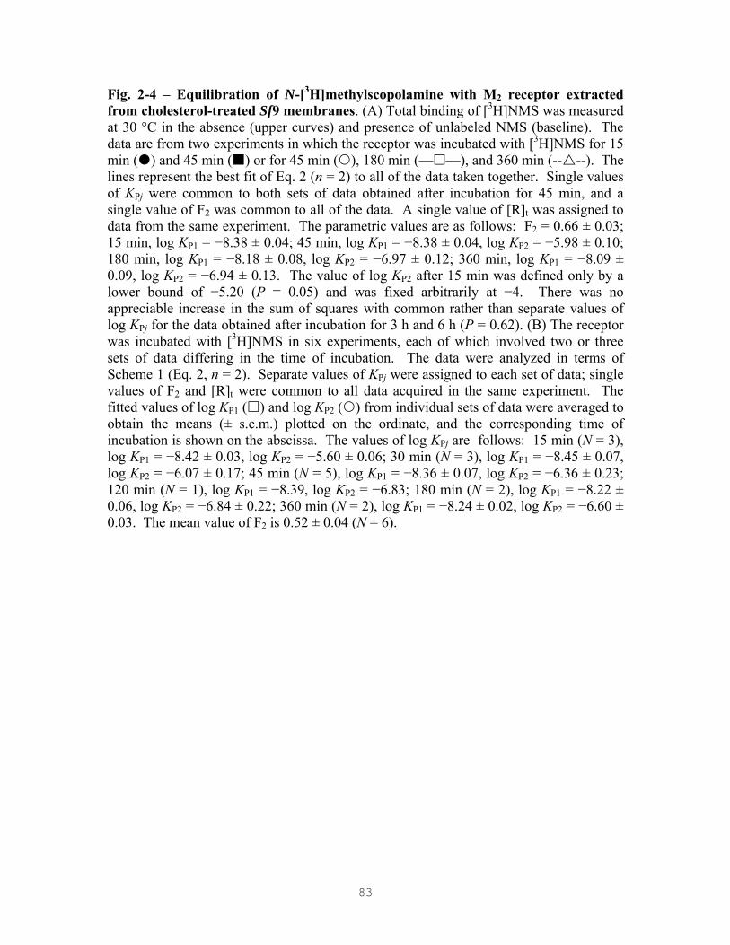
83
Fig. 2-4 – Equilibration of N-[3H]methylscopolamine with M2 receptor extracted from cholesterol-treated Sf9 membranes. (A) Total binding of [3H]NMS was measured at 30 °C in the absence (upper curves) and presence of unlabeled NMS (baseline). The data are from two experiments in which the receptor was incubated with [3H]NMS for 15 min ( ) and 45 min ( ) or for 45 min ( ), 180 min (— —), and 360 min (-- --). The lines represent the best fit of Eq. 2 (n = 2) to all of the data taken together. Single values of KPj were common to both sets of data obtained after incubation for 45 min, and a single value of F2 was common to all of the data. A single value of [R]t was assigned to data from the same experiment. The parametric values are as follows: F2 = 0.66 ± 0.03; 15 min, log KP1 = −8.38 ± 0.04; 45 min, log KP1 = −8.38 ± 0.04, log KP2 = −5.98 ± 0.10; 180 min, log KP1 = −8.18 ± 0.08, log KP2 = −6.97 ± 0.12; 360 min, log KP1 = −8.09 ± 0.09, log KP2 = −6.94 ± 0.13. The value of log KP2 after 15 min was defined only by a lower bound of −5.20 (P = 0.05) and was fixed arbitrarily at −4. There was no appreciable increase in the sum of squares with common rather than separate values of log KPj for the data obtained after incubation for 3 h and 6 h (P = 0.62). (B) The receptor was incubated with [3H]NMS in six experiments, each of which involved two or three sets of data differing in the time of incubation. The data were analyzed in terms of Scheme 1 (Eq. 2, n = 2). Separate values of KPj were assigned to each set of data; single values of F2 and [R]t were common to all data acquired in the same experiment. The fitted values of log KP1 ( ) and log KP2 ( ) from individual sets of data were averaged to obtain the means (± s.e.m.) plotted on the ordinate, and the corresponding time of incubation is shown on the abscissa. The values of log KPj are follows: 15 min (N = 3), log KP1 = −8.42 ± 0.03, log KP2 = −5.60 ± 0.06; 30 min (N = 3), log KP1 = −8.45 ± 0.07, log KP2 = −6.07 ± 0.17; 45 min (N = 5), log KP1 = −8.36 ± 0.07, log KP2 = −6.36 ± 0.23; 120 min (N = 1), log KP1 = −8.39, log KP2 = −6.83; 180 min (N = 2), log KP1 = −8.22 ± 0.06, log KP2 = −6.84 ± 0.22; 360 min (N = 2), log KP1 = −8.24 ± 0.02, log KP2 = −6.60 ± 0.03. The mean value of F2 is 0.52 ± 0.04 (N = 6).

84
Fig. 2-5 – Binding to M2 receptor extracted from native Sf9 membranes, analyzed empirically in terms of Scheme 1. Incubations were carried out at 30 °C (A, B) and 4 °C (C, D). (A, C) Total binding of [3H]QNB (closed symbols, ) or [3H]NMS (open symbols, ) was measured in the absence (upper curves) and presence of unlabeled NMS (baseline). The time of incubation with [3H]QNB was 2 h at 30 °C (A) and 12 h at 4 °C (C); that with [3H]NMS was 45 min at 30 °C (A) and 6 h, 12 h, or 18 h at 4 °C (C). Each experiment included assays with both radioligands taken in parallel, and different symbols denote data from different experiments (e.g., , ; , ; , ; etc) (N = 6). (B, D) Total binding was measured at near-saturating (B, , 32–38 nM, N = 3; D, , 27–29 nM, N = 3) and subsaturating (B, , 1–2 nM, N = 3) concentrations of [3H]QNB and graded concentrations of unlabeled NMS. The time of incubation was 2 h at 30 °C and 12 h at 4 °C. The lines indicate the best fit of Scheme 1 to the pooled data represented in panels A and B (n = 2) and, in a separate analysis, to the pooled data represented in panels C and D (n = 1). The parametric values and further details regarding the analyses are given in Table 2-2. Values plotted at the lower end of the x-axis in panels B and D indicate binding in the absence of NMS. The mean values of the Hill coefficient obtained for unlabeled NMS are as follows: B, 0.82 ± 0.06 at 32–38 nM [3H]QNB, 1.07 ± 0.05 at 1–2 nM [3H]QNB; D, 1.17 ± 0.14 at 27–29 nM [3H]QNB.

85
Fig. 2-6 – Binding to M2 receptor extracted from cholesterol-treated Sf9 membranes, analyzed empirically in terms of Scheme 1. Incubations were carried out at 30 °C (A, B) and 4 °C (C, D). (A, C) Total binding of [3H]QNB (closed symbols) or [3H]NMS (open symbols), was measured in the absence (upper curves) or presence of unlabeled NMS (baseline). The time of incubation with [3H]QNB was 2 h at 30 °C (A) and 12 h at 4 °C (C); that with [3H]NMS was 3 h at 30 °C (A) and 6 h at 4 °C (C). The results from 3 independent experiments are shown in each panel, and different symbols denote data from different experiments (i.e., , ; , ; , ). (B, D) Total binding was measured at near-saturating (B, , 30–35 nM, N = 10; D, , 28–30 nM, N = 2) and subsaturating (B, , 1–2 nM, N = 7; D, , 1–2 nM, N = 2) concentrations of [3H]QNB and graded concentrations of unlabeled NMS. The time of incubation was 2 h at 30 °C (B) and 12 h at 4 °C (D). The solid lines represent the best fits of Scheme 1 (n = 2) to the pooled data represented in panels A and B and, in a separate analysis, to the pooled data represented in panels C and D. The parametric values and further details regarding the analyses are given in Table 2-2. The dotted lines were simulated as described in the text and illustrate discrepancies in the affinity of NMS as estimated under different conditions.

86
Fig. 2-7 – Comparison of Schemes 1 and 2 for goodness of fit to data from cholesterol-treated Sf9 membranes. The data represented in the corresponding left- and right-hand panels of Figure 2-6 were pooled and analyzed in terms of Schemes 1 and 2 to obtain the global sum of squares at different values of n (A, binding at 30 °C; B, binding at 4 °C) (hatched bars, Scheme 1; grey bars, Scheme 2). The value from each analysis is plotted as a percentage of the value obtained when n was taken as 1, which is the same for both models. All parameters were assigned throughout in a mechanistically consistent manner, in contrast to the empirical assignments depicted in Figure 2-6; thus, all relevant data shared single values KPj, KAj, and Fj (Scheme 1) and of KP, f0j, KA, fi0, and fij (Scheme 2). In each case, single values of [R]t were assigned to data from assays performed in parallel within the same experiment. Further details are described in the text and in the legend to Figure 2-9. The dashed line in each panel indicates the relative sum of squares from an unconstrained, empirical analysis in terms of Scheme 1 (A, n = 2; B, n = 3). The fitted curves from Scheme 2 with n = 6 are shown in Figure 2-9.

87
Fig. 2-8 – Optimal number of interacting sites for the fit of Scheme 2 to data from cholesterol-treated membranes. The value of AICC was calculated from the weighted sum of squares and the degrees of freedom at each value of n (A, binding at 30 °C; B, binding at 4 °C). When a cooperativity factor was fixed arbitrarily at a specific value, or when two or more cooperativity factors were subsumed in a single parametric value, the restriction was disregarded when calculating the degrees of freedom. The probability that one of two models is correct was calculated from the difference in AICC, and the complementary estimates of P (%) are shown joined by a horizontal line at the tops of the corresponding bars.

88
Fig. 2-9 – Binding to M2 receptor extracted from cholesterol-treated Sf9 membranes, analyzed in terms of Scheme 2. Incubations were carried out at 30 °C (A, B) or 4 °C (C, D). The data are the same as those shown in Figure 2-6. (A, C) Total binding of [3H]QNB (closed symbols) or [3H]NMS (open symbols) was measured in the absence (upper curves) or presence of unlabeled NMS (baseline). (B, D) Total binding was measured at near-saturating ( ) and sub-saturating ( ) concentrations of [3H]QNB and graded concentrations of unlabeled NMS. The lines represent the best fit of Scheme 2 (n = 6) to the pooled data obtained at 30 °C (panels A and B) and, in a separate analysis, to those obtained at 4 °C (panels B and C). All parameters were assigned to enforce mechanistic consistency. Single values of KP and f0j ([3H]QNB), KA and fi0 ([3H]NMS and NMS) and fij ([3H]QNB and NMS) were common to all relevant data in both panels. The fitted values of log KP and log KA, respectively, are as follows: 30 °C, –9.51 ± 0.36 and –7.85 ± 0.06; 4 °C, –9.18 ± 0.39 and –7.73 ± 0.09. Single values of [R]t were assigned to data acquired in parallel: that is, to data obtained with [3H]QNB and [3H]NMS (A, C) and to data obtained with unlabeled NMS at both concentrations of [3H]QNB (B, D) in the same experiment. Further details are described in the text and in the legend to Figure 2-7.

89
CHAPTER 3
EFFECT OF CHOLESTEROL ON THE BINDING OF ANTAGONISTS TO THE MUSCARINIC
CHOLINERGIC RECEPTOR EXTRACTED FROM PORCINE ATRIA
90
3.1 Abstract
Muscarinic receptors were extracted from native and cholesterol-depleted
sarcolemmal membranes in cholate–NaCl and compared for the binding of
quinuclidinylbenzilate (QNB) and N-methylscopolamine (NMS). Methyl-β-cyclodextrin
was used to remove cholesterol, which was reduced 13-fold to levels comparable to those
in Sf9 cells. Both preparations exhibited the same or similar capacity for [3H]QNB and
[3H]NMS, and two classes of receptor were required to describe the data. With receptor
from native membranes, 22% of the sites exhibited an anomalously weak affinity for
[3H]NMS, while the balance were of higher affinity. Similar estimates of the affinity
were inferred from the inhibitory effect of unlabeled NMS on the binding of [3H]QNB,
and the interactions therefore appear to be competitive. Receptor from cholesterol-
depleted membranes revealed a similar pattern, including sites of anomalously weak
affinity for [3H]NMS, but the affinities estimated from assays at graded concentrations of
[3H]NMS differed significantly from those inferred from the inhibitory effect of the
unlabeled analogue on [3H]QNB, suggesting that the receptor is capable of cooperative
interactions. Depletion of cholesterol also was accompanied by a threefold decrease in
the affinity of [3H[QNB. These results indicate that removal of cholesterol from
cholesterol-rich sarcolemmal membranes does not necessarily have an effect opposite to
that of adding cholesterol to cholesterol-poor Sf9 membranes. In the present
investigation, however, the receptor from native myocardial membranes failed to exhibit
the non-competitive effects described previously. It therefore appears that cholesterol is
not the sole determinant of the latter.

91
3.2 Introduction
In recent years, much evidence has been gathered for the existence of oligomers of G
protein-coupled receptors in native [110,147] and heterologous [225,227] systems using
various biochemical and biophysical techniques [277]. Although such oligomers now
appear to be ubiquitous, the prevailing view has been that the receptors are mutually
independent and that signaling involves a ligand-regulated interconversion between free
receptor (R) and free G protein or subunits thereof on the one hand and a 1:1 heteromeric
RG complex on the other [80,137]. For many years, the dispersion of affinities revealed
in the binding of agonists generally has been rationalized, at least qualitatively, in terms
of such an arrangement. The multiple states of affinity therefore are assumed to be
induced by the G protein, but this notion has proven difficult to sustain on quantitative
grounds [85,110,125].
In an alternative view, the dispersion of affinities revealed in the binding of agonists
derives from cooperativity or perhaps asymmetry among the constituent components of
an oligomer. Owing to the relationship between the multiple affinities of agonists and
efficacy, such a view implies that interactions within the oligomer lie in the mechanistic
pathway of signaling. The determinants of cooperativity and the cooperative potential of
oligomers are therefore questions that relate to our understanding of signaling via G
protein-coupled receptors.
It previously has been shown that the ligand-binding properties of the muscarinic
receptor extracted from the sarcolemmal membranes of porcine atria differed in different
detergents; that is, the the binding of antagonists was competitive in digitonin-cholate and
non-competitive in cholate-NaCl or Lubrol-PX [145]. Digitonin is a plant glycoalkaloid
saponin which is known to form water-insoluble, 1:1 complexes with cholesterol [278],
and it has been suggested that the difference in ligand-binding characteristics can be
attributed to the ability of digitonin to sequester cholesterol from the receptor [147].
Also, it has been shown that M2 receptor extracted from native Sf9 membranes in
cholate–NaCl bound muscarinic antagonists in a wholly competitive manner. Such
effects in cholate−NaCl were in contrast to the non-competitive behavior observed
92
previously in extracts of porcine atria, but the membranes of Sf9 cells contain
comparatively low levels of cholesterol. Treatment of Sf9 membranes with exogenous
cholesterol regained the pattern of non-competitive effects in the binding of muscarinic
antagonists that was observed with atrial extracts. Thus, the apparent capacity for
[3H]QNB exceeded that for [3H]NMS, while the specific binding of [3H]QNB was
inhibited fully at comparatively low concentrations of unlabelled NMS.
In the present investigation, sarcolemmal membranes were depleted of cholesterol in
a reversal of the process employed with Sf9 membranes. Although the capacity was
essentially the same for [3H]QNB and [3H]NMS, the sites appeared heterogeneous to both
ligands, and the inhibition of [3H]QNB by unlabelled NMS was not strictly competitive.
Since the receptor from native membranes bound NMS and QNB in an essentially
competitive but otherwise similar manner, cholesterol had comparatively little effect on
the binding properties of the cardiac muscarinic receptor in the present investigation.
3.3 Materials and Methods
Ligands, detergents, and other materials
N-[3H]Methylscopolamine was purchased as the chloride salt from NEN Life Science
Products (lot 3589315, 71.6 Ci/mmol) and as the bromide salt from Amersham
Pharmacia Biotech (batch 38, 80 Ci/mmol). Mass spectra provided by the manufacturer
indicated that the samples from Amersham Biosciences were devoid of contaminating
scopolamine. The purity of material from PerkinElmer Life Sciences has been confirmed
previously [147]. (–)-[3H]Quinuclidinylbenzilate was purchased from Amersham
Biosciences (batch B-50, 48 Ci/mmol). Unlabeled NMS hydrobromide was purchased
from Sigma-Aldrich, and scopolamine hydrobromide was from RBI-Sigma.

93
Sodium cholate was purchased from Sigma-Aldrich. Solubilized receptor was
concentrated as required by means of Centricon-10 and Centriprep-30 filters (Amicon)
purchased from Millipore. HEPES and Tris were obtained as the free bases from
Boehringer Mannheim and Sigma-Aldrich (Trizma), respectively. EDTA and EGTA
were obtained as the free acids from British Drug Houses or Bioshop Canada. Bacitracin,
all protease inhibitors, and Sephadex G-50 were from Sigma-Aldrich. Other chemicals
were obtained from the sources shown below. Protein was estimated by means of
bicinchoninic acid using the BCA Protein Assay Kit from Pierce. Bovine serum albumin
was taken as the standard.
Membrane preparation and solubilization of M2 muscarinic receptor
Sarcolemmal membranes were collected from a sucrose gradient and spun down in buffer
A (20 mM imidazole, 1 mM Na2+-EDTA, 0.1 mM PMSF, 0.02% (w/v) sodium azide, 1
mM benzamidine, 2 µg/mL pepstatin A, 0.2 µg/mL leupeptin, 200 µg/mL bacitracin, pH
7.60 with HCl) essentially as described previously [110]. The gradient typically yielded
81–287 mg of protein from about 350–375 g of left plus right atria. The sarcolemmal
membranes were washed twice to remove imidazole and endogenous ligands. The
washing buffer was the same as that to be used in subsequent steps (i.e., buffer B for
solubilization in cholate–NaCl, 20 mM NaH2PO4, 20 mM NaCl, 1 mM EGTA, 0.1 mM
PMSF, pH 7.40 with NaOH). The product from the sucrose gradient was suspended in
the appropriate buffer (120 mL) with one burst of a Brinkmann Polytron (setting 5, 5 s),
and the suspension then was centrifuged for 40 min at 4 ºC and 100,000 × g. This step
was repeated, and the pellets from the second spin were stored at –75 ºC.
Cholesterol was removed from the sarcolemmal membranes of porcine atria by
meand of methyl-β-cyclodextrin [246]. Briefly, methyl-β-cyclodextrin (Sigma-Aldrich)
was added to buffer C (20 mM KH2PO4, 20 mM NaCl, 1 mM EDTA, 0.1 mM PMSF, and
NaOH to pH 7.60), and the mixture was vortexed and sonicated periodically in a bath

94
sonicator (Haake SWB20) until a clear solution was obtained at a stock concentration of
300 g/L.
To remove cholesterol from sarcolemmal membranes of porcine atria, prepared as
described above, the frozen pellets were thawed on ice and suspended in buffer C. A
portion of the homogenate was supplemented with methyl-β-cyclodextrin such that the
final concentration of methyl-β-cyclodextrin was 100 g/L with respect to the total volume
of the suspension; the balance was supplemented with buffer C alone. The final
concentration of total protein was 1.5 g/L. Each suspension then was shaken on a
horizontal shaker for 3 h at room temperature and centrifuged for 45 min at 4 ºC and
100,000 × g. Pellets were stored at –75 ºC until required for solubilization.
The amount of cholesterol in native and cholesterol-depleted sarcolemmal
membranes from porcine atria was quantified using a cholesterol assay kit from
BioVision.
Solubilization in sodium cholate plus NaCl was carried out as described previously
[147] in a procedure modified from that described in an earlier report [279]. Washed
sarcolemmal membranes were thawed on ice and suspended in buffer D (40 mM
NaH2PO4, 1 M NaCl, 1 mM EGTA, 0.16% sodium cholate, 0.1 mM PMSF, NaOH to pH
7.40, 2 mg of protein/mL), and the mixture was shaken for 1 h at room temperature. The
suspension then was diluted 1:1 with a buffer (40 mM NaH2PO4, 1 mM EGTA, NaOH to
pH 7.40) and centrifuged for 40 min at 4 ºC and 100,000 × g. The supernatant fraction
was stored on ice until required for the binding assays. The final concentrations of
cholate and NaCl were 0.08% and 0.5 M, respectively.
Binding assays
The radioligand and any unlabeled ligand were dissolved in buffer E (20 mM HEPES, 20
mM NaCl, 1 mM EDTA, 5 mM MgSO4, 0.1 mM PMSF, and NaOH to pH 7.40)
supplemented with cholate plus NaCl at concentrations of 0.1% and 20 mM, respectively.

95
An aliquot of the ligand-containing solution (48 µL) was added to the solubilized
receptor (5 µL), and the reaction mixture was incubated at 30 ºC. The time of incubation
for [3H]NMS was 45 min, and the time of incubation for all samples containing [3H]QNB
was 2 h.
To terminate the reaction and separate the bound radioligand, an aliquot of the
sample (50 µL) was applied to a column of Sephadex G-50 Fine (0.8 cm × 6.5 cm). The
column was pre-equilibrated and eluted with buffer F (20 mM HEPES, 20 mM NaCl, 1
mM EDTA, 5 mM MgSO4, and NaOH to pH 7.40) supplemented with cholate plus NaCl
at a concentration of 0.017% and 20 mM, respectively. All of the eluant up to and
including the void volume was collected (1.40 mL) and assayed for radioactivity.
Binding assays generally were carried out within one week of solubilization.
Nonspecific binding was taken throughout as binding in the presence of 1 mM unlabeled
NMS and increased linearly with the concentration of either radioligand. To compare the
binding of [3H]NMS and [3H]QNB, the two radioligands were assayed in parallel;
similarly, the inhibitory effect of unlabeled NMS on the binding of [3H]QNB was
measured in parallel at two concentrations of the latter.
Analysis of data
All data were analyzed with total binding taken as the dependent variable (Bobsd) and with
the total concentrations of all ligands taken as the independent variables. Any subsequent
manipulations were for the purpose of presentation only and did not alter the relationship
between the data and the fitted curve. Levels of specific binding (Bsp), estimates of total
receptor ([R]t), and estimates of maximal specific binding (Bmax) are presented as the
concentration in the binding assay (pM); similarly, the concentrations of ligands denote
the total molar concentration in the binding assay. Assays at graded concentrations of
either radioligand were performed with and without unlabeled NMS (1 mM), and both
sets of data shared a single estimate of nonspecific binding in the analyses described
below.

96
For data acquired at graded concentrations of [3H]QNB or [3H]NMS, the Hill
equation was formulated as shown in Eq. 1. The variable [P]t represents the total
concentration of the radioligand. The quantity Bsp represents specific binding at the
corresponding value of [P]t, and the parameter Bmax represents maximal specific binding.
The parameter K is the concentration of unbound radioligand at half-maximal occupancy,
and nH is the corresponding Hill coefficient. The parameter NS represents the fraction of
unbound radioligand that appears as nonspecific binding. Equation 1 was solved
numerically in the manner described previously [248].
H
H H
t spobsd max t sp
t sp
([P] )NS([P] )
([P] )
n
n n
BB B B
K B−
= + −+ −
(1)
For data acquired at graded concentrations of unlabeled NMS, the Hill equation was
formulated as H H H[A] 0 [A] t( ) /( [A] )n n n
obsdB B B K K= →∞= − + [A] .B →∞+ The variable [A]t is
the total concentration of unlabeled ligand, and the parameters [A] 0B = and [A]B →∞
represent the asymptotic levels of binding when [A] = 0 and as [A] → ∞ .
Mechanistic analyses were performed in terms of intrinsic heterogeneity (Scheme 1).
Estimates of total binding were fitted according to the equation Bobsd = Bsp + NS([P]t −
Bsp), in which P represents the radioligand and other quantities are as described above for
Eq. 1. The value of Bsp was computed according to Scheme 1.
Scheme 1 represents an intrinsically heterogeneous system that comprises n classes
of noninterconverting and mutually independent sites (Rj, j = 1, 2, ..., n). Those of type j
constitute the fraction Fj of all sites (i.e., Fj = [Rj]t/[R]t, where [Rj]t = [Rj] + [ARj] + [PRj],
and [R]t = t1[R ]n
jj=∑ ). The ligands P and A compete for Rj with the equilibrium
dissociation constants KPj and KAj, respectively. Total specific binding of a radiolabeled
probe was calculated according to Eq. 2, and the required values of [PRj] were obtained
as described below.
sp1[PR ]
n
jj
B=
= ∑ (2)

97
The values of [PRj] in Eq. 2 were calculated from the expansions in terms of the total
concentration of R and the free concentrations of A and P. The latter were computed
numerically from the corresponding implicit equations for [A]t and [P]t as described
previously [248].
Analyses in terms of Scheme 1 involved data combined from several experiments.
Scheme 1 was applied empirically and in a mechanistically consistent manner. In the
case of the latter, parametric values were shared among different sets of data in strict
accord with the premises of the model. In such analyses, ligand P represents [3H]QNB,
and ligand A represents both isotopic forms of NMS. Thus, the parameters KPj (Scheme
1) denote the affinity of [3H]QNB, and KAj (Scheme 1) denote the affinity of labeled and
unlabeled NMS. When Scheme 1 was applied in an empirical manner, different sets of
data were permitted separate values of parameters that ought to be invariant (i.e., Table 3-
2, Fig. 3-2). In such analyses, ligand P represents the radiolabeled probe—either
[3H]QNB or [3H]NMS as indicated, and ligand A represents unlabeled NMS.
The results of analyses involving multiple sets of data from replicated experiments
have been presented in each case with reference to a single fitted curve. To obtain the
values plotted on the y-axis, estimates of Bobsd were adjusted according to the expression
{ }obsd obsd' (x , a) / (x ,a)i iB B f f= [132]. The function f represents the fitted model, and
the vectors xi and a represent the independent variables at point i and the parameters,
respectively. Individual values of B'obsd at the same xi were averaged to obtain the mean
and standard error plotted in the figure. Specific binding (Bsp) was calculated as B'obsd
less the fitted estimate of nonspecific binding at the same concentration of unbound
radioligand.
Statistical procedures

98
All parameters were estimated by nonlinear regression [250]. Affinities were optimized
throughout on a logarithmic scale (i.e., log KPj). Values of [R]t were assigned separately
to data from separate experiments.
Weighting of the data, tests for significance, and other statistical procedures were
performed as described elsewhere [81,132,248]. Fits involving nested models, such as
Scheme 1 at different values of n, were compared by means of the F-statistic [248].
Weighted residuals were of comparable magnitude within each set of data. In
simultaneous analyses, individual sets of data generally made comparable contributions
to the total weighted sum of squares; thus, the fits were not dominated by the data from
one experiment or group of experiments. Mean values calculated from two or more
individual estimates of a parameter or other quantity are presented together with the
standard error. For parametric values derived from a single analysis of one or more sets
of data, the errors were estimated from the diagonal elements of the covariance matrix.
Such values reflect the range within which the weighted sum of squares is essentially the
same.
3.4 Results
Effect of cholesterol on apparent capacity and affinity
Sarcolemmal membranes from porcine atria were depleted of cholesterol via treatment
with methyl-β-cyclodextrin at a final concentration of 66.7 mg/mL with respect to the
total amount of protein. The level of cholesterol in treated membranes was 13-fold less
than that in native sarcolemmal membranes (Fig. 3-1).
In extracts of cholesterol-depleted membranes, the apparent capacity for [3H]NMS
after incubation for 45 min at 30 ºC was 94% of that for [3H]QNB in terms of the Hill
equation; in extracts of native sarcolemmal membranes, the apparent capacity for
[3H]NMS was 80% of that for [3H]QNB (Table 3-1). The difference in capacity reported
here for native sarcolemmal membranes is less than that found previously with the

99
muscarinic receptor from porcine atria. In the earlier study, [3H]NMS labeled only 56%
of the sites labeled by [3H]QNB, and the shortfall was attributed to cooperativity in the
binding of [3H]NMS [147].
There was a 2.5-fold decrease in the overall potency of [3H]QNB when cholesterol
was removed from the sarcolemmal membranes of porcine atria (native, log EC50 = −9.33
± 0.06; CHL-depleted, log EC50 = −8.92 ± 0.09), whereas the potency of [3H]NMS was
essentially unchanged (native, log EC50 = −8.03 ± 0.03; CHL-depleted, log EC50 = −8.02
± 0.11) (Table 3-1).
Assessment of the data in terms of Scheme 1
The data were assessed empirically in terms of Scheme 1, in that the affinity of NMS was
estimated separately for the radiolabeled form (KPj) and for the unlabeled analogue (KAj)
at each concentration of [3H]QNB. If the model describes the system, the measured
affinity for a particular class of sites is expected to be the same regardless of the nature of
the assay: that is, regardless of whether the variable is radiolabeled or unlabeled NMS or
whether the latter is examined at a lower or higher concentration of [3H]QNB. Any
difference constitutes a discrepancy between the model and the data. The affinity of
[3H]QNB (KPj) was taken as the same throughout, as was the value of F2 when two
classes of sites were required. The latter restriction is consistent with the assumption of
noninterconverting sites, which is implicit in Scheme 1. These constraints are consistent
with the data, as indicated by the lack of effect on the sum of squares (P = 0.96), and they
permit a direct comparison of the affinities estimated for NMS.
With muscarinic receptor from native sarcolemmal membranes, at least two classes
of sites were required to describe the pooled data in panels A and B of Figure 3-2 (P <
0.00001), and the parametric values are listed in Table 3-2. The total concentration of
receptor was estimated as a single parameter for all data acquired in parallel, either with
[3H]NMS and [3H]QNB or with unlabeled NMS at two concentrations of [3H]QNB.
There was little or no increase in the weighted sum of squares with one estimate rather

100
than two for the affinity of unlabeled NMS obtained at sub-saturating and near-saturating
concentrations of [3H]QNB (P = 0.018, Table 3-2). Similarly, there was little effect on
the sum of squares when mechanistic consistency was enforced by reducing the number
of parameters for the affinity of NMS from three to one at each class of sites (P = 0.0055,
Table 3-2). Binding therefore was essentially competitive, although the small differences
among the values of KPj and KAj for NMS may indicate weak cooperative effects.
With muscarinic receptor from cholesterol-depleted membranes, at least two classes
of sites were required to describe the pooled data in panels C and D of Figure 3-2 (P =
0.00002), and the parametric values are listed in Table 3-2. The total concentration of
receptor was estimated as a single parameter for all data acquired in parallel, either with
[3H]NMS and [3H]QNB or with unlabeled NMS at two concentrations of [3H]QNB.
There was no appreciable increase in the weighted sum of squares with one estimate
rather than two for the affinity of unlabeled NMS obtained at sub-saturating and near-
saturating concentrations of [3H]QNB (P = 0.051, Table 3-2). There was a significant
increase in the sum of squares when mechanistic consistency was enforced by reducing
the number of parameters for the affinity of NMS from three to one (14%, P < 0.00001).
Although the differences in affinity are comparatively small (≤ 4.5-fold), the level of
significance suggests that cooperative interactions were somewhat stronger than those
observed with receptor from native membranes.
3.5 Discussion
It previously has been shown that [3H]NMS labels only about 50% of the sites labeled by
[3H]QNB in preparations of muscarinic receptor extracted from sarcolemmal membranes
in cholate–NaCl [147]. In terms of Scheme 1, the sites inaccessible to [3H]NMS implied
a value of KP2 that exceeded the highest concentration of radioligand used in the assays;
moreover, that value was at least 70-fold greater than the value of KA2 inferred from the
inhibition of [3H]QNB. Such a discrepancy implies that NMS inhibits at sites to which it
cannot bind, a non-competitive effect that could be described quantitatively in terms of

101
cooperativity among at least four interacting sites, presumably within a tetramer. A
similar pattern has been observed more recently with M2 receptor purified from Sf9 cells
and reconstituted into phospholipid membranes, where the receptor was shown to exist
exclusively as a tetramer [148].
In the present investigation, only 22% of the sites labeled by [3H]NMS in
sarcolemmal extracts prepared in cholate–NaCl were of anomalously weak affinity for
the radioligand; moreover, the value of KP2 was 4–5-fold smaller than that observed
previously (cf. log KP2 = −6.11, Table 3-2; log KP2 = −5.45, Ref. [147]). Owing to these
differences in the binding of [3H]NMS, the estimates of affinity derived for the
radioligand in the present investigation are consistent with those derived from the
inhibitory effect of unlabeled NMS on [3H]QNB, and binding to the receptor extracted
from native membranes was essentially competitive. The reason for the absence of non-
competitive effects is unclear, although it may relate in part to differences in the level of
cholesterol. Whereas cholesterol previously was over 20-fold more abundant in
sarcolemmal membranes than in Sf9 membranes, the difference in the present
investigation was only 13-fold. The distribution of cholesterol within the cell is
unknown, and the overall difference may mask larger differences in regions important to
the functioning of the receptor.
Muscarinic receptor extracted from cholesterol-depleted membranes resembled that
extracted from native membranes, despite a level of cholesterol that was comparable to
the level in Sf9 membranes. About 25% of the sites were of anomalously weak affinity
for [3H]NMS, and the values of both KP1 and KP2 approximated the corresponding values
of KA1 and KA2 inferred from the inhibitory effect of unlabeled NMS on the binding of
[3H]QNB. The interactions therefore appeared to be largely competitive, although the
differences between KPj and KAj were sufficiently large to suggest that there was a non-
competitive component (P < 0.00001).
The present results indicate that depletion of cholesterol was essentially without
effect on the mechanism whereby antagonists bind to M2 receptor extracted from
cholesterol-rich porcine sarcolemmal membranes, in contrast to the changes observed

102
when cholesterol was added to native Sf9 membranes [227]. Binding was competitive or
nearly so throughout, although the removal of cholesterol may have unmasked weak non-
competitive effects that otherwise were latent. Since the cholesterol-depleted preparation
from heart resembled the preparation from Sf9 membranes, the lack of an effect derived
from the absence of non-competitive behavior in the preparation from native sarcolemmal
membranes. These considerations suggest that the manifestation of cooperative effects
can differ among different myocardial preparations for reasons that remain unclear.
They also indicate that cooperativity is not determined exclusively by the average level of
cholesterol within the membrane.
Although cholesterol was without appreciable effect on the mechanism of binding,
the overall affinity of [3H]QNB was about fourfold weaker in the cholesterol-depleted
preparation, as determined from the values of KP1 and KP2 estimated in terms of Scheme
1. That difference is consistent with a sixfold increase in affinity that was obtained when
cholesterol was added to Sf9 membranes [227]. It therefore appears that the effects of
cholesterol on the mechanism of binding and on affinity are independent; that is, the
state of the receptor in which binding is cooperative with respect to NMS and QNB is not
necessarily the state of lower affinity for those ligands.
The propensity of [3H]QNB to bind with higher affinity to M2 receptor from
cholesterol-enriched preparations recalls the existence of G protein-coupled receptors in
multiple states that differ in their ligand-binding preferences for agonists and antagonists.
An explicit scheme for such an arrangement presupposes that the receptor interconverts
between an inactive state (R) and an active state (R*) (Scheme 3). The preference of a
ligand for one state over the other shifts the equilibrium accordingly; thus, ligands with
higher affinity for the R* state are agonists, and ligands that are indifferent or prefer the R
state are antagonists. This context has served as the basis for a further examination of the
effect of cholesterol on the affinity of muscarinic ligands, as described in Chapter 4.
3.6 Acknowledgements

103
The managers and staff of Quality Meat Packers Limited are gratefully acknowledged for
their generous donation of porcine atria. This investigation was supported by the
Canadian Institutes of Health Research (MOP43990) and the Heart and Stroke
Foundation of Ontario (T4914, T5650).

104
Table 3-1 − Empirical characterization of specific binding
N-[3H]Methylscopolamine [3H]Quinuclidinylbenzilate Preparation
Temp. (°C) log K nH Bmax (pM) log K nH Bmax (pM)
Relative capacity
Native (4,4) 30 −8.03 ± 0.03 0.99 ± 0.07 162−269 −9.33 ± 0.06 0.91 ± 0.02 236−299 0.80 ± 0.05
CHL-depleted (5,5) 30 −8.02 ± 0.11 0.87 ± 0.07 27−348 −8.92 ± 0.09 0.92 ± 0.05 34−311 0.94 ± 0.06 M2 receptor was extracted from native and cholesterol-depleted sarcolemmal membranes of porcine atria. Binding was measured at graded concentrations of [3H]NMS and [3H]QNB taken in parallel. The time of incubation was 0.75 h for assays containing [3H]NMS alone and 2 h. for assays containing [3H]QNB. The number of sets of data is shown in parentheses ([3H]NMS, [3H]QNB). Each set was analyzed independently in terms of the Hill equation, and the individual estimates of log K and nH were averaged to obtain the means (± S.E.M.) listed in the table. The relative capacity for [3H]NMS and [3H]QNB was calculated from parallel estimates of Bmax (i.e., Bmax, [ 3H]NMS/Bmax, [ 3H]QNB), and the individual values were averaged to obtain the mean (± S.E.M.) listed in the table.

105
Table 3-2 − Affinities of NMS for M2 receptor extracted from native and cholesterol-depleted sarcolemmal membranes of porcine atria, estimated empirically in terms of Scheme 1
Protocol NMS
Affinity
Preparation Temp. (°C) Ligands
[[3H]QNB] (nM)
Time of incubation
(h) log KL1 log KL2 L
Native membranes 30 [3H]NMS (4) 0.75 −8.07 ± 0.03 −6.11 ± 0.44 P
NMS/[3H]QNB (3) 0.2−0.4 2 −8.16 ± 0.08 −6.10 ± 0.56 A
NMS/[3H]QNB (5) 20−37 2 −7.92 ± 1.53 −7.06 ± 5.68 A
CHL-depleted membranes 30 [3H]NMS (5) 0.75 −8.20 ± 0.04 −6.47 ± 0.29 P
NMS/[3H]QNB (4) 1−2 2 A
NMS/[3H]QNB (10) 18−24 2 −7.55 ± 0.12 a −6.60 ± 0.73 a
A The data illustrated in the paired left- and right-hand panels of Figure 3-2 were pooled (i.e., panels A and B, panels C and D) and analyzed according to Scheme 1 (n = 1 or 2) to obtain the parametric values listed in the table. NMS was present either as the radioligand (L ≡ P, left-hand panel) or as the unlabeled analogue (L ≡ A, right-hand panel). QNB was present only as the radioligand. The number of experiments is shown in parentheses. Two classes of sites were required throughout, and a single value of F2 was common to all of the data in both panels. Affinities were estimated as follows: for [3H]QNB, single values of KPj were common to all of the relevant data in both panels; for NMS, values of KPj were assigned to data obtained with the radioligand (left-hand panel), and separate or common values of KAj were assigned to data obtained with the unlabeled analogue at the two concentrations of [3H]QNB (right-hand panel). Values of [R]t were assigned as described in the text. The fitted value of F2 and the values of KPj for [3H]QNB are as follows: receptor from native porcine atria (n = 2), F2 = 0.22 ± 0.03, log KP1 = −9.54 ± 0.03, log KP2 = −8.65 ± 0.14; receptor from CHL-depleted porcine atria (n = 2), F2 = 0.25 ± 0.04, log KP1 = −9.04 ± 0.04, log KP2 = −7.66 ± 0.23. a There is no appreciable increase in the weighted sum of squares with one estimate rather than two for the affinity of unlabeled NMS at each class of sites at both concentrations of [3H]QNB (P = 0.05).

106
Fig. 3-1 – Levels of cholesterol in membranes from porcine sarcolemma, Sf9 cells and CHO cells. Membranes of Sf9 cells were supplemented with cholesterol via an inclusion complex with methyl-β-cyclodextrin after harvest and homogenization (0.5 mM with respect to cholesterol). Sarcolemmal membranes of porcine atria were depleted of cholesterol via incubation with methyl-β-cyclodextrin alone (66.7 mg of MβCD/mg of protein). The levels of cholesterol were as follows (mg of cholesterol per mg of protein): native Sf9 membranes, 0.07 ± 0.01 (N = 7); treated Sf9 membranes, 2.58 ± 0.33 (N = 5); native porcine membranes, 0.94 ± 0.20 (N = 3); depleted porcine membranes, 0.07 ± 0.02 (N = 3). The level in membranes prepared from Chinese hamster ovary cells was 0.17 ± 0.01 (N = 3).

107
Fig. 3-2 – Binding to M2 receptor extracted from native (panels A and B) and cholesterol-depleted (panels C and D) sarcolemmal membranes of porcine atria, analyzed empirically in terms of Scheme 1. Incubations were carried out at 30 °C. (A and C). Total binding of [3H]QNB (closed symbols) or [3H]NMS (open symbols), was measured in the absence (upper curves) or presence of unlabeled NMS (baseline). The time of incubation was 2 h and 45 min for samples containing [3H]QNB and [3H]NMS, respectively. The results from four (panel A) and five (panel C) independent experiments are shown in each panel, and different symbols denote data from different experiments. (B and D) Total binding was measured at near-saturating (B, , 20–37 nM, N = 5; D, , 18–24 nM, N = 10) and subsaturating (B, , 0.2–0.4 nM, N = 3; D, , 1–2 nM, N = 4) concentrations of [3H]QNB and graded concentrations of unlabeled NMS. The time of incubation was 2 h. The lines represent the best fits of Scheme 1 to the pooled data represented in panels A and B (n = 2) and, in a separate analysis, to the pooled data represented in panels C and D (n = 1). The parametric values and further details regarding the analyses are given in Table 3-2.

108
CHAPTER 4
MECHANISTIC BASIS FOR THE EFFECT OF CHOLESTEROL ON THE AFFINITY OF AGONISTS
AND ANTAGONISTS FOR THE M2 MUSCARINIC CHOLINERGIC RECEPTOR

109
4.1 Abstract
N-Ethylmaleimide (NEM) has been used together with cholesterol to examine the
effect of the latter on the binding of ligands to the M2 muscarinic cholinergic receptor.
Upon treatment with NEM, receptors extracted from native and cholesterol-treated Sf9
membranes in cholate-NaCl underwent an increase in their apparent affinity for agonists
and a decrease in that for antagonists. Treatment of Sf9 membranes with cholesterol-
methyl-β-cyclodextrin increased the affinity of the solubilized receptor for agonists and
antagonists, irrespective of NEM. The data were in good agreement with a model in
which the receptor interconverts spontaneously between an agonist-specific state akin to
the active state (R*) and an antagonist-specific, inactive state (R). Two equivalent fits
emerged from the analyses. In the first, NEM shifted the equilibrium between R and R*
toward the latter without affecting affinity per se, and cholesterol increased the affinity of
all ligands for both states without affecting the position of the equilibrium. In the second,
cholesterol shifted the equilibrium without affecting affinity per se, and NEM increased
the affinity of agonists while decreasing that of antagonists. Previous studies on the
purified M2 receptor have shown that NEM acts exclusively to favor R* over R [Sum
C.S. et al. (2002) J. Biol. Chem. 277, 36188−36203]; the effect of cholesterol therefore
can be attributed to an increase in the affinity of each ligand for both states. Although the
alternative solution can be rejected in this case, its existence is not self-evident and
illustrates the insight to be gained from mechanistically explicit models.

110
4.2 Introduction
Muscarinic and other G protein-coupled receptors interconvert spontaneously between at
least two states: an active state favored by agonists (R*) and an inactive state favored by
inverse agonists (R) [137,280–282]. The distribution of receptors between those states
can be modulated via the G protein, and the allosteric interaction between agonists and
guanyl nucleotides regulates signaling across the membrane [97,283,284]. Other factors
that can or may affect the distribution include sodium and magnesium [283,285–289],
mutations that lead to constitutive activity [280,290], sulfhydryl-specific reagents [228],
and extraction in detergent [212,228,291,292].
The structural basis for the difference between R and R* remains unclear. It also is
not clear how that interconversion relates to the GTP-sensitive dispersion of affinities
revealed in the binding of agonists, although both phenomena are linked to efficacy
[91,293,294], nor is it understood how the heterogeneity discerned by agonists emerges
from a single class or subtype of receptor. If the system is at thermodynamic
equilibrium, a population of identical and mutually independent sites is expected to
appear homogeneous (e.g., nH = 1) irrespective of the number of conformational states
accessible to a single molecule [85,248]. It has been suggested that heterogeneity is
induced by G proteins interacting transiently with the receptor to yield a ligand-sensitive
distribution of coupled and uncoupled states (i.e., R + G º RG) [80,137,294]. Such
schemes become problematic when applied quantitatively [85,125], however, and it may
be that the RG complex is less transient than assumed in the models
[110,111,119,295,296]. To reconcile these observations, it has been suggested that the
interconverting species is an oligomer in which the observed heterogeneity derives from
cooperativity or asymmetry with respect to the binding of the agonist [81,110,148].
The multiple states of GPCRs have exhibited some variability in their sensitivity to
the sulfhydryl reagent N-ethylmaleimide. In the case of muscarinic receptors in
membranes from rat brain, the reagent was shown early on to cause an apparent
interconversion from a state of low affinity for agonists to a state of higher affinity
[94,149]; in other studies, however, it was found to mimic and thereby to pre-empt the
111
interconversion from high affinity to low affinity brought about by GTP [297]. The latter
effect also has been found repeatedly with the muscarinic receptor in membranes from rat
heart (e.g, Ref. [298]).
Such apparent inconsistencies may be a consequence of different experimental
conditions and their effect on the interaction between the receptor and the G protein,
which would be expected to affect the binding patterns revealed by agonists. In the case
of the cardiac muscarinic receptor, the effect of N-ethylmaleimide was clarified in an
examination of the purified receptor devoid of G protein [228]. Under those conditions,
alkylation favored a thermally unstable state of high affinity for agonists and low affinity
for antagonists over a comparatively stable state of low affinity for agonists and high
affinity for antagonists. N-Ethylmaleimide and agonists therefore appear to drive the
receptor toward the same state, at least in the absence of G protein.
Sulfhydryl-specific reagents have been useful probes of the relationship between
structure and function [299–303]. Such reagents have been tagged with environmentally
sensitive fluorescent or spin labels and used to detect the conformational change between
the inactive and active states [299–301]. Results from such studies have led to the
suggestion that activation involves the rotation or tilting of the sixth transmembrane helix
[280,299,301–303].
Evidence for the existence of multiple interconverting states also has emerged from
constitutive activity as seen, for example, in mutants of the β2 adrenergic receptor
[137,151,304] and the M5 muscarinic receptor [305]. In the latter case, the ligand-
regulated activity of 13 constitutively active mutants differing only at position 465 in the
sixth helix could be accounted for by shifts in a single equilibrium between an active and
an inactive state [280]. Similarly, the notion of spontaneously interconverting states is
consistent with the constitutive activity and related properties of M1–M4 muscarinic
receptors overexpressed in Chinese hamster ovary cells [306] and of M5 muscarinic
receptors coexpressed with comparatively large amounts of Gq [305].

112
The ligand-binding properties of G protein-coupled receptors also can be affected by
modifications to their lipid environment, as indicated by the changes that occur when the
receptor is extracted in detergent. Upon solubilization in digitonin−cholate, muscarinic
receptors extracted from Sf9 cells, porcine cerebral cortex, and porcine atria have been
shown to undergo a decrease in their apparent affinity for antagonists; high-affinity
binding was restored upon their reconstitution into brain membranes or into liposomes
prepared from cholesteryl hemisuccinate, phosphatidylcholine, and phosphatidylinositol
[291,292]. Similarly, delipidation of the β-adrenergic receptor from erythrocyte
membranes through solubilization in deoxycholate abolished virtually all binding of the
antagonist [125I]iodocyanopindolol; upon reconstitution with soybean lipids, the receptor
bound agonists and antagonists with affinities similar to those of the receptor in native
membranes [212]. Such effects suggest that the binding properties are sensitive to lipids
acting either directly on the receptor or via their contribution to properties of the
membrane.
Cholesterol is essential to many eukaryotic organisms, in which 65–90% of cellular
free cholesterol can reside in the plasma membrane at an average concentration of 200–
300 µg/mg protein [214]. It has been shown to modulate the properties of many
membrane proteins, including G protein-coupled receptors [214]. In the case of the M2
muscarinic receptor, the antagonist [3H]quinuclidinylbenzilate was found to bind sixfold
more tightly to receptor extracted from cholesterol-treated Sf9 membranes than to that
from native membranes [227]. Moreover, the former preparation was more resistant to
thermal instability than the latter. The effect of cholesterol therefore differs from that of
N-ethylmaleimide, which was found to favor a thermally unstable state of high affinity
for agonists and low affinity for antagonists [228]. This comparison raises that
possibility that cholesterol acts directly on the muscarinic and perhaps other G protein-
coupled receptors to modulate the equilibrium between the active and inactive states,
shifting the distribution toward the latter.
In the present study, N-ethylmaleimide has been examined for its effect on the
affinity of agonists and antagonists for the M2 muscarinic receptor extracted from native
and cholesterol-treated Sf9 membranes. The data are quantitatively consistent with a

113
model in which the receptor interconverts spontaneously between a state of high affinity
for antagonists and low affinity for agonists and a state of low affinity for antagonists and
high affinity for agonists. Whereas N-ethylmaleimide shifts the equilibrium toward the
state favored by agonists, cholesterol increases the affinity of all ligands for both states.
4.3 Materials and Methods
Ligands, detergents, and other materials
(−)-[3H]Quinuclidinylbenzilate (QNB) was purchased from Amersham Biosciences
(batch B-49, 49 Ci/mmol; batch B-50, 48 Ci/mmol). Oxotremorine-M, carbamylcholine
(carbachol) chloride, arecoline hydrobromide, pilocarpine hydrochloride, and N-
methylscopolamine hydrobromide were purchased from Sigma-Aldrich.
Sodium cholate was purchased from Sigma-Aldrich. Solubilized receptor was
concentrated as required by means of Centricon-10 and Centriprep-30 filters (Amicon)
purchased from Millipore. HEPES and Tris were obtained as the free bases from
Boehringer Mannheim and Sigma-Aldrich (Trizma), respectively. EDTA and EGTA
were obtained as the free acids from British Drug Houses or Bioshop Canada. N-
Ethylmaleimide (ultra grade), bacitracin, all protease inhibitors, and Sephadex G-50 were
from Sigma-Aldrich. Protein was estimated by means of bicinchoninic acid using the
BCA Protein Assay Kit from Pierce. Bovine serum albumin was taken as the standard.
Preparation of M2 muscarinic receptor, addition of cholesterol, and treatment with N-
ethylmaleimide
FLAG-tagged M2 muscarinic receptor was expressed in baculoviral-infected Sf9 cells,
and the harvested membranes were stored at −75 °C. Native membranes were
supplemented with exogenous cholesterol by means of an inclusion complex with
methyl-β-cyclodextrin. Sarcolemmal membranes were prepared from porcine atria.

114
Further details concerning these and related procedures have been described previously
[110,147,155,227,246].
Cholesterol was assayed using the Cholesterol/Cholesteryl Ester Quantitation Kit
from BioVision Research Products. The level in untreated Sf9 membranes was about 7%
of that in sarcolemmal membranes and increased to about 270% upon treatment of the
membranes with the cholesterol-methyl-β-cyclodextrin inclusion complex. The amounts
in absolute terms were as follows (mg of cholesterol per mg of protein): native Sf9
membranes, 0.07 ± 0.01 (N = 7); treated Sf9 membranes, 2.58 ± 0.33 (N = 5); porcine
sarcolemmal membranes, 0.94 ± 0.20 (N = 3).
Receptor was solubilized from native and cholesterol-treated Sf9 membranes as
described previously [147,227]. The extraction was performed in sodium cholate plus
NaCl at final concentrations of 0.08% and 0.5 M, respectively. Solubilised material was
divided into aliquots of 1–2 mL and stored at −75 °C until required.
For reaction with N-ethylmaleimide, aliquots of solubilized extract were thawed and
supplemented with the reagent at a final concentration of 10 mM. Unless otherwise
indicated, the reaction was carried out on ice for 2 h and 24 h with receptor from native
and cholesterol-treated Sf9 membranes, respectively. Following the period of alkylation,
aliquots containing the the treated receptor were introduced directly into the binding
assays. A fresh solution of N-ethylmaleimide was prepared for each experiment, and
controls lacking the reagent were prepared in an otherwise identical manner. Further
details regarding the treatment with N-ethylmaleimide have been described previously
[228].
Binding assays
All assays were performed on receptor extracted from Sf9 membranes. The radioligand
and any unlabeled ligand were dissolved in Buffer A (20 mM HEPES, 20 mM NaCl, 1
mM EDTA, 5 mM MgSO4, 0.1 mM PMSF, and NaOH to pH 7.40) supplemented with

115
cholate plus NaCl at a concentration of 0.1% and 20 mM, respectively. An aliquot of the
ligand-containing solution (48 µL) was added to the solubilized receptor (5 µL), and the
reaction mixture was incubated for 2 h at 30 ˚C. M2 receptor extracted from native and
cholesterol-treated Sf9 membranes has been shown to equilibrate with
[3H]quinuclinylbenzilate and accompanying ligands under those conditions [227].
To terminate the reaction and separate the bound radioligand, an aliquot of the
sample (50 µL) was applied to a column of Sephadex G-50 Fine (0.8 cm 6.5 cm). The
column was pre-equilibrated and eluted with Buffer B (20 mM HEPES, 20 mM NaCl, 1
mM EDTA, 5 mM MgSO4, and NaOH to pH 7.40) supplemented with cholate plus NaCl
at a concentration of 0.017% and 20 mM, respectively. All of the eluant up to and
including the void volume was collected (1.60 mL) and assayed for radioactivity.
Binding assays generally were carried out within one week of solubilization.
Nonspecific binding was taken throughout as binding in the presence of 1 mM unlabeled
N-methylscopolamine and increased linearly with the concentration of
[3H]quinuclidinylbenzilate. The inhibitory effect of one or more of oxotremorine-M,
carbachol, arecoline, and pilocarpine on the binding of [3H]quinuclidinylbenzilate was
assayed in parallel with that of N-methylscopolamine, which was included throughout as
a control.
Analysis of data
All data were analyzed with total binding taken as the dependent variable (Bobsd) and with
the total concentrations of all ligands taken as the independent variables. Any subsequent
manipulations were for the purpose of presentation only and did not alter the relationship
between the data and the fitted curve. Levels of specific binding (Bsp), estimates of total
receptor ([R]t), and estimates of maximal specific binding (Bmax) are presented as the
concentration in the binding assay (pM); similarly, the concentrations of ligands denote
the total molar concentration in the binding assay.

116
For data acquired at graded concentrations of [3H]quinuclidinylbenzilate, the Hill
equation was formulated as shown in Eq. (1).
H
H H
max t spobsd t sp
t sp
([P] )NS([P] )
([P] )
n
n n
B BB B
K B−
= + −+ −
(1)
The variable [P]t represents the total concentration of the radioligand. The quantity Bsp
represents specific binding at the corresponding value of [P]t, and the parameter Bmax
represents maximal specific binding. The parameter K is the concentration of unbound
radioligand at half-maximal occupancy, and nH is the corresponding Hill coefficient. The
parameter NS represents the fraction of unbound radioligand that appears as nonspecific
binding. Eq. (1) was solved numerically in the manner described previously [248]. For
data on the binding of [3H]quinuclidinylbenzilate at graded concentrations of an
unlabeled ligand, the Hill equation was formulated as shown in Eq. (2).
∞→∞→= ++
−= ]A[t
]A[0]A[obsd HH
H
]A[)( B
KKBBB nn
n
(2)
The variable [A]t is the total concentration of unlabeled ligand, and K is the value of
[A]t at which the specific binding of the radioligand is reduced by 50%. The parameters
B[A]=0 and B[A]→∞ represent the asymptotic levels of binding when [A]=0 and
[A]→∞.
Mechanistic analyses were performed in terms of Schemes 1 and 3, in which an
unlabeled ligand (A) and [3H]quinuclidinylbenzilate (P) compete for a population of
mutually independent sites. In Scheme 1, the receptor exists in a single state to which
ligands A and P bind with equilibrium dissociation constants KA and KP, respectively
(e.g., KA = [A][R]/[AR], KP = [P][R]/PR]). In Scheme 3, the receptor can exist in two
spontaneously interconverting states designated R and R*. The ligands A and P bind to R
with equilibrium dissociation constants KA and KP, respectively, and the relative affinity
of the ligand for the two states is designated αA and αP (i.e., αA = K*A/KA, K*A =
[A][R*]/[AR*]; αP = K*P/KP, K*P = [P][R*]/[PR*]). The parameter KR represents the
117
relative concentrations of R and R* at equilibrium in the absence of ligand (i.e., [R]/[R*]
= KR).
Schemes 1 and 3 were fitted to the data by means of the equation Bobsd = Bsp +
NS([P]t – Bsp), in which P represents the radioligand and other quantities are as
described above for Eq. (1). The value of Bsp was computed according to Eq. (3) or Eq.
(4), respectively.
]PR[sp =B (3)
]PR[]PR[sp∗+=B (4)
The values of [PR] and of [PR]+[PR*] were calculated from the expansions in terms
of the total concentration of R and the free concentrations of A and P. The latter were
computed numerically from the corresponding implicit equations for [A]t and [P]t.
Further details concerning the formulation of Schemes 1 and 3 have been described
elsewhere [228,248].
Separation on Sephadex G-50 yields low levels of nonspecific binding [147,227],
which represented about 0.06% of unbound [3H]quinuclidinylbenzilate with all
preparations used in the present investigation. At the concentrations of
[3H]quinuclidinylbenzilate used to examine the inhibitory effect of other ligands (i.e., 15–
27 nM), nonspecific binding never exceeded 8% of binding in the absence of the
unlabeled ligand. Nonspecific binding in such experiments was taken as the fitted
asymptote at saturating concentrations of the unlabeled ligand, which was poorly defined
under some conditions with some ligands. A single value of NS therefore was common
to data acquired in parallel, which included values at graded concentrations of N-
methylscopolamine in addition to those obtained with one or more other ligands as
described above.
The results of analyses involving multiple sets of data from replicated experiments
have been presented in each case with reference to a single fitted curve. To obtain the
values plotted on the y-axis, estimates of Bobsd were adjusted according to the expression

118
{ }obsd obsd' ( , / ( , )i iB B f f x a= x a [81]. The function f represents the fitted model, and the
vectors xi and a represent the independent variables at point i and the fitted parameters
for the set of data under consideration; ix and a are the corresponding vectors in which
values that differ from experiment to experiment have been replaced by the means for all
experiments associated with the fitted curve. Individual values of 'obsdB at the same xi
were averaged to obtain the mean and standard error plotted in the figure. Specific
binding (Bsp) was calculated as 'obsdB less the fitted estimate of nonspecific binding at the
same concentration of unbound radioligand.
Statistical procedures
All parameters were estimated by nonlinear regression. Affinities and equilibrium
constants were optimized throughout on a logarithmic scale [i.e., log KL, log αL (L ≡ A or
P), log KR]. In analyses of data from two or more experiments, the data from each
experiment were assigned separate values of [R]t and NS. Other parameters were shared
as described in the legends to the tables and figures.
Fits of nested models were compared by means of the F-statistic. Weighted residuals
were of comparable magnitude within each set of data; in simultaneous analyses,
individual sets of data generally made comparable contributions to the total weighted sum
of squares. Mean values calculated from two or more individual estimates of a parameter
or other quantity are presented together with the standard error. For parametric values
derived from a single analysis of one or more sets of data, the errors were estimated from
the diagonal elements of the covariance matrix.
Further details regarding the optimization of parameters, weighting of the data, tests
for significance, and other statistical procedures have been described previously [81,248].

119
4.4 Results
Reaction of the M2 receptor with N-ethylmaleimide
N-Ethylmaleimide reduced the apparent affinity of [3H]quinuclidinylbenzilate and
increased that of oxotremorine-M for M2 receptor extracted from native and cholesterol-
treated Sf9 membranes. The time-dependence of the effect is illustrated in Fig. 4-1. With
the receptor from native membranes, the value of K for [3H]quinuclidinylbenzilate (Eq. 1)
increased to a maximum within the first hour of alkylation and then remained unchanged
for up to 6 h (Fig. 4-1A). The effect was mirrored in the value of K for oxotremorine-M
(Eq. 2), which decreased to a minimum in a parallel manner (Fig. 4-1B). Essentially the
same pattern was found with receptor from cholesterol-treated membranes, where a
higher or lower value of K was attained within 6 h and remained unchanged for at least
24 h.
Attainment of a plateau in the value of K suggests that alkylation of the receptor is
complete. In accord with this interpretation, the affinity of [3H]quinuclidinylbenzilate for
receptor extracted from native membranes was the same after incubation for 30 min with
N-ethylmaleimide irrespective of whether or not an additional amount of the reagent was
added to the reaction mixture after 15 min. It has been shown previously that the affinity
of the alkylated receptor for [3H]quinuclidinylbenzilate is the same regardless of whether
or not unreacted N-ethylmaleimide is removed immediately prior to the binding assay
[228].
Preparations of the M2 receptor extracted from native Sf9 membranes are known to
undergo a time-dependent loss of sites during incubation at 30 °C, an effect that is
avoided if the membranes are supplemented with cholesterol or if the preparation is
maintained at 4 °C [227]. In previous studies, the reaction with N-ethylmaleimide was
conducted in an ice bath for a period 24 h [228]. The receptor remains functionally
viable under those conditions, and the same procedure was used for receptor from
cholesterol-treated membranes in the present investigation. In the case of receptor from

120
native membranes, the time of alkylation was reduced to 2 h in order to avoid any
possibility of thermal inactivation.
Effects of N-ethylmaleimide and cholesterol, assessed in terms of the Hill equation and
Scheme 1
Treatment with cholesterol and N-ethylmaleimide led to four preparations of M2 receptor,
each of which was characterized for the binding of [3H]quinuclidinylbenzilate and five
unlabeled ligands to obtain the data illustrated in Figure 4-2. Receptor extracted from
native Sf9 membranes appeared homogeneous with respect to all six ligands, as indicated
by Hill coefficients near or indistinguishable from 1 (Table 4-1). The value of nH
underwent little or no change upon pretreatment of the membranes with cholesterol.
Treatment with N-ethylmaleimide tended to decrease the value of nH, at least with
receptor from native membranes, although the effect was comparatively small in most
cases. The data for all ligands in all preparations were described to at least a first
approximation when the value of nH was taken as 1, and this constraint was implicit in
subsequent analyses in terms of Schemes 1 and 3. The possible consequences of
heterogeneity were considered in attempts to fit an expanded version of Scheme 3 that
comprised multiple classes of receptor (i.e., Scheme 3 in Ref. 228), but the parameters
tended to be highly correlated and poorly defined.
Cholesterol and N-ethylmaleimide affected the affinity of each ligand in a
characteristic manner that can be quantified empirically in terms of Scheme 1, in which
the receptor is assumed to exist in a single state. The data were analyzed in concert to
obtain estimates of the 24 parameters that denote the affinity of each ligand for each
preparation of receptor. The values are listed in Table 4-2, and the fitted curves are
compared with the data in Figure 4-2. With receptor from native or cholesterol-treated
membranes, treatment with N-ethylmaleimide increased the affinity of agonists and
decreased that of antagonists. With native or N-ethylmaleimide-treated receptor, agonists
and antagonists bound with higher affinity to receptor from cholesterol-treated

121
membranes than to that from native membranes. These effects suggest a pattern in which
cholesterol increased affinity for all ligands whereas N-ethylmaleimide favored an
agonist-specific state over an antagonist-specific state. Such an arrangement is not
explicit in Scheme 1, and the data therefore were examined in terms of the two-state
receptor depicted in Scheme 3.
Effects of N-ethylmaleimide and cholesterol, assessed in terms of Scheme 3
With six ligands and four preparations of receptor, an unconstrained application of
Scheme 3 requires 48 parameters to describe the affinity for R (KL) and the relative
affinity for R and R* (αL) of each ligand in each preparation. Different preparations also
may differ in the distribution of vacant receptors between R and R* (KR), resulting in a
total of 52 such parameters overall. A series of analyses therefore was performed in
which the number of parameters was reduced to the minimum that was consistent with
the data. The baseline for consistency was taken as a sum of squares equal or comparable
to that obtained with Scheme 1, applied as described above (Fig. 4-2, Table 4-2).
Neither cholesterol nor N-ethylmaleimide appeared to affect the relative affinities for
R and R*. In a test of all ligands taken together, there was no appreciable increase in the
weighted sum of squares with a single value of αL for each ligand rather than with
separate values for each of the four preparations of receptor (P > 0.1). With that
constraint, two distinct but equivalent scenarios emerged with respect to the effects of
cholesterol and N-ethylmaleimide on KL and KR. The parametric values are listed in
Table 4-3, and in each case the fitted curves are virtually superimposable with those
obtained with Scheme 1 and illustrated in Fig. 4-2.
In the first scenario, N-ethylmaleimide is without effect on affinity (i.e., KL) but
shifts the equilibrium between R and R* toward the latter (i.e., a decrease in KR), while
cholesterol is without effect on the equilibrium between R and R* but causes a decrease
in the dissociation constant of each ligand for R (Table 4-3A). Since the values of αL are
the same in all preparations, cholesterol also causes a decrease in the dissociation

122
constants for R* (i.e., αLKL). Thus, the data are well described with only two values of
KR: one for the native receptor and one for receptor treated with N-ethylmaleimide;
similarly, the data require only two values of KL for each ligand: one for the receptor
from native membranes and one for that from membranes supplemented with cholesterol.
These constraints can be applied without an appreciable increase in the sum of squares
over that from the wholly unconstrained analysis or that from the analysis with six values
of αL (P ≥ 0.08). There is an increase of at least twofold upon the further assumpution
that N-ethylmaleimide is without effect on KR or that cholesterol is without effect on KL
for any ligand (P < 0.00001).
In the second scenario, cholesterol is without effect on affinity (i.e., KL) but shifts the
equilibrium between R and R* (i.e., KR), while N-ethylmaleimide increases the affinity of
agonists and decreases that of antagonists without affecting the equilibrium between the
two states (Table 4-3B). There accordingly are two values of KR: one for receptor from
native membranes and one for that from membranes supplemented with cholesterol;
similarly, there are two values of KL for each ligand: one for the native receptor and one
for receptor treated with N-ethylmaleimide. These constraints have little effect on the
sum of squares (P ≥ 0.025), whereas there is an increase of 1.6-fold or more upon the
further assumption that cholesterol is without effect on KR or that N-ethylmaleimide is
without effect on KL (P < 0.00001).
In either application of Scheme 3, the constraints permitted by the data reduced the
total number of parameters corresponding to KL, αL, and KR from 52 to 20. The
minimum sum of squares was virtually the same irrespective of whether KR was deemed
sensitive to N-ethylmaleimide (WSSQ = 127,200, dof = 1,517) or cholesterol (WSSQ =
127,000, dof = 1517), and the value was comparable to that obtained with Scheme 1 with
24 parameters corresponding to KP and KA (WSSQ = 125,500, dof = 1,513). Scheme 3
therefore provides a fit equivalent to that of Scheme 1 but requires four fewer parameters.
The two analyses summarized in Table 4-3 were performed with the values of log KR
fixed at 3 and −1 with respect to treatment with N-ethylmaleimide or the addition of
cholesterol. In the first case, the native receptor is almost wholly in the R state (log KR =

123
3), and the alkylated receptor is predominantly in the R* state (log KR = −1); in the
second, the receptor from native membranes is wholly in the R state (log KR = 3), and
that from cholesterol-treated membranes is predominantly in the R* state (log KR = −1).
The need for such assignments was dictated by uncertainty over the absolute values
of KR, which were highly correlated in each analysis. To determine the domains of
acceptable values, the weighted sum of squares was mapped as described below with
respect to the values of log KR for unreacted (log KR(−)) and alkylated (log KR(+)) receptors
in the first case and for receptor from native (log KR(−)) and cholesterol-treated (log KR(+))
membranes in the second. The values of log KR selected for the analyses summarized in
Table 4-3 lie within the domain defined by those combinations that yield the minimum in
the sum of squares, and the fitted values of other parameters (i.e., log KL and log αL) are
the same for all combinations of log KR(−) and log KR(+) within that domain.
Effect of N-ethylmaleimide on KR in Scheme 3
The dependence of the sum of squares on the value of log KR(−) is illustrated in Figs. 4-3A
and 4-3B for analyses in which log KR(+) was fixed at −2 and 2, respectively. Both maps
show a peak when KR is assumed to be unaffected by N-ethylmaleimide: that is, when
log KR(−) equals log KR(+). When log KR(−) exceeds KR(+), the sum of squares decreases to
a minimum of 127,200 when log KR(+) is −2 (Fig. 4-3A) and 129,300 when log KR(+) is 2
(Fig. 4-3B). When log KR(−) is less than KR(+), the sum of squares decreases to a
minimum that is markedly higher than that obtained when log KR(−) exceeds KR(+).
The results for all combinations of log KR(−) and log KR(+) between −4 and 4 are
illustrated in Fig. 4-4. Best fits of the model are obtained when log KR(−) is 2 or more and
log KR(+) is 0 or less, as defined by the depression at the left of the figure (WSSQ =
127,200). At those values of KR, the receptors exist almost wholly in the R state before
treatment with N-ethylmaleimide ([R] > 100[R*]) and predominantly in the R* state
thereafter ([R] ≤ [R*]). A marginally higher plateau emerges when log KR(+) exceeds 0
while remaining less than log KR(−) by 2 units or more (WSSQ = 129,200). Reasonable

124
agreement therefore requires only that N-ethylmaleimide effect a 100-fold increase in the
fraction of receptors in the R* state, regardless of the magnitude of that fraction before or
after alkylation.
Another plateau, located at the right of Fig. 4-4 (WSSQ = 156,300), corresponds to
the opposite arrangement in which native receptors are predominately in the R* state and
interconvert to the R state upon treatment with N-ethylmaleimide (i.e., log KR(−) ≤ −1 and
log KR(+) ≥ 2). The sum of squares exceeds that at the minimum by 23% (P < 0.00001),
and the data therefore permit a clear distinction between the two possible effects of
alkylation. Such a domain constitutes a false minimum that could be misidentified as the
best fit in the absence of a map such as that presented in the figure.
Effect of cholesterol on KR in Scheme 3
The dependence of the sum of squares on the value of log KR(−) is illustrated in Figs. 4-3C
and 4-3D for analyses in which log KR(+) was fixed at −2 and 2, respectively. The pattern
is similar to that described above for the effect of N-ethylmaleimide, in that both maps
show a peak when KR is assumed to be unaffected by the addition of cholesterol (i.e., log
KR(−) = log KR(+), WSSQ = 211,500). Unlike N-ethylmaleimide, however, cholesterol is
found not to discriminate in its effect on KR; that is, the sum of squares is essentially the
same regardless of whether the addition of cholesterol is assumed to favor R over R* (log
KR(−) < log KR(+), WSSQ = 127,500) or R* over R (log KR(−) > log KR(+), WSSQ =
127,000).
The results for all combinations of log KR(−) and log KR(+) are illustrated in Fig. 4-5,
where the two possible effects of cholesterol are seen to be nearly symmetrical. If
cholesterol favors R, best fits are obtained at values of KR within the domain defined by
the depression at the right [i.e., log KR(−) ≤ −1, and log KR(+) > log (100KR(−))]. If
cholesterol favors R*, best fits lie within the domain defined by the depression at the left
[i.e., log KR(−) ≥ 2, and log KR(+) < log (KR(−)/100)]. The fitted values of KL and αL listed
in Table 4-3B are for the situation in which cholesterol favors R* over R. Those listed in
125
Table 4-4 are for the alternative possibility in which cholesterol favors R over R*, and
they are the same for all combinations of log KR(−) and log KR(+) within the domain
corresponding to the minimum. Whereas N-ethylmaleimide increases the affinity of
agonists and decreases that of antagonists in both cases, the values of KL are smaller for
all ligands when cholesterol is assumed to shift the equilibrium toward R (cf. Tables 4-3B
and 4).
4.5 Discussion
Cholesterol is at least tenfold more abundant in sarcolemmal membranes from porcine
atria than in membranes from Sf9 cells, with consequences for the binding properties of
the M2 muscarinic cholinergic receptor [227]. Receptor extracted from sarcolemmal
membranes in cholate–NaCl has been shown to exhibit a pattern of non-competitive
effects wherein N-[3H]methylscopolamine labels only about one half of the sites labeled
by [3H]quinuclidinylbenzilate, yet binding at near-saturating concentrations of the latter is
inhibited fully at comparatively low concentrations of unlabeled N-methylscopolamine.
In contrast, receptor extracted from native Sf9 membranes exhibits the same capacity for
N-[3H]methylscopolamine and [3H]quinuclidinylbenzilate, and the inhibition of the latter
by unlabeled N-methylscopolamine is essentially competitive. Upon the enrichment of
Sf9 membranes with cholesterol, the solubilized receptor exhibits the difference in
capacity and attendant non-competitive effects characteristic of that extracted from
porcine atria. A quantitative analysis of those effects suggested that cholesterol is
required for the manifestation of cooperativity in the binding of antagonists to at least
four interacting sites, presumably within a tetramer or larger oligomer [227].
The propensity of a ligand to favor the active (R*) or the inactive form (R) of a
GPCR derives from the difference in its affinity for the two states. Whereas agonists
bind with higher affinity to R*, antagonists either are indifferent (i.e., classical
antagonists) or bind with higher affinity to R (i.e., inverse agonists). A shift in the
equilibrium toward R* therefore is expected to increase the apparent affinity of agonists
126
and to decrease that of inverse agonists; similarly, a shift toward R is expected to
decrease the affinity of agonists and to increase that of inverse agonists. Purified M2
receptor devoid of G protein has been shown to interconvert between agonist- and
antagonist-specific states that resemble R and R*, suggesting that the process is intrinsic
to the receptor [228].
The effect of cholesterol on cooperativity in the binding of antagonists was
accompanied by a sixfold increase in the apparent affinity of [3H]quinuclidinylbenzilate
and a somewhat smaller, twofold increase in that of N-[3H]methylscopolamine [227].
Cholesterol also prevented a thermal inactivation that otherwise occurs with G protein-
coupled receptors in their active state, particularly in the absence of an orthosteric ligand
[e.g., 137,227,228,300]. Such effects suggest that cholesterol may act on the equilibrium
between R and R*, shifting the distribution of sites toward the former. To examine that
possibility, M2 receptors extracted from native and cholesterol-treated Sf9 membranes
were compared with respect to their affinity for [3H]quinuclidinylbenzilate, N-
methylscopolamine and four muscarinic agonists. The effect of cholesterol was compared
with that of the thiol reagent N-ethylmaleimide, which is known to drive the equilibrium
toward R* and therefore provides a point of reference (Ref. [228], and references cited
therein).
An empirical analysis of the data in terms of Scheme 1 indicated that N-
ethylmaleimide increases the apparent affinity of agonists and decreases that of
antagonists irrespective of cholesterol, whereas cholesterol increases the affinity of all
ligands irrespective of N-ethylmaleimide (Table 4-2). The effect of N-ethylmaleimide
agrees qualitatively with the notion that alkylation causes a net interconversion of
receptors from the inactive to the active state. Such an arrangement is consistent the
results obtained with Scheme 3 (Table 4-3A), which yields a fit equivalent to that of
Scheme 1 and constitutes an explicit description of the supposed equilibrium between the
two states. Thus, N-ethylmaleimide is seen to increase the value of KR, thereby favoring
R* over R and hence the binding of agonists over that of antagonists, whereas cholesterol
increases the affinity per se of all ligands for both states (i.e., KL and αLKL).

127
In the case of Scheme 3, however, the analyses identified another region of
parameter space in which the fit is virtually identical to that described above. In this
second scenario, cholesterol is seen to affect the value of KR, which can be either
decreased (Table 4-3A) or increased (Table 4-4), while N-ethylmaleimide acts to increase
the affinity of agonists and to decrease that of antagonists. This interpretation is not self-
evident from the results in Table 4-2, and its emergence illustrates the limitations of an
empirical description such as that provided by Scheme 1. The present data therefore are
ambiguous when taken alone, but alkylation by N-ethylmaleimide is known from
independent studies to cause a net interconversion of sites for R to R* without affecting
either KL or αL [e.g., 228]. It follows that the effect of cholesterol is to increase the
affinity of agonists and antagonists for both states.
The conditions of the present experiments were selected to avoid behavior that
cannot be accommodated by Scheme 3. Under some of the conditions described here,
the M2 receptor exhibits a heterogeneity that is detected as a shortfall in its apparent
capacity for N-[3H]methylscopolamine [147,148,227]. Assays with N-
[3H]methylscopolamine also reveal a time-dependent loss of sites when the radioligand is
incubated at 30 °C with receptor purified from porcine atria [228] or extracted from
native Sf9 membranes [227]. Such effects were avoided through the use of
[3H]quinuclidinylbenzilate, which labeled all sites with the same or similar affinity (Fig.
4-2F) and has been shown to protect the receptor from thermal inactivation [227,228].
The latter effect can be attributed to the slow kinetics of binding encountered with
quinuclidinylbenzilate and the reduced likelihood of finding the receptor in a labile,
unliganded state. Scheme 3 therefore provided at least a first approximation of the
system and served to distinguish the effect of cholesterol from the known effect of N-
ethylmaleimide.
Lipids can affect the activity of membrane proteins through direct interactions or by
influencing the biophysical properties of the surrounding membrane. The latter effects
depend upon the composition of lipids within an annulus that forms around the protein;
the former arise from the binding of so-called non-annular lipids at specific sites [307].
Both effects have been implicated in the influence of cholesterol on the ligand-binding

128
properties of membrane proteins, including G protein-coupled receptors, and related
events [214]. The influence of cholesterol on aspects of G protein-mediated signaling has
been linked to its concentration in liquid-ordered domains known as lipid rafts, which
also are enriched in G protein-coupled receptors, G proteins, and effectors [308]. The
colocalization of different constitutents within such domains has implications for the
assembly of specific complexes before and during signaling [308]. In the case of the β2-
adrenergic receptor, localization has been associated with reduced mobility and hence
reduced signaling through Gs [309,310].
Rhodopsin has been shown to undergo both direct and indirect regulation by
cholesterol (Ref. [214], and references cited therein). A direct effect is suggested by the
cholesterol-sensitive transfer of resonance energy between tryptophanyl residues of
rhodopsin and the fluorescent sterol cholestatrienol, and it has been estimated that one
molecule of cholesterol interacts with one molecule of rhodopsin. An indirect effect is
suggested by the sensitivity of the conformational equilibrium between metarhodopsin I
and metarhodopsin II to the level of cholesterol in the membrane; that is, increasing the
amount of cholesterol shifts the equilibrium towards that of metarhodopsin I, which is the
inactive photointermediate. This effect was attributed to a reduction in the free volume of
the bilayer, since the formation of metarhodopsin II has been shown to be accompanied
by volume expansion of the protein. Cholesterol also has been found to protect rhodopsin
from thermal inactivation, and it therefore has been suggested to serve a functional role in
stabilizing the protein during trafficking to the plasma membrane. A similar protective
effect has been observed with the M2 muscarinic and other G protein-coupled receptors
[227,269].
In the present investigation, the effects of cholesterol were monitored in mixed
micellar solutions obtained by solubilizing the membranes in cholate–NaCl. Sodium
cholate does not form a precipitable complex with cholesterol [311], in contrast to
digitonin, and any influence of the membrane on the receptor presumably was lost upon
solubilization. It therefore appears that the increased affinity of muscarinic ligands in
extracts of cholesterol-treated membranes derives from a direct interaction between
cholesterol and the receptor itself.

129
Non-annular lipids affect the activity of membrane-embedded proteins in the manner
of cofactors, and it has been suggested that they are located between transmembrane α-
helices or at protein-protein interfaces [307]. In several cases, they have been shown to
co-purify with the protein and have been resolved in high-resolution structures of proteins
or protein complexes such as cytochrome c oxidase, the potassium channel,
bacteriorhodopsin, the β2-adrenergic receptor, and the A2A adenosine receptor [307 and
references cited therein].
Early evidence for the existence of non-annular sites came from studies on the effects
of cholesterol on the Ca2+-ATPase of sarcoplasmic reticulum [223]. The sterol increased
enzymatic activity without affecting the thickness of the lipid bilayer and therefore
appeared to act directly on the protein [312]. Cholesterol also is required for the
functioning of the nicotinic acetylcholine receptor, and five to ten non-annular binding
sites have been suggested to reside in the interstices of the five receptor subunits [313].
There was no correlation between the effect of cholesterol on the action of the ion
channel and changes in membrane fluidity, again pointing to a direct interaction with the
protein [314,315].
Molecular modeling studies of the oxytocin receptor have suggested a site of action
for cholesterol on the surface of transmembrane segments 5 and 6 [276]. A recent crystal
structure of the β2-adrenergic receptor bearing T4 lysozyme in place of the third
intracellular loop has revealed a parallel dimer; at the interface between the two
protomers, six molecules of cholesterol and two covalently-bound molecules of palmitic
acid form a two-fold symmetric sheet [71]. The functional significance of that
cholesterol remains unclear, however, owing in part to uncertainty over the physiological
significance of the dimer itself. A later structure of the same construct shows the receptor
as an anti-parallel dimer. No cholesterol appears at the interface, but each monomer of
the receptor binds two molecules of cholesterol within a pocket formed by
transmembrane helices 1−4 [316].
The general increase in affinity brought about by cholesterol in the present
investigation recalls a similar effect described previously for other G protein-coupled

130
receptors (Ref. 227, and references cited therein). Those earlier studies were carried out
on receptors in membrane fragments, and a contribution from annular lipids therefore
cannot be ruled out; nonetheless, it seems likely that the effect derived at least in part
from structured cholesterol of the sort identified in recent crystallographic studies. Thus,
cholesterol has been shown to increase the affinity of antagonists for membrane-bound
oxytocin, µ-opioid, and 5-HT1A receptors. Similarly, agonists bound with weaker affinity
to the oxytocin, galanin, and cholecystokinin receptors when cholesterol was removed
from the plasma membranes of uterine myometrium and Chinese hamster ovary cells,
respectively; high affinity-binding was re-established when cholesterol was restored to
the depleted membranes. The agonist [3H]DAMGO bound with higher affinity to the µ-
opioid receptor in the plasma membrane of yeast upon the removal of ergosterol or the
addition of cholesterol. In the case of the µ-opioid and oxytocin receptors, the increase in
affinity for agonists was shown to be independent of the G protein.
Whereas cholesterol acts to enhance stability and increase affinity through a direct
interaction with the receptor, at least under the conditions described here, there is no
effect on correlates of efficacy. The increase in affinity in terms of Scheme 3 was similar
in magnitude for agonists and antagonists (Table 4-3A). Also, the effect on agonists was
independent of endogenous G proteins: membranes from Sf9 cells expressing the M2
receptor exhibit no agonist-stimulated binding of [35S]GTPγS [317,318], and they are
immunonegative to antibodies that recognize αi1, αi2, αi3, and αo [317]. Lack of signaling
through the M2 receptor contrasts with the activity of the M1, M3, and M5 receptors,
which have been shown to increase the levels of inositol-1,4,5-trisphosphate and
intracellular calcium when expressed in Sf9 cells [319,320]. Finally, the contrast with N-
ethylmaleimide indicates that the nature of the effect achieved by cholesterol differs from
that of an agent known to perturb the distribution of receptors between the active and
inactive states. If the direct interaction of cholesterol with the receptor is a determinant
of activity, perhaps through the facilitation of cooperative effects [227], it involves
properties that have not been measured or affected in the present investigation.

131
4.6 Acknowledgements
We thank Rabindra V. Shivnaraine for helpful discussions during the preparation of the
manuscript. This investigation was supported by the Canadian Institutes of Health
Research (MOP43990) and the Heart and Stroke Foundation of Ontario (T5650, T6280).
A. T. C. was the recipient of an Ontario Graduate Scholarship.
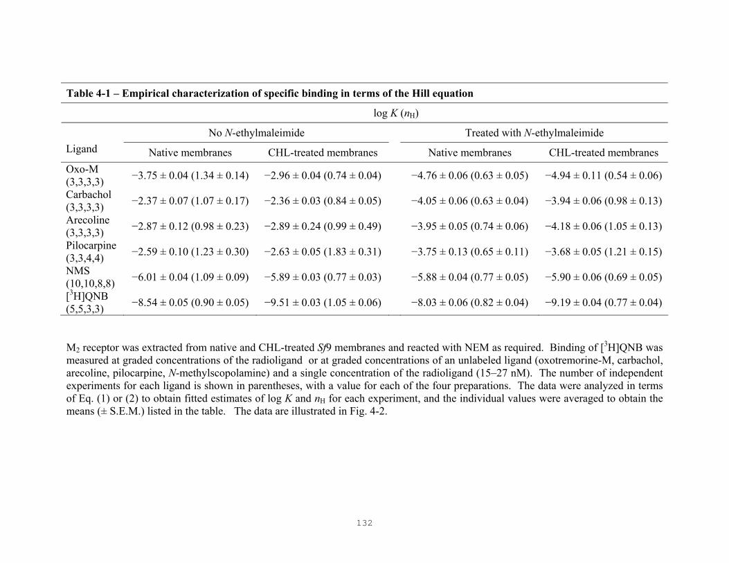
132
Table 4-1 – Empirical characterization of specific binding in terms of the Hill equation
log K (nH)
No N-ethylmaleimide Treated with N-ethylmaleimide Ligand Native membranes CHL-treated membranes Native membranes CHL-treated membranes Oxo-M (3,3,3,3) −3.75 ± 0.04 (1.34 ± 0.14) −2.96 ± 0.04 (0.74 ± 0.04) −4.76 ± 0.06 (0.63 ± 0.05) −4.94 ± 0.11 (0.54 ± 0.06)
Carbachol (3,3,3,3) −2.37 ± 0.07 (1.07 ± 0.17) −2.36 ± 0.03 (0.84 ± 0.05) −4.05 ± 0.06 (0.63 ± 0.04) −3.94 ± 0.06 (0.98 ± 0.13)
Arecoline (3,3,3,3) −2.87 ± 0.12 (0.98 ± 0.23) −2.89 ± 0.24 (0.99 ± 0.49) −3.95 ± 0.05 (0.74 ± 0.06) −4.18 ± 0.06 (1.05 ± 0.13)
Pilocarpine (3,3,4,4) −2.59 ± 0.10 (1.23 ± 0.30) −2.63 ± 0.05 (1.83 ± 0.31) −3.75 ± 0.13 (0.65 ± 0.11) −3.68 ± 0.05 (1.21 ± 0.15)
NMS (10,10,8,8) −6.01 ± 0.04 (1.09 ± 0.09) −5.89 ± 0.03 (0.77 ± 0.03) −5.88 ± 0.04 (0.77 ± 0.05) −5.90 ± 0.06 (0.69 ± 0.05)
[3H]QNB (5,5,3,3) −8.54 ± 0.05 (0.90 ± 0.05) −9.51 ± 0.03 (1.05 ± 0.06) −8.03 ± 0.06 (0.82 ± 0.04) −9.19 ± 0.04 (0.77 ± 0.04)
M2 receptor was extracted from native and CHL-treated Sf9 membranes and reacted with NEM as required. Binding of [3H]QNB was measured at graded concentrations of the radioligand or at graded concentrations of an unlabeled ligand (oxotremorine-M, carbachol, arecoline, pilocarpine, N-methylscopolamine) and a single concentration of the radioligand (15–27 nM). The number of independent experiments for each ligand is shown in parentheses, with a value for each of the four preparations. The data were analyzed in terms of Eq. (1) or (2) to obtain fitted estimates of log K and nH for each experiment, and the individual values were averaged to obtain the means (± S.E.M.) listed in the table. The data are illustrated in Fig. 4-2.

133
Table 4-2 – Affinities of agonists and antagonists, estimated in terms of Scheme 1
log KL
No N-ethylmaleimide Treated with N-ethylmaleimide
Ligand Native
Membranes CHL-treated membranes
Native membranes CHL-treated membranes
Oxotremorine-M −4.80 ± 0.03 (3) −4.96 ± 0.04 (3) −5.36 ± 0.10 (3) −6.47 ± 0.19 (3)
Carbachol −3.37 ± 0.04 (3) −4.39 ± 0.04 (3) −4.66 ± 0.10 (3) −5.60 ± 0.15 (3)
Arecoline −3.74 ± 0.04 (3) −4.82 ± 0.10 (3) −4.67 ± 0.09 (3) −5.82 ± 0.18 (3)
Pilocarpine −3.54 ± 0.04 (3) −4.54 ± 0.07 (3) −4.58 ± 0.12 (4) −5.37 ± 0.19 (4)
N-Methylscopolamine −7.05 ± 0.02 (10) −7.81 ± 0.04 (10) −6.72 ± 0.09 (8) −7.53 ± 0.12 (8)
[3H]Quinuclidinylbenzilate −8.60 ± 0.02 (5) −9.57 ± 0.03 (5) −8.34 ± 0.09 (3) −9.29 ± 0.06 (3) The data illustrated in all panels of Fig. 4-2 were pooled and analyzed in concert according to Scheme 1 to obtain the value of log KL (L ≡ P or A) for each ligand in each preparation of receptor. The number of independent experiments is shown in parentheses. Data obtained in all experiments on the same preparation of receptor were assigned a single value of log KP for [3H]QNB (L ≡ P) and a single value of KA for each unlabeled ligand (L ≡ A), and the fitted values are listed in the table. Most experiments involved the characterization of at least two ligands, and assays were performed on aliquots from the same batch of receptor. All data from the same experiment therefore were assigned single values of [R]t and NS.

134
Table 4-3 – Affinities of agonists and antagonists, estimated in terms of Scheme 3
A. KR affected by NEM; KL affected by CHL B. KR affected by CHL; KL affected by NEM
log KR (−NEM) = 3.0 log KR (+NEM) = −1.0
log KR (−CHL) = 3.0 log KR (+CHL) = −1.0
log KL (±NEM) log KL (±CHL)
Ligand
log αL (±CHL, ±NEM) −CHL +CHL ∆log KL
log αL (±CHL, ±NEM) −NEM +NEM ∆log KL
Oxo-M −0.61 ± 0.08 −4.79 ± 0.03 −4.99 ± 0.04 −0.20 −0.11 ± 0.05 −4.81 ± 0.03 −5.47 ± 0.07 −0.66
Carbachol −1.25 ± 0.08 −3.37 ± 0.04 −4.40 ± 0.04 −1.03 −0.97 ± 0.06 −3.39 ± 0.04 −4.68 ± 0.07 −1.29
Arecoline −0.91 ± 0.07 −3.73 ± 0.04 −4.84 ± 0.09 −1.11 −1.06 ± 0.10 −3.75 ± 0.04 −4.71 ± 0.07 −0.96
Pilocarpine −0.96 ± 0.10 −3.54 ± 0.04 −4.53 ± 0.07 −0.99 −0.94 ± 0.08 −3.55 ± 0.04 −4.57 ± 0.09 −1.02
NMS 0.46 ± 0.08 −7.05 ± 0.02 −7.82 ± 0.04 −0.77 −0.70 ± 0.04 −7.07 ± 0.02 −6.77 ± 0.06 0.30
[3H]QNB 0.40 ± 0.07 −8.61 ± 0.02 −9.58 ± 0.03 −0.97 −0.92 ± 0.04 −8.62 ± 0.02 −8.38 ± 0.06 0.24 The data illustrated in all panels of Fig. 4-2 were pooled and analyzed in concert according to Scheme 3. A total of 102 sets of data were obtained with the six ligands, and the number of independent experiments per ligand is shown in Table 4-2. Two analyses were performed, and the fitted curves in each case are virtually superimposable with those illustrated in Fig. 4-2. In the first analysis (A), the value of log KR was set at 3.0 and −1.0 for all data acquired with unreacted receptor and NEM-treated receptor, respectively; single values of log KL were assigned to the data obtained with the same ligand in all experiments performed on receptor from native membranes on the one hand and from CHL-treated membranes on the other, irrespective of NEM. In the second analysis (B), the value of log KR was set at 3.0 and −1.0 for all data acquired with receptor from native and cholesterol-treated membranes, respectively; single values of log KL were assigned to the data obtained with the same ligand in all experiments performed on unreacted receptor on the one hand and on NEM-treated receptor, irrespective of cholesterol. In both analyses, single values of log αL were common to all of the data relevant to each ligand, irrespective of NEM and cholesterol. The radioligand was present throughout, and the values of log KL and log αL shown for [3H]QNB were common to all of the data. Single values of [R]t and NS were assigned to all data acquired in the same experiment. The weighted sum of squares was 127,200 in (A) and 127,000 in (B).

135
Table 4-4 – Affinities of agonists and antagonists, estimated in terms of Scheme 3
KR affected by CHL; KL affected by NEM
log KR (−CHL) = −3.0 log KR (+CHL) = 1.0
log KL (±CHL)
Ligand
log αL (±CHL, ±NEM) −NEM +NEM ∆log KL
Oxo-M 0.25 ± 0.05 −5.02 ± 0.04 −5.66 ± 0.08 −0.64
Carbachol 1.09 ± 0.06 −4.45 ± 0.04 −5.71 ± 0.08 −1.26
Arecoline 1.18 ± 0.10 −4.90 ± 0.09 −5.82 ± 0.11 −0.92
Pilocarpine 1.05 ± 0.08 −4.58 ± 0.07 −5.55 ± 0.11 −0.97
NMS 0.83 ± 0.04 −7.87 ± 0.04 −7.53 ± 0.06 0.34
[3H]QNB 1.03 ± 0.04 −9.63 ± 0.03 −9.34 ± 0.05 0.29 The data illustrated in all panels of Fig. 4-2 were pooled and analyzed according to Scheme 3. The parameters were assigned as described for the analysis represented in Table 4-3B, and the two calculations differed only in the values of log KR for the receptor from native and cholesterol-treated membranes. The weighted sum of squares was 127,500.

136
Fig. 4-1 – Effect of N-ethylmaleimide on the apparent affinity of oxotremorine-M and [3H]quinuclidinylbelzilate for M2 receptor extracted from native and cholesterol-treated Sf9 membranes. Extracts from native ( ) and cholesterol-treated ( ) membranes were reacted with NEM (10 mM) for the times shown on the abscissa. Total binding to untreated (t = 0) and NEM-treated samples then was measured at graded concentrations of [3H]QNB (A) or at a near-saturating concentration of [3H]QNB (21–27 nM) and graded concentrations of oxotremorine-M (B). The data were analyzed in terms of Eq. (1) or Eq. (2) to obtain the individual values of K and nH for each experiment, and the mean values of log K from 1–5 experiments at each time are plotted in the figure (A, EC50; B, IC50). The data obtained with each ligand and each preparation of receptor were analyzed in concert to test for the significance of any time-dependent effect of NEM. In each case, there is no appreciable increase in the sum of squares with single values of log K and nH for all of the data rather than with a separate value of each parameter for the 1–5 sets of data obtained after each time of incubation (P ≥ 0.27).

137
Fig. 4-2

138
Fig. 4-2 – Effect of cholesterol and N-ethylmaleimide on the binding of agonists and antagonists to M2 receptor extracted from Sf9 membranes. Receptor from native ( ,
) and cholesterol-treated membranes ( , ) was characterized without further treatment ( , ) and after reaction with NEM ( , ). (A–E) Total binding was measured at a single concentration of [3H]QNB (15–27 nM) and graded concentrations of the unlabeled ligand identified on the abscissa. Points shown at the lower end of the abscissa indicate binding in the absence of unlabeled ligand. (F) Total binding was measured at graded concentrations of [3H]QNB either alone (upper curves) or in the presence of 1 mM unlabeled NMS (baseline). The lines indicate the best fit of Scheme 1 to the data represented in all panels taken together, and the parametric values are listed in Table 4-2. Further details regarding the analysis are given in in the legend to Table 4-2.

139
Fig. 4-3 – Fit of Scheme 3 mapped with respect to KR as affected by N-ethylmaleimide and cholesterol. The data represented in all panels of Fig. 4-2 were pooled and analyzed according to Scheme 3 to obtain the weighted sum of squares plotted on the ordinate. Parameters were assigned as described in the legend to Table 4-3, with the value of log KR taken as follows for the two scenarios with respect to NEM and cholesterol. (A, B) The value log KR was fixed at −2 (A) or +2 (B) for NEM-treated receptor, and the corresponding value for unreacted receptor was fixed as shown on the abscissa (cf. Table 4-3A). (C, D) The value of log KR was fixed at −2 (C) or +2 (D) for receptor from cholesterol-treated membranes, and the corresponding value for receptor from native membranes was fixed as shown on the abscissa (cf. Table 4-3B).

140
Fig. 4-4 – Domain of acceptable parametric values for an effect of N-ethylmaleimide on KR in Scheme 3. Analyses such as those represented in Figs. 4-3A and 4-3B were conducted with the values of log KR taken as shown for data acquired with unreacted receptor (−NEM) and with receptor treated with N-ethylmaleimide (+NEM). The weighted sum of squares from each analysis is plotted on the z-axis. The parametric values listed in Table 4-3A are from the analysis in which log KR was fixed at 3 (−NEM) and −1 (+NEM).

141
Fig. 4-5 – Domain of acceptable parametric values for an effect of cholesterol on KR in Scheme 3. Analyses such as those represented in Figs. 4-3C and 4-3D were conducted with the values of log KR taken as shown for data acquired with receptor from native (−CHL) and cholesterol-treated membranes (+CHL). The weighted sum of squares from each analysis is plotted on the z-axis. The parametric values listed in Table 4-3B are from the analysis in which log KR was fixed at 3 (−CHL) and −1 (+CHL); those listed in Table 4-4 are from the analysis in which log KR was fixed at −3 (−CHL) and 1 (+CHL).

142
CHAPTER 5
GENERAL DISCUSSION
143
5.1 Insights from the Thesis
The mechanistic basis of GPCR-mediated signaling is encoded in the nucleotide-
modulated dispersion of affinities revealed in the binding of agonists. Quantitative
measures of such dispersions previously have been shown to correlate with efficacy or
intrinsic activity [86,90,91,321], but the underlying cause remains unclear. The binding
patterns obtained for agonists at most G protein-coupled receptors are inherently
ambiguous in that data typically are acquired at a single concentration of the radioligand,
and there is insufficient information to distinguish among mechanistic schemes that
predict a dispersion of affinities for a system at thermodynamic equilibrium.
The dispersion could arise in at least three ways, as follows: first, two or more
classes of distinct, non-interconverting, and mutually independent sites may differ in their
affinity for the ligand (multi-site model); second, intrinsically identical sites may exist in
two or more interconverting states arising from transient association with other,
stoichiometrically limiting proteins within the membrane (ternary complex model); and
third, successive equivalents of the ligand may bind to interacting sites in a cooperative
manner (cooperativity).
It previously has been shown that schemes based on the first two scenarios cannot
describe radioligand-binding data from G protein-mediated systems in a mechanistically
consistent manner and therefore are not tenable as a mechanistic basis for signaling
[83,85,86,125]. When formulated mathematically, those schemes are described by
rational functions only if one can disregard the equations of state for all interacting
species except the receptor; that is, binding to the receptor should not deplete the free
concentration of one or more ligands [248]. At odds with this restriction is the
requirement, in schemes based on the ternary complex model, that the G protein must be
limiting for the model to give rise to a dispersion of affinities in the binding of agonists
[85]. Moreover, there is an important distinction between the binding patterns that arise
from the multi-site and ternary complex models. For a mixture of independent and
dissimilar sites, points of inflection measured on the abscissa of a semilogarithmic plot
can be related directly to the equilibrium dissociation constants of the ligand for the

144
different classes of sites. With the ternary model, however, increases in the concentration
of agonist are accompanied by changes in the concentration of uncoupled G protein;
accordingly, the apparent affinity of an agonist varies with its concentration [85]. A
consequence of such changes is that at no point is there a simple relationship between the
concentration of the drug and its equilibrium dissociation constant for uncoupled and G-
protein coupled receptor.
Although the multi-site model is not tenable as a mechanistic basis for signaling, it
does not suffer from the complications found with the ternary complex model which arise
from the mutual depletion of receptor and G protein. It therefore can be applied
empirically when the aim is simply to draw a curve through a series of experimentally
acquired points [248]. In so doing, it provides a good first approximation of the data, and
the fit may resemble that of the elusive true model. The multi-site model and models
based on cooperativity share the same functional form in that they are both rational
functions. They accordingly are indistinguishable under certain conditions. For example,
a sum of hyperbolic terms can describe both the multi-site model and negative
cooperativity when there is only one independent variable. Moreover, the latter is
indistinguishable from the special case of the former in which the capacity of each
subclass is some multiple of a basic unit. The data are ambiguous, but the choice is
limited when the number of interacting sites equals 2; the possibilities increase as the
oligomer becomes more complex. For instance, a tetramer would be indistinguishable
from 1, 2, 3 or 4 classes of mutually independent sites, depending upon the degree to
which each equivalent of ligand affected the affinity of the next. The ambiguity can be
avoided by increasing the number of independent variables [87]. In binding studies, a
convenient and informative procedure is to characterize the inhibitory behavior of an
unlabeled ligand at two or more concentrations of the radiolabeled probe; the data then
can be analyzed in concert to distinguish between competing mechanistic proposals that
otherwise are equivalent.
G protein-coupled receptors are membrane proteins that transduce signals across
biological membranes, and it therefore seems likely that their local environment
influences the manner by which they operate. Cholesterol is well known to be an integral

145
part of cellular membranes, particularly those of eukaryotes where it previously has been
shown to exert its influence on the functioning of many membrane proteins both directly
and indirectly [307]. Its physicochemical effects have been well characterized for many
membrane systems. In recent years, it has been identified as a key component of
membrane microdomains such as caveolae and lipid rafts, both of which have been
suggested to serve as platforms for various signaling systems [215,322,323]. In the
present investigation, cholesterol has been identified as a determinant of cooperativity in
that the M2 muscarinic cholinergic receptor was found to exhibit non-competitive effects
in the binding of muscarinic antagonists in the presence of the sterol.
Cooperativity implies the existence of oligomers, and questions regarding the role of
those structures in signal transduction relate in part to the size of the basic functional unit
and to its structural integrity during signaling. The ability of cholesterol to elicit non-
competitive effects from the M2 receptor raises the question of whether cholesterol does
so by facilitating the formation of oligomers or by modulating the cooperative properties
within already formed complexes.
5.2 Cooperativity
In the present investigation, M2 muscarinic receptor from native membranes of Sf9 cells
bound muscarinic antagonists in an essentially competitive manner that could be
approximated by Scheme 1. The apparent difference in capacity for [3H]NMS and
[3H]QNB at 30 °C can be attributed to thermal instability and was avoided when the
assays were performed at 4 °C. When the data are analyzed simultaneously and
mechanistic consistency is enforced in terms of Scheme 1, there are no discrepancies in
the affinity of NMS as estimated from data obtained at graded concentrations of
[3H]NMS and from those obtained at graded concentrations of unlabeled NMS at near-
saturating and sub-saturating concentrations of [3H]QNB. The competitive behavior that
is exhibited by M2 receptor from native Sf9 membranes is in contrast to that previously
observed with M2 receptor extracted from sarcolemmal membranes of porcine atria [147].

146
The difference in the ligand-binding properties of the M2 receptor from the two
preparations can be attributed to the level of cholesterol that is present in the membrane
prior to extraction and solubilization of the protein in cholate-NaCl.
When native Sf9 membranes were supplemented with cholesterol, receptors from
those preparations resembled receptors from porcine atria; that is, [3H]NMS labeled only
a little more than one half of the sites labeled by [3H]QNB, yet comparatively low
concentrations of unlabeled NMS inhibited the binding of [3H]QNB at all of the sites.
NMS therefore appears to be inhibitory at sites to which it does not bind, a non-
competitive effect that emerges as discrepancies in the affinity of NMS in terms of
Scheme 1. Moreover, unlike receptors from native Sf9 membranes, those from
cholesterol-treated membranes are thermally stable and the non-competitive effects occur
at both 30 °C and 4 °C. The inability of Scheme 1 to provide a consistent account of the
combined data from either temperature cannot be overcome by increasing the number of
independent sites, which indicates that the model is inadequate at any level of
complexity. With six or more interacting sites in terms of Scheme 2, the model returns a
fit that is equivalent, according to the global sum of squares, to that obtained from an
unconstrained application of Scheme 1 which therefore suggests that models based on
cooperativity are tenable.
When sarcolemmal membranes from porcine atria were depleted of cholesterol, the
receptors resembled those in extracts from native Sf9 membranes in that [3H]NMS
labeled essentially the same number of sites as did [3H]QNB. Nevertheless, there was a
relatively small fraction of sites (25%, Table 3-2) at which the affinity of [3H]NMS was
anomalously weak; also, the affinities estimated from assays at graded concentrations of
[3H]NMS differed significantly from those inferred from the inhibitory effect of the
unlabeled analogue on [3H]QNB, suggesting that the receptor remains capable of
cooperative interactions. The discrepancies are smaller, however, than those found with
receptor extracted from cholesterol-treated Sf9 membranes or from native sarcolemmal
membranes of porcine atria [147]. The non-competitive component of the overall
interaction therefore appears to be small relative to the competitive contribution. Small
discrepancies in the affinity of NMS also were observed with receptors from native Sf9

147
membranes, which also exhibited no shortfall in capacity when the binding assays were
performed at 4 °C.
It previously has been shown that the ligand-binding properties of the M2 receptor
differ in different detergents [147]. For example, when M2 receptor from porcine atria is
extracted in digitonin-cholate, binding of muscarinic antagonists is competitive. In
contrast, when the receptor is extracted in cholate-NaCl, it exhibits non-competitive
effects in the binding of ligands. Digitonin is a plant glycoalkaloid saponin which is
known to form water-insoluble, 1:1 complexes with cholesterol [278]. It has been shown
that although treatment of membranes with digitonin does not physically deplete the
membrane of cholesterol, it does effectively reduce cholesterol-receptor interactions
[324,325]. This suggests that receptors solubilized in digitonin-cholate are largely devoid
of cholesterol relative to those solubilized in cholate-NaCl, which may account for the
differences in their ligand-binding properties. In the present investigation, receptor from
native and cholesterol-treated Sf9 membranes, and receptor from native and cholesterol-
depleted sarcolemmal membranes of porcine atria all were extracted in cholate-NaCl.
The manifestation of cooperativity therefore appears to depend upon the presence of
cholesterol in the membranes of both preparations prior to the extraction and
solubilization of the receptor into detergent.
Oligomers of the M2 receptor do not appear to be induced during or after extraction
in cholate-NaCl, nor can their cooperative properties be attributed to factors other than
cholesterol. Receptors from native and cholesterol-treated Sf9 membranes were extracted
and assayed in the same buffers and exhibited the same electrophoretic mobility, either
before or after cross-linking, yet only receptor from treated membranes exhibited marked
non-competitive behavior. Moreover, the effects of cholesterol were monitored in mixed
micellar solutions obtained by solubilization of the lipid bilayer. It therefore seems likely
that the non-competitive behavior found in extracts of cholesterol-treated Sf9 membranes
derives from cholesterol acting directly on the receptor rather than indirectly via the
membrane. The manifestation of cooperativity therefore appears to arise from a specific
effect of the sterol on already formed oligomeric complexes.

148
G protein-coupled receptors are known to exist in multiple states. It has been shown
that the binding of agonists to the cardiac muscarinic receptor can be described in terms
of an oligomer that interconverts spontaneously, and in a guanyl nucleotide-sensitive
manner, between two states that differ in their cooperative properties [81]. In the present
investigation, the manner by which cholesterol modulates the manifestation of
cooperativity in oligomers of the M2 receptor has been examined in a context similar to
that adopted for guanyl nucleotides; that is, cholesterol may perturb the known
conformational equilibrium of the receptor between R and R* by driving the system
towards R, which generally is accepted to be the inactive state of the receptor. The effect
of cholesterol was compared with that of NEM, a sulfhydryl reagent that is known to
drive the receptor towards the active state (R*) and therefore served to calibrate the
system. The effects of cholesterol and NEM on conformational equilibria were
monitored through the ligand-binding preferences of the M2 receptor for agonists and
antagonists.
In terms of Scheme 1, agonists bound more tightly to receptors treated with NEM
than to untreated receptors; antagonist exhibited the opposite preference in that they
bound more tightly to unalkylated receptors than to those treated with NEM. In contrast,
both agonists and antagonists bound more tightly to receptors from cholesterol-treated Sf9
membranes than to receptors from native membranes. The opposing effects of NEM on
the affinity of agonists and antagonists suggest that NEM drives the conformational
equilibrium towards R* whereas cholesterol simply appears to increase the affinity of all
the ligands and seems to have no effect on the conformational equilibrium.
The seemingly clear effect of cholesterol on the affinity of muscarinic ligands and
that of NEM on conformational equilibria revealed by Scheme 1 is potentially deceptive,
however, inasmuch as the model does not describe the proposed mechanism in an explicit
manner. When the Scheme 1 is expanded to Scheme 3, which allows for the spontaneous
interconversion of the receptor between two states, the data were found to be ambiguous,
notwithstanding the pattern that emerged from the analysis according to Scheme 1. In
terms of Scheme 3, the data can be described equally well by an effect of cholesterol on
either the interconversion or affinity per se, with an accompanying effect of NEM on

149
either affinity or the interconversion. Since NEM is known from other data to favor R*
over R [137,151,228,280,304], cholesterol appears to increase the affinity of all the
ligands for the receptor.
It has been demonstrated in the present investigation that M2 receptors from Sf9 cells,
which behave competitively, can be made to resemble those from porcine atria, which
behave non-competitively, by treatment of Sf9 membranes with cholesterol. The manner
by which cholesterol gives rise to the observed non-competitive effects was examined in
the context of an equilibrium between two states akin to the inactive and active states,
which it was thought might differ in the degree to which they exhibit cooperative effects.
There was no effect on the position of the specific equilibrium known to be affected by
NEM; rather, cholesterol acted to increase the affinity of all ligands irrespective of their
identity as agonists and antagonists. It follows that cholesterol and NEM do not appear to
regulate the same equilibrium in an opposing manner, and the state favored by cholesterol
does not appear to be the same as the inactive state favored by inverse agonists.
Cholesterol is necessary as a determinant of cooperativity in the M2 muscarinic receptor,
however, and it remains to be seen whether or not its presence is a general requirement
for non-competitive effects in oligomers of other G protein-coupled receptors.
5.3 Functional Role of Cholesterol
In the present investigation, methyl-β-cyclodextrin was used to adjust the amount of
cholesterol in membranes of Sf9 cells and porcine atria, supplementing the former and
depleting the latter. α-, β-, and γ-cyclodextrins are cyclic oligomers of glucose consisting
of six, seven, and eight D-glucopyranose units, respectively [326]. The glucose units are
linked via α-glycosidic bonds such that the external surface of the molecule is hydrophilic
and the internal cavity is hydrophobic. Hydrophobic molecules of appropriate size can be
accommodated within the internal cavity through the formation of inclusion complexes,
thus making cyclodextrins excellent carriers of lipid molecules. It has been shown that
the methylated form of β-cyclodextrins is most suited for studies of cholesterol. It is least

150
susceptible to cholesterol precipitation upon dilution, and it appears to be the most
effective in loading cholesterol into cells over a short period of time. It also is most
effective in enriching and depleting cells with cholesterol at much lower concentrations
than other modified forms of β-cyclodextrins [327].
It is the ratio of the amount of cholesterol to that of cyclodextrin that determines
whether the inclusion complex acts as a cholesterol donor or a cholesterol acceptor [328].
Moreover, the efficiency of cholesterol transfer from the inclusion complex to biological
membranes has been shown to depend on the cyclodextrin-to-cholesterol molar ratio, the
cyclodextrin-cholesterol concentration, and the duration of exposure of biological
membranes to the inclusion complex. In the present study, membranes of Sf9 cells were
successfully enriched with cholesterol by incubation with the inclusion complex at a
cholesterol concentration of 0.5 mM for 30 min. It has been shown that methyl-β-
cyclodextrin becomes saturated with cholesterol at molar ratios of approximately 20-to-1
[327]. The concentrations employed in the present work were sufficient to enrich the
membranes of Sf9 cells with cholesterol to levels that are comparable to or greater than
those in sarcolemmal membranes of porcine atria. Similar concentrations of cholesterol
within the inclusion complex and a similar duration of exposure also were employed in
the enrichment of plasma membranes of guinea pig myometrium and Sf9 cells in studies
with the oxytocin receptor [246,263]. In those studies, the levels of cholesterol were
increased two- and sixteen-fold, respectively. It has been shown that the enrichment
procedure varies for different systems, and exposure to inclusion complexes which
contain saturating amounts of cholesterol generally leads to levels of cholesterol that
exceed those in controls [328].
β-Cyclodextrins also are efficient in depleting cholesterol from biological
membranes, and it has been estimated that 80-90% of total cellular cholesterol can be
removed when cells are exposed to high concentrations of methyl-β-cyclodextrin (5–10
mM) for a prolonged period of time (>2 h) [329,330]. The removal of cholesterol from
the membrane has been suggested to occur by diffusion. A molecule of cyclodextrin is
thought to move into the immediate vicinity of the plasma membrane, where molecules
of cholesterol can diffuse directly into its hydrophobic pocket. This arrangement makes

151
cyclodextrins much more efficient than other lipid carriers such as high density
lipoproteins, in that there is no need for cholesterol to desorb completely through the
aqueous phase before being picked up by the acceptor molecule. It has been shown that
the activation energy for the transfer of cholesterol to cyclodextrin (7 kcal/mol) is much
lower than that for transfer to a phospholipid-absorbing particle (20 kcal/mol) [331].
Similar to the procedures for cholesterol enrichment, the precise concentration and
incubation time that is required for sufficient cholesterol depletion varies for different
systems [328]. In the present study, sarcolemmal membranes of porcine atria were
treated with methyl-β-cyclodextrin at a final concentration of 75 mM for 6 h. The same
concentration was used to deplete the plasma membranes of guinea-pig myometrium in
studies with the oxytocin receptor. Although this concentration is much higher than those
generally employed in other studies [328], the present conditions were effective in
lowering the amount of cholesterol to levels that are comparable to those of native
membranes of Sf9 cells, apparently with no adverse effects on binding. The observation
that receptors from Sf9 membranes can be made to resemble those from heart suggests
that the effects of cholesterol are reversible.
Prolonged exposure of cells to cholesterol-methyl-β-cyclodextrin can lead to toxicity
[327], and the growth of Sf9 cells in the present investigation was compromised at levels
of the inclusion complex greater than about 0.09 mM with respect to cholesterol. Sf9
membranes were therefore treated with the inclusion complex after harvest of the cells
but prior to solubilization in detergent. The ligand-binding properties of solubilized M2
receptors from porcine atria have been shown to depend in part on the detergent. The
differential capacity for [3H]NMS and [3H]QNB and the related non-competitive effects
observed in cholate-NaCl and Lubrol-PX were not found in digitonin-cholate, where the
two ligands appear to compete directly for all of the sites [147]. Moreover, the effects of
the detergents appear to be reversible in that digitionin can recover those sites that are
latent upon extraction of the receptor in cholate-NaCl. Digitonin is a plant glycoalkaloid
saponin which forms water-insoluble complexes with cholesterol [278] and has been
shown to be less effective than sodium cholate in extracting lipids from biological
membranes [332,333]. It follows that molecules of cholesterol may be removed from

152
non-annular sites within the supposed oligomer of the M2 receptor upon extraction in
digitonin-cholate. The difference in the cooperative properties of the receptor between
the solubilized preparations may therefore depend upon the amount of cholesterol that
remains associated with the protein. In the resolved crystal structure of the β2-adrenergic
receptor, six molecules of cholesterol and two molecules of palmitic acid have been
found to form a two-fold symmetric sheet between two receptor molecules [71].
Molecular modeling has suggested that the interaction of cholesterol with the oxytocin
receptor involves residues on the surface of transmembrane segments 5 and 6 [276].
Solubilization of the M2 receptor presumably eliminates any indirect effect of the
membrane environment on the functioning of the protein, and it therefore seems likely
that the non-competitive behavior found in extracts of cholesterol-treated Sf9 membranes
derives from cholesterol acting directly on the receptor. Moreover, receptor from porcine
atria can be made to exhibit largely competitive behavior in the binding of antagonists
simply by depleting sarcolemmal membranes of cholesterol prior to solubilization in
cholate-NaCl. Although there is no longer a difference in capacity between [3H]NMS
and [3H]QNB in those preparations, there are small but significant discrepancies in the
affinity estimated for the radiolabeled and unlabeled forms of NMS. The discrepancy is
much smaller, however, than that in extracts of receptor from native sarcolemmal
membranes from which cholesterol was not removed [147].
The ability of cholesterol to crystallize along with the β2-adrenergic receptor
indicates that the two are tightly coupled, and the minor non-competitive component that
is detected in preparations of cholesterol-depleted porcine atria suggests that residual
amounts of cholesterol may remain bound to the receptor even after treatment with very
high concentrations of methyl-β-cyclodextrin. Small but significant discrepancies also
were detected with M2 receptor from native Sf9 membranes when the binding assays were
performed at 4 °C in order to avoid thermal instability and a decrease in the capacity for
[3H]NMS. Such discrepancies also may derive from weak or residual cooperativity
associated with low levels of cholesterol. Such residual effects suggest that the level of
cholesterol in the membranes of mammalian cells greatly exceeds what is required for the
proper functioning of some receptors.

153
Cholesterol has been shown to occupy non-annular sites within membrane proteins
or within multi-subunit complexes of membrane proteins [222,307]. Its direct influence
at those sites is distinct from the more general effects that arise from its influence on the
membrane environment. This is the case for the CA2+-ATPase [223], rhodopsin [224],
the nicotinic acetylcholine receptor [314,315], the oxytocin receptor [246], the µ-opioid
receptor [267], and the galanin receptor [264] to name a few. In the case of the latter
three, the functional role of cholesterol was related to its effect in promoting the high-
affinity state for the binding of agonists, which was independent of the interaction
between those receptors and their cognate G proteins. The present results demonstrate
that cholesterol is required for the manifestation of cooperativity in binding of antagonists
to the M2 receptor. There also is a general increase in the affinity of muscarinic ligands,
both agonists and antagonists, that appears to be independent of the known
interconversion of the receptor between an inactive and an active state. The relationship
between the increase in ligand-affinity and the manifestation of cooperativity is not clear.
Activated G protein-coupled receptors are comparatively unstable. For example,
constitutively active mutants of the β2-adrenergic receptor [137,334] and the α2A-
adrenergic receptor [335,336] have been reported to undergo spontaneous inactivation
more rapidly than the corresponding wild-type receptors. With the M2 muscarinic
receptor, it has been shown that either purification or solubilization of the protein from
the membrane leaves the receptor in a labile form akin to the active state [228]. In the
present investigation, M2 receptors from native Sf9 membranes have been found to be
thermally unstable at 30 °C. Such instability is not observed with receptor from
cholesterol-treated Sf9 membranes at the same temperature, which indicates that
cholesterol offers some protection. The stabilizing effect of cholesterol on the M2
receptor resembles that on the oxytocin receptor, which was protected against proteolytic
degradation and against inactivation by heat and extremes of pH [269]. NEM previously
has been shown to drive the conformational equilibrium of the M2 receptor further
towards the active state and to react more readily when G protein-coupled receptors are in
their agonist-specific state [228]. Thus, the instability exhibited by solubilized M2
receptors from native Sf9 membranes suggests that those receptors are in the active state,

154
and the protective effect of cholesterol against thermal inactivation raises the possibility
that receptors from cholesterol-treated membranes are in the inactive state. It previously
has been shown, however, that sufficient concentrations of agonists or antagonists can
protect the M2 receptor against thermal inactivation [228]. This is consistent with the
present observation that treatment with cholesterol, and hence its protective effect, does
not arise from a shift in the equilibrium distribution of receptors from the active to the
inactive state.
Ligands can serve as pharmacological chaperones by assisting in the maturation and
transport of G protein-coupled receptors to the cellular surface [337]. In the case of the
M2 receptor, it has been shown that solubilization in digitonin-cholate leads to active
monomers and inactive oligomers [225]. Moreover, QNB has been found to preserve the
functionality of oligomers, in that treatment of Sf9 cells with QNB after harvest led to a
gain of function in that oligomers were retained by the affinity-column and purified in an
active state [225]. QNB therefore appears to increase the number of oligomers that were
functional in the membrane or remain so upon solubilization; accordingly, it may
promote oligomerization in the membrane, slow or prevent the fragmentation of pre-
existing oligomers, or exert a protective effect on their activity.
In the present investigation, exogenous cholesterol also was added to Sf9 cells after
harvest in a manner similar to that of QNB. Solubilization has been shown to alter the
affinities of various ligands for muscarinic receptors expressed in Sf9 cells [292].
Similarly, detergents have been shown to modulate the binding properties of muscarinic
receptors from mammalian sources, and the measured affinity for some ligands can vary
20-fold from one detergent to another (Ref. 147 and references cited therein). It also has
been suggested that oligomers of the M2 receptor from Sf9 cells may be unusually
sensitive to dissociation or inactivation owing to the absence of an additional element that
imparts stability in other systems [225]. Cholesterol may therefore act in a manner
similar to that of the chaperone-like effect of QNB; that is, it may promote
oligomerization in the membrane, slow or prevent the fragmentation of pre-existing
oligomers, or exert a protective effect on their activity in the sense of providing the
necessary conditions for the manifestation of cooperativity.

155
The present data indicate that cholesterol acts to increase the affinity of all the
ligands, which suggests that it reverses the general lowering effect of solubilization on
the affinity of the M2 receptor. Moreover, cholesterol appears to have no effect on the
oligomeric status of the M2 receptor, as there was no difference in the cross-linking
pattern obtained with receptors from native and cholesterol-treated Sf9 membranes. It
therefore seems unlikely that cholesterol acts to promote oligomerization of the receptor
or to reduce the fragmentation of a native oligomer. Detergent-related effects on binding
point to differences in cooperative interactions among linked sites, presumably within an
oligomer [147]. M2 receptors were extracted in cholate-NaCl, which preserves the
specific association between cholesterol and the protein and the higher affinity for
ligands; resistance to thermal inactivation and the manifestation of cooperativity in the
M2 muscarinic receptor can therefore be attributed to cholesterol in the present work.

156
CHAPTER 6
FUTURE DIRECTIONS

157
6.1 Specificity in the Effects of Cholesterol on Cooperativity and Affinity
In the work presented here, cholesterol was identified as a determinant of cooperativity in
the M2 muscarinic receptor. Cholesterol also was shown to protect the receptor from
thermal instability and to increase the affinity of the receptor for both agonists and
antagonists.
The minimum level of cholesterol that is required for the manifestation of non-
competitive effects in the binding of muscarinic antagonists remains unclear. Native Sf9
membranes were not completely devoid of cholesterol, yet binding was competitive.
Moreover, specific binding was lost altogether when native Sf9 membranes were treated
with methyl-β-cyclodextrin alone, which presumably reduced further the amount of
cholesterol from levels that were low at the outset. These observations suggest that
native Sf9 membranes contain a level of cholesterol that is necessary for ligand-binding
but insufficient for the manifestation of cooperativity. That in turn suggests that
cholesterol can affect the receptor in different ways, perhaps via different sites. It
therefore would be useful to supplement native membranes with graded amounts of
cholesterol in order to determine the level at which binding becomes non-competitive.
Cooperativity can be monitored as the magnitude of the apparent difference in capacity
for [3H]NMS and [3H]QNB.
A related issue here is the increase effected by cholesterol in the affinity of
muscarinic agonists and antagonists, which implies a cholesterol-regulated
interconversion between states distinct from the active and inactive states revealed in
phenomena such as constitutive activity and the effects of NEM. It remains unclear
whether the induction of cooperativity and the increase in affinity are separate and
independent effects of cholesterol or two manifestations of the same interaction between
the lipid and the receptor. It has been suggested that the effects of guanyl nucleotides on
the binding of agonists—which are a correlate of efficacy—derive from differences in
cooperativity within a tetramer that interconverts between an inactive and an active state
[81]. If so, the effects of cholesterol on cooperativity and affinity may arise from
different interactions. A comparison of the relationship between the level of cholesterol

158
and the appearance of cooperativity on the one hand and the increase in affinity on the
other may shed light on this question.
Detergent-solubilized receptor has been used throughout the present investigation,
which suggests that the effects of cholesterol derive from a direct interaction between the
lipid and the receptor. It therefore would be of interest to probe the specificity of those
effects by examining analogues of cholesterol such as ergosterol, stigmasterol and 5-
cholesten-3-one.
Studies on the dose-dependence of sterol-related effects will require a greater degree
of control than has been exercised to date over the levels of cholesterol in native Sf9
membranes. A portion of the cholesterol in Sf9 cells is acquired from fetal bovine serum
present in the culture medium. Recent studies in our laboratory have shown that the cells
can be adapted to grow and to express the M2 receptor in serum-free media devoid of or
supplemented with cholesterol as required. Such cultures can be expected to yield levels
of cholesterol that are more consistent, particularly at the lowest levels. There also may
be differences between the properties of receptor extracted from cells grown in the
presence of cholesterol and those of receptor extracted from membranes supplemented
with cholesterol after the cells have been harvested. Other concerns include the thermal
instability that has been found with receptor extracted from membranes containing low
levels of cholesterol; experiments with such preparations are likely to be more
straightforward if carried out at 4 °C.
6.2 Oligomeric Status of the Receptor
Electrophoretic mobility and the modeling of non-competitive effects both suggest that
the M2 receptor extracted from Sf9 membranes in cholate–NaCl is obtained as a hexamer.
That is in contrast to previous observations that the receptor is a tetramer when extracted
from sarcolemmal membranes of porcine atria [110,147] and when purified from Sf9 cells
and reconstituted in phospholipid vesicles [148]. This discrepancy ought to be
159
reconciled, as it seems likely that there is only one oligomeric state of functional
relevance.
The solubilized extract in cholate-NaCl may contain a mixture of oligomeric states
such as monomers, dimers, and tetramers, the electrophoretic mobility notwithstanding.
In such a case, an artifactually large number of interacting sites might be required for a
model such as Scheme 2 to describe the data. To investigate this possibility, the data
from cholesterol-treated Sf9 membranes can be re-analyzed in terms of models that
include a single oligomeric species of the receptor plus a population of monomers (e.g.,
148). The AIC score can then be re-calculated for the various models, and it can be
determined whether or not a hexavalent form remains the optimal size consistent with the
data. Models that incorporate more than one oligomeric species also could be examined.
In the absence of additional data, however, it seems likely that such calculations would be
compromised by the emergence of correlated and undefined parameters.
The major band detected upon gel electrophoresis of solubilized receptor extracted
from both native and cholesterol-treated Sf9 membranes migrated as a hexamer. Upon
cross-linking of both preparations, the samples migrated as a broad band of which the
leading edge corresponded to a hexamer. The samples were from unprocessed extracts,
however, and other proteins may have associated with the receptor to yield a fortuitous
agreement between electrophoretic mobility on the one hand and the results of
mechanistic analyses on the other. To probe for the presence of such proteins, samples of
the receptor could be extracted from the gels and subjected to a proteomic screen by
means of mass spectrometry. Positive results could be confirmed by means of western
blotting with appropriate antibodies. To avoid detecting unassociated components that
co-migrated with the cross-linked receptor, the latter could be purified on an antagonist-
affinity column (e.g., 110) or, in the case of the hexahistidyl-tagged receptor now
available in our laboratory, on a chelating column.
There are limits to what can be achieved by applying quantitative models and
biochemical assays to comparatively uncontrolled preparations such as the extracts used
in the present investigation. The oligomeric status of the M2 receptor and its attendant

160
ligand-binding properties both can be examined under more defined conditions by
reconstituting the purified receptor into phospholipid vesicles prepared with and without
cholesterol or other sterols. It previously has been shown that the M2 receptor can be
purified as monomers, which then can be reconstituted exclusively as tetramers within
cholesterol-containing phospholipid vesicles [148]. Because the receptor is highly
purified, its oligomeric status can be identified unambiguously from the mobilility of
cross-linked material during electrophoresis. Whereas monomers were found to bind
muscarinic antagonists in a competitive manner, the reconstituted tetramers exhibited the
shortfall in capacity for [3H]NMS and related non-competitive effects characteristic of
receptors extracted from porcine sarcolemmal membranes or cholesterol-treated Sf9
membranes.
The reconstituted preparation is a promising system for further studies on the
relationship between cholesterol, oligomeric status and functional properties such as
cooperativity and affinity. The present results suggest that purified monomers
reconstituted in the absence of cholesterol will form tetramers to which antagonists bind
in a competitive manner. Stepwise increases in the level of cholesterol will permit the
investigation of such questions as the relationship between changes in affinity on the one
hand and the emergence of non-competitive effects on the other. Subsequent work may
take advantage of the observation that reconstituted tetramers survive solubilization of the
vesicles [148] and more recent evidence that they can be stabilized, if necessary, by
cross-linking without affecting the binding properties. The extent to which cholesterol-
related effects are retained after solubilization will help to distinguish direct interactions
from effects determined by the properties of the membrane.
Finally, reconstitution with and without cholesterol may yield insight into the
relationship between sterols and oligomeric status. The present data suggest that the
existence of oligomers is independent of cholesterol, but even the native preparations
examined to date contained residual levels. If reconstitution of the receptor in the
absence of cholesterol were to yield monomers, dimers or a mixture of oligomeric states,
it would point to a role for cholesterol in maintaining both the oligomer per se and the
properties that derive therefrom.

161
APPENDIX
ASSESSING THE OLIGOMERIC STATUS OF THE M2 MUSCARINIC RECEPTOR
USING ATOMIC FORCE MICROSCOPY

162
INTRODUCTION
The atomic force microscope (AFM) was invented in 1986 by Gerd Binnig, Calvin Quate
and Christoph Gerber. It is an instrument that gathers information with the use of an
atomically sharp probe attached to the end of a flexibile cantilever, which makes physical
contact with the specimen as it is scanned across the surface. The tip of the probe is often
made of silicon or silicon nitride and has a radius of curvature of the order of nanometres.
An early example of the capability of this emerging technology when applied to the
imaging of biological systems achieved lateral and vertical resolutions of about 0.5 nm
and 0.1 nm, respectively [338]. When the tip is brought close to the surface of the
sample, forces between the tip and the sample lead to a deflection of the cantilever. The
magnitude of the deflection is determined by the underlying topographical features of the
sample surface, and it typically is measured using a laser spot reflected from the top of
the cantilever onto an array of photodiodes. In contrast to the more traditional
approaches of examining biological structure, such as electron microscopy and X-ray
crystallography, atomic force microscopy can be applied directly to biological specimens
in near-native conditions. In the case of membrane proteins, they can be examined while
embedded in the lipid bilayer and immersed in a physiological salt solution [339].
Atomic force microscopy therefore can be used to assess the size of membrane proteins
and to resolve their sub-substructural features. It also can be used to monitor changes in
the overall lateral organization of those proteins within native and artificial membranes.
Various membrane proteins have now been examined using atomic force
microscopy, and they include rhodopsin [184], bacteriorhodopsin [340], halorhodopsin
[341], porin OmpF [342], staphylococcal α-hemolysin [343], aquaporins [344], rotors
from various F0 ATP synthases [345], and bacterial light-harvesting complexes [346].
Most of these high-resolution topographs were initially achieved for those proteins that
were crystallized in two dimensions, which prompted early suggestions that a crystalline
assembly was a pre-requisite for observing membrane proteins at sub-nanometer
resolution by the AFM. Later images of the G protein-coupled receptor rhodopsin, which
was obtained from the rod outer segments of native mouse disc membranes, demonstrated
that a comparable spatial resolution also could be achieved from non-crystalline

163
assemblies. In that work, it was shown that rhodopsin occurs in extensive arrays of what
appears to be rows of dimers [184]. Moreoever, atomic force microscopy has revealed
that those single-layered discs were 7–8 nm thick and circular in shape, from which it
was estimated that, on average, rhodopsin molecules protruded from the surface by a little
more than 1 nm.
The M2 muscarinic receptor has been shown to bind antagonists in a competitive
manner behave when extracted from membranes containing low levels of cholesterol and
non-competitively when extracted from membranes containing significantly higher levels
of the sterol. The latter behavior implies the existence of oligomers, which have been
detected independently by the co-immunoprecipitation of differently tagged M2 receptors
expressed in Sf9 cells [155,225,226] and by bioluminescence resonance energy transfer
[347]. Moreover, receptors from untreated and cholesterol-treated Sf9 membranes
exhibited the same electrophoretic mobility, either before or after cross-linking with BS3,
even though only the receptor from treated membranes exhibited marked non-competitive
behavior. It follows that receptors from native Sf9 membranes also occur as oligomers
but require the presence of sufficient cholesterol for the non-competitive effects to be
manifested. In the present work, atomic force microscopy has been employed in an
attempt to assess the oligomeric status in various preparations of solubilized and purified
extracts of the M2 receptor and also to identify factors that may affect its state of
oligomerization.
MATERIALS AND METHODS
Development of substrate
A general procedure for the reconstitution of purified M2 receptor into liposomes
involves a mixture of phosphatidylcholine (PC), phosphatidylserine (PS), and cholesterol
(CHL) (all from Sigma-Aldrich) at a molar ratio of 4.5:4.5:1.0, respectively. The lipid
mixtures were dissolved in chloroform and dried uniformly under argon gas to a thin film
which was stored at –20 °C prior to use. Unless otherwise stated, the dried film was

164
rehydrated with 1.8 mL of HEN buffer (20 mM HEPES, 0.83 mM EDTA, 160 mM NaCl,
pH = 8.0) at a final total lipid concentration of 1 mM, and the mixture was sonicated until
a clear, homogenous solution was obtained. This lipid mixture was initially used to
create a pre-formed film on the surface of the mica onto which various preparations of the
M2 receptor could be plated (e.g., solubilized, purified, and reconstituted receptor).
For clarity, lipid vesicles alone refer to those samples where the dried lipid film is
rehydrated with HEN buffer and are not mixed with preparations of the receptor.
Reconstituted lipid vesicles alone refer to those samples where the dried lipid film is
rehydrated with HEN buffer which are not mixed with preparations of the receptor and
are passaged down a column of Sephadex G-50. Reconstituted receptor refers to those
samples where the dried lipid film is rehydrated with HEN buffer which are mixed with
preparations of the receptor followed by passaging down the column of Sephadex G-50.
Preparations of the M2 muscarinic receptor
M2 muscarinic receptor was detergent-solubilized and affinity-purified from sarcolemmal
membranes of porcine atria as described previously [110]. The concentration of digitonin
in solubilized and purified samples of the receptor was 0.86% and 0.1%, respectively.
Reconstitution of the purified receptor into phospholipid vesicles also was carried out
essentially as described previously [148]. Unless otherwise stated, the dried film of lipid
(phosphatidyl choline:phosphatidyl serine:cholesterol = 4.5:4.5:1.0) was suspended in 1.8
mL of HEN buffer containing 0.18% deoxycholate (Sigma-Aldrich) and 0.04% sodium
cholate (Sigma-Aldrich). The purified receptor was incubated with carbachol (10 mM)
for 15 min at 4 °C, mixed with the lipid suspension (100 µL) and applied to a column of
Sephadex G-50 (0.8 × 5.0 cm) pre-equilibrated with HEN buffer.
AFM imaging

165
Solution tapping mode atomic force microscopy images were acquired at ambient
temperature on a Digital Instruments IIIa Multimode scanning probe microscope (SPM,
Digital Instruments/Veeco) equipped with an “E” scanner. Scanning was performed
using 100−200 µm long oxide-sharpened silicon nitride V-shaped cantilevers with a
nominal spring constant of 0.06−0.58 N/m, as indicated by the manufacturer. The AFM
cantilevers were irradiated with UV light prior to use to remove any adventitious organic
contaminants. A contact/tapping mode fluid cell was sealed against a freshly cleaved
muscovite mica substrate with a silicone O-ring. The fluid cell was fitted with inlet and
outlet tubing to allow exchange of solution in the cell during imaging. Prior to injection
of the sample, HEN buffer supplemented with calcium chloride (final [Ca2+] = 10 mM)
was flowed into the cell and over the surface of the mica, which generally served as the
negative control or as the reference image for all subsequent experiments. Unless
otherwise stated, samples also were supplemented with calcium chloride (final [Ca2+] =
10.7 mM) prior to their injection into the fluid cell. The injection was followed by a 30
min waiting period to allow the sample to adsorb onto the surface of the mica, which was
then washed with a solution EDTA (25 mM) to chelate the calcium ions and also to
remove any loosely bound material.
RESULTS
The initial substrate, which is a lipid mixture of PC, PS and CHL suspended in HEN
buffer, formed a lipid bilayer with an average thickness of 6.0 ± 0.3 nm on the surface of
the mica (Fig. A-1A, Fig. A1-B, N = 2). Unpurified M2 receptor solubilized in digitonin-
cholate (0.86%/0.17%) was reconstituted in this lipid mixture, and the resultant receptor-
containing vesicles were collected and laid over the initial lipid bilayer that was already
present on the mica. The addition of receptor-containing vesicles resulted in the
disruption of the pre-formed lipid bilayer, which may have been due to the presence of
significant amounts of digitionin in the solubilized extract (Fig. A1-C).

166
To probe for possible disruptive effects of digitonin, reconstituted lipid vesicles
lacking both receptor and detergent were prepared by passage of the mixture of lipids
down a column of Sephadex G-50. The images were examined under the AFM, and
preliminary results indicated that the reconstituted vesicles were capable of forming a
bilayer on the mica surface without the prior application of an initial lipid film. That
bilayer had a thickness of about 6.1 ± 0.4 nm (Fig. A1-D, N = 2), which was comparable
to the thickness of the initial substrate formed from a lipid mixture that had not been
processed on Sephadex G-50. When reconstituted lipid vesicles containing receptor
solubilized in digitonin-cholate (0.86%/0.17%) were laid directly onto mica, however, no
bilayer structures can be identified (Fig. A1-C). It follows that a constituent of the
receptor preparation, which seems likely to be the detergent, hinders the formation of the
lipid bilayer.
The possible disruptive effect of digitonin was examined further with samples of the
purified M2 receptor, in which the concentration of digitonin (0.1%) is about 8-fold less
than that in unprocessed extracts (0.86%). When reconstituted lipid vesicles lacking
receptor were layered on mica and then overlaid with a solution of purified M2 receptor
from porcine atria, the preformed lipid bilayer remained intact (Fig. A1-E). To determine
if the retention of the initial bilayer was due to the much lower digitonin content in the
purified receptor sample, receptor purified from porcine atria was reconstituted in lipid
vesicles for examination under the AFM. When that sample was imaged, however, no
discernible bilayer structures were present. Instead, the surface of the mica contained
numerous non-specific globular structures (Fig. A1-F). Such structures may be vesicles
that have failed to fuse with one another and adopt a planar, bilayer arrangement on the
surface of the mica. This lack of membrane fusion could be the result of lipid vesicles
that were too small.
Preliminary studies by Riyad Raghu, a summer student in our laboratory, have
shown that repetitive freezing and thawing of lipid vesicles increased their mean diameter
from 30 ± 11 nm to 260 ± 60 nm (N = 80), and the enlarged vesicles yielded films with a
thickness of about 5 nm. M2 receptor purified from porcine atria therefore was
reconstituted in lipid vesicles, and the sample was subjected to five freeze-thaw cycles in

167
a dry ice-acetone bath. Prior to imaging with the AFM, the samples were first examined
under an electron microscope to assess whether or not the procedure had produced larger
vesicles. There appears to be no significant difference in size between those lipid
vesicles that underwent five freeze-thaw cycles (Fig. A-2A, mean diameter > 100 nm)
and those that did not (Fig. A-2B, mean diameter > 100 nm).
Although the freeze-thaw procedure failed to increase vesicular size, the vesicles
from these two preparations were larger than those obtained by Riyad Raghu prior to his
freeze-thaw procedure. It appears that the formation of larger vesicles in the present
investigation was due to the reconstitution procedure rather than to the freeze-thaw
cycles, as lipid vesicles mixed with purified M2 receptor that had not undergone the
reconstitution procedure, but were subjected to the cycles of freezing and thawing failed
to yield vesicles with a mean diameter larger than 100 nm (Fig. A-2C). This also is
evident in samples of lipid vesicles alone, which had not undergone reconstitution but
were subjected to five cycles of freezing and thawing, and which gave liposomes that
were relatively smaller in size (Fig. A2-D).
When examined with the AFM, those vesicles that were relatively small in size under
the EM appeared to give better lipid bilayers than did larger vesicles. For example, lipids
that were not subjected to the reconstitution procedure but were subjected to the freeze-
thaw cycles gave relatively small vesicles under the EM (Fig. A-2D). An aliquot of this
sample readily formed an extensive lipid bilayer on the mica (Fig. A-2E). The existence
of that bilayer can be discerned by the appearance of several holes on its surface, through
which the underlying mica can be seen. An extensive bilayer also appeared when the
injection of an aliquot of this same sample was followed by an injection of purified
receptor (Fig. A-2F). Again, the presence of several holes confirms the presence of the
lipid bilayer, as the holes reveal the surface of the mica substrate underneath.
In the case of the image in Figure A-2F, it is difficult to ascribe any of the elevated
features found on the surface of the lipid bilayer to the purified M2 receptor. Various
constituents could give rise to such features when topography alone is being monitored.
For example, they may derive from the receptor, aggregates of lipid or a mixture of both.

168
Given that a rise in topography as monitored by the AFM can derive from almost
anything on the surface of the mica, it will be difficult to identify which of the elevated
features are, in fact, receptor despite the purity of the preparation. It is difficult to
ascertain whether or not the purified receptor successfully embedded within the pre-
formed lipid bilayer already on the mica or if the vesicles remained on top following
injection and subsequently were flushed away. The latter scenario seems likely given the
lack of propensity of receptor-containing reconstituted vesicles to fuse with one another
during the freeze-thaw cycles and from their inability to form a bilayer when injected
directly on the mica surface (Fig. A-1F). An identifiable recurring pattern on the surface
of the lipid bilayer may be required in order to distinguish between the presence of the
receptor and that of non-specific lipid aggregates. Thus, a sample of purified M2
receptor in a two-dimensional crystalline array is what likely is necessary for
visualization and identification of the protein under the AFM.
Lipid vesicles that had undergone reconstitution with purified receptor and the cycles
of freezing and thawing were larger (Fig. A-2A). Although larger, these vesicles did not
give as extensive a bilayer as those that were not subjected to the reconstitution
procedure. In contrast to the numerous and predominantly globular structures observed
previously with a similar sample (Fig. A-1F), this batch was able to give more flattened
but isolated bilayer structures on the surface of the mica, perhaps owing to their larger
size (Fig. A-2G). The difference between the extent of the bilayer structures formed by
lipids that did and did not undergo reconstitution can be seen in Figs. A2-G and A-2F,
respectively. The former preparation was injected into the AFM subsequently followed
by the injection of the latter. It can be seen that the lipids from the former preparation
occur largely as isolated structures (Fig. A2-G), whereas subsequent injection of the latter
preparation resulted in the disappearance of these isolated lipid islands, which were
replaced by a massive lipid bilayer on the surface of the mica (Fig. A2-H).

169
DISCUSSION
In the present work, formation of a lipid bilayer on the surface of the mica was achieved
from samples of either unreconstituted or reconstituted lipid vesicles that did not contain
receptor. Two preparations of the receptor were reconstituted in lipid vesicles; that is,
one from a crude solubilized-extract and another from a purified receptor-preparation.
The latter preparation contains a much lower concentration of digitonin than does that of
the former. When a sample of digitonin-solubilized receptor that was reconstituted in
lipid vesicles was overlaid over an existing lipid bilayer on the surface of the mica, the
lipid bilayer was disrupted (Fig. A-1C). In contrast, when a sample of purified receptor
that was reconstituted in lipid vesicles was overlaid over an existing lipid bilayer, the
underlying bilayer structure was preserved. The disruption may be due to the significant
amounts of digitonin that is present in the solubilized extract.
Although a sample of reconstituted, purified receptor does not cause bilayer
disruption, it does result in the appearance of large aggregates on the surface of the
bilayer which suggests that the receptor-containing lipid vesicles have failed to fuse with
the pre-formed lipid bilayer that is already present on the mica (Fig. A-1E). Therefore,
the step of laying an initial lipid bilayer on the mica was skipped, and the purified
receptor-containing lipid vesicles were laid directly onto the mica surface. It, however,
did not produce a flat lipid bilayer on the surface of the mica which suggested that the
receptor-containing vesicles have failed to fuse (Fig. A-1F).
It initially was presumed that the lack of membrane fusion may be due to the small
size of the purified receptor-containing vesicles. Those samples were therefore subjected
to freeze-thaw cycles in an attempt to facilitate fusion and thus create larger vesicles that
would form a bilayer when laid onto the surface of the mica. However, such attempts at
creating larger vesicles did not appear to work. What is clear from the present study is
that those vesicles that do not contain receptor were able to form an identifiable lipid
bilayer on the mica irrespective of the reconstitution procedure while those vesicles that
do contain receptor did not. It therefore appears that a constituent belonging to the
receptor preparation which may be the receptor itself is hindering bilayer formation. It

170
also may again be due to the digitonin which is still present in the purified receptor
preparation, albeit in a much lower concentration than that from the solubilized receptor-
extract.
There are likely many factors that affect membrane fusion. It previously has been
suggested that molecular shape plays an important consideration in membrane modeling;
that is, the physical dimensions of a membrane component can influence its phase
preference upon hydration [348]. By taking into account the interaction free energies,
molecular geometry, and entropy of lipid systems, theoreticians have developed a
dimensionless packing parameter, S, that is useful in determining the size and shape of
lipid aggregates upon hydration. For instance, it had been predicted from the values of S
and shown experimentally that single-chain lipids with the shape of an inverted cone or a
wedge form micelles; in contrast, double-chain lipids such as phosphatidylcholines,
which have large head groups, fluid chains and a shape resembling that of a cylinder,
form bilayers. Cholesterol and some double-chain lipids with small head groups, which
have a shape resembling that of a truncated cone, have been shown to adopt hexagonal
phases upon hydration [349]. In terms of these criteria, it has been shown that cylindrical
and wedge-shaped molecules were essential for spontaneous vesiculation [350] and
bilayer stabilization [351,352], and that molecules having complementary molecular
shapes also are able to form bilayer structures.
It is possible that either digitonin or the receptor or both alter the nature of the
packing of phospholipids within vesicles, thus affecting the overall shape and size of the
latter. This therefore could have consequences for their propensity to fuse and to adopt a
planar, bilayer arrangement when adsorbed onto the surface of the mica. A method that
can monitor the order of molecular packing and the fluidity of membranes is that of
fluorescence anisotropy, which is directly proportional to the former and inversely
proportional to the latter. With this technique, digitonin previously has been shown to
cause biphasic changes in membrane fluidity; that is, at lower concentrations, digitonin
significantly increased the fluidity of membranes, but at higher concentrations, the
anisotropy of fluorescence polarization started to increase until it finally formed micelles
that were more rigid than the natural membranes [353]. In the present investigation, the

171
amount of digitonin in samples of purified receptor may have been high enough to cause
a rigidifying effect on lipid vesicles. Therefore, although the level of digitonin in
samples of purified receptor may not have been sufficient to cause bilayer disruption per
se, as it did with samples of solubilized receptor-extracts (Fig. A1-C), it also may not be
sufficiently low to increase membrane fluidity and allow for vesicle fusion (Fig. A1-F).
Moreover, digitonin also has been shown to form a tight complex with cholesterol [354],
and this sequestering property of the detergent also could have unwanted effects on
bilayer formation and vesicle fusion.
The M2 receptor itself also may influence the ability of the vesicles to fuse together
and adopt a planar arrangement on the surface of the mica. It previously has been
suggested that the presence of hydrophobic mismatch—that is, a mismatch in length
between the hydrophobic part of membrane-spanning proteins and the thickness of the
hydrophobic segment of the lipid bilayer—has consequences not only on protein
conformation and activity but also on membrane structure and organization [355].
For instance, in studies of gramicidin embedded within bilayers of
phosphatidylcholine of differing thickness, it has been shown that there was a significant
increase in the average lipid chain order for those phospholipids with relatively shorter
backbones [356,357]. It also has been shown that such stretching or disordering of the
acyl chains is a means by which lipids adapt to the presence of a hydrophobic mismatch
[357], where the lipids directly adjacent to the protein are perturbed the most and those
further in the bulk are perturbed the least [358]. From the work of Riyad Raghu and in
the present investigation, an estimate of about 5–6 nm has been obtained for the thickness
of the lipid bilayer that is plated on the surface of the mica. In comparison, an estimate of
about 7–8 nm was obtained for the thickness of single-layered mouse disc membranes
from the surface of which rhodopsin protruded by another 1 nm. If the M2 receptor is
assumed to have dimensions similar to those of rhodopsin, there potentially could be a 4
nm discrepancy in thickness between the receptor and the bilayer that is plated on the
mica. Thus, the potential for hydrophobic mismatch is highly likely, which therefore
would result in changes in lipid chain order.

172
Hydrophobic mismatch also can affect the melting transition temperature of lipid
bilayers. It has been shown that peptides or proteins with relatively long hydrophobic
segments stabilize the thicker gel phase whereas those that are relatively short stabilize
the fluid phase. For example, bacteriorhodopsin has been shown to induce an increase in
the phase transition temperature in bilayers of PC with short acyl chain lengths, but a
decrease in those bilayers with relatively longer acyl chains [359]. Similar to G protein-
coupled receptors, bacteriorhodopsin also contains seven transmembrane helices, and
thus the effect of the former on the phase transition temperature of lipid bilayers is likely
similar to that of the latter. In the present work, the preparation of the lipid suspensions,
the reconstitution of the lipid vesicles and the imaging of the lipid bilayers with the AFM
were all done at room temperature. Since the effect of temperature on the phase
transitions of the lipids was not examined, the effect of the M2 receptor on this process is
unknown.
Although biological membranes have an overall bilayer organization, they contain
large amounts of lipids that preferably adopt non-lamellar structures such as that of the
inverted HII phase [360]. Moreover, the presence of membrane proteins, which could
create a hydrophobic mismatch with their surrounding lipid environment, can facilitate
the formation of these non-lamellar phases. For example, even though
phosphatidylcholine is a typical bilayer-forming lipid, it has been shown that the presence
of high concentrations of gramicidin A can induce the formation of a non-lamellar, HII
phase [361]. In the present work, although the M2 receptor may not promote the
formation of a non-lamellar phase per se, it does appear to support the adoption of non-
lamellar structures, in the sense that the presence of the receptor preparation hinders the
formation of planar bilayer structures on the surface of the mica.
Again through hydrophobic mismatch, the presence of membrane proteins also can
induce preferential protein-lipid interactions, which therefore can influence the formation
of lipid microdomains [362,363]. For example, in mixtures of di-C12:0-PC and di-
C18:0-PC, bacteriorhodopsin has been shown to have a preference for the former when
both lipids are in the gel state, but a preference for the latter when both are in the fluid
state [364]. In the present investigation, the M2 receptor was reconstituted in two types of

173
phospholipid, each of which came from natural sources and therefore contained a mixture
of lipid species of different acyl chain lengths. Moreover, cholesterol also was included
in the reconstituted sample, creatinig the potential for microdomain formation based not
only on lipid type but also on lipid size.
The presence of membrane proteins therefore can affect the properties of the lipid
bilayer in a manner analogous to the known effects of the latter on the function of the
former. The influence of membrane proteins on physico-chemical factors such as lipid
chain order, phase transition temperature, and the formation of non-lamellar phases and
lipid microdomains likely will have consequences for vesicle fusion and overall
membrane organization. Most of the work discussed above on the effects of hydrophobic
mismatch was conducted in model membrane systems composed of a single lipid
component. In the present investigation, however, the M2 receptor was reconstituted in
vesicles formed from heterogeneous preparations of phosphatidylcholine and
phosphatidylserine. The effect of the receptor on its lipid environment is therefore likely
to be complex; nonetheless, reconstituted vesicles that contained purified M2 receptor
(Figs. A-2A and A-2B) were generally larger than those which were not subjected to the
reconstitution procedure but were mixed with purified receptor (Fig. A-2C) and those
which were neither subjected to reconstitution nor mixed with purified receptor (Fig. A-
2D). These results show that the receptor is not incorporated into the liposomes simply
by mixing a solution of purified receptor with a lipid suspension. Thus, the increase in
size reflects the incorporation of the receptor within the vesicles upon passage of the
mixture down a column of Sephadex G-50, a process that presumably is accompanied by
a degree of re-organization of the surrounding lipid molecules to accommodate the
presence of the protein. As noted above, such adjustments in the molecular packing of
the lipid molecules will affect the physico-chemical properties of the resultant vesicles,
perhaps with consequences for membrane fusion and bilayer organization.
The intent behind the use of lipids obtained from natural sources in the development
of a substrate amenable for studies with the AFM was to adopt an established procedure
for the reconstitution of purified M2 receptor was reconstituted in lipid vesicles for use in
biochemical and pharmacological assays. In that way, any structural information

174
gathered from atomic force microscopy could be assessed and related directly to data
obtained from the other techniques.
There did not appear to be any major problems in terms of obtaining a bilayer on the
surface of the mica either with a suspension of PC:PS:CHL or with reconstituted vesicles
alone. The difficulties arise when the receptor was introduced into the preparation, either
in purified form or as an unprocessed extract. Direct injection of the purified receptor
into the AFM appeared to preserve the underlying lipid bilayer that already was present
on the surface of the mica (Fig. A-1E). This was in contrast to the result obtained upon
injection of the extract, which caused a major disruption of the pre-existing lipid bilayer
on the mica surface (Fig. A-1C). Thus, the purified receptor preparation was preferred
not only because it preserved the underlying lipid bilayer but also because, in a purified
sample, the M2 receptor would essentially be the predominant protein in the preparation,
which would make interpretation of the AFM data much simpler.
The heterogeneity of the lipids, in terms of the lengths of their acyl chains, did not
pose a problem with regard to the formation of a bilayer as long as the sample did not
contain receptor. Thus, the failure to form a lipid bilayer cannot be attributed solely to
the heterogeneity of the lipids; rather, it appears to result from the digitonin or the
receptor protein itself, as both are likely to affect the physico-chemical properties of the
surrounding lipids. The disruptive effect of digitonin appears to have been dampened by
employing purified receptor preparations which contain lower levels of digitonin rather
than solubilized extracts. The reasons for the inability of reconstituted vesicles
containing purified receptor to form bilayers on the mica are not clear. Perhaps the
failure to form bilayers stems from the hydrophobic mismatch between the lipids and the
protein, which likely gives rise to altered membrane and vesicular properties. One way to
simplify the system would be to use pure synthetic lipids in the reconstitution procedure
and thereby to avoid the heterogeneity inherent in natural products. The receptor also
could be reconstituted in one lipid at a time to probe for specificity in the ability to form a
bilayer.

175
FIGURE A-1

176
Fig. A-1 − Development of substrate and Incorporation of the M2 receptor. (A) Mica. (B) A lipid mixture consisting of phosphatidylcholine (PC), phosphatidylserine (PS) and cholesterol (CHL) at a molar ratio of 4.5:4.5:1.0, respectively, and rehydrated with HEN buffer (HEPES, 20 mM; Na2+-EDTA, 0.83 mM; NaCl, 160 mM) at a total lipid concentration of 1 mM. The lipid mixture was supplemented with CaCl2 to a final concentration of 10.7 mM. (C) FLAG-tagged M2 receptor solubilised in digitonin–cholate (0.86%/0.17%) and reconstituted with PC:PS:CHL (HEN buffer plus 10.7 mM CaCl2) at a total lipid concentration of 1 mM. (D) Reconstituted PC:PS:CHL alone (no receptor, HEN buffer, 10.7 mM CaCl2, 1 mM total lipid). (E) A sample of purified M2 receptor (digitonin–cholate, 0.10%/0.02%) reconstituted in PC:PS:CHL (HEN buffer, 10.7 mM CaCl2, 1 mM total lipid)) was overlaid on top of the preformed lipid bilayer in Figure A-1D. (F) Purified M2 receptor (digitonin–cholate, 0.10%/0.02%) reconstituted in PC:PS:CHL (HEN buffer, 10.7 mM CaCl2, 1 mM total lipid) was laid directly onto the mica surface (i.e., without a preformed lipid bilayer).

177
FIGURE A-2
178
Fig. A-2 − Effect of repetitive freezing and thawing on vesicular size assessed by electron microscopy and atomic force microscopy. Images obtained with the electron microscope (Panels A–D) were taken at 50,000 magnification, and those obtained with the atomic force microscope (Panels E–H) were taken from an area having a scan size of 5 µm 5 µm. (A) Purified M2 receptor (digitonin–cholate, 0.10%/0.02%) reconstituted in phosphatidylcholine (PC), phosphatidylserine (PS), and cholesterol (CHL) (PC:PS:CHL at a molar ratio of 4.5:4.5:1.0) was subjected to five freeze-thaw cycles in a dry ice-acetone bath. The black horizontal bar demarcates a width of 100 nm. (B and G) Purified M2 receptor reconstituted in PC:PS:CHL (4.5:4.5:1.0) which had not undergone repetitive freezing and thawing. (C and F) Purified M2 receptor was mixed with PC:PS:CHL (4.5:4.5:1.0) and subjected to five freeze-thaw cycles. (D and E) PC:PS:CHL (4.5:4.5:1.0) alone, which had not undergone the reconstitution procedure but was subjected to five freeze-thaw cycles. (H) The image was obtained by injecting an aliquot of the sample from Figure A-2G and subsequently injecting an aliquot of the sample from Figure A-2F.

179
REFERENCES 1. Bennett, M. R. (2000) The concept of transmitter receptors: 100 years on,
Neuropharmacology 39, 523–546.
2. Ehrlich, P. (1900) Croonian Lecture. Proceedings of the Royal Society of London LXVI, pp 424-448.
3. Langley, J. N. (1878) On the Physiology of the Salivary Secretion: Part II. On the Mutual Antagonism of Atropin and Pilocarpin, having especial reference to their relations in the Sub-maxillary Gland of the Cat, J. Physiol 1, 339–369.
4. Maehle, A. H. (2005) The quantification and differentiation of the drug receptor theory, c. 1910-1960, Ann. Sci. 62, 479–500.
5. Langley, J. N. (1907) On the contraction of muscle, chiefly in relation to the presence of "receptive" substances: Part I, J. Physiol 36, 347–384.
6. Ehrlich, P. (1960) The Collected Papers of Paul Ehrlich, in Chemotherapy (Himmelweit, F., Ed.) 505–518, Pergamon Press, London.
7. Maehle, A. H., Prull, C. R., and Halliwell, R. F. (2002) The emergence of the drug receptor theory, Nat. Rev. Drug Discov. 1, 637–641.
8. Straub, W. (1907) Pflugers Archiv fur die gesamte Physiologie 119, 127–151.
9. Hill, A. V. (1909) The mode of action of nicotine and curari, determined by the form of the contraction curve and the method of temperature coefficients, J. Physiol 39, 361–373.
10. Clark, A. J. (1926) The reaction between acetyl choline and muscle cells, J. Physiol 61, 530–546.
11. Clark, A. J. (1926) The antagonism of acetyl choline by atropine, J. Physiol 61, 547–556.
12. Clark, A. J. (1933) The mode of action of drugs on cells Edward Arnold and Co., London.
13. Clark, A. J. (1937) General Pharmacology Verlag von Julius Springer, Berlin.
14. Gaddum, J. (1937) J Physiol 89, 7P–9P.
15. Schild, H. O. (1947) The use of drug antagonists for the identification and classification of drugs, Br. J. Pharmacol. Chemother. 2, 251–258.
16. Schild, H. O. (1949) pAx and competitive drug antagonism, Br. J. Pharmacol. Chemother. 4, 277–280.

180
17. Schild, H. O. (1957) Drug antagonism and pAx, Pharmacol. Rev. 9, 242–246.
18. Runlakshana, O. and Schild, H. O. (1959) Some quantitative uses of drug antagonists, Br. J. Pharmacol. Chemother. 14, 48–58.
19. Wolfe, E., Barger, A., and Benison, S. (2000) Walter B Cannon, Science and Society, 308–332, Boston Medical Library and Harvard University Press, Cambridge.
20. Ahlquist, R. P. (1948) A study of the adrenotropic receptors, Am. J. Physiol 153, 586–600.
21. Ariens, E. J. (1954) Affinity and intrinsic activity in the theory of competitive inhibition. I. Problems and theory, Arch. Int. Pharmacodyn. Ther. 99, 32–49.
22. Stephenson, R. P. (1956) A modification of receptor theory, Br. J. Pharmacol. Chemother. 11, 379–393.
23. Furchgott, R. F. (1954) Dibenamine blockade in strips of rabbit aorta and its use in differentiating receptors, J. Pharmacol. Exp. Ther. 111, 265–284.
24. Furchgott, R. F. (1955) The pharmacology of vascular smooth muscle, Pharmacol. Rev. 7, 183–265.
25. Furchgott, R. F. (1966) The use of beta-haloalkylamines in the differentiation of receptors and in the determination of dissociation constants of receptor-agonist complexes, in Advances in Drug Research (Harper, N. J. and Simmonds, A. B., Eds.) 21–55, New York.
26. Nickerson, M. (1956) Receptor occupancy and tissue response, Nature 178, 697–698.
27. Lefkowitz, R. J. (2004) Historical review: a brief history and personal retrospective of seven-transmembrane receptors, Trends Pharmacol. Sci. 25, 413–422.
28. Paton, W. D. and Rang, H. P. (1965) The uptake of atropine and related drugs by intestinal smooth muscle of the guinea-pig in relation to acetylcholine receptors, Proc. R. Soc. Lond B Biol. Sci. 163, 1–44.
29. Gill, E. W. and Rang, H. P. (1966) An alkylating derivative of benzilylcholine with specific and long-lasting parasympatholytic activity, Mol. Pharmacol. 2, 284–297.
30. Miledi, R., Molinoff, P., and Potter, L. T. (1971) Isolation of the cholinergic receptor protein of Torpedo electric tissue, Nature 229, 554–557.

181
31. Alexander, R. W., Williams, L. T., and Lefkowitz, R. J. (1975) Identification of cardiac beta-adrenergic receptors by (minus) [3H]alprenolol binding, Proc. Natl. Acad. Sci. U. S. A 72, 1564–1568.
32. Sutherland, E. W. (1971) Cyclic AMP. An Introduction, (Robison, G., Ed.) 5–13, Academic Press.
33. Walsh, D. A., Perkins, J. P., and Krebs, E. G. (1968) An adenosine 3',5'-monophosphate-dependant protein kinase from rabbit skeletal muscle, J. Biol. Chem. 243, 3763–3765.
34. Fischer, E. H. and Krebs, E. G. (1966) Relationship of structure to function of muscle phosphorylase, Fed. Proc. 25, 1511–1520.
35. Rodbell, M., Birnbaumer, L., Pohl, S. L., and Krans, H. M. (1971) The glucagon-sensitive adenyl cyclase system in plasma membranes of rat liver. V. An obligatory role of guanylnucleotides in glucagon action, J. Biol. Chem. 246, 1877–1882.
36. Caron, M. G., Srinivasan, Y., Pitha, J., Kociolek, K., and Lefkowitz, R. J. (1979) Affinity chromatography of the beta-adrenergic receptor, J. Biol. Chem. 254, 2923–2927.
37. Shorr, R. G., Lefkowitz, R. J., and Caron, M. G. (1981) Purification of the beta-adrenergic receptor. Identification of the hormone binding subunit, J. Biol. Chem. 256, 5820–5826.
38. Benovic, J. L., Shorr, R. G., Caron, M. G., and Lefkowitz, R. J. (1984) The mammalian beta 2-adrenergic receptor: purification and characterization, Biochemistry 23, 4510–4518.
39. Regan, J. W., Nakata, H., DeMarinis, R. M., Caron, M. G., and Lefkowitz, R. J. (1986) Purification and characterization of the human platelet alpha 2-adrenergic receptor, J. Biol. Chem. 261, 3894–3900.
40. Lomasney, J. W., Leeb-Lundberg, L. M., Cotecchia, S., Regan, J. W., DeBernardis, J. F., Caron, M. G., and Lefkowitz, R. J. (1986) Mammalian alpha 1-adrenergic receptor. Purification and characterization of the native receptor ligand binding subunit, J. Biol. Chem. 261, 7710–7716.
41. Cerione, R. A., Sibley, D. R., Codina, J., Benovic, J. L., Winslow, J., Neer, E. J., Birnbaumer, L., Caron, M. G., and Lefkowitz, R. J. (1984) Reconstitution of a hormone-sensitive adenylate cyclase system. The pure beta-adrenergic receptor and guanine nucleotide regulatory protein confer hormone responsiveness on the resolved catalytic unit, J. Biol. Chem. 259, 9979–9982.

182
42. May, D. C., Ross, E. M., Gilman, A. G., and Smigel, M. D. (1985) Reconstitution of catecholamine-stimulated adenylate cyclase activity using three purified proteins, J. Biol. Chem. 260, 15829–15833.
43. Nathans, J. and Hogness, D. S. (1983) Isolation, sequence analysis, and intron-exon arrangement of the gene encoding bovine rhodopsin, Cell 34, 807–814.
44. Kubo, T., Fukuda, K., Mikami, A., Maeda, A., Takahashi, H., Mishina, M., Haga, T., Haga, K., Ichiyama, A., Kangawa, K., et al. (1986) Cloning, sequencing and expression of complementary DNA encoding the muscarinic acetylcholine receptor, Nature 323, 411–416.
45. Kubo, T., Maeda, A., Sugimoto, K., Akiba, I., Mikami, A., Takahashi, H., Haga, T., Haga, K., Ichiyama, A., Kangawa, K., et al. (1986) Primary structure of porcine cardiac muscarinic acetylcholine receptor deduced from the cDNA sequence, FEBS Lett. 209, 367–372.
46. Dixon, R. A., Kobilka, B. K., Strader, D. J., Benovic, J. L., Dohlman, H. G., Frielle, T., Bolanowski, M. A., Bennett, C. D., Rands, E., Diehl, R. E., Mumford, R. A., Slater, E. E., Sigal, I. S., Caron, M. G., Lefkowitz, R. J., and Strader, C. D. (1986) Cloning of the gene and cDNA for mammalian beta-adrenergic receptor and homology with rhodopsin, Nature 321, 75–79.
47. Kobilka, B. K., Matsui, H., Kobilka, T. S., Yang-Feng, T. L., Francke, U., Caron, M. G., Lefkowitz, R. J., and Regan, J. W. (1987) Cloning, sequencing, and expression of the gene coding for the human platelet alpha 2-adrenergic receptor, Science 238, 650–656.
48. Sunahara, R. K., Niznik, H. B., Weiner, D. M., Stormann, T. M., Brann, M. R., Kennedy, J. L., Gelernter, J. E., Rozmahel, R., Yang, Y. L., Israel, Y., and . (1990) Human dopamine D1 receptor encoded by an intronless gene on chromosome 5, Nature 347, 80–83.
49. Milligan, G. and Kostenis, E. (2006) Heterotrimeric G-proteins: a short history, Br. J. Pharmacol. 147 Suppl 1, S46–S55.
50. Takeda, S., Kadowaki, S., Haga, T., Takaesu, H., and Mitaku, S. (2002) Identification of G protein-coupled receptor genes from the human genome sequence, FEBS Lett. 520, 97–101.
51. Rompler, H., Staubert, C., Thor, D., Schulz, A., Hofreiter, M., and Schoneberg, T. (2007) G protein-coupled time travel: evolutionary aspects of GPCR research, Mol. Interv. 7, 17–25.
52. Robinson, P. R., Cohen, G. B., Zhukovsky, E. A., and Oprian, D. D. (1992) Constitutively active mutants of rhodopsin, Neuron 9, 719–725.
183
53. Parma, J., Duprez, L., Van, S. J., Cochaux, P., Gervy, C., Mockel, J., Dumont, J., and Vassart, G. (1993) Somatic mutations in the thyrotropin receptor gene cause hyperfunctioning thyroid adenomas, Nature 365, 649–651.
54. Landis, C. A., Masters, S. B., Spada, A., Pace, A. M., Bourne, H. R., and Vallar, L. (1989) GTPase inhibiting mutations activate the alpha chain of Gs and stimulate adenylyl cyclase in human pituitary tumours, Nature 340, 692–696.
55. Ma, P. and Zemmel, R. (2002) Value of novelty?, Nat. Rev. Drug Discov. 1, 571–572.
56. Bockaert, J. and Pin, J. P. (1999) Molecular tinkering of G protein-coupled receptors: an evolutionary success, EMBO J. 18, 1723–1729.
57. Wess, J. (1998) Molecular basis of receptor/G-protein-coupling selectivity, Pharmacol. Ther. 80, 231–264.
58. Fredriksson, R., Lagerstrom, M. C., Lundin, L. G., and Schioth, H. B. (2003) The G-protein-coupled receptors in the human genome form five main families. Phylogenetic analysis, paralogon groups, and fingerprints, Mol. Pharmacol. 63, 1256–1272.
59. Hayflick, J. S. (2000) A family of heptahelical receptors with adhesion-like domains: a marriage between two super families, J. Recept. Signal. Transduct. Res. 20, 119–131.
60. Harmar, A. J. (2001) Family-B G-protein-coupled receptors, Genome Biol. 2, REVIEWS3013.
61. Qanbar, R. and Bouvier, M. (2003) Role of palmitoylation/depalmitoylation reactions in G-protein-coupled receptor function, Pharmacol. Ther. 97, 1–33.
62. O'Brien, P. J., St Jules, R. S., Reddy, T. S., Bazan, N. G., and Zatz, M. (1987) Acylation of disc membrane rhodopsin may be nonenzymatic, J. Biol. Chem. 262, 5210–5215.
63. Ferguson, S. S., Zhang, J., Barak, L. S., and Caron, M. G. (1998) Molecular mechanisms of G protein-coupled receptor desensitization and resensitization, Life Sci. 62, 1561–1565.
64. Tsao, P. I. and von Zastrow, M. (2001) Diversity and specificity in the regulated endocytic membrane trafficking of G-protein-coupled receptors, Pharmacol. Ther. 89, 139–147.
65. Coughlin, S. R. (1994) Expanding horizons for receptors coupled to G proteins: diversity and disease, Curr. Opin. Cell Biol. 6, 191–197.

184
66. Dohlman, H. G., Thorner, J., Caron, M. G., and Lefkowitz, R. J. (1991) Model systems for the study of seven-transmembrane-segment receptors, Annu. Rev. Biochem. 60, 653–688.
67. Palczewski, K., Kumasaka, T., Hori, T., Behnke, C. A., Motoshima, H., Fox, B. A., Le Trong, I, Teller, D. C., Okada, T., Stenkamp, R. E., Yamamoto, M., and Miyano, M. (2000) Crystal structure of rhodopsin: A G protein-coupled receptor, Science 289, 739–745.
68. Park, J. H., Scheerer, P., Hofmann, K. P., Choe, H. W., and Ernst, O. P. (2008) Crystal structure of the ligand-free G-protein-coupled receptor opsin, Nature 454, 183–187.
69. Scheerer, P., Park, J. H., Hildebrand, P. W., Kim, Y. J., Krauss, N., Choe, H. W., Hofmann, K. P., and Ernst, O. P. (2008) Crystal structure of opsin in its G-protein-interacting conformation, Nature 455, 497–502.
70. Warne, T., Serrano-Vega, M. J., Baker, J. G., Moukhametzianov, R., Edwards, P. C., Henderson, R., Leslie, A. G., Tate, C. G., and Schertler, G. F. (2008) Structure of a beta1-adrenergic G-protein-coupled receptor, Nature 454, 486–491.
71. Cherezov, V., Rosenbaum, D. M., Hanson, M. A., Rasmussen, S. G., Thian, F. S., Kobilka, T. S., Choi, H. J., Kuhn, P., Weis, W. I., Kobilka, B. K., and Stevens, R. C. (2007) High-resolution crystal structure of an engineered human beta2-adrenergic G protein-coupled receptor, Science 318, 1258–1265.
72. Rasmussen, S. G., Choi, H. J., Rosenbaum, D. M., Kobilka, T. S., Thian, F. S., Edwards, P. C., Burghammer, M., Ratnala, V. R., Sanishvili, R., Fischetti, R. F., Schertler, G. F., Weis, W. I., and Kobilka, B. K. (2007) Crystal structure of the human beta2 adrenergic G-protein-coupled receptor, Nature 450, 383–387.
73. Jaakola, V. P., Griffith, M. T., Hanson, M. A., Cherezov, V., Chien, E. Y., Lane, J. R., Ijzerman, A. P., and Stevens, R. C. (2008) The 2.6 angstrom crystal structure of a human A2A adenosine receptor bound to an antagonist, Science 322, 1211–1217.
74. Ballesteros, J. A., Shi, L., and Javitch, J. A. (2001) Structural mimicry in G protein-coupled receptors: implications of the high-resolution structure of rhodopsin for structure-function analysis of rhodopsin-like receptors, Mol. Pharmacol. 60, 1–19.
75. Lu, Z. L., Saldanha, J. W., and Hulme, E. C. (2002) Seven-transmembrane receptors: crystals clarify, Trends Pharmacol. Sci. 23, 140–146.
76. Scheer, A., Costa, T., Fanelli, F., De Benedetti, P. G., Mhaouty-Kodja, S., Abuin, L., Nenniger-Tosato, M., and Cotecchia, S. (2000) Mutational analysis of the highly conserved arginine within the Glu/Asp-Arg-Tyr motif of the alpha(1b)-

185
adrenergic receptor: effects on receptor isomerization and activation, Mol. Pharmacol. 57, 219–231.
77. Seifert, R. and Wenzel-Seifert, K. (2002) Constitutive activity of G-protein-coupled receptors: cause of disease and common property of wild-type receptors, Naunyn Schmiedebergs Arch. Pharmacol. 366, 381–416.
78. Birdsall, N. J., Burgen, A. S., and Hulme, E. C. (1978) The binding of agonists to brain muscarinic receptors, Mol. Pharmacol. 14, 723–736.
79. Munson, P. J. and Rodbard, D. (1980) Ligand: a versatile computerized approach for characterization of ligand-binding systems, Anal. Biochem. 107, 220–239.
80. De Lean, A., Stadel, J. M., and Lefkowitz, R. J. (1980) A ternary complex model explains the agonist-specific binding properties of the adenylate cyclase-coupled beta-adrenergic receptor, J. Biol. Chem. 255, 7108–7117.
81. Chidiac, P., Green, M. A., Pawagi, A. B., and Wells, J. W. (1997) Cardiac muscarinic receptors. Cooperativity as the basis for multiple states of affinity, Biochemistry 36, 7361–7379.
82. Feldman, H. A. (1972) Mathematical theory of complex ligand-binding systems of equilibrium: some methods for parameter fitting, Anal. Biochem. 48, 317–338.
83. Wong, H. M., Sole, M. J., and Wells, J. W. (1986) Assessment of mechanistic proposals for the binding of agonists to cardiac muscarinic receptors, Biochemistry 25, 6995–7008.
84. Sokolovsky, M., Gurwitz, D., and Kloog, J. (1983) Biochemical characterization of the muscarinic receptors, Adv. Enzymol. Relat Areas Mol. Biol. 55, 137–196.
85. Lee, T. W., Sole, M. J., and Wells, J. W. (1986) Assessment of a ternary model for the binding of agonists to neurohumoral receptors, Biochemistry 25, 7009–7020.
86. Kent, R. S., De Lean, A., and Lefkowitz, R. J. (1980) A quantitative analysis of beta-adrenergic receptor interactions: resolution of high and low affinity states of the receptor by computer modeling of ligand binding data, Mol. Pharmacol. 17, 14–23.
87. Sinkins, W. G. and Wells, J. W. (1993) G protein-linked receptors labeled by [3H]histamine in guinea pig cerebral cortex. II. Mechanistic basis for multiple states of affinity [corrected], Mol. Pharmacol. 43, 583–594.
88. Waelbroeck, M., Robberecht, P., Chatelain, P., and Christophe, J. (1982) Rat cardiac muscarinic receptors. I. Effects of guanine nucleotides on high- and low-affinity binding sites, Mol. Pharmacol. 21, 581–588.

186
89. Jim, K., Bolger, G. T., Triggle, D. J., and Lambriecht, G. (1982) Muscarinic receptors in guinea pig ileum. A study by agonist--3H-labelled antagonist competition, Can. J. Physiol Pharmacol. 60, 1707–1714.
90. Potter, L. T. and Ferrendelli, C. A. (1989) Affinities of different cholinergic agonists for the high and low affinity states of hippocampal M1 muscarine receptors, J. Pharmacol. Exp. Ther. 248, 974–978.
91. Ehlert, F. J. (1985) The relationship between muscarinic receptor occupancy and adenylate cyclase inhibition in the rabbit myocardium, Mol. Pharmacol. 28, 410–421.
92. Cerione, R. A., Codina, J., Benovic, J. L., Lefkowitz, R. J., Birnbaumer, L., and Caron, M. G. (1984) The mammalian beta 2-adrenergic receptor: reconstitution of functional interactions between pure receptor and pure stimulatory nucleotide binding protein of the adenylate cyclase system, Biochemistry 23, 4519–4525.
93. Florio, V. A. and Sternweis, P. C. (1985) Reconstitution of resolved muscarinic cholinergic receptors with purified GTP-binding proteins, J. Biol. Chem. 260, 3477–3483.
94. Ehlert, F. J., Roeske, W. R., and Yamamura, H. I. (1980) Regulation of muscarinic receptor binding by guanine nucleotides and N-ethylmaleimide, J. Supramol. Struct. 14, 149–162.
95. Hulme, E. (1980) Neurotransmitters and their Receptors, (Littauer, U., Dudai, Y., Silman, I., Teichberg, V., and Vogel, Z., Eds.) 241–250, Wiley, Chichester.
96. Hulme, E. C., Berrie, C. P., Birdsall, N. J., and Burgen, A. S. (1981) Two populations of binding sites for muscarinic antagonists in the rat heart, Eur. J. Pharmacol. 73, 137–142.
97. Burgisser, E., De Lean, A., and Lefkowitz, R. J. (1982) Reciprocal modulation of agonist and antagonist binding to muscarinic cholinergic receptor by guanine nucleotide, Proc. Natl. Acad. Sci. U. S. A 79, 1732–1736.
98. Hosey, M. M. (1982) Regulation of antagonist binding to cardiac muscarinic receptors, Biochem. Biophys. Res. Commun. 107, 314–321.
99. Smith, S. K. and Limbird, L. E. (1981) Solubilization of human platelet alpha-adrenergic receptors: evidence that agonist occupancy of the receptor stabilizes receptor--effector interactions, Proc. Natl. Acad. Sci. U. S. A 78, 4026–4030.
100. Limbird, L. E., Gill, D. M., and Lefkowitz, R. J. (1980) Agonist-promoted coupling of the beta-adrenergic receptor with the guanine nucleotide regulatory protein of the adenylate cyclase system, Proc. Natl. Acad. Sci. U. S. A 77, 775–779.

187
101. Kilpatrick, B. F. and Caron, M. G. (1983) Agonist binding promotes a guanine nucleotide reversible increase in the apparent size of the bovine anterior pituitary dopamine receptors, J. Biol. Chem. 258, 13528–13534.
102. Matesic, D. F., Manning, D. R., Wolfe, B. B., and Luthin, G. R. (1989) Pharmacological and biochemical characterization of complexes of muscarinic acetylcholine receptor and guanine nucleotide-binding protein, J. Biol. Chem. 264, 21638–21645.
103. Matesic, D. F., Manning, D. R., and Luthin, G. R. (1991) Tissue-dependent association of muscarinic acetylcholine receptors with guanine nucleotide-binding regulatory proteins, Mol. Pharmacol. 40, 347–353.
104. Haga, K., Haga, T., and Ichiyama, A. (1986) Reconstitution of the muscarinic acetylcholine receptor. Guanine nucleotide-sensitive high affinity binding of agonists to purified muscarinic receptors reconstituted with GTP-binding proteins (Gi and Go), J. Biol. Chem. 261, 10133–10140.
105. Ueda, H., Harada, H., Nozaki, M., Katada, T., Ui, M., Satoh, M., and Takagi, H. (1988) Reconstitution of rat brain mu opioid receptors with purified guanine nucleotide-binding regulatory proteins, Gi and Go, Proc. Natl. Acad. Sci. U. S. A 85, 7013–7017.
106. Fan, L. Q., Gioannini, T. L., Wolinsky, T., Hiller, J. M., and Simon, E. J. (1995) Functional reconstitution of a highly purified mu-opioid receptor protein with purified G proteins in liposomes, J. Neurochem. 65, 2537–2542.
107. Fujioka, T., Inoue, F., Sumita, S., and Kuriyama, M. (1988) Reconstitution of partially purified opioid receptors with a GTP-binding protein, Biochem. Biophys. Res. Commun. 156, 54–60.
108. Ohara, K., Haga, K., Berstein, G., Haga, T., Ichiyama, A., and Ohara, K. (1988) The interaction between D-2 dopamine receptors and GTP-binding proteins, Mol. Pharmacol. 33, 290–296.
109. Freissmuth, M., Selzer, E., and Schutz, W. (1991) Interactions of purified bovine brain A1-adenosine receptors with G-proteins. Reciprocal modulation of agonist and antagonist binding, Biochem. J. 275 ( Pt 3), 651–656.
110. Wreggett, K. A. and Wells, J. W. (1995) Cooperativity manifest in the binding properties of purified cardiac muscarinic receptors, J. Biol. Chem. 270, 22488–22499.
111. Ma, A. W., Pawagi, A. B., and Wells, J. W. (2008) Heterooligomers of the muscarinic receptor and G proteins purified from porcine atria, Biochem. Biophys. Res. Commun. 374, 128–133.

188
112. Munshi, R., Pang, I. H., Sternweis, P. C., and Linden, J. (1991) A1 adenosine receptors of bovine brain couple to guanine nucleotide-binding proteins Gi1, Gi2, and Go, J. Biol. Chem. 266, 22285–22289.
113. Chalecka-Franaszek, E., Weems, H. B., Crowder, A. T., Cox, B. M., and Cote, T. E. (2000) Immunoprecipitation of high-affinity, guanine nucleotide-sensitive, solubilized mu-opioid receptors from rat brain: coimmunoprecipitation of the G proteins G(alpha o), G(alpha i1), and G(alpha i3), J. Neurochem. 74, 1068–1078.
114. Georgoussi, Z., Milligan, G., and Zioudrou, C. (1995) Immunoprecipitation of opioid receptor-Go-protein complexes using specific GTP-binding-protein antisera, Biochem. J. 306 ( Pt 1), 71–75.
115. Fitzgerald, T. J., Uhing, R. J., and Exton, J. H. (1986) Solubilization of the vasopressin receptor from rat liver plasma membranes. Evidence for a receptor X GTP-binding protein complex, J. Biol. Chem. 261, 16871–16877.
116. Nobles, M., Benians, A., and Tinker, A. (2005) Heterotrimeric G proteins precouple with G protein-coupled receptors in living cells, Proc. Natl. Acad. Sci. U. S. A 102, 18706–18711.
117. Gales, C., Van Durm, J. J., Schaak, S., Pontier, S., Percherancier, Y., Audet, M., Paris, H., and Bouvier, M. (2006) Probing the activation-promoted structural rearrangements in preassembled receptor-G protein complexes, Nat. Struct. Mol. Biol. 13, 778–786.
118. Frank, M., Thumer, L., Lohse, M. J., and Bunemann, M. (2005) G Protein activation without subunit dissociation depends on a G{alpha}(i)-specific region, J. Biol. Chem. 280, 24584–24590.
119. Bunemann, M., Frank, M., and Lohse, M. J. (2003) Gi protein activation in intact cells involves subunit rearrangement rather than dissociation, Proc. Natl. Acad. Sci. U. S. A 100, 16077–16082.
120. Neubig, R. R. (1994) Membrane organization in G-protein mechanisms, FASEB J. 8, 939–946.
121. Neubig, R. R., Gantzos, R. D., and Brasier, R. S. (1985) Agonist and antagonist binding to alpha 2-adrenergic receptors in purified membranes from human platelets. Implications of receptor-inhibitory nucleotide-binding protein stoichiometry, Mol. Pharmacol. 28, 475–486.
122. Gonzalez-Maeso, J., Rodriguez-Puertas, R., and Meana, J. J. (2002) Quantitative stoichiometry of G-proteins activated by mu-opioid receptors in postmortem human brain, Eur. J. Pharmacol. 452, 21–33.
123. Alousi, A. A., Jasper, J. R., Insel, P. A., and Motulsky, H. J. (1991) Stoichiometry of receptor-Gs-adenylate cyclase interactions, FASEB J. 5, 2300–2303.

189
124. Sternweis, P. C. and Robishaw, J. D. (1984) Isolation of two proteins with high affinity for guanine nucleotides from membranes of bovine brain, J. Biol. Chem. 259, 13806–13813.
125. Green, M. A., Chidiac, P., and Wells, J. W. (1997) Cardiac muscarinic receptors. Relationship between the G protein and multiple states of affinity, Biochemistry 36, 7380–7394.
126. Wreggett, K. A. and De Lean, A. (1984) The ternary complex model. Its properties and application to ligand interactions with the D2-dopamine receptor of the anterior pituitary gland, Mol. Pharmacol. 26, 214–227.
127. Leung, E., Jacobson, K. A., and Green, R. D. (1990) Analysis of agonist-antagonist interactions at A1 adenosine receptors, Mol. Pharmacol. 38, 72–83.
128. Neubig, R. R., Gantzos, R. D., and Thomsen, W. J. (1988) Mechanism of agonist and antagonist binding to alpha 2 adrenergic receptors: evidence for a precoupled receptor-guanine nucleotide protein complex, Biochemistry 27, 2374–2384.
129. Ehlert, F. J. (1987) Coupling of muscarinic receptors to adenylate cyclase in the rabbit myocardium: effects of receptor inactivation, J. Pharmacol. Exp. Ther. 240, 23–30.
130. Ehlert, F. J. and Rathbun, B. E. (1990) Signaling through the muscarinic receptor-adenylate cyclase system of the heart is buffered against GTP over a range of concentrations, Mol. Pharmacol. 38, 148–158.
131. Minton, A. P. and Sokolovsky, M. (1990) A model for the interaction of muscarinic receptors, agonists, and two distinct effector substances, Biochemistry 29, 1586–1593.
132. Chidiac, P. and Wells, J. W. (1992) Effects of adenyl nucleotides and carbachol on cooperative interactions among G proteins, Biochemistry 31, 10908–10921.
133. Tota, M. R., Kahler, K. R., and Schimerlik, M. I. (1987) Reconstitution of the purified porcine atrial muscarinic acetylcholine receptor with purified porcine atrial inhibitory guanine nucleotide binding protein, Biochemistry 26, 8175–8182.
134. Jacobs, S. and Cuatrecasas, P. (1976) The mobile receptor hypothesis and "cooperativity" of hormone binding. Application to insulin, Biochim. Biophys. Acta 433, 482–495.
135. Lavine, N., Ethier, N., Oak, J. N., Pei, L., Liu, F., Trieu, P., Rebois, R. V., Bouvier, M., Hebert, T. E., and Van Tol, H. H. (2002) G protein-coupled receptors form stable complexes with inwardly rectifying potassium channels and adenylyl cyclase, J. Biol. Chem. 277, 46010–46019.
190
136. Rebois, R. V. and Hebert, T. E. (2003) Protein complexes involved in heptahelical receptor-mediated signal transduction, Receptors. Channels 9, 169–194.
137. Samama, P., Cotecchia, S., Costa, T., and Lefkowitz, R. J. (1993) A mutation-induced activated state of the beta 2-adrenergic receptor. Extending the ternary complex model, J. Biol. Chem. 268, 4625–4636.
138. Samama, P., Pei, G., Costa, T., Cotecchia, S., and Lefkowitz, R. J. (1994) Negative antagonists promote an inactive conformation of the beta 2-adrenergic receptor, Mol. Pharmacol. 45, 390–394.
139. Limbird, L. E., Meyts, P. D., and Lefkowitz, R. J. (1975) Beta-adrenergic receptors: evidence for negative cooperativity, Biochem. Biophys. Res. Commun. 64, 1160–1168.
140. Sinkins, W. G., Kandel, M., Kandel, S. I., Schunack, W., and Wells, J. W. (1993) G protein-linked receptors labeled by [3H]histamine in guinea pig cerebral cortex. I. Pharmacological characterization [corrected], Mol. Pharmacol. 43, 569–582.
141. Steinberg, G. H., Eppel, J. G., Kandel, M., Kandel, S. I., and Wells, J. W. (1985) H2 histaminic receptors in rat cerebral cortex. 1. Binding of [3H]histamine, Biochemistry 24, 6095–6107.
142. Steinberg, G. H., Kandel, M., Kandel, S. I., and Wells, J. W. (1985) H2 histaminic receptors in rat cerebral cortex. 2. Inhibition of [3H]histamine by H2 antagonists, Biochemistry 24, 6107–6115.
143. Steinberg, G. H., Kandel, M., Kandel, S. I., and Wells, J. W. (1985) H2 histaminic receptors in rat cerebral cortex. 3. Inhibition of [3H]histamine by H2 agonists, Biochemistry 24, 6115–6125.
144. Boyer, J. L., Martinez-Carcamo, M., Monroy-Sanchez, J. A., Posadas, C., and Garcia-Sainz, J. A. (1986) Guanine nucleotide-induced positive cooperativity in muscarinic-cholinergic antagonist binding, Biochem. Biophys. Res. Commun. 134, 172–177.
145. Henis, Y. I. and Sokolovsky, M. (1983) Muscarinic antagonists induce different receptor conformations in rat adenohypophysis, Mol. Pharmacol. 24, 357–365.
146. Mattera, R., Pitts, B. J., Entman, M. L., and Birnbaumer, L. (1985) Guanine nucleotide regulation of a mammalian myocardial muscarinic receptor system. Evidence for homo- and heterotropic cooperativity in ligand binding analyzed by computer-assisted curve fitting, J. Biol. Chem. 260, 7410–7421.
147. Park, P. S., Sum, C. S., Pawagi, A. B., and Wells, J. W. (2002) Cooperativity and oligomeric status of cardiac muscarinic cholinergic receptors, Biochemistry 41, 5588–5604.

191
148. Ma, A. W., Redka, D. S., Pisterzi, L. F., Angers, S., and Wells, J. W. (2007) Recovery of oligomers and cooperativity when monomers of the m(2) muscarinic cholinergic receptor are reconstituted into phospholipid vesicles, Biochemistry 46, 7907–7927.
149. Aronstam, R. S., Hoss, W., and Abood, L. G. (1977) Conversion between configurational states of the muscarinic receptor in rat brain, Eur. J. Pharmacol. 46, 279–282.
150. Aronstam, R. S., Abood, L. G., and Hoss, W. (1978) Influence of sulfhydryl reagents and heavy metals on the functional state of the muscarinic acetylcholine receptor in rat brain, Mol. Pharmacol. 14, 575–586.
151. Leff, P. (1995) The two-state model of receptor activation, Trends Pharmacol. Sci. 16, 89–97.
152. Ehlert, F. J. (1986) 'Inverse agonists', cooperativity and drug action at benzodiazepine receptors, Trends Pharmacol. Sci. 7, 28–32.
153. Angers, S., Salahpour, A., and Bouvier, M. (2002) Dimerization: an emerging concept for G protein-coupled receptor ontogeny and function, Annu. Rev. Pharmacol. Toxicol. 42, 409–435.
154. Ng, G. Y., O'Dowd, B. F., Caron, M., Dennis, M., Brann, M. R., and George, S. R. (1994) Phosphorylation and palmitoylation of the human D2L dopamine receptor in Sf9 cells, J. Neurochem. 63, 1589–1595.
155. Park, P., Sum, C. S., Hampson, D. R., Van Tol, H. H., and Wells, J. W. (2001) Nature of the oligomers formed by muscarinic m2 acetylcholine receptors in Sf9 cells, Eur. J. Pharmacol. 421, 11–22.
156. Zeng, F. Y. and Wess, J. (1999) Identification and molecular characterization of m3 muscarinic receptor dimers, J. Biol. Chem. 274, 19487–19497.
157. Hebert, T. E., Moffett, S., Morello, J. P., Loisel, T. P., Bichet, D. G., Barret, C., and Bouvier, M. (1996) A peptide derived from a beta2-adrenergic receptor transmembrane domain inhibits both receptor dimerization and activation, J. Biol. Chem. 271, 16384–16392.
158. Karpa, K. D., Lin, R., Kabbani, N., and Levenson, R. (2000) The dopamine D3 receptor interacts with itself and the truncated D3 splice variant d3nf: D3-D3nf interaction causes mislocalization of D3 receptors, Mol. Pharmacol. 58, 677–683.
159. Cvejic, S. and Devi, L. A. (1997) Dimerization of the delta opioid receptor: implication for a role in receptor internalization, J. Biol. Chem. 272, 26959–26964.

192
160. Pfeiffer, M., Koch, T., Schroder, H., Klutzny, M., Kirscht, S., Kreienkamp, H. J., Hollt, V., and Schulz, S. (2001) Homo- and heterodimerization of somatostatin receptor subtypes. Inactivation of sst(3) receptor function by heterodimerization with sst(2A), J. Biol. Chem. 276, 14027–14036.
161. Romano, C., Yang, W. L., and O'Malley, K. L. (1996) Metabotropic glutamate receptor 5 is a disulfide-linked dimer, J. Biol. Chem. 271, 28612–28616.
162. Yesilaltay, A. and Jenness, D. D. (2000) Homo-oligomeric complexes of the yeast alpha-factor pheromone receptor are functional units of endocytosis, Mol. Biol. Cell 11, 2873–2884.
163. White, J. H., Wise, A., Main, M. J., Green, A., Fraser, N. J., Disney, G. H., Barnes, A. A., Emson, P., Foord, S. M., and Marshall, F. H. (1998) Heterodimerization is required for the formation of a functional GABA(B) receptor, Nature 396, 679–682.
164. Jones, K. A., Borowsky, B., Tamm, J. A., Craig, D. A., Durkin, M. M., Dai, M., Yao, W. J., Johnson, M., Gunwaldsen, C., Huang, L. Y., Tang, C., Shen, Q., Salon, J. A., Morse, K., Laz, T., Smith, K. E., Nagarathnam, D., Noble, S. A., Branchek, T. A., and Gerald, C. (1998) GABA(B) receptors function as a heteromeric assembly of the subunits GABA(B)R1 and GABA(B)R2, Nature 396, 674–679.
165. Kaupmann, K., Malitschek, B., Schuler, V., Heid, J., Froestl, W., Beck, P., Mosbacher, J., Bischoff, S., Kulik, A., Shigemoto, R., Karschin, A., and Bettler, B. (1998) GABA(B)-receptor subtypes assemble into functional heteromeric complexes, Nature 396, 683–687.
166. Jordan, B. A. and Devi, L. A. (1999) G-protein-coupled receptor heterodimerization modulates receptor function, Nature 399, 697–700.
167. Gomes, I., Jordan, B. A., Gupta, A., Trapaidze, N., Nagy, V., and Devi, L. A. (2000) Heterodimerization of mu and delta opioid receptors: A role in opiate synergy, J. Neurosci. 20, RC110.
168. George, S. R., Fan, T., Xie, Z., Tse, R., Tam, V., Varghese, G., and O'Dowd, B. F. (2000) Oligomerization of mu- and delta-opioid receptors. Generation of novel functional properties, J. Biol. Chem. 275, 26128–26135.
169. Gines, S., Hillion, J., Torvinen, M., Le Crom, S., Casado, V., Canela, E. I., Rondin, S., Lew, J. Y., Watson, S., Zoli, M., Agnati, L. F., Verniera, P., Lluis, C., Ferre, S., Fuxe, K., and Franco, R. (2000) Dopamine D1 and adenosine A1 receptors form functionally interacting heteromeric complexes, Proc. Natl. Acad. Sci. U. S. A 97, 8606–8611.

193
170. AbdAlla, S., Lother, H., and Quitterer, U. (2000) AT1-receptor heterodimers show enhanced G-protein activation and altered receptor sequestration, Nature 407, 94–98.
171. Jordan, B. A., Trapaidze, N., Gomes, I., Nivarthi, R., and Devi, L. A. (2001) Oligomerization of opioid receptors with beta 2-adrenergic receptors: a role in trafficking and mitogen-activated protein kinase activation, Proc. Natl. Acad. Sci. U. S. A 98, 343–348.
172. Hovius, R., Vallotton, P., Wohland, T., and Vogel, H. (2000) Fluorescence techniques: shedding light on ligand-receptor interactions, Trends Pharmacol. Sci. 21, 266–273.
173. Angers, S., Salahpour, A., Joly, E., Hilairet, S., Chelsky, D., Dennis, M., and Bouvier, M. (2000) Detection of beta 2-adrenergic receptor dimerization in living cells using bioluminescence resonance energy transfer (BRET), Proc. Natl. Acad. Sci. U. S. A 97, 3684–3689.
174. Pfleger, K. D. and Eidne, K. A. (2005) Monitoring the formation of dynamic G-protein-coupled receptor-protein complexes in living cells, Biochem. J 385, 625–637.
175. Heim, R. and Tsien, R. Y. (1996) Engineering green fluorescent protein for improved brightness, longer wavelengths and fluorescence resonance energy transfer, Curr. Biol. 6, 178–182.
176. Overton, M. C. and Blumer, K. J. (2000) G-protein-coupled receptors function as oligomers in vivo, Curr. Biol. 10, 341–344.
177. McVey, M., Ramsay, D., Kellett, E., Rees, S., Wilson, S., Pope, A. J., and Milligan, G. (2001) Monitoring receptor oligomerization using time-resolved fluorescence resonance energy transfer and bioluminescence resonance energy transfer. The human delta -opioid receptor displays constitutive oligomerization at the cell surface, which is not regulated by receptor occupancy, J. Biol. Chem. 276, 14092–14099.
178. Ramsay, D., Carr, I. C., Pediani, J., Lopez-Gimenez, J. F., Thurlow, R., Fidock, M., and Milligan, G. (2004) High-affinity interactions between human alpha1A-adrenoceptor C-terminal splice variants produce homo- and heterodimers but do not generate the alpha1L-adrenoceptor, Mol. Pharmacol. 66, 228–239.
179. Rocheville, M., Lange, D. C., Kumar, U., Sasi, R., Patel, R. C., and Patel, Y. C. (2000) Subtypes of the somatostatin receptor assemble as functional homo- and heterodimers, J. Biol. Chem. 275, 7862–7869.
180. Dinger, M. C., Bader, J. E., Kobor, A. D., Kretzschmar, A. K., and Beck-Sickinger, A. G. (2003) Homodimerization of neuropeptide y receptors

194
investigated by fluorescence resonance energy transfer in living cells, J. Biol. Chem. 278, 10562–10571.
181. Latif, R., Graves, P., and Davies, T. F. (2002) Ligand-dependent inhibition of oligomerization at the human thyrotropin receptor, J. Biol. Chem. 277, 45059–45067.
182. Rocheville, M., Lange, D. C., Kumar, U., Patel, S. C., Patel, R. C., and Patel, Y. C. (2000) Receptors for dopamine and somatostatin: formation of hetero-oligomers with enhanced functional activity, Science 288, 154–157.
183. Wang, L. and Sigworth, F. J. (2006) Cryo-EM and single particles, Physiology. (Bethesda. ) 21, 13–18.
184. Fotiadis, D., Liang, Y., Filipek, S., Saperstein, D. A., Engel, A., and Palczewski, K. (2003) Atomic-force microscopy: Rhodopsin dimers in native disc membranes, Nature 421, 127–128.
185. Fotiadis, D., Liang, Y., Filipek, S., Saperstein, D. A., Engel, A., and Palczewski, K. (2004) The G protein-coupled receptor rhodopsin in the native membrane, FEBS Lett. 564, 281–288.
186. Fotiadis, D., Jastrzebska, B., Philippsen, A., Muller, D. J., Palczewski, K., and Engel, A. (2006) Structure of the rhodopsin dimer: a working model for G-protein-coupled receptors, Curr. Opin. Struct. Biol. 16, 252–259.
187. Klammt, C., Schwarz, D., Eifler, N., Engel, A., Piehler, J., Haase, W., Hahn, S., Dotsch, V., and Bernhard, F. (2007) Cell-free production of G protein-coupled receptors for functional and structural studies, J. Struct. Biol. 158, 482–493.
188. Maggio, R., Vogel, Z., and Wess, J. (1993) Coexpression studies with mutant muscarinic/adrenergic receptors provide evidence for intermolecular "cross-talk" between G-protein-linked receptors, Proc. Natl. Acad. Sci. U. S. A 90, 3103–3107.
189. Monnot, C., Bihoreau, C., Conchon, S., Curnow, K. M., Corvol, P., and Clauser, E. (1996) Polar residues in the transmembrane domains of the type 1 angiotensin II receptor are required for binding and coupling. Reconstitution of the binding site by co-expression of two deficient mutants, J. Biol. Chem. 271, 1507–1513.
190. Hebert, T. E., Loisel, T. P., Adam, L., Ethier, N., Onge, S. S., and Bouvier, M. (1998) Functional rescue of a constitutively desensitized beta2AR through receptor dimerization, Biochem. J. 330 ( Pt 1), 287–293.
191. Vance, D. E. and Van den Bosch, H. (2000) Cholesterol in the year 2000, Biochim. Biophys. Acta 1529, 1–8.

195
192. Goluszko, P. and Nowicki, B. (2005) Membrane cholesterol: a crucial molecule affecting interactions of microbial pathogens with mammalian cells, Infect. Immun. 73, 7791–7796.
193. Yeagle, P. L. (1985) Cholesterol and the cell membrane, Biochim. Biophys. Acta 822, 267–287.
194. Vlodavsky, I. and Sachs, L. (1974) Difference in the cellular cholesterol to phospholipid ratio in normal lymphocytes and lymphocytic leukaemic cells, Nature 250, 67–68.
195. Maguire, P. A. and Druse, M. J. (1989) The influence of cholesterol on synaptic fluidity, dopamine D1 binding and dopamine-stimulated adenylate cyclase, Brain Res. Bull. 23, 69–74.
196. Sinensky, M. (1980) Adaptive alteration in phospholipid composition of plasma membranes from a somatic cell mutant defective in the regulation of cholesterol biosynthesis, J Cell Biol. 85, 166–169.
197. Dorling, P. R. and Le Page, R. N. (1973) A rapid high yield method for the preparation of rat liver cell plasma membranes, Biochim. Biophys. Acta 318, 33–40.
198. Ashworth, L. A. and Green, C. (1966) Plasma membranes: phospholipid and sterol content, Science 151, 210–211.
199. Nelson, G. J. (1967) Composition of neutral lipids from erythrocytes of common mammals, J Lipid Res. 8, 374–379.
200. Demel, R. A., London, Y., Geurts van Kessel, W. S., Vossenberg, F. G., and van Deenen, L. L. (1973) The specific interaction of myelin basic protein with lipids at the air-water interface, Biochim. Biophys. Acta 311, 507–519.
201. Bloch, K. E. (1983) Sterol structure and membrane function, CRC Crit Rev. Biochem. 14, 47–92.
202. Bloch, K. E. (1979) Speculations on the evolution of sterol structure and function, CRC Crit Rev. Biochem. 7, 1–5.
203. Brown, D. A. and London, E. (1998) Functions of lipid rafts in biological membranes, Annu. Rev. Cell Dev. Biol. 14, 111–136.
204. Vist, M. R. and Davis, J. H. (1990) Phase equilibria of cholesterol/dipalmitoylphosphatidylcholine mixtures: 2H nuclear magnetic resonance and differential scanning calorimetry, Biochemistry 29, 451–464.

196
205. Chong, P. L. (1994) Evidence for regular distribution of sterols in liquid crystalline phosphatidylcholine bilayers, Proc. Natl. Acad. Sci. U. S. A 91, 10069–10073.
206. Ahn, K. W. and Sampson, N. S. (2004) Cholesterol oxidase senses subtle changes in lipid bilayer structure, Biochemistry 43, 827–836.
207. van Dijck, P. W. (1979) Negatively charged phospholipids and their position in the cholesterol affinity sequence, Biochim. Biophys. Acta 555, 89–101.
208. Silvius, J. R. (2003) Role of cholesterol in lipid raft formation: lessons from lipid model systems, Biochim. Biophys. Acta 1610, 174–183.
209. Ali, M. R., Cheng, K. H., and Huang, J. (2007) Assess the nature of cholesterol-lipid interactions through the chemical potential of cholesterol in phosphatidylcholine bilayers, Proc. Natl. Acad. Sci. U. S. A 104, 5372–5377.
210. Demel, R. A., Jansen, J. W., van Dijck, P. W., and van Deenen, L. L. (1977) The preferential interaction of cholesterol with different classes of phospholipids, Biochim. Biophys. Acta 465, 1–10.
211. Lange, Y., D'Alessandro, J. S., and Small, D. M. (1979) The affinity of cholesterol for phosphatidylcholine and sphingomyelin, Biochim. Biophys. Acta 556, 388–398.
212. Kirilovsky, J. and Schramm, M. (1983) Delipidation of a beta-adrenergic receptor preparation and reconstitution by specific lipids, J. Biol. Chem. 258, 6841–6849.
213. Attwood, P. V. and Gutfreund, H. (1980) The application of pressure relaxation to the study of the equilibrium between metarhodopsin I and II from bovine retinas, FEBS Lett. 119, 323–326.
214. Burger, K., Gimpl, G., and Fahrenholz, F. (2000) Regulation of receptor function by cholesterol, Cell Mol. Life Sci. 57, 1577–1592.
215. Chini, B. and Parenti, M. (2004) G-protein coupled receptors in lipid rafts and caveolae: how, when and why do they go there?, J. Mol. Endocrinol. 32, 325–338.
216. Heerklotz, H. (2002) Triton promotes domain formation in lipid raft mixtures, Biophys. J 83, 2693–2701.
217. Munro, S. (2003) Lipid rafts: elusive or illusive?, Cell 115, 377–388.
218. Keller, S., Tsamaloukas, A., and Heerklotz, H. (2005) A quantitative model describing the selective solubilization of membrane domains, J Am. Chem Soc 127, 11469–11476.

197
219. Foster, L. J., De Hoog, C. L., and Mann, M. (2003) Unbiased quantitative proteomics of lipid rafts reveals high specificity for signaling factors, Proc. Natl. Acad. Sci. U. S. A 100, 5813–5818.
220. Huang, J., Buboltz, J. T., and Feigenson, G. W. (1999) Maximum solubility of cholesterol in phosphatidylcholine and phosphatidylethanolamine bilayers, Biochim. Biophys. Acta 1417, 89–100.
221. Bloch, K. (1991) Cholesterol: Evolution of Structure and Function, in Biochemistry of Lipids, Lipoproteins and Membranes (Vance, D. and Vance, J., Eds.) 363–381, Elsevier, Amsterdam.
222. Lee, A. G. (2003) Lipid-protein interactions in biological membranes: a structural perspective, Biochim. Biophys. Acta 1612, 1–40.
223. Simmonds, A. C., East, J. M., Jones, O. T., Rooney, E. K., McWhirter, J., and Lee, A. G. (1982) Annular and non-annular binding sites on the (Ca2+ + Mg2+)-ATPase, Biochim. Biophys. Acta 693, 398–406.
224. Albert, A. D., Young, J. E., and Yeagle, P. L. (1996) Rhodopsin-cholesterol interactions in bovine rod outer segment disk membranes, Biochimica et Biophysica Acta-Biomembranes 1285, 47–55.
225. Park, P. S. and Wells, J. W. (2003) Monomers and oligomers of the M2 muscarinic cholinergic receptor purified from Sf9 cells, Biochemistry 42, 12960–12971.
226. Park, P. S. and Wells, J. W. (2004) Oligomeric potential of the M2 muscarinic cholinergic receptor, J. Neurochem. 90, 537–548.
227. Colozo, A. T., Park, P. S., Sum, C. S., Pisterzi, L. F., and Wells, J. W. (2007) Cholesterol as a determinant of cooperativity in the M(2) muscarinic cholinergic receptor, Biochem. Pharmacol. 74, 236–255.
228. Sum, C. S., Park, P. S., and Wells, J. W. (2002) Effects of N-ethylmaleimide on conformational equilibria in purified cardiac muscarinic receptors, J. Biol. Chem. 277, 36188–36203.
229. Milligan, G. (2001) Oligomerisation of G-protein-coupled receptors, J. Cell Sci. 114, 1265–1271.
230. Park, P. S., Filipek, S., Wells, J. W., and Palczewski, K. (2004) Oligomerization of G protein-coupled receptors: past, present, and future, Biochemistry 43, 15643–15656.
231. Bulenger, S., Marullo, S., and Bouvier, M. (2005) Emerging role of homo- and heterodimerization in G-protein-coupled receptor biosynthesis and maturation, Trends Pharmacol. Sci. 26, 131–137.

198
232. Birnbaumer, L., Abramowitz, J., and Brown, A. M. (1990) Receptor-effector coupling by G proteins, Biochim. Biophys. Acta 1031, 163–224.
233. Berrie, C. P., Birdsall, N. J., Hulme, E. C., Keen, M., and Stockton, J. M. (1984) Solubilization and characterization of guanine nucleotide-sensitive muscarinic agonist binding sites from rat myocardium, Br. J. Pharmacol. 82, 853–861.
234. Ross, E. M., Maguire, M. E., Sturgill, T. W., Biltonen, R. L., and Gilman, A. G. (1977) Relationship between the beta-adrenergic receptor and adenylate cyclase, J. Biol. Chem. 252, 5761–5775.
235. Stiles, G. L., Caron, M. G., and Lefkowitz, R. J. (1984) Beta-adrenergic receptors: biochemical mechanisms of physiological regulation, Physiol Rev. 64, 661–743.
236. Haga, K. and Haga, T. (1985) Purification of the muscarinic acetylcholine receptor from porcine brain, J. Biol. Chem. 260, 7927–7935.
237. Levitzki, A. (1986) Beta-adrenergic receptors and their mode of coupling to adenylate cyclase, Physiol Rev. 66, 819–854.
238. Peterson, G. L., Rosenbaum, L. C., Broderick, D. J., and Schimerlik, M. I. (1986) Physical properties of the purified cardiac muscarinic acetylcholine receptor, Biochemistry 25, 3189–3202.
239. Limbird, L. E. and Lefkowitz, R. J. (1976) Negative cooperativity among beta-adrenergic receptors in frog erythrocyte membranes, J. Biol. Chem. 251, 5007–5014.
240. Pizard, A., Marchetti, J., Allegrini, J., Alhenc-Gelas, F., and Rajerison, R. M. (1998) Negative cooperativity in the human bradykinin B2 receptor, J. Biol. Chem. 273, 1309–1315.
241. Vivo, M., Lin, H., and Strange, P. G. (2006) Investigation of cooperativity in the binding of ligands to the D(2) dopamine receptor, Mol. Pharmacol. 69, 226–235.
242. Lavoie, C. and Hebert, T. E. (2003) Pharmacological characterization of putative beta1-beta2-adrenergic receptor heterodimers, Can. J. Physiol Pharmacol. 81, 186–195.
243. El-Asmar, L., Springael, J. Y., Ballet, S., Andrieu, E. U., Vassart, G., and Parmentier, M. (2005) Evidence for negative binding cooperativity within CCR5-CCR2b heterodimers, Mol. Pharmacol. 67, 460–469.
244. Armstrong, D. and Strange, P. G. (2001) Dopamine D2 receptor dimer formation: evidence from ligand binding, J. Biol. Chem. 276, 22621–22629.

199
245. Marheineke, K., Grunewald, S., Christie, W., and Reilander, H. (1998) Lipid composition of Spodoptera frugiperda (Sf9) and Trichoplusia ni (Tn) insect cells used for baculovirus infection, FEBS Lett. 441, 49–52.
246. Klein, U., Gimpl, G., and Fahrenholz, F. (1995) Alteration of the myometrial plasma membrane cholesterol content with beta-cyclodextrin modulates the binding affinity of the oxytocin receptor, Biochemistry 34, 13784–13793.
247. Rasband, W. S., ImageJ, U.S. National Institutes of Health, Bethesda, Maryland, USA, http://rsb.info.nih.gov/ij,1997–2006.
248. Wells, J. W. (1992) Analysis and interpretation of binding at equilibrium, in Receptor-ligand interactions: a practical approach (Hulme, E. C., Ed.) 289–395, Oxford University Press, Oxford.
249. Sum, C. S., Pyo, N., and Wells, J. W. (2001) Apparent capacity of cardiac muscarinic receptors for different radiolabeled antagonists, Biochem. Pharmacol. 62, 829–851.
250. Marquardt, D. W. (1963) An Algorithm for Least-Squares Estimation of Nonlinear Parameters, J. Soc. Ind. Appl. Math. 11, 431–441.
251. Motulsky, H. J. and Christopoulos, A. (2003) Fitting Models to Biological Data using Linear and Nonlinear Regression. A practical guide to curve fitting. Graphpad Software Inc., San Diego.
252. Gaudin, Y., Barge, A., Ebel, C., and Ruigrok, R. W. (1995) Aggregation of VSV M protein is reversible and mediated by nucleation sites: implications for viral assembly, Virology 206, 28–37.
253. Majhi, P. R., Ganta, R. R., Vanam, R. P., Seyrek, E., Giger, K., and Dubin, P. L. (2006) Electrostatically driven protein aggregation: beta-lactoglobulin at low ionic strength, Langmuir 22, 9150–9159.
254. Klco, J. M., Lassere, T. B., and Baranski, T. J. (2003) C5a receptor oligomerization. I. Disulfide trapping reveals oligomers and potential contact surfaces in a G protein-coupled receptor, J. Biol. Chem. 278, 35345–35353.
255. Salahpour, A., Bonin, H., Bhalla, S., Petaja-Repo, U., and Bouvier, M. (2003) Biochemical characterization of beta2-adrenergic receptor dimers and oligomers, Biol. Chem. 384, 117–123.
256. Liang, Y., Fotiadis, D., Filipek, S., Saperstein, D. A., Palczewski, K., and Engel, A. (2003) Organization of the G protein-coupled receptors rhodopsin and opsin in native membranes, J. Biol. Chem. 278, 21655–21662.
200
257. Kwok-Keung, F. B. and Stryer, L. (1980) Photolyzed rhodopsin catalyzes the exchange of GTP for bound GDP in retinal rod outer segments, Proc. Natl. Acad. Sci. U. S. A 77, 2500–2504.
258. Gray-Keller, M. P., Biernbaum, M. S., and Bownds, M. D. (1990) Transducin activation in electropermeabilized frog rod outer segments is highly amplified, and a portion equivalent to phosphodiesterase remains membrane-bound, J. Biol. Chem. 265, 15323–15332.
259. Monod, J., Wyman, J., and Changeaux, J. P. (1965) On the Nature of Allosteric Transitions: A Plausible Model, J. Mol. Biol. 12, 88–118.
260. Sourjik, V. and Berg, H. C. (2004) Functional interactions between receptors in bacterial chemotaxis, Nature 428, 437–441.
261. Bray, D. and Duke, T. (2004) Conformational spread: the propagation of allosteric states in large multiprotein complexes, Annu. Rev. Biophys. Biomol. Struct. 33, 53–73.
262. van Holde, K. E., Miller, K. I., and van Olden, E. (2000) Allostery in very large molecular assemblies, Biophys. Chem. 86, 165–172.
263. Gimpl, G., Klein, U., Reilander, H., and Fahrenholz, F. (1995) Expression of the human oxytocin receptor in baculovirus-infected insect cells: high-affinity binding is induced by a cholesterol-cyclodextrin complex, Biochemistry 34, 13794–13801.
264. Pang, L., Graziano, M., and Wang, S. (1999) Membrane cholesterol modulates galanin-GalR2 interaction, Biochemistry 38, 12003–12011.
265. Reversi, A., Rimoldi, V., Brambillasca, S., and Chini, B. (2006) Effects of cholesterol manipulation on the signaling of the human oxytocin receptor, Am. J. Physiol Regul. Integr. Comp Physiol 291, R861–R869.
266. Chen, X. and Resh, M. D. (2002) Cholesterol depletion from the plasma membrane triggers ligand-independent activation of the epidermal growth factor receptor, J. Biol. Chem. 277, 49631–49637.
267. Lagane, B., Gaibelet, G., Meilhoc, E., Masson, J. M., Cezanne, L., and Lopez, A. (2000) Role of sterols in modulating the human mu-opioid receptor function in Saccharomyces cerevisiae, Journal of Biological Chemistry 275, 33197–33200.
268. Pucadyil, T. J. and Chattopadhyay, A. (2005) Cholesterol modulates the antagonist-binding function of hippocampal serotonin1A receptors, Biochim. Biophys. Acta 1714, 35–42.
269. Gimpl, G. and Fahrenholz, F. (2002) Cholesterol as stabilizer of the oxytocin receptor, Biochim. Biophys. Acta 1564, 384–392.

201
270. Simons, K. and Ikonen, E. (1997) Functional rafts in cell membranes, Nature 387, 569–572.
271. Guzzi, F., Zanchetta, D., Cassoni, P., Guzzi, V., Francolini, M., Parenti, M., and Chini, B. (2002) Localization of the human oxytocin receptor in caveolin-1 enriched domains turns the receptor-mediated inhibition of cell growth into a proliferative response, Oncogene 21, 1658–1667.
272. Rimoldi, V., Reversi, A., Taverna, E., Rosa, P., Francolini, M., Cassoni, P., Parenti, M., and Chini, B. (2003) Oxytocin receptor elicits different EGFR/MAPK activation patterns depending on its localization in caveolin-1 enriched domains, Oncogene 22, 6054–6060.
273. Feron, O., Han, X., and Kelly, R. A. (1999) Muscarinic cholinergic signaling in cardiac myocytes: dynamic targeting of M2AChR to sarcolemmal caveolae and eNOS activation, Life Sci. 64, 471–477.
274. Feron, O., Smith, T. W., Michel, T., and Kelly, R. A. (1997) Dynamic targeting of the agonist-stimulated m2 muscarinic acetylcholine receptor to caveolae in cardiac myocytes, J. Biol. Chem. 272, 17744–17748.
275. Mitchell, D. C., Straume, M., Miller, J. L., and Litman, B. J. (1990) Modulation of metarhodopsin formation by cholesterol-induced ordering of bilayer lipids, Biochemistry 29, 9143–9149.
276. Politowska, E., Kazmierkiewicz, R., Wiegand, V., Fahrenholz, F., and Ciarkowski, J. (2001) Molecular modelling study of the role of cholesterol in the stimulation of the oxytocin receptor, Acta Biochim. Pol. 48, 83–93.
277. Salahpour, A., Angers, S., and Bouvier, M. (2000) Functional significance of oligomerization of G-protein-coupled receptors, Trends Endocrinol. Metab 11, 163–168.
278. Severs, N. J. and Robenek, H. (1983) Detection of microdomains in biomembranes. An appraisal of recent developments in freeze-fracture cytochemistry, Biochim. Biophys. Acta 737, 373–408.
279. Carson, S. (1982) Cholate-salt solubilisation of bovine brain muscarinic receptors, Biochem. Pharmacol. 31, 1806–1809.
280. Spalding, T. A., Burstein, E. S., Wells, J. W., and Brann, M. R. (1997) Constitutive activation of the m5 muscarinic receptor by a series of mutations at the extracellular end of transmembrane 6, Biochemistry 36, 10109–10116.
281. Gether, U. and Kobilka, B. K. (1998) G protein-coupled receptors. II. Mechanism of agonist activation, J. Biol. Chem. 273, 17979–17982.

202
282. Kobilka, B. K. and Deupi, X. (2007) Conformational complexity of G-protein-coupled receptors, Trends Pharmacol. Sci. 28, 397–406.
283. Rosenberger, L. B., Yamamura, H. I., and Roeske, W. R. (1980) Cardiac muscarinic cholinergic receptor binding is regulated by Na+ and guanyl nucleotides, J. Biol. Chem. 255, 820–823.
284. Birnbaumer, L., Codina, J., Mattera, R., Cerione, R. A., Hildebrandt, J. D., Sunyer, T., Rojas, F. J., Caron, M. G., Lefkowitz, R. J., and Iyengar, R. (1985) Regulation of hormone receptors and adenylyl cyclases by guanine nucleotide binding N proteins, Recent Prog. Horm. Res. 41, 41–99.
285. Harden, T. K., Meeker, R. B., and Martin, M. W. (1983) Interaction of a radiolabeled agonist with cardiac muscarinic cholinergic receptors, J. Pharmacol. Exp. Ther. 227, 570–577.
286. McMahon, K. K. and Hosey, M. M. (1985) Agonist interactions with cardiac muscarinic receptors. Effects of Mg2+, guanine nucleotides, and monovalent cations, Mol. Pharmacol. 28, 400–409.
287. Horstman, D. A., Brandon, S., Wilson, A. L., Guyer, C. A., Cragoe, E. J., Jr., and Limbird, L. E. (1990) An aspartate conserved among G-protein receptors confers allosteric regulation of alpha 2-adrenergic receptors by sodium, J. Biol. Chem. 265, 21590–21595.
288. Shiozaki, K. and Haga, T. (1992) Effects of magnesium ion on the interaction of atrial muscarinic acetylcholine receptors and GTP-binding regulatory proteins, Biochemistry 31, 10634–10642.
289. Peterson, G. L., Toumadje, A., Johnson, W. C., Jr., and Schimerlik, M. I. (1995) Purification of recombinant porcine m2 muscarinic acetylcholine receptor from Chinese hamster ovary cells. Circular dichroism spectra and ligand binding properties, J. Biol. Chem. 270, 17808–17814.
290. Smit, M. J., Vischer, H. F., Bakker, R. A., Jongejan, A., Timmerman, H., Pardo, L., and Leurs, R. (2007) Pharmacogenomic and structural analysis of constitutive g protein-coupled receptor activity, Annu. Rev. Pharmacol. Toxicol. 47, 53–87.
291. Berstein, G., Haga, T., and Ichiyama, A. (1989) Effect of the lipid environment on the differential affinity of purified cerebral and atrial muscarinic acetylcholine receptors for pirenzepine, Mol. Pharmacol. 36, 601–607.
292. Rinken, A. (1995) Subtype-specific changes in ligand binding properties after solubilization of muscarinic receptors from baculovirus-infected Sf9 insect cell membranes, J. Pharmacol. Exp. Ther. 272, 8–14.

203
293. Colquhoun, D. (1998) Binding, gating, affinity and efficacy: the interpretation of structure-activity relationships for agonists and of the effects of mutating receptors, Br. J Pharmacol. 125, 924–947.
294. Kenakin, T. (2002) Drug efficacy at G protein-coupled receptors, Annu. Rev. Pharmacol. Toxicol. 42, 349–379.
295. Gales, C., Van Durm, J. J., Schaak, S., Pontier, S., Percherancier, Y., Audet, M., Paris, H., and Bouvier, M. (2006) Probing the activation-promoted structural rearrangements in preassembled receptor-G protein complexes, Nat. Struct. Mol. Biol. 13, 778–786.
296. Lambert, N. A. (2008) Dissociation of heterotrimeric g proteins in cells, Sci. Signal. 1, re5.
297. Korn, S. J., Martin, M. W., and Harden, T. K. (1983) N-Ethylmaleimide-induced alteration in the interaction of agonists with muscarinic cholinergic receptors of rat brain, J. Pharmacol. Exp. Ther. 224, 118–126.
298. Dunlap, J. and Brown, J. H. (1984) Differences and similarities in muscarinic receptors of rat heart and retina: effects of agonists, guanine nucleotides, and N-ethylmaleimide, J. Neurochem. 43, 214–220.
299. Ghanouni, P., Steenhuis, J. J., Farrens, D. L., and Kobilka, B. K. (2001) Agonist-induced conformational changes in the G-protein-coupling domain of the beta 2 adrenergic receptor, Proc. Natl. Acad. Sci. U. S. A 98, 5997–6002.
300. Gether, U., Lin, S., Ghanouni, P., Ballesteros, J. A., Weinstein, H., and Kobilka, B. K. (1997) Agonists induce conformational changes in transmembrane domains III and VI of the beta2 adrenoceptor, EMBO J. 16, 6737–6747.
301. Farrens, D. L., Altenbach, C., Yang, K., Hubbell, W. L., and Khorana, H. G. (1996) Requirement of rigid-body motion of transmembrane helices for light activation of rhodopsin, Science 274, 768–770.
302. Rasmussen, S. G., Jensen, A. D., Liapakis, G., Ghanouni, P., Javitch, J. A., and Gether, U. (1999) Mutation of a highly conserved aspartic acid in the beta2 adrenergic receptor: constitutive activation, structural instability, and conformational rearrangement of transmembrane segment 6, Mol. Pharmacol. 56, 175–184.
303. Javitch, J. A., Fu, D., Liapakis, G., and Chen, J. (1997) Constitutive activation of the beta2 adrenergic receptor alters the orientation of its sixth membrane-spanning segment, J. Biol. Chem. 272, 18546–18549.
304. Chidiac, P., Hebert, T. E., Valiquette, M., Dennis, M., and Bouvier, M. (1994) Inverse agonist activity of beta-adrenergic antagonists, Mol. Pharmacol. 45, 490–499.

204
305. Burstein, E. S., Spalding, T. A., Brauner-Osborne, H., and Brann, M. R. (1995) Constitutive activation of muscarinic receptors by the G-protein Gq, FEBS Lett. 363, 261–263.
306. Jakubik, J., Bacakova, L., el-Fakahany, E. E., and Tucek, S. (1995) Constitutive activity of the M1-M4 subtypes of muscarinic receptors in transfected CHO cells and of muscarinic receptors in the heart cells revealed by negative antagonists, FEBS Lett. 377, 275–279.
307. Lee, A. G. (2004) How lipids affect the activities of integral membrane proteins, Biochim. Biophys. Acta 1666, 62–87.
308. Ostrom, R. S. and Insel, P. A. (2004) The evolving role of lipid rafts and caveolae in G protein-coupled receptor signaling: implications for molecular pharmacology, Br. J. Pharmacol. 143, 235–245.
309. Xiang, Y., Rybin, V. O., Steinberg, S. F., and Kobilka, B. (2002) Caveolar localization dictates physiologic signaling of beta 2-adrenoceptors in neonatal cardiac myocytes, J. Biol. Chem. 277, 34280–34286.
310. Pontier, S. M., Percherancier, Y., Galandrin, S., Breit, A., Gales, C., and Bouvier, M. (2008) Cholesterol-dependent separation of the beta2-adrenergic receptor from its partners determines signaling efficacy: insight into nanoscale organization of signal transduction, J. Biol. Chem. 283, 24659–24672.
311. Mitra, S. and Dungan, S. R. (2001) Cholesterol solubilization in aqueous micellar solutions of quillaja saponin, bile salts, or nonionic surfactants, J. Agric. Food Chem. 49, 384–394.
312. Ding, J., Starling, A. P., East, J. M., and Lee, A. G. (1994) Binding sites for cholesterol on Ca(2+)-ATPase studied by using a cholesterol-containing phospholipid, Biochemistry 33, 4974–4979.
313. Jones, O. T., Eubanks, J. H., Earnest, J. P., and Mcnamee, M. G. (1988) A minimum number of lipids are required to support the functional properties of the nicotinic acetylcholine receptor, Biochemistry 27, 3733–3742.
314. Sunshine, C. and Mcnamee, M. G. (1994) Lipid modulation of nicotinic acetylcholine receptor function: the role of membrane lipid composition and fluidity, Biochim. Biophys. Acta 1191, 59–64.
315. Fernandezballester, G., Castresana, J., Fernandez, A. M., Arrondo, J. L. R., Ferragut, J. A., and Gonzalezros, J. M. (1994) A Role for Cholesterol As A Structural Effector of the Nicotinic Acetylcholine-Receptor, Biochemistry 33, 4065–4071.
316. Hanson, M. A., Cherezov, V., Griffith, M. T., Roth, C. B., Jaakola, V. P., Chien, E. Y., Velasquez, J., Kuhn, P., and Stevens, R. C. (2008) A specific cholesterol

205
binding site is established by the 2.8 A structure of the human beta2-adrenergic receptor, Structure. 16, 897–905.
317. Heitz, F., McClue, S. J., Harris, B. A., and Guenet, C. (1995) Expression of human M2 muscarinic receptors in Sf9 cells: characterisation and reconstitution with G-proteins, J. Recept. Signal. Transduct. Res. 15, 55–70.
318. Heitz, F., Nay, C., and Guenet, C. (1997) Expression of functional human muscarinic M2 receptors in different insect cell lines, J. Recept. Signal. Transduct. Res. 17, 305–317.
319. Kukkonen, J. P., Nasman, J., Ojala, P., Oker-Blom, C., and Akerman, K. E. (1996) Functional properties of muscarinic receptor subtypes Hm1, Hm3 and Hm5 expressed in Sf9 cells using the baculovirus expression system, J. Pharmacol. Exp. Ther. 279, 593–601.
320. Kukkonen, J. P., Nasman, J., Rinken, A., Dementjev, A., and Akerman, K. E. (1998) Pseudo-noncompetitive antagonism of M1, M3, and M5 muscarinic receptor-mediated Ca2+ mobilization by muscarinic antagonists, Biochem. Biophys. Res. Commun. 243, 41–46.
321. Birdsall, N. J. M., Burgen, A. S. V., and Hulme, E. C. (1977) Correlation between the binding properties and pharmacological responses of muscarinic receptors., Adv. Behav. Biol. 24, 25–33.
322. Patel, H. H., Murray, F., and Insel, P. A. (2008) Caveolae as organizers of pharmacologically relevant signal transduction molecules, Annu. Rev. Pharmacol. Toxicol. 48, 359–391.
323. Patel, H. H., Murray, F., and Insel, P. A. (2008) G-protein-coupled receptor-signaling components in membrane raft and caveolae microdomains, Handb. Exp. Pharmacol.167–184.
324. Elias, P. M., Goerke, J., Friend, D. S., and Brown, B. E. (1978) Freeze-fracture identification of sterol-digitonin complexes in cell and liposome membranes, J Cell Biol. 78, 577–596.
325. Nishikawa, M., Nojima, S., Akiyama, T., Sankawa, U., and Inoue, K. (1984) Interaction of digitonin and its analogs with membrane cholesterol, J Biochem. 96, 1231–1239.
326. Ohtani, Y., Irie, T., Uekama, K., Fukunaga, K., and Pitha, J. (1989) Differential effects of alpha-, beta- and gamma-cyclodextrins on human erythrocytes, Eur. J. Biochem. 186, 17–22.
327. Christian, A. E., Haynes, M. P., Phillips, M. C., and Rothblat, G. H. (1997) Use of cyclodextrins for manipulating cellular cholesterol content, J. Lipid Res. 38, 2264–2272.

206
328. Zidovetzki, R. and Levitan, I. (2007) Use of cyclodextrins to manipulate plasma membrane cholesterol content: evidence, misconceptions and control strategies, Biochim. Biophys. Acta 1768, 1311–1324.
329. Kilsdonk, E. P., Yancey, P. G., Stoudt, G. W., Bangerter, F. W., Johnson, W. J., Phillips, M. C., and Rothblat, G. H. (1995) Cellular cholesterol efflux mediated by cyclodextrins, J Biol. Chem. 270, 17250–17256.
330. Levitan, I., Christian, A. E., Tulenko, T. N., and Rothblat, G. H. (2000) Membrane cholesterol content modulates activation of volume-regulated anion current in bovine endothelial cells, J Gen. Physiol 115, 405–416.
331. Yancey, P. G., Rodrigueza, W. V., Kilsdonk, E. P., Stoudt, G. W., Johnson, W. J., Phillips, M. C., and Rothblat, G. H. (1996) Cellular cholesterol efflux mediated by cyclodextrins. Demonstration Of kinetic pools and mechanism of efflux, J Biol. Chem. 271, 16026–16034.
332. Aveldano, M. I. (1995) Phospholipid solubilization during detergent extraction of rhodopsin from photoreceptor disk membranes, Arch. Biochem. Biophys. 324, 331–343.
333. Banerjee, P., Joo, J. B., Buse, J. T., and Dawson, G. (1995) Differential solubilization of lipids along with membrane proteins by different classes of detergents, Chem. Phys. Lipids 77, 65–78.
334. Gether, U., Ballesteros, J. A., Seifert, R., Sanders-Bush, E., Weinstein, H., and Kobilka, B. K. (1997) Structural instability of a constitutively active G protein-coupled receptor. Agonist-independent activation due to conformational flexibility, J Biol. Chem. 272, 2587–2590.
335. Wilson, M. H. and Limbird, L. E. (2000) Mechanisms regulating the cell surface residence time of the alpha 2A-adrenergic receptor, Biochemistry 39, 693–700.
336. Ren, Q., Kurose, H., Lefkowitz, R. J., and Cotecchia, S. (1993) Constitutively active mutants of the alpha 2-adrenergic receptor, J. Biol. Chem. 268, 16483–16487.
337. Bernier, V., Bichet, D. G., and Bouvier, M. (2004) Pharmacological chaperone action on G-protein-coupled receptors, Curr. Opin. Pharmacol. 4, 528–533.
338. Muller, D. J., Heymann, J. B., Oesterhelt, F., Moller, C., Gaub, H., Buldt, G., and Engel, A. (2000) Atomic force microscopy of native purple membrane, Biochim. Biophys. Acta 1460, 27–38.
339. Engel, A. and Muller, D. J. (2000) Observing single biomolecules at work with the atomic force microscope, Nat. Struct. Biol. 7, 715–718.

207
340. Muller, D. J., Buldt, G., and Engel, A. (1995) Force-induced conformational change of bacteriorhodopsin, J Mol. Biol. 249, 239–243.
341. Cisneros, D. A., Oesterhelt, D., and Muller, D. J. (2005) Probing origins of molecular interactions stabilizing the membrane proteins halorhodopsin and bacteriorhodopsin, Structure. 13, 235–242.
342. Schabert, F. A., Henn, C., and Engel, A. (1995) Native Escherichia coli OmpF porin surfaces probed by atomic force microscopy, Science 268, 92–94.
343. Czajkowsky, D. M., Sheng, S., and Shao, Z. (1998) Staphylococcal alpha-hemolysin can form hexamers in phospholipid bilayers, J Mol. Biol. 276, 325–330.
344. Fotiadis, D., Hasler, L., Muller, D. J., Stahlberg, H., Kistler, J., and Engel, A. (2000) Surface tongue-and-groove contours on lens MIP facilitate cell-to-cell adherence, J Mol. Biol. 300, 779–789.
345. Seelert, H., Poetsch, A., Dencher, N. A., Engel, A., Stahlberg, H., and Muller, D. J. (2000) Structural biology. Proton-powered turbine of a plant motor, Nature 405, 418–419.
346. Fotiadis, D., Qian, P., Philippsen, A., Bullough, P. A., Engel, A., and Hunter, C. N. (2004) Structural analysis of the reaction center light-harvesting complex I photosynthetic core complex of Rhodospirillum rubrum using atomic force microscopy, J Biol. Chem. 279, 2063–2068.
347. Goin, J. C. and Nathanson, N. M. (2006) Quantitative analysis of muscarinic acetylcholine receptor homo- and heterodimerization in live cells: regulation of receptor down-regulation by heterodimerization, J Biol. Chem 281, 5416–5425.
348. Kumar, V. V. (1991) Complementary molecular shapes and additivity of the packing parameter of lipids, Proc. Natl. Acad. Sci. U. S. A 88, 444–448.
349. Israelachvili, J. (1976) J Chem Soc Faraday Trans II 72, 1525–1568.
350. Hauser, H. (1989) Mechanism of spontaneous vesiculation, Proc. Natl. Acad. Sci. U. S. A 86, 5351–5355.
351. Kumar, V. V., Malewicz, B., and Baumann, W. J. (1989) Lysophosphatidylcholine stabilizes small unilamellar phosphatidylcholine vesicles. Phosphorus-31 NMR evidence for the "wedge" effect, Biophys. J 55, 789–792.
352. Lasch, J., Hoffmann, J., Omelyanenko, W. G., Klibanov, A. A., Torchilin, V. P., Binder, H., and Gawrisch, K. (1990) Interaction of Triton X-100 and octyl glucoside with liposomal membranes at sublytic and lytic concentrations. Spectroscopic studies, Biochim. Biophys. Acta 1022, 171–180.

208
353. Kopanchuk, S. and Rinken, A. (2001) The changes in membrane fluidity duringthe micelle formation determines the efficiency of the solubilization ofmuscarinic receptors, Proc. Estonian Acad. Sci. Chem. 50, 229–240.
354. Menzies, G. S., Howland, K., Rae, M. T., and Bramley, T. A. (1999) Stimulation of specific binding of [3H]-progesterone to bovine luteal cell-surface membranes: specificity of digitonin, Mol. Cell Endocrinol. 153, 57–69.
355. Killian, J. A. (1998) Hydrophobic mismatch between proteins and lipids in membranes, Biochim. Biophys. Acta 1376, 401–415.
356. Watnick, P. I., Chan, S. I., and Dea, P. (1990) Hydrophobic mismatch in gramicidin A'/lecithin systems, Biochemistry 29, 6215–6221.
357. de Planque, M. R., Greathouse, D. V., Koeppe, R. E., Schafer, H., Marsh, D., and Killian, J. A. (1998) Influence of lipid/peptide hydrophobic mismatch on the thickness of diacylphosphatidylcholine bilayers. A 2H NMR and ESR study using designed transmembrane alpha-helical peptides and gramicidin A, Biochemistry 37, 9333–9345.
358. Fattal, D. R. and Ben-Shaul, A. (1993) A molecular model for lipid-protein interaction in membranes: the role of hydrophobic mismatch, Biophys. J 65, 1795–1809.
359. Piknova, B., Perochon, E., and Tocanne, J. F. (1993) Hydrophobic mismatch and long-range protein/lipid interactions in bacteriorhodopsin/phosphatidylcholine vesicles, Eur. J Biochem. 218, 385–396.
360. de Kruijff, B. (1997) Lipid polymorphism and biomembrane function, Curr. Opin. Chem Biol. 1, 564–569.
361. Killian, J. A. (1992) Gramicidin and gramicidin-lipid interactions, Biochim. Biophys. Acta 1113, 391–425.
362. Tocanne, J. F., Cezanne, L., Lopez, A., Piknova, B., Schram, V., Tournier, J. F., and Welby, M. (1994) Lipid domains and lipid/protein interactions in biological membranes, Chem Phys. Lipids 73, 139–158.
363. Sperotto, M. M. and Mouritsen, O. G. (1993) Lipid enrichment and selectivity of integral membrane proteins in two-component lipid bilayers, Eur. Biophys. J 22, 323–328.
364. Dumas, F., Sperotto, M. M., Lebrun, M. C., Tocanne, J. F., and Mouritsen, O. G. (1997) Molecular sorting of lipids by bacteriorhodopsin in dilauroylphosphatidylcholine/distearoylphosphatidylcholine lipid bilayers, Biophys. J 73, 1940–1953.