Embed Size (px)
Citation preview
Translational Cancer Mechanisms and Therapy
Modulation of Target Antigen Density ImprovesCAR T-cell Functionality and PersistenceSneha Ramakrishna1, Steven L. Highfill2, Zachary Walsh1,3,4, Sang M Nguyen1,Haiyan Lei1, Jack F. Shern1, Haiying Qin1, Ira L. Kraft1, Maryalice Stetler-Stevenson5,Constance M.Yuan5, Jennifer D. Hwang6, Yang Feng6, Zhongyu Zhu6,Dimiter Dimitrov6, Nirali N. Shah1, and Terry J. Fry1,4
Abstract
Purpose: Chimeric antigen receptor T-cell (CART) therapytargeting CD22 induces remission in 70% of patients withrelapsed/refractory acute lymphoblastic leukemia (ALL).However, the majority of post-CD22 CART remissions areshort and associated with reduction in CD22 expression. Weevaluate the implications of low antigen density on the activityof CD22 CART and proposemechanisms to overcome antigenescape.
Experimental Design: Using ALL cell lines with variableCD22 expression, we evaluate the cytokine profile, cytotoxic-ity, and in vivo CART functionality in the setting of low CD22expression. We develop a high-affinity CD22 chimeric antigenreceptor (CAR) as an approach to improve CAR sensitivity. Wealso assess Bryostatin1, a therapeutically relevant agent, toupregulate CD22 and improve CAR functionality.
Results: We demonstrate that low CD22 expression nega-tively impacts in vitro and in vivo CD22 CART functionality andimpairs in vivoCART persistence. Moreover, low antigen expres-sion on leukemic cells increases na€�ve phenotype of persistingCART. Increasing CAR affinity does not improve response tolow-antigen leukemia. Bryostatin1 upregulates CD22 on leu-kemiaand lymphomacell lines for1week following single-doseexposure, and improves CART functionality and in vivo persis-tence. While Bryostatin1 attenuates IFNg production by CART,overall in vitro and in vivo CART cytotoxicity is not adverselyaffected. Finally, administration of Bryostain1 with CD22 CARresults in longer duration of in vivo response.
Conclusions:Wedemonstrate that target antigenmodulationis a promising strategy to improve CD22 CAR efficacy andremission durability in patients with leukemia and lymphoma.
IntroductionWhile overall survival for pediatric B-cell acute lymphoblastic
leukemia (ALL) treated with risk-adapted, multi-agent chemo-therapeutic regimens is greater than 85% at 5 years, patients whorelapse lack therapeutic options offering high likelihood ofcure (1, 2). For these patients, chimeric antigen receptor T cells
(CART) targeting CD19 can induce remissions in a high percent-age of patients and is potentially curative (3–5). However, longerfollow-up data has now shown that a portion of the patientsachieving remission will subsequently relapse with either poorCART persistence or loss of the targeted CD19 epitope (5–8). Wedeveloped a chimeric antigen receptor (CAR) targeting CD22 (9),an alternative clinically validated ALL antigen (10), and demon-strated a 70% remission induction rate at biologically activedoses, including activity in patients relapsing with CD19 CAR–resistant ALL (11). Similar to CD19 CART, remission durability isreduced in a substantial number of patients achieving remissionafter CD22 CART due to poor CAR persistence or altered targetantigen expression (11). However, in contrast to most resistantleukemias relapsing after CD19 CAR or blinatumomab, relapseafter the CD22 CAR typically occurs with sustained, but dimin-ished, cell surface CD22 expression on the leukemia (11).
As experience with CAR therapy expands, thresholds of antigenexpression required to activate CARs have been identified inpreclinical models (12–15). At lower target antigen site densities,CARs are less functional, producing less cytokines such as IFNg orIL2, despite augmentationwith costimulatorymolecules or altera-tions of CART binding affinity (13). Importantly, this thresholdfor activation is relatively high compared with that required foractivation through the T-cell receptor (16–18). However, thefunctional ramifications of this site density limitation are notfully understood and the implications of site density on in vivoCART cell behavior have not been evaluated. We have extensivelystudied CD22 site density on a variety of B-cell malignancies, aswell as normal B cells (19). While normal B cell CD22 site densityvalues are approximately 104 molecules/cell (19), mean ALL
1Pediatric Oncology Branch, National Cancer Institute/Center for CancerResearch, National Institutes of Health, Bethesda, Maryland. 2Cell ProcessingSection, Department of Transfusion Medicine, National Institutes of HealthClinical Center, Bethesda, Maryland. 3Colgate University, Hamilton, New York.4Department of Pediatrics, University of Colorado Denver and Children's Hos-pital Colorado, Aurora, Colorado. 5Laboratory of Pathology, National CancerInstitute/Center for Cancer Research, National Institutes of Health, Bethesda,Maryland. 6Protein Interactions Section, Cancer and Inflammation Program,National Cancer Institute/Center for Cancer Research, National Institutes ofHealth, Frederick, Maryland.
Note: Supplementary data for this article are available at Clinical CancerResearch Online (http://clincancerres.aacrjournals.org/).
Current address for S. Ramakrishna: Stanford University, Stanford, California;current address for Z. Zhu, Lentigen Technology Inc., Gaithersburg, Maryland;and current address for D. Dimitrov, University of Pittsburgh Medical School,Pittsburgh, Pennsylvania.
Corresponding Author: Terry J. Fry, University of Colorado Anschutz MedicalCampus, Aurora, CO 80207. Phone: 303-247-7293; Fax: 720-777-7339; E-mail:[email protected]
Clin Cancer Res 2019;XX:XX–XX
doi: 10.1158/1078-0432.CCR-18-3784
�2019 American Association for Cancer Research.
ClinicalCancerResearch
www.aacrjournals.org OF1
Cancer Research. on December 29, 2019. © 2019 American Association forclincancerres.aacrjournals.org Downloaded from
Published OnlineFirst May 20, 2019; DOI: 10.1158/1078-0432.CCR-18-3784
CD22 site density is a log lower, with a wide range in expression,from 102–104 (20). In addition, specific ALL subtypes such asMLL-rearranged ALL have even lower CD22 site density (20).These characteristics of CD22 make it a clinically relevant modelof reduced antigen expression to evaluate the effects of site densityon CART efficacy.
With the goal of developing approaches to overcome immuneescape, we systematically evaluated the impact of CD22 sitedensity on CART functionality. Our data demonstrates reducedCART activity, shortened CART persistence, and altered CARTphenotype following in vivo exposure to leukemia with lowerantigen site density. We identified a therapeutically feasibleapproach to enhance CART efficacy through increasing site den-sity using Bryostatin1, a natural product originally isolated fromthe marine bryozoan Bugula neritina previously shown to mod-ulate Protein Kinase C (PKC; ref. 21). We found that Bryostatin1,previously shown to upregulate CD22 expression in chroniclymphocytic leukemia (CLL; ref. 22), increases CD22 expressionon pre-B ALL and diffuse large B-cell lymphoma (DLBCL), andimproves antileukemiaCART response,memory T-cell formation,and durability of response. This is the first report to validate targetantigen modulation as a clinically feasible and relevant approachto enhance the efficacy and durability of CART therapy.
Materials and MethodsClinical trial and patient data
Patients screened for enrollment on CD19 CART(NCT02028455) or CD22 CART (NCT02315612) trials at theNCI were evaluated for site density of both CD19 and CD22antigens on the surface of leukemic blasts, measured using flowcytometric methods as described previously, particularly relevantto CD22 detection(REF: PMID: 20872890). Both protocols wereapproved by the NCI Institutional Review Board and the NIHRecombinant DNA Advisory Committee and were in accordancewith U.S. Common Rule. All subjects provided written informedconsent, or parental permission with minor assent was obtainedwhenappropriate. Patients treatedon theCD22CAR trial alsohadserial evaluations for CD22 at the time of enrollment and atrelapse. In addition, 4 CD22 CART trial patients were evaluatedfor CAR T-cell expansion over time by flow cytometry.
Cell lines, patient-derived xenografts, and healthy donorlymphocytes
B-ALL cell linesused includedNalm6GFP luciferase transduced,obtained from Dr. Crystal Mackall, Pediatric Oncology Branch,NCI, NIH, Bethesda, MD, 2008, and SEM GFP luciferase trans-duced and Kopn8 GFP luciferase transduced, obtained fromDr. SarahTasian,Children'sHospital ofPhladelphia, Philadelphia,PA, 2014. Previously generated CRISPR variants of Nalm6-GFPþ
cell line were used, including CD22neg, CD22lo, and CD22hi (11).DLBCL cell lines used included Pfeiffer and Toledo, obtained fromATCC in June 2017. All cell lines were routinely tested for Myco-plasma by Luminescence Mycoplasma Test (Cambrex MycoAlert),last tested in 2016. All experiments were done within 2 weeks ofthawing cell line. Leukemia and lymphoma cell lines werecultured in RPMI medium supplemented with 10% heat-inactivated FBS, 100 U/mL penicillin, 100 U/mL streptomycin,2 mmol/L L-glutamine, and 10 mmol/L HEPES. Human healthydonor peripheral blood mononuclear cells were obtained fromthe Department of Transfusion Medicine at the NIH under aninstitutional review board–approved protocol and were frozenin 10% FBS and stored in liquid nitrogen for future use.
The patient-derived xenograft (PDX) in this study was devel-oped from a patient who relapsed with CD22-low leukemiafollowing CD22 CART. The PDX cell line was created by injecting1� 106 to 10� 106 patient ALL cells intravenously intoNSGmice(NOD scid gamma, NOD.Cg-Prkdcscid Il2rgtm1Wjl/SzJ; JacksonImmunoResearch Laboratories). After the second passage, the cellline was transduced with a lenti-GFP-Luc virus and sorted for theleukemia cells expressing GFP luciferase.
Generation of site density modelAs described previously, site density model cell lines were
developed through CRISPR/Cas9 editing to remove CD22,followed by lentiviral reinsertion of CD22 and single-cell clon-ing (11). For additional details, please see SupplementaryMaterials and Methods.
Generation of human CD22 CAR T cellsLentiviral vectors encoding CD22 CAR were produced by
transient transfection of 293T cells. Using Lipofectamine 3000(Life Technologies), 293T cells were transfected with plasmidsencoding packing and envelope vectors (pMDLg/pRRE, pMD.2G,pRSV-Rev, p3000), as well as a plasmid encoding the CD22 CAR.Viral supernatant was harvested from transfected cells at 24, 48,and 72hours after transfection, spun for 10minutes at 3,000 RPMto remove cell debris, and frozen at�80�C. Human lymphocyteswere then thawed at 1 � 106/mL and activated for 48 hours inAIM-V media with 40 IU/mL IL2 and a 3:1 ratio of CD3/CD28microbeads (Life Technologies) per cell. T cells were then resus-pended at 2� 106/mL in 10mL lentiviral supernatant, 5mL AIM-V, 100 IU/mL IL2, and 10 mg/mL protamine sulfate, and subse-quently spun at 2,000 � g for 2 hours at 32�C in 6-well plates.Plates were then incubated overnight at 37�C, and the process wasrepeated for a second day. After the second overnight incubation,CD3/CD28 microbeads were removed, T cells were resuspendedat 0.3� 106/mL in AIM-V with 100 IU/mL IL2, and were culturedfor an additional 48–72 hours before use in experiments. Aftergeneration, T cells (both CAR and mock) were cultured in AIM-Vmedium supplemented with 5% heat-inactivated FBS, 100 U/mLpenicillin, 100 U/mL streptomycin, 2 mmol/L L-glutamine,10 mmol/L HEPES, and 100 IU/mL IL2.
Translational Relevance
CD22 chimeric antigen receptor (CAR) T cells induce remis-sion in approximately 70% of patients with relapse andrefractory acute lymphoblastic leukemia, including those withCD19 antigen loss after CD19-targeted immunotherapy.However, most will relapse with reduced CD22 antigen den-sity. Here, we describe the implications of low antigen densityon the activity of CD22CAR T cells. Using in vivomodeling, wedemonstrate reduced activation, expansion, and persistence inCAR T cells when exposed to leukemia with low CD22 expres-sion. Finally, we identify that Bryostatin1, a drug already safelyadministered to humans, can increase CD22 expression levels,resulting in improved durability ofCAR response.On the basisof these findings, we propose that Bryostatin1 may be com-bined with CD22 CAR therapy in patients to enhance dura-bility of responses.
Ramakrishna et al.
Clin Cancer Res; 2019 Clinical Cancer ResearchOF2
Cancer Research. on December 29, 2019. © 2019 American Association forclincancerres.aacrjournals.org Downloaded from
Published OnlineFirst May 20, 2019; DOI: 10.1158/1078-0432.CCR-18-3784

Nalm6 xenograft and PDX in vivo studiesCD22 CAR functionality and antileukemic efficacy were eval-
uated in vivo in Nalm6 xenograft or PDX models with NOD.Cg-Prkdcscid Il2rgtm1Wjl/SzJ (NSG) mice (The Jackson Laboratoryand bred in-house) ages 6–10weeks. Mice received 1� 106GFPþLuciferaseþ tumor cells (Nalm6,Nalm6CRISPR variant, or PDX)intravenously on day 0. On day 3, mice received CD22-CAR–transduced T cells or mock-transduced T cells intravenously at theindicated quantity. To monitor leukemia burden, luciferin-D(Caliper Life Sciences) was injected into mice intraperitoneallyand imaged 4 minutes later using In Vivo Imaging System (IVIS)Technology (Caliper Life Sciences). Bioluminescent signalflux (luminescence) for mice was measured using Living ImageVersion 4.1 Software (Caliper Life Science). All animal studieswere conducted in accordance with an approved animal protocol(PB-027-2-M) with the NCI and University of Colorado DenverAnimal Care and Use Committees.
Bryostatin1 treatmentFor all in vitro experiments and tumor/CAR pretreatment prior
to intravenous injection, Bryostatin1was administered to cells at aconcentration of 1 ng/mL (1 nmol/L). DMSO vehicle control wasadministered to control-treated cells at an equal volume. Prior tococulture or intravenous injection, Bryostatin1- and DMSO-treated cells were washed three times in sterile PBS. For all in vivoexperiments, Bryostatin1 was diluted in PBS and administered viaintraperitoneal injection at 40 mg/kg (1 mg/25 g mouse). Forcontrol-treated groups, an equal volume of DMSO was dilutedin PBS and administered in the same manner.
RNA sequencing and data analysisSixteen RNA sequencing (RNA-seq) samples were pooled and
sequencedononeNextSeqhighoutput runusing Illumina TruSeqPoly A RNA kit v3 with paired end sequencing. All samples hadgreater than 30 million reads pass filter reads with a base callquality of above 95% of bases with Q30 and above. Reads of thesamples were trimmed for adapters and low-quality bases usingTrimmomatic software before alignment with the referencegenome Human-hg19 and the annotated transcripts usingSTAR. The mapping quality statistics were calculated using Picardsoftware and library complexity wasmeasured in terms of uniquefragments in the mapped reads using Picard MarkDuplicateutility. The Partek Flow informatics pipeline was used to generatefold change and differentially expressed gene data, as well as fordata visualization. Gene set enrichment analysis (GSEA) of thedifferentially expressed genes was performed as described previ-ously and using standard parameters.
Flow cytometryFACS analysis of cell surface CAR and protein expression was
performed using an LSR II Fortessa Flow Cytometer (BD Bios-ciences). CD22 CARwas detected by incubation with 22-Fc (R&DSystems), followed by incubation with human IgG-specific PE-F(ab)2 (Thermo Fisher Scientific). The following human mAbswere used for detectionof cell surface proteins: CD22-APC,CD22-PE, CD19-Pacific Blue, CD45-PerCP/Cy5.5, CD3-APC/Cy7, PD1-PE/Cy7, LAG3-APC, TIM3-Pacific Blue, CD8-APC, CD8-PE/Cy7,CD45RA-APC, CD45RO-PE/Cy7, CCR7-Pacific Blue, CD4-PacificBlue, andCD69-APC (all fromBioLegend). CD22 site density wasdetermined using Quantibrite-PE Beads (BD Biosciences) usingmethods described previously (PMID: 20872890). Dead cells
were identified using eFluor 506 fixable viability dye (ThermoFisher Scientific). GFP-expressing leukemia was identifiedthrough the FITC channel.
In vitro cytokine and leukemia clearance assaysCytokine production assays. CAR or mock T cells were washed toremove IL2 and resuspended in RPMImedium. A total of 1� 105
effector cells were then cocultured with tumor cells in RPMI at a1:1 effector-to-target ratio in 96-well plates and incubated at 37�Cfor 20hours. Plateswere spun at 1,200RPMfor 6minutes to pelletthe cells, and cytokine concentrations in the culture supernatantsweremeasured using IL2 ELISA (R&D systems), IFNg ELISA (R&Dsystems), or Granzyme B ELISA (Thermo Fisher Scientific) kitsaccording to the manufacturer's directions.
Leukemia clearance assays. 1 � 105 CAR or mock T cells werecocultured with tumor cells in RPMI at a 1:1 effector to target ratioin 96-well plates. After initiation of coculture, plates were incu-bated in an IncuCyte ZOOM and imaged for green object con-fluence (indicating GFP positive leukemia confluence) every 3–6 hours for up to 40 hours.
Statistical analysisELISA and in vitro flow cytometry results were compared by
student unpaired t test. CAR protein expression changes in bonemarrow, spleen, and peripheral blood were calculated using theMann–Whitney test.
ResultsDecreased CD22 site density on pre-B-cell ALL impacts CD22-directed CART functionality
At baseline, CD22 site density on ALL blasts is significantlylower than CD19 site density (23). We have previously shownthat leukemic blasts frompatientswith refractory ALL treatedwithCD22 CART therapy demonstrated a significant decline in CD22site density at relapse compared with pretreatment (11), and wenow validate these findings that 8 of 20 CD22-CAR–treatedpatients relapsed with a 50% reduction of CD22 expression(Fig. 1A; median site density pre-CAR 2,714, post-CAR median1,435). Patients with lower initial blast CD22 site density tendedto have decreased CD22 CAR expansion (Fig. 1B). Moreover, thepatients with lower initial CD22 site densities were only able tomaintain stabilization of disease (SD), whereas patients withhigher initial site density achieved minimal residual disease(MRD)–negative complete response (CR; Fig. 1B). Collectively,these observations suggest that the lower and dynamic expressionof CD22, as compared with CD19, impacts CART activity.
To determine whether CD22 site density directly impacts CARTfunction, we generated Nalm6 ALL cell lines with varying expres-sion of CD22, and selected four specific cell lines to represent arange of clinically relevant antigen expressions [CD22 negative(CD22neg), CD22 low (CD22lo, 621 molecules/cell), parentalNalm6 (Nalm6, 1,998 molecules/cell), and CD22 high (CD22hi,12,007 molecules/cell; ref. 11; Supplementary Fig. S1A]. In cocul-ture assays, healthy, donor-derived CD22 CART produced incre-mentally decreased IFNg , IL2, andGranzymeB in response to lowerCD22 antigen site densities (ref. 11; Supplementary Fig. S1B). Toaccount for donor variability affecting CART functionality, weassessed the CD22 CAR products from 17 patients enrolled onour CD22 CART trial, which confirmed the gradation of IL2production relative to site density (Fig. 1D). There was a consistent
Target Antigen Density Modulation Affects CAR Functionality
www.aacrjournals.org Clin Cancer Res; 2019 OF3
Cancer Research. on December 29, 2019. © 2019 American Association forclincancerres.aacrjournals.org Downloaded from
Published OnlineFirst May 20, 2019; DOI: 10.1158/1078-0432.CCR-18-3784
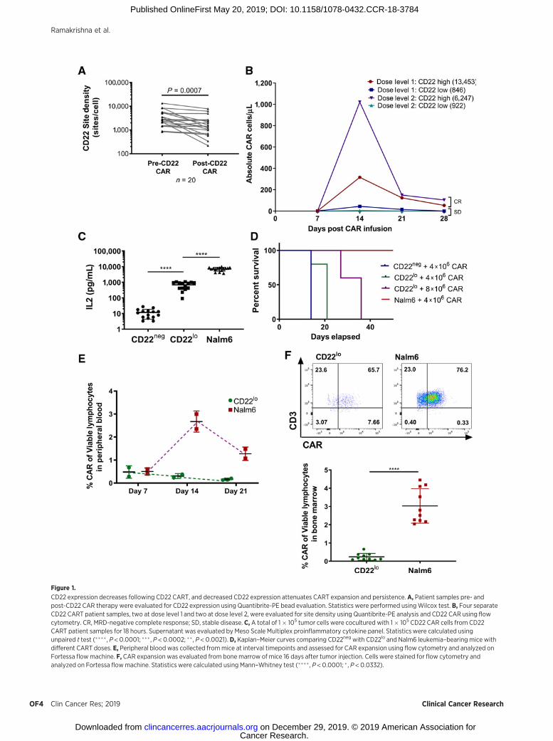
Figure 1.
CD22 expression decreases following CD22 CART, and decreased CD22 expression attenuates CART expansion and persistence.A, Patient samples pre- andpost-CD22 CAR therapy were evaluated for CD22 expression using Quantibrite-PE bead evaluation. Statistics were performed usingWilcox test. B, Four separateCD22 CART patient samples, two at dose level 1 and two at dose level 2, were evaluated for site density using Quantibrite-PE analysis and CD22 CAR using flowcytometry. CR, MRD-negative complete response; SD, stable disease. C, A total of 1� 105 tumor cells were cocultured with 1� 105 CD22 CAR cells from CD22CART patient samples for 18 hours. Supernatant was evaluated by Meso Scale Multiplex proinflammatory cytokine panel. Statistics were calculated usingunpaired t test (���� , P < 0.0001; ��� , P < 0.0002; �� , P < 0.0021). D, Kaplan–Meier curves comparing CD22neg with CD22lo and Nalm6 leukemia–bearing mice withdifferent CART doses. E, Peripheral blood was collected frommice at interval timepoints and assessed for CAR expansion using flow cytometry and analyzed onFortessa flowmachine. F, CAR expansion was evaluated from bone marrow of mice 16 days after tumor injection. Cells were stained for flow cytometry andanalyzed on Fortessa flowmachine. Statistics were calculated using Mann–Whitney test (���� , P < 0.0001; � , P < 0.0332).
Ramakrishna et al.
Clin Cancer Res; 2019 Clinical Cancer ResearchOF4
Cancer Research. on December 29, 2019. © 2019 American Association forclincancerres.aacrjournals.org Downloaded from
Published OnlineFirst May 20, 2019; DOI: 10.1158/1078-0432.CCR-18-3784
pattern of diminished production of multiple T-cell cytokines bypatient-derived CD22 CART products in response to low-CD22–expressing cell lines (Supplementary Fig. S1C).
Site density affects CD22 CART activation, efficacy, expansion,and phenotype
While low CD22 site expression did not affect CART-mediatederadication of ALL in vitro, in vivo clearance of CD22lo ALL by CD22CARTwas impaired comparedwith clearance of parental Nalm6, asshown previously (11). Moreover, eradication of CD22lo ALL wasnot rescued by increased dose of CD22 CART (Fig. 1D). Impor-tantly, diminished ability for CD22 CART to clear CD22lo ALL wasassociated with poor early expansion of CART (Fig. 1E), consistentwith our observations in patients treated with the CD22 CART(Fig. 1B). Finally, CD22 CARTs were significantly reduced in thebonemarrowofCD22lo leukemia–bearingmice at day 16afterCARinjection as compared with parental Nalm6-bearing mice (Fig. 1F).Together, these findings demonstrate that CD22 CARTs are lesseffective at clearing CD22lo ALL due to ineffective CAR expansion.
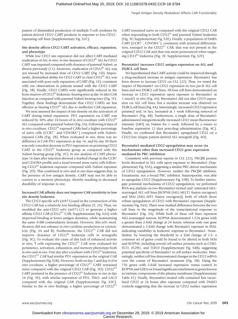
We next assessed the impact of site density on the activation ofCART during initial expansion. PD1 expression on CART wasreduced by 30% after 24 hours of in vitro coculture with CD22lo
ALL comparedwith parental Nalm6 (Fig. 2A). Following 8 days ofin vitro coculture, CD22lo-exposed CARs had a higher percentageof na€�ve cells (CCR7þ and CD45RAþ) compared with Nalm6-exposed CARs (Fig. 2B). When evaluated in vivo, there was nodifference in CAR PD1 expression at day 16, and by day 30 therewas only amodest decrease in PD1 expression onpersisting CD22CART in the CD22lo leukemia group as compared with theNalm6-bearing group (Fig. 2C). In vivo analysis of CART pheno-type 16 days after injection showed amarked change in the CCR7and CD45RA profile and a trend toward more na€�ve cells follow-ing CD22lo leukemia exposure compared with Nalm6 exposure(Fig. 2D). This combined in vitro and in vivo data suggests that, inthe presence of low antigen density, CART may not be able toconvert to a memory phenotype, thereby resulting in decreaseddurability of response in vivo.
Increased CAR affinity does not improve CAR sensitivity to lowsite density leukemia
The CD22-specific scFv (m971) used in the construction of theCD22 CAR has a relatively low binding affinity (9, 24). Thus, wemodified the anti-CD22 scFv (m971-L7) to generate a higheraffinity CD22 CAR (CD22V1 CAR; Supplementary Fig. S2A) withimproved binding at lower antigen densities, while maintainingthe same 41BB costimulatory domain. However, this scFv mod-ification did not enhance in vitro cytokine production or cytotox-icity (Fig. 3A and B). Furthermore, the CD22V1 CAR did notimprove clearance of CD22lo leukemia cells in xenografts(Fig. 3C). To evaluate the cause of this lack of enhanced activityin vitro, T cells expressing the CD22V1 CAR were evaluated forpersistence, activation, exhaustion, and memory phenotype bothin vitro and in vivo. One day after coculture with CD22lo leukemia,the CD22V1 CAR had similar PD1 expression as the original CAR(Supplementary Fig. S2B).However, both on day 1 andday 8 of invitro coculture, a higher percentage of CD22V1 CART remainedna€�ve compared with the original CD22 CAR (Fig. 3D). CD22V1
CART persisted in the presence of CD22lo leukemia in vivo at day16 (Fig. 3E), with similar expression of PD1, TIM3, and LAG3compared with the original CAR (Supplementary Fig. S2C).Similar to the in vitro findings, a higher percentage of CD22V1
CART remained na€�ve as compared with the original CD22 CARwhen responding to both CD22lo and parental Nalm6 leukemia(Fig. 3F; Supplementary Fig. S2E). Finally, a population of EMRACART (CCR7�, CD45RAþ), consistent with terminal differentia-tion, emerged in the CD22V1 CAR, that was not present in theoriginal CD22 CAR and that was more pronounced when target-ing CD22lo leukemia (Fig. 3F; Supplementary Fig. S2F).
Bryostatin1 increases CD22 antigen expression on ALL andDLBCL cell lines
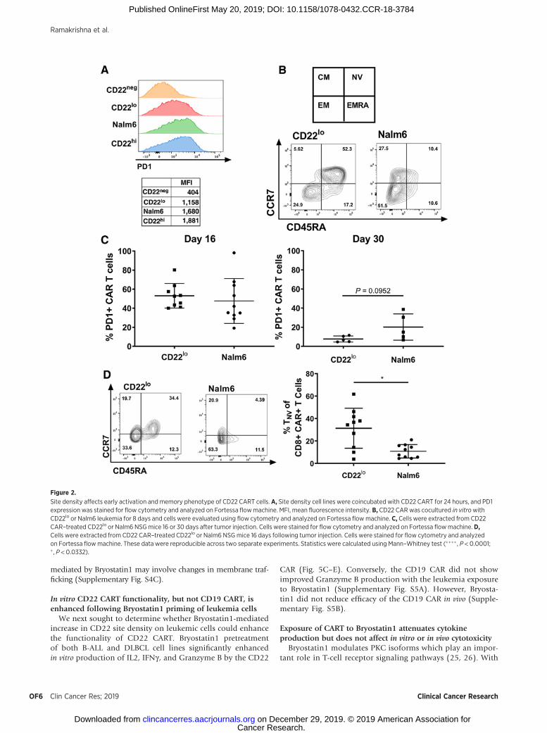
We hypothesized that CART activity could be improved througha drug-mediated increase in antigen expression. Bryostatin1 hasbeen shown to increase CD22 on CLL (22). Thus, we tested theimpact of Bryostatin1 on CD22 expression in two pre-B ALL celllines and two DLBCL cell lines. All four cell lines demonstrated anincrease in CD22 expression upon exposure to Bryostatin1 at1 nmol/L in vitro (Fig. 4A). Bryostatin1 did not alter CD19 expres-sion on ALL cell lines, but a modest increase was observed onDLBCLcell lines (Fig. 4A). Interestingly, increasedCD22 expressionpersisted and, in fact, increased at 1 week following removal ofBryostatin1 (Fig. 4B). Furthermore, a single dose of Bryostatin1administered intraperitoneally increased CD22 mean fluorescenceintensity (MFI) on Nalm6 for 1 week, followed by a return tobaseline expression 12 days post-drug administration (Fig. 4C).Finally, we confirmed that Bryostatin1 upregulated CD22 on aCD22-low relapse patient-derived ALL xenograft (Fig. 4D).
Bryostatin1-mediated CD22 upregulation may occur viamechanisms other than increased CD22 gene expressionmediated by PKC inhibition
Consistent with previous reports in CLL (22), PKCbII proteinlevels decreased in ALL cells upon exposure to Bryostatin1 (Sup-plementary Fig. S3A), suggesting a similar PKC-driven mechanismof CD22 upregulation. However, neither the PKCbII inhibitor,Enzastaurin, nor a broad PKC inhibitor, Staurosporine, was ableto upregulate CD22 (Supplementary Fig. S3B). To further interro-gate potential mechanisms of CD22 upregulation, we performedRNA-seq analysis on two Bryostatin1-treated and -untreated MLL-rearranged ALL cell lines [KOPN8 (MLL-MLLT1 fusion oncogene)and SEM (MLL-AFF1 fusion oncogene)], both of which showrobust upregulation of CD22 with Bryostatin1 exposure (Supple-mentary Fig. S4A). There were marked differences between the twocell lines in the magnitude of the transcriptional response toBryostatin1 (Fig. 5A). While both of these cell lines representMLL-rearranged tumors, KOPN8 demonstrated 1,524 genes witha greater than 2-fold change at 24 hours, whereas only one genedemonstrated a 2-fold change with Bryostain1 exposure in SEM,indicating variability in leukemic response to Bryostatin1. None-theless, by lowering the threshold to a fold change of 1.5, acommon set of genes could be found to be altered in both SEMand KOPN8, including several cell surface proteins such as CD82,FLT1, FGFR1, and TLR10 (Supplementary Fig. S4B), suggestingpotential specificity of Bryostatin1 to cell surface molecules. Inter-estingly, neither cell line demonstrated changes in theCD22mRNAover the course of Bryostatin1 treatment (Fig. 5B). Using the756 genes with 2-fold increased expression versus control inKOPN8andGSEAwe foundsignificant enrichment ingenesknownas intrinsic components of the plasmamembrane (SupplementaryTable S1). Finally, Bryostatin1-treated cells contained less vacuo-lized CD22 at 24 hours after exposure compared with DMSOcontrols suggesting that the increase in CD22 surface expression
Target Antigen Density Modulation Affects CAR Functionality
www.aacrjournals.org Clin Cancer Res; 2019 OF5
Cancer Research. on December 29, 2019. © 2019 American Association forclincancerres.aacrjournals.org Downloaded from
Published OnlineFirst May 20, 2019; DOI: 10.1158/1078-0432.CCR-18-3784
mediated by Bryostatin1 may involve changes in membrane traf-ficking (Supplementary Fig. S4C).
In vitro CD22 CART functionality, but not CD19 CART, isenhanced following Bryostatin1 priming of leukemia cells
We next sought to determine whether Bryostatin1-mediatedincrease in CD22 site density on leukemic cells could enhancethe functionality of CD22 CART. Bryostatin1 pretreatmentof both B-ALL and DLBCL cell lines significantly enhancedin vitro production of IL2, IFNg , and Granzyme B by the CD22
CAR (Fig. 5C–E). Conversely, the CD19 CAR did not showimproved Granzyme B production with the leukemia exposureto Bryostatin1 (Supplementary Fig. S5A). However, Bryosta-tin1 did not reduce efficacy of the CD19 CAR in vivo (Supple-mentary Fig. S5B).
Exposure of CART to Bryostatin1 attenuates cytokineproduction but does not affect in vitro or in vivo cytotoxicity
Bryostatin1 modulates PKC isoforms which play an impor-tant role in T-cell receptor signaling pathways (25, 26). With
Figure 2.
Site density affects early activation andmemory phenotype of CD22 CART cells. A, Site density cell lines were coincubated with CD22 CART for 24 hours, and PD1expression was stained for flow cytometry and analyzed on Fortessa flowmachine. MFI, mean fluorescence intensity. B, CD22 CARwas cocultured in vitrowithCD22lo or Nalm6 leukemia for 8 days and cells were evaluated using flow cytometry and analyzed on Fortessa flowmachine. C, Cells were extracted from CD22CAR–treated CD22lo or Nalm6 NSGmice 16 or 30 days after tumor injection. Cells were stained for flow cytometry and analyzed on Fortessa flowmachine. D,Cells were extracted from CD22 CAR–treated CD22lo or Nalm6 NSGmice 16 days following tumor injection. Cells were stained for flow cytometry and analyzedon Fortessa flowmachine. These data were reproducible across two separate experiments. Statistics were calculated using Mann–Whitney test (���� , P < 0.0001;� , P < 0.0332).
Ramakrishna et al.
Clin Cancer Res; 2019 Clinical Cancer ResearchOF6
Cancer Research. on December 29, 2019. © 2019 American Association forclincancerres.aacrjournals.org Downloaded from
Published OnlineFirst May 20, 2019; DOI: 10.1158/1078-0432.CCR-18-3784

Figure 3.
A total of 1� 105 tumor cells were cocultured with 1� 105 mock, CD22, or CD22V1 CAR and assessed for IFNg and IL2 cytokines by ELISA from cell culturesupernatants [statistics were calculated using paired t test (� , P < 0.0332); A], or Annexin V staining was assessed over time using IncuCyte ZOOM (B). Thesedata are representative of two separate experiments and were consistent across two different effector-to-target ratios. C,NSGmice were injected with 1� 106
GPF-positive CD22neg, CD22lo, or Nalm6 tumor cells on day 0. On day 3, 5� 106 CD22 or CD22V1 CAR were injected for treatment. Mice were imaged using IVIStechnology and luciferin-D intraperitoneal injections. Luminescence quantification is shown on the right. These data are representative of two separateexperiments.D, CD22 or CD22V1 CART was coincubated with tumor cells. On days 1 and 8, CAR was harvested, stained for flow cytometry, and analyzed onFortessa flowmachine. Statistics were calculated using unpaired t test (�, P < 0.0332). E and F,Mice were injected with CD22lo or Nalm6 leukemia on day 0. Atotal of 5� 106 CD22 CAR T cells were administered on day 3, and mice were sacrificed on day 16. Bone marrow cells were stained for flow cytometry andanalyzed on Fortessa flowmachine. Statistics were calculated using unpaired t test (���� , P < 0.0001; � , P < 0.0332).
Target Antigen Density Modulation Affects CAR Functionality
www.aacrjournals.org Clin Cancer Res; 2019 OF7
Cancer Research. on December 29, 2019. © 2019 American Association forclincancerres.aacrjournals.org Downloaded from
Published OnlineFirst May 20, 2019; DOI: 10.1158/1078-0432.CCR-18-3784
Figure 4.
Bryostatin1 upregulates CD22, but not CD19, and CD22 increased expression is durable for 1 week after drug exposure.A, Four cell lines were coincubated with1 nmol/L Bryostatin1 for 24 hours and analyzed using flow cytometry 1 day after Bryostatin1 exposure. Site density was analyzed through use of standardizedQuantibrite-PE beads. Statistics were calculated using unpaired t test (��� , P < 0.0002; �� , P < 0.0021; � , P < 0.0332). B, Cell lines were coincubated with 1 nmol/LBryostatin1 for 24 hours, washed, and analyzed using flow cytometry at 1 and 7 days after Bryostatin1 exposure. MFI fold change¼ CD22 MFIBryostatin1/CD22MFIDMSO. C, NSGmice were injected with 1� 106 GPF-positive Nalm6 leukemia cells on day 0. Bryostatin1 was administered at 0.8 mg/kg on day 3. Mice weresacrificed 7 and 12 days after Bryostatin1 injection, and CD22 was evaluated through flow cytometry. MFI fold change¼ CD22 MFIBryostatin1/CD22 MFIDMSO.D, CD22-low relapse PDX cell line was cultured in vitrowith 1 nmol/L Bryostatin1 for 24 hours and measured for CD22 expression using CD22 antibody.
Ramakrishna et al.
Clin Cancer Res; 2019 Clinical Cancer ResearchOF8
Cancer Research. on December 29, 2019. © 2019 American Association forclincancerres.aacrjournals.org Downloaded from
Published OnlineFirst May 20, 2019; DOI: 10.1158/1078-0432.CCR-18-3784
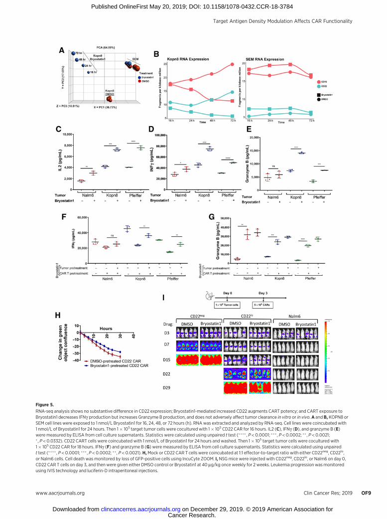
Figure 5.
RNA-seq analysis shows no substantive difference in CD22 expression; Bryostatin1-mediated increased CD22 augments CART potency; and CART exposure toBryostatin1 decreases IFNg production but increases Granzyme B production, and does not adversely affect tumor clearance in vitro or in vivo. A and B, KOPN8 orSEM cell lines were exposed to 1 nmol/L Bryostatin1 for 16, 24, 48, or 72 hours (h). RNAwas extracted and analyzed by RNA-seq. Cell lines were coincubated with1 nmol/L of Bryostatin1 for 24 hours. Then 1� 105 target tumor cells were cocultured with 1� 105 CD22 CAR for 16 hours. IL2 (C), IFNg (D), and granzyme B (E)were measured by ELISA from cell culture supernatants. Statistics were calculated using unpaired t test (���� , P < 0.0001; ��� , P < 0.0002; �� , P < 0.0021;� , P < 0.0332). CD22 CART cells were coincubated with 1 nmol/L of Bryostatin1 for 24 hours and washed. Then 1� 105 target tumor cells were cocultured with1� 105 CD22 CAR for 18 hours. IFNg (F) and granzyme B (G) were measured by ELISA from cell culture supernatants. Statistics were calculated using unpairedt test (���� , P < 0.0001; ��� , P < 0.0002; ��, P < 0.0021). H,Mock or CD22 CAR T cells were coincubated at 1:1 effector-to-target ratio with either CD22neg, CD22lo,or Nalm6 cells. Cell death was monitored by loss of GFP-positive cells using IncuCyte ZOOM. I, NSGmice were injected with CD22neg, CD22lo, or Nalm6 on day 0,CD22 CAR T cells on day 3, and then were given either DMSO control or Bryostatin1 at 40 mg/kg once weekly for 2 weeks. Leukemia progression was monitoredusing IVIS technology and luciferin-D intraperitoneal injections.
Target Antigen Density Modulation Affects CAR Functionality
www.aacrjournals.org Clin Cancer Res; 2019 OF9
Cancer Research. on December 29, 2019. © 2019 American Association forclincancerres.aacrjournals.org Downloaded from
Published OnlineFirst May 20, 2019; DOI: 10.1158/1078-0432.CCR-18-3784
the ultimate goal of utilizing Bryostatin1 in combination withCAR therapy, we evaluated the direct impact of Bryostatin1on CD22 CAR functionality, independent of its effects onleukemic site density. CD22 CAR pretreated with Bryostatin1produced attenuated levels of IFNg when subsequently cocul-tured with ALL or DLBCL cell lines relative to DMSO controlpretreated CD22 CAR, but produced significantly increasedlevels of Granzyme B (Fig. 5F and G). In vitro cytotoxicitykinetics of Bryostain1-exposed CART was similar to DMSO-exposed CART (Fig. 5H). To evaluate the combined effect ofBryostatin1 on leukemic CD22 site density and CART func-tionality we repeated functional assays using Bryostatin1-treated leukemia with Bryostatin1-exposed CART. AttenuatedIFNg production by CART-exposed cells was restored (Fig. 5F)and Granzyme B production was augmented (Fig. 5G) in thepresence of Bryostatin1 pretreated leukemia cells. Finally, weevaluated Bryostatin1 effects on in vivo CAR activity againstCD22lo leukemia, which does not modulate CD22 expressionfollowing Bryostatin1 exposure (Supplementary Fig. S4D).Bryostatin1 did not impact the ability of the CD22 CAR toattenuate progression of the CD22lo leukemia or CD22 CARability to clear the Nalm6 leukemia in vivo (Fig. 5I; Supple-mentary Fig. S6A). Together, these findings demonstrate thatexposing CART to Bryostatin1 affects IFNg and Granzyme Bproduction, but does not negatively impact overall CARTfunctionality in vivo.
Bryostatin1 affects CART persistence and memory phenotype,and improves durability of leukemic response
Finally, we evaluated the impact of Bryostatin1-mediatedincrease in CD22 site density on in vivo CAR persistence andfunctionality using the parental Nalm6, which does notexpress sufficient CD22 to maximize cytokine response (Sup-plementary Fig. S1) and does modulate CD22 expression inresponse to Bryostatin1 (Fig. 4). We tested the administrationof Bryostatin1 as a "priming therapy" prior to CART infusion.Mice were injected with Bryostatin1 pretreated tumor cells,followed by CART injection. Seven days after CD22 CARinjection, T-cell phenotype was evaluated. Within the CD8þ
CART population, we found significant cumulative enrichmentof central memory (CCR7þ, CD45RA�) and effector memory(CCR7�, CD45RA�) cells, and fewer na€�ve cells, in the spleensof mice that received Bryostatin1 pretreated Nalm6 (Fig. 6A).With Bryostatin1 exposure, CART demonstrated similar acti-vation without evidence of exhaustion, as evidenced by stablePD1 expression, without increase in TIM3 or LAG3 (Supple-mentary Fig. S5A). At 30 days, mice treated with Bryostatin1after CART injection had slightly increased CD22 CART inbone marrow (Fig. 6B). We then tested Bryostatin1 adminis-tration for 2 weeks after subcurative dose of CD22 CARTinfusion in Nalm6 (Fig. 6C; Supplementary Fig. S6B) and SEM(Fig. 6C; Supplementary Fig. S6C), demonstrating that Bryos-tatin1 improved durability of remission, extending beyondcessation of Bryostatin1. Finally, we evaluated the effects ofBryostatin1 administration following CD22 CART infusion theCD22-low relapse PDX model, demonstrating improved abil-ity to clear leukemia (Fig. 6D; Supplementary Fig. S6D).Collectively, these results demonstrate that Bryostatin1 iseffective as both a priming therapy to increase antigen expres-sion prior to CAR T-cell infusion and as a potential rescuefollowing emergence of CD22 CAR–resistant leukemia.
DiscussionCART therapy has provided patients with relapsed or refractory
pre-B-cell ALL a potentially curative therapeutic option, butremission duration is shortened in a high percentage of patientsdue to changes in target antigen expression. Furthermore, recentstudies have begun to identify limitations in CAR activity in thesetting of low antigen site density (11–15, 27–33). Our recentclinical experiencewith aCD22CAR supports low antigen densityas a mechanism of escape from CAR-targeted therapy (11). Here,we evaluate the characteristics of CAR failure in the setting of lowsite density, focusing on the effects on CAR function and persis-tence. Moreover, we provide the first preclinical evidence tosupport the use of drug-mediated antigen modulation to over-come such limitations. Our data establishes two important find-ings: (i) low site density on tumor cells results in significantchanges in the persistence and phenotype of CART, and (ii)drug-mediated increase in target antigen site density improvesCAR T-cell functionality and length of remission.
We first assessed the effects of site density on short-term CARTfunctionality, and observed a reduction in cytokine production astarget antigen expression declines, consistent with previous stud-ies (12, 15, 32, 33). We further demonstrate that the CD22 CARdelayed in vivo progression of, but failed to clear, low site densityleukemia. Although activation and exhaustionmarkers, includingPD1, TIM3, and LAG3, were not significantly different on CARTexposed to low site density leukemia, lower site density leukemiainduced a significant decrease in numbers of persisting CART,with a more na€�ve memory phenotype, suggesting diminishedT-cell expansion and conversion to memory phenotype. Whetherthis finding represents a recruitment of less CAR-expressing T cellsinto the expansion pool or less potent activation of the samepercentage of CAR-expressing T cells will require further testing.This is the first clear evidence that site density not only affectsshort-term activity of CART, but also affects long-termpersistence,with implications for the durability of CART-induced remissions.However, as these findings were in a xenograft model, it isimpossible to ascertain the exact implications for CAR persistencein an immunocompetent host, in which nonleukemic expressionof CAR antigen could impact sustained expansion of the CART.Thus, these findings require confirmation in immunocompetentmodels or in patients.
We next proceeded to evaluate potential strategies to over-come the limitations on cellular therapy imposed by lowantigen density, through either improving CAR functionalityor augmenting tumor sensitivity. We attempted to improveCAR functionality through enhancing the affinity of the CD22CAR, thereby improving sensitivity to low site density.Although binding affinity can improve the efficacy of solubleantibody-based therapeutics (34), our data does not demon-strate improvement in CAR functionality with increased scFvaffinity. Interestingly, while CD22V1 CAR had a larger na€�veT-cell population, especially against CD22lo leukemia, it alsodemonstrated a more pronounced terminally differentiatedeffector memory population (CCR7�, CD45RAþ), suggestingthat when the T cell is activated by the CD22V1 CAR, it maybecome terminally differentiated rather than converting to amemory phenotype. While this data is supported by experiencewith some CARs (35), the complexity of individual CARconstructs may preclude the ability to generalize thesefindings (28, 36).
Ramakrishna et al.
Clin Cancer Res; 2019 Clinical Cancer ResearchOF10
Cancer Research. on December 29, 2019. © 2019 American Association forclincancerres.aacrjournals.org Downloaded from
Published OnlineFirst May 20, 2019; DOI: 10.1158/1078-0432.CCR-18-3784

Figure 6.
Bryostatin1 treatment pre-CAR infusion alters T-cell phenotype without T-cell exhaustion, and post-CAR infusion improves durability of remission in vivo. A,Nalm6 cells were exposed to 1 nmol/L Bryostatin1 for 24 hours, then injected into mice on day 0. CD22 CAR T cells were administered on day 3, and mice weresacrificed on day 10. Bonemarrow cells were stained for flow cytometry and analyzed on Fortessa flowmachine. Statistics were calculated using unpaired t test(� , P < 0.0332). B,NSGmice were injected with Nalm6 on day 0, CD22 CART on day 3, and then were given either DMSO control or Bryostatin1 at 40 mg/kg onceweekly for 2 weeks. Mice were sacrificed 30 days after tumor injection. Cells were stained for flow cytometry and analyzed on Fortessa flowmachine. C, NSGmice were injected with 1� 106 GPF-positive Nalm6 or SEM tumor cells on day 0. On day 3, either 3� 106 (Nalm6) or 2� 106 (SEM) mock or CD22 CARwereinjected for treatment. Mice were given 40 mg/kg of Bryostatin1 or DMSO once weekly for 2 weeks. Mice were imaged using IVIS technology and luciferin-Dintraperitoneal injections. D,NSGmice were injected with 1� 106 GPF-positive PDX tumor cells on day 0. On day 42, 3� 106 Mock or CD22 CARwere injected fortreatment. Mice were given 40 mg/kg of Bryostatin1 or DMSO once weekly for 2 weeks starting on day 45. Mice were imaged using IVIS technology and luciferin-D intraperitoneal injections.
www.aacrjournals.org Clin Cancer Res; 2019 OF11
Target Antigen Density Modulation Affects CAR Functionality
Cancer Research. on December 29, 2019. © 2019 American Association forclincancerres.aacrjournals.org Downloaded from
Published OnlineFirst May 20, 2019; DOI: 10.1158/1078-0432.CCR-18-3784
To improve tumor sensitivity, we found that Bryostatin1 upre-gulatedCD22 expression in ALL andDLBCL cell lines, aswell as ina CD22-low relapse PDX, thereby improving CART cytokineproduction and memory phenotype. As a modulator of PKC,Bryostatin1 has variable effects on PKC protein levels based onexposure parameters (37–41). In CLL cell lines, Bryostatin1-mediated upregulation of CD22 correlated with decreasedPKC (22). We found that Bryostatin1 was associated withdecreased PKCbII, but PKC inhibitors did not provide the sameupregulation of CD22 as Bryostatin1. We also found that CD22mRNA levels were not significantly altered in Bryostatin1-treatedALL cell lines and that multiple cell surface molecules wereupregulated. These findings suggest that Bryostatin1 has a muchmore complex mechanism of upregulation of CD22, but furtherevaluation will be needed to elucidate the exact mechanism ofCD22 upregulation.
We demonstrate that CAR functionality can be indirectlyimproved via Bryostatin1-mediated increase in CD22 expression.However, Bryostatin1 has been previously implicated in directlyaffecting T-cell function (25, 26). Although Bryostatin1 attenu-ated IFNg production by CD22 CART, Granzyme B productionwas increased and therewas ultimately no adverse effect on in vitroor in vivo cytotoxicity. The net effect of Bryostatin1 on CARTfunctionality and antigen expression level resulted in overallaugmented cytokine production and cytotoxicity, and improveddurability of response. This indicates that Bryostatin1 could beadministered during CART expansion or to rescue patients fol-lowing post-CART relapse resulting from reduced antigen expres-sion.However, we alsopostulate that Bryostatin1 couldbeused toprime tumors prior to CART therapy based on our observationthat site density affects CAR T-cell persistence.
While combination therapies are the cornerstone of oncologictreatments, CART has not yet been combined with other drugswith the intent of augmenting sensitivity to low antigen levels.Prior preclinical studies have shown improved CAR activity withdrug modulation (14, 42); however, to our knowledge, ourfindings are the first to describe how drug-induced increasedantigen expression can improve CAR T-cell activity. Bryostatin1has been used in clinical trials for oncologic patients with morethan 1,400 individuals treated, including one phase I pediatricclinical trial. The pediatric phase I study showed the drug to bewell-tolerated, but recommended increasing the dose adminis-tration in subsequent trials (43).While adult phase I and II studieshave established the MTD, the measured effect was tumorresponse and PKC inhibition rather than surface protein expres-sion. Our data supports the concept of using Bryostatin1 tomodulate target antigen density in combination with a targetedimmunotherapy such as CART, but clinical experience is neededin pediatric patients specifically characterizing CD22 antigenexpression as a biomarker to establish Bryostatin1 dosing.
Taken together, our observations contribute to an under-standing of the complexity in developing effective CAR ther-apies in tumors where lower expression is a characteristic of thetargeted antigen. Because CD22 expression has been targetedsuccessfully by immunotherapy and is likely comparable with
many other antigens currently being evaluated (12, 28), CD22represents a broadly applicable and clinically relevant modelantigen. As such, the effects of antigen expression on CARpersistence and phenotype described here may be pertinentto many other potential target antigens. Finally, our findingswith Bryostatin1 support the initiation of trials testing whetherdrug-mediated upregulation of antigen expression can imp-rove efficacy and remission durability following targetedimmunotherapy.
Disclosure of Potential Conflicts of InterestNo potential conflicts of interest were disclosed.
DisclaimerThe content is solely the responsibility of the authors and does not neces-
sarily represent the official views of the University of PittsburghMedical Center.The content of this article does not necessarily reflect the views of policies of theDepartment of Health and Human Services, nor does mention of trade names,commercial products, or organizations imply endorsement by the U.S.Government.
Authors' ContributionsConception and design: S. Ramakrishna, S.L. Highfill, H. Qin, T.J. FryDevelopment of methodology: S. Ramakrishna, Z. Walsh, S.M. Nguyen,H. Qin, M. Stetler-Stevenson, Y. Feng, Z. Zhu, T.J. FryAcquisition of data (provided animals, acquired and managed patients,provided facilities, etc.): S. Ramakrishna, Z. Walsh, S.M. Nguyen, H. Qin,M. Stetler-Stevenson, C.M. Yuan, J.D. Hwang, D. Dimitrov, N.N. ShahAnalysis and interpretation of data (e.g., statistical analysis, biostatistics,computational analysis): S. Ramakrishna, Z. Walsh, S.M. Nguyen, H. Lei,J.F. Shern, H.Qin, I.L. Kraft, M. Stetler-Stevenson, C.M. Yuan, N.N. Shah, T.J. FryWriting, review, and/or revision of the manuscript: S. Ramakrishna,S.L. Highfill, Z. Walsh, J.F. Shern, H. Qin, I.L. Kraft, M. Stetler-Stevenson,C.M. Yuan, J.D. Hwang, D. Dimitrov, N.N. Shah, T.J. FryAdministrative, technical, or material support (i.e., reporting or organizingdata, constructing databases): J.F. Shern, J.D. Hwang, Y. Feng, Z. Zhu,N.N. ShahStudy supervision: T.J. Fry
AcknowledgmentsThe authors would like to thank John Buckley for his assistance with murine
experiments and Dr. Michael Kruhlak for his aid in confocal microscopy. Theauthors would also like to thank Drs. Crystal Mackall, Patrick Brown, RimasOrentas, and Elena Sotillo for their feedback and comments. This work wassupported by the Intramural Research Program at theNIH and by a St. Baldrick'sFoundation - Stand Up To Cancer Pediatric Cancer Dream Team TranslationalResearch Grant (grant no. SU2C-AACR-DT-27-17). Stand Up To Cancer is adivision of the Entertainment Industry Foundation. Research grants are admin-istered by the American Association for Cancer Research, the Scientific Partner ofSU2C. This work was supported by the Intramural Research Program at NIH.Research reported in this article was supported in part by funds provided by theUniversity of Pittsburgh Medical Center (UPMC) as part of the UPMC ImmuneTransplant and Therapy Center.
The costs of publication of this articlewere defrayed inpart by the payment ofpage charges. This article must therefore be hereby marked advertisement inaccordance with 18 U.S.C. Section 1734 solely to indicate this fact.
Received November 21, 2018; revised April 1, 2019; accepted May 15, 2019;published first May 20, 2019.
References1. Pui CH, Yang JJ, Hunger SP, Pieters R, Schrappe M, Biondi A, et al.
Childhood acute lymphoblastic leukemia: progress through collaboration.J Clin Oncol 2015;33:2938–48.
2. Smith MA, Altekruse SF, Adamson PC, Reaman GH, Seibel NL. Declin-ing childhood and adolescent cancer mortality. Cancer 2014;120:2497–506.
Ramakrishna et al.
Clin Cancer Res; 2019 Clinical Cancer ResearchOF12
Cancer Research. on December 29, 2019. © 2019 American Association forclincancerres.aacrjournals.org Downloaded from
Published OnlineFirst May 20, 2019; DOI: 10.1158/1078-0432.CCR-18-3784
3. Davila ML, Riviere I, Wang X, Bartido S, Park J, Curran K, et al. Efficacy andtoxicity management of 19–28z CAR T cell therapy in B cell acute lym-phoblastic leukemia. Sci Transl Med 2014;6:224ra25.
4. Maude SL, FreyN, ShawPA, Aplenc R, Barrett DM, BuninNJ, et al. Chimericantigen receptor T cells for sustained remissions in leukemia. N Engl J Med2014;371:1507–17.
5. Lee DW, Kochenderfer JN, Stetler-Stevenson M, Cui YK, Delbrook C,Feldman SA, et al. T cells expressing CD19 chimeric antigen receptors foracute lymphoblastic leukaemia in children and young adults: a phase 1dose-escalation trial. Lancet 2015;385:517–28.
6. Grupp SA, Kalos M, Barrett D, Aplenc R, Porter DL, Rheingold SR, et al.Chimeric antigen receptor-modified T cells for acute lymphoid leukemia.N Engl J Med 2013;368:1509–18.
7. Maude SL, Teachey DT, Porter DL, Grupp SA. CD19-targeted chimericantigen receptor T-cell therapy for acute lymphoblastic leukemia. Blood2015;125:4017–23.
8. Gardner R,WuD, Cherian S, FangM, Hanafi LA, Finney O, et al. Acquisitionof a CD19-negative myeloid phenotype allows immune escape of MLL-rearranged B-ALL fromCD19CAR-T-cell therapy. Blood 2016;127:2406–10.
9. HasoW, LeeDW, ShahNN, Stetler-StevensonM, YuanCM, Pastan IH, et al.Anti-CD22-chimeric antigen receptors targeting B-cell precursor acutelymphoblastic leukemia. Blood 2013;121:1165–74.
10. Yilmaz M, Richard S, Jabbour E. The clinical potential of inotuzumabozogamicin in relapsed and refractory acute lymphocytic leukemia.Ther Adv Hematol 2015;6:253–61.
11. Fry TJ, Shah NN, Orentas RJ, Stetler-Stevenson M, Yuan CM, Ramak-rishna S, et al. CD22-targeted CAR T cells induce remission in B-ALL thatis naive or resistant to CD19-targeted CAR immunotherapy. Nat Med2018;24:20–8.
12. Walker AJ, Majzner RG, Zhang L, Wanhainen K, Long AH, Nguyen SM, et al.Tumor antigen and receptor densities regulate efficacy of a chimeric antigenreceptor targeting anaplastic lymphomakinase.Mol Ther 2017;25:2189–201.
13. ChmielewskiM,HombachAA, AbkenH.CD28 cosignalling does not affectthe activation threshold in a chimeric antigen receptor-redirected T-cellattack. Gene Ther 2011;18:62–72.
14. Yoshida T, Mihara K, Takei Y, Yanagihara K, Kubo T, Bhattacharyya J, et al.All-trans retinoic acid enhances cytotoxic effect of T cells with an anti-CD38chimeric antigen receptor in acute myeloid leukemia. Clin Transl Immu-nology 2016;5:e116.
15. Watanabe K, Terakura S, Martens AC, van Meerten T, Uchiyama S, Imai M,et al. Target antigen density governs the efficacy of anti-CD20-CD28-CD3zeta chimeric antigen receptor-modified effector CD8þ T cells. J Immunol2015;194:911–20.
16. Huang J, Brameshuber M, Zeng X, Xie J, Li QJ, Chien YH, et al. A singlepeptide-major histocompatibility complex ligand triggers digital cytokinesecretion in CD4(þ) T cells. Immunity 2013;39:846–57.
17. Sykulev Y, Joo M, Vturina I, Tsomides TJ, Eisen HN. Evidence that a singlepeptide-MHC complex on a target cell can elicit a cytolytic T cell response.Immunity 1996;4:565–71.
18. Srivastava S, Riddell SR. Engineering CAR-T cells: design concepts.Trends Immunol 2015;36:494–502.
19. Jasper GA, Arun I, Venzon D, Kreitman RJ, Wayne AS, Yuan CM, et al.Variables affecting the quantitation of CD22 in neoplastic B cells.Cytometry B Clin Cytom 2011;80:83–90.
20. Shah NN, Stevenson MS, Yuan CM, Richards K, Delbrook C, Kreitman RJ,et al. Characterization of CD22 expression in acute lymphoblastic leuke-mia. Pediatr Blood Cancer 2015;62:964–9.
21. Pettit GR, Herald CL, Doubek DL, Herald DL, Arnold E, Clardy J. Isolationand structure of bryostatin 1. J Am Chem Soc 1982;104:6846–8.
22. Biberacher V, Decker T, Oelsner M, Wagner M, Bogner C, Schmidt B, et al.The cytotoxicity of anti-CD22 immunotoxin is enhanced by bryostatin 1 inB-cell lymphomas through CD22 upregulation and PKC-bII depletion.Haematologica 2012;97:771–9.
23. Qin H, Ramakrishna S, Nguyen S, Fountaine TJ, Ponduri A, Stetler-Stevenson M, et al. Preclinical development of bivalent chimeric antigenreceptors targeting both CD19 and CD22. Mol Ther Oncolytics 2018;11:127–37.
24. Xiao X, Ho M, Zhu Z, Pastan I, Dimitrov DS. Identification and charac-terization of fully human anti-CD22 monoclonal antibodies. MAbs 2009;1:297–303.
25. Tuttle TM,BethkeKP, IngeTH,McCradyCW,PettitGR, BearHD.Bryostatin1-activated T cells can traffic andmediate tumor regression. J SurgRes 1992;52:543–8.
26. Drexler HG, Gignac SM, Jones RA, Scott CS, Pettit GR, Hoffbrand AV.Bryostatin 1 induces differentiation of B-chronic lymphocytic leukemiacells. Blood 1989;74:1747–57.
27. Weijtens ME, Hart EH, Bolhuis RL. Functional balance between T cellchimeric receptor density and tumor associated antigen density: CTLmediated cytolysis and lymphokine production. Gene Ther 2000;7:35–42.
28. Turatti F, Figini M, Balladore E, Alberti P, Casalini P, Marks JD, et al.Redirected activity of human antitumor chimeric immune receptors isgoverned by antigen and receptor expression levels and affinity of inter-action. J Immunother 2007;30:684–93.
29. James SE, Greenberg PD, Jensen MC, Lin Y, Wang J, Till BG, et al. Antigensensitivity of CD22-specific chimeric TCR is modulated by target epitopedistance from the cell membrane. J Immunol 2008;180:7028–38.
30. AnurathapanU,ChanRC,HindiHF,Mucharla R, Bajgain P,Hayes BC, et al.Kinetics of tumor destruction by chimeric antigen receptor-modifiedT cells. Mol Ther 2014;22:623–33.
31. Caruso HG, Hurton LV, Najjar A, Rushworth D, Ang S, Olivares S, et al.Tuning sensitivity of CAR to EGFR density limits recognition of normaltissue while maintaining potent antitumor activity. Cancer Res 2015;75:3505–18.
32. Hombach AA, Gorgens A, Chmielewski M, Murke F, Kimpel J, Giebel B,et al. Superior therapeutic index in lymphoma therapy: CD30(þ) CD34(þ) hematopoietic stem cells resist a chimeric antigen receptor T-cell attack.Mol Ther 2016;24:1423–34.
33. Hegde M, Mukherjee M, Grada Z, Pignata A, Landi D, Navai SA, et al.Tandem CAR T cells targeting HER2 and IL13Ralpha2 mitigate tumorantigen escape. J Clin Invest 2016;126:3036–52.
34. Adler MJ, Dimitrov DS. Therapeutic antibodies against cancer.Hematol Oncol Clin North Am 2012;26:447–81.
35. Arcangeli S, Rotiroti MC, Bardelli M, Simonelli L, Magnani CF, Biondi A,et al. Balance of anti-CD123 chimeric antigen receptor binding affinity anddensity for the targeting of acute myeloid leukemia. Mol Ther 2017;25:1933–45.
36. LynnRC, Feng Y, Schutsky K, PoussinM, Kalota A,DimitrovDS, et al. High-affinity FRb-specific CAR T cells eradicate AML andnormalmyeloid lineagewithout HSC toxicity. Leukemia 2016;30:1355–64.
37. IsakovN, GalronD,Mustelin T, Pettit GR, Altman A. Inhibition of phorbolester-induced T cell proliferation by bryostatin is associated with rapiddegradation of protein kinase C. J Immunol 1993;150:1195–204.
38. Lee HW, Smith L, Pettit GR, Bingham Smith J. Dephosphorylation ofactivated protein kinase C contributes to downregulation by bryostatin.Am J Physiol 1996;271:C304–11.
39. Grant S. Modulation of ara-C induced apoptosis in leukemia by the PKCactivator bryostatin 1. Front Biosci 1997;2:d242–52.
40. Grant S, Jarvis WD, Swerdlow PS, Turner AJ, Traylor RS, Wallace HJ, et al.Potentiation of the activity of 1-beta-D-arabinofuranosylcytosine by theprotein kinase C activator bryostatin 1 in HL-60 cells: association withenhanced fragmentation of mature DNA. Cancer Res 1992;52:6270–8.
41. Jarvis WD, Povirk LF, Turner AJ, Traylor RS, Gewirtz DA, Pettit GR, et al.Effects of bryostatin 1 and other pharmacological activators of proteinkinase C on 1-[beta-D-arabinofuranosyl]cytosine-induced apoptosis inHL-60 human promyelocytic leukemia cells. Biochem Pharmacol 1994;47:839–52.
42. Mihara K, Yoshida T, Ishida S, Takei Y, Kitanaka A, Shimoda K, et al. All-trans retinoic acid and interferon-alpha increase CD38 expression on adultT-cell leukemia cells and sensitize them to T cells bearing anti-CD38chimeric antigen receptors. Blood Cancer J 2016;6:e421.
43. Weitman S, Langevin AM, Berkow RL, Thomas PJ, Hurwitz CA, Kraft AS,et al. A phase I trial of bryostatin-1 in children with refractory solid tumors:a Pediatric Oncology Group study. Clin Cancer Res 1999;5:2344–8.
www.aacrjournals.org Clin Cancer Res; 2019 OF13
Target Antigen Density Modulation Affects CAR Functionality
Cancer Research. on December 29, 2019. © 2019 American Association forclincancerres.aacrjournals.org Downloaded from
Published OnlineFirst May 20, 2019; DOI: 10.1158/1078-0432.CCR-18-3784
Published OnlineFirst May 20, 2019.Clin Cancer Res Sneha Ramakrishna, Steven L. Highfill, Zachary Walsh, et al. Functionality and PersistenceModulation of Target Antigen Density Improves CAR T-cell
Updated version
10.1158/1078-0432.CCR-18-3784doi:
Access the most recent version of this article at:
Material
Supplementary
http://clincancerres.aacrjournals.org/content/suppl/2019/05/18/1078-0432.CCR-18-3784.DC1Access the most recent supplemental material at:
E-mail alerts related to this article or journal.Sign up to receive free email-alerts
Subscriptions
Reprints and
To order reprints of this article or to subscribe to the journal, contact the AACR Publications
Permissions
Rightslink site. (CCC)Click on "Request Permissions" which will take you to the Copyright Clearance Center's
.http://clincancerres.aacrjournals.org/content/early/2019/06/12/1078-0432.CCR-18-3784To request permission to re-use all or part of this article, use this link
Cancer Research. on December 29, 2019. © 2019 American Association forclincancerres.aacrjournals.org Downloaded from
Published OnlineFirst May 20, 2019; DOI: 10.1158/1078-0432.CCR-18-3784